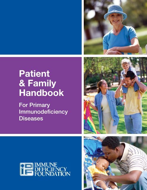
IDF Patient & Family Handbook for Primary Immunodeficiency ... - IDFA
IDF Patient & Family Handbook for Primary Immunodeficiency ... - IDFA
IDF Patient & Family Handbook for Primary Immunodeficiency ... - IDFA

Create successful ePaper yourself
Turn your PDF publications into a flip-book with our unique Google optimized e-Paper software.
<strong>Patient</strong><br />
& <strong>Family</strong><br />
<strong>Handbook</strong><br />
For <strong>Primary</strong><br />
<strong>Immunodeficiency</strong><br />
Diseases
This book contains general medical in<strong>for</strong>mation which cannot be applied safely to any individual case. Medical knowledge and<br />
practice can change rapidly. There<strong>for</strong>e, this book should not be used as a substitute <strong>for</strong> professional medical advice.<br />
FOURTH EDITION<br />
COPYRIGHT 1987, 1993, 2001, 2007 IMMUNE DEFICIENCY FOUNDATION<br />
Copyright 2007 by Immune Deficiency Foundation, USA.<br />
Readers may redistribute this article to other individuals <strong>for</strong> non-commercial use, provided that the text, html codes, and this<br />
notice remain intact and unaltered in any way. The <strong>Patient</strong> & <strong>Family</strong> <strong>Handbook</strong> may not be resold, reprinted or redistributed<br />
<strong>for</strong> compensation of any kind without prior written permission from Immune Deficiency Foundation. If you have any questions<br />
about permission, please contact: Immune Deficiency Foundation, 40 West Chesapeake Avenue, Suite 308, Towson, MD<br />
21204, USA; or by telephone at 1-800-296-4433.
<strong>Patient</strong> &<br />
<strong>Family</strong> <strong>Handbook</strong><br />
For <strong>Primary</strong> <strong>Immunodeficiency</strong> Diseases<br />
4th Edition<br />
This publication has been made possible<br />
through a generous grant from<br />
Baxter Healthcare Corporation<br />
40 West Chesapeake Avenue, Suite 308<br />
Towson, Maryland 21204<br />
800-296-4433<br />
www.primaryimmune.org<br />
idf@primaryimmune.org
EDITORS<br />
R. Michael Blaese, MD<br />
Immune Deficiency Foundation<br />
Towson, MD<br />
Jerry A. Winkelstein, MD<br />
Johns Hopkins University<br />
School of Medicine<br />
Baltimore, MD<br />
Katherine A. Antilla, MAEd<br />
Immune Deficiency Foundation<br />
Towson, MD<br />
ASSOCIATE EDITORS<br />
CONTRIBUTORS<br />
Christine M. Belser<br />
Immune Deficiency Foundation<br />
Towson, MD<br />
Melvin Berger, MD, PhD<br />
Rainbow Babies &<br />
Children’s Hospital<br />
Cleveland, OH<br />
Francisco A. Bonilla, MD, PhD<br />
Boston Children’s Hospital<br />
Boston, MA<br />
Marcia Boyle, MS<br />
Immune Deficiency Foundation<br />
Towson, MD<br />
Rebecca H. Buckley, MD<br />
Duke University<br />
School of Medicine<br />
Durham, NC<br />
Mary Ellen Conley, MD<br />
St. Jude<br />
Children’s Research Hospital<br />
Memphis, TN<br />
Charlotte Cunningham-Rundles,<br />
MD, PhD<br />
Mt. Sinai Medical Center<br />
New York, NY<br />
Robert Dash<br />
Baxter Healthcare Corporation<br />
Deerfield, IL<br />
Carol Ernst, RN, OCN<br />
Mercy Anderson Hospital<br />
Cincinnati, OH<br />
Thomas A. Fleisher, MD<br />
National Institutes of Health<br />
Bethesda, MD<br />
Michael Frank, MD<br />
Duke University<br />
School of Medicine<br />
Durham, NC<br />
Ramsay Fuleihan, MD<br />
Northwestern University<br />
Chicago, IL<br />
Serrie L. Krash, MS<br />
Immune Deficiency Foundation<br />
Towson, MD<br />
Howard M. Lederman, MD, PhD<br />
Johns Hopkins University<br />
School of Medicine<br />
Baltimore, MD<br />
Harry Malech, MD<br />
National Institutes of Health<br />
Bethesda, MD<br />
Steven Miles, MD<br />
All Seasons Allergy, Asthma<br />
and Immunology Center<br />
Woodlands, TX<br />
Hans D. Ochs, MD<br />
University of Washington<br />
School of Medicine<br />
Seattle, WA<br />
Jordan S. Orange, MD, PhD<br />
Children’s Hospital of Philadelphia<br />
Philadelphia, PA<br />
Kenneth Paris, MD, MPH<br />
Louisiana State University<br />
New Orleans, LA<br />
Jennifer M. Puck, MD<br />
University of Cali<strong>for</strong>nia<br />
San Francisco, CA<br />
John W. Seymour, PhD, LMFT<br />
Mankato State University<br />
Mankato, MN<br />
Ricardo U. Sorensen, MD<br />
Louisiana State University<br />
New Orleans, LA<br />
E. Richard Stiehm, MD<br />
University of Cali<strong>for</strong>nia<br />
Los Angeles, CA<br />
Kathleen Sullivan, MD, PhD<br />
Children’s Hospital of Philadelphia<br />
Philadelphia, PA
<strong>Patient</strong> and <strong>Family</strong> <strong>Handbook</strong><br />
i<br />
Preface<br />
The first edition of the <strong>Patient</strong> & <strong>Family</strong> <strong>Handbook</strong> was written nearly two decades ago in response to<br />
requests from patients with primary immunodeficiency diseases, their families and their physicians. We<br />
hoped that it would help patients and their families to learn more about the immune system, the primary<br />
immunodeficiency diseases, currently available therapies and possible future treatments. Since then, tens<br />
of thousands of copies have been distributed to patients and their families!<br />
This fourth edition, like those editions that have come be<strong>for</strong>e, has been inspired by the many new and<br />
exciting advances in the diagnosis and therapy of the primary immunodeficiencies. Many chapters in this<br />
edition are new and all of the existing chapters have been revised to include important new in<strong>for</strong>mation.<br />
We hope that the first chapter will be useful to all who read this book. It explains how the immune system<br />
works and how the failure of the immune system leads to primary immunodeficiency diseases. Individual<br />
chapters on most of the specific primary immunodeficiencies follow. We have included three new chapters<br />
on disorders that were not covered in previous editions and updated the existing chapters to include<br />
in<strong>for</strong>mation on new diagnostic tools, more precise clinical in<strong>for</strong>mation, and new therapies. The General<br />
Care chapter has been updated to reflect new nutritional guidance and conveys commonsense guidelines<br />
that the patient and family may find useful. The chapter on Specific Medical Therapy has been revised to<br />
reflect recent advances in treatments available to the primary immunodeficient individual, including newer<br />
methods of treatment with immunoglobulin. A new chapter dedicated to the adolescent with a primary<br />
immunodeficiency has been added, making three chapters that deal with issues relating to patients<br />
from infancy through adult life. The Health Insurance chapter has been expanded and updated to reflect<br />
changes in reimbursement and insurance; it should be a good place to start when trying to understand<br />
this complex and important area. Resources includes additional in<strong>for</strong>mation available both in printed <strong>for</strong>m<br />
and on the Internet. Finally, the Glossary offers definitions of the more common, and possibly confusing,<br />
medical terms.<br />
A few words on how to use this book: this book is not a substitute <strong>for</strong> a dialogue between the patient, his/<br />
her family, their physician, and other members of the healthcare team. Rather, it is intended to provide the<br />
patient and family with tools to enhance the communication process and to understand the in<strong>for</strong>mation<br />
they receive from the healthcare team. Most importantly, this book is not intended to suggest diagnostic<br />
approaches or to recommend specific therapy <strong>for</strong> any patient. Each patient’s condition and treatment is<br />
unique and the management of their illness should be customized to their individual medical needs.<br />
We thank all those individuals who contributed to this book: those who wrote specific chapters, the<br />
members of the Medical Advisory Committee who reviewed the chapters, the Board of Trustees and the<br />
staff of the Immune Deficiency Foundation who made this book a high priority, and finally, the patients and<br />
their families whose suggestions will make this edition even better than the last three!<br />
The Editors<br />
Baltimore 2007
ii<br />
<strong>Patient</strong> and <strong>Family</strong> <strong>Handbook</strong><br />
The Immune Deficiency Foundation<br />
The Immune Deficiency Foundation (<strong>IDF</strong>) was founded in 1980 by parents of children with primary<br />
immunodefiency diseases and their physicians. At that time, there were no educational materials or<br />
programs <strong>for</strong> patients and no public advocacy initiatives. One of the greatest challenges faced by people<br />
who find themselves or their children diagnosed with a primary immunodeficiency is getting the right<br />
in<strong>for</strong>mation at the right time. To fill this void, one of the most important publications developed by <strong>IDF</strong> is<br />
the <strong>Patient</strong> & <strong>Family</strong> <strong>Handbook</strong> and we are proud to offer this fourth edition. We know that this book has<br />
served as the basis of understanding primary immunodeficiency diseases <strong>for</strong> over two decades and we<br />
are pleased to present this updated version.<br />
A few years ago, Kinsey Moore, then an eighth grader, had the assignment of writing an essay on what<br />
book she would choose to be. Kinsey wrote:<br />
“The book I would want to be turned into is the Immune Deficiency Foundation (<strong>IDF</strong>) <strong>Family</strong> <strong>Handbook</strong>.<br />
When I was born, I was very ill and almost died several times. In all of my baby pictures, I have cords and<br />
wires connected to me. One time when I was in the hospital and I had a life-threatening infection <strong>for</strong> the<br />
third time that month, my mom walked into the library and saw a little blue and white book poking off<br />
the shelf. It was the <strong>IDF</strong>’s family handbook. Everything in that book applied to me. When my mom told<br />
the doctors, they did not believe her. After two years of going to different doctors, I was finally diagnosed<br />
when I was four. This book saved my life. I would want to become this book so I could save more people<br />
in my situation. My family knows what it is like to feel lost and not know whether I would live till tomorrow,<br />
but this book gave us hope.”<br />
We hope this handbook gives you hope, knowledge and empowerment to help cope with the challenges<br />
of living with a primary immunodeficiency disease. As a patient-focused organization dedicated to our<br />
community, we encourage you to contact <strong>IDF</strong> to help meet your needs. Whether you want to talk to<br />
a peer support volunteer, need assistance from a patient advocate or want to attend an educational<br />
meeting, know that <strong>IDF</strong> is the place to turn <strong>for</strong> help and in<strong>for</strong>mation.<br />
Marcia Boyle<br />
President & Founder<br />
Immune Deficiency Foundation
<strong>Patient</strong> and <strong>Family</strong> <strong>Handbook</strong><br />
iii<br />
About the Immune Deficiency Foundation<br />
The Immune Deficiency Foundation, founded in 1980, is the national non-profit patient organization<br />
dedicated to improving the diagnosis and treatment of patients with primary immunodeficiency diseases<br />
through research, education and advocacy.<br />
Educational Publications<br />
• <strong>Patient</strong> & <strong>Family</strong> <strong>Handbook</strong> <strong>for</strong> <strong>Primary</strong> <strong>Immunodeficiency</strong> Diseases<br />
• Our Immune System<br />
• A Guide <strong>for</strong> School Personnel on <strong>Primary</strong> Immune Deficiency Diseases<br />
• Diagnostic and Clinical Care Guidelines <strong>for</strong> <strong>Primary</strong> <strong>Immunodeficiency</strong> Diseases<br />
• <strong>IDF</strong> Guide <strong>for</strong> Nurses on Immunoglobulin Therapy <strong>for</strong> <strong>Primary</strong> <strong>Immunodeficiency</strong> Diseases<br />
• <strong>IDF</strong> Advocate—newsletter<br />
• <strong>Primary</strong> Immune Tribune—e-newsletter<br />
Services <strong>for</strong> <strong>Patient</strong>s and Families<br />
• <strong>Patient</strong> Advocacy—inquiries related to diagnosis, treatment, health insurance, peer support and<br />
literature requests<br />
• <strong>IDF</strong> Educational Meetings—local and regional patient meetings, national conference<br />
• <strong>IDF</strong> Volunteer Network—Peer Support, Grassroots Advocacy and Fundraising<br />
• Student Scholarships—post-secondary education<br />
Services <strong>for</strong> Medical Professionals<br />
• Consulting Immunologist Program (877-666-0866) provides physicians with a free consult or second<br />
opinion on patients with primary immunodeficiency diseases<br />
• LeBien Visiting Professor Program offers Grand Rounds and clinical presentations at medical<br />
institutions throughout North America<br />
• United States <strong>Immunodeficiency</strong> Network (USIDNET). <strong>IDF</strong> administers this National Institute of Health<br />
contract <strong>for</strong> research and mentoring <strong>for</strong> primary immunodeficiency diseases<br />
• National Registries of <strong>Primary</strong> <strong>Immunodeficiency</strong> Diseases<br />
Public Policy Initiatives<br />
• Advocacy ef<strong>for</strong>ts on public policy issues at national and state levels by monitoring issues that are<br />
critical to patients<br />
• <strong>IDF</strong> Grassroots Advocacy Program mobilizes the primary immunodeficiency community to contact their<br />
government representatives to promote healthcare legislation that will positively affect the community.<br />
• Advocacy <strong>for</strong> increased funding <strong>for</strong> research on primary immunodeficiency diseases<br />
• Work with other organizations on quality of care initiatives <strong>for</strong> users of plasma products<br />
www.primaryimmune.org<br />
800-296-4433
iv<br />
<strong>Patient</strong> and <strong>Family</strong> <strong>Handbook</strong><br />
Contents<br />
Chapter 1 The Immune System and <strong>Primary</strong> <strong>Immunodeficiency</strong> Diseases . . . . . . . . . . . . . . . . .1<br />
Chapter 2 Common Variable Immune Deficiency . . . . . . . . . . . . . . . . . . . . . . . . . . . . . . . . . . .11<br />
Chapter 3 X-Linked Agammaglobulinemia . . . . . . . . . . . . . . . . . . . . . . . . . . . . . . . . . . . . . . . .15<br />
Chapter 4 Selective IgA Deficiency . . . . . . . . . . . . . . . . . . . . . . . . . . . . . . . . . . . . . . . . . . . . . .19<br />
Chapter 5 Severe Combined Immune Deficiency . . . . . . . . . . . . . . . . . . . . . . . . . . . . . . . . . . .23<br />
Chapter 6 Chronic Granulomatous Disease . . . . . . . . . . . . . . . . . . . . . . . . . . . . . . . . . . . . . . .30<br />
Chapter 7 Wiskott-Aldrich Syndrome . . . . . . . . . . . . . . . . . . . . . . . . . . . . . . . . . . . . . . . . . . . .36<br />
Chapter 8 Hyper IgM Syndromes . . . . . . . . . . . . . . . . . . . . . . . . . . . . . . . . . . . . . . . . . . . . . . .42<br />
Chapter 9 DiGeorge Syndrome . . . . . . . . . . . . . . . . . . . . . . . . . . . . . . . . . . . . . . . . . . . . . . . .46<br />
Chapter 10 IgG Subclass Deficiency and Specific Antibody Deficiency . . . . . . . . . . . . . . . . . . . .50<br />
Chapter 11 Ataxia Telangiectasia . . . . . . . . . . . . . . . . . . . . . . . . . . . . . . . . . . . . . . . . . . . . . . . .54<br />
Chapter 12 Hyper IgE Syndrome . . . . . . . . . . . . . . . . . . . . . . . . . . . . . . . . . . . . . . . . . . . . . . . .58<br />
Chapter 13 Complement Deficiencies. . . . . . . . . . . . . . . . . . . . . . . . . . . . . . . . . . . . . . . . . . . . .62<br />
Chapter 14 Other Important <strong>Primary</strong> <strong>Immunodeficiency</strong> Diseases . . . . . . . . . . . . . . . . . . . . . . . .66<br />
Chapter 15 Inheritance . . . . . . . . . . . . . . . . . . . . . . . . . . . . . . . . . . . . . . . . . . . . . . . . . . . . . . . .71<br />
Chapter 16 Laboratory Tests . . . . . . . . . . . . . . . . . . . . . . . . . . . . . . . . . . . . . . . . . . . . . . . . . . .79<br />
Chapter 17 General Care . . . . . . . . . . . . . . . . . . . . . . . . . . . . . . . . . . . . . . . . . . . . . . . . . . . . . .84<br />
Chapter 18 Specific Medical Therapy . . . . . . . . . . . . . . . . . . . . . . . . . . . . . . . . . . . . . . . . . . . . .92<br />
Chapter 19 Infants and Children with <strong>Primary</strong> <strong>Immunodeficiency</strong> Diseases . . . . . . . . . . . . . . . .104<br />
Chapter 20 Adolescents with <strong>Primary</strong> <strong>Immunodeficiency</strong> Diseases . . . . . . . . . . . . . . . . . . . . . .111<br />
Chapter 21 Adults with <strong>Primary</strong> <strong>Immunodeficiency</strong> Diseases . . . . . . . . . . . . . . . . . . . . . . . . . .119<br />
Chapter 22 Health Insurance . . . . . . . . . . . . . . . . . . . . . . . . . . . . . . . . . . . . . . . . . . . . . . . . . .125<br />
Glossary . . . . . . . . . . . . . . . . . . . . . . . . . . . . . . . . . . . . . . . . . . . . . . . . . . . . . . . .134<br />
Resources . . . . . . . . . . . . . . . . . . . . . . . . . . . . . . . . . . . . . . . . . . . . . . . . . . . . . . .138
The Immune System and<br />
<strong>Primary</strong> <strong>Immunodeficiency</strong><br />
Diseases<br />
chapter<br />
1<br />
The immune system is composed of a variety of different cell types<br />
and proteins. Each component per<strong>for</strong>ms a special task aimed at<br />
recognizing and/or reacting against <strong>for</strong>eign material.
2 The Immune System And <strong>Primary</strong> <strong>Immunodeficiency</strong> Diseases<br />
Components of the Immune System<br />
The immune system is composed of a variety of<br />
different cell types and proteins. Each component<br />
per<strong>for</strong>ms a special task aimed at recognizing<br />
<strong>for</strong>eign material (antigens) and/or reacting<br />
against <strong>for</strong>eign material. For some components,<br />
recognition of the material as <strong>for</strong>eign to the body is<br />
their primary and only function. Other components<br />
function primarily to react against the <strong>for</strong>eign<br />
material. Still, other components function to both<br />
recognize and react against <strong>for</strong>eign materials.<br />
These <strong>for</strong>eign materials, or antigens, include<br />
microorganisms that cause infections (such as<br />
bacteria and viruses), pollen, and transplanted<br />
organs from other individuals. Since the functions<br />
of the immune system are so critical to survival,<br />
many of them can be per<strong>for</strong>med by more than<br />
one component of the system. This redundancy<br />
acts as a back-up mechanism. There<strong>for</strong>e, if<br />
one component of the whole system is missing<br />
or functioning poorly, another component can<br />
partially take over at least some of its functions.<br />
The major components of the immune system are:<br />
• B-lymphocytes<br />
• T-lymphocytes<br />
• NK cells<br />
• Phagocytes (Macrophages and Neutrophils)<br />
• Complement<br />
B-lymphocytes<br />
B-lymphocytes (sometimes called B-cells) are<br />
specialized cells of the immune system whose<br />
major function is to produce antibodies (also<br />
called immunoglobulins or gammaglobulins).<br />
B-lymphocytes develop from primitive cells<br />
(stem cells) in the bone marrow (see Figure 2).<br />
When mature, B-lymphocytes can be found in<br />
the bone marrow, lymph nodes, spleen, some<br />
areas of the intestine, and to a lesser extent,<br />
in the bloodstream. When B-lymphocytes are<br />
stimulated by a <strong>for</strong>eign material (antigens), they<br />
respond by maturing into another cell type called<br />
plasma cells. Plasma cells are the mature cells that<br />
actually produce the antibodies. Antibodies, the<br />
major product of plasma cells, find their way into<br />
the bloodstream, tissues, respiratory secretions,<br />
intestinal secretions, and even tears. Antibodies<br />
are highly specialized serum protein molecules.<br />
For every <strong>for</strong>eign antigen, there are antibody<br />
molecules specifically designed <strong>for</strong> that antigen.<br />
For example, like a lock and key, there are<br />
antibody molecules that physically fit the<br />
poliovirus, others that are aimed specifically at<br />
the bacteria that cause diphtheria and still others<br />
that match the measles virus. The variety of<br />
different antibody molecules is so extensive that<br />
B-lymphocytes have the ability to produce them<br />
against virtually all possible microorganisms in our<br />
environment. When antibody molecules recognize<br />
the microorganism as <strong>for</strong>eign, they physically<br />
attach to the microorganism and set off a complex<br />
chain of reactions involving other components of<br />
the immune system (see Figure 3) that eventually<br />
destroy the microorganism. The chemical names<br />
<strong>for</strong> antibody proteins are “immunoglobulins” or<br />
“gammaglobulins.” Antibodies vary from molecule<br />
to molecule with respect to which microorganisms<br />
they bind. They can also vary with respect to their<br />
specialized functions in the body (see Figure 4).<br />
This kind of variation in specialized function is<br />
determined by the antibody’s chemical structure,<br />
which in turn determines the class of the antibody<br />
(or immunoglobulin). There are four major classes<br />
of antibodies or immunoglobulins:<br />
• Immunoglobulin G (IgG)<br />
• Immunoglobulin A (IgA)<br />
• Immunoglobulin M (IgM)<br />
• Immunoglobulin E (IgE)<br />
Each immunoglobulin class has special chemical<br />
characteristics that provide it with specific<br />
advantages. For example, antibodies in the IgG<br />
fraction are <strong>for</strong>med in large quantities, last <strong>for</strong> over<br />
a month and travel from the blood stream to the<br />
tissues easily. The IgG class is the only class of<br />
immunoglobulins which crosses the placenta and<br />
passes immunity from the mother to the newborn.<br />
Antibodies of the IgA fraction are produced<br />
near mucus membranes and find their way<br />
into secretions such as tears, bile, saliva, and<br />
mucus, where they protect against infection in the<br />
respiratory tract and intestines.<br />
Antibodies of the IgM class are the first antibodies<br />
<strong>for</strong>med in response to infection. They are important<br />
in protection during the early days of an infection.<br />
Antibodies of the IgE class are responsible <strong>for</strong><br />
allergic reactions.
The Immune System And <strong>Primary</strong> <strong>Immunodeficiency</strong> Diseases 3<br />
CHAPTER 1; FIGURE 1<br />
Major Organs of the Immune System<br />
A<br />
D<br />
B<br />
E<br />
C<br />
F<br />
G<br />
A. Thymus: The thymus is an organ located in the upper chest. Immature<br />
lymphocytes leave the bone marrow and find their way to the thymus<br />
where they are “educated” to become mature T-lymphocytes.<br />
B. Liver: The liver is the major organ responsible <strong>for</strong> synthesizing proteins<br />
of the complement system. In addition, it contains large numbers of<br />
phagocytic cells which ingest bacteria in the blood as it passes through<br />
the liver.<br />
C. Bone Marrow: The bone marrow is the location where all cells of the<br />
immune system begin their development from primitive stem cells.<br />
D. Tonsils: Tonsils are collections of lymphocytes in the throat.<br />
E. Lymph Nodes: Lymph nodes are collections of B-lymphocytes and<br />
T-lymphocytes throughout the body. Cells congregate in lymph nodes<br />
to communicate with each other.<br />
F. Spleen: The spleen is a collection of T-lymphocytes, B-lymphocytes<br />
and monocytes. It serves to filter the blood and provides a site <strong>for</strong><br />
organisms and cells of the immune system to interact.<br />
G. Blood: Blood is the circulatory system that carries cells and proteins of<br />
the immune system from one part of the body to another.
4 The Immune System And <strong>Primary</strong> <strong>Immunodeficiency</strong> Diseases<br />
Components of the Immune System continued<br />
Antibodies protect the host against infection in<br />
a number of different ways. For example, some<br />
microorganisms, such as viruses, must attach<br />
to body cells be<strong>for</strong>e they can cause an infection,<br />
but antibody bound to the surface of a virus can<br />
interfere with the virus’s ability to attach to the host<br />
cell. In addition, antibody attached to the surface<br />
of some microorganisms can cause the activation<br />
of a group of proteins, called the complement<br />
system, that directly kills the bacteria or viruses.<br />
Antibody-coated bacteria are also much easier<br />
<strong>for</strong> phagocytic cells to ingest and kill than bacteria<br />
that are not coated with antibody. All of these<br />
actions of antibodies prevent microorganisms from<br />
successfully invading body tissues and causing<br />
serious infections. The long life of B-lymphocytes<br />
enables us to retain immunity to viruses and<br />
bacteria that infected us many years ago. For<br />
example, once a person has been infected with<br />
chicken pox, he or she will seldom catch it again<br />
because they retain the B-lymphocytes and<br />
antibodies <strong>for</strong> many years and the antibodies<br />
prevent infection a second time.<br />
T-lymphocytes<br />
T-lymphocytes (sometimes called T-cells) are<br />
another type of immune cell. T-lymphocytes do not<br />
produce antibody molecules. The specialized roles<br />
of T-lymphocytes are to directly attack <strong>for</strong>eign<br />
antigens such as viruses, fungi, or transplanted<br />
tissues, and to act as regulators of the immune<br />
system. T-lymphocytes develop from stem cells in<br />
the bone marrow. Early in fetal life, the immature<br />
cells migrate to the thymus, a specialized organ<br />
of the immune system in the chest. Within the<br />
thymus, immature lymphocytes develop into<br />
mature T-lymphocytes (the “T” stands <strong>for</strong> the<br />
thymus). The thymus is essential <strong>for</strong> this process,<br />
and T-lymphocytes cannot develop if the fetus<br />
does not have a thymus. Mature T-lymphocytes<br />
leave the thymus and populate other organs<br />
of the immune system, such as the spleen,<br />
lymph nodes, bone marrow, and blood. Each<br />
T-lymphocyte reacts with a specific antigen, just<br />
as each antibody molecule reacts with a specific<br />
antigen. In fact, T-lymphocytes have molecules<br />
on their surfaces that are similar to antibodies<br />
and recognize antigens. The variety of different<br />
T-lymphocytes is so extensive that the body has<br />
T-lymphocytes that can react against virtually<br />
any antigen.<br />
T-lymphocytes also vary in their function. There<br />
are “killer” or cytotoxic T-lymphocytes, helper<br />
T-lymphocytes, and regulatory T-lymphocytes.<br />
Each has a different role to play in the immune<br />
system. Killer, or cytotoxic, T-lymphocytes are<br />
the T-lymphocytes which per<strong>for</strong>m the actual<br />
destruction of the invading microorganism. Killer<br />
T-lymphocytes protect the body from certain<br />
bacteria and viruses that have the ability to survive<br />
and even reproduce within the body’s own cells.<br />
Killer T-lymphocytes also respond to <strong>for</strong>eign<br />
tissues in the body, such as a transplanted kidney.<br />
Killer T-lymphocytes migrate to the site of an<br />
infection or the transplanted tissues. Once there,<br />
the killer cell directly binds to its target and kills it.<br />
Helper T-lymphocytes assist B-lymphocytes in<br />
producing antibody and assist killer T-lymphocytes<br />
in their attack on <strong>for</strong>eign substances. The helper<br />
T-lymphocyte “helps” or enhances the function of<br />
B-lymphocytes, causing them to produce more<br />
antibodies more quickly and switch from the<br />
production of IgM to IgG and IgA.<br />
Regulatory T-lymphocytes suppress or turn off<br />
other T-lymphocytes. Without regulatory cells, the<br />
immune system would keep working even after<br />
an infection had been cured and overreact to the<br />
infection. Regulatory T-lymphocytes act as the<br />
thermostat of the lymphocyte system to keep it<br />
turned on just enough—not too much and not<br />
too little.<br />
NK Cells<br />
NK cells are so named because they easily<br />
kill cells infected with viruses. They are said<br />
to be Natural Killer (NK) cells as they do not<br />
require the same thymic education process that<br />
T-lymphocytes require. NK cells are derived from<br />
the bone marrow and are present in relatively<br />
low numbers in the bloodstream and in tissues.<br />
They are extremely important in defending against<br />
viruses and many people believe that they act to<br />
prevent cancer.<br />
They act to kill viruses by attaching to the cell<br />
that contains the virus and injecting it with a<br />
killer potion of chemicals. They are particularly<br />
important in the defense against herpes viruses.<br />
This family of viruses includes the traditional cold<br />
sore <strong>for</strong>m of herpes as well as Epstein Barr virus<br />
(the cause of most infectious mononucleosis) and<br />
the varicella virus which causes chicken pox.
The Immune System And <strong>Primary</strong> <strong>Immunodeficiency</strong> Diseases 5<br />
CHAPTER 1; FIGURE 2<br />
Cells of the Immune System<br />
D<br />
B<br />
A B<br />
C<br />
S<br />
Thymus<br />
Bone Marrow<br />
PMN M<br />
RBC Platelet DC<br />
I J K L M<br />
PC<br />
T-CD4<br />
T-CD8<br />
G H<br />
IgG<br />
IgM<br />
IgE<br />
IgA<br />
F<br />
E<br />
A Bone Marrow: The site in the body where<br />
most of the cells of the immune system<br />
are produced as immature or stem cells<br />
B Stem Cells: These cells have the<br />
potential to differentiate and mature into<br />
the different cells of the immune system<br />
C Thymus: An organ located in the chest<br />
which instructs immature lymphocytes to<br />
become mature T-lymphocytes<br />
D B-Lymphocytes: These lymphocytes<br />
arise in the bone marrow and differentiate<br />
into plasma cells which in turn produce<br />
immunoglobulins (antibodies)<br />
E Effector/Cytotoxic: These lymphocytes<br />
arise in the bone marrow but migrate to<br />
the thymus where they are instructed to<br />
mature into T-lymphocytes<br />
F T-helper Lymphocytes: These specialized<br />
lymphocytes “help” other T-lymphocytes<br />
and B-lymphocytes to per<strong>for</strong>m their<br />
functions<br />
G Plasma Cells: These cells develop from<br />
B-lymphocytes and are the cells that<br />
make immunoglobulin<br />
H Immunoglobulins: These highly specialized<br />
protein molecules, also known as<br />
antibodies, fit <strong>for</strong>eign antigens, such as<br />
polio, like a lock and key. Their variety is<br />
so extensive that they can be produced to<br />
match all possible microorganisms in our<br />
environment<br />
I Polymorphonuclear (PMN) Cell: A type of<br />
phagocytic cell found in the blood stream<br />
J Monocytes: A type of phagocytic cell<br />
found in the blood stream which develops<br />
into a macrophage when it migrates to<br />
tissues<br />
K Red Blood Cells: The cells in the blood<br />
stream which carry oxygen from the lungs<br />
to the tissues<br />
L Platelets: Small cells in the blood stream<br />
which are important in blood clotting<br />
M Dendritic Cells: Important cells in presenting<br />
antigen to immune system cells
6 The Immune System And <strong>Primary</strong> <strong>Immunodeficiency</strong> Diseases<br />
Components of the Immune System continued<br />
Phagocytes<br />
Phagocytes are specialized cells of the immune<br />
system whose primary function is to ingest and kill<br />
microorganisms. These cells, like the others in the<br />
immune system, develop from primitive stem cells<br />
in the bone marrow. When mature, they migrate to<br />
all tissues of the body but are especially prominent<br />
in the bloodstream, spleen, liver, lymph nodes,<br />
and lungs.<br />
There are several different types of phagocytic<br />
cells. Polymorphonuclear leukocytes (neutrophils<br />
or granulocytes) are found in the bloodstream and<br />
can migrate into sites of infection within a matter<br />
of minutes. It is this phagocytic cell that increases<br />
in number in the bloodstream during infection<br />
and is in large part responsible <strong>for</strong> an elevated<br />
white blood cell count during infection. It also is<br />
the phagocytic cell that leaves the bloodstream<br />
and accumulates in the tissues during the first<br />
few hours of infection, and is responsible <strong>for</strong> the<br />
<strong>for</strong>mation of “pus.”<br />
Monocytes, another type of phagocytic cell, are<br />
also found circulating in the bloodstream. They<br />
also line the walls of blood vessels in organs<br />
like the liver and spleen. Here they capture<br />
microorganisms as they pass by in the blood.<br />
When monocytes leave the bloodstream and<br />
enter the tissues, they change shape and size and<br />
become macrophages. Macrophages are essential<br />
<strong>for</strong> killing fungi and the class of bacteria to which<br />
tuberculosis belongs (mycobacteria).<br />
Phagocytic cells serve a number of critical<br />
functions in the body’s defense against infection.<br />
They have the ability to leave the bloodstream and<br />
move into the tissues to the site of infection. Once<br />
at the site of infection, they ingest the invading<br />
microorganisms. Ingestion of microorganisms<br />
by phagocytic cells is made easier when the<br />
microorganisms are coated with either antibody or<br />
complement or both. Once the phagocytic cell has<br />
engulfed or ingested the microorganism, it initiates<br />
a series of chemical reactions within the cell which<br />
result in the death of the microorganism.<br />
Complement<br />
The complement system is composed of 30<br />
proteins, which function in an ordered and<br />
integrated fashion to defend against infection<br />
and produce inflammation. Most proteins in the<br />
complement system are produced in the liver.<br />
The complement components must be converted<br />
from inactive <strong>for</strong>ms to activated <strong>for</strong>ms in order<br />
to per<strong>for</strong>m their protective functions. In some<br />
instances, microorganisms must first combine<br />
with antibody in order to activate complement.<br />
In other cases, the microorganisms can activate<br />
complement without the need <strong>for</strong> antibody. As<br />
mentioned above, one of the proteins of the<br />
complement system coats microorganisms to<br />
make them more easily ingested by phagocytic<br />
cells. Other components of complement act to<br />
send out chemical signals to attract phagocytic<br />
cells to the sites of infection.<br />
When the complement system is assembled on<br />
the surface of some microorganisms, a complex<br />
is created which can puncture the microorganism<br />
and cause it to burst.
The Immune System And <strong>Primary</strong> <strong>Immunodeficiency</strong> Diseases 7<br />
CHAPTER 1; FIGURE 3<br />
Normal Anti-bacterial Action<br />
A<br />
Key:<br />
Neutrophil<br />
Antibody<br />
B<br />
Bacteria<br />
Complement<br />
In most instances, bacteria are destroyed by the cooperative ef<strong>for</strong>ts of<br />
phagocytic cells, antibody and complement.<br />
A. Neutrophil (Phagocytic Cell) Engages Bacteria (Microbe): The<br />
microbe is coated with specific antibody and complement. The<br />
phagocytic cell then begins its attack on the microbe by attaching to<br />
the antibody and complement molecules.<br />
B. Phagocytosis Of The Microbe: After attaching to the microbe, the<br />
phagocytic cell begins to ingest the microbe by extending itself around<br />
the microbe and engulfing it.<br />
C. Destruction Of The Microbe: Once the microbe is ingested, bags of<br />
enzymes or chemicals are discharged into the vacuole where they kill<br />
the microbe.<br />
C
8 The Immune System And <strong>Primary</strong> <strong>Immunodeficiency</strong> Diseases<br />
Examples of How the Immune System<br />
Fights Infections<br />
Bacteria<br />
Our bodies are covered with bacteria and our<br />
environment contains bacteria on most surfaces.<br />
Our skin and internal mucous membranes act<br />
as a physical barrier and prevent infection with<br />
those bacteria in most cases. When the skin or<br />
mucous membranes are broken due to disease,<br />
inflammation, or injury, bacteria can enter the<br />
body. An infecting bacteria is usually coated with<br />
complement and antibody once it enters the<br />
tissues and this allows the neutrophil to easily<br />
recognize the bacteria as <strong>for</strong>eign. The neutrophil<br />
then engulfs the bacteria and destroys it. When<br />
the antibody, complement, and neutrophils are<br />
all functioning normally, this is typically the end<br />
of the process. When the number of bacteria is<br />
overwhelming, or there are defects in antibody,<br />
complement, and/or neutrophils, recurrent<br />
bacterial infections can occur.<br />
Viruses<br />
Most of us are exposed to viruses frequently. The<br />
way our bodies defend against viruses is slightly<br />
different than how we fight bacteria. Viruses<br />
can only survive and multiply inside our cells.<br />
This allows them to hide somewhat from our<br />
immune system. When a virus infects a cell, the<br />
cell releases chemicals to alert other cells to the<br />
infection and prevent other cells from becoming<br />
infected. Many viruses have outsmarted this<br />
protective strategy and they continue to spread<br />
the infection. Circulating T-cells become alerted<br />
to the infection and migrate to the site where they<br />
kill the cells harboring the virus. This is a very<br />
destructive manner to kill the virus since many<br />
of our own cells are sacrificed in the process.<br />
Nevertheless, it is an efficient mechanism to<br />
eradicate the virus. Our bodies have a back-up<br />
strategy so that we do not have to go through the<br />
process of T-lymphocytes killing so many cells<br />
each time we are infected. At the same time, the<br />
T-lymphocytes are killing the virus, they are also<br />
instructing the B-lymphocytes to make antibody.<br />
When we are exposed to the same virus a second<br />
time, the antibody will prevent the infection.
The Immune System And <strong>Primary</strong> <strong>Immunodeficiency</strong> Diseases 9<br />
CHAPTER 1; FIGURE 4<br />
Immunoglobulin Structure<br />
A<br />
B<br />
Each class or type of immunoglobulin shares properties in common with<br />
the others. They all have antigen binding sites which combine specifically<br />
with the <strong>for</strong>eign antigen.<br />
A. IgG: IgG is the major immunoglobulin class in the body and is found in<br />
the blood stream as well as in tissues.<br />
B. Secretory IgA: Secretory IgA is composed of two IgA molecules joined<br />
by a J-chain and attached to a secretory piece. These modifications<br />
allow the secretory IgA to be secreted into mucus, intestinal juices and<br />
tears where it protects those areas from infection.<br />
C. IgM: IgM is composed of five immunoglobulin molecules attached to<br />
each other. It is <strong>for</strong>med very early in infection and activates complement<br />
very easily.<br />
C
10 The Immune System And <strong>Primary</strong> <strong>Immunodeficiency</strong> Diseases<br />
The Immune System and <strong>Primary</strong><br />
<strong>Immunodeficiency</strong> Diseases<br />
When part of the immune system is either absent<br />
or its function is hampered, an immune deficiency<br />
disease may result. An immune deficiency disease<br />
may be caused either by an intrinsic (inborn)<br />
defect in the cells of the immune system or an<br />
extrinsic (coming from the outside) environmental<br />
factor or agent. In the case of an inborn or<br />
congenital defect, the immune deficiency disease<br />
is a primary immune deficiency disease. When<br />
the damage is caused by an extrinsic <strong>for</strong>ce, such<br />
as an environmental factor or agent, it is called<br />
a secondary immune deficiency disease. For<br />
example, AIDS is a secondary immune deficiency<br />
disease caused by the HIV virus. Secondary<br />
immune deficiencies can also be caused by<br />
irradiation, chemotherapy, malnutrition, and burns.<br />
The secondary immune deficiencies are not<br />
discussed in this handbook.<br />
The primary immunodeficiency diseases are a<br />
group of disorders caused by basic defects in<br />
immune function that are intrinsic to, or inherent in,<br />
the cells and tissues of the immune system. There<br />
are over 150 primary immunodeficiency diseases.<br />
Some are relatively common, while others are<br />
quite rare. Some affect a single cell or protein of<br />
the immune system and others may affect more<br />
than one component of the immune system.<br />
Although primary immunodeficiency diseases may<br />
differ from one another in many ways, they share<br />
one important feature. They all result from a defect<br />
in one of the functions of the normal immune<br />
system. The primary immunodeficiencies result<br />
from defects in T-lymphocytes, B-lymphocytes, NK<br />
cells, phagocytic cells or the complement system.<br />
Most of them are inherited diseases and may run<br />
in families, such as X-linked agammaglobulinemia<br />
(XLA) or Severe Combined <strong>Immunodeficiency</strong><br />
(SCID). Other primary immunodeficiencies, such<br />
as Common Variable <strong>Immunodeficiency</strong> (CVID)<br />
and Selective IgA Deficiency are not always<br />
inherited in a clear cut or predictable fashion. In<br />
these disorders, the cause is unknown but the<br />
interaction of genetic and environmental factors<br />
may play a role in their causation.<br />
Because one of the most important functions of<br />
the normal immune system is to protect us against<br />
infection, patients with primary immunodeficiency<br />
diseases commonly have an increased susceptibility<br />
to infection. This increased susceptibility to infection<br />
may include too many infections, infections<br />
that are difficult to clear, or unusually severe<br />
infections. The infections may be located in the<br />
sinuses (sinusitis), the bronchi (bronchitis), the<br />
lung (pneumonia) or the intestinal tract (infectious<br />
diarrhea). Another function of the immune system<br />
is to discriminate between the individual (“self”) and<br />
<strong>for</strong>eign material (“non-self”), such as microorganisms,<br />
pollen or even a transplanted kidney from another<br />
individual. In some immunodeficiency diseases, the<br />
immune system is unable to discriminate between<br />
“self” and “non-self.”<br />
There<strong>for</strong>e, in addition to an increased susceptibility<br />
to infection, patients with immune deficiencies may<br />
have autoimmune diseases in which their immune<br />
system attacks their own cells or tissues as if they<br />
were <strong>for</strong>eign or “non-self.” There are also a few<br />
types of immune deficiencies in which the ability<br />
to respond to an infection is intact, but the ability<br />
to regulate that response is abnormal. Examples<br />
of this are autoimmune lymphoproliferative<br />
syndrome (ALPS) and IPEX (immunodeficiency,<br />
polyendocrinopathy, X-linked syndrome).<br />
<strong>Primary</strong> immunodeficiency diseases can occur in<br />
individuals of any age. The original descriptions of<br />
these diseases were in children, but as medical<br />
experience has grown, many adolescents<br />
and adults have been diagnosed with primary<br />
immunodeficiency diseases. This is partly due<br />
to the fact that some of the disorders, such as<br />
Common Variable <strong>Immunodeficiency</strong> Disease<br />
and Selective IgA Deficiency, may have their initial<br />
clinical presentation in adult life. Another factor<br />
is that effective therapy exists <strong>for</strong> nearly all of the<br />
disorders and patients who were diagnosed in<br />
infancy and childhood now reach adult life as<br />
productive members of society.<br />
Finally, the primary immunodeficiency diseases<br />
were originally felt to be very rare. However, they<br />
are more common than originally thought. In fact,<br />
Selective IgA deficiency, occurs as often as one in<br />
500 individuals, translating into 500,000 patients in<br />
the United States alone. With so many individuals<br />
affected with primary immunodeficiencies, it is no<br />
surprise that research in the field of immunology<br />
is advancing rapidly. Each year brings better<br />
diagnostic strategies, and hope <strong>for</strong> more cures.
Common Variable<br />
Immune Deficiency<br />
chapter<br />
2<br />
Common Variable Immune Deficiency is a disorder characterized<br />
by low levels of serum immunoglobulins (antibodies) and an<br />
increased susceptibility to infections. The genetic causes of the<br />
low levels of serum immunoglobulins are not known in most cases.<br />
It is a relatively common <strong>for</strong>m of immunodeficiency, hence, the<br />
word “common.” The degree and type of deficiency of serum<br />
immunoglobulins, and the clinical course, varies from patient to<br />
patient, hence, the word “variable.”
12 Common Variable Immune Deficiency<br />
Definition of Common Variable Immune Deficiency<br />
Common Variable Immune Deficiency (CVID) is<br />
a disorder characterized by low levels of serum<br />
immunoglobulins (antibodies) and an increased<br />
susceptibility to infections. The exact cause<br />
of the low levels of serum immunoglobulins is<br />
usually not known. It is a relatively common<br />
<strong>for</strong>m of immunodeficiency, hence, the word<br />
“common.” The degree and type of deficiency of<br />
serum immunoglobulins, and the clinical course,<br />
varies from patient to patient, hence, the word<br />
“variable.” In some patients, there is a decrease in<br />
both IgG and IgA; in others, all three major types<br />
(IgG, IgA and IgM) of immunoglobulins may be<br />
decreased. The clinical signs and symptoms also<br />
vary from severe to mild. Frequent and unusual<br />
infections may first occur during early childhood,<br />
adolescence or adult life. In the majority of<br />
patients, the diagnosis is not made until the 3rd or<br />
4th decade of life. However, about 20% of patients<br />
have symptoms of disease or are found to be<br />
immunodeficient under the age of 16.<br />
Due to the relatively late onset of symptoms<br />
and diagnosis, other names that have been<br />
used <strong>for</strong> this disorder include “acquired”<br />
agammaglobulinemia, “adult onset”<br />
agammaglobulinemia, or “late onset”<br />
hypogammaglobulinemia. The term “acquired<br />
immunodeficiency” is now used to refer to a<br />
syndrome caused by the AIDS virus (HIV) and<br />
should not be used <strong>for</strong> individuals with CVID as<br />
these two disorders are very different.<br />
The causes of CVID are largely unknown although<br />
recent studies have shown the involvement of<br />
a small group of genes in some of the patients<br />
(see chapter titled Inheritance). Over the past<br />
few decades, studies on the cells of the immune<br />
system in patients with CVID have revealed a<br />
spectrum of lymphocyte abnormalities. Most<br />
patients appear to have normal numbers of<br />
B-lymphocytes, but they fail to undergo normal<br />
maturation into plasma cells capable of making the<br />
different types of immunoglobulins and antibodies.<br />
Other patients lack enough function from helper<br />
T-lymphocytes necessary <strong>for</strong> a normal antibody<br />
response. A third group of patients have excessive<br />
numbers of cytotoxic T-lymphocytes, although the<br />
role of these cells in the disease is unclear.<br />
Clinical Presentation of Common Variable<br />
Immune Deficiency<br />
Both males and females may have CVID. Some<br />
patients have symptoms in the first few years of life<br />
while many patients may not develop symptoms<br />
until the second or third decade, or even later. The<br />
presenting features of most patients with CVID<br />
are recurrent infections involving the ears, sinuses,<br />
nose, bronchi and lungs. When the lung infections<br />
are severe and occur repeatedly, permanent<br />
damage to the bronchial tree may occur and<br />
a chronic condition of the bronchi (breathing<br />
tubes) develops, causing widening and scarring<br />
of these structures. This condition is known as<br />
bronchiectasis. The organisms commonly found in<br />
these infections are bacteria that are widespread<br />
in the population and that often cause pneumonia<br />
(Haemophilus influenzae, pneumococci, and<br />
staphylococci). The purpose of treatment of lung<br />
infections is to prevent their recurrence and the<br />
accompanying chronic damage to lung tissue.<br />
A regular cough in the morning that produces<br />
yellow or green sputum may suggest the presence<br />
of chronic infection or bronchiectasis (widening,<br />
scarring and inflammation of the bronchi).<br />
<strong>Patient</strong>s with CVID may also develop enlarged<br />
lymph nodes in the neck, the chest or abdomen.<br />
The specific cause is unknown, but enlarged<br />
lymph nodes may be driven by infection, immune<br />
dysregulation, or both. Similarly, enlargement of<br />
the spleen is relatively common, as is enlargement<br />
of collections of lymphocytes in the walls of the<br />
intestine called Peyer’s patches.<br />
Although patients with CVID have a depressed<br />
antibody response and low levels of immunoglobulin<br />
in their blood (hypogammaglobulinemia), some of<br />
the antibodies that are produced by these patients<br />
may attack their own tissues (autoantibodies).<br />
These autoantibodies may attack and destroy<br />
blood cells (e.g. red cells, white cells or platelets).<br />
Although, most individuals with CVID present first<br />
with recurrent bacterial infections, in about 20% of<br />
cases the first manifestation of the immune defect<br />
is a finding of very low platelets in the blood, or<br />
perhaps severe anemia due to destruction of red<br />
cells. The autoantibodies may also cause arthritis<br />
or endocrine disorders, such as thyroid disease.
Common Variable Immune Deficiency 13<br />
Clinical Presentation of Common Variable Immune Deficiency continued<br />
Some patients with CVID who may not be<br />
receiving optimal immunoglobulin replacement<br />
therapy may also develop a painful inflammation<br />
of one or more joints. This condition is called<br />
polyarthritis. In the majority of these cases, the<br />
joint fluid does not contain bacteria. To be certain<br />
that the arthritis is not caused by a treatable<br />
infection; the joint fluid may be removed by<br />
needle aspiration and studied <strong>for</strong> the presence of<br />
bacteria. In some instances, a bacterium called<br />
Mycoplasma may be the cause and can be difficult<br />
to diagnose. The typical arthritis associated with<br />
CVID may involve the larger joints such as knees,<br />
ankles, elbows and wrists. The smaller joints (i.e.<br />
the finger joints) are rarely affected. Symptoms<br />
of joint inflammation usually disappear with<br />
adequate immunoglobulin therapy and appropriate<br />
antibiotics. In some patients, however, arthritis<br />
may occur even when the patient is receiving<br />
adequate immunoglobulin replacement.<br />
Some patients with CVID report gastrointestinal<br />
complaints such as abdominal pain, bloating,<br />
nausea, vomiting, diarrhea and weight loss. Careful<br />
evaluation of the digestive organs may reveal<br />
malabsorption of fat and certain sugars. If a small<br />
sample (biopsy) of the bowel mucosa is obtained,<br />
characteristic changes may be seen. These<br />
changes are helpful in diagnosing the problem and<br />
treating it. In some patients with digestive problems,<br />
a small parasite called Giardia lamblia has been<br />
identified in the biopsies and in the stool samples.<br />
Eradication of these parasites by medication may<br />
eliminate the gastrointestinal symptoms.<br />
Finally, patients with CVID may have an increased<br />
risk of cancer, especially cancer of the lymphoid<br />
system, skin and gastrointestinal tract.<br />
<strong>Patient</strong>s with CVID do not have physical<br />
abnormalities unless complications have<br />
developed. Some patients with CVID may have<br />
an enlarged spleen and lymph nodes. If chronic<br />
lung disease has developed, the patient may have<br />
a reduced ability to exercise and decreased vital<br />
capacity (the maximum amount of air that can<br />
be taken into the lung voluntarily). Involvement of<br />
the gastrointestinal tract may, in some instances,<br />
interfere with normal growth in children or lead to<br />
weight loss in adults.<br />
Diagnosis of Common Variable<br />
Immune Deficiency<br />
CVID is suspected in children or adults who<br />
have a history of recurrent infections involving<br />
ears, sinuses, bronchi, and lungs. The diagnosis<br />
is confirmed by finding low levels of serum<br />
immunoglobulins, including IgG, IgA and usually<br />
IgM. <strong>Patient</strong>s who have received complete<br />
immunizations against polio, measles, diphtheria<br />
and tetanus will usually have very low or absent<br />
antibody levels to one or more of these vaccines.<br />
Immunization with other vaccines, such as the<br />
pneumococcal vaccine, is done to define the<br />
degree of immunodeficiency. In some instances,<br />
these tests help the physician decide if the patient<br />
will benefit from immunoglobulin replacement<br />
therapy. The number of T-lymphocytes may also<br />
be determined and their function tested in samples<br />
of blood. With special laboratory techniques, it is<br />
possible to determine if B-lymphocytes produce<br />
antibody in a test tube (tissue culture) and if<br />
T-lymphocytes have normal functions.
14 Common Variable Immune Deficiency<br />
Genetics and Inheritance of Common Variable<br />
Immune Deficiency<br />
Due to the unclear genetic nature of CVID, a clear<br />
pattern of inheritance has not been defined. In<br />
some instances, more than one family member<br />
is found to be deficient in one or more types of<br />
immunoglobulins. For example, it is not unusual<br />
<strong>for</strong> one family member to have CVID while another<br />
may have selective IgA deficiency (see chapter<br />
titled Selective IgA Deficiency).<br />
In the past few years, mutations in several different<br />
genes have been found to be associated with<br />
CVID. These include inducible co-stimulatory<br />
(ICOS) in one family and a protein on B-cells<br />
(CD19) in several families as causes of autosomal<br />
recessive CVID. Mutations in a cell receptor (TACI)<br />
<strong>for</strong> two factors (BAFF or APRIL) needed <strong>for</strong> normal<br />
growth and regulation of B-cells have also been<br />
found in about 10% of patients with CVID. A<br />
causative role of these mutations in the immune<br />
defect is not yet clear since some of these<br />
mutations can be found in people with normal<br />
immunoglobulins.<br />
Treatment <strong>for</strong> Common Variable<br />
Immune Deficiency<br />
The treatment of CVID is similar to that of<br />
other disorders characterized by low levels<br />
of serum immunoglobulins. In the absence<br />
of a significant T-lymphocyte defect or organ<br />
damage, immunoglobulin replacement therapy<br />
almost always brings improvement of symptoms.<br />
Immunoglobulin is extracted from a large pool<br />
of human plasma consisting mostly of IgG and<br />
containing all the important antibodies present in<br />
the normal population (see chapter titled Specific<br />
Medical Therapy).<br />
<strong>Patient</strong>s with chronic sinusitis or chronic lung<br />
disease may also require long-term treatment<br />
with broad-spectrum antibiotics. If mycoplasma<br />
or chlamydia infections are suspected, antibiotics<br />
specific <strong>for</strong> those organisms may be indicated. If<br />
bronchiectasis has developed, physical therapy<br />
and daily postural drainage are needed to remove<br />
the secretions from the lungs and bronchi.<br />
<strong>Patient</strong>s with gastrointestinal symptoms and<br />
malabsorption are evaluated <strong>for</strong> the presence of<br />
Giardia lamblia, rotavirus and a variety of other<br />
gastrointestinal infections.<br />
Most patients with immunodeficiency and<br />
arthritis respond favorably to treatment with<br />
immunoglobulin replacement therapy.<br />
Expectations <strong>for</strong> Common Variable<br />
Immune Deficiency <strong>Patient</strong>s<br />
Immunoglobulin replacement therapy combined<br />
with antibiotic therapy has greatly improved the<br />
outlook of patients with CVID. The aim of the<br />
treatment is to keep the patient free of infections<br />
and to prevent the development of chronic lung<br />
disease. The outlook <strong>for</strong> patients with CVID<br />
depends on how much damage has occurred<br />
to their lungs or other organs be<strong>for</strong>e diagnosis<br />
and treatment with immunoglobulin replacement<br />
therapy and how successfully infections can be<br />
prevented in the future by using immunoglobulin<br />
and antibiotic therapy.
X-Linked<br />
Agammaglobulinemia<br />
chapter<br />
3<br />
The basic defect in X-Linked Agammaglobulinemia is a failure of<br />
B-lymphocyte precursors to mature into B-lymphocytes and<br />
ultimately plasma cells. Since they lack the cells that are responsible<br />
<strong>for</strong> producing immunoglobulins, these patients have severe<br />
deficiencies of immunoglobulins.
16 X-Linked Agammaglobulinemia<br />
Definition of X-Linked Agammaglobulinemia<br />
X-Linked Agammaglobulinemia (XLA) was<br />
first described in 1952 by Dr. Ogden Bruton.<br />
This disease, sometimes called Bruton’s<br />
Agammaglobulinemia or Congenital<br />
Agammaglobulinemia, was one of the first<br />
immunodeficiency diseases to be identified. XLA<br />
is an inherited immunodeficiency disease in which<br />
patients lack the ability to produce antibodies,<br />
proteins that make up the gamma globulin or<br />
immunoglobulin fraction of blood plasma.<br />
Antibodies are an integral part of the body’s<br />
defense mechanism against certain microorganisms<br />
(e.g. bacteria, viruses). Antibodies are important<br />
in the recovery from infections and also protect<br />
against getting certain infections more than once.<br />
There are antibodies specifically designed to<br />
combine with each and every microorganism—<br />
much like a lock and key. When microorganisms,<br />
such as bacteria, land on a mucus membrane<br />
or enter the body, antibody molecules specific<br />
<strong>for</strong> that microorganism stick to the surface of the<br />
microorganism. Antibody bound to the surface<br />
of a microorganism can have one or more effects<br />
that are beneficial to the person. For example,<br />
some microorganisms must attach to body cells<br />
be<strong>for</strong>e they can cause an infection and antibody<br />
prevents the microorganism from “sticking” to the<br />
cells. Antibody attached to the surface of some<br />
microorganisms will also cause the activation of<br />
other body defenses (such as a group of blood<br />
proteins called serum complement) which can<br />
directly kill the bacteria or viruses. Finally, antibody<br />
coated bacteria are much easier <strong>for</strong> white blood<br />
cells (phagocytes) to ingest and kill than bacteria<br />
which are not coated with antibody. All of these<br />
actions prevent microorganisms from invading body<br />
tissues where they may cause serious infections<br />
(see chapter titled The Immune System and <strong>Primary</strong><br />
<strong>Immunodeficiency</strong> Diseases).<br />
The basic defect in XLA is an inability of the patient<br />
to produce antibodies. Antibodies are proteins that<br />
are produced by specialized cells in the body, called<br />
the plasma cells (see chapter titled The Immune<br />
System and <strong>Primary</strong> <strong>Immunodeficiency</strong> Diseases).<br />
The development of plasma cells proceeds in an<br />
orderly fashion from stem cells located in the bone<br />
marrow. The stem cells give rise to immature<br />
lymphocytes called pro-B-lymphocytes.<br />
Pro-B-lymphocytes give rise to Pre-B-lymphocytes,<br />
which in turn give rise to B-lymphocytes. Each<br />
B-lymphocyte bears on its cell surface a sample<br />
of the immunoglobulin that it is able to produce.<br />
This cell surface immunoglobulin can bind<br />
<strong>for</strong>eign substances, called antigens. When the<br />
B-lymphocyte comes into contact with its specific<br />
antigen, like the pneumococcus or tetanus toxoid,<br />
it matures into an antibody secreting plasma cell.<br />
Each B-cell makes a slightly different antibody (or<br />
immunoglobulin) to allow the body to respond to<br />
millions of different <strong>for</strong>eign substances.<br />
Most patients with XLA have B-lymphocyte<br />
precursors, but very few of these go on to become<br />
B-lymphocytes. As a result, the underlying defect<br />
in XLA is a failure of B-lymphocyte precursors<br />
to mature into B-cells. <strong>Patient</strong>s with XLA have<br />
mutations in the gene that is necessary <strong>for</strong> the<br />
normal development of B-lymphocytes. This gene,<br />
discovered in 1993, is named BTK, or Bruton’s<br />
Tyrosine Kinase, in honor of the discoverer of the<br />
disorder, Dr. Ogden Bruton. As the name of the<br />
disorder suggests, the BTK gene is located on the<br />
X chromosome.
X-Linked Agammaglobulinemia 17<br />
Clinical Presentation of X-Linked<br />
Agammaglobulinemia<br />
<strong>Patient</strong>s with X-Linked Agammaglobulinemia (XLA)<br />
are prone to develop infections because they lack<br />
antibodies. The infections frequently occur at or<br />
near the surfaces of mucus membranes, such as<br />
the middle ear, sinuses and lungs, but in some<br />
instances can also involve the bloodstream or<br />
internal organs. As a result, patients with XLA may<br />
have infections that involve the sinuses (sinusitis),<br />
the eyes (conjunctivitis), the ears (otitis), the nose<br />
(rhinitis), the airways to the lung (bronchitis) or the<br />
lung itself (pneumonia). Gastrointestinal infections<br />
can also be a problem, especially with the parasite<br />
Giardia. Giardia may cause abdominal pain,<br />
diarrhea, poor growth or loss of serum proteins<br />
like gamma globulin. Some patients with XLA also<br />
have problems with skin infections.<br />
In patients without antibodies, any of these infections<br />
may invade the bloodstream and spread to other<br />
organs deep within the body, such as the bones,<br />
joints or brain. Infections in XLA patients are usually<br />
caused by microorganisms that are killed or<br />
inactivated very effectively by antibodies in normal<br />
people. The most common bacteria that cause<br />
infection are the pneumococcus, the streptococcus,<br />
the staphylococcus and Hemophilus influenzae.<br />
Some specific kinds of viruses may also cause<br />
serious infections in these patients.<br />
On physical examination, most patients with XLA<br />
have very small tonsils and lymph nodes (the<br />
glands in your neck). This is because most of the<br />
bulk of tonsils and lymph nodes is made up of<br />
B-lymphocytes. In the absence of B-lymphocytes,<br />
these tissues are reduced in size.<br />
Diagnosis of X-Linked Agammaglobulinemia<br />
The diagnosis of XLA should be considered in any<br />
boy with recurrent or severe bacterial infections,<br />
particularly if he has small or absent tonsils and<br />
lymph nodes.<br />
The first screening test should be an evaluation<br />
of serum immunoglobulins. In most patients with<br />
XLA all of the immunoglobulins (IgG, IgM and<br />
IgA) are markedly reduced or absent. However,<br />
there are exceptions; some patients make<br />
some IgM or IgG. In addition, unaffected babies<br />
make only small quantities of immunoglobulins<br />
in the first few months of life, making it difficult<br />
to distinguish an unaffected baby with a normal<br />
delay in immunoglobulin production from a<br />
baby with a true immunodeficiency. If the serum<br />
immunoglobulins are low or if the physician<br />
strongly suspects the diagnosis of XLA, the<br />
number of B-cells in the peripheral blood should<br />
be tested. A low percentage of B-cells (nearly<br />
absent) in the blood is the most characteristic and<br />
reliable laboratory finding in patients with XLA.<br />
If a baby boy has a brother, maternal cousin or<br />
maternal uncle with XLA, the newborn baby is at<br />
risk to have XLA and his family and his physicians<br />
should immediately determine the percentage<br />
of B-cells in the blood so that treatment can be<br />
started be<strong>for</strong>e an affected infant gets sick.<br />
The diagnosis of XLA can be confirmed by<br />
demonstrating the absence of BTK protein in<br />
monocytes or platelets or by the detection of a<br />
mutation in BTK in DNA. Almost every family has<br />
a different mutation in BTK; however, members of<br />
the same family usually have the same mutation.
18 X-Linked Agammaglobulinemia<br />
Inheritance of X-Linked Agammaglobulinemia<br />
X-Linked Agammaglobulinemia (XLA) is a genetic<br />
disease and can be inherited or passed on in a<br />
family. It is inherited as an X-linked recessive trait.<br />
(For more in<strong>for</strong>mation on how X-Linked recessive<br />
traits are inherited, see chapter titled Inheritance.)<br />
It is important to know the type of inheritance so<br />
the family can better understand why a child has<br />
been affected, the risk that subsequent children<br />
may be affected and the implications <strong>for</strong> other<br />
members of the family.<br />
Now that the precise gene that causes XLA has<br />
been identified, it is possible to test the female<br />
siblings (sisters) of a patient with XLA, and other<br />
female relatives such as the child’s maternal aunts,<br />
to determine if they are carriers of the disease.<br />
Carriers of XLA have no symptoms, but have a<br />
50% chance of transmitting the disease to each of<br />
their sons (see chapter titled Inheritance). In some<br />
instances, it is also possible to determine if a fetus<br />
of a carrier female will be born with XLA. Currently,<br />
these genetic tests are being per<strong>for</strong>med in only a<br />
few laboratories.<br />
Treatment <strong>for</strong> X-Linked Agammaglobulinemia<br />
At this time, there is no way to cure patients who<br />
have X-Linked Agammaglobulinemia (XLA). The<br />
defective gene cannot be repaired or replaced, nor<br />
can the maturation of B-lymphocyte precursors<br />
to B-lymphocytes and plasma cells be induced.<br />
However, patients with XLA can be given some of<br />
the antibodies that they are lacking. The antibodies<br />
are supplied in the <strong>for</strong>m of immunoglobulins (or<br />
gamma globulins), and can be given directly<br />
into the blood stream (intravenously) or under<br />
the skin (subcutaneously) (see chapter titled<br />
Specific Medical Therapy). The immunoglobulin<br />
preparations contain antibodies that substitute <strong>for</strong><br />
the antibodies that the XLA patient can not make<br />
himself. They contain antibodies to a wide variety<br />
of microorganisms. Immunoglobulin is particularly<br />
effective in preventing the spread of infections<br />
into the bloodstream and to deep body tissues or<br />
organs. Some patients benefit from the use of oral<br />
antibiotics every day to protect them from infection<br />
or to treat chronic sinusitis or chronic bronchitis.<br />
<strong>Patient</strong>s with XLA should not receive any<br />
live viral vaccines, such as live polio, or the<br />
measles, mumps, rubella (MMR) vaccine.<br />
Although uncommon, it is possible that live<br />
vaccines (particularly the oral polio vaccine) in<br />
agammaglobulinemia patients can transmit the<br />
diseases that they were designed to prevent.<br />
Expectations <strong>for</strong> X-Linked<br />
Agammaglobulinemia <strong>Patient</strong>s<br />
Most X-Linked Agammaglobulinemia (XLA)<br />
patients who receive immunoglobulin on a<br />
regular basis will be able to lead relatively<br />
normal lives. They do not need to be isolated or<br />
limited in their activities. Active participation in<br />
team sports should be encouraged. Infections<br />
may require some extra attention from time to<br />
time, but children with XLA can participate in all<br />
regular school and extracurricular activities, and<br />
when they become adults can have productive<br />
careers and families. A full active lifestyle is to be<br />
encouraged and expected.
Selective IgA Deficiency<br />
chapter<br />
4<br />
Individuals with Selective IgA Deficiency lack IgA, but usually have<br />
normal amounts of the other types of immunoglobulins. Most<br />
affected people have no illness as a result. Others may develop a<br />
variety of significant clinical problems. Selective IgA Deficiency is<br />
relatively common in Caucasians.
20 Selective IgA Deficiency<br />
Definition of Selective IgA Deficiency<br />
Selective IgA Deficiency is the complete absence<br />
of the IgA class of immunoglobulins in the blood<br />
serum and secretions. There are five types<br />
(classes) of immunoglobulins or antibodies in<br />
the blood: IgG, IgA, IgM, IgD, and IgE. The<br />
immunoglobulin class present in the largest<br />
amount in blood is IgG, followed by IgM and<br />
IgA. IgD is much lower, and IgE is present in only<br />
minute amounts in the blood.<br />
Of these immunoglobulin classes, it is primarily<br />
IgM and IgG that protect us internally from<br />
infection. It is also important that the body is<br />
protected at surfaces that come into contact<br />
with the environment. These sites are the<br />
mucosal surfaces, which include: mouth, ears,<br />
sinuses and nose, throat, airways within the<br />
lung, gastrointestinal tract, eyes, and genitalia.<br />
IgA antibodies are transported in secretions<br />
to mucosal surfaces and play a major role in<br />
protecting these surfaces from infection. Other<br />
immunoglobulin classes are also found in<br />
secretions at mucosal surfaces, but not in nearly<br />
the same amount as IgA. This is why IgA is known<br />
as the secretory antibody. If our mucosal surfaces<br />
were spread out they would cover an area equal to<br />
one and one-half tennis courts, so the importance<br />
of IgA in protecting our mucosal surfaces cannot<br />
be overstated.<br />
IgA has some special chemical characteristics. It<br />
is present in secretions as two antibody molecules<br />
attached by a component called the J chain (“J”<br />
<strong>for</strong> “joining”). In order <strong>for</strong> these antibodies to be<br />
secreted, they must also be attached to another<br />
molecule called the secretory piece. The IgA unit<br />
that protects the mucosal surfaces is actually<br />
composed of two IgA molecules joined by the J<br />
chain and attached to the secretory piece.<br />
Although individuals with Selective IgA Deficiency<br />
do not produce IgA, they do produce all the other<br />
immunoglobulin classes. In addition, the function<br />
of their T-lymphocytes, phagocytic cells and<br />
complement system are normal. Hence, the term<br />
Selective IgA Deficiency.<br />
The causes of Selective IgA Deficiency are<br />
unknown. It is likely that there are a variety of<br />
causes <strong>for</strong> Selective IgA Deficiency and the cause<br />
may vary from individual to individual.<br />
Low serum IgA, like absent serum IgA, is also<br />
relatively common. Similarly, most people with<br />
low serum IgA have no apparent illness. Some<br />
people with low serum IgA have a clinical course<br />
very similar to people with common variable<br />
immunodeficiency (see chapter titled Common<br />
Variable Immune Deficiency).<br />
Clinical Features of Selective IgA Deficiency<br />
Selective IgA Deficiency is one of the most<br />
common primary immunodeficiency diseases.<br />
Studies have indicated that as many as one in<br />
every five hundred people have Selective IgA<br />
Deficiency. Many of these individuals appear<br />
healthy, or have relatively mild illnesses and<br />
are generally not sick enough to be seen by a<br />
doctor and may never be discovered to have IgA<br />
deficiency. In contrast, there are individuals with<br />
Selective IgA Deficiency who have significant<br />
illnesses. Currently, it is not understood why some<br />
individuals with IgA deficiency have almost no<br />
illness while others are very sick. Also, it is not<br />
known precisely what percent of individuals with<br />
IgA deficiency will eventually develop complications;<br />
estimates range from 25% to 50% over 20 years<br />
of observation. Studies have suggested that<br />
some patients with IgA deficiency may be missing<br />
a fraction of their IgG (the IgG2 and/or IgG4<br />
subclasses), and that may be part of the explanation<br />
of why some patients with IgA deficiency are more<br />
susceptible to infection than others.<br />
A common problem in IgA deficiency is<br />
susceptibility to infections. This is seen in about<br />
half of the patients with IgA deficiency that come<br />
to medical attention. Recurrent ear infections,<br />
sinusitis, bronchitis and pneumonia are the<br />
most common infections seen in patients with<br />
Selective IgA Deficiency. Some patients also have<br />
gastrointestinal infections and chronic diarrhea.<br />
The occurrence of these kinds of infections is<br />
easy to understand since IgA protects mucosal<br />
surfaces. These infections may become chronic.<br />
Furthermore, the infection may not completely<br />
clear with treatment, and patients may have to<br />
remain on antibiotics <strong>for</strong> longer than usual.
Selective IgA Deficiency 21<br />
Clinical Features of Selective IgA Deficiency continued<br />
A second major problem in IgA deficiency is the<br />
occurrence of autoimmune diseases. These are<br />
found in about 25% to 33% of patients who seek<br />
medical help. In autoimmune diseases, individuals<br />
produce antibodies or T-lymphocytes which react<br />
with their own tissues with resulting inflammation and<br />
damage. Some of the more frequent autoimmune<br />
diseases associated with IgA deficiency are:<br />
Rheumatoid Arthritis, Systemic Lupus Erythematosis<br />
and Immune Thrombocytopenic Purpura (ITP).<br />
These autoimmune diseases may cause sore and<br />
swollen joints of the hands or knees, a rash on<br />
the face, anemia (a low red blood cell count) or<br />
thrombocytopenia (a low platelet count). Other kinds<br />
of autoimmune disease may affect the endocrine<br />
system and/or the gastrointestinal system.<br />
Allergies may also be more common among<br />
individuals with Selective IgA Deficiency than<br />
among the general population. These occur in<br />
about 10-15% of these patients. The types of<br />
allergies vary. Asthma is one of the common<br />
allergic diseases that occurs with Selective IgA<br />
Deficiency. It has been suggested that asthma<br />
may be more severe, and less responsive to<br />
therapy, in individuals with IgA deficiency than it<br />
is in normal individuals. Another type of allergy<br />
associated with IgA deficiency is food allergy, in<br />
which patients have reactions to certain foods.<br />
Symptoms associated with food allergies are<br />
diarrhea or abdominal cramping. It is not certain<br />
whether there is an increased incidence of allergic<br />
rhinitis (hay fever) or eczema in Selective IgA<br />
Deficiency.<br />
<strong>Patient</strong>s with IgA Deficiency are often considered<br />
to be at increased risk of anaphylactic reactions<br />
when they receive blood products (including IVIG)<br />
that contain some IgA. This is thought to be due<br />
to IgG (or possibly IgE) anti-IgA antibodies which<br />
may be found in some of these people. However,<br />
it has been observed that many patients with IgA<br />
deficiency do not have adverse reactions to blood<br />
products or IVIG. There is no agreement among<br />
experts in this field regarding the magnitude of the<br />
risk of these types of reactions in IgA deficiency, or<br />
the need <strong>for</strong> caution or measurement of anti-IgA<br />
antibodies be<strong>for</strong>e administration of blood or IVIG<br />
to these individuals.<br />
Diagnosis of Selective IgA Deficiency<br />
The diagnosis of Selective IgA Deficiency is usually<br />
suspected because of either chronic or recurrent<br />
infections, allergies, autoimmune diseases, chronic<br />
diarrhea, or some combination of these problems.<br />
The diagnosis is established when tests of the<br />
patient’s blood serum demonstrate absence of<br />
IgA with normal levels of the other major classes<br />
of immunoglobulins (IgG and IgM). Most patients<br />
make antibodies normally. An occasional patient<br />
may also have IgG2 and/or IgG4 subclass<br />
deficiency and associated antibody deficiency<br />
(see chapter titled IgG Subclass Deficiency and<br />
Specific Antibody Deficiency). The numbers and<br />
functions of T-lymphocytes are normal. Several<br />
other tests that may be important include a<br />
complete blood count, measurement of lung<br />
function, and urinalysis. Other tests that may be<br />
obtained in specific patients include measurement<br />
of thyroid function, measurement of kidney<br />
function, measurements of absorption of nutrients<br />
by the GI tract, and the test <strong>for</strong> antibodies directed<br />
against the body’s own tissues (autoantibodies).
22 Selective IgA Deficiency<br />
Treatment of Selective IgA Deficiency<br />
It is not currently possible to replace IgA in IgA<br />
deficient patients, although research toward<br />
purification of human IgA is ongoing. However,<br />
it remains to be seen if replacement of IgA by<br />
any route (IV, oral, or topical) will be beneficial<br />
<strong>for</strong> humans with IgA deficiency. Treatment of<br />
the problems associated with Selective IgA<br />
Deficiency should be directed toward the particular<br />
problem. For example, patients with chronic or<br />
recurrent infections need appropriate antibiotics.<br />
Ideally, antibiotic therapy should be directed<br />
at the specific organism causing the infection.<br />
Un<strong>for</strong>tunately, it is not always possible to identify<br />
these organisms, and the use of broad-spectrum<br />
antibiotics may be necessary. Certain patients who<br />
have chronic sinusitis or chronic bronchitis may<br />
need to stay on long term preventive antibiotic<br />
therapy. It is important that the doctor and the<br />
patient communicate closely so that appropriate<br />
decisions can be reached <strong>for</strong> therapy.<br />
As mentioned above, some patients with IgA<br />
deficiency also have IgG2 and/or IgG4 subclass<br />
deficiency and/or a deficiency of antibody<br />
production. However, these laboratory findings do<br />
not always predict a greater frequency or severity of<br />
infections. For patients with IgA and IgG2 deficiency<br />
who do not respond adequately to antibiotics, the<br />
use of replacement gamma globulin may be helpful<br />
in diminishing the frequency of infections (see<br />
chapter titled Specific Medical Therapy).<br />
There are a variety of therapies <strong>for</strong> the treatment<br />
of autoimmune diseases. Anti-inflammatory drugs,<br />
such as aspirin or ibuprofen, are used in diseases<br />
that cause joint inflammation. Steroids may be<br />
helpful in a variety of autoimmune diseases. If<br />
autoimmune disease results in an abnormality of<br />
the endocrine system, replacement therapy with<br />
hormones may be necessary. Treatment of the<br />
allergies associated with IgA deficiency is similar<br />
to treatment of allergies in general. It is not known<br />
whether immunotherapy (allergy shots) is helpful in<br />
the allergies associated with Selective IgA Deficiency,<br />
although there is no evidence of any increased risk<br />
associated with this therapy in these patients.<br />
The most important aspect of therapy in IgA<br />
deficiency is close communication between<br />
the patient (and/or the patient’s family) and the<br />
physician so that problems can be recognized and<br />
treated as soon as they arise.<br />
Expectations <strong>for</strong> Selective IgA Deficiency <strong>Patient</strong>s<br />
Although Selective IgA Deficiency is usually<br />
one of the milder <strong>for</strong>ms of immunodeficiency,<br />
it may result in severe disease in some people.<br />
There<strong>for</strong>e, it is difficult to predict the long-term<br />
outcome in a given patient with Selective IgA<br />
Deficiency. In general, the prognosis in Selective<br />
IgA Deficiency depends on the prognosis of the<br />
associated diseases. It is important <strong>for</strong> physicians<br />
to continually assess and reevaluate patients<br />
with Selective IgA Deficiency <strong>for</strong> the existence of<br />
associated diseases and the development of more<br />
extensive immunodeficiency. For example, rarely,<br />
IgA deficiency will progress to become Common<br />
Variable <strong>Immunodeficiency</strong> with its deficiencies of<br />
IgG and/or IgM. The physician should be notified<br />
of anything unusual, especially fever, productive<br />
cough, skin rash or sore joints. The key to a good<br />
prognosis is adequate communication with the<br />
physician and the initiation of therapy as soon as<br />
disease processes are recognized.
Severe Combined<br />
<strong>Immunodeficiency</strong><br />
chapter<br />
5<br />
Severe Combined <strong>Immunodeficiency</strong> is an uncommon primary<br />
immunodeficiency in which there is combined absence of T-lymphocyte<br />
and B-lymphocyte function. There are a number of different genetic<br />
defects that can cause Severe Combined <strong>Immunodeficiency</strong>. These<br />
defects lead to extreme susceptibility to very serious infections. This<br />
condition is generally considered to be the most serious of the primary<br />
immunodeficiencies. Fortunately, effective treatments, such as bone<br />
marrow transplantation, exist that can cure the disorder and the future<br />
holds the promise of gene therapy.
24<br />
Severe Combined <strong>Immunodeficiency</strong><br />
Definition of Severe Combined <strong>Immunodeficiency</strong><br />
Severe combined immunodeficiency (SCID,<br />
pronounced “skid”) is a rare, potentially fatal<br />
syndrome of diverse genetic cause in which<br />
there is combined absence of T-lymphocyte and<br />
B-lymphocyte function (and in many cases also<br />
natural killer, or NK lymphocyte function). These<br />
defects lead to extreme susceptibility to serious<br />
infections. There are currently twelve known<br />
genetic causes of SCID. Although they vary<br />
with respect to the specific defect that causes<br />
the immunodeficiency, some of their laboratory<br />
findings and their pattern of inheritance, these all<br />
have severe deficiencies in both T-cell and B-cell<br />
function (see chapter titled Inheritance).<br />
Deficiency of the Common<br />
Gamma Chain of the T-cell<br />
Receptor<br />
The most common <strong>for</strong>m of SCID, affecting nearly<br />
45% of all cases, is due to a mutation in a gene<br />
on the X chromosome that encodes a component<br />
(or chain) shared by the T-cell growth factor<br />
receptor and other growth factor receptors. This<br />
component is referred to as cg, <strong>for</strong> common<br />
gamma chain. Mutations in this gene result in<br />
very low T-lymphocyte and NK-lymphocyte<br />
counts, but the B-lymphocyte count is high<br />
(a so-called T-, B+, NK-phenotype). Despite the<br />
high number of B-lymphocytes, there is no<br />
B-lymphocyte function since the T-cells are not<br />
able to “help” the B-cells to function normally (see<br />
chapter titled The Immune System and <strong>Primary</strong><br />
<strong>Immunodeficiency</strong> Diseases). This deficiency is<br />
inherited as an X-linked recessive trait. Only males<br />
have this type of SCID, but females may carry the<br />
gene and have a 1 in 2 chance (50%) of passing it<br />
on to each son.<br />
Adenosine Deaminase<br />
Deficiency<br />
Another type of SCID is caused by mutations in a<br />
gene that encodes an enzyme called adenosine<br />
deaminase (ADA). ADA is essential <strong>for</strong> the<br />
metabolic function of a variety of body cells, but<br />
especially T-cells. The absence of this enzyme<br />
leads to an accumulation of toxic metabolic<br />
by-products within lymphocytes that cause the<br />
cells to die. ADA deficiency is the second most<br />
common cause of SCID, accounting <strong>for</strong> 15% of<br />
cases. Babies with this type of SCID have the<br />
lowest total lymphocyte counts of all, and T, B and<br />
NK-lymphocyte counts are all very low. This <strong>for</strong>m<br />
of SCID is inherited as an autosomal recessive<br />
trait. Both boys and girls can be affected.<br />
Deficiency of the Alpha Chain<br />
of the IL-7 Receptor<br />
Another <strong>for</strong>m of SCID is due to mutations in a<br />
gene on chromosome 5 that encodes another<br />
growth factor receptor component, the alpha<br />
chain of the IL-7 receptor (IL-7Ra). When T, B and<br />
NK cell counts are done, infants with this type<br />
have B and NK cells, but no T-cells. However, the<br />
B-cells don’t work because of the lack of T-cells.<br />
IL-7Ra deficiency is the third most common cause<br />
of SCID accounting <strong>for</strong> 11% of SCID cases. It is<br />
inherited as an autosomal recessive trait. Both<br />
boys and girls can be affected.<br />
Deficiency of Janus Kinase 3<br />
Another type of SCID is caused by a mutation<br />
in a gene on chromosome 19 that encodes an<br />
enzyme found in lymphocytes called Janus kinase<br />
3 (Jak3). This enzyme is necessary <strong>for</strong> function<br />
of the above-mentioned cg. Thus, when T, B and<br />
NK-lymphocyte counts are done, infants with this<br />
type look very similar to those with X-linked SCID,<br />
i.e. they are T-, B+, NK-. Since this <strong>for</strong>m of SCID<br />
is inherited as an autosomal recessive trait both<br />
boys and girls can be affected. Jak3 deficiency<br />
accounts <strong>for</strong> less than 10% of cases of SCID.<br />
Deficiencies of CD3 Chains<br />
Three other <strong>for</strong>ms of SCID are due to mutations<br />
in the genes that encode three of the individual<br />
protein chains that make up another component<br />
of the T-cell receptor complex, CD3. These SCIDcausing<br />
gene mutations result in deficiencies of<br />
CD3d, e or z chains. These deficiencies are also<br />
inherited as autosomal recessive traits.<br />
Deficiency of CD45<br />
Another type of SCID is due to mutations in the<br />
gene encoding CD45, a protein found on the<br />
surface of all white cells that is necessary <strong>for</strong> T-cell<br />
function. This deficiency is also inherited as an<br />
autosomal recessive trait.
Severe Combined <strong>Immunodeficiency</strong><br />
25<br />
Definition of Severe Combined <strong>Immunodeficiency</strong> continued<br />
Other Causes of SCID<br />
Four more types of SCID <strong>for</strong> which the molecular<br />
cause is known are those due to mutations<br />
in genes that encode proteins necessary <strong>for</strong><br />
the development of the immune recognition<br />
receptors on T- and B-lymphocytes. These are:<br />
recombinase activating genes 1 and 2 (RAG1 and<br />
RAG2) deficiency (in some instances also known<br />
as Ommen’s Syndrome), Artemis deficiency and<br />
Ligase 4 deficiency. Infants with these types of<br />
SCID lack T- and B-lymphocytes but have NK<br />
lymphocytes, i.e. they have a T-B-NK+ phenotype.<br />
These deficiencies are all inherited as autosomal<br />
recessive traits.<br />
Finally, there are probably other SCID-causing<br />
mutations that have not yet been identified.<br />
Less Severe Combined<br />
Immunodeficiencies<br />
There is another group of genetic disorders of<br />
the immune system that results in combined<br />
immunodeficiencies that usually do not reach<br />
the level of clinical severity to qualify as severe<br />
combined immunodeficiency. A list of several of<br />
these disorders follows, although there may be<br />
additional syndromes that qualify <strong>for</strong> inclusion as<br />
combined immunodeficiency (CID) that are not<br />
listed. These disorders include Bare Lymphocyte<br />
syndrome (MHC class-II deficiency); purine<br />
nucleoside phosphorylase (PNP) deficiency;<br />
ZAP70 deficiency; CD25 deficiency; Cartilage-Hair<br />
Hypoplasia; and MHC class I deficiency.<br />
Clinical Presentation of Severe Combined<br />
<strong>Immunodeficiency</strong><br />
An excessive number of infections is the most<br />
common presenting symptom of infants with<br />
SCID. These infections are not usually the same<br />
sorts of infections that normal children have,<br />
e.g., frequent colds. The infections of the infant<br />
with SCID can be much more serious and even<br />
life threatening and may include pneumonia,<br />
meningitis or bloodstream infections. The widespread<br />
use of antibiotics even <strong>for</strong> minimal infections has<br />
changed the pattern of presentation of SCID, so<br />
the doctor seeing the infant must have a high<br />
index of suspicion in order to detect this condition.<br />
Organisms that cause infections in normal children<br />
may cause infections in infants with SCID, or<br />
they may be caused by organisms or vaccines<br />
which are usually not harmful in children who have<br />
normal immunity. Among the most dangerous is<br />
an organism called Pneumocystis jiroveci which<br />
can cause a rapidly fatal pneumonia (PCP) if not<br />
diagnosed and treated promptly. Another very<br />
dangerous organism is the chickenpox virus<br />
(varicella). Although chickenpox is annoying and<br />
causes much discom<strong>for</strong>t in healthy children,<br />
it usually is limited to the skin and mucous<br />
membranes and resolves in a matter of days. In<br />
the infant with SCID, it can be fatal because it<br />
doesn’t resolve and can then infect the lung, liver<br />
and the brain. Cytomegalovirus (CMV), which<br />
nearly all of us carry in our salivary glands, may<br />
cause fatal pneumonia in infants with SCID.<br />
Other dangerous viruses <strong>for</strong> SCID infants are<br />
the cold sore virus (Herpes simplex), adenovirus,<br />
parainfluenza 3, Epstein-Barr virus (EBV or the<br />
infectious mononucleosis virus), polioviruses, the<br />
measles virus (rubeola) and rotavirus.<br />
Since vaccines that infants receive <strong>for</strong> chickenpox,<br />
measles and rotavirus are live virus vaccines, infants<br />
with SCID can contract infections from those<br />
viruses through the immunizations. If it is known<br />
that someone in the family has had SCID in the<br />
past, or currently has SCID, these vaccines should<br />
not be given to new babies born into the family<br />
until SCID has been ruled out in those babies.<br />
Fungal (yeast) infections may be very difficult to<br />
treat. As an example, candida fungal infections of<br />
the mouth (thrush), are common in most babies<br />
but usually disappear spontaneously or with<br />
simple oral medication. In contrast, <strong>for</strong> the child<br />
with SCID, oral thrush usually persists despite<br />
all medication; it may improve but it doesn’t<br />
go completely away or recurs as soon as the<br />
medication is stopped. The diaper area may also<br />
be involved. Occasionally, candida pneumonia,<br />
abscesses, esophageal infection or even<br />
meningitis may develop in SCID infants.
26<br />
Severe Combined <strong>Immunodeficiency</strong><br />
Clinical Presentation of Severe Combined <strong>Immunodeficiency</strong> continued<br />
Persistent diarrhea resulting in failure to thrive<br />
is a common problem in children with SCID. It<br />
may lead to severe weight loss and malnutrition.<br />
The diarrhea may be caused by the same<br />
bacteria, viruses or parasites which affect normal<br />
children. However, in the case of SCID, the<br />
organisms are very difficult to get rid of once they<br />
become established.<br />
The skin may be involved in children with SCID.<br />
The skin may become chronically infected with<br />
the same fungus (candida) that infects the mouth<br />
and causes thrush. SCID infants may also have a<br />
rash that is mistakenly diagnosed as eczema, but<br />
is actually caused by a reaction of the mother’s<br />
T-cells (that entered the SCID baby’s circulation<br />
be<strong>for</strong>e birth) against the baby’s tissues. This<br />
reaction is called graft-versus-host disease (GVHD).<br />
Diagnosis of Severe Combined <strong>Immunodeficiency</strong><br />
The diagnosis is usually first suspected in children<br />
because of the above clinical features. However,<br />
in some instances there has been a previous child<br />
with SCID in the family and this positive family<br />
history may prompt the diagnosis even be<strong>for</strong>e the<br />
child develops any symptoms. One of the easiest<br />
ways to diagnose this condition is to count the<br />
peripheral blood lymphocytes in the child (or those<br />
in the cord blood). This is done by two tests; the<br />
complete blood count and the manual differential<br />
(or a count of the percentage of each different type<br />
of white cell in the blood), from which the doctor<br />
can calculate the absolute lymphocyte count (or<br />
total number of lymphocytes in the blood). There<br />
are usually more than 4000 lymphocytes (per<br />
cubic millimeter) in normal infant blood in the first<br />
year of life, 70% of which are T-cells. Since SCID<br />
infants have no T-cells, they usually have many<br />
fewer lymphocytes than this. The average <strong>for</strong> all<br />
types of SCID is 1500 lymphocytes (per cubic<br />
millimeter). If a low lymphocyte count is found, this<br />
should be confirmed by repeating the test once<br />
more. If the count is still low, then tests that count<br />
T-cells and measure T-cell function should be done<br />
promptly to confirm or exclude the diagnosis.<br />
The different types of lymphocytes can be<br />
identified with special stains and counted. In this<br />
way, the number of total T-lymphocytes, helper<br />
T-lymphocytes, killer T-lymphocytes, B-lymphocytes<br />
and NK-lymphocytes can be counted. Since<br />
there are other conditions that can result in lower<br />
than normal numbers of the different types of<br />
lymphocytes, the most important tests are those<br />
of T-cell function. The most definitive test to<br />
examine the function of the T-lymphocytes is to<br />
place blood lymphocytes in culture tubes, treat<br />
them with various stimulants and then incubate<br />
them <strong>for</strong> several days. Normal T-lymphocytes react<br />
to these stimulants by undergoing cell division.<br />
In contrast, lymphocytes from patients with SCID<br />
usually do not react to these stimuli.<br />
Immunoglobulin levels are usually very low in<br />
SCID. Most commonly (but not always), all<br />
immunoglobulin classes are depressed (i.e. IgG,<br />
IgA, IgM and IgE). Since IgG from the mother<br />
passes into the baby’s blood through the placenta,<br />
it will be present in the newborn’s and young<br />
infant’s blood at nearly normal levels. There<strong>for</strong>e,<br />
the immunoglobulin deficiency may not be<br />
recognized <strong>for</strong> several months until the transferred<br />
maternal IgG has been metabolized away.
Severe Combined <strong>Immunodeficiency</strong><br />
27<br />
Diagnosis of Severe Combined <strong>Immunodeficiency</strong> continued<br />
The diagnosis of SCID can also be made in<br />
utero (be<strong>for</strong>e the baby is born) if there has been<br />
a previously affected infant in the family and<br />
if the molecular defect has been identified. If<br />
mutational analysis had been completed on the<br />
previously affected infant, a diagnosis can be<br />
determined <strong>for</strong> the conceptus (an embryo or fetus<br />
with surrounding tissues). This can be done by<br />
molecular testing of cells from a chorionic villous<br />
sampling (CVS) or from an amniocentesis, where<br />
a small amount of fluid (which contains fetal<br />
cells) is removed from the uterine cavity. Even<br />
if the molecular abnormality has not been fully<br />
characterized in the family, there are tests that can<br />
rule out certain defects. For example, adenosine<br />
deaminase deficiency can be ruled in or out by<br />
enzyme analyses on the above-mentioned CVS<br />
or amnion cells. If there is documentation that the<br />
<strong>for</strong>m of SCID is inherited as an X-linked recessive<br />
trait and the conceptus is a female, she would not<br />
be affected.<br />
In a majority of cases, unless termination of the<br />
pregnancy is a consideration if the fetus is affected,<br />
the diagnosis is best made at birth on cord blood<br />
lymphocytes. This is because there is some risk<br />
to the fetus by the above procedures if blood is<br />
collected <strong>for</strong> lymphocyte studies while he or she is<br />
in utero.<br />
Early diagnosis, be<strong>for</strong>e the infant has had a<br />
chance to develop any infections, is extremely<br />
valuable since bone marrow transplants given in<br />
the first 3 months of life have a 96% success rate.<br />
In fact, screening all newborns to detect SCID<br />
soon after birth is technically possible because of<br />
recent scientific advances. In fact, pilot programs<br />
to test the feasibility of newborn screening in<br />
all infants are planned. If simple screening tests<br />
were done on the cord blood of all babies born in<br />
the United States, all infants with SCID could be<br />
diagnosed at birth and transplantation could be<br />
accomplished shortly after that with expectation of<br />
a greater than 96% success rate.<br />
Inheritance of Severe Combined<br />
<strong>Immunodeficiency</strong><br />
All types of SCID are probably due to genetic<br />
defects. These defects can be inherited from the<br />
parents or can be due to new mutations that arise<br />
in the affected infant. As already noted, the defect<br />
can be inherited either as an X-linked (sex-linked)<br />
defect where the gene is inherited from the mother<br />
or as one of multiple types of autosomal recessive<br />
defects (see previous section on the causes SCID)<br />
where both parents carry a defective gene. The<br />
reader should turn to the chapter titled Inheritance<br />
to more fully understand how autosomal recessive<br />
and sex-linked recessive diseases are inherited,<br />
the risks <strong>for</strong> having other children with the disease<br />
and how these patterns of inheritance affect other<br />
family members. Parents should seek genetic<br />
counseling so that they are aware of the risks of<br />
future pregnancies.<br />
It should be emphasized that there is no right<br />
or wrong decision about having more children.<br />
The decision must be made in light of the special<br />
factors involved in the family structure, the basic<br />
philosophy of the parents, their religious beliefs<br />
and background, their perception of the impact of<br />
the illness upon their lives, and the lives of all the<br />
members of the family. There are countless factors<br />
that may be different <strong>for</strong> each family.
28<br />
Severe Combined <strong>Immunodeficiency</strong><br />
General Treatment of Severe Combined<br />
<strong>Immunodeficiency</strong><br />
Infants with this life-threatening condition<br />
need all the support and love that parents can<br />
provide. They may have to tolerate repeated<br />
hospitalizations which, in turn, may be associated<br />
with painful procedures. Parents need to call upon<br />
all of their inner resources to learn to handle the<br />
anxiety and stress of this devastating problem.<br />
They must have well-defined and useful coping<br />
mechanisms and support groups. The demands<br />
on the time and energies of the parents caring<br />
<strong>for</strong> a patient with SCID can be overwhelming. If<br />
there are siblings, parents must remember that<br />
they need to share their love and care with them.<br />
Parents also need to spend energy in maintaining<br />
their own relationship with each other. If the stress<br />
of the child’s illness and treatment destroys the<br />
family structure, a successful therapeutic outcome<br />
<strong>for</strong> the patient is a hollow victory indeed.<br />
The infant with SCID needs to be isolated from<br />
children outside the family, especially from young<br />
children. If there are siblings who attend daycare,<br />
religious school, kindergarten or grade school, the<br />
possibility of bringing chickenpox into the home<br />
represents the greatest danger. Fortunately, this<br />
threat is being diminished by the widespread use<br />
of the chickenpox vaccine (Varivax). Nevertheless,<br />
the parents need to alert the school authorities as<br />
to this danger, so that they can be notified if and<br />
when chickenpox is in the school. If the siblings<br />
have been vaccinated or have had chickenpox,<br />
there is no danger. If the siblings have a close<br />
exposure and they have not been vaccinated<br />
nor had chickenpox themselves, they should live<br />
in another house during the incubation period<br />
(11 to 21 days). Examples of close contacts <strong>for</strong><br />
the sibling would be sitting at the same reading<br />
table, eating together or playing with a child who<br />
breaks out in the “pox” anytime within 72 hours of<br />
that exposure. If the sibling breaks out with “pox”<br />
at home and exposes the patient, the patient<br />
should receive varicella immunoglobulin (VZIG) or<br />
immunoglobulin replacement therapy immediately.<br />
If, despite this, the SCID infant breaks out with<br />
“pox”, he or she should be given intravenous<br />
acyclovir in the hospital <strong>for</strong> 5-7 days. Children who<br />
have been vaccinated with live polio vaccine may<br />
excrete live virus which could be dangerous to<br />
the SCID infant. There<strong>for</strong>e, children who come in<br />
contact with the patient (such as siblings) should<br />
receive the killed polio vaccine.<br />
Usually the infant with SCID should not be taken<br />
to public places (daycare nurseries, church<br />
nurseries, doctors’ offices, etc.) where they are<br />
likely to be exposed to other young children who<br />
could be harboring infectious agents. Contact with<br />
relatives should also be limited, especially those<br />
with young children. Neither elaborate isolation<br />
procedures nor the wearing of masks or gowns<br />
by the parents is necessary at home. However,<br />
frequent hand-washing is essential.<br />
Although no special diets are helpful, nutrition is<br />
nevertheless very important. In some instances,<br />
the child with SCID cannot absorb food normally,<br />
which in turn can lead to poor nutrition. As a<br />
result, in some instances the child may need<br />
continuous intravenous feedings to maintain<br />
normal nutrition. Sick children generally have poor<br />
appetites, so maintaining good nutrition may not<br />
be possible in the usual fashion (see chapter titled<br />
General Care).<br />
Death from infection with Pneumocystis jiroveci, a<br />
widespread organism which rarely causes infection<br />
in normal individuals, but causes pneumonia in<br />
SCID patients, used to be a common occurrence<br />
in this syndrome. Pneumonia from this organism<br />
can be prevented by prophylactic treatment with<br />
trimethoprim-sulfamethoxazole. All infants with<br />
SCID should receive this preventive treatment until<br />
their T-cell defect has been corrected.<br />
Live virus vaccines and non-irradiated blood<br />
or platelet transfusions are dangerous. If<br />
you or your doctor suspect that your child has a<br />
serious immunodeficiency, you should not allow<br />
rotavirus, chickenpox, mumps, measles, live virus<br />
polio or BCG vaccinations to be given to your child<br />
until their immune status has been evaluated. As<br />
mentioned above, the patient’s siblings should not<br />
receive live poliovirus vaccine or the new rotavirus<br />
vaccine. If viruses in the other live virus vaccines<br />
are given to the patient’s siblings, they are not<br />
likely to be shed or transmitted from the sibling<br />
to the patient. The exception to this could be the<br />
chickenpox vaccine if the sibling develops a rash<br />
with blisters.<br />
If your SCID infant needs to have a blood or<br />
platelet transfusion, your infant should always get<br />
irradiated (CMV-negative, leukocyte-depleted)<br />
blood or platelets. This precaution is necessary<br />
in order to prevent fatal GVHD from T-cells in<br />
blood products and to prevent your infant from<br />
contracting an infection with CMV.
Severe Combined <strong>Immunodeficiency</strong><br />
29<br />
Specific Therapy <strong>for</strong> Severe Combined<br />
<strong>Immunodeficiency</strong><br />
Immunoglobulin (IVIG) replacement therapy should<br />
be given to SCID infants who are more than 3<br />
months of age and/or who have already had<br />
infections. Although immunoglobulin therapy will<br />
not restore the function of the deficient T-cells, it<br />
does replace the missing antibodies resulting from<br />
the B-cell defect and is, there<strong>for</strong>e, of some benefit.<br />
For patients with SCID due to ADA deficiency,<br />
replacement therapy with a modified <strong>for</strong>m of<br />
the enzyme (from a cow, called PEG-ADA) has<br />
been used with some success. The immune<br />
reconstitution effected by PEG-ADA is not a<br />
permanent cure and requires 2 subcutaneous<br />
injections weekly <strong>for</strong> the rest of the child’s life.<br />
PEG-ADA treatment is not recommended if the<br />
patient has an HLA-matched sibling available as a<br />
donor <strong>for</strong> a marrow transplant.<br />
The most successful therapy <strong>for</strong> SCID is immune<br />
reconstitution by bone marrow transplantation.<br />
Bone marrow transplantation <strong>for</strong> SCID is best<br />
per<strong>for</strong>med at medical centers that have had<br />
experience with SCID and its optimal treatment<br />
and where there are pediatric immunologists<br />
overseeing the transplant. In a bone marrow<br />
transplant, bone marrow cells from a normal donor<br />
are given to the immunodeficient patient to replace<br />
the defective lymphocytes of the patient’s immune<br />
system with the normal cells of the donor’s<br />
immune system. The goal of transplantation in<br />
SCID is to correct the immune dysfunction. This<br />
contrasts with transplantation in cancer patients,<br />
where the goal is to eradicate the cancer cells and<br />
drugs suppressing the immune system are used<br />
heavily in that type of transplant.<br />
The ideal donor <strong>for</strong> a SCID infant is a perfectly<br />
HLA-type matched normal brother or sister.<br />
Lacking that, techniques have been developed<br />
over the past three decades that permit good<br />
success with half-matched related donors<br />
(such as a mother or a father). Pre-transplant<br />
chemotherapy is usually not necessary. Several<br />
hundred marrow transplants have been per<strong>for</strong>med<br />
in SCID infants over the past 30 years, with an<br />
overall survival rate of 60-70%. However, the<br />
outcomes are better if the donor is a matched<br />
sibling (>85% success rate) and if the transplant<br />
can be per<strong>for</strong>med soon after birth or less than 3.5<br />
months of life (>96% survival even if only halfmatched).<br />
HLA-matched bone marrow or cord<br />
blood transplantation from unrelated donors has<br />
also been used successfully to treat SCID.<br />
There does not appear to be any advantage to<br />
in utero marrow stem cell transplantation over<br />
transplantaion per<strong>for</strong>med immediately after birth.<br />
Moreover, the mother would probably not be able<br />
to be used as the donor since anesthesia would<br />
cause some risk to the fetus, the procedures carry<br />
risk to both mother and fetus, and there would be<br />
no way to detect GVHD.<br />
Finally, another type of treatment that has been<br />
explored over the past two decades is gene<br />
therapy. There have been successful cases of<br />
gene therapy in both X-linked and ADA-deficient<br />
SCID. However, research in this area is still being<br />
conducted to make this treatment safer. One<br />
cannot per<strong>for</strong>m gene therapy unless the abnormal<br />
gene is known, hence the importance of making a<br />
molecular diagnosis.<br />
Expectations <strong>for</strong> Severe Combined<br />
<strong>Immunodeficiency</strong> <strong>Patient</strong>s<br />
Severe combined immunodeficiency syndrome<br />
is generally considered to be the most serious<br />
of the primary immunodeficiencies. Without a<br />
successful bone marrow transplant or gene<br />
therapy, the patient is at constant risk <strong>for</strong> a severe<br />
or fatal infection. With a successful bone marrow<br />
transplant, the patient’s own defective immune<br />
system is replaced with a normal immune system,<br />
and normal T-lymphocyte function is restored.<br />
The first bone marrow transplantation <strong>for</strong> SCID<br />
was per<strong>for</strong>med in 1968. That patient is alive and<br />
well today.
chapter<br />
6<br />
Chronic Granulomatous<br />
Disease<br />
Chronic Granulomatous Disease (CGD) is a genetically determined<br />
(inherited) disease characterized by an inability of the body’s<br />
phagocytic cells (also called phagocytes) to make hydrogen<br />
peroxide and other oxidants needed to kill certain microorganisms.
Chronic Granulomatous Disease 31<br />
Definition of Chronic Granulomatous Disease<br />
Chronic Granulomatous Disease (CGD) is<br />
a genetically determined (inherited) disease<br />
characterized by an inability of the body’s<br />
phagocytic cells (also called phagocytes) to<br />
make hydrogen peroxide and other oxidants<br />
needed to kill certain microorganisms. As a result<br />
of this defect in phagocytic cell killing, patients<br />
with CGD have an increased susceptibility to<br />
infections caused by certain bacteria and fungi.<br />
The condition is also associated with an excessive<br />
accumulation of immune cells into aggregates<br />
called granulomas (hence the name of the disease)<br />
at sites of infection or other inflammation.<br />
The term “phagocytic” (from the Greek, phagein,<br />
“to eat”) is a general term used to describe any<br />
white blood cell in the body that can surround<br />
and ingest microorganisms into tiny membrane<br />
packets in the cell. The membranes packets<br />
(also called phagosomes) are filled with digestive<br />
enzymes and other antimicrobial substances.<br />
In general, there are two main categories of<br />
phagocytic cells found in the blood, neutrophils<br />
and monocytes. Neutrophils (also called<br />
granulocytes or polymorphonuclear leukocytes<br />
[PMNs]) comprise 50-70% of all circulating white<br />
blood cells and are the first responders to bacterial<br />
or fungal infections. Neutrophils are short lived,<br />
lasting only about three days in the tissues after<br />
they kill microorganisms. Monocytes are the other<br />
type of phagocyte, making up about 1-5% of<br />
circulating white blood cells. Monocytes entering<br />
the tissues can live a long time, slowly changing<br />
into cells called macrophages or dendritic cells<br />
which help to deal with infections.<br />
Phagocytes look very much like amoeba in that<br />
they can easily change shape and crawl out of<br />
blood vessels and into tissues, easily squeezing<br />
between other cells. They can sense the presence<br />
of bacteria or fungus pathogens that cause an<br />
infection in the tissues and then, crawl rapidly<br />
to the site of infection. When the phagocytes<br />
arrive at the site of infection, they approach the<br />
microorganism and attempt to engulf it and<br />
contain it inside a pinched off piece of membrane<br />
that <strong>for</strong>ms a sort of bubble, or membrane packet,<br />
inside the cell called a phagosome. The cell is<br />
then triggered to dump packets of digestive<br />
enzymes and other antimicrobial substances<br />
into the phagosome. The cell also produces<br />
hydrogen peroxide and other toxic oxidants that<br />
are secreted directly into the phagosome. The<br />
hydrogen peroxide works together with the other<br />
substances to kill and digest the infection microbe.<br />
Although phagocytes from patients with CGD<br />
can migrate normally to sites of infection, ingest<br />
infecting microbes and even dump digestive<br />
enzymes and other antimicrobial substances into<br />
the phagosome, they lack the enzyme machinery<br />
to make hydrogen peroxide and other oxidants.<br />
Thus, CGD patients’ phagocytes can defend<br />
against some types of infections, but not infections<br />
which specifically require hydrogen peroxide <strong>for</strong><br />
control of the infection. Their defect in defense<br />
against infection is limited to only certain bacteria<br />
and fungi. CGD patients do have normal immunity<br />
to most viruses and to some kinds of bacteria and<br />
fungi. This is why CGD patients are not infected<br />
all the time. Instead they may go months to years<br />
without infections, and then experience episodes<br />
of severe life-threatening infections with microbes<br />
that especially require hydrogen peroxide <strong>for</strong><br />
control. CGD patients make normal amounts and<br />
types of antibodies, so that unlike patients with<br />
inherited defects in lymphocyte function, CGD<br />
patients are not particularly susceptible to viruses.<br />
In summary, the phagocytic cells of patients with<br />
CGD fail to produce hydrogen peroxide, but<br />
otherwise retain many other types of antimicrobial<br />
activities. This leads CGD patients to be<br />
susceptible to infections with a special subset of<br />
bacteria and fungi. They have normal antibody<br />
production, normal T-cell function, and a normal<br />
complement system; in short, much of the rest of<br />
their immune system is normal.
32 Chronic Granulomatous Disease<br />
Clinical Presentation of Chronic<br />
Granulomatous Disease<br />
Children with Chronic Granulomatous Disease<br />
(CGD) are usually healthy at birth. Then, sometime<br />
in their first few months or years of life, they may<br />
develop recurrent bacterial or fungal infections.<br />
The most common presentation of CGD in<br />
infancy is a skin or bone infection with bacteria<br />
called Serratia marcescens. In fact, any infant<br />
with a major soft tissue or bone infection with<br />
this particular organism is usually tested <strong>for</strong> CGD.<br />
Similarly, if an infant develops an infection with an<br />
unusual fungus called Aspergillis, this may result in<br />
testing <strong>for</strong> CGD.<br />
The infections in CGD may involve any organ<br />
system or tissue of the body, but the skin, lungs,<br />
lymph nodes, liver, bones and occasionally brain<br />
are the usual sites of infection. Infected lesions<br />
may have prolonged drainage, delayed healing<br />
and residual scarring. Infection of a lymph node is<br />
a common problem in CGD, often requiring either<br />
drainage of the lymph node or in many cases<br />
surgical removal of the affected lymph node to<br />
achieve cure of the infection.<br />
Pneumonia is a recurrent and common problem<br />
in patients with CGD. Almost 50% of pneumonias<br />
in CGD patients are caused by fungi, particularly<br />
Aspergillus. Other organisms such as Burkholderia<br />
cepacia, Serratia marcescens, Klebsiella<br />
pneumoniae and Nocardia also commonly cause<br />
pneumonia. Fungal pneumonias may come on<br />
very slowly, initially only causing a general sense<br />
of fatigue, and only later causing cough or chest<br />
pain. Surprisingly, many fungal pneumonias<br />
do not cause fever in the early phases of the<br />
infection. By contrast bacterial infections usually<br />
present acutely with fever and cough. Nocardia<br />
in particular, causes high fevers and can also<br />
result in lung abscesses that can destroy parts of<br />
lung tissue. Since pneumonias can be caused by<br />
so many different organisms and it is important<br />
to catch these infections early and treat them<br />
aggressively <strong>for</strong> a long period of time, it is critical<br />
to seek medical attention early. There should be<br />
a low threshold <strong>for</strong> getting a chest X-ray or even<br />
a computerized tomography (CAT) scan of the<br />
chest, followed by other diagnostic procedures<br />
to make a specific diagnosis. Treatment often<br />
requires more than one antibiotic and effective<br />
treatment to cure an infection may require many<br />
weeks of antibiotics.<br />
Liver abscess can also occur in patients with<br />
CGD. This may present with generalized malaise,<br />
but often is associated with mild pain over the liver<br />
area of the abdomen. Imaging scans are required<br />
<strong>for</strong> diagnosis and needle biopsy is necessary to<br />
determine the organism causing the abscess.<br />
Staphylococcus causes about 90% of liver<br />
abscesses. Often the liver abscesses do not <strong>for</strong>m<br />
a large easily drained pocket of pus, but instead<br />
<strong>for</strong>ms a hard lump or granuloma and multiple tiny<br />
abscesses in the liver. This solid mass of infection<br />
may require surgical removal <strong>for</strong> cure.<br />
Osteomyelitis (bone infections) frequently involves<br />
the small bones of the hands and feet, but can<br />
involve the spine, particularly in cases that spread<br />
from an infection in the lungs, such as with fungi<br />
such as Aspergillus.<br />
There are many potent new antibacterial and<br />
antifungal antibiotics, many of them very active in<br />
oral <strong>for</strong>m, which treat CGD infections. Due to this,<br />
there has been significant improvement in the rates<br />
of successful cure of infection without significant<br />
organ damage from the infection. However, this<br />
requires prompt diagnosis of the infection and<br />
prolonged administration of antibiotics.<br />
Some infections may result in the <strong>for</strong>mation of<br />
localized, swollen collections of infected tissue.<br />
In some instances, these swellings may cause<br />
obstruction of the intestine or urinary tract. They<br />
often contain microscopic groups of cells called<br />
granulomas. In fact, it is the granuloma <strong>for</strong>mation<br />
that was the basis <strong>for</strong> the name of the disease.<br />
Granulomas can also <strong>for</strong>m without any clear<br />
infection cause and may result in sudden blockage<br />
of the urinary system in small children. In fact,<br />
about 20% of CGD patients develop some type<br />
of inflammatory bowel disease as a result of CGD<br />
granulomas and in some cases is indistinguishable<br />
from Crohn’s Disease.
Chronic Granulomatous Disease 33<br />
Diagnosis of Chronic Granulomatous Disease<br />
Because the most common genetic type of CGD<br />
affects only boys, it may be mistakenly assumed that<br />
CGD cannot affect girls. However, there are several<br />
genetic types of CGD, and some do affect girls. In<br />
fact, about 15% of all CGD patients are girls.<br />
CGD can vary in its severity and there is a certain<br />
amount of chance as to when a patient with<br />
CGD develops a severe infection. For this reason<br />
there are some CGD patients that may not have<br />
an infection that draws attention to the disease<br />
until late adolescence or even adulthood. While<br />
it is more common to see infections that lead<br />
to diagnosis in early childhood, surprisingly, the<br />
average age of diagnosis of boys with CGD is<br />
about three years old and of girls, seven years<br />
old. It is important <strong>for</strong> pediatricians and internists<br />
caring <strong>for</strong> adolescents and young adults not to<br />
completely dismiss the possibility of a diagnosis of<br />
CGD in a young adult patient who gets pneumonia<br />
with an unusual organism like the Aspergillus<br />
fungus. Any patient of any age who presents with<br />
an Aspergillus, Nocardia or Burkholderia cepacia<br />
pneumonia, a staphylococcus liver abscess or<br />
pneumonia, or a bone infection with Serratia<br />
marcescens should be tested to rule out CGD.<br />
These are the usual combinations of organisms<br />
and sites of infection that commonly trigger testing<br />
<strong>for</strong> CGD. By contrast, merely having an occasional<br />
staphylococcal infection of the skin is not<br />
particularly a special sign of CGD, nor is recurrent<br />
middle ear infections, though CGD patients can<br />
also suffer from these problems.<br />
The most accurate test <strong>for</strong> CGD measures the<br />
production of hydrogen peroxide in phagocytic<br />
cells. Hydrogen peroxide produced by normal<br />
phagocytes oxidizes a chemical called<br />
Dihydrorhodamine, making it fluorescent and the<br />
fluorescence is measured with a sophisticated<br />
machine. In contrast, phagocytic cells from<br />
CGD patients cannot produce enough hydrogen<br />
peroxide to make the dihydrorhodamine fluoresce.<br />
There are other types of tests still used to<br />
diagnose CGD, such as the Nitroblue Tetrazolium<br />
(NBT) slide test. The NBT is a visual test in which<br />
phagocytes producing oxidants turn blue and are<br />
scored manually using a microscope. It is more<br />
prone to human subjective assessment and can<br />
lead to false negatives, occasionally missing the<br />
diagnosis of CGD in patients with mild <strong>for</strong>ms<br />
of CGD, where cells may turn slightly blue, but<br />
actually are abnormal.<br />
Once a diagnosis of CGD is made, there are<br />
a few specialized labs that can confirm the genetic<br />
sub-type of CGD.<br />
Inheritance Pattern of Chronic<br />
Granulomatous Disease<br />
Chronic Granulomatous Disease (CGD) is a<br />
genetically determined disease and can be<br />
inherited or passed on in families. There are two<br />
patterns <strong>for</strong> transmission. One <strong>for</strong>m of the disease<br />
affects about 75% of cases and is inherited in<br />
a sex-linked (or X-linked) recessive manner;<br />
i.e. it is carried on the “X” chromosome. Three<br />
other <strong>for</strong>ms of the disease are inherited in an<br />
autosomal recessive fashion. They are carried on<br />
chromosomes other than the “X” chromosome<br />
(see chapter titled Inheritance). It is important to<br />
understand the type of inheritance so families can<br />
understand why a child has been affected, the risk<br />
that subsequent children may be affected, and the<br />
implications <strong>for</strong> other members of the family.
34 Chronic Granulomatous Disease<br />
Treatment of Chronic Granulomatous Disease<br />
A mainstay of therapy is the early diagnosis<br />
of infection and prompt, aggressive use of<br />
appropriate antibiotics. Initial therapy with<br />
antibiotics aimed at the most likely offending<br />
organisms may be necessary while waiting <strong>for</strong><br />
results of cultures. A careful search <strong>for</strong> the cause<br />
of infection is important so that sensitivity of the<br />
microorganism to antibiotics can be determined.<br />
Intravenous antibiotics are usually necessary <strong>for</strong><br />
treating serious infections in CGD patients and<br />
clinical improvement may not be obvious <strong>for</strong> a<br />
number of days in spite of treatment with the<br />
appropriate antibiotics. In the past, granulocyte<br />
transfusions have been used <strong>for</strong> some CGD<br />
patients when aggressive antibiotic therapy fails<br />
and the infection is life-threatening. Fortunately,<br />
this is not commonly needed any more because of<br />
the availability of newer, more potent antibacterial<br />
and antifungal antibiotics.<br />
Some patients with CGD have such frequent<br />
infections, especially as young children, that<br />
continuous daily administered oral antibiotics<br />
(prophylaxis) are often recommended. CGD<br />
patients who receive prophylactic antibiotics may<br />
have infection-free periods and prolonged intervals<br />
between serious infections. The most effective<br />
antibiotic to prevent bacterial infection in CGD is<br />
a <strong>for</strong>mulation of the combination of trimethoprim<br />
and sulfamethoxasole, sometimes also called<br />
co-trimazole or under the trade names, Bactrim<br />
or Septra. This reduces frequency of bacterial<br />
infection by almost 70%. It is a safe and effective<br />
agent <strong>for</strong> CGD patients because it provides<br />
coverage <strong>for</strong> most of the bacterial pathogens<br />
that cause infection in CGD, but does not have<br />
much effect on normal bowel flora, leaving most<br />
of the normal protective bacterial ecology of the<br />
gut in place. The other important thing about<br />
co-trimazole prophylaxis is that it does not seem<br />
to wane in its effectiveness. This is because the<br />
bacteria that it protects against in CGD are not<br />
normally found in patients except in the setting<br />
of an actual infection. So, the antibiotic does not<br />
usually cause the organisms it is protecting against<br />
to become resistant.<br />
A natural product of the immune system, gamma<br />
interferon, is also used to treat patients with<br />
CGD to boost their immune system. <strong>Patient</strong>s<br />
with CGD who are treated with gamma interferon<br />
have been shown to have more than 70% fewer<br />
infections, and when infections do occur, they<br />
may be less serious (see chapter titled Specific<br />
Medical Therapy). CGD patients are not deficient<br />
in gamma interferon, and gamma interferon is not<br />
a cure <strong>for</strong> CGD. It augments immunity in a number<br />
of general ways that partially compensate <strong>for</strong> the<br />
defect in hydrogen peroxide production. Gamma<br />
interferon may have side effects such as causing<br />
fever, nightmares, fatigue and problems with<br />
concentration. Antipyretics such as ibuprofren may<br />
help. Some patients choose not to take gamma<br />
interferon because they do not like injections,<br />
because of the cost or because they have<br />
unacceptable side effects. There is some evidence<br />
that even doses of gamma interferon lower than<br />
the standard recommendation may provide<br />
some protection against infection. Due to this, a<br />
number of experts in the field have suggested that<br />
patients, who decide not to take gamma interferon<br />
<strong>for</strong> any of the above reasons, should at least first<br />
consider trying lower or less frequent dosing. In<br />
particular, side effects are usually dose dependent<br />
and may be decreased or eliminated by lowering<br />
the dose of gamma interferon which will likely still<br />
provide infection prophylaxis even at the lower<br />
dose or frequency of administration.<br />
More recently it has been demonstrated that daily<br />
doses of the oral antifungal agent, Itraconazole,<br />
can reduce the frequency of fungal infections in<br />
CGD. Maximum infection prophylaxis <strong>for</strong> CGD<br />
involves treatment with daily oral doses of cotrimazole<br />
and itraconazole together, plus three<br />
times weekly injections of gamma interferon.<br />
On this regimen, the average rate of infection<br />
in CGD patients drops to an average of one<br />
severe infection approximately every four years.<br />
Of course individual genetic factors and chance<br />
result in some CGD patients having more frequent<br />
infections while others have infections even less<br />
frequently than every four years.
Chronic Granulomatous Disease 35<br />
Treatment of Chronic Granulomatous Disease (continued)<br />
CGD can be cured by successful bone marrow<br />
transplant, but most CGD patients do not seek<br />
this option. This may be because they lack a<br />
fully tissue matched normal sibling, or because<br />
they are doing well enough with conventional<br />
therapy that it is inappropriate to assume the risks<br />
associated with transplantation. But it is important<br />
<strong>for</strong> that subset of CGD patients who do have<br />
constant problems with life threatening infections<br />
to know that bone marrow transplantation could<br />
be a treatment option. Gene therapy is not yet an<br />
option to cure CGD. However, some laboratories<br />
are working on this new therapy and gene therapy<br />
might be an option in the future.<br />
Many physicians suggest that swimming should<br />
be confined to well chlorinated pools. Fresh<br />
water lakes in particular and even salt water<br />
swimming may expose patients to organisms<br />
which are not virulent (or infectious) <strong>for</strong> normal<br />
swimmers but may be infectious <strong>for</strong> CGD<br />
patients. Aspergillus is present in many samples<br />
of marijuana, so CGD patients should be<br />
discouraged from smoking marijuana.<br />
A major risk to CGD patients is the handling of<br />
garden mulch (shredded moldy tree bark); this<br />
type of exposure is the cause of a grave and<br />
life-threatening <strong>for</strong>m of full lung acute inhalation<br />
Aspergillus pneumonia. Households with CGD<br />
patients should never use garden mulch at all if<br />
possible and CGD patients should remain indoors<br />
during its application in neighboring yards. Once<br />
the mulch is settled firmly on the ground and is not<br />
being spread or raked, it is much less of a danger<br />
to CGD patients. <strong>Patient</strong>s should also avoid<br />
turning manure or compost piles, repotting house<br />
plants, cleaning cellars or garages, per<strong>for</strong>ming<br />
demolition <strong>for</strong> construction, dusty conditions, and<br />
spoiled or moldy grass and hay (including avoiding<br />
“hay rides”). Since early treatment of infections is<br />
very important, patients are urged to consult their<br />
physicians about even minor infections.<br />
Expectations <strong>for</strong> the Chronic<br />
Granulomatous Disease <strong>Patient</strong><br />
The quality of life <strong>for</strong> many patients with Chronic<br />
Granulomatous Disease (CGD) has improved<br />
remarkably with knowledge of the phagocytic<br />
cell abnormality and appreciation of the need<br />
<strong>for</strong> early, aggressive antibiotic therapy when<br />
infections occur. Remarkable improvements in<br />
morbidity and mortality have occurred in the last<br />
20 years. The great majority of CGD children can<br />
expect to live into adulthood, and many adult<br />
CGD patients hold responsible jobs, get married<br />
and have children. However, most CGD patients<br />
remain at significant risk <strong>for</strong> infections and must<br />
take their prophylaxis and be vigilant to seek early<br />
diagnosis and treatment <strong>for</strong> possible infection.<br />
Recurrent hospitalization may be required in CGD<br />
patients since multiple tests are often necessary<br />
to locate the exact site and cause of infections,<br />
and intravenous antibiotics are usually needed<br />
<strong>for</strong> treatment of serious infections. Disease-free<br />
intervals are increased by prophylactic antibiotics<br />
and treatment with gamma interferon. Serious<br />
infections tend to occur less frequently when<br />
patients reach their teenage years. Again it must<br />
be emphasized that many patients with CGD<br />
complete high school, attend college, and are<br />
carrying on relatively normal lives.
chapter<br />
7<br />
Wiskott-Aldrich Syndrome<br />
Wiskott-Aldrich syndrome is a primary immunodeficiency disease<br />
involving both T- and B-lymphocytes. In addition, the blood cells that<br />
help control bleeding, called platelets are also affected. The classic <strong>for</strong>m<br />
of Wiskott-Aldrich syndrome has a characteristic pattern of findings that<br />
include an increased tendency to bleed caused by a reduced number of<br />
platelets, recurrent bacterial, viral and fungal infections and eczema of<br />
the skin. With the identification of the gene responsible <strong>for</strong> this disorder,<br />
we now recognize that milder <strong>for</strong>ms of the disease also occur that<br />
express some, but not all, of the above symptoms.
Wiskott-Aldrich Syndrome 37<br />
Definition of Wiskott-Aldrich Syndrome<br />
In 1937, Dr. Wiskott described three brothers<br />
with low platelet counts (thrombocytopenia),<br />
bloody diarrhea, eczema and recurrent ear<br />
infections. Seventeen years later, in 1954, Dr.<br />
Aldrich demonstrated that this syndrome was<br />
inherited as an X-linked recessive trait (see chapter<br />
titled Inheritance). In the 1950s and 60s, the<br />
features of the underlying immunodeficiency were<br />
identified, and Wiskott-Aldrich syndrome joined<br />
the list of primary immunodeficiency diseases.<br />
Wiskott-Aldrich syndrome (WAS) is a primary<br />
immunodeficiency disease involving both T- and<br />
B-lymphocytes. Platelets—blood cells responsible<br />
<strong>for</strong> controlling bleeding—are also severely affected.<br />
In its classic <strong>for</strong>m, WAS has a characteristic<br />
pattern of findings that include:<br />
1. Increased tendency to bleed caused by a<br />
significantly reduced number of platelets<br />
2. Recurrent bacterial, viral and fungal infections<br />
3. Eczema of the skin<br />
In addition, long term observations of patients<br />
with WAS have revealed an increased incidence of<br />
malignancies, including lymphoma and leukemia,<br />
and an increased incidence of a variety of<br />
autoimmune diseases in many patients.<br />
The WAS is caused by mutations (or mistakes)<br />
in the gene which produce a protein named<br />
in honor of the disorder, the Wiskott-Aldrich<br />
Syndrome Protein (WASP). The WASP gene is<br />
located on the short arm of the X chromosome.<br />
The majority of these mutations are “unique.”<br />
This means that almost every family has its own<br />
characteristic mutation of the WASP gene. If<br />
the mutation is severe and interferes almost<br />
completely with the gene’s ability to produce the<br />
WAS protein, the patient has the classic, more<br />
severe <strong>for</strong>m of WAS. In contrast, if there is some<br />
production of mutated WAS protein, a milder <strong>for</strong>m<br />
of the disorder may result.<br />
Clinical Presentation of Wiskott-Aldrich Syndrome<br />
The clinical presentation of Wiskott-Aldrich<br />
syndrome (WAS) varies from patient to patient.<br />
Some patients present with all three classic<br />
manifestations, including low platelets and<br />
bleeding, immunodeficiency and infection, and<br />
eczema. Other patients present with low platelet<br />
counts and bleeding. In past years, the patients<br />
who presented with just low platelet counts were<br />
felt to have a different disease called X-linked<br />
thrombocytopenia (XLT). After the identification<br />
of the WAS gene, it was realized that both the<br />
WAS and X-linked thrombocytopenia are due to<br />
mutations of the same gene, and thus are different<br />
clinical <strong>for</strong>ms of the same disorder. The initial<br />
clinical manifestations of WAS may be present<br />
soon after birth or develop in the first year of life.<br />
These early clinical signs are directly related to any<br />
or all of the classic clinical triad including bleeding<br />
because of the low platelet count, itchy and scaly<br />
skin rashes and eczema and/or infections because<br />
of the underlying immunodeficiency.<br />
Bleeding Tendency with Wiskott-Aldrich Syndrome<br />
A reduced number of platelets of small size is a<br />
characteristic hallmark of all patients with WAS.<br />
Since WAS is the only disorder where small<br />
platelets are found, their presence is a useful<br />
diagnostic test <strong>for</strong> the disease. Bleeding into the<br />
skin caused by the thrombocytopenia may cause<br />
pinhead sized bluish-red spots, called petechiae, or<br />
they may be larger and resemble bruises. Affected<br />
boys may also have bloody bowel movements<br />
(especially during infancy), bleeding gums, and<br />
prolonged nose bleeds. Hemorrhage into the brain<br />
is a dangerous complication and some physicians<br />
recommend that toddlers with very low platelet<br />
counts (
38 Wiskott-Aldrich Syndrome<br />
Infections with Wiskott-Aldrich Syndrome<br />
Due to a profound deficiency of T- and<br />
B-lymphocyte function, infections are common<br />
in classic WAS and may involve all classes of<br />
microorganisms. These infections may include<br />
upper and lower respiratory infections such as<br />
otitis media, sinusitis and pneumonia. More severe<br />
infections such as sepsis (bloodstream infection<br />
or “blood poisoning”), meningitis and severe viral<br />
infections are less frequent. Infrequently, patients<br />
with classic WAS may develop pneumonia with<br />
pneumocystis jiroveci (carinii). The skin may also<br />
become infected with various bacteria as a result of<br />
intense scratching of areas involved with eczema.<br />
A viral infection of the skin called molluscum<br />
contagiosum is also commonly seen in WAS.<br />
Eczema with Wiskott-Aldrich Syndrome<br />
Eczema is commonly found in patients with classic<br />
WAS. In infants, the eczema may resemble “cradle<br />
cap”, a severe diaper rash, or be generalized,<br />
occurring on the body and/or extremities. In older<br />
boys, eczema may be limited to the skin creases<br />
around the front of the elbow, around the wrist and<br />
neck and behind the knees or the eczema may<br />
involve much of the total skin area. Since eczema<br />
is extremely itchy (pruritic), affected boys often<br />
scratch themselves until they bleed, even while<br />
asleep. In extreme cases, the eczema may cause<br />
so much reddened skin inflammation that the boys<br />
“radiate” heat to the environment and have difficulty<br />
maintaining normal body temperature. Eczema may<br />
also be mild or absent in some patients.<br />
Autoimmune Manifestations with<br />
Wiskott-Aldrich Syndrome<br />
A problem observed frequently in infants, as<br />
well as adults with WAS is a high incidence<br />
of “autoimmune-like” symptoms. The word<br />
“autoimmune” describes conditions that appear<br />
to be the result of a dysregulated immune<br />
system reacting against part of the patient’s own<br />
body. Among the most common autoimmune<br />
manifestations observed in WAS patients is a type<br />
of blood vessel inflammation (vasculitis) associated<br />
with fever and skin rash on the extremities—<br />
sometimes worsened following episodes of<br />
exercise. Another autoimmune disorder is anemia<br />
caused by antibodies that destroy the patient’s<br />
own red blood cells (hemolytic anemia). The low<br />
platelets can also be worsened by autoimmunity<br />
in which the patient makes antibodies to attack<br />
his remaining platelets (commonly called ITP or<br />
idiopathic thrombocytopenic purpura). Some<br />
patients have a more generalized disorder in<br />
which there may be high fevers in the absence<br />
of infection, associated with swollen joints,<br />
tender lymph glands, kidney inflammation, and<br />
gastrointestinal symptoms such as diarrhea.<br />
Occasionally, inflammation of arteries (vasculitis)<br />
occurring primarily in the muscles, heart, brain or<br />
other internal organs develops and causes a wide<br />
range of symptoms. These autoimmune episodes<br />
may last only a few days or may occur in waves<br />
over a period of many years and may be difficult<br />
to treat.
Wiskott-Aldrich Syndrome 39<br />
Malignancies with Wiskott-Aldrich Syndrome<br />
Malignancies can occur in young children, in<br />
adolescents and adults with WAS. Most of these<br />
malignancies involve the B-lymphocytes resulting<br />
in lymphoma or leukemia.<br />
Diagnosis of Wiskott-Aldrich Syndrome<br />
Due to the wide spectrum of findings, the<br />
diagnosis of Wiskott-Aldrich syndrome (WAS)<br />
should be considered in any boy presenting with<br />
unusual bleeding and bruises, congenital or early<br />
onset thrombocytopenia and small platelets.<br />
The characteristic platelet abnormalities, low<br />
numbers and small size, are almost always<br />
already present in the cord blood of newborns.<br />
The simplest and most useful test to diagnose<br />
WAS is to obtain a platelet count and to carefully<br />
determine the platelet size. WAS platelets are<br />
significantly smaller than normal platelets. In older<br />
children, over the age of two years, a variety of<br />
immunologic abnormalities can also be identified<br />
and used to support the diagnosis. Certain<br />
types of serum antibodies are characteristically<br />
low or absent in boys with WAS. They often<br />
have low levels of antibodies to blood group<br />
antigens (isohemagglutinins; e.g. antibodies<br />
against type A or B red cells) and fail to produce<br />
antibodies against certain vaccines that contain<br />
polysaccharides or complex sugars such as<br />
the vaccine against streptococcus pneumonae<br />
(pneumovax). Skin tests to assess T-lymphocyte<br />
function may show a negative response and<br />
laboratory tests of T-lymphocyte function may<br />
be abnormal. The diagnosis is confirmed by<br />
demonstrating a decrease or absence of the WAS<br />
protein in blood cells or by the presence of a<br />
mutation within the WASP gene. These tests are<br />
done in a few specialized laboratories and require<br />
blood or other tissue.<br />
Inheritance of Wiskott-Aldrich Syndrome<br />
WAS is inherited as an X-linked recessive disorder<br />
(see chapter titled Inheritance). Only boys are<br />
affected with this disease. Since this is an inherited<br />
disease transmitted as an X-linked recessive trait,<br />
there may be brothers or maternal uncles (the<br />
patient’s mother’s brother) with similar findings. It<br />
is possible that the family history may be entirely<br />
negative because of small family size or because<br />
of the occurrence of a new mutation. It is felt<br />
that about 1/3 of newly diagnosed WAS patients<br />
result from a new mutation occurring at the time<br />
of conception. If the precise mutation of WASP is<br />
known in a given family, it is possible to per<strong>for</strong>m<br />
prenatal DNA diagnosis on cells obtained by<br />
amniocentesis or chorionic villus sampling.
40 Wiskott-Aldrich Syndrome<br />
Treatment of Wiskott-Aldrich Syndrome<br />
All children with serious chronic illness need the<br />
support of the parents and family. The demands on<br />
the parents of boys with WAS and the decisions<br />
they have to make may be overwhelming. Progress<br />
in nutrition and antimicrobial therapy, prophylactic<br />
use of immunoglobulin replacement therapy and<br />
bone marrow transplantation have significantly<br />
improved the life expectancy of patients with<br />
WAS. Due to increased blood loss, iron deficiency<br />
anemia is common and iron supplementation is<br />
often necessary.<br />
When there are symptoms of infection, a thorough<br />
search <strong>for</strong> bacterial, viral and fungal infections<br />
is necessary to determine the most effective<br />
antimicrobial treatment. Since patients with WAS<br />
have abnormal antibody responses to vaccines<br />
and to invading microorganisms, the prophylactic<br />
administration of immunoglobulin replacement<br />
therapy may be indicated <strong>for</strong> those patients who<br />
suffer from frequent bacterial infections. It should<br />
be noted that if the patient has low platelet counts,<br />
most physicians would prescribe intravenous<br />
immunoglobulin therapy because the injection<br />
of subcutaneous immunoglobulin may cause<br />
bleeding. Immunoglobulin replacement therapy is<br />
particularly important if the patient has been treated<br />
with splenectomy (surgical removal of the spleen).<br />
The eczema can be severe and persistent,<br />
requiring constant care. Excessive bathing should<br />
be avoided because frequent baths can cause<br />
drying of the skin and make the eczema worse.<br />
Bath oils should be used during the bath and<br />
a moisturizing cream should be applied after<br />
bathing, and several times daily to areas of dry<br />
skin/eczema. Steroid creams applied sparingly<br />
to areas of chronic inflammation are often helpful<br />
but their overuse should be avoided. Do not use<br />
strong steroid creams, e.g. fluorinated steroids, on<br />
the face. If certain foods make the eczema worse<br />
and if known food allergies exist, attempts should<br />
be made to remove the offending food items.<br />
Platelet transfusions may be used in some<br />
situations to treat the low platelet count and<br />
bleeding. For example, if serious bleeding cannot<br />
be stopped by conservative measures, platelet<br />
transfusions are indicated. Hemorrhages into<br />
the brain usually require immediate platelet<br />
transfusions. Surgical removal of the spleen (a<br />
lymphoid organ in the abdomen that “filters the<br />
blood”) has been per<strong>for</strong>med in WAS patients and<br />
has been shown to correct the low platelet count,<br />
or thrombocytopenia, in over 90% of the cases.<br />
Splenectomy does not cure the other features<br />
of WAS and should only be used to control<br />
thrombocytopenia in patients with particularly<br />
low platelet counts. The ability of high dose<br />
immunoglobulin replacement therapy to raise the<br />
platelet count in WAS boys has been shown to<br />
improve considerably once the spleen has been<br />
removed. Removal of the spleen increases the<br />
susceptibility of WAS patients to certain infections,<br />
especially infections of the blood stream and<br />
meningitis caused by encapsulated bacteria like<br />
S pneumonae or H influenzae. If splenectomy is<br />
used, it is imperative that the child be placed on<br />
prophylactic antibiotics and possibly immunoglobulin<br />
replacement therapy, potentially, <strong>for</strong> the rest of their<br />
lives to prevent these serious infections.<br />
The symptoms of autoimmune diseases<br />
may require treatment with drugs that further<br />
suppress the patient’s immune system. High<br />
dose immunoglobulin replacement therapy and<br />
systemic steroids may correct the problem and it<br />
is important that the steroid dose be reduced to<br />
the lowest level that will control symptoms as soon<br />
as possible.<br />
As with all children with primary immunodeficiency<br />
diseases involving T-lymphocytes and/or<br />
B-lymphocytes, boys with WAS should not receive<br />
live virus vaccines since there is a possibility that<br />
a vaccine strain of the virus may cause disease.<br />
Complications of chicken pox infection occur<br />
occasionally and may be prevented by early<br />
treatment following exposure with antiviral drugs,<br />
high dose immunoglobulin replacement therapy or<br />
Herpes Zoster Hyper Immune Serum.
Wiskott-Aldrich Syndrome 41<br />
Treatment of Wiskott-Aldrich Syndrome continued<br />
The only “permanent cure” <strong>for</strong> WAS is bone<br />
marrow transplantation or cord blood stem cell<br />
transplantation (see chapter titled Specific Medical<br />
Therapy) and the search <strong>for</strong> an HLA-matched<br />
donor should be undertaken as soon as the<br />
diagnosis of WAS has been established.<br />
Because patients with WAS have some<br />
residual T-lymphocyte function in spite of their<br />
immunodeficiency, immunosuppressive drugs<br />
and/or total body irradiation are required to<br />
“condition” the patient be<strong>for</strong>e transplantation.<br />
If the affected boy has healthy siblings with<br />
the same parents, the entire family should be<br />
tissue typed to determine whether there is an<br />
HLA-identical sibling (a good tissue match) who<br />
could serve as bone marrow transplant donor.<br />
The results with HLA-identical sibling donor bone<br />
marrow transplantation in WAS are excellent with<br />
an overall success (cure) rate of 80-90%. This<br />
procedure is the treatment of choice <strong>for</strong> boys<br />
with significant clinical findings of the WAS. The<br />
decision to per<strong>for</strong>m an HLA-matched sibling<br />
bone marrow transplant in patients with milder<br />
clinical <strong>for</strong>ms, such as isolated thrombocytopenia,<br />
is more difficult and should be discussed with<br />
an experienced immunologist. Success with<br />
matched-unrelated donor (MUD) transplants has<br />
improved substantially over the past two decades.<br />
Fully matched-unrelated donor transplants are<br />
now almost as successful as matched sibling<br />
transplants if they are per<strong>for</strong>med while the patient<br />
is under 5-6 years of age and be<strong>for</strong>e they have<br />
acquired a significant complication such as<br />
severe viral infections or cancer. The success<br />
rate of fully matched-unrelated donor transplants<br />
decreases with age making the decision to<br />
transplant teenagers or adults with WAS difficult.<br />
Cord blood stem cells, fully or partially matched,<br />
have successfully been used <strong>for</strong> immune<br />
reconstitution and the correction of platelet<br />
abnormalities in a few WAS patients and may be<br />
considered if a matched sibling or fully matched<br />
unrelated donor is not available. In contrast to the<br />
excellent outcome of HLA-matched transplants,<br />
haploidentical bone marrow transplantation (the<br />
use of a parent as a donor) has been much less<br />
successful than are HLA-matched transplants.<br />
Expectations <strong>for</strong> Wiskott-Aldrich<br />
Syndrome <strong>Patient</strong>s<br />
Three decades ago, the classic Wiskott-Aldrich<br />
syndrome was one of the most severe primary<br />
immunodeficiency disorders with a life<br />
expectancy of only 2-3 years. Although it remains<br />
a serious disease in which life-threatening<br />
complications may occur, many affected males<br />
go through puberty and enter adulthood, live<br />
productive lives and have families of their own.<br />
The oldest bone marrow transplanted patients<br />
are now in their twenties and thirties and seem<br />
to be cured, without developing malignancies or<br />
autoimmune diseases.
chapter<br />
8<br />
Hyper IgM Syndrome<br />
<strong>Patient</strong>s with the Hyper IgM Syndrome have an inability to switch<br />
their antibody (immunoglobulin) production from IgM to IgG, IgA,<br />
and IgE. As a result, patients have decreased levels of IgG and IgA<br />
and normal or elevated levels of IgM. A number of different genetic<br />
defects can cause the Hyper IgM Syndrome. The most common<br />
<strong>for</strong>m is inherited as an X-chromosome linked trait and affects only<br />
boys. Most of the other <strong>for</strong>ms are inherited as autosomal recessive<br />
traits and affect both girls and boys.
Hyper IgM Syndrome 43<br />
Definition of Hyper IgM Syndrome<br />
<strong>Patient</strong>s with Hyper IgM (HIM) syndrome have<br />
an inability to switch production of antibodies<br />
of the IgM type to antibodies of the IgG, IgA, or<br />
IgE type. As a result, patients with this primary<br />
immunodeficiency disease have decreased levels<br />
of serum IgG and IgA and normal or elevated<br />
levels of IgM. B-lymphocytes can produce<br />
IgM antibodies on their own, but they require<br />
interactive help from T-lymphocytes in order to<br />
switch antibody production from IgM to IgG, IgA<br />
and IgE. The hyper IgM syndrome results from a<br />
variety of genetic defects that affect this interaction<br />
between T-lymphocytes and B-lymphocytes.<br />
The most common <strong>for</strong>m of hyper IgM syndrome<br />
results from a defect or deficiency of a protein that is<br />
found on the surface of activated T-lymphocytes.<br />
The affected protein is called “CD40 ligand”<br />
because it binds to a protein on B-lymphocytes<br />
called CD40. CD40 ligand is made by a gene<br />
on the X-chromosome. There<strong>for</strong>e, this primary<br />
immunodeficiency disease is inherited as an<br />
X-linked recessive trait and usually found only<br />
in boys. As a consequence of their deficiency in<br />
CD40 ligand, affected patients’ T-lymphocytes are<br />
unable to instruct B-lymphocytes to switch their<br />
production of immunoglobulins from IgM to IgG,<br />
IgA and IgE. In addition, CD40 ligand is important<br />
<strong>for</strong> other T-lymphocyte functions, and there<strong>for</strong>e,<br />
patients with X-linked hyper IgM syndrome (XHIM)<br />
also have a defect in some of the protective<br />
functions of their T-lymphocytes.<br />
Other <strong>for</strong>ms of HIM syndrome are inherited as<br />
autosomal recessive traits (see chapter titled<br />
Inheritance) and have been observed in females<br />
and males. The molecular bases <strong>for</strong> some of the<br />
other <strong>for</strong>ms of HIM have been discovered. These<br />
<strong>for</strong>ms of HIM syndrome result from defects in<br />
the genes that are involved in the CD40 signaling<br />
pathway. Genetic defects in CD40 are very rare<br />
and have been described in few families. The<br />
resulting disease is almost identical to XHIM<br />
because although the CD40 ligand is present on<br />
T-lymphocytes, the CD40 found on B-lymphocytes<br />
and other cells of the immune system is either not<br />
present or does not function normally. Two other<br />
genes (AID and UNG) have been identified that<br />
are necessary <strong>for</strong> B-lymphocytes to switch their<br />
antibody production from IgM to IgG, IgA or IgE.<br />
Defects in both of these genes have been found<br />
in patients with HIM syndrome. Since the function<br />
of these genes is limited to antibody switching,<br />
the other T-lymphocyte functions of CD40 ligand<br />
are not affected, and these patients are less likely<br />
to have infections caused by organisms that are<br />
controlled by T-cells.<br />
Finally, a defect in another X-linked gene that<br />
is necessary <strong>for</strong> the activation of the signaling<br />
molecule NF-qB has been identified in a <strong>for</strong>m<br />
of HIM that is associated with a skin condition<br />
called ectodermal dysplasia. <strong>Patient</strong>s have<br />
immunodeficiency with sparse hair and conical<br />
teeth among other abnormalities. NF-qB is<br />
activated by CD40 and is necessary <strong>for</strong> the<br />
signaling pathway that results in antibody<br />
switching. NF-qB is also activated by other<br />
signaling pathways that are important in fighting<br />
infections. There<strong>for</strong>e, these affected boys are<br />
susceptible to a variety of serious infections.<br />
Clinical Presentation of Hyper IgM Syndrome<br />
Most patients with Hyper IgM (HIM) syndrome<br />
develop clinical symptoms during their first or<br />
second year of life. The most common problem<br />
is an increased susceptibility to infection including<br />
recurrent upper and lower respiratory tract<br />
infections. The most frequent infective agents<br />
are bacteria. A variety of other microorganisms<br />
can also cause serious infections. For example,<br />
Pneumocystis jiroveci (carinii) pneumonia, an<br />
opportunistic infection, is relatively common during<br />
the first year of life and its presence may be the<br />
first clue that the child has the X-linked <strong>for</strong>m of<br />
HIM syndrome (XHIM). Lung infections may also<br />
be caused by viruses such as Cytomegalovirus<br />
and fungi such as Cryptococcus. Gastrointestinal<br />
complaints, most commonly diarrhea and<br />
malabsorption, have also been reported in<br />
some patients. One of the major organisms<br />
causing gastrointestinal symptoms in XHIM is<br />
Cryptosporidium that may cause sclerosing<br />
cholangitis, a severe disease of the liver.<br />
Approximately half of the patients with XHIM<br />
syndrome develop neutropenia (low white blood<br />
cell count), either transiently or persistently. The<br />
cause of the neutropenia is unknown, although<br />
most patients respond to treatment with the
44 Hyper IgM Syndrome<br />
Clinical Presentation of Hyper IgM Syndrome contined<br />
colony stimulating factor, G-CSF. Neutropenia<br />
is often associated with oral ulcers, proctitis<br />
(inflammation and ulceration of the rectum) and<br />
skin infections. Enlargement of the lymph nodes<br />
is seen more frequently in patients with autosomal<br />
recessive HIM syndrome than in most of the other<br />
primary immunodeficiency diseases.<br />
As a result, patients often have enlarged tonsils,<br />
a big spleen and liver, and enlarged lymph<br />
nodes. Autoimmune disorders may also occur in<br />
patients with HIM syndrome. Their manifestations<br />
may include chronic arthritis, low platelet<br />
counts (thrombocytopenia), hemolytic anemia,<br />
hypothyroidism, and kidney disease.<br />
Diagnosis of Hyper IgM Syndrome<br />
The diagnosis of X-linked Hyper IgM (XHIM)<br />
syndrome should be considered in any boy<br />
presenting with hypogammaglobulinemia,<br />
characterized by low or absent IgG and IgA and<br />
normal or elevated IgM levels. Failure to express<br />
CD40 ligand on activated T-cells is a characteristic<br />
finding. However, some patients with other<br />
<strong>for</strong>ms of immunodeficiency may have a markedly<br />
depressed expression of CD40 ligand while their<br />
CD40 ligand gene is perfectly normal. There<strong>for</strong>e,<br />
the final diagnosis of XHIM syndrome depends on<br />
the identification of a mutation affecting the CD40<br />
ligand gene. This type of DNA analysis can be<br />
done in several specialized laboratories.<br />
The autosomal recessive <strong>for</strong>ms of HIM can be<br />
suspected if a patient has the characteristics of<br />
XHIM but is either a female patient and/or has a<br />
normal CD40 ligand gene with normal expression<br />
on activated T-lymphocytes.<br />
Ectodermal Dysplasia with <strong>Immunodeficiency</strong>,<br />
another X-linked <strong>for</strong>m of HIM, can be suspected<br />
in a patient who has features of ectodermal<br />
dysplasia (e.g. sparse hair and conical teeth) and<br />
recurrent infections, normal or elevated IgM and<br />
low IgG, IgA and IgE.<br />
The diagnosis of the different <strong>for</strong>ms of autosomal<br />
recessive HIM or of Ectodermal Dysplasia with<br />
<strong>Immunodeficiency</strong> can be confirmed by mutation<br />
analysis of the genes known to cause these disorders.<br />
Inheritance of Hyper IgM Syndrome<br />
X-linked Hyper IgM (XHIM) and Ectodermal<br />
Dysplasia with <strong>Immunodeficiency</strong> are inherited<br />
as X-linked recessive disorders. As a result, only<br />
boys are affected. See chapter titled Inheritance<br />
<strong>for</strong> more complete in<strong>for</strong>mation on how X-linked<br />
recessive disorders are passed on from generation<br />
to generation. Since these are inherited diseases,<br />
transmitted as an X-linked recessive trait, there<br />
may be brothers or maternal uncles (mother’s<br />
brothers) who have similar clinical findings. As in<br />
other X-linked disorders, there also may be no<br />
other affected members of the family.<br />
Since the autosomal recessive <strong>for</strong>ms of HIM require<br />
that the gene on both chromosomes be affected,<br />
they are less frequent than the X-linked conditions.<br />
If the precise mutation in the affected gene is<br />
known in a given family, it is possible to make a<br />
prenatal diagnosis or test family members to see if<br />
they are carriers of the mutation.
Hyper IgM Syndrome 45<br />
Treatment of Hyper IgM Syndrome<br />
<strong>Patient</strong>s with Hyper IgM (HIM) syndrome have a<br />
severe deficiency in IgG. Regular treatment with<br />
immunoglobulin replacement therapy every 3 to<br />
4 weeks is effective in decreasing the number<br />
of infections (see chapter titled Specific Medical<br />
Therapy). The immunoglobulin replaces the<br />
missing IgG and often results in a reduction<br />
or normalization of the serum IgM level. Since<br />
patients with the XHIM syndrome also have a<br />
marked susceptibility to Pneumocystis jiroveci<br />
(carinii) pneumonia, many physicians feel it is<br />
important to initiate prophylactic or preventative<br />
treatment <strong>for</strong> Pneumocystis jiroveci pneumonia<br />
by starting affected infants on trimethoprimsulfamethoxazole<br />
(Bactrim, Septra) prophylaxis<br />
as soon as the diagnosis of XHIM syndrome is<br />
made. Sometimes, neutropenia may improve<br />
during treatment with IVIG. <strong>Patient</strong>s with persistent<br />
neutropenia may also respond to granulocyte<br />
colony stimulating factor (G-CSF) therapy.<br />
However, G-CSF treatment is only necessary<br />
in selected patients and long-term treatment<br />
with G-CSF is usually not recommended. Boys<br />
with HIM, similar to other patients with primary<br />
immunodeficiency diseases, should not receive<br />
live virus vaccines since there is a remote<br />
possibility that the vaccine strain of the virus may<br />
cause disease. It is also important to reduce the<br />
possibility of drinking water that is contaminated<br />
with Cryptosporidium because exposure to this<br />
organism may cause severe gastrointestinal<br />
symptoms and chronic liver disease. The family<br />
should be proactive and contact the authorities<br />
responsible <strong>for</strong> the local water supply and ask if<br />
the water is safe and tested <strong>for</strong> Cryptosporidium.<br />
<strong>Patient</strong>s with XHIM syndrome have defects in<br />
T-lymphocyte function in addition to their antibody<br />
deficiency, and patients with Ectodermal Dysplasia<br />
with <strong>Immunodeficiency</strong> also have defects in<br />
other aspects of their immune system. Treatment<br />
with immunoglobulin may not fully protect these<br />
patients against all infections. In recent years,<br />
bone marrow transplantation or cord blood stem<br />
cell transplantation have been advocated (see<br />
chapter titled Specific Medical Therapy). More<br />
than a dozen patients with XHIM have received<br />
an HLA identical sibling bone marrow transplant<br />
with excellent success. Thus, a permanent cure<br />
<strong>for</strong> this disorder is possible. Cord blood stem cell<br />
transplants, fully or partially matched, have also<br />
been successfully per<strong>for</strong>med, resulting in complete<br />
immune reconstitution. Matched unrelated donor<br />
(MUD) transplants are nearly as successful as<br />
matched sibling transplants. Since patients<br />
with the XHIM syndrome may have strong T-cell<br />
responses against organ transplants, including<br />
bone marrow transplants, immunosuppressive<br />
drugs or low dose irradiation are usually required.<br />
Expectation <strong>for</strong> the Hyper IgM Syndrome <strong>Patient</strong><br />
Although patients with the Hyper IgM syndrome<br />
may have defects in the production of IgG and<br />
IgA antibodies and in some aspects of their<br />
T-lymphocyte function (XHIM), a number of<br />
effective therapies exist which allow these children<br />
to grow into happy and successful adults.
chapter<br />
9<br />
DiGeorge Syndrome<br />
DiGeorge Syndrome is a primary immunodeficiency disease which<br />
is caused by abnormal migration and development of certain cells<br />
and tissues during fetal development. As part of the developmental<br />
defect, the thymus gland may be affected and T-lymphocyte<br />
production may be impaired, resulting in recurrent infections.
DiGeorge Syndrome 47<br />
Definition of DiGeorge Syndrome<br />
The DiGeorge Syndrome is a primary<br />
immunodeficiency disease which is caused by<br />
abnormal migration and development of certain<br />
cells and tissues during growth and differentiation<br />
of the fetus. Different tissues and organs often<br />
arise from a single group of embryonic cells.<br />
Although the tissues and organs that ultimately<br />
develop from a single group of embryonic cells<br />
may appear to be unrelated in the fully <strong>for</strong>med<br />
child, they are related in that they have developed<br />
from the same embryonic or fetal tissues.<br />
Although many different organs may be involved in<br />
the DiGeorge Syndrome, they all evolve from the<br />
same embryonic cells.<br />
Most, but not all patients with the DiGeorge<br />
Syndrome have a small deletion in a specific<br />
part of chromosome number 22 at position<br />
22q11.2. Another name <strong>for</strong> this syndrome is the<br />
chromosome 22q11.2 deletion syndrome. <strong>Patient</strong>s<br />
with the DiGeorge Syndrome do not all show the<br />
same organ involvement. A given organ may be<br />
uninvolved, or so mildly involved that the organ<br />
appears to be normal. Thus, patients with the<br />
DiGeorge Syndrome may not all have the same<br />
organs involved or the same severity. <strong>Patient</strong>s<br />
with the DiGeorge Syndrome may have any or all<br />
of the following:<br />
Facial Appearance<br />
Affected children may have an underdeveloped<br />
chin, eyes with heavy eyelids, ears that are rotated<br />
back and defective upper portions of their ear<br />
lobes. These facial characteristics vary greatly from<br />
child to child and may not be very prominent in<br />
many affected children.<br />
Parathyroid Gland Abnormalities<br />
Affected children may have underdeveloped<br />
parathyroid glands (hypoparathyroidism). The<br />
parathyroids are small glands found in the front of<br />
the neck near the thyroid gland (hence the name<br />
“parathyroid”). They function to control the normal<br />
metabolism and blood levels of calcium. Children<br />
with the DiGeorge Syndrome may have trouble<br />
maintaining normal levels of calcium, and this<br />
may cause them to have seizures (convulsions).<br />
In some cases, the parathyroid abnormality is<br />
relatively mild or not present at all. The parathyroid<br />
defect often becomes less severe with time.<br />
Heart Defects<br />
Affected children may have a variety of heart<br />
(or cardiac) defects. For the most part, these<br />
anomalies involve the aorta and the part of the<br />
heart from which the aorta develops. As with other<br />
organs affected in the DiGeorge Syndrome, heart<br />
defects vary from child to child. In some children,<br />
heart defects may be very mild or absent.<br />
Thymus Gland Abnormalities<br />
Affected infants and children may have<br />
abnormalities of their thymus. The thymus gland<br />
is normally located in the upper area of the front<br />
of the chest. The thymus begins its development<br />
high in the neck during the first three months of<br />
fetal development. As the thymus matures and<br />
gets bigger, it drops down into the chest to its<br />
ultimate location under the breastbone and over<br />
the heart.<br />
The thymus controls the development and<br />
maturation of one kind of lymphocyte, the<br />
T-lymphocyte (“T” <strong>for</strong> “Thymus”) (see chapter<br />
titled The Immune System and <strong>Primary</strong> Immune<br />
Deficiency Diseases). The size of the thymus<br />
affects the number of T-lymphocytes that can<br />
develop. <strong>Patient</strong>s with a small thymus produce<br />
fewer T-lymphocytes than someone with a<br />
normally sized thymus. T-lymphocytes are<br />
essential <strong>for</strong> resistance to certain viral and fungal<br />
infections. Some T-lymphocytes, the cytotoxic<br />
T-lymphocytes, directly kill viruses. T-lymphocytes<br />
also help B-lymphocytes to develop into plasma<br />
cells and produce immunoglobulins or antibodies.<br />
<strong>Patient</strong>s with the DiGeorge Syndrome may have<br />
poor T-cell production compared to their peers,<br />
and as a result, they may have an increased<br />
susceptibility to viral, fungal and bacterial infections.<br />
As with the other defects in the DiGeorge<br />
Syndrome, the T-lymphocyte defect varies from<br />
patient to patient. In addition, small or mild<br />
deficiencies may disappear with time.<br />
Miscellaneous Clinical Features<br />
In addition to the above features, patients with<br />
the DiGeorge Syndrome may occasionally have<br />
a variety of other developmental abnormalities<br />
including cleft palate, poor function of the palate,<br />
delayed acquisition of speech and difficulty in<br />
feeding and swallowing. In addition, some patients<br />
have learning disabilities, behavioral problems,<br />
and hyperactivity.
48<br />
DiGeorge Syndrome<br />
Diagnosis of DiGeorge Syndrome<br />
The diagnosis of the DiGeorge Syndrome is<br />
usually made on the basis of signs and symptoms<br />
that are present at birth or develop soon<br />
after birth. Some children may have the facial<br />
features that are characteristic of the DiGeorge<br />
Syndrome. Affected children may also show signs<br />
of low blood calcium levels as a result of their<br />
hypoparathyroidism. This may show up as low<br />
blood calcium on a routine blood test, or the infant<br />
may be “jittery” or have seizures (convulsions) as<br />
a result of the low calcium. Affected children may<br />
also show signs and symptoms of a heart defect.<br />
They may have a heart murmur that shows up<br />
on a routine physical exam, they may show signs<br />
of heart failure, or they may have low oxygen<br />
content of their arterial blood and appear “blue” or<br />
cyanotic. Finally, affected children may show signs<br />
of infection because of the underdevelopment of<br />
their thymus gland and low T-lymphocyte levels.<br />
Some children have signs or symptoms at birth or<br />
while they are still in the hospital nursery. Others<br />
may not show signs or symptoms until they are<br />
a few weeks or months older. Some children and<br />
adults are diagnosed at a much older age due<br />
to speech delay, qualitative speech problems, or<br />
feeding problems.<br />
There is a great deal of variation in the DiGeorge<br />
Syndrome from child to child. In some children,<br />
all of the different organs and tissues are<br />
affected. These children have the characteristic<br />
facial characteristics, low blood calcium from<br />
hypoparathyroidism, heart defects and a<br />
deficiency in their T-lymphocyte number and<br />
function. In other children, all of the different<br />
organs and tissues may not be affected, and<br />
the organs and tissues that are affected may be<br />
affected to different degrees.<br />
Not only do children with the DiGeorge Syndrome<br />
differ in the organs and tissues that are affected,<br />
but they also differ in terms of how severely a<br />
given organ or tissue is affected.<br />
In the past, the diagnosis of the DiGeorge<br />
Syndrome was usually made when at least three<br />
of the characteristic findings described above were<br />
present. Un<strong>for</strong>tunately, this caused many mild<br />
cases to be missed. In recent years, the genetic<br />
test has been more widely used. Over 90% of<br />
patients with the clinical diagnosis of DiGeorge<br />
Syndrome have a small deletion of a specific<br />
portion of chromosome number 22 at position<br />
22q11.2. This can be identified in a number<br />
of ways, but the most common way is a FISH<br />
analysis (<strong>for</strong> Fluorescent In Situ Hybridization). Use<br />
of a FISH analysis test has made the diagnosis<br />
of DiGeorge Syndrome more precise and more<br />
common. Approximately 1 in 4000 babies have<br />
DiGeorge Syndrome or chromosome 22q11.2<br />
deletion syndrome. For patients who do not have<br />
the deletion, the diagnosis continues to rely on the<br />
characteristic combination of clinical features.
DiGeorge Syndrome<br />
49<br />
Therapy <strong>for</strong> DiGeorge Syndrome<br />
Therapy <strong>for</strong> DiGeorge syndrome is aimed at<br />
correcting the defects in the organs or tissues that<br />
are affected. There<strong>for</strong>e, therapy depends on the<br />
nature of the different defects and their severity.<br />
Treatment of the low calcium and<br />
hypoparathyroidism may involve calcium<br />
supplementation and replacement of the missing<br />
parathyroid hormone. A heart (or cardiac) defect<br />
may require medications or corrective surgery<br />
to improve the function of the heart. If surgery is<br />
required, the exact nature of the surgery depends<br />
on the nature of the heart defect. Surgery can<br />
be per<strong>for</strong>med be<strong>for</strong>e any immune defects are<br />
corrected. It is important that all the precautions<br />
that are usually taken with children with T-cell<br />
immunodeficiencies be observed, such as<br />
irradiating all blood products to prevent<br />
graft-vs.-host disease and ensuring the blood<br />
products are free of potentially harmful viruses (see<br />
chapter titled Specific Medical Therapy).<br />
As mentioned above, the immunologic defect in<br />
T-lymphocyte function varies from child to child.<br />
There<strong>for</strong>e, the need <strong>for</strong> therapy of the T-lymphocyte<br />
defect also varies. Many children with the<br />
DiGeorge Syndrome have perfectly normal<br />
T-lymphocyte functions and require no therapy <strong>for</strong><br />
immunodeficiency. Other children initially have mild<br />
defects in T- lymphocyte function which improve<br />
as they grow older. In most cases of the DiGeorge<br />
Syndrome, the small amount of thymus that is<br />
present provides adequate T-lymphocyte function.<br />
Rarely, the thymus is so small that adequate<br />
numbers of T-cells do not develop. In these cases,<br />
a special <strong>for</strong>m of bone marrow transplantation or a<br />
thymus transplant may be per<strong>for</strong>med.<br />
In some children with the DiGeorge Syndrome,<br />
the T-lymphocyte defect is significant enough to<br />
cause the B-lymphocytes to fail to make sufficient<br />
antibodies. This occurs because antibodies are<br />
produced by B-lymphocytes under the direction<br />
of a specific subset of T-lymphocytes (see<br />
chapter titled The Immune System and <strong>Primary</strong><br />
<strong>Immunodeficiency</strong>). When the B-cells are affected,<br />
this most often results in a delay in the production<br />
of antibodies. Rarely, children may require<br />
immunoglobulin replacement therapy.<br />
As described in the preceding paragraphs, not all<br />
children with DiGeorge Syndrome require therapy<br />
<strong>for</strong> their immunodeficiency. Approximately 80%<br />
of the patients with the chromosome 22q11.2<br />
deletion have diminished T-cell numbers. However,<br />
less than 0.2% have an immunodeficiency that<br />
requires a bone marrow transplant or a thymus<br />
transplant. The majority of children with an<br />
immunodeficiency have a mild to moderate deficit in<br />
the number of T-cells. These patients usually do not<br />
require transplantation; however, strategies aimed<br />
at prevention of the bacterial infections can often be<br />
quite helpful. This may include antibiotic prophylaxis<br />
and adequate treatment of any allergies. Allergies<br />
appear to be increased in patients with the<br />
DiGeorge Syndrome. They may contribute to<br />
the infections and are treated with the same<br />
medications used in other patients with allergies.<br />
Approximately 10% of patients with the<br />
chromosome 22q11.2 deletion, and an unknown<br />
number of patients with the DiGeorge Syndrome<br />
without the deletion, have an autoimmune<br />
disease. This occurs when the immune system<br />
makes a mistake and tries to fight its own body.<br />
It is not known why this happens in people with<br />
T-lymphocyte problems. The most common<br />
autoimmune diseases in the DiGeorge Syndrome<br />
are idiopathic thrombocytopenia purpura<br />
(antibodies against platelets), autoimmune<br />
hemolytic anemia (antibodies against red blood<br />
cells), juvenile/adult arthritis and autoimmune<br />
disease of the thyroid gland.<br />
Expectations <strong>for</strong> the DiGeorge Syndrome <strong>Patient</strong><br />
The outlook <strong>for</strong> a child with DiGeorge Syndrome<br />
depends on the degree to which the child is<br />
affected in all organ systems. The severity of heart<br />
disease is usually the most important determining<br />
factor. As mentioned above, most children have<br />
a mild to moderate deficit in T-cell production<br />
that often improves with age. Overall, the outlook<br />
<strong>for</strong> the infection pattern is optimistic as most<br />
patients do not suffer from recurrent infections in<br />
adulthood. Nevertheless, approximately one third<br />
of adults have minor recurrent infections.
chapter<br />
10<br />
IgG Subclass Deficiency<br />
and Specific Antibody<br />
Deficiency<br />
There are five classes of immunoglobulins: IgG, IgA, IgM, IgD, and<br />
IgE. The IgG class of immunoglobulins is itself composed of four<br />
different subtypes of IgG molecules called the IgG subclasses.<br />
<strong>Patient</strong>s who lack, or have very low levels of, one or two IgG<br />
subclasses, but whose other immunoglobulin levels are normal, are<br />
said to have a selective IgG subclass deficiency.
IgG Subclass Deficiency and Specific Antibody Deficiency 51<br />
Definition of Subclass Deficiency<br />
Antibodies are made of proteins called<br />
immunoglobulins. There are five types or classes<br />
of immunoglobulin: IgG, IgA, IgM, IgD and IgE<br />
(see chapter titled The Immune System and<br />
<strong>Primary</strong> <strong>Immunodeficiency</strong> Diseases). Most of<br />
the antibodies in the blood and the fluid that<br />
bathes the tissues and cells of the body are of<br />
the IgG class. The IgG class of antibodies is<br />
itself composed of four different subtypes of IgG<br />
molecules called the IgG subclasses. These are<br />
designated IgG1, IgG2, IgG3 and IgG4. <strong>Patient</strong>s<br />
who suffer recurrent infections because they<br />
lack, or have very low levels of, one or two IgG<br />
subclasses, but whose other immunoglobulin<br />
levels, including total IgG, are normal, are said to<br />
have a selective IgG subclass deficiency.<br />
While all the IgG subclasses contain antibodies,<br />
each subclass serves somewhat different<br />
functions in protecting the body against infection.<br />
For example, the IgG1 and IgG3 subclasses<br />
are rich in antibodies against proteins such<br />
as the toxins produced by the diphtheria and<br />
tetanus bacteria, as well as antibodies against<br />
viral proteins. In contrast, antibodies against the<br />
polysaccharide (complex sugar) coating (capsule)<br />
of certain disease-producing bacteria (e.g. the<br />
pneumococcus and Haemophilus influenzae) are<br />
predominantly of the IgG2 type. Some of the IgG<br />
subclasses can easily cross the placenta and enter<br />
the unborn infant’s bloodstream, while others do<br />
not. Antibodies of certain IgG subclasses interact<br />
readily with the complement system, while others<br />
interact poorly, if at all, with the complement proteins.<br />
Thus, an inability to produce antibodies of a specific<br />
subclass may render the individual susceptible to<br />
certain kinds of infections but not others.<br />
The IgG circulating in the bloodstream is 60-70%<br />
IgG1, 20-30% IgG2, 5-8% IgG3 and 1-3% IgG4.<br />
The amount of the different IgG subclasses<br />
present in the bloodstream varies with age. For<br />
example, IgG1 and IgG3 reach normal adult levels<br />
by 5-7 years of age while IgG2 and IgG4 levels<br />
rise more slowly, reaching adult levels at about<br />
10 years of age. In young children, the ability to<br />
make antibodies to the polysaccharide coatings<br />
of bacteria, antibodies that are most commonly<br />
of the IgG2 subclass, develops more slowly than<br />
the ability to make antibodies to proteins. These<br />
factors must be taken into account be<strong>for</strong>e an<br />
individual is considered to be abnormal, either<br />
by virtue of having a low IgG subclass level or an<br />
inability to make a specific type of antibody.<br />
Clinical Presentation of Subclass Deficiency<br />
Recurrent ear infections, sinusitis, bronchitis and<br />
pneumonia are the most frequently observed<br />
illnesses in patients with IgG subclass deficiencies.<br />
Both males and females may be affected. Some<br />
patients will show an increased frequency of<br />
infection beginning in their second year of life. In<br />
other patients the onset of infections may occur<br />
later. Often a child with IgG subclass deficiency<br />
will first come to the physician’s attention because<br />
of recurrent ear infections. Somewhat later in<br />
life, recurrent or chronic sinusitis, bronchitis<br />
and/or pneumonia may make their appearance.<br />
In general, the infections suffered by patients<br />
with selective IgG subclass deficiencies are not<br />
as severe as those suffered by patients who<br />
have combined deficiencies of IgG, IgA and IgM<br />
(<strong>for</strong> example X-linked agammaglobulinemia or<br />
common variable immunodeficiency). Very rarely,<br />
IgG subclass deficient patients have suffered<br />
recurrent episodes of meningitis or bacterial<br />
infections of the bloodstream (e.g. sepsis).<br />
Selective IgG1 subclass deficiency is very<br />
rare. IgG2 subclass deficiency is the most<br />
frequent subclass deficiency in children, while<br />
IgG3 subclass deficiency is the most common<br />
deficiency seen in adults. IgG4 deficiency most<br />
often occurs in association with IgG2 deficiency.<br />
The significance of isolated, or selective, IgG4<br />
deficiency is unclear at this time. Since many<br />
normal children under 10 years of age have<br />
undetectable IgG4, IgG4 deficiency is generally<br />
not accepted as a diagnosis in this age group.
52<br />
IgG Subclass Deficiency and Specific Antibody Deficiency<br />
Diagnosis of Subclass Deficiency<br />
IgG subclass deficiency may be suspected in<br />
children and adults who have a history of recurrent<br />
infections of the ears, sinuses, bronchi and/or<br />
lungs. An individual is considered to have a<br />
selective IgG subclass deficiency if one or more<br />
of the IgG subclass levels in their blood is below<br />
the normal range <strong>for</strong> age and the levels of other<br />
immunoglobulins (i.e. total IgG, IgA and IgM) are<br />
normal or near normal.<br />
An individual may have very low levels or absence<br />
of one or more IgG subclasses and yet the total<br />
amount of IgG in their blood may be normal or<br />
near normal. There<strong>for</strong>e, in order to make the<br />
diagnosis of selective IgG subclass deficiency,<br />
measurement of IgG subclasses is required along<br />
with measurement of serum IgG, IgA, and IgM.<br />
It is important to consider that the concentrations<br />
of IgG subclasses vary up or down over time and<br />
the normal ranges used in different laboratories<br />
also vary. Normal values are usually “defined” as<br />
those values between two standard deviations<br />
below and above the average <strong>for</strong> that person’s<br />
age. Un<strong>for</strong>tunately, that may give the impression<br />
to some physicians and patients that individuals<br />
below the second standard deviation are abnormal<br />
when 2.5% of normal individuals fall below this<br />
level. The finding of a mild IgG subclass deficiency<br />
should prompt re-evaluation over a period of<br />
months be<strong>for</strong>e calling that person abnormal.<br />
Subclass deficiencies need to be interpreted by<br />
taking into account the clinical status of the patient<br />
as well as the ability to produce specific antibodies<br />
in response to childhood vaccines.<br />
IgG subclass deficiencies may accompany<br />
IgA deficiency (see chapter titled Selective IgA<br />
Deficiency). Combined deficiencies of IgA with<br />
IgG2 and IgG4 deficiency are frequently observed.<br />
IgG2 and IgG4 deficiency as well as IgA and<br />
IgE deficiency also occur in association with<br />
Ataxia Telangiectasia (see chapter titled Ataxia<br />
Telangiectasia).<br />
Many patients with selective IgG2 subclass<br />
deficiency or IgA and IgG2 deficiency are unable<br />
to produce protective levels of antibody when<br />
immunized with unconjugated polysaccharide<br />
vaccines against Streptococcus pneumoniae (the<br />
pneumococcus) or Haemophilus influenzae bacteria.<br />
Some patients may also be unable to produce<br />
protective levels of antibody when immunized with<br />
polysaccharides that are also conjugated to proteins.<br />
<strong>Patient</strong>s with IgG subclass deficiencies usually make<br />
normal amounts of antibodies to protein vaccines<br />
such as the diphtheria and tetanus toxoids in the<br />
routine DPT immunizations.<br />
<strong>Patient</strong>s with IgG subclass deficiencies have<br />
normal numbers of B and T-lymphocytes and their<br />
T-lymphocytes function normally when tested.<br />
An additional subset of patients have normal<br />
immunoglobulin levels and normal IgG subclasses,<br />
yet fail to produce protective antibody levels<br />
in response to infections with Streptococcus<br />
pneumoniae or to vaccines against this bacteria.<br />
These patients are thought to have a Specific<br />
Antibody Deficiency (SAD) and are usually grouped<br />
along with patients with IgG subclass deficiency.<br />
Natural History of Subclass Deficiency<br />
The natural history of patients with selective IgG<br />
subclass deficiency is not completely understood.<br />
Selective IgG subclass deficiencies occur more<br />
often in children than in adults and the type of<br />
subclass deficiency in children (i.e. predominantly<br />
IgG2) differs from that most commonly seen in<br />
adults (i.e. IgG3). These findings suggested that at<br />
least some children may “outgrow” their subclass<br />
deficiencies. In fact, recent studies have shown<br />
that many, but not all, children who were subclass<br />
deficient during early childhood (i.e. under the age<br />
of 5 years) develop normal subclass levels as well<br />
as the ability to make antibodies to polysaccharide<br />
vaccines as they get older. However, IgG subclass<br />
deficiencies may persist in some children as well<br />
as in adults and in some instances a selective<br />
IgG subclass deficiency may evolve into common<br />
variable immunodeficiency. At the present time,<br />
it is not possible to determine which patients will<br />
have the transient type of subclass deficiency<br />
and in which patients the subclass deficiency<br />
may be permanent or the <strong>for</strong>erunner of a more<br />
wide-ranging immunodeficiency, such as common<br />
variable immunodeficiency. For these reasons,<br />
periodic reevaluation of immunoglobulin and IgG<br />
subclass levels is necessary.
IgG Subclass Deficiency and Specific Antibody Deficiency<br />
53<br />
Inheritance of Subclass Deficiency<br />
No clear-cut pattern of inheritance has been<br />
observed in the IgG subclass deficiencies.<br />
Occasionally, two individuals with IgG subclass<br />
deficiency may be found in the same family. In<br />
some families IgG subclass deficiencies have been<br />
found in some family members while other family<br />
members may have IgA deficiency or common<br />
variable immunodeficiency.<br />
Treatment of Subclass Deficiency<br />
<strong>Patient</strong>s with IgG subclass deficiency frequently<br />
suffer recurrent or chronic infections of the ears,<br />
sinuses, bronchi and lungs. Treatment of these<br />
infections usually requires antibiotics. One goal of<br />
treatment is to prevent permanent damage to the<br />
ears and lungs that might result in hearing loss or<br />
chronic lung disease. Another goal is to maintain<br />
patients as symptom-free as possible so that they<br />
may pursue the activities of daily living such as<br />
school or work. Sometimes antibiotics may be<br />
used <strong>for</strong> prevention (i.e. prophylaxis) of infections<br />
in patients who are unusually susceptible to ear or<br />
sinus infections.<br />
For immunodeficiency diseases in which patients<br />
are unable to produce adequate levels of the<br />
major immunoglobulin classes (i.e. IgG, IgA and<br />
IgM) and fail to make antibodies against proteins<br />
as well as polysaccharide antigens (<strong>for</strong> example,<br />
X-linked Agammaglobulinemia and Common<br />
Variable <strong>Immunodeficiency</strong>), immunoglobulin<br />
replacement therapy is clearly needed (see<br />
chapter titled Specific Medical Therapy). The use<br />
of immunoglobulin replacement therapy in patients<br />
with IgG subclass deficiencies is not as clear cut<br />
as it is <strong>for</strong> X-linked agammaglobulinemia and<br />
Common Variable <strong>Immunodeficiency</strong> patients.<br />
<strong>Patient</strong>s with IgG subclass deficiency and/or<br />
specific antibody deficiency have a more limited<br />
antibody and immunoglobulin deficiency than<br />
patients with X-linked Agammaglobulinemia<br />
and Common Variable <strong>Immunodeficiency</strong>. For<br />
patients in whom infections and symptoms can<br />
be controlled with antibiotics, immunoglobulin<br />
replacement therapy may not be necessary.<br />
However, <strong>for</strong> patients whose infections cannot<br />
be readily controlled with antibiotics, or have<br />
abnormal antibody responses, immunoglobulin<br />
replacement therapy may be considered.<br />
Since many young children appear to outgrow their<br />
IgG subclass deficiencies as they get older, it is<br />
important to reevaluate the patient to determine if<br />
the subclass deficiency is still present. Reevaluation<br />
requires discontinuation of immunoglobulin<br />
replacement and at least 4-6 months of<br />
observation be<strong>for</strong>e IgG levels are re-tested. If the<br />
subclass deficiency has resolved, immunoglobulin<br />
replacement therapy may be discontinued and the<br />
patient observed. If the deficiency has persisted,<br />
immunoglobulin therapy may be re-instituted.<br />
In teenagers and adults, disappearance of the<br />
subclass deficiency is less likely.<br />
Expectations <strong>for</strong> the Subclass Deficiency <strong>Patient</strong><br />
The outlook <strong>for</strong> patients with IgG subclass<br />
deficiency is generally good. Many children appear<br />
to outgrow their deficiency as they get older. For<br />
those patients <strong>for</strong> whom the deficiency persists, the<br />
use of antibiotics and, in certain circumstances, the<br />
use of immunoglobulin replacement therapy may<br />
prevent serious infections and the development<br />
of impaired lung function, hearing loss or injury to<br />
other organ systems.
chapter<br />
11<br />
Ataxia Telangiectasia<br />
Ataxia Telangiectasia (A-T) is a primary immunodeficiency disease<br />
which affects a number of different organs in the body. It is<br />
characterized by: neurologic abnormalities resulting in an unsteady<br />
gait (ataxia), dilated blood vessels (telangiectasia) of the eyes and<br />
skin, a variable immunodeficiency involving both cellular<br />
(T-lymphocyte) and humoral (B-lymphocytes) immune responses<br />
and a predisposition to cancer.
Ataxia Telangiectasia<br />
55<br />
Definition of Ataxia Telangiectasia<br />
Ataxia Telangiectasia (A-T) is a primary<br />
immunodeficiency disease which affects a number<br />
of different organs in the body. It is characterized<br />
by: neurologic abnormalities resulting in an<br />
unsteady gait (ataxia), dilated blood vessels<br />
(telangiectasia) of the eyes and skin, a variable<br />
immunodeficiency involving both cellular<br />
(T-lymphocyte) and humoral (B-lymphocytes)<br />
immune responses and a predisposition to cancer.<br />
Clinical Presentation of Ataxia Telangiectasia<br />
The first presenting symptom is generally ataxia,<br />
a medical term used to describe an unsteady<br />
gait. Children with Ataxia Telangiectasia (A-T) may<br />
sway when they stand or sit and they wobble or<br />
stagger when they walk. Ataxia usually results<br />
from neurologic abnormalities affecting a part of<br />
the brain (the cerebellum) that controls balance.<br />
A-T first becomes apparent when the child begins<br />
to walk, typically between 12 and 18 months of<br />
age. At this early point in time, many children are<br />
thought to have cerebral palsy or an undefined<br />
neurologic disorder. The specific diagnosis of A-T<br />
may be difficult to make when symptoms first<br />
appear. Later, neurological symptoms include<br />
abnormalities in eye movements, including rapidly<br />
alternating twitches of the eyes (nystagmus) and<br />
difficulty in initiating voluntary eye movements<br />
(oculomotor apraxia). <strong>Patient</strong>s with A-T also<br />
develop difficulty using the muscles needed <strong>for</strong><br />
speech (dysarthria) and swallowing. Often, the<br />
diagnosis of A-T is only suspected when the<br />
neurologic problems start to become progressively<br />
worse, typically at age 5-6 years.<br />
Dilated blood vessels (telangiectasia) become<br />
apparent after the onset of the ataxia, generally<br />
between 2 and 8 years of age. Telangiectasia<br />
usually occurs on the white portion of the eye<br />
(bulbar conjunctiva) but may also be found on the<br />
ears, neck and extremities. However, telangiectasia<br />
does not develop in all people with A-T.<br />
Another clinical feature of A-T is an increased<br />
susceptibility to infections. This symptom is a<br />
major feature in some individuals. Infections most<br />
commonly involve the lungs and sinuses and<br />
are usually caused by bacteria or viruses. The<br />
infections are, at least in part, due to the variable<br />
immunodeficiency seen in A-T. Another factor that<br />
may contribute to lung infections is the swallowing<br />
dysfunction that results in aspiration with solid<br />
food and liquid going down the passageway to the<br />
lungs (the trachea) instead of the passageway to<br />
the stomach (the esophagus).<br />
<strong>Patient</strong>s with A-T may have defects in both their<br />
T-lymphocyte system and B-lymphocyte system.<br />
They may have reduced numbers of T-lymphocytes<br />
in their blood. These abnormalities in T-lymphocytes<br />
are usually associated with a small or immature<br />
thymus gland. The low number of T-lymphocytes<br />
generally does not increase the patient’s<br />
susceptibility to infection. Most patients with A-T<br />
produce some antibody responses against <strong>for</strong>eign<br />
antigens, such as microorganisms, but some of<br />
these responses may be impaired, particularly<br />
those responses directed against the large sugar<br />
molecules (polysaccharides) found on the outside<br />
of bacteria that cause respiratory infections. These<br />
disordered antibody responses may be associated<br />
with low levels of immunoglobulins—especially<br />
deficiencies of IgA, IgE and IgG subclasses<br />
(see chapter titled IgG Subclass Deficiency and<br />
Specific Antibody Deficiency). Finally, patients with<br />
A-T have an increased risk <strong>for</strong> developing cancer,<br />
particularly cancers of the immune system, such<br />
as lymphoma and leukemia.
56<br />
Ataxia Telangiectasia<br />
Diagnosis of Ataxia Telangiectasia<br />
The diagnosis of Ataxia Telangiectasia (A-T) is<br />
usually based on characteristic clinical findings<br />
and supported by laboratory tests. Once all of<br />
the clinical signs and symptoms of A-T have<br />
become obvious in an older child or young adult,<br />
the diagnosis is relatively easy. The most difficult<br />
time to diagnose A-T is during the period when<br />
neurologic symptoms are first apparent (early<br />
childhood) and the typical telangiectasia has<br />
not yet appeared. During this period, a history<br />
of recurrent infections and typical immunologic<br />
findings can be suggestive of the diagnosis. One<br />
of the most helpful laboratory tests used to assist<br />
in the diagnosis of A-T is the measurement of<br />
alpha-fetoprotein levels in the blood. This is a<br />
protein that is usually produced only during fetal<br />
development but may persist at high blood levels<br />
in some conditions (such as A-T) after birth. The<br />
vast majority of A-T patients (>95%) have elevated<br />
levels of serum alpha-fetoprotein. When other<br />
causes of elevations of alpha-fetoprotein are<br />
eliminated, elevated alpha-fetoprotein in the blood,<br />
in association with the characteristic signs and<br />
symptoms, makes the diagnosis of A-T a virtual<br />
certainty.<br />
Other diagnostic tests include:<br />
1. Detection of the protein (ATM) made by the A-T<br />
gene using a western blot<br />
2. Measurement of cellular damage (cell death or<br />
chromosomal breakage) after exposure of cells<br />
to x-rays in the laboratory<br />
3. Sequencing (reading the spelling) of the A-T<br />
gene (ATM)<br />
Inheritance of Ataxia Telangiectasia<br />
Ataxia Telangiectasia is inherited as an autosomal<br />
recessive disorder (see chapter titled Inheritance).<br />
The gene responsible <strong>for</strong> A-T has been identified<br />
and is found on the long arm of chromosome<br />
11 at 11q22-23. It controls the production of<br />
a phosphatidylinositol-3-kinase-like enzyme<br />
involved in cellular responses to stress, DNA<br />
damage and cell cycle control. The identification<br />
of the specific gene responsible <strong>for</strong> A-T has made<br />
carrier detection and prenatal diagnosis possible,<br />
though it can be done only in a few specialized<br />
laboratories and is very expensive.<br />
General Treatment <strong>for</strong> Ataxia Telangiectasia<br />
There is no cure <strong>for</strong> any of the problems in<br />
Ataxia Telangiectasia (A-T), and treatment is<br />
largely supportive. <strong>Patient</strong>s of all ages should be<br />
encouraged to participate in as many activities<br />
as possible. Children should be able to attend<br />
school on a regular basis, but most will eventually<br />
need full-time classroom aides. Progressive eye<br />
movement abnormalities make reading difficult,<br />
but listening skills do not deteriorate. As a result,<br />
it is helpful to introduce books-on-tape at a young<br />
age to foster development of listening skills.<br />
Computers are also helpful learning aides that<br />
can be easily adapted to the specific needs of an<br />
individual who has problems with eye and hand<br />
coordination. Physical and occupational therapists<br />
should be included in the treatment team to<br />
prevent the development of stiffness in muscles<br />
and to maintain functional mobility.<br />
A prompt diagnosis should be sought <strong>for</strong> all<br />
suspected infections and specific therapy<br />
instituted. For patients who have normal levels<br />
of serum immunoglobulins and normal antibody<br />
responses to vaccines, immunization with<br />
influenza (flu) and pneumococcal vaccines may be<br />
helpful. For patients with total IgG, or IgG subclass<br />
deficiencies, and/or patients who have problems<br />
making normal antibody responses to vaccines,<br />
immunoglobulin replacement therapy may be<br />
indicated. In an ef<strong>for</strong>t to decrease exposure to the<br />
flu, all household members should receive the flu<br />
vaccine every fall.
Ataxia Telangiectasia<br />
57<br />
General Treatment <strong>for</strong> Ataxia Telangiectasia continued<br />
Special attention should be paid to the lungs.<br />
A-T patients have difficulty taking deep breaths<br />
and coughing to clear mucus from the airways.<br />
They may benefit from daily chest physiotherapy<br />
or use of a therapy vest. If chronic lung disease<br />
develops, a lung specialist should be consulted<br />
about the use of intermittent antibiotic prophylaxis,<br />
inhaled medicines to decrease airway inflammation<br />
or constriction and the need <strong>for</strong> supplemental<br />
oxygen while sleeping. Many A-T patients develop<br />
problems with chewing and swallowing. Those<br />
who aspirate (have food and liquids entering their<br />
windpipe and lungs) may improve when thin liquids<br />
are eliminated from their diet. In some individuals,<br />
a tube from the stomach to the outside of the<br />
abdomen (gastrostomy tube) may be necessary to<br />
eliminate the need <strong>for</strong> swallowing large volumes of<br />
liquids and to decrease the risk of aspiration.<br />
Diagnostic X-rays should be limited because of<br />
the theoretical risk that the X-rays may cause<br />
chromosomal damage. In general, X-rays should<br />
only be done if the result will influence therapy and<br />
there is no other way to obtain the in<strong>for</strong>mation that<br />
the X-ray will provide.<br />
Specific Therapy <strong>for</strong> Ataxia Telangiectasia<br />
Specific therapy <strong>for</strong> the neurologic problems<br />
of A-T is not possible at the present time. The<br />
use of thymic transplants, thymic hormones<br />
and bone marrow transplantation has not led to<br />
improvement. Similarly, there is no evidence that<br />
any specific supplemental nutritional therapy is<br />
beneficial. However, now that the gene has been<br />
identified and the gene’s normal function is being<br />
studied, hopefully new and specific therapy may<br />
become available.<br />
Expectations <strong>for</strong> the Ataxia Telangiectasia <strong>Patient</strong><br />
In general, Ataxia Telangiectasia (A-T) follows a<br />
progressive course. It must be stressed that the<br />
course of the disease can be quite variable and<br />
it is difficult to predict the course in any given<br />
individual. Even within families, where the specific<br />
genetic defect is the same, there can be great<br />
variability in the type and severity of different<br />
neurologic problems and immunodeficiency.<br />
The course of the disease in most patients<br />
is characterized by progressive neurologic<br />
deterioration. Many patients are confined to a<br />
wheelchair in their teens. Infections of the lungs<br />
(bronchitis or pneumonia) and sinuses (sinusitis)<br />
are common and may damage the lungs even<br />
if treated promptly. Malignancies or cancers are<br />
also more common in patients with A-T. They can<br />
be treated but require modifications of standard<br />
chemotherapy protocols. For example, A-T patients<br />
should never receive radiation therapy <strong>for</strong> cancer.<br />
It should be emphasized, that although the above<br />
course is the most typical, the course of A-T<br />
varies considerably from patient to patient. Some<br />
patients have been able to attend college and live<br />
independently, and some have lived into the fifth<br />
decade of life.
chapter<br />
12<br />
Hyper IgE Syndrome<br />
Hyper-IgE syndrome (HIES) is a complex primary immunodeficiency<br />
disorder characterized by a spectrum of abnormalities related to the<br />
immune system, bones, connective tissue and teeth.
Hyper IgE Syndrome 59<br />
Definition of Hyper IgE Syndrome<br />
Hyper-IgE syndrome (HIES) is a complex primary<br />
immunodeficiency disorder characterized by a<br />
spectrum of abnormalities related to the immune<br />
system, bones, connective tissue and teeth. The<br />
cause of HIES is not known. The disease is also<br />
known as Job Syndrome because skin boils, a<br />
hallmark of the syndrome, are reminiscent of the<br />
biblical character Job, who was smitten by Satan<br />
“with sore boils from the sole of his foot unto his<br />
crown.” HIES was initially defined as a triad of clinical<br />
problems involving skin boils, severe episodes of<br />
pneumonia and very high serum IgE levels.<br />
History of Hyper IgE Syndrome<br />
Davis, Schaller and Wedgewood (1966) first<br />
reported two red-haired, fair-skinned girls with<br />
many episodes of pneumonia, eczema-like<br />
rashes and recurrent skin boils remarkable <strong>for</strong><br />
their lack of surrounding warmth, redness, or<br />
tenderness. The syndrome was further defined<br />
and clarified by Buckley et al. (1972), who noted<br />
similar infectious problems in two boys who also<br />
had distinctive facial appearances and extremely<br />
elevated IgE levels. Following this report, elevated<br />
IgE was found in the two girls from the initial<br />
report, showing that Job syndrome and Buckley<br />
syndrome represented the same condition.<br />
Clinical Presentation of Hyper IgE Syndrome<br />
Within the past decade, a large and comprehensive<br />
study of the clinical features of HIES revealed a more<br />
complete clinical picture of HIES involving the<br />
immune system, the skeleton and dentition (teeth).<br />
This study also showed that the severity of the<br />
different clinical findings varied with age and from<br />
individual to individual, even within the same family.<br />
Immune System<br />
There are a number of clinical features of HIES that<br />
relate to underlying abnormalities in the immune<br />
system. These include eczema, abscesses,<br />
pneumonia, infections with a fungus called candida,<br />
IgE values in the blood serum that are extremely<br />
high and high numbers of a type of white blood<br />
cell known as eosinophils. In the first month of life,<br />
rashes are described in three quarters of HIES<br />
cases. Most often, the rashes occur in the creases<br />
behind the ears, the back, buttocks and scalp.<br />
Staphylococcus aureus is the most common cause<br />
of the abscesses or boils. Unlike boils in people<br />
with normal immunity, those in HIES patients are<br />
often not “hot”, red, or painful. Consequently, they<br />
may not be recognized and treated promptly.<br />
Upper respiratory infections—sinusitis, otitis media,<br />
otitis externa and mastoiditis—are also frequent.<br />
Fortunately, following institution of prophylactic<br />
anti-staphylococcal antibiotics, abscesses, or<br />
boils, occur less commonly and respiratory<br />
infections decrease.<br />
A clinical hallmark of HIES is recurrent pneumonia.<br />
By early adulthood, more than 50% of patients<br />
have had three or more X-ray proven pneumonias.<br />
For reasons that are not understood, pneumonias<br />
in HIES cause destruction of lung tissue, resulting in<br />
cavities in the lung (pneumatoceles) or scarring and<br />
thickening of the lower air passages (bronchiectasis).<br />
Another chronic infectious problem in HIES are<br />
chronic and recurrent candida fungal infections of<br />
mucus membranes (such as the mouth and throat)<br />
and the nail beds.<br />
Skeleton and Connective Tissue<br />
Skeletal abnormalities and a characteristic facial<br />
appearance in HIES were recognized in the<br />
original reports, but the fair skin and red hair in the<br />
original cases turned out to be just a coincidence.<br />
<strong>Patient</strong>s often resemble each other, having a<br />
prominent <strong>for</strong>ehead and chin, broad nose and<br />
thickened facial skin; these features evolve during<br />
adolescence. Lax joints are also typical.
60<br />
Hyper IgE Syndrome<br />
Clinical Presentation of Hyper IgE Syndrome continued<br />
Bone fractures occur with seemingly insignificant<br />
trauma, and bone density may be reduced.<br />
Curvature of the spine (scoliosis) is common and<br />
needs to be monitored as children with HIES<br />
grow so it can be treated if necessary. Fused skull<br />
bones (craniosynostosis), and extra or abnormally<br />
<strong>for</strong>med ribs or vertebrae are also found more often<br />
than in the general population.<br />
Teeth<br />
Retention of primary (or baby) teeth in HIES<br />
patients appears to be quite a consistent finding.<br />
Reduced resorption of primary tooth roots<br />
leads to failure to shed primary teeth, which in<br />
turn prevents the appropriate eruption of the<br />
permanent teeth. After an X-ray to make sure the<br />
permanent teeth are present, children who have<br />
had retained primary teeth extracted have had<br />
normal eruption of their permanent teeth.<br />
Other Clinical Findings<br />
<strong>Patient</strong>s with HIES are also at increased risk <strong>for</strong><br />
malignancies, especially lymphoma. Systemic<br />
lupus erythematosus and other autoimmune<br />
diseases have also been associated with HIES.<br />
Diagnosis of Hyper IgE Syndrome<br />
In the absence of a known gene or definitive<br />
test <strong>for</strong> HIES, the diagnosis must be made on a<br />
combination of clinical and laboratory findings. An<br />
elevated level of serum IgE alone is not sufficient<br />
to make the diagnosis since patients with certain<br />
conditions such as severe allergic skin rashes<br />
occasionally have IgE levels in the HIES range<br />
without having HIES. An IgE of over 2,000 IU/ml<br />
(normal adult value is less than 100 IU/ml) has<br />
been used as a cutoff level <strong>for</strong> HIES when other<br />
features including boils and pneumonia are present.<br />
In infants, in whom normal IgE levels are very low,<br />
an IgE of 10 times the age-appropriate level is a<br />
reasonable guide <strong>for</strong> HIES. It should be noted that<br />
in some adults with HIES, IgE may decrease and<br />
even become normal. The presence of the other<br />
clinical features involving the skeleton and teeth can<br />
be very useful in supporting the clinical diagnosis.<br />
Other than IgE level, laboratory tests are not helpful<br />
in diagnosing HIES, and even high IgE levels are<br />
not specific, since these can be found in other<br />
conditions. Many studies have focused on the<br />
immune aspects of HIES, such as the migration<br />
of neutrophils toward damaged or infected tissue.<br />
However, no specific immune defect has been<br />
found consistently in all patients with HIES.<br />
Inheritance of Hyper IgE Syndrome<br />
Autosomal dominant HIES<br />
HIES is very rare, with only around 200 published<br />
cases. It occurs in males and females of all ethnic<br />
groups with apparently equal frequency. Most<br />
families with more than one affected person are<br />
consistent with autosomal dominant inheritance. In<br />
this <strong>for</strong>m of inheritance, the presence of an abnormal<br />
gene on only one of the patient’s two autosomes<br />
(non-sex chromosomes) causes the disease (see<br />
chapter titled Inheritance). The abnormal gene on<br />
one chromosome “dominates” the normal gene<br />
on the other chromosome and the disease occurs.<br />
HIES in some, but not all of these families, have<br />
been linked to markers on chromosome 4, a region<br />
suggested by an abnormal chromosome in a single<br />
patient with a sporadic HIES.<br />
Autosomal recessive HIES<br />
A small number of patients from consanguineous<br />
families with severe pneumonia, abscesses,<br />
eczema, high IgE and eosinophilia appear to<br />
have an autosomal recessive <strong>for</strong>m of HIES. In<br />
addition to their bacterial infections, these patients
Hyper IgE Syndrome<br />
61<br />
Inheritance of Hyper IgE Syndrome continued<br />
also have viral infections including Molluscum<br />
contagiosum, Herpes simplex and recurrent<br />
Varicella zoster. No lung cysts occurred in patients<br />
with this <strong>for</strong>m of HIES, although the incidence<br />
of pneumonia was the same, and many died in<br />
childhood with neurological complications.<br />
Treatment of Hyper IgE Syndrome<br />
Skin care and prompt treatment of infections<br />
are the most important elements of HIES<br />
management. Skin colonization precedes<br />
infection. Topical antibacterials and oral antibiotic<br />
treatment are often effective preventive measures.<br />
When eczema is severe, topical moisturizing creams<br />
and limited topical steroids can help achieve healing.<br />
Antiseptic treatments of the skin greatly reduce<br />
the bacterial burden in the skin without leading to<br />
emergence of antibiotic resistant bacteria.<br />
Skin abscesses may require incision and drainage,<br />
but can largely be prevented with continuous oral<br />
antibiotics. The role of prophylactic antibiotics<br />
has not been rigorously investigated, but there is<br />
general consensus in favor of use of antibiotics<br />
against Staphylococcus aureus in HIES.<br />
Candidiasis of the fingernails, mouth or vagina in<br />
HIES rarely disseminates and responds to oral<br />
triazole antifungals, which have been of great benefit<br />
to patients with HIES. Although the over-use<br />
of antibiotics and antifungals is discouraged in<br />
general with “normal“ patients, due to concerns<br />
about selection <strong>for</strong> resistant organisms, the<br />
under-use in HIES leaves this group at risk <strong>for</strong><br />
infections that are debilitating and dangerous.<br />
A remarkable feature of HIES is how well the<br />
patient may feel (and appear) when they have an<br />
infection. For example, even with evidence of a<br />
significant infection on physical examination and<br />
X-ray evidence of pneumonia, an HIES patient<br />
may deny feeling sick and may not see the need<br />
<strong>for</strong> invasive diagnostic testing or prolonged<br />
therapy. Moreover, doctors unfamiliar with HIES<br />
are hesitant to believe that patients who do not<br />
appear very ill, and appear about the same as<br />
usual, are really quite ill.<br />
Finding the organisms causing an infection cannot<br />
be overemphasized. Lung abscesses may require<br />
drainage or resection, but surgery is difficult in<br />
HIES because patients’ remaining lung tissue often<br />
fails to expand to fill the chest cavity. Prolonged<br />
chest tube drainage and intensive IV antibiotic<br />
treatment may be needed. There<strong>for</strong>e, pulmonary<br />
surgery in HIES should not be undertaken lightly,<br />
and ideally it should be per<strong>for</strong>med at a medical<br />
center with experience with the disease.<br />
Following the resolution of acute pneumonias,<br />
pulmonary cysts or cavities <strong>for</strong>m what serve<br />
as a focus <strong>for</strong> colonization with Pseudomonas<br />
aeruginosa, Aspergillus and other fungal species.<br />
These super infections can be a difficult aspect of<br />
HIES. Potential management strategies include<br />
continuous treatment with antifungal drugs and/or,<br />
aerosolised antibiotics.<br />
Although individual case reports have suggested<br />
benefit from interferon, immunoglobulin<br />
replacement therapy, G-CSF or other treatments,<br />
a general role <strong>for</strong> immune reconstitution and/<br />
or immune modulators in HIES is unproven.<br />
It has been previously suggested that since<br />
immunodeficiency is a central part of HIES,<br />
bone marrow transplantation might be curative.<br />
However, in the two instances in which it has<br />
been per<strong>for</strong>med, the results have not been<br />
encouraging enough to recommend bone marrow<br />
transplantation <strong>for</strong> most patients.<br />
Expectations <strong>for</strong> Hyper IgE Syndrome <strong>Patient</strong>s<br />
<strong>Patient</strong>s with HIES require constant vigilance with<br />
regard to infections and chronic lung disease.<br />
With early diagnosis and treatment of infections,<br />
most patients with HIES lead full lives, becoming<br />
productive adults.
chapter<br />
13<br />
Complement Deficiencies<br />
Complement is the term used to describe a group of serum<br />
proteins that are critically important in our defense against infection.<br />
There are deficiencies of each of the individual components of<br />
complement. <strong>Patient</strong>s with complement deficiencies encounter<br />
clinical problems that depend on the role of the specific<br />
complement protein in normal function.
Complement Deficiencies<br />
63<br />
Definition of Complement Deficiencies<br />
Complement is the term used to describe a group<br />
of serum proteins that are critically important in our<br />
defense against infection (see chapter titled The<br />
Immune System and <strong>Primary</strong> <strong>Immunodeficiency</strong><br />
Diseases). The complement system includes at<br />
least 30 proteins. The first nine of these proteins<br />
were given numbers as their names, such as C1,<br />
C2, C3, etc. As more proteins were discovered<br />
they were named with letters, such as Factor B and<br />
Factor D. Still, others were given more descriptive<br />
names such as C1 Inhibitor.<br />
These proteins act together to provide critical<br />
help in our defense against infection in a number<br />
of ways. One of the proteins, C3, acts to coat<br />
bacteria so that the bacteria are more easily<br />
ingested, or eaten, by white blood cells. Others,<br />
C5, C6, C7, C8 and C9, assemble on the surface<br />
of a certain kind of bacteria and punch holes in<br />
the bacteria, causing them to rupture and die.<br />
Finally, small fragments of two of the complement<br />
proteins, C3 and C5, can cause an increase in<br />
blood supply and the attraction of white blood<br />
cells to local areas of infection, both of which are<br />
needed to clear an infection.<br />
The complement proteins act in a cascade,<br />
one protein activating or stimulating the next in<br />
a specific sequence of reactions, much like a<br />
series of standing dominoes pushing each other<br />
over. A protein that recognizes the invading<br />
microorganism, such as immunoglobulin<br />
(antibody) often starts the cascade of complement<br />
proteins. There are 3 known major pathways of<br />
complement activation. These are the classical<br />
pathway, the lectin pathway, and the alternative<br />
pathway. The classical pathway, the first to be<br />
discovered, is triggered by the interaction of<br />
antibodies (immunoglobulin) with microorganisms,<br />
such as bacteria. In the lectin pathway, Mannose<br />
Binding Lectin (MBL) binds to the sugars on a<br />
bacterial surface and activates the complement<br />
system. Like the lectin pathway, the alternative<br />
pathway doesn’t need antibody or immunoglobulin<br />
to be activated. These three complement<br />
pathways all lead to the activation of the<br />
components of complement, although each does<br />
so in a slightly different way.<br />
Some proteins of the complement system<br />
regulate the degree to which the system is<br />
activated and keep it under control so it doesn’t<br />
activate excessively and overreact to invading<br />
microorganisms. The control, or regulatory,<br />
proteins such as C1 Inhibitor (C1INH) are also<br />
critically important in stopping the complement<br />
system from over reacting to trivial stimuli, such as<br />
minor trauma.<br />
There are deficiencies in each of the individual<br />
components of complement. For example, there<br />
are individuals deficient in C2, individuals deficient<br />
in C3, individuals deficient in C5, individuals<br />
deficient in C1 Inhibitor, etc.<br />
Clinical Presentation of Complement Deficiencies<br />
Deficiencies of the Third<br />
Component of Complement<br />
(C3) and those proteins of<br />
the complement system that<br />
activate C3<br />
The third component of complement (C3) is<br />
the protein that coats bacteria and makes<br />
them more susceptible to being eaten by a<br />
certain kind of white blood cell, phagocytic cells<br />
(see chapter titled The Immune System and<br />
<strong>Primary</strong> <strong>Immunodeficiency</strong> Diseases). <strong>Patient</strong>s<br />
who are deficient in C3 or the proteins that are<br />
necessary to activate C3 (e.g. C1, C2 and C4)<br />
are susceptible to a variety of bacterial infections.<br />
Although patients with deficiencies of C3, C1 or<br />
C4 are quite rare, patients with deficiencies of C2<br />
are more common, occurring as frequently as 1 in<br />
10,000 in the general population.<br />
Interestingly, these patients also have a higher<br />
than expected prevalence of some so-called<br />
“autoimmune diseases” such as systemic lupus<br />
erythematosis (SLE or Lupus) or rheumatoid<br />
arthritis. Although the reasons <strong>for</strong> this association<br />
of these autoimmune diseases with these<br />
complement system deficiencies are unknown,<br />
some complement deficient patients may have<br />
more difficulty with autoimmune diseases than<br />
they do with infection.
64<br />
Complement Deficiencies<br />
Clinical Presentation of Complement Deficiencies continued<br />
Deficiencies of C5, C6,<br />
C7, C8 or C9<br />
Individuals who are deficient in any one of<br />
these components of complement are only<br />
susceptible to one family of bacteria. This<br />
includes the organism that causes an important<br />
<strong>for</strong>m of meningitis, Neisseria meningitidis, and<br />
the organism that causes gonorrhea, Neisseria<br />
gonorrhoeae. These late acting complement<br />
proteins are involved in <strong>for</strong>ming holes in<br />
membranes. It is thought that the hole-<strong>for</strong>ming<br />
complement proteins may be significant in<br />
protecting against these organisms. Although<br />
patients deficient in any of these components<br />
possess the opsonic protein, C3, it does not<br />
appear to be sufficient to provide protection<br />
against these specific organisms.<br />
Deficiency of C1 Inhibitor<br />
There are individuals who are missing, or have<br />
an abnormality of, the C1 inhibitor, a significant<br />
regulator of the complement system. C1 inhibitor<br />
has been shown to have inhibitory activity in the<br />
complement system, the clotting system, the<br />
kinin generating system (a system that generates<br />
another inflammatory peptide, Bradykinin), and<br />
the fibrinolytic system (the system that dissolves<br />
blood clots). <strong>Patient</strong>s with C1 inhibitor deficiency<br />
often have the illness, Hereditary Angioedema.<br />
Angioedema refers to swelling in tissues under<br />
the skin or mucus membranes that are not itchy.<br />
This swelling can affect the hands, feet, bowel,<br />
mouth and airway. When the swelling affects the<br />
skin, there is localized swelling, usually without any<br />
redness or itching. If the swelling affects the wall of<br />
the bowel, it causes extreme abdominal pain. The<br />
swelling of the airways can be especially serious<br />
since the swelling can compromise the patient’s<br />
ability to breath. The swelling, or angioedema,<br />
usually lasts up to three days.<br />
Diagnosis of Complement Deficiencies<br />
A variety of laboratory tests are used to diagnose<br />
patients with deficiencies of individual complement<br />
proteins or components. Initially, the ability of the<br />
patient’s complement system to function as a<br />
whole is examined by seeing if the whole cascade<br />
is capable of punching a hole in red blood cells.<br />
There are different ways to test the integrity<br />
of the whole cascade, but the most common<br />
is CH50 assay (see chapter titled Laboratory<br />
Tests). If the integrity of the cascade is abnormal,<br />
a search is made to determine which of the<br />
components is not present or not functioning in<br />
the proper fashion. Tests of individual components<br />
are relatively sophisticated and not per<strong>for</strong>med in<br />
every laboratory. These tests check <strong>for</strong> either the<br />
presence of the individual complement protein in<br />
the patient’s blood serum or <strong>for</strong> the ability of the<br />
individual complement proteins to function properly.<br />
Inheritance of Complement Deficiencies<br />
Most of the complement proteins and regulators<br />
are inherited as autosomal recessive genes;<br />
this means that there are two copies of each<br />
gene present, one contributed by each parent<br />
(see chapter titled Inheritance). There are two<br />
exceptions:<br />
1. A deficiency of Properdin, is inherited as an<br />
X-linked recessive trait.<br />
2. C1 Inhibitor Deficiency (or Hereditary<br />
Angioedema) requires the presence of only<br />
one abnormal gene out of the two genes <strong>for</strong><br />
this protein to produce the disease. When the<br />
presence of one abnormal gene “dominates”<br />
over the normal gene, it is called autosomal<br />
dominant inheritance. In this case, the presence<br />
of the one normal gene does not produce<br />
sufficient C1 inhibitor to prevent patients from<br />
having Hereditary Angioedema attacks.
Complement Deficiencies<br />
65<br />
Treatment of Complement Deficiencies<br />
Deficiencies of the Third<br />
Component of Complement<br />
(C3) and those proteins of<br />
the complement system that<br />
activate C3 and Deficiencies of<br />
C5, C6, C7, C8 or C9<br />
At this time, it is not possible to replace the<br />
missing components of the complement system.<br />
In general, these proteins have rapid turnover<br />
and often must be made by the body on a daily<br />
basis. There<strong>for</strong>e, long-term replacement therapy<br />
is not an option since injections of highly purified<br />
components would be required almost every day<br />
and the proteins are difficult to purify. <strong>Patient</strong>s<br />
with abnormalities that are associated with a<br />
high frequency of infection are usually helped by<br />
immunization when available and, occasionally, are<br />
treated with prophylactic antibiotics.<br />
Deficiency of C1 Inhibitor<br />
There has been extensive research in recent years<br />
on the treatment of Hereditary Angioedema and<br />
there are a number of companies developing<br />
therapies <strong>for</strong> this illness. For several decades it<br />
has been known that impeded androgens can be<br />
highly effective in treating patients with Hereditary<br />
Angioedema. Impeded androgens, such as<br />
Danazol or Oxandrolone, are androgens in which<br />
the masculinizing effect of the drug is minimized.<br />
In Europe, the plasma protein that is deficient<br />
in Hereditary Angioedema, C1 inhibitor, reliably<br />
terminates attacks and is available <strong>for</strong> therapeutic<br />
administration. It appears that it will become<br />
available in North America as well.<br />
Expectations <strong>for</strong> Complement Deficiency <strong>Patient</strong>s<br />
Most patients with complement deficiencies can<br />
expect to become productive adults if they are<br />
recognized as having the deficiency and treated<br />
early and vigorously.
chapter<br />
14<br />
Other Important <strong>Primary</strong><br />
<strong>Immunodeficiency</strong> Diseases<br />
In addition to the major primary immunodeficiencies described in<br />
other chapters, there are other less common, but well-described,<br />
immunodeficiencies. These less common disorders can be classified<br />
into four categories:<br />
Less common antibody deficiencies<br />
Less common cellular deficiencies<br />
Less common phagocytic cell deficiencies<br />
Less common innate immune defects
Other Important <strong>Primary</strong> <strong>Immunodeficiency</strong> Diseases 67<br />
Less Common Antibody Deficiencies<br />
Similar to the patients described in the chapters<br />
on X-linked Agammaglobulinemia (XLA), Hyper<br />
IgM Syndrome, Selective IgA Deficiency, Common<br />
Variable <strong>Immunodeficiency</strong>, IgG Subclass<br />
Deficiency and Specific Antibody Deficiency,<br />
individuals with less common antibody deficiencies<br />
usually present with upper respiratory infections<br />
or infections of the sinuses or lungs. Laboratory<br />
studies show low immunoglobulins and/or<br />
deficient antibody function. The patients often<br />
improve with antibiotics but get sick again when<br />
these are discontinued. These illnesses include the<br />
following disorders:<br />
Autosomal Recessive<br />
Agammaglobulinemia<br />
These patients resemble patients with XLA including<br />
a profound deficiency of immune globulins,<br />
antibodies and B-cells, but their BTK gene, the<br />
gene defective in XLA, is normal. Several different<br />
genetic abnormalities have been described. Each<br />
of these abnormalities is inherited as an autosomal<br />
recessive trait (see chapter titled Inheritance).<br />
There<strong>for</strong>e, both males and females can be affected<br />
with these deficiencies. These patients should be<br />
treated with immunoglobulin replacement therapy.<br />
Antibody Deficiency<br />
with Normal or Elevated<br />
Immunoglobulins<br />
These patients have severe infections similar to<br />
patients with Common Variable <strong>Immunodeficiency</strong>,<br />
but their immunoglobulin levels are normal or<br />
elevated. They have decreased antibody levels<br />
to most vaccine antigens, both protein and<br />
polysaccharide, which differentiate them from<br />
selective antibody deficiency patients.<br />
Selective IgM Deficiency<br />
These patients have low IgM (less than 30 mg/dl<br />
in adults, less than 20 mg/dl in children) with<br />
recurrent infections that are often severe. There<br />
are variable antibody responses. Some patients<br />
are asymptomatic. This disease may fit into the<br />
group of disorders called Common Variable<br />
Immunodeficiencies.<br />
Selective IgE Deficiency<br />
IgE is the allergy antibody. It is typically very low<br />
(< 5 IU/ml) in up to 10 percent of the patients<br />
attending an allergy clinic. Most of the patients<br />
are not ill, but recurrent respiratory infections have<br />
been described in some patients.<br />
<strong>Immunodeficiency</strong> with<br />
Thymoma (Good’s Syndrome)<br />
This primary immunodeficiency is associated<br />
with a benign thymic tumor. Good’s Syndrome<br />
is usually first suspected when a thymic tumor<br />
is seen on a chest X-ray. Most patients are<br />
adults. Removal of the thymic tumor does not<br />
cure the immunodeficiency although it may help<br />
other symptoms.<br />
Antibody Deficiency with<br />
Transcobalamin II Deficiency<br />
Transcobalamin 2 is a protein that transports<br />
vitamin B12 to the tissues from the gastrointestinal<br />
tract. A hereditary deficiency is associated with<br />
anemia, failure to thrive, low white cell counts and<br />
hypogammaglobulinemia. It can be treated with<br />
B12 injections.<br />
Warts, Hypogammaglobulinemia,<br />
Infection, Myelokathexis<br />
(WHIM) Syndrome<br />
WHIM is an autosomal recessive disorder (see<br />
chapter titled Inheritance) with severe warts,<br />
recurrent bacterial and viral infections and low,<br />
but not absent, immunoglobulins and neutropenia<br />
(low granulocytes). The latter is due to failure of<br />
the bone marrow to release granulocytes into the<br />
blood stream (myelokathexis). WHIM is caused by a<br />
defective gene <strong>for</strong> CXCR4, a chemokine protein that<br />
regulates leukocyte movement. Treatment includes<br />
immunoglobulin replacement therapy and G-CSF<br />
(see chapter titled Specific Medical Therapy).
68<br />
Other Important <strong>Primary</strong> <strong>Immunodeficiency</strong> Diseases<br />
Less Common <strong>Primary</strong> Cellular Immunodeficencies<br />
Drug-Induced Antibody<br />
Deficiency<br />
Several pharmaceuticals may depress<br />
immunoglobulin and antibody levels, and this<br />
may result in recurrent infections. The chief<br />
drugs implicated include high-dose steroid<br />
drugs (particularly when given intravenously),<br />
anticonvulscent drugs (Dilantin and others),<br />
anti-inflammatory drugs used <strong>for</strong> arthritis, and<br />
the monoclonal antibody, Rituximab (Rituxan).<br />
The latter drug targets B-cells, the precursor<br />
of the antibody-producing plasma cells.<br />
In rare instances, severe and permanent<br />
hypogammaglobulinemia can occur with drug<br />
therapy, but usually the hypogammaglobulinemia<br />
reverses when the drug is discontinued.<br />
Cellular immunodeficiencies discussed in<br />
previous chapters included severe combined<br />
immunodeficiency, ataxia telangiectasia,<br />
Wiskott-Aldrich syndrome and the DiGeorge<br />
syndrome. Some patients with less common<br />
cellular immunodeficiencies also have severe<br />
immunodeficiency with early onset and significant<br />
morbidity and mortality while others have mild<br />
problems. All have some defect of their T-cell<br />
(cellular) immune system, recognized by<br />
deep-seated infections, viral and fungal infections,<br />
and tuberculosis and other mycobacterial<br />
infections. Most of the other, less common T-cell<br />
deficiencies described below are relatively rare.<br />
Chronic Mucocutaneous<br />
Candidiasis (CMC)<br />
CMC is characterized by persistent Candida (fungus)<br />
infections of the mucous membranes, scalp, skin<br />
and nails, but not of the blood stream or internal<br />
organs (i.e. not systemic candidiasis). CMC is usually<br />
congenital and often hereditary, with onset in infancy<br />
manifested by persistent oral Candida infections<br />
(thrush). Later, the nails and skin become chronically<br />
infected. These infections respond to anti-Candida<br />
treatment but recur when the treatment stops.<br />
CMC is associated with a selective T-cell<br />
deficiency to Candida and a few related fungi, but<br />
otherwise their immune system is fine. The most<br />
common abnormal laboratory test is a negative<br />
delayed hypersensitivity skin test to Candida<br />
antigen despite widespread Candida infection.<br />
One hereditary <strong>for</strong>m of CMC is the<br />
APECED Syndrome (autosomal recessive<br />
polyendocrinopathy-candidiasis-ectodermal<br />
dysplasia) associated with multiple endocrine<br />
problems (eg hypothyroidism or Addison disease)<br />
due to an AIRE gene defect on chromosome 21.<br />
A few CMC patients develop severe hepatitis<br />
or bronchiectasis. Treatment requires life-long<br />
antifungal medicines.<br />
Cartilage Hair Hypoplasia (CHH)<br />
CHH is an autosomal recessive immunodeficiency<br />
associated with dwarfism. It is particularly<br />
common among the Amish because of family<br />
intermarriage. Most patients have very fine<br />
brittle hair and an unusual susceptibility to viral<br />
infections. The immunodeficiency is variable<br />
and usually involves both antibody and cellular<br />
immunity. Some patients have been treated by<br />
bone marrow transplantation, but this will not<br />
correct their hereditary short stature.<br />
X-linked Lymphoproliferative<br />
(XLP) Syndrome<br />
XLP is characterized by life-long vulnerability to<br />
Epstein-Barr virus (EBV) infection, which can lead<br />
to severe and fatal infectious mononucleosis,<br />
lymph node cancers (lymphomas), combined<br />
immunodeficiency and, less commonly, aplastic<br />
anemia or vasculitis. XLP is associated with a<br />
defect on the X chromosome termed SH2DIA.<br />
This defect affects males. The mothers of the<br />
affected males and possibly some of their sisters<br />
are carriers (see chapter titled Inheritance).<br />
Most XLP patients do well until they are exposed<br />
to EBV. Then, they become seriously ill with fever,<br />
swollen lymph nodes, enlarged liver and spleen,<br />
and hepatitis. If they recover, they go on to<br />
develop one of the above-named problems. Some<br />
patients are misdiagnosed with common variable<br />
immunodeficiency. Early recognition is crucial<br />
since the disease can be cured by bone marrow<br />
or cord blood transplantation. Immunoglobulin<br />
replacement therapy is often used, but this will not<br />
prevent the EBV infection.
Other Important <strong>Primary</strong> <strong>Immunodeficiency</strong> Diseases<br />
69<br />
Less Common <strong>Primary</strong> Cellular Immunodeficencies continued<br />
X-linked Immune Dysregulation<br />
with Polyendocrinopathy (IPEX)<br />
Syndrome<br />
IPEX is characterized by multiple autoimmune<br />
endocrine diseases (particularly diabetes and<br />
thyroid problems), chronic diarrhea and a rash<br />
resembling eczema. IPEX is associated with<br />
abnormalities of a gene on the X chromosome<br />
termed FOXP3. These boys have activated T-cells<br />
which stimulate autoimmune problems. Early<br />
immunosuppressive medications (cyclosporin or<br />
tacrolimus) followed by bone marrow transplantion<br />
are commonly used as treatments.<br />
Interferon-g/IL-12 Pathway<br />
Deficiencies<br />
These deficiencies are genetic disorders<br />
characterized by a special susceptibility to<br />
mycobacteria (the family of bacteria which cause<br />
tuberculosis and related infections) and salmonella<br />
infections. Many of the infants become ill as a<br />
result of a live BCG tuberculosis vaccination, given<br />
routinely at birth in many countries (not USA). Other<br />
patients have skin infections, swollen lymph nodes<br />
or blood stream infections with an enlarged liver and<br />
spleen. The illness results from a genetic inability<br />
to make interferon and/or IL-12, two proteins that<br />
are especially important in helping to kill these<br />
bacteria within the white blood cells. Several genetic<br />
<strong>for</strong>ms and several different molecular pathways are<br />
responsible. Treatment includes antibiotics and bone<br />
marrow transplantation.<br />
Natural Killer Cell Deficiency<br />
This is a rare disorder characterized by recurrent<br />
herpes virus infection and a selective deficiency<br />
of natural killer (NK) cells. Natural killer cells are<br />
lymphocytes (about 10 percent of the circulating<br />
lymphocytes) that are neither T- nor B-cells.<br />
Natural killer cells kill tumors and viral-infected<br />
cells and represent an early defense against<br />
cancer and viral infection. These patients may<br />
have recurrent or chronic herpes infections such<br />
as cold sores, severe Epstein-Barr virus infection,<br />
or varicella (chickenpox). Many of the patients<br />
require continuous anti-viral medicines.<br />
Less Common Phagocytic Cell Deficiencies<br />
The chief phagocytic white blood cell is the<br />
polymorphonuclear granulocyte (also known as<br />
neutrophil). To be effective, the neutrophil must<br />
move to a site of infection, ingest the organism<br />
and then kill the organism (see chapter titled<br />
The Immune System and <strong>Primary</strong><br />
<strong>Immunodeficiency</strong> Diseases).<br />
Neutropenias<br />
Neutropenias are disorders characterized by low<br />
numbers of granulocytes, usually defined as a<br />
neutrophil count of less than 500 cells/ul (normal is<br />
more than 2000 cells/ul). Depending on its severity<br />
and duration, neutropenia can lead to serious and<br />
fatal infection or intermittent infection of the skin,<br />
mucus membranes, bones, lymph nodes, liver,<br />
spleen or blood stream (sepsis).<br />
Neutropenia can occur at birth and can be<br />
life-long. One <strong>for</strong>m, termed severe congenital<br />
neutropenia (Kostmann syndrome), is an<br />
autosomal recessive disorder. This disorder is<br />
associated with a gene abnormality of G-CSFR or<br />
the receptor <strong>for</strong> G-CSF, a cytokine that stimulates<br />
granulocyte growth. These infants require G-CSF<br />
and may have bone marrow transplantation.<br />
Another <strong>for</strong>m of neutropenia is cyclic neutropenia<br />
which is an autosomal dominant disorder in which<br />
the neutropenia occurs every 2 to 4 weeks and<br />
lasts about a week. It is associated with a gene<br />
defect termed ELA-2.<br />
A third <strong>for</strong>m, benign chronic neutropenia, has<br />
low but not life-threatening neutropenia and is<br />
often asymptomatic. Treatment <strong>for</strong> all of these<br />
disorders may include antibiotics <strong>for</strong> infections,<br />
prophylactic antibiotics, G-CSF injections and<br />
bone marrow transplantation.
70<br />
Other Important <strong>Primary</strong> <strong>Immunodeficiency</strong> Diseases<br />
Less Common Phagocytic Cell Deficiencies continued<br />
Several primary immunodeficiencies have an<br />
associated neutropenia. These immunodeficiecies<br />
include X-linked hyper-IgM syndrome, X-linked<br />
agammaglobulinemia, and WHIM syndrome.<br />
Some of these patients acquire an autoimmune<br />
antibody to their own neutrophils. This antibody<br />
causes neutropenia due to accelerated destruction<br />
of the neutrophils.<br />
Phagocyte Killing Defects<br />
Several rare phagocyte defects have an inability<br />
to kill organisms similar to patients with chronic<br />
granulomatous disease (CGD) (see chapter<br />
titled Chronic Granulomatous Disease). They<br />
should be suspected in patients who seem to<br />
have CGD but tests <strong>for</strong> that disorder are normal.<br />
These include enzyme defects or deficiencies<br />
of glucose-6-phosphate dehydrogenase,<br />
myeloperoxidase, glutathione reductase and<br />
glutathione synthetase.<br />
Specific Granule Deficiency<br />
Specific granule deficiency is associated with<br />
killing defects and decreased granules within their<br />
neutrophils.<br />
Glycogen Storage Disease<br />
Type Ib<br />
Glycogen storage disease type Ib is a disorder<br />
with neutropenia, poor granulocyte killing, a large<br />
liver and low blood sugar. It is due to a defect of<br />
the enzyme glucose-6 phosphate transporter 1<br />
with accumulation of glycogen in the liver.<br />
b-actin Deficiency<br />
b-actin Deficiency is associated with poor<br />
granulocyte movement (chemotaxis) and<br />
recurrent infection. b-actin is a structural protein<br />
that allows cell movement. Some patients with<br />
chemotactic disorders have severe periodontitis<br />
and early tooth loss. Three of these syndromes are<br />
termed Papillon-Lefebre syndrome, prepubertail<br />
periodontitis, and juvenile periodontitis.<br />
Less Common Innate Immune Defects<br />
Innate immunity includes those body defenses that<br />
are present at birth and do not increase following<br />
microbial exposure or immunization.<br />
Toll-like Receptor (TLRs)<br />
Defects<br />
Toll-like receptors are proteins present on the<br />
surface of many leukocytes that react with proteins<br />
present on many microbes. Upon contact with an<br />
organism these TLRs send internal messages to<br />
the nucleus of the cell to secrete cytokines, which<br />
stimulate the immune system, and kill invading<br />
microorganisms.<br />
Several immunodeficiencies have been recently<br />
described in which cellular proteins that should<br />
transmit the message from the TLRs to the<br />
nucleus are abnormal, resulting in failure of<br />
cytokines to be produced in response to bacterial<br />
infection. One of these is a disorder termed IRAK-4<br />
deficiency. Another is ectodermal dysplasia with<br />
immunodeficiency (EDA-ID), an X-linked disorder<br />
associated with a defect of a gene termed NEMO<br />
which encodes an enzyme (IKK-g) necessary<br />
<strong>for</strong> nuclear signaling (see chapter titled Hyper<br />
IgM Syndrome). Many of these latter patients<br />
have defects of sweating, sparse hair, abnormal<br />
dentition, and an antibody deficiency.<br />
Mannose-binding Lectin (MBL)<br />
Deficiency<br />
MLB is a deficiency of a circulating protein that<br />
allows microbes to activate the complement<br />
system. A hereditary deficiency of MBL is<br />
associated with recurrent severe infections.<br />
A partial deficiency may aggravate other<br />
problems such as cystic fibrosis, HIV or lupus<br />
erythematosus.
Inheritance<br />
chapter<br />
15<br />
Many diseases are genetic in origin and consequently, are passed<br />
on in families. Most of the immunodeficiency diseases are inherited<br />
in one of two different modes of inheritance: X-linked recessive or<br />
autosomal recessive. Laboratory studies and family history can be<br />
helpful in establishing the possible role of genes or chromosomes<br />
in a particular primary immunodeficiency disease and may help to<br />
identify a particular pattern of inheritance.
72<br />
Inheritance<br />
Inheritance of <strong>Primary</strong> <strong>Immunodeficiency</strong> Diseases<br />
Most of our physical and chemical characteristics<br />
are passed along from parents to children.<br />
Examples of these include the color of our eyes,<br />
our hair color, and the chemicals that determine<br />
our blood type. In the same manner, many of the<br />
primary immunodeficiency diseases are inherited,<br />
or passed on, in families. The chemical structures<br />
that are responsible <strong>for</strong> these characteristics, and<br />
the tens of thousands of other characteristics that<br />
make an individual unique are called genes. These<br />
genes are packaged on long, string-like structures<br />
called chromosomes. Every cell in the body<br />
contains all the chromosomes and consequently,<br />
all of the genes necessary <strong>for</strong> life.<br />
Each of our cells contains 23 pairs of chromosomes,<br />
hence, 23 sets of gene pairs. One of each pair of<br />
chromosomes is inherited from our mother while<br />
the other is inherited from our father. Since genes<br />
are on these chromosomes, we also inherit one<br />
gene (or message) <strong>for</strong> a certain characteristic (such<br />
as eye color) from our mother and one gene <strong>for</strong> the<br />
same characteristic from our father.<br />
During egg and sperm production, the total number<br />
of 46 parental chromosomes (23 pairs) is divided in<br />
half. One chromosome of each pair, and only one,<br />
is normally passed on in each egg or sperm. When<br />
fertilization of the egg occurs, the 23 chromosomes<br />
contained in the egg combine with the 23<br />
chromosomes in the sperm to restore the total<br />
number to 46. In this way each parent contributes<br />
half of his/her genetic in<strong>for</strong>mation to each offspring.<br />
All of the chromosomes except the sex<br />
chromosomes are called autosomes and are<br />
numbered from 1-22 according to size. One<br />
additional pair of chromosomes determines the<br />
sex of the individual. These are called the sex<br />
chromosomes and are of two types, X and Y<br />
chromosomes. As shown in Figure 1, females<br />
have two X chromosomes, and males have an X<br />
and a Y chromosome. As a result of having two<br />
X chromosomes, females can only produce eggs<br />
that have an X chromosome. In contrast, since<br />
men have both an X and Y chromosome, half of<br />
the sperm produced will contain an X chromosome<br />
and half will carry a Y chromosome. The sex of the<br />
baby is determined by which type of sperm fertilizes<br />
the egg. If the sperm that fertilizes (or combines<br />
with) the egg carries an X chromosome, the child<br />
that results will be a female. If the sperm carries a Y<br />
chromosome, the child that results will be a male.<br />
CHAPTER 15; FIGURE 1<br />
The Sex Chromosomes
Inheritance<br />
73<br />
Types of Inheritance<br />
Many diseases are genetic in origin and are<br />
passed on in families. Most of the primary<br />
immunodeficiency diseases are inherited in one<br />
of two different modes of inheritance; X-linked<br />
recessive or autosomal recessive. Rarely, the<br />
inheritance is autosomal dominant. Laboratory<br />
studies can be helpful in establishing the possible<br />
role of genes or chromosomes in a particular<br />
primary immunodeficiency disease. In addition,<br />
family history in<strong>for</strong>mation may help to identify<br />
a particular pattern of inheritance, as can<br />
comparisons to other families with similar problems.<br />
Consult the appropriate handbook chapter or your<br />
physician to learn whether a particular immune<br />
deficiency disease is genetic, and if so, what <strong>for</strong>m<br />
of inheritance is involved.<br />
X-linked Recessive Inheritance<br />
One type of single gene disorder involves those<br />
genes located on the X chromosome. Since women<br />
have two X chromosomes, they usually do not have<br />
problems when a gene on one X chromosome<br />
does not work properly. This is because they have a<br />
second X chromosome that usually carries a normal<br />
gene and compensates <strong>for</strong> the abnormal gene<br />
on the affected X chromosome. Men have only<br />
one X chromosome, which is paired with their<br />
male-determining Y chromosome. The Y<br />
chromosome does not carry much active genetic<br />
in<strong>for</strong>mation. There<strong>for</strong>e, if there is an abnormal gene<br />
on the X chromosome, the paired Y chromosome<br />
has no normal gene to compensate <strong>for</strong> the<br />
abnormal gene on the affected X chromosome, and<br />
the boy (man) has the disorder. This special type of<br />
inheritance is called X-linked recessive.<br />
In this <strong>for</strong>m of inheritance, a family history of several<br />
affected males may be found. The disease is<br />
passed on from females (mothers) to males (sons).<br />
While the males are affected with the disease, the<br />
carrier females are generally asymptomatic and<br />
healthy even though they carry the gene <strong>for</strong> the<br />
disease because they carry a normal gene on the<br />
other X chromosome. The diagram in Figure 2<br />
illustrates how this kind of inheritance operates in<br />
the usual situation.<br />
X-linked agammaglobulinemia is used as the<br />
specific example. Parents in the situation shown<br />
in Figure 2 can have 4 different types of children<br />
with respect to X-linked agammaglobulinemia.<br />
CHAPTER 15; FIGURE 2<br />
X-linked Recessive Inheritance—Carrier Mother
74<br />
Inheritance<br />
Types of Inheritance continued<br />
The X chromosome is diagrammed as an “X.”<br />
An X chromosome that carries the gene <strong>for</strong><br />
agammaglobulinemia is represented by an “AX.”<br />
A normal X chromosome is represented by an “XN.”<br />
A “Y” represents a Y chromosome.<br />
The mother, who is a carrier, can produce two<br />
kinds of eggs—one containing an X chromosome<br />
carrying the agammaglobulinemia gene (AX), and<br />
one containing an X chromosome with a normal<br />
gene (XN). The father, who is unaffected, can<br />
produce two kinds of sperm—one containing a<br />
normal X chromosome (XN), and one containing a<br />
Y chromosome.<br />
If the egg containing the agammaglobulinemia X<br />
chromosome (AX) combines with (or is fertilized by)<br />
the sperm containing the normal X chromosome,<br />
then a daughter who is a carrier (AX/XN) is<br />
produced. The gene <strong>for</strong> agammaglobulinemia is<br />
balanced out by the normal gene on the other X<br />
chromosome.<br />
If the egg containing the agammaglobulinemia<br />
X chromosome (AX) combines with the sperm<br />
containing the Y chromosome (Y), then a male<br />
who is affected with agammaglobulinemia (AX/<br />
Y) is produced. In this case, there is no gene<br />
Examples of <strong>Primary</strong> <strong>Immunodeficiency</strong><br />
Diseases with X-linked Recessive<br />
Inheritance:<br />
X-Linked Agammaglobulinemia<br />
Wiskott-Aldrich Syndrome<br />
Severe Combined <strong>Immunodeficiency</strong> (one <strong>for</strong>m)<br />
Hyper IgM syndrome (two <strong>for</strong>ms)<br />
X-Linked Lymphoproliferative Disease<br />
Chronic Granulomatous Disease (one <strong>for</strong>m)<br />
on the Y chromosome that corresponds to<br />
the agammaglobulinemia gene, and only the<br />
agammaglobulinemia gene is active in the child.<br />
If the egg containing the normal X chromosome<br />
(XN) combines with the sperm containing the<br />
normal X chromosome (XN) then a normal female<br />
(XN/XN) is produced. In this case the child does not<br />
carry the agammaglobulinemia gene.<br />
Finally if the egg containing the normal X<br />
chromosome (XN) combines with the sperm<br />
containing the Y chromosome (Y), then a normal<br />
male (XN/Y) results.<br />
CHAPTER 15; FIGURE 3<br />
X-linked Recessive Inheritance—Affected Father
Inheritance<br />
75<br />
Types of Inheritance continued<br />
The chances <strong>for</strong> a given egg combining with a<br />
given sperm are completely random. According<br />
to the laws of probability, the chance <strong>for</strong> any given<br />
pregnancy of a carrier female to result in each of<br />
these outcomes is as follows:<br />
Carrier female—1 in 4 chance or 25%<br />
Agammaglobulinemia male—1 in 4 chance or 25%<br />
Normal female—1 in 4 chance or 25%<br />
Normal male—1 in 4 chance or 25%<br />
It should be noted that the outcome of one<br />
pregnancy is not influenced by the outcome of a<br />
previous pregnancy. Just as in coin flipping, the fact<br />
that you get a “heads” on your first toss doesn’t<br />
mean you will get a “tails” on the next. Similarly, if<br />
you have a son with agammaglobulinemia with your<br />
first pregnancy you are not guaranteed to have an<br />
unaffected child with your second pregnancy; your<br />
chances of having a son with agammaglobulinemia<br />
are still 1 in 4 (25%) with each pregnancy.<br />
In most of the X-linked primary immunodeficiency<br />
diseases, carrier females can be identified by<br />
laboratory tests if the mutation in a given family<br />
has been determined. Consult with your physician<br />
or genetic counselor to learn if carrier detection is<br />
available in your specific situation.<br />
With earlier diagnosis and improved therapy,<br />
many young men with X-linked disorders, such<br />
as agammaglobulinemia, are reaching adult<br />
life and having children of their own. Figure 3<br />
illustrates the kind of children that a man with<br />
X-linked agammaglobulinemia would have if he<br />
married a woman who did not carry the gene <strong>for</strong><br />
agammaglobulinemia. As can be seen in Figure 3, all<br />
of the daughters of an affected male would be carrier<br />
females and none of the sons would be affected.<br />
Autosomal Recessive<br />
Inheritance<br />
If a primary immunodeficiency disease can only<br />
occur if two abnormal genes (one from each parent)<br />
are present in the patient, then the disorder is<br />
inherited as an autosomal recessive disorder. If an<br />
individual inherits only one gene <strong>for</strong> the disorder;<br />
then he or she carries the gene <strong>for</strong> the disorder but<br />
does not have the disorder itself.<br />
CHAPTER 15; FIGURE 4<br />
Autosomal Recessive Inheritance—SCID Example
76<br />
Inheritance<br />
Types of Inheritance continued<br />
In this <strong>for</strong>m of inheritance, males and females are<br />
affected with equal frequency. Both parents carry<br />
the gene <strong>for</strong> the disease although they themselves<br />
are healthy. Figure 4 illustrates how this kind of<br />
inheritance operates in the usual situation. One<br />
<strong>for</strong>m of severe combined immunodeficiency<br />
disease (SCID) is used as the specific example.<br />
As illustrated in Figure 4, these parents, each of<br />
whom is a carrier, can have 3 different types of<br />
children with respect to SCID. The chromosome<br />
carrying the gene <strong>for</strong> SCID is diagrammed as a<br />
vertical line with the initials SCID next to it. The<br />
normal chromosome is diagrammed as a vertical<br />
line with the initial “N” next to it. The mother can<br />
produce two kinds of eggs—one containing the<br />
chromosome carrying the SCID gene and one<br />
containing a chromosome carrying the normal<br />
gene. Similarly, the father can produce two kinds<br />
of sperm—one kind containing the chromosome<br />
carrying the SCID gene and the other containing<br />
the chromosome carrying the normal gene. If an<br />
egg containing the SCID chromosome combines<br />
with a sperm containing the SCID chromosome,<br />
then a child with SCID is produced; in this case<br />
the child has two genes <strong>for</strong> SCID and no normal<br />
genes to counteract them. If an egg containing the<br />
chromosome carrying the SCID gene combines with<br />
a sperm containing a normal chromosome then a<br />
carrier child results; in this case the gene <strong>for</strong> SCID<br />
is balanced by a normal gene and the child is well,<br />
but still carries the gene <strong>for</strong> SCID. Similarly, if an<br />
egg containing the normal chromosome combines<br />
with a sperm containing the chromosome carrying<br />
the SCID gene, a carrier child is also produced.<br />
Examples of Autosomal Recessive<br />
Inheritance:<br />
Severe Combined <strong>Immunodeficiency</strong><br />
(several <strong>for</strong>ms)<br />
Chronic Granulomatous Disease (several <strong>for</strong>ms)<br />
Ataxia Telangiectasia<br />
Finally, if an egg containing the normal chromosome<br />
combines with a sperm containing the normal<br />
chromosome, a normal child who is neither a carrier<br />
nor has the disease is produced.<br />
The chances <strong>for</strong> a given egg to combine with a<br />
given sperm are completely random. According<br />
to the laws of probability, the chance <strong>for</strong> any<br />
pregnancy of carrier parents to result in each of the<br />
following outcomes is as follows:<br />
Affected child—1 in 4 chance or 25%<br />
Carrier child—2 in 4 chance or 50%<br />
Normal child—1 in 4 chance or 25%<br />
Again, it should be noted that the outcome of one<br />
pregnancy is not influenced by the outcome of a<br />
previous pregnancy. Just as in coin flipping, the fact<br />
that you get a “heads” on your first toss doesn’t<br />
mean you will get a “tails” on your next. Similarly,<br />
if you have a child with SCID with your first<br />
pregnancy you are not guaranteed a normal child<br />
or a carrier child with your second pregnancy; your<br />
chances of having a child with SCID are still 25%<br />
or 1 in 4 with each pregnancy.<br />
Carrier Testing<br />
In many primary immunodeficiency disorders,<br />
carrier parents can be identified by laboratory<br />
tests. Consult with your physician or genetic<br />
counselor to learn if carrier detection is available<br />
in your specific situation.
Inheritance<br />
77<br />
Reproductive Options<br />
After the birth of a child with a special problem,<br />
many families face complicated decisions about<br />
future pregnancies. The risk of recurrence and the<br />
burden of the disorder are two important factors<br />
in those decisions. For instance, if a problem is<br />
unlikely to occur again, the couple may proceed<br />
with another pregnancy even if the first child’s<br />
problem is serious. Or if the risk of recurrence<br />
is high, but good treatment is available, the<br />
couple may be willing to try again. On the other<br />
hand, when both the risk and the burden are<br />
high, the circumstances may seem unfavorable<br />
to some families. It should be emphasized that<br />
these decisions are personal. Although important<br />
in<strong>for</strong>mation can be gained from speaking to a<br />
pediatrician, immunologist, obstetrician and/or<br />
genetic counselor, ultimately the parents should<br />
decide which option to choose.<br />
There is a number of options available regarding<br />
family planning <strong>for</strong> families with family members<br />
with genetically determined (inherited) primary<br />
immunodeficiency diseases. In some situations,<br />
prenatal testing of a fetus in the uterus can<br />
determine whether the infant will be affected<br />
by the primary immunodeficiency disease.<br />
Chorionic villus sampling (CVS) or amniocentesis<br />
can be per<strong>for</strong>med to obtain a fetal sample <strong>for</strong><br />
chromosome, gene or biochemical testing. CVS<br />
is usually scheduled at 10-13 weeks of pregnancy<br />
and involves the retrieval of a tiny sample<br />
of the developing placenta from the womb.<br />
Amniocentesis is typically per<strong>for</strong>med at 16-17<br />
weeks of pregnancy and involves the withdrawal<br />
of fluid that surrounds the fetus. Both procedures<br />
have a small risk of miscarriage that should be<br />
balanced against the benefits of the testing.<br />
Chromosome studies can be per<strong>for</strong>med on<br />
cells from CVS or amniocentesis. In addition<br />
to determining the chromosome number and<br />
structure, this study will identify the sex of<br />
the fetus. For conditions that are X-linked,<br />
identification of the sex will help determine<br />
whether the fetus could be affected by the<br />
disease (if male) or a possible carrier (if female).<br />
The fetal sample can also be used to provide DNA<br />
(deoxyribonucleic acid) <strong>for</strong> gene testing. There are<br />
two main types of DNA studies: direct and indirect.<br />
For some of the primary immunodeficiency<br />
diseases, specific gene changes, or mutations,<br />
can be identified in affected individuals. If the<br />
specific change, or mutation, is known in the<br />
affected family member who has the disorder,<br />
the mutation can then be tested <strong>for</strong> in the DNA<br />
from a fetal sample obtained during a subsequent<br />
pregnancy. This direct testing of the DNA <strong>for</strong> a<br />
specific mutation is the most accurate <strong>for</strong>m of<br />
DNA testing. If a specific mutation has not been<br />
identified, or cannot be identified, a family linkage<br />
study may be possible to follow the mutated<br />
gene’s transmission through the family. Normal<br />
DNA variations near the gene in question, called<br />
polymorphisms or markers, can be identified in<br />
some families. The inheritance of these markers<br />
near the gene of concern can be used to<br />
determine whether the gene has been passed on<br />
to the fetus.<br />
Finally, <strong>for</strong> some conditions, biochemical<br />
measurement of a particular enzyme or protein in<br />
the fetal cells may provide an alternative method of<br />
testing <strong>for</strong> the disorder. Absence or severe deficiency<br />
of the enzyme produced by the gene mutation<br />
would indicate the presence of the disorder.<br />
In certain situations, other prenatal testing<br />
techniques may provide in<strong>for</strong>mation about the<br />
risk of an affected fetus. A detailed sonogram<br />
at 16-18 weeks of pregnancy can often identify<br />
the sex of the fetus. This in<strong>for</strong>mation can be<br />
helpful to families deciding whether to undergo<br />
amniocentesis <strong>for</strong> an X-linked disorder. For some<br />
families, testing chorionic villus or amniotic fluid<br />
cells will not provide the proper in<strong>for</strong>mation about<br />
the fetus’s status, but testing of the fetus’s blood<br />
will provide the proper in<strong>for</strong>mation. This procedure<br />
can be per<strong>for</strong>med after 18 weeks of pregnancy<br />
and involves the insertion of a needle into the<br />
fetus’s umbilical cord or liver vein to withdraw a<br />
small amount of blood <strong>for</strong> testing.<br />
If an affected fetus is identified through prenatal<br />
testing, the couple can then decide whether they<br />
wish to continue the pregnancy.
78<br />
Inheritance<br />
Reproductive Options continued<br />
Some couples at risk <strong>for</strong> autosomal recessive<br />
disorders elect to use donor sperm through a<br />
process called artificial insemination. Alternatively,<br />
in both autosomal recessive and X-linked<br />
recessive disorders, donor eggs can be used. The<br />
risk <strong>for</strong> an affected child is reduced substantially<br />
by using a donor, as the donor would be unlikely<br />
to be a carrier of the same condition. Finally, <strong>for</strong><br />
certain conditions, testing of the early embryo<br />
may be possible after in vitro fertilization<br />
(conception outside the womb). This process,<br />
called pre-implantation diagnosis, allows <strong>for</strong> those<br />
embryos unaffected with the genetic condition to<br />
be transferred to the woman’s uterus. Afterwards<br />
the child is carried like any other until birth.<br />
Although this type of procedure is not yet readily<br />
available <strong>for</strong> any of the primary immunodeficiency<br />
diseases, it may be accessible in the future.<br />
Some couples may choose to adopt a child, if they<br />
do not wish to attempt a pregnancy themselves.<br />
Although this process can be frustrating and<br />
lengthy, many couples are successful in locating<br />
a baby or child to join their family. Finally, the<br />
option of maintaining the current family size<br />
may seem best to some couples. This may be<br />
because the possibility of having an affected child<br />
is unacceptable or because the demands of the<br />
current family are high. Expansion of the family just<br />
may not be desired.<br />
Careful consideration of these options is important<br />
be<strong>for</strong>e decisions can be reached. In addition,<br />
periodic consultation with the medical staff can<br />
be helpful in keeping current with recent medical<br />
advances that could potentially provide more<br />
in<strong>for</strong>mation <strong>for</strong> your family. Once again, it should<br />
be emphasized that these decisions are personal.<br />
Although important in<strong>for</strong>mation can be gained<br />
from speaking to your pediatrician, immunologist,<br />
obstetrician and/or genetic counselor, ultimately the<br />
parents should decide which option they choose.
Laboratory Tests<br />
chapter<br />
16<br />
Laboratory studies are essential to evaluate the immune system<br />
to determine the presence of primary immunodeficiency disease.<br />
This chapter will focus on basic approaches in using the laboratory,<br />
limitations in using this data and a general concept of how to<br />
interpret laboratory data.
80<br />
Laboratory Tests<br />
Laboratory Evaluation of the Immune System<br />
Laboratory studies are essential to evaluate the<br />
immune system to determine the presence of<br />
primary immunodeficiency disease. The laboratory<br />
evaluation of a person’s immune system is usually<br />
prompted by an individual experiencing some<br />
clinical problems such as a recurrent and/or<br />
chronic infection. In<strong>for</strong>mation regarding the types of<br />
organisms, the sites of infection and the therapies<br />
required to effectively treat the individual’s infection<br />
often help focus the laboratory studies. It is critical<br />
to recognize that it is the patient’s medical history<br />
and physical exam that direct the appropriate<br />
choice of laboratory tests.<br />
This chapter will focus on basic approaches<br />
in using the laboratory, limitations in using this<br />
data and a general concept of how to interpret<br />
laboratory data.<br />
Normal Versus Abnormal Laboratory Values<br />
An important aspect of interpreting any laboratory<br />
value, especially those relating to the immune<br />
system, is what values are considered normal<br />
and what values are considered abnormal. To<br />
determine what is “normal,” samples are obtained<br />
from a group of healthy individuals, who most<br />
often are adults and equally divided between<br />
males and females.<br />
Once the test is given to these “normal”<br />
individuals, the results can be used to determine<br />
what the “normal” range is <strong>for</strong> these tests using<br />
a variety of statistical approaches or tools. One<br />
of the more common statistical measurements<br />
is called a 95% confidence interval, which is a<br />
calculated range that includes 95% of the “normal”<br />
results. Other statistical approaches that can be<br />
used include calculating a mean and standard<br />
deviation <strong>for</strong> the results from the “normal”<br />
individuals. In all of these calculations, the tested<br />
“normal” group is viewed as representing the<br />
general “normal” population. The range generated<br />
from the “normal” group can be used to decide<br />
if a result from a patient is “normal” or “not<br />
normal.” It is important to note that by definition<br />
when the “normal range” is set to include a 95%<br />
confidence interval, 5% of the remaining samples<br />
from “normal” individuals are outside this (normal)<br />
range; 2.5% will have values above the range and<br />
2.5% will have values below the range.<br />
Using the measurement of height as an example,<br />
normal individuals can be just above or just below<br />
a normal range (or 95% confidence interval) and<br />
still be normal. Someone 1 inch taller than the<br />
95% confidence interval is not necessarily a giant<br />
and someone 1 inch shorter is not necessarily<br />
a dwarf. In fact, by definition, 2.5% of normal<br />
individuals will fall below the 95% confidence limit<br />
and 2.5% will fall above!<br />
The fact that 5% of otherwise normal healthy<br />
individuals will fall outside the normal range is<br />
important when looking at laboratory results—<br />
finding a value outside of the reference range<br />
does not automatically represent an abnormality.<br />
The clinical relevance of an “abnormal” laboratory<br />
finding must be based on the clinical history as well<br />
as the size of the difference from the normal range.<br />
Another important issue to consider <strong>for</strong> the proper<br />
interpretation of laboratory results is that the data<br />
must be compared to the appropriate normal<br />
group or reference range. This is a crucial issue <strong>for</strong><br />
tests of immune function related to age because<br />
the immune system undergoes substantial<br />
development during childhood. The range of test<br />
values that are “normal” in infancy will probably<br />
be quite different when the child is 2 or 20 years<br />
old. Consequently, all studies in children must be<br />
compared to age-related reference ranges. If the<br />
laboratory reporting test results does not provide<br />
age specific in<strong>for</strong>mation, it is important to consult<br />
with a specialist who can suggest appropriate<br />
age-specific reference ranges. Optimally, this<br />
should be provided by the laboratory per<strong>for</strong>ming<br />
the tests, but if these are not available, it is<br />
acceptable to interpret laboratory results using<br />
published age-specific reference ranges.<br />
The actual laboratory tests chosen should be based<br />
on clinical history and physical examination. The<br />
laboratory work-up can be broken up into approaches<br />
used to evaluate immune disorders characterized<br />
as antibody deficiencies, cellular (T-cell) defects,<br />
neutrophil disorders and complement deficiencies.<br />
These four major categories of tests <strong>for</strong> immune<br />
deficiencies are described below. In<strong>for</strong>mation about<br />
evaluative approaches, tests used to screen <strong>for</strong><br />
abnormalities and more sophisticated testing used<br />
to better characterize the disorder are included.
Laboratory Tests<br />
81<br />
Major Categories of Tests<br />
Laboratory Evaluation For<br />
Antibody Deficiency<br />
The standard screening tests <strong>for</strong> antibody<br />
deficiency are measurement of quantitative<br />
immunoglobulin levels in the blood serum. These<br />
consist of IgG, IgA, and IgM levels. The results<br />
must be compared to age-specific reference<br />
ranges, taking into consideration the substantial<br />
changes in immunoglobulin levels during infancy<br />
and childhood.<br />
There are also tests <strong>for</strong> specific antibody<br />
production. These tests measure how well the<br />
immunoglobulins that are present in the blood<br />
serum function as antibodies aimed at specific<br />
antigens such as bacteria and viruses. In this<br />
approach, the fact that the patient has been<br />
immunized with common vaccines, including<br />
those that have antigens made of proteins (e.g.<br />
tetanus toxoid, diphtheria toxoid) and those with<br />
carbohydrate antigens (e.g. Pneumovax, Hib<br />
vaccine) is used to see how well the patient is<br />
able to <strong>for</strong>m specific antibodies against these<br />
various types of antigens. In some instances,<br />
the patient may have already been immunized<br />
with these vaccines as part of their normal care,<br />
while in other instances the patient may need to<br />
be immunized or re-immunized with the intent<br />
of examining their response after a specific time<br />
interval. The use of the two different types of<br />
vaccines is necessary because certain patients<br />
with recurrent infections (and normal or near<br />
normal quantitative immunoglobulin levels) have<br />
been identified with an abnormality in the response<br />
to carbohydrate antigens, but a normal response<br />
to protein antigens. It is worth noting that during<br />
the maturing of the immune system, the response<br />
to carbohydrate antigen vaccines lags behind<br />
the response to protein antigen vaccines. The<br />
interpretation of vaccine responses is best done<br />
by a physician who deals with immunodeficient<br />
patients on a regular basis.<br />
The ability to evaluate the antibody response in a<br />
patient receiving immunoglobulin replacement is<br />
far more difficult. This is because immunoglobulin<br />
is rich in most of the specific antibodies that<br />
are generated following immunizations. When<br />
immunized with common vaccines, it is difficult<br />
to tell the difference between the antibody<br />
provided by the immunoglobulin treatment and<br />
any that might have been made by the patient.<br />
The solution to this is to immunize with vaccines<br />
that are not normally encountered by the general<br />
population and there<strong>for</strong>e are unlikely to be present<br />
in immunoglobulin preparations. Uncommon<br />
vaccines, such as typhoid or rabies vaccine, can<br />
provide these new antigens. It is important to<br />
note that in a patient with a previously confirmed<br />
defect in antibody production, stopping therapy<br />
to recheck <strong>for</strong> antibody levels and immunization<br />
response is unnecessary and may place the<br />
patient at risk of acquiring an infection during the<br />
period when the treatment is stopped.<br />
Additional studies used to evaluate patients<br />
with antibody deficiencies include measuring<br />
the different types of lymphocytes in the blood<br />
by staining those cells with chemicals that can<br />
identify the different types of cells. A commonly<br />
used test is called flow cytometry that can identify<br />
B-cells present in the circulation (e.g. CD19 and<br />
CD20 positive cells). The B-cell is the lymphocyte<br />
that has the ability to become the antibody<br />
factory. Certain immune disorders associated<br />
with antibody deficiency have an absence of<br />
B-cells (e.g. X-linked agammaglobulinemia) as a<br />
characteristic feature.<br />
In addition, analysis of DNA <strong>for</strong> mutations that are<br />
associated with a particular disease can be used<br />
to confirm a particular diagnosis (e.g. the gene<br />
encoding Bruton tyrosine kinase [BTK] associated<br />
with X-linked agammaglobulinemia). Finally, there<br />
are studies done in specialized laboratories that<br />
involve culture systems to assess immunoglobulin<br />
production in response to a variety of different stimuli.<br />
Evaluation of Cellular<br />
(T-Cell) Immunity<br />
The laboratory evaluation of cellular or T-cell<br />
immunity focuses on determining the number of<br />
T-cells and evaluating the function of these cells.<br />
The simplest test to evaluate possible decreased<br />
or absent T-cells is a complete blood count<br />
(CBC) and differential to establish the total<br />
blood (absolute) lymphocyte count. This is a<br />
reasonable method to access <strong>for</strong> diminished T-cell<br />
numbers, since normally about three-quarters<br />
of the circulating lymphocytes are T-cells and a<br />
reduction in T-lymphocytes will usually cause a<br />
reduction in the total number of lymphocytes, or<br />
total lymphocyte count. This can be confirmed by<br />
using flow cytometry with reagents that identify<br />
either the entire population of T-cells (e.g. CD3) or<br />
subpopulations of T-cells (e.g. CD4 and CD8 cells).<br />
The measurement of the number of T-cells is often<br />
accompanied by cell culture studies that evaluate
82<br />
Laboratory Tests<br />
Major Categories of Tests continued<br />
the functional capacity of T-cells. Most frequently<br />
this is done by measuring the ability of the T-cells<br />
to respond to different types of stimuli including<br />
mitogens (e.g. phytohemaglutinin [PHA]) and<br />
antigens (e.g. tetanus toxoid, candida antigen).<br />
The T-cell response to stimuli can be measured<br />
by observing whether the T-cells begin to divide<br />
and grow (called proliferation) and/or whether<br />
they produce various chemical mediators called<br />
cytokines (e.g. gamma interferon). There are<br />
an increasing variety of functional tests that are<br />
available to evaluate T-cell function, all aimed at<br />
providing data that allows quantitative assessment<br />
of T-cell functional status. However, it remains<br />
somewhat difficult to interpret the diagnostic<br />
significance of T-cell functional data that falls in<br />
between the extremes of markedly diminished and<br />
entirely normal function.<br />
Many of the primary cellular deficiencies are<br />
associated with genetic defects. This is particularly<br />
true with severe combined immune deficiency<br />
(SCID) where more than 10 different genetic causes<br />
<strong>for</strong> SCID have been identified. These can all be<br />
evaluated using current technology <strong>for</strong> mutation<br />
analysis and this approach provides the most<br />
accurate means to establish the definitive diagnosis.<br />
Evaluation of Neutrophil<br />
Immunity<br />
The laboratory evaluation of the neutrophil<br />
begins by obtaining a series of white blood cell<br />
counts (WBC) with differentials. The WBC and<br />
differentials will determine if there is a decline in<br />
the absolute neutrophil count (neutropenia). This<br />
is the most common abnormal laboratory finding<br />
with a clinical history that suggests defective<br />
neutrophil immunity. More than one WBC is<br />
necessary to rule out cyclic neutropenia.<br />
A careful review of the blood smear is<br />
important to rule out certain diseases that are<br />
associated with abnormalities in the structure<br />
of the neutrophil, or the way it looks under the<br />
microscope. An elevated IgE level may also<br />
suggest the diagnosis of Job’s (hyper IgE)<br />
syndrome. If these initial screening tests of<br />
neutrophil numbers are normal, testing would<br />
then focus on two possible primary immune<br />
disorders: chronic granulomatous disease (CGD)<br />
and leukocyte adhesion deficiency (LAD). Both of<br />
these disorders have normal or elevated numbers<br />
of neutrophils and each of these disorders has<br />
distinctive features that can help to direct the<br />
appropriate evaluation.<br />
Laboratory testing to diagnose CGD relies on<br />
the evaluation of a critical function of neutrophils<br />
that kills certain bacteria and fungi—the creation<br />
of reactive oxygen. This leads to a process<br />
called the oxidative burst that can be measured<br />
using a number of different methods including<br />
a simple dye reduction test called the Nitroblue<br />
Tetrazolium (NBT) test. In addition, a more recent<br />
testing approach uses flow cytometry to measure<br />
the oxidative burst of activated neutrophils<br />
that have been preloaded with a specific dye<br />
(dihyrorhodamine 123 or DHR) referred to as the<br />
DHR test. There is a third evaluation used by<br />
some laboratories called a chemiluminescence<br />
test. The DHR test has been used <strong>for</strong> more<br />
than ten years, and it is extremely sensitive in<br />
making the diagnosis. As a result of its excellent<br />
per<strong>for</strong>mance, this test has become the standard<br />
in most laboratories supporting clinics that see<br />
CGD patients regularly. The best confirmation<br />
of the specific type of CGD is suggested by the<br />
results of the DHR test, but requires confirmation<br />
by either specifically evaluating <strong>for</strong> protein<br />
involved or its related gene mutation underlying<br />
the disease.<br />
Laboratory testing <strong>for</strong> the most common <strong>for</strong>m<br />
of LAD Type 1 involves flow cytometry testing to<br />
determine the presence of a specific protein on<br />
the surface of neutrophils (and other leukocytes).<br />
This protein is part of a set of surface receptors<br />
that <strong>for</strong>m the Beta-2 integrins, proteins that<br />
are necessary <strong>for</strong> normal neutrophil motility<br />
or movement. When absent or significantly<br />
decreased, the movement of neutrophils to<br />
sites of infection is hampered and produces<br />
a large increase in the number of these cells in<br />
the circulation.<br />
Laboratory Evaluation of<br />
Complement<br />
The finest screening test <strong>for</strong> deficiencies in<br />
the complement system is the total hemolytic<br />
complement assay or CH50. In situations with a<br />
defect in one complement component, the CH50<br />
will be almost completely absent. Specialized<br />
complement laboratories can provide additional<br />
testing that will identify the specific complement<br />
component that is defective. There are some<br />
extremely rare conditions in which there are<br />
defects in another complement pathway. These<br />
can be screened <strong>for</strong> by using a functional test<br />
directed specifically at this pathway, the AP50 test.
Laboratory Tests<br />
83<br />
Summary<br />
Laboratory testing plays a central role in the<br />
evaluation of the immune system. All results must<br />
be compared to appropriate reference ranges<br />
to avoid misinterpretation. An accurate medical<br />
history, family history and physical examination<br />
are critical in developing the best strategy <strong>for</strong><br />
laboratory evaluation, and the orderly use of<br />
laboratory testing is strongly recommended. This<br />
typically begins with screening tests, followed<br />
by more sophisticated (and costly) tests that<br />
are chosen based on the initial test results. The<br />
range of laboratory testing available to evaluate<br />
the immune system continues to expand. This<br />
has been driven in part by the recognition of new<br />
clinical syndromes associated with recurrent and<br />
or chronic infections. It is the direct link between<br />
the clinical findings and laboratory testing that<br />
has extended our understanding of immune<br />
deficiency diseases. The continuation of this trend<br />
and laboratory testing of the future will likely be<br />
even more sophisticated and help provide further<br />
answers to the underlying basis of the expanding<br />
range of primary immunodeficiencies.
chapter<br />
17<br />
General Care<br />
This chapter provides in<strong>for</strong>mation pertinent to the general care<br />
of the primary immunodeficient patient in the home, at school, at<br />
work, and at play. General measures, both those that maximize<br />
the body’s resistance to infection and those that reduce the risk<br />
of acquiring an infection from the environment are included.<br />
In addition, specific illnesses are discussed in terms of their<br />
characteristic symptoms and supportive therapies.
General Care<br />
85<br />
General Health Measures<br />
Nutrition<br />
An adequate diet provides nutrients essential <strong>for</strong><br />
normal growth and development, body repair<br />
and maintenance. While good dietary habits<br />
are important <strong>for</strong> everyone, they are extremely<br />
important <strong>for</strong> the primary immunodeficient<br />
individual. Children, in particular, need a balanced<br />
diet to grow and develop normally. Dietary<br />
guidelines <strong>for</strong> Americans encourage eating a<br />
variety of foods, maintaining an ideal body weight,<br />
consuming adequate starch and fiber and limiting<br />
the intake of fat, cholesterol, sugar, salt and alcohol.<br />
There are a number of “fad” diets and other diet<br />
recommendations that claim to boost immune<br />
resistance or help fight disease. It is very important<br />
to consider any unusual diet very carefully and to<br />
partner with your physician in this decision. The only<br />
truly proven dietary measure <strong>for</strong> increasing immunity<br />
is to have a balanced diet with adequate nutrition.<br />
CHAPTER 17; FIGURE 1<br />
Digestive System<br />
Special Medical Diets<br />
In times of infection, illness, or food intolerance, the<br />
normal diet may need to be modified. Talk to your<br />
doctor to learn when these diets are indicated.<br />
Clear Liquid Diet<br />
A clear liquid diet may be used when there is<br />
a severe intolerance to food during infection or<br />
illness, or when nausea, vomiting and diarrhea<br />
are present. Since it is nutritiously inadequate,<br />
a clear liquid diet is usually only used <strong>for</strong> one<br />
to two days. The main purpose of a clear liquid<br />
diet is to replace lost fluids. Suggested fluids<br />
include: electrolyte replacement solutions such as<br />
Pedialyte and Gatorade , fat-free broth, strained<br />
vegetable broth, strained citrus juices, plain<br />
Jell-O , and fruit ices.<br />
Full Liquid Diet<br />
A full liquid diet may be ordered when there is<br />
difficulty in chewing or swallowing solid foods,<br />
as in pharyngitis, or when advancing from clear<br />
liquids. When properly planned, this diet can be<br />
nutritious and used <strong>for</strong> extended periods of time.<br />
A full liquid diet includes all foods that are liquid at<br />
room and body temperatures. Eggs (soft-cooked),<br />
strained meats, fruits and vegetables, ice cream,<br />
milkshakes and creamed soups are examples of<br />
food which can be added to the diet.<br />
Soft Diet<br />
A soft or bland diet is the transitional step between<br />
a liquid and a regular diet. Suggested foods<br />
include those that are easily chewed, swallowed<br />
and digested. Foods to be avoided include those<br />
that have high fiber content, are rich and highly<br />
flavored or are fried and greasy.<br />
In some circumstances, if patients are not able<br />
to eat or drink normally, or if they can eat but are<br />
unable to absorb nutrients adequately from their<br />
stomach and intestines, there are procedures to<br />
assist them in maintaining adequate nutrition.
86 General Care<br />
Special Dietary Procedures<br />
Enteral Nutrition<br />
Enteral nutrition is used when an individual with<br />
a normally functioning gastrointestinal system<br />
is unable, or refuses, to eat sufficient liquids to<br />
maintain hydration and sufficient foods to meet<br />
energy needs. Two common methods of providing<br />
enteral nutrition are with the use of a nasogastric<br />
tube or a gastrostomy tube. A nasogastric tube<br />
involves placement of a small flexible plastic<br />
tube through the nose, into the esophagus and<br />
then to the stomach. A prescribed amount of<br />
liquid feeding (containing essential nutrients) is<br />
administered through the tube continuously or at<br />
regular intervals. A gastrostomy tube involves the<br />
placement of a feeding tube into the stomach or<br />
small intestine.<br />
Total Parenteral Nutrition<br />
Total parenteral nutrition (TPN) (or intravenous<br />
hyperalimentation) is a term used to describe<br />
methods of delivering all essential nutrients,<br />
fluids and calories directly into the blood stream.<br />
Total parenteral nutrition is used to maintain the<br />
nutritional status of an individual who is very ill,<br />
malnourished, or whose gastrointestinal function<br />
is inadequate. The TPN solution usually contains<br />
protein, carbohydrates, electrolytes, vitamins,<br />
water and trace minerals. Fats may be supplied in<br />
a separate solution. Various types of intravenous<br />
catheters are used to administer the solutions<br />
through a large vein. Nutrients are introduced<br />
directly into the blood stream, bypassing the<br />
stomach and intestinal tract.<br />
Nutritional Supplements<br />
It is important to consider the large number<br />
of nutritional supplements that are available.<br />
These include a wide variety of vitamins, meal<br />
replacements, herbal remedies, botanicals,<br />
probiotics and naturopathic medicines. Many of<br />
these are marketed with claims to improve various<br />
aspects of your health. Since these items are not<br />
considered “drugs” by the United States Food<br />
and Drug Administration, the claims made by the<br />
companies that produce these items do not need to<br />
be based upon scientific data as medicines must.<br />
For this reason, extreme caution is recommended<br />
when adding these types of treatments to your<br />
regimen. Importantly, some of these supplements<br />
can even be harmful or interact directly with<br />
prescription medicines you are taking. The use of<br />
certain supplements, however, is supported by<br />
scientific evidence under specific circumstances.<br />
You should feel com<strong>for</strong>table in discussing the use of<br />
these treatments with your physician.<br />
Hygiene<br />
General principles of good hygiene are essential <strong>for</strong><br />
patients with immunodeficiency and their families.<br />
Hand washing be<strong>for</strong>e meals, after outings, and<br />
after using the toilet should become routine. It is<br />
essential to remember that to be truly effective,<br />
hands must be washed vigorously with soap<br />
and water <strong>for</strong> at least 15 seconds (try timing this<br />
sometime). When hands are not visually dirty,<br />
alcohol-based hand gels are an effective alternative.<br />
These have the advantage of being able to<br />
neutralize germs, are portable and can be applied<br />
rapidly. The regular use of hand gels has been<br />
shown to reduce the occurrence of colds in healthy<br />
people, and there is no reason to believe that this<br />
would not apply to immunodeficient patients as<br />
well. Individually wrapped and disposable hand<br />
wipes are excellent <strong>for</strong> school lunches and <strong>for</strong><br />
outings. For younger children, periodic washing of<br />
toys may be beneficial. Cuts and scrapes should be<br />
cleansed, and a first-aid cream applied.<br />
Some individuals with a primary immunodeficiency are<br />
prone to tooth decay. Regular visits to the dentist and<br />
proper brushing and flossing are to be encouraged.<br />
Individuals with a primary immunodeficiency<br />
should avoid exposure to people who are ill with<br />
an infection. During periods of influenza outbreaks,<br />
crowded areas such as shopping centers and<br />
movie theaters should be avoided by patients with<br />
severe combined immunodeficiency disease and<br />
patients who already have developed significant<br />
lung damage.
General Care 87<br />
Exercise<br />
Participation in age appropriate physical activities<br />
should be encouraged. Hobbies and sports<br />
promote physical fitness and provide an excellent<br />
outlet <strong>for</strong> energy and stress. Swimming, biking,<br />
running and walking promote lung function,<br />
muscle development, strength and endurance.<br />
In general, people who are physically fit and<br />
participate in regular exercise get sick less than<br />
people who do not exercise. In some studies,<br />
people who exercise regularly were even found<br />
to have stronger immune systems. Although<br />
this has not been directly tested in primary<br />
immunodeficiencies, exercise can be a fun,<br />
rewarding and useful part of your routine.<br />
Some patients with a primary immunodeficiency<br />
may have problems controlling bleeding. In these<br />
cases, the types of exercises in which the patients<br />
can safely participate should be discussed with<br />
their physician.<br />
Sleep<br />
Sleep is an essential requirement <strong>for</strong> good<br />
health. An appropriate amount of sleep each<br />
night that is consistent from day-to-day is highly<br />
recommended. Although there have not been<br />
specific studies of sleep habits in patients with<br />
primary immunodeficiency, erratic sleep has been<br />
shown to have negative effects on the immune<br />
system in other types of patients. For this reason,<br />
viewing sleep as a part of your therapeutic<br />
program is very important. Some helpful guidelines<br />
include: 1) trying to go to sleep and wake up<br />
at roughly the same time each day; 2) trying to<br />
avoid late nights; 3) avoiding consumption of<br />
caffeine (such as caffeinated coffee, sodas or<br />
tea) in the evening; 4) trying to minimize potential<br />
disturbances during the night; 5) avoiding long<br />
naps during the day that could interfere with your<br />
regular sleep schedule; and 6) planning your<br />
schedule around a night that will include an<br />
age-appropriate amount of sleep. Children aged<br />
three years and below also require naps during the<br />
day that should be considered an essential part of<br />
their sleep schedule (see table).<br />
Age Appropriate Nightly Sleep<br />
(adapted from Pediatrics. 2003. vol 111, page 302-307)<br />
Age<br />
Average nighttime<br />
sleep duration<br />
(hours)<br />
Average daytime<br />
sleep duration<br />
(hours)<br />
6mos 11 3 1/2<br />
1yr 12 2<br />
2yr 11 1/2 2<br />
3yr 11 2<br />
4yr 11 1<br />
6yr 11 0<br />
8yr 10 1/2 0<br />
10yr 10 0<br />
13yr 9 0<br />
16yr+ 8 0<br />
Stress<br />
The common notion that people get sick more<br />
often when they are under increased stress is<br />
supported by scientific data. Some studies also<br />
show that stress negatively affects the immune<br />
system. There are also scientific studies that show<br />
reducing stress can improve immune function.<br />
Since some of these measures can be low-risk,<br />
they may be worth considering. These include<br />
massage therapy, biofeedback, meditation and<br />
hobbies. These interventions have advantages<br />
that may or may not apply to particular patients.<br />
You should partner with your physician or<br />
healthcare team in considering or choosing<br />
specific interventions (some may actually be<br />
covered by your medical insurance). However,<br />
regular exercise and sleep are perhaps the most<br />
important stress-reducing measures and should<br />
be taken seriously.
88 General Care<br />
General Care During Specific Illness<br />
This section provides basic in<strong>for</strong>mation about<br />
some of the illnesses patients may experience<br />
that may not require hospitalization. Medical terms<br />
often used in association with these illnesses are<br />
defined and their characteristic symptoms are<br />
described. General supportive measures designed<br />
to provide relief of symptoms and prevention of<br />
complications are also provided.<br />
These illnesses are grouped according to the body<br />
“system” involved. These systems include the<br />
visual (eyes), the auditory (ears), the respiratory<br />
(nose, throat, lungs) and the gastrointestinal<br />
(stomach, intestines). It is important to stress the<br />
need <strong>for</strong> physician communication and supervision<br />
<strong>for</strong> an individual with a primary immunodeficiency<br />
during any illness. The frequency of even “minor”<br />
illnesses should be reported because they can<br />
influence the preventive therapy the physician feels<br />
is necessary (i.e. immunoglobulin, antibiotics).<br />
The goals of medical treatment and supportive<br />
care of any primary immunodeficient individual<br />
are to reduce the frequency of infections, prevent<br />
complications and prevent an acute infection from<br />
becoming chronic.<br />
The patient, family and physician must work together<br />
as a unit if these goals are to be accomplished.<br />
Visual System<br />
Conjunctivitis<br />
Conjunctivitis (pink eye) is an inflammation or<br />
infection of the lining of the eyelid and of the<br />
membrane covering the outer layer of the eyeball<br />
(conjunctiva). It can be caused by bacteria, viruses<br />
or chemical irritants such as smoke or soap.<br />
Conjunctivitis may occur by itself, or in association<br />
with other illnesses, such as the common cold.<br />
The symptoms commonly associated with<br />
conjunctivitis are redness and swelling of the<br />
eyelids, tearing, and discharge of pus. These<br />
symptoms are usually accompanied by itching,<br />
burning, and discom<strong>for</strong>t from light. In the morning,<br />
it is not unusual to find the eyelids “stuck” together<br />
from the discharge that has dried during the<br />
night. These secretions are best loosened by<br />
placing a wash cloth soaked in warm water on<br />
each eye. After a few minutes, gently clean each<br />
eye, working from the inner corner to the outer<br />
corner of the eye. Meticulous hand washing is<br />
necessary <strong>for</strong> anyone coming in contact with the<br />
eye discharge in order to prevent the spread of<br />
the infection. It may be necessary to be seen by<br />
a physician to determine the type of conjunctivitis<br />
involved, and the type of treatment required.<br />
Auditory System<br />
Otitis Media<br />
Otitis Media is an infection of the middle ear<br />
and is usually caused by bacteria or viruses. A<br />
small tube called the Eustachian tube connects<br />
the middle ear with the back of the throat and<br />
nose. In the infant and small child, the tube is<br />
shorter and straighter than in the adult, providing<br />
a ready path <strong>for</strong> bacteria and viruses to gain<br />
entrance into the middle ear. In some infections<br />
and allergies, this tube may actually swell and<br />
close, preventing drainage from the middle ear.<br />
The characteristic symptom associated with otitis<br />
media is pain, caused by irritation of the nerve<br />
endings in the inflamed ear. A baby or young<br />
child may indicate pain by crying, head rolling, or<br />
pulling at the infected ear(s). The older child or<br />
adult may describe the pain as being sharp and<br />
piercing. Restlessness, irritability, fever, nausea,<br />
and vomiting may also be present. Pressure in<br />
the infected ear drum tends to increase when the<br />
individual is in a flat position. This explains why<br />
pain is often more severe at night, causing the<br />
individual to wake up frequently. As fluid pressure<br />
increases within the ear drum, pain becomes more<br />
severe and the ear drum may actually rupture.<br />
The appearance of pus or bloody drainage in the<br />
ear canal is an indication of a possible ear drum<br />
rupture. Although pain is usually relieved when<br />
the ear drum ruptures, the infection still exists.<br />
Whenever an ear infection is suspected, the<br />
patient should be seen by a physician. Antibiotic<br />
therapy is begun in order to stop the infection and<br />
prevent hearing impairment. Decongestants may<br />
also be prescribed to shrink mucous membranes,<br />
promoting better drainage from the middle ear.<br />
A follow-up examination may be per<strong>for</strong>med<br />
in approximately ten days to be sure that the<br />
infection has cleared and that no residual fluid<br />
remains behind the ear drum.
General Care 89<br />
General Care During Specific Illness continued<br />
Respiratory System<br />
The following respiratory illnesses will be<br />
discussed in terms of definitions and symptoms.<br />
Because the general care of the patient during<br />
these illnesses is similar, it will be handled as a<br />
single discussion at the end of the section.<br />
Rhinitis<br />
Rhinitis is a term used to describe an inflammation<br />
of the nose. It is usually caused by bacteria,<br />
viruses, chemical irritants and/or allergens.<br />
Symptoms may include sneezing, difficulty in<br />
breathing through the nose, and nasal discharge.<br />
The nasal discharge may vary from thin and<br />
watery, to thick and yellow or green.<br />
Pharyngitis<br />
Pharyngitis is a term used to describe an<br />
inflammation of the throat (sore throat). It is usually<br />
caused by a bacterial or viral infection. Symptoms<br />
include a raw or tickling sensation in the back of<br />
the throat and difficulty swallowing. Temperature<br />
may be normal or elevated. Sore throats caused by<br />
streptococcus (strep throat) can, in rare incidences,<br />
cause serious complications such as rheumatic<br />
fever. Whenever you or your child complains of a<br />
sore throat, your doctor should be contacted.<br />
Temperature may be within normal limits or slightly<br />
elevated. Repeated or prolonged episodes of<br />
acute sinusitis may lead to chronic sinusitis.<br />
Croup<br />
Croup is a general term used to describe an<br />
infection in children which causes narrowing of<br />
the air passages leading to the lungs such as the<br />
larynx, trachea and bronchi. Croup can be caused<br />
by viruses or bacteria. The child’s temperature<br />
may be normal or slightly elevated. The onset of<br />
croup may be sudden or occur gradually. In some<br />
instances, the onset occurs at night and the child<br />
may awaken with a tight “barking” cough. Breathing<br />
is difficult due to the narrowing of the trachea<br />
(windpipe). Croup can be a frightening experience<br />
<strong>for</strong> both the parents and child. Un<strong>for</strong>tunately, the<br />
child’s anxiety may increase the severity of the<br />
symptoms. It is important <strong>for</strong> the parents to remain<br />
as calm and as reassuring as possible.<br />
CHAPTER 17; FIGURE 2<br />
Common Sites of Infection<br />
Acute Sinusitis<br />
Sinusitis is a term used to describe an<br />
inflammation of one or more of the sinuses (see<br />
Figure 2). The sinuses are small cavities, lined with<br />
mucous membranes, located in the facial bones<br />
surrounding the nasal cavities. The purpose of the<br />
sinuses is thought to be to decrease the weight<br />
of the skull and to give resonance and timbre<br />
to the voice. The basic causes of sinusitis are<br />
the blockage of normal routes of sinus drainage<br />
and the spread of infections from the nasal<br />
passages. Headache, aching in the <strong>for</strong>ehead<br />
and cheekbones and tenderness over the face in<br />
these same areas are characteristic symptoms.<br />
In addition, there may be pain in and around<br />
the eyes and in the teeth of the upper jaw. The<br />
pain and headache associated with sinusitis is<br />
typically more pronounced in the morning due<br />
to accumulated secretions in the sinuses during<br />
sleep. Being in an upright position during the day<br />
facilitates sinus drainage providing temporary<br />
relief. Depending on the amount of post-nasal<br />
drainage, cough, throat irritation, bad breath<br />
and decreased appetite may also be present.
90 General Care<br />
General Care During Specific Illness continued<br />
Always notify your physician when you suspect your<br />
child has croup. Your physician may recommend<br />
that the child be seen immediately.<br />
Acute Coryza (Common Cold)<br />
Acute coryza (common cold) is an acute<br />
inflammation of the upper respiratory tract (nose<br />
and throat or nasopharynx). Early symptoms<br />
include a dry tickling sensation in the throat,<br />
followed by sneezing, coughing and increased<br />
amounts of nasal discharge. There may also be<br />
symptoms of fatigue, chills, fever and general<br />
aches and discom<strong>for</strong>t.<br />
Influenza (Flu)<br />
Influenza (flu) is a term used to describe a highly<br />
contagious respiratory infection which is caused<br />
by three closely related viruses. Influenza may<br />
occur sporadically or in epidemics. Usually<br />
epidemics occur every two to four years and<br />
develop rapidly because of the short incubation<br />
period. The incubation period includes the time<br />
a person is exposed to an infecting agent to the<br />
time symptoms of the illness appear. Symptoms<br />
of the flu include sudden onset of high fever;<br />
chills, headache, weakness, fatigue, rhinitis, and<br />
muscular soreness. Vomiting and diarrhea may<br />
also be present with one type of influenza.<br />
Acute Bronchitis<br />
Acute Bronchitis is an inflammation of the bronchi<br />
(the major branches off the trachea or windpipe).<br />
It often accompanies or follows an upper<br />
respiratory tract infection, such as the common<br />
cold. Symptoms include fever and cough. At the<br />
onset, the cough is dry, but gradually becomes<br />
productive (producing mucus).<br />
Pneumonia<br />
Pneumonia is an acute infection of the lungs and<br />
can be caused by bacteria, viruses, and fungi.<br />
Symptoms include chills, high fever, cough,<br />
and chest pain associated with breathing and<br />
coughing. In some cases nausea, vomiting, and<br />
diarrhea may also occur. <strong>Patient</strong>s who develop<br />
pneumonia must be treated by a physician since<br />
permanent lung damage may develop if it is not<br />
treated aggressively.<br />
General Care of the Individual with<br />
Respiratory Illness<br />
The treatment of respiratory infections is directed<br />
toward the relief of symptoms and the prevention<br />
of complications. Your doctor may prescribe<br />
a medication to relieve fever and general body<br />
aches. Antibiotics may be prescribed to control<br />
infections of bacterial origin and/or to prevent<br />
complications. Expectorants may be prescribed<br />
to liquefy (water down) mucus secretions.<br />
Decongestants to shrink swollen mucous<br />
membranes may also be ordered. Fluids should be<br />
encouraged, and drinking a variety of beverages is<br />
important. Beverages served with crushed ice can<br />
be soothing to a sore throat. Warm beverages,<br />
such as tea, may promote nasal drainage and<br />
relieve chest tightness.<br />
During the acute phase of any illness, there may<br />
be an initial loss of appetite. You or your child<br />
should not be <strong>for</strong>ced to eat, and large meals<br />
should not be offered. It is often better to offer<br />
small frequent feedings of liquid and soft foods.<br />
Once the appetite returns, a high-caloric, high<br />
protein diet, to replace the proteins lost during the<br />
acute phase of the illness, should be offered (see<br />
section in this chapter titled Nutrition).<br />
General com<strong>for</strong>t measures also include rinsing<br />
the mouth with plain water at regular intervals.<br />
This will relieve the dryness and “bad taste” that<br />
often accompanies illness and mouth breathing.<br />
A vaporizer is helpful in increasing room humidity.<br />
If you use a vaporizer; it must be kept clean, to<br />
prevent contamination with molds and bacteria.<br />
A petroleum jelly coating can provide relief<br />
and protection to irritated lips and nose. Body<br />
temperature fluctuations may be associated with<br />
periods of perspiration. Bed linens and clothing<br />
should be changed as often as necessary,<br />
and your child should be protected from drafts<br />
and chills. Finally, adequate rest is important.<br />
If persistent coughing or post nasal drip interferes<br />
with rest, elevation of the head and shoulders<br />
with extra pillows during periods of sleep should<br />
be attempted.<br />
The individual should be encouraged to cover the<br />
mouth and nose when sneezing and coughing.<br />
Soiled tissues should be promptly discarded.<br />
Frequent hand washing is essential to prevent<br />
the spread of the infection. In some cases of<br />
bronchitis and pneumonia (depending on the<br />
age and level of understanding) coughing and<br />
breathing deeply at regular intervals should be<br />
encouraged. Coughing protects the lungs by<br />
removing mucus and <strong>for</strong>eign particles from the<br />
air passages. Deep breathing promotes full<br />
expansion of the lungs, reducing the risk of further<br />
complications. In some situations, a physician<br />
may order chest postural drainage, chest<br />
physiotherapy, or sinus postural drainage.
General Care 91<br />
General Care During Specific Illness continued<br />
Gastrointestinal System<br />
Diarrhea<br />
Diarrhea is characterized by frequent, loose, watery<br />
bowel movements (stools). Diarrhea may be caused<br />
by viral, bacterial, or parasitic infections. Certain<br />
medications may also cause diarrhea. Diarrhea<br />
may be mild to severe in nature. Whether it is mild<br />
or severe depends on the frequency of stools,<br />
their volume, how loose they are, the presence<br />
or absence of fever and how much fluid the<br />
child or adult can take and retain by mouth. The<br />
significance of diarrhea is related to the amount<br />
of body fluids lost and the severity of dehydration<br />
which develops. Infants and young children are<br />
at a greater risk <strong>for</strong> dehydration than older children<br />
and adults.<br />
Symptoms of dehydration include:<br />
Poor skin turgor (loss of elasticity)<br />
Dry, parched lips, mouth and tongue<br />
Thirst<br />
Decreased urinary output<br />
In infants, depressed (sunken) fontanelles (soft<br />
spots) when the child is lying down<br />
A sunken appearance to the eyes<br />
Behavioral changes ranging from increased<br />
restlessness to extreme weakness.<br />
The general care of diarrhea focuses on the<br />
replacement of lost body fluids and the prevention<br />
of dehydration. When diarrhea is mild, changes in<br />
the diet and increased fluid intake may compensate<br />
<strong>for</strong> fluid losses. The doctor may suggest giving the<br />
infant or young child clear liquids. If clear liquids<br />
are tolerated (no vomiting) and the frequency and<br />
volume of stools decrease, you may be instructed<br />
to offer diluted <strong>for</strong>mula or milk.<br />
An infant may find com<strong>for</strong>t in a pacifier. Sucking<br />
may help relieve abdominal cramping. Burping<br />
is still necessary to expel any swallowed air. The<br />
older child and adult may be encouraged to drink<br />
fluids such as weak tea, Gatorade , bouillon and<br />
“flattened” soft drinks. If nausea and vomiting are<br />
present, offer the older child and adult ice chips<br />
and popsicles. Fluids taken too quickly, or in too<br />
large of an amount, may precipitate vomiting. If<br />
these fluids are tolerated, gradually offer small sips<br />
of other fluids. Bland foods, such as rice cereal,<br />
yogurt, and low fat cottage cheese can slowly be<br />
added to the diet (see section titled Nutrition).<br />
General com<strong>for</strong>t measures include coating the<br />
rectal area with a petroleum jelly preparation.<br />
This will help protect the skin and reduce irritation<br />
from frequent diarrheal stools. Soiled diapers and<br />
clothing should be changed immediately. The<br />
older child and adult may be encouraged to rinse<br />
his mouth with water regularly. This helps to relieve<br />
mouth dryness and “bad taste” associated with<br />
illness and is especially important after vomiting.<br />
In infectious diarrhea, several measures are used<br />
to reduce the chances of spreading the illness to<br />
other family members. Frequent hand washing<br />
is essential <strong>for</strong> everyone. It may be easier <strong>for</strong> the<br />
infected person to use disposable cups, dishes,<br />
and utensils. Soiled diapers, clothing and linens<br />
should be kept separate and washed separately<br />
from other family laundry. Bathrooms should<br />
be cleaned with a disinfectant solution as often<br />
as necessary.<br />
Use of the Internet<br />
As many patients and their families have access<br />
to the internet, it is very important to carefully<br />
consider the source of any in<strong>for</strong>mation available.<br />
Although there are many valuable resources on<br />
the internet, including nearly all of the scientific<br />
data that helps guide expert care of primary<br />
immunodeficient patients, there are also just as<br />
many personal opinions and unproven testimonials.<br />
Talk to your physician and healthcare team<br />
partners about finding good sources of in<strong>for</strong>mation<br />
on the internet and in interpreting other in<strong>for</strong>mation<br />
you may have found.
chapter<br />
18<br />
Specific Medical Therapy<br />
There are several specific medical therapies available <strong>for</strong> patients<br />
with primary immunodeficiency diseases. Effective therapies <strong>for</strong><br />
these disorders are a reality <strong>for</strong> most patients, improving their<br />
productivity and allowing most of them to lead active lives, pursue<br />
careers and have families.
Specific Medical Therapy<br />
93<br />
Introduction<br />
There are several specific medical therapies<br />
available <strong>for</strong> patients with primary immunodeficiency<br />
diseases. Effective therapies <strong>for</strong> these disorders are<br />
a reality <strong>for</strong> most patients, improving their quality<br />
of life and allowing them to become productive<br />
members of society. In this chapter, five established<br />
therapies (immunoglobulin replacement, stem cell<br />
transplantation, granulocyte-colony stimulating<br />
factor, gamma interferon and adenosine deaminase<br />
replacement) and an experimental type of therapy<br />
(gene therapy) will be considered. Their specific<br />
indications, dose and treatment regimens, and<br />
risk/benefit ratios should be discussed with your<br />
physician.<br />
Immunoglobulin Therapy<br />
The term, immunoglobulin, refers to the fraction<br />
of blood plasma that contains “immunoglobulins”<br />
or “antibodies.” Individuals who are unable to<br />
produce adequate amounts of immunoglobulins<br />
or antibodies, such as patients with X-linked<br />
agammaglobulinemia, common variable<br />
immunodeficiency, hyper-IgM syndromes or other<br />
<strong>for</strong>ms of hypogammaglobulinemia, may benefit<br />
from replacement therapy with immunoglobulin. It<br />
is important to understand that the immunoglobulin<br />
that is given partly replaces what the body should<br />
be making, but it does not help the patient’s own<br />
immune system make more. Un<strong>for</strong>tunately, the<br />
immunoglobulin only provides temporary protection.<br />
Most antibodies, whether produced by the patient’s<br />
own immune system or given in the <strong>for</strong>m of<br />
immunoglobulin, are used up or “metabolized” by the<br />
body. Approximately 1/2 of the infused antibodies<br />
are metabolized over 3 to 4 weeks, so repeat doses<br />
are required at regular intervals. Depending on the<br />
route of administration, this may be done by giving<br />
small infusions under the skin as often as every 2 or<br />
3 days, or larger intravenous infusions once every 3<br />
or 4 weeks. Since it only replaces the missing end<br />
product, but does not correct the defect in antibody<br />
production, immunoglobulin replacement is usually<br />
necessary <strong>for</strong> the patient’s whole life.<br />
As explained in other chapters of this handbook<br />
(see chapter titled The Immune System and<br />
<strong>Primary</strong> <strong>Immunodeficiency</strong> Disease), B-lymphocytes<br />
mature into plasma cells, which manufacture<br />
antibodies and release them into the bloodstream.<br />
There are literally millions of different antibodies<br />
in every normal person, but because there are so<br />
many different germs, no one person has made<br />
antibodies to every germ. The best way to ensure<br />
that the immunoglobulin will contain a wide variety<br />
of antibodies is to combine, or “pool”, blood from<br />
many individuals.<br />
To commercially prepare immunoglobulin<br />
that can be given to patients with a primary<br />
immunodeficiency, the immunoglobulin must first<br />
be purified (extracted) from the plasma, or liquid<br />
part, of the blood of normal healthy individuals.<br />
Each donor must be acceptable as a blood<br />
donor according to the strict rules en<strong>for</strong>ced by<br />
the American Association of Blood Banks and<br />
the U.S. Food and Drug Administration (FDA).<br />
Donors are screened <strong>for</strong> travel or behavior that<br />
might increase the risk of acquiring an infectious<br />
disease. All immunoglobulin licensed in the U.S.<br />
is made from plasma collected in America. The<br />
blood or plasma from each donor is carefully<br />
tested <strong>for</strong> evidence of transmissible diseases,<br />
such as AIDS or hepatitis, and any sample that is<br />
even suspected of having one of those diseases<br />
is discarded. Plasma is collected from tens of<br />
thousands of donors, and then pooled together.<br />
The first step in immunoglobulin production is to<br />
remove all the red and white blood cells. This is<br />
frequently done right as it comes out of the donor’s<br />
arm by a process called plasmapheresis, which<br />
returns the red and white cells directly back to the<br />
donor. Then, the immunoglobulins are chemically<br />
purified from the liquid plasma in a series of steps<br />
usually involving treatment with alcohol. This<br />
process results in the purification of antibodies<br />
of the immunoglobulin G (IgG) class; only trace<br />
amounts of IgA and IgM remain in the final product<br />
(see chapter titled The Immune System and <strong>Primary</strong><br />
<strong>Immunodeficiency</strong> Disease). The purification<br />
process removes blood proteins other than IgG<br />
and is also very effective at killing viruses and other<br />
germs that may be in the blood.<br />
Immunoglobulin was first used to prevent<br />
infectious diseases in World War II and it was first<br />
given <strong>for</strong> primary immune deficiency in 1952. Until<br />
the early 1980’s, the only <strong>for</strong>m that was available
94 Specific Medical Therapy<br />
Immunoglobulin Therapy continued<br />
was usually given by deep injection into muscle<br />
(intramuscular or IM). Immunoglobulin products<br />
<strong>for</strong> intramuscular injection continue to be used to<br />
give normal individuals a boost of antibodies after<br />
exposure to some specific diseases such<br />
as measles or hepatitis, or be<strong>for</strong>e they travel to<br />
areas where those diseases are prevalent. The<br />
amount of immunoglobulin needed to prevent<br />
these diseases is small, only about five ml<br />
(one tsp.). Un<strong>for</strong>tunately, patients with a primary<br />
immunodeficiency require frequent injections with<br />
much larger doses of immunoglobulin. These<br />
intramuscular injections were very painful, and<br />
only modest amounts of immunoglobulin could<br />
be given in this way. There simply wasn’t enough<br />
room inside the muscle <strong>for</strong> more.<br />
In the early 1980’s, new manufacturing processes<br />
were developed to make immunoglobulin<br />
preparations that could be safely injected<br />
intravenously, or directly into the vein. There are<br />
now several immunoglobulin preparations licensed<br />
in the U.S. <strong>for</strong> intravenous (IV) use. For the most<br />
part, the products are equivalent in antibody<br />
activity. However, there are some minor differences,<br />
which may make one particular preparation<br />
more suitable <strong>for</strong> a given individual. Most of the<br />
products that can be given intravenously contain<br />
some type of sugar or amino acids which help<br />
preserve the IgG molecules and prevent them<br />
from sticking together to <strong>for</strong>m aggregates, which<br />
can cause severe side effects. Although these<br />
additives are harmless <strong>for</strong> most people, some of<br />
them may cause problems <strong>for</strong> specific individuals.<br />
Your doctor is your best source of in<strong>for</strong>mation<br />
about which product is best <strong>for</strong> you. IV infusions<br />
are usually given once every three or four weeks.<br />
This results in a very high “peak” IgG level in the<br />
blood right after the dose is given and may leave a<br />
relatively low IgG level in the blood at the “trough”<br />
just be<strong>for</strong>e the next dose is due.<br />
Another route <strong>for</strong> giving immunoglobulin is to inject<br />
it relatively slowly, directly under the skin. This is<br />
known as subcutaneous infusion, and is done with<br />
a much smaller needle than is used <strong>for</strong> IV, and a<br />
small pump that can be worn on the belt. It is an<br />
alternative <strong>for</strong> those patients who have difficulty<br />
getting venous access and <strong>for</strong> some who have<br />
adverse reactions to intravenous immunoglobulin.<br />
Typically, subcutaneous infusions are given once<br />
or twice a week by the patient (or by a parent or<br />
partner) at home. Since small doses are given<br />
more frequently than is usually done with IV<br />
therapy, the “peaks” and “troughs” tend to level<br />
out. Subcutaneous therapy may be preferred by<br />
patients who have side effects from the high peaks<br />
or feel “washed-out” or weak be<strong>for</strong>e their next IV<br />
dose would be due. Subcutaneous infusions might<br />
also be preferred by patients who have trouble<br />
getting IVs started, and by those who prefer treating<br />
themselves at home on their own schedule.<br />
Purified immunoglobulin has been used <strong>for</strong> nearly<br />
50 years and has an excellent safety record. There<br />
has never been a case of AIDS due to the use<br />
of immunoglobulin. However, in 1993, be<strong>for</strong>e we<br />
had good tests <strong>for</strong> the presence of the Hepatitis<br />
C virus, there was an outbreak of hepatitis C<br />
associated with one of the immunoglobulin<br />
preparations. Since that time, all immunoglobulin<br />
preparations are treated with special steps which<br />
are included to specifically kill viruses such as<br />
the AIDS virus and the hepatitis viruses. Some<br />
processes treat the immunoglobulin with solvent<br />
and detergent to dissolve the envelopes of the<br />
viruses. Others pasteurize with heat or use acid<br />
treatment to kill them. These methods have been<br />
shown by FDA tests to destroy all AIDS and<br />
hepatitis viruses, and most manufacturers now<br />
per<strong>for</strong>m several of these steps on every batch to<br />
make the chance of any virus getting through as<br />
low as possible.<br />
Most patients usually tolerate the intravenous<br />
immunoglobulin products very well. They can be<br />
administered either in an outpatient clinic or in the<br />
patient’s own home. A typical infusion will take two<br />
to four hours from start to finish. Some patients<br />
may tolerate certain preparations more quickly,<br />
while others may take longer to receive their<br />
dose of IgG. Use of intravenous products allows<br />
physicians to give larger doses of immunoglobulin<br />
than could be given intramuscularly. In fact, doses<br />
can be given that are large enough to keep the<br />
IgG levels in the patient’s serum in the normal<br />
range, even just be<strong>for</strong>e the next infusion when the<br />
level would be lowest. Most patients have no side<br />
effects from the IV infusions, but sometimes<br />
low-grade fever or headaches occur. These<br />
symptoms can usually be alleviated or eliminated<br />
by infusing the immunoglobulin at a slower<br />
rate and/or by giving acetaminophen, aspirin<br />
or other common medications an hour or so<br />
be<strong>for</strong>e infusion. Less often, patients experience<br />
hives, chest tightness or wheezing. These<br />
symptoms usually respond to antihistamines<br />
such as diphenhydramine (Benadryl ) and/or<br />
asthma medications like albuterol. Headaches<br />
may occasionally be severe, especially in patients<br />
with a history of migraine. These headaches may<br />
occur during the infusion or as long as three to five
Specific Medical Therapy 95<br />
General Care During Specific Illness continued<br />
days later. Some patients with the more severe<br />
and persistent headaches have been found to<br />
have an increase in the number of white blood<br />
cells in the cerebral-spinal fluid, which surrounds<br />
the brain. This is known as aseptic meningitis.<br />
The cause of this apparent inflammation is not<br />
known, but it is not an infection and patients<br />
have not had permanent injury. It is bothersome,<br />
and usually requires treatment. In some patients<br />
merely changing brands of IVIG will eliminate the<br />
problem. In some cases, it is necessary to treat<br />
with a steroid, be<strong>for</strong>e, during, or after the infusion.<br />
You should notify your doctor if you experience<br />
headaches that do not respond to standard<br />
medications such as acetaminophen.<br />
The dose of immunoglobulin varies from patient<br />
to patient. In part, the dose is determined by<br />
the patient’s condition and weight. It is also<br />
determined by measuring the level of IgG in the<br />
patient’s blood at some interval after infusion,<br />
and by determining how well a given dose of<br />
immunoglobulin treats or prevents symptoms in<br />
an individual patient. Studies have shown that<br />
patients with chronic sinusitis and chronic lung<br />
diseases such as bronchitis do better when given<br />
higher doses of immunoglobulin. Some patients,<br />
who lose IgG molecules from their digestive tracts<br />
or kidneys, may require more frequent doses.<br />
It is important to remember that although our<br />
current immunoglobulin products are very good,<br />
they do not duplicate exactly what nature normally<br />
provides. The manufactured immunoglobulin is<br />
almost pure IgG, so essentially no IgA or IgM is<br />
transferred to the patient. The specific protective<br />
functions of these immunoglobulins are there<strong>for</strong>e<br />
not replaced. The IgA on the mucosal surfaces of<br />
the respiratory tract is not being replaced, which<br />
may be part of the reason that antibody deficient<br />
patients remain somewhat more susceptible<br />
to respiratory infections, even though they are<br />
receiving enough immunoglobulin to maintain<br />
normal or near-normal blood levels of IgG.<br />
Hematopoietic Stem Cell Transplantation<br />
Transplantation of stem cells from a normal donor<br />
to a recipient with a primary immunodeficiency is<br />
a highly specialized procedure that can be used<br />
to treat some primary immunodeficiency diseases.<br />
A “stem cell” is a type of cell that can produce<br />
descendants that branch into different types of<br />
cells, such as Blymphocytes and Tlymphocytes. It<br />
can also make more stem cells and continuously<br />
regenerate the pool of stem cells. Traditionally,<br />
stem cells <strong>for</strong> the immune system were obtained<br />
from bone marrow. This process was called<br />
“bone marrow transplantation.” Now, purification<br />
techniques allow separation of stem cells from<br />
peripheral blood, and blood obtained from the<br />
placenta at birth (“cord blood”). Cord blood, in<br />
particular, has been shown to provide an excellent<br />
alternative source of stem cells <strong>for</strong> the immune<br />
and blood systems. The stem cells that give rise<br />
to the lymphocytes and other cells of the immune<br />
system also make blood cells. They are called<br />
“Hematopoietic” stem cells (HSC). The process<br />
of taking stem cells from one person and putting<br />
them into another is there<strong>for</strong>e called “HSCT”<br />
or hematopoietic stem cell transplantation. The<br />
primary immunodeficiency diseases <strong>for</strong> which<br />
HSCT is most commonly per<strong>for</strong>med include<br />
those diseases that are characterized by deficient<br />
T-lymphocytes or combined deficiencies of<br />
T-lymphocytes and B-lymphocytes. HSCT<br />
is most often used to treat severe combined<br />
immune deficiency (SCID). HSCT has also been<br />
used in some patients to treat other primary<br />
immunodeficiency diseases such as the<br />
Wiskott-Aldrich syndrome, hyper-IgM syndromes,<br />
and chronic granulomatous disease.<br />
As mentioned in the chapter titled The Immune<br />
System and <strong>Primary</strong> <strong>Immunodeficiency</strong> Diseases,<br />
bone marrow is the organ in which immature cells<br />
of the immune system, the stem cells, divide and<br />
begin the developmental journey on the road to<br />
becoming mature T-lymphocytes, B-lymphocytes,<br />
and macrophages and neutrophils. In the normal<br />
fetus, there are so many stem cells that they<br />
spill out of the bone marrow which makes fetal<br />
blood and umbilical cord blood rich sources of<br />
stem cells. The transplantation of HSCs from a<br />
“normal” individual to an individual with a primary<br />
immunodeficiency has the potential to replace the<br />
deficient immune system of the patient with<br />
a normal immune system.
96 Specific Medical Therapy<br />
Hematopoietic Stem Cell Transplantation continued<br />
There are two potential obstacles that must<br />
be overcome <strong>for</strong> this to succeed. The first<br />
obstacle is that, except <strong>for</strong> the children with<br />
the most complete <strong>for</strong>m of severe combined<br />
immunodeficiency, the patient (the recipient or<br />
host) will have enough immune function remaining<br />
to recognize the transplanted marrow as <strong>for</strong>eign,<br />
react against it and reject it. This first problem<br />
is called graft rejection. Thus, most patients<br />
other than those with SCID must be treated with<br />
chemotherapy and/or radiation therapy, even<br />
if they do not have cancer, in order to further<br />
weaken their own residual immune system to<br />
prevent it from rejecting the transplanted HSCs.<br />
Another similar situation occurs when the<br />
recipient’s bone marrow is full of its own, defective<br />
stem cells. In that case, the grafted cells may<br />
not find any place to establish themselves and<br />
there may be a “failure of engraftment.” In this<br />
situation, chemotherapy and/or radiation must<br />
also be given to reduce the number of defective<br />
stem cells in the recipient’s bone marrow to “make<br />
room” <strong>for</strong> the new stem cells to engraft. Although<br />
the chemotherapy and/or radiation therapy<br />
prevents the patient (recipient) from rejecting the<br />
transplanted HSCs, it may cause serious side<br />
effects. These include loss of all of the cells of the<br />
bone marrow, including the red cells that carry<br />
oxygen, the white cells that help fight infection,<br />
and the platelets that help the blood clot. There is<br />
a very high risk of contracting a serious infection<br />
during the weeks immediately after a transplant.<br />
The chemotherapy also may cause severe<br />
blistering of the mouth or other mucus membranes<br />
that makes eating and drinking impossible. It<br />
is because of these serious complications that<br />
transplantation is reserved <strong>for</strong> those patients with<br />
the most severe immune defects.<br />
The second obstacle arises from the fact that the<br />
transplanted stem cells (or graft) carry the immune<br />
system of the donor. Mature T-cells may be carried<br />
along with the stem cells and/or may develop<br />
from the graft and may recognize the recipient<br />
(host) patient’s tissues as <strong>for</strong>eign. The grafted<br />
immune system can react against and attack<br />
tissues in the recipient (or “host”). This problem<br />
is called “graft vs. host disease (GvHD).” Often,<br />
medicines such as steroids and cyclosporine,<br />
which suppress inflammation and T-cell activation,<br />
are given to prevent and/or treat GvHD. In order to<br />
overcome the dual problems of graft rejection by<br />
the host and, more importantly, graft versus host<br />
disease, doctors try to find a “match” in which<br />
certain proteins called “transplantation,” “HLA,” or<br />
“histocompatibility” antigens are the same on the<br />
cells of the donor and recipient (see paragraph<br />
below). Alternatively, the bone marrow or other<br />
HSC preparation can be treated to remove most<br />
of the mature T-lymphocytes, which are the ones<br />
that cause most GvHD. Un<strong>for</strong>tunately, depletion<br />
of T-cells may cause delays in the development<br />
of the new immune system or incomplete<br />
engraftment.<br />
Selection of the Donor<br />
A “matched” HSCT is one that uses a donor<br />
whose tissue (or transplantation) antigens are very<br />
similar or identical to those of the recipient. These<br />
transplantation antigens are called histocompatibility<br />
antigens because they determine whether the<br />
transplanted tissue is “compatible” with the donor.<br />
In humans, these histocompatibility antigens are<br />
referred to as HLA antigens.<br />
Each of us has our own collection of these<br />
histocompatibility antigens on most of our cells<br />
including the cells of our immune system and<br />
bone marrow, as well as on cells in most other<br />
tissues including skin, liver and lungs. The exact<br />
structure of these HLA antigens is determined<br />
by a series of genes clustered on the sixth<br />
(6th) human chromosome. Since the genes <strong>for</strong><br />
histocompatibility antigens are closely clustered<br />
on the chromosome they are usually inherited as a<br />
single unit called a haplotype. There are so many<br />
different <strong>for</strong>ms of these histocompatibility antigens<br />
that each person’s haplotype (or collection of<br />
HLA antigens) is relatively unique. Since people<br />
who are closely related (like brothers and sisters)<br />
share many genes, they may also share their gene<br />
clusters (their haplotypes) which determine their<br />
histocompatibility antigens.<br />
Since all of us have two number 6 chromosomes,<br />
we each have two haplotypes encoding the<br />
HLA antigens. Within each haplotype the<br />
histocompatibility gene cluster contains four major<br />
groups (or loci) designated HLA-A, HLA-B, HLA-C<br />
and HLA-D, with many different specificities<br />
possible in each of these four different groups or<br />
loci. There are so many different specific antigens<br />
possible <strong>for</strong> each of the four different HLA groups<br />
the chance that two unrelated individuals would<br />
have identical haplotypes is low. However, since<br />
haplotypes tend to be inherited as a unit, the<br />
chance that an individual’s brother or sister would<br />
have the same haplotype is relatively high.
Specific Medical Therapy 97<br />
Hematopoietic Stem Cell Transplantation continued<br />
The four loci in each haplotype are clustered together<br />
on each of the 6th chromosomes (see Figure 1<br />
below). The ways that parents could pass them<br />
on to their children are also shown in the diagram.<br />
In almost all cases, the four genes making up the<br />
haplotype on one chromosome stay together when<br />
the paired chromosomes are separated and one<br />
member of each pair goes into one individual egg or<br />
sperm cell. Like any other genetic characteristic, the<br />
choice of which of the two number 6 chromosomes<br />
goes into any given sperm or egg cell occurs in a<br />
random fashion. Whichever one of each parent’s<br />
chromosomes is passed on to a child determines<br />
each of the two haplotypes the child will have.<br />
As with any other genetic characteristic, each<br />
parent passes on only one chromosome and only<br />
one set of transplantation genes (haplotype) to each<br />
child. There are only four possible combinations<br />
of haplotypes in the children. Each of the four<br />
combinations is represented in Figure 1.<br />
In the usual situation, a brother or sister of the patient<br />
is selected as the donor. Each sibling has a 25%<br />
chance of having the same transplantation genes<br />
and being a perfect match <strong>for</strong> the patient since there<br />
are only four possible combinations of genes. Due to<br />
the laws of probability and the fact that most families<br />
have limited numbers of children, fewer than 25% of<br />
patients have a sibling which is a “match.” There has<br />
been a major ef<strong>for</strong>t to develop alternative methods<br />
<strong>for</strong> giving transplants to patients who do not have a<br />
matched donor in their own family.<br />
CHAPTER 18; FIGURE 1<br />
Selection of the Donor<br />
Matched Unrelated Donors<br />
If an HLA identical matched sibling donor is not<br />
available, one alternative is to try to find a suitable<br />
“matched” donor through one of the worldwide<br />
computer-based registries of individuals who<br />
have volunteered to serve as bone marrow<br />
donors. The National Marrow Donor Program<br />
in the United States has listings of hundreds of<br />
thousands of individuals who have provided a<br />
blood sample to have their HLA type measured.<br />
Successful transplants <strong>for</strong> patients with a primary<br />
immunodeficiency using donors found through<br />
this and other registries have saved the lives of<br />
many patients over the past 20 years with results<br />
using a fully matched unrelated donor (MUD<br />
donor) now approaching the success rate <strong>for</strong><br />
transplants using sibling matches. There have<br />
also been many patients successfully transplanted<br />
using donors that were not fully matched. The<br />
success rate <strong>for</strong> such transplants has not been<br />
as high as with those full matches and diminishes<br />
with the greater the degree of mismatch between<br />
donor and patient. With mismatched transplants,<br />
other measures must be added to the transplant<br />
procedure in ef<strong>for</strong>t to protect the patient from<br />
graft versus host disease or GvHD that may occur<br />
when the T-cells in the graft recognize the new<br />
host as <strong>for</strong>eign and begin to attack.<br />
Another source of HSC that may be used<br />
<strong>for</strong> transplantation in patients with primary<br />
immunodeficiency is umbilical cord blood. In the<br />
growing fetus, the HSC frequently leaves the<br />
marrow and are found circulating in high numbers<br />
in the blood. At the time of birth, the placenta is<br />
recovered, the blood that is remaining is removed<br />
and the HSC isolated and banked. These cord<br />
blood HSC may then be HLA typed and used<br />
<strong>for</strong> transplantation. Since cord blood contains<br />
fewer mature T-lymphocytes than the marrow<br />
or blood of adult donors, sometimes cord blood<br />
transplants have been successful even though<br />
the degree of match between donor and patient<br />
was not very good. A limitation of cord blood HSC<br />
transplantation is if only a small amount of cord<br />
blood is obtained, there may not be a sufficient<br />
number of HSC to treat a larger child or adult.<br />
T-cell Depletion<br />
Another alternative approach <strong>for</strong> the transplantation<br />
of a patient who does not have a matched sibling<br />
donor is to remove the mature T-lymphocytes<br />
from the stem cells be<strong>for</strong>e infusing them into the<br />
patient. In this way, the infused cells are less able
98 Specific Medical Therapy<br />
Hematopoietic Stem Cell Transplantation continued<br />
to recognize the patient (host) as <strong>for</strong>eign, and the<br />
graft versus host reaction is markedly reduced and<br />
sometimes eliminated. Although the mature<br />
T-lymphocytes have been removed from the grafted<br />
stem cells, T-lymphocytes of donor origin can still<br />
develop from those stem cells and reconstitute the<br />
patient’s T-lymphocyte immunity. The risk of graft<br />
versus host disease from these T-lymphocytes is<br />
markedly reduced because these cells develop<br />
inside the new host from immature precursor cells<br />
in the grafted marrow. Like a person’s own T-cells,<br />
they are “educated” during their maturation to<br />
ignore or “tolerate” the histocompatibility antigens<br />
on the cells of the recipient (host). Since it takes<br />
longer <strong>for</strong> new T-cells to mature and learns to<br />
work with other cells in the recipient, engraftment<br />
from T-cell depleted HSCTs is usually slower than<br />
with matched HSCTs, and complete immunologic<br />
reconstitution may not occur. Occasionally, more<br />
than one transplant has to be per<strong>for</strong>med to help<br />
create a fully functioning new immune system in<br />
the recipient. Unmatched donors <strong>for</strong> this kind of<br />
transplant will usually be one of the recipient’s<br />
parents (called haploidentical transplants), since<br />
they share one half of the transplantation antigens<br />
of their child (see Figure 1). Some centers use<br />
this approach frequently <strong>for</strong> treatment of SCID<br />
babies while other centers believe that the search<br />
<strong>for</strong> a matched unrelated donor is the first choice<br />
option. It is also possible to do T-cell depletion in<br />
situations where less than fully matched unrelated<br />
donors are used.<br />
The Procedure of Bone Marrow<br />
Transplantation<br />
Bone marrow transplantation is accomplished by<br />
removing bone marrow from the pelvic bones.<br />
Bone marrow is removed by drawing the marrow<br />
up through a needle which is about 1/8 of an<br />
inch in diameter. Only two teaspoons are taken<br />
from each puncture site because, if more is<br />
taken, the sample is diluted with the blood which<br />
flows through the bone marrow space. Bringing<br />
blood with the bone marrow increases the risk<br />
of carrying over the mature T-cells which cause<br />
GvHD. Usually, two teaspoons are taken <strong>for</strong> each<br />
two pounds of the recipient’s body weight. The<br />
average donor might have only a few punctures<br />
per<strong>for</strong>med to get enough stem cells <strong>for</strong> a baby,<br />
but over 100 punctures may be required to<br />
get enough stem cells <strong>for</strong> a teen or full sized<br />
adult. The procedure may be per<strong>for</strong>med under<br />
general anesthesia or under spinal anesthesia.<br />
The discom<strong>for</strong>t after the procedure varies from<br />
donor to donor. Nearly everyone will require some<br />
type of pain control medication <strong>for</strong> two to three<br />
days, but most are not required to stay in the<br />
hospital overnight, and are able to return to full<br />
activity shortly. Due to the ability of stem cells to<br />
regenerate them, being a donor does not deplete<br />
or damage one’s immune system.<br />
After removal from the donor, the bone marrow is<br />
passed through a fine sieve to remove any small<br />
particles of bone and then placed in a sterile<br />
plastic bag. The cells are given to the patient with<br />
a primary immunodeficiency through a needle into<br />
a vein in the same manner as a blood transfusion.<br />
Results of Bone Marrow<br />
Transplantation<br />
Bone marrow transplantation between HLA<br />
matched siblings has been successfully employed<br />
in the treatment of immunodeficiency since 1968.<br />
The first child to receive a transplant, a patient<br />
with SCID, is still alive, healthy and has a family<br />
of his own. This case suggests that, as best as<br />
can be determined, the graft is very long lasting<br />
and appears to be permanent. In the usual<br />
patient with a primary immunodeficiency, bone<br />
marrow transplantation involving a “matched”<br />
marrow has minimal graft versus host disease<br />
and is associated with an overall success rate of<br />
as high as 90%. Many of these recipients can be<br />
considered cured and will be free from any signs<br />
of their primary immunodeficiency. However, a<br />
great deal depends on the health of the patient<br />
at the time of the transplant. If the patient is in<br />
relatively good health, free from infection at the<br />
time of the transplantation and does not have lung<br />
damage from previous infections, the outlook is<br />
very good. The chances of a successful transplant<br />
in SCID with full recovery by the recipient will be<br />
best if the transplant is done within the first month<br />
of life. Many patients who have diseases such as<br />
Wiskott-Aldrich syndrome, chronic granulomatous<br />
disease, or hyper IgM syndromes who require<br />
chemotherapy be<strong>for</strong>e the transplant to allow<br />
engraftment of the new bone marrow can also be<br />
cured by HSCT. Here again, the initial health of the<br />
patient is extremely important and the best survival<br />
is in children transplanted under the age of five<br />
who are relatively free of infections and who do not<br />
have pre-existing lung or liver damage.
Specific Medical Therapy 99<br />
Granulocyte-Colony Stimulating Factor (G-CSF)<br />
Cells that are derived from stem cells in the<br />
bone marrow can take several different paths or<br />
“lineages” as they develop into mature cells in<br />
the blood. The particular pathway any given cell<br />
will follow is partly determined by its exposure to<br />
“growth factors” or chemical signals from other<br />
cells that tell the bone marrow what is needed. For<br />
example, a growth factor called “erythropoietin”<br />
is necessary <strong>for</strong> production of the red blood cells<br />
that carry oxygen in the blood.<br />
The types of white blood cells that eat and kill<br />
bacteria are called granulocytes. The most<br />
important type of granulocyte is the neutrophil<br />
or poly. If a patient does not make enough<br />
of this kind of white blood cell, or if they are<br />
destroyed in the blood stream or the spleen,<br />
the condition is called neutropenia. The most<br />
important growth factor which helps stem cells<br />
in the bone marrow produce neutrophils is called<br />
granulocyte-colony stimulating factor (G-CSF).<br />
It got this name because it was first discovered<br />
as a protein which caused stem cells to develop<br />
into colonies of granulocytes in laboratory culture<br />
experiments. The gene <strong>for</strong> G-CSF has been<br />
isolated, replicated and put into cells in test tubes,<br />
which then produce the human protein. This is<br />
purified and can be injected into patients to raise<br />
their white blood cell counts. G-CSF is available<br />
as Neupogen ® and in a slightly modified <strong>for</strong>m<br />
called Neulasta ® . These growth factors are most<br />
commonly used <strong>for</strong> cancer patients whose bone<br />
marrow has been suppressed by chemotherapy.<br />
They are also used in patients with a primary<br />
immunodeficiency that have had bone marrow<br />
transplants, in order to get the new marrow<br />
to produce white blood cells faster. G-CSF is<br />
also used in patients who do not make enough<br />
granulocytes because of defects in their own bone<br />
marrow or autoimmune diseases. Neupogen ®<br />
is usually kept in the refrigerator and is given by<br />
subcutaneous injection at home, several times a<br />
week, or everyday in some cases. The dose must<br />
be adjusted according to the resulting rise in the<br />
white blood cell count. Side effects may include<br />
local reactions at the injection sites, pain in the<br />
bones, and potentially, serious allergic reactions.<br />
Gamma-Interferon<br />
Phagocytic cells (neutrophils, monocytes,<br />
macrophages and eosinophils) of patients with<br />
chronic granulomatous disease (CGD) are not<br />
able to kill certain types of bacteria and fungi (see<br />
chapter titled Chronic Granulomatous Disease).<br />
Gamma Interferon is a protein the immune system<br />
uses to stimulate the phagocytes to kill bacteria<br />
more efficiently, amongst other effects (see next<br />
paragraph). CGD patients who are given gamma<br />
interferon three times weekly by subcutaneous<br />
injection have approximately 70% fewer serious<br />
infections than patients not receiving gamma<br />
interferon. When patients taking gamma interferon<br />
do have infections, they require less time in the<br />
hospital. Benefit from gamma interferon is most<br />
evident in children under ten years of age, but all<br />
age groups benefit to some degree.<br />
Interferon is a substance that is found naturally<br />
in the body. It is called “interferon” because it<br />
was originally discovered to interfere with virus<br />
growth. Several different types of interferon have<br />
been identified and it has been shown that they<br />
exert numerous effects on the immune system.<br />
The types of interferons are named alpha, beta<br />
and gamma. Gamma interferon is related to alpha<br />
and beta interferon <strong>for</strong> its antiviral activities. In<br />
addition, gamma interferon is a potent stimulus <strong>for</strong><br />
phagocytic cells and can help make up <strong>for</strong> the fact<br />
that CGD cells do not make hydrogen peroxide<br />
properly. Gamma interferon improves the bacterial<br />
killing by the phagocytic cells.<br />
Gamma interferon is supplied in a single dose vial<br />
of 0.5 ml. The dose <strong>for</strong> each patient is determined<br />
by body surface area, which means that both<br />
height and weight are considered. For small<br />
children, the surface area is not a reliable method,<br />
so their dose is based only on their weight. The<br />
vials must be kept refrigerated, but not frozen, and<br />
should not be shaken. There is no preservative<br />
in the gamma interferon preparation, so opened<br />
vials should be discarded after twelve hours at<br />
room temperature. Expired vials should not be<br />
used. Gamma interferon is given at home as a<br />
subcutaneous (under the skin) injection three<br />
times a week (such as Monday, Wednesday, and<br />
Friday). The preferred sites <strong>for</strong> injection are in the<br />
thighs and arms. These injections are similar to<br />
giving insulin to diabetics. Used syringes should
100 Specific Medical Therapy<br />
Gamma-Interferon continued<br />
be disposed of in an approved needle waste box<br />
and the full box returned to the physician or local<br />
emergency room <strong>for</strong> proper disposal. Needles<br />
and syringes should not be discarded in your<br />
household trash.<br />
Common side effects of gamma interferon<br />
include fever, muscle aches, headaches, and<br />
occasionally chills. Taking the interferon at bedtime<br />
can minimize side effects. If headaches persist<br />
the next morning, the drug should be given<br />
earlier in the evening. If the severity of the side<br />
effects is unacceptable, it may be appropriate to<br />
reduce the dose, but this should be determined<br />
by the physician. If no other side effects are<br />
seen but fevers suddenly follow the gamma<br />
interferon injection, this should be reported to<br />
the physician. In some instances fevers following<br />
gamma interferon can be a sign of a sub-clinical<br />
(or hidden) infection. A few patients experience<br />
symptoms of depression from gamma interferon,<br />
and if depression occurs, consult your physician.<br />
<strong>Patient</strong>s with a history of seizures should not take<br />
gamma interferon.<br />
PEG-ADA<br />
Deficiency of the enzyme adenosine deaminase<br />
(ADA) causes a rare, life threatening <strong>for</strong>m of severe<br />
combined immunodeficiency disease (SCID) (see<br />
chapter titled Severe Combined <strong>Immunodeficiency</strong>).<br />
About one in a million children are born with<br />
ADA deficiency. Cells of the immune system<br />
(lymphocytes) are more dependent on ADA <strong>for</strong><br />
their development and proper functioning than are<br />
most other types of cells. When ADA is missing, a<br />
substance called deoxyadenosine builds up. This is<br />
toxic to the developing immune system, but it does<br />
not hurt other types of cells as much. Most ADA<br />
deficient infants lack both T- and B-lymphocytes<br />
and begin to get repeated, serious infections of<br />
the skin, respiratory and digestive tracts soon after<br />
birth. However, there are milder cases, in which the<br />
onset of serious illness may be delayed <strong>for</strong> months<br />
or even a few years. The complete deficiency of<br />
ADA results in SCID. Although antibiotics and<br />
regular treatment with intravenous gamma globulin<br />
are helpful, ADA-deficiency SCID that is not treated<br />
specifically is usually fatal by two years of age if<br />
immune function is not restored. Like other <strong>for</strong>ms<br />
of SCID, ADA deficiency can be cured by HSCT<br />
from a matched donor with the same tissue type<br />
as the patient (usually a brother or sister). Although<br />
some experts may differ, most now recommend<br />
that ADA SCID patients receive chemotherapy<br />
“conditioning” prior to their transplant. This<br />
is because in many cases there is significant<br />
resistance to the transplanted HSC from engrafting<br />
in this <strong>for</strong>m of SCID that may be overcome by the<br />
“conditioning” treatment.<br />
There are three approaches to treating ADA<br />
deficient SCID patients who do not have<br />
a “matched” donor: “partially matched” or<br />
haploidentical HSCT, enzyme replacement, and<br />
experimental gene therapy. The various methods<br />
of carrying out bone marrow transplantation and<br />
some experimental approaches to gene therapy<br />
are discussed earlier in this chapter. This section<br />
will deal with enzyme replacement therapy. The<br />
rapid elimination of purified enzymes by the body<br />
made enzyme replacement <strong>for</strong> ADA deficiency<br />
impractical until it was discovered that linking<br />
a large molecule called polyethylene glycol, or<br />
PEG, to the enzyme could greatly prolong its life<br />
and there<strong>for</strong>e its effectiveness after injection. A<br />
clinical trial of PEG-ADA (ADAGEN ® ), using ADA<br />
purified from cows, was begun in April 1986 in<br />
a critically ill child who had failed to benefit from<br />
two haploidentical marrow transplants. Based<br />
on the results with this patient and others treated<br />
subsequently, PEG-ADA was approved <strong>for</strong><br />
treatment of ADA deficiency by the US Food and<br />
Drug Administration in March 1990. A teaspoon<br />
of PEG-ADA has as much ADA activity as a billion<br />
normal T-lymphocytes. Intramuscular injection<br />
of this amount or less of PEG-ADA once or<br />
twice a week maintains enough ADA activity in<br />
the bloodstream of patients to largely eliminate<br />
the toxic effects of deoxyadenosine that cause<br />
the immune deficiency. This gives the immune<br />
system a chance to recover over a period of<br />
several weeks to a few months. Continued weekly<br />
injections of PEG-ADA are then necessary to<br />
maintain clinical improvement.
Specific Medical Therapy 101<br />
PEG-ADA continued<br />
Immune function (as measured in the laboratory)<br />
improves in nearly all patients, but the degree of<br />
improvement varies, ranging from very little to<br />
nearly normal. However, clinical benefit is more<br />
uni<strong>for</strong>m and is evident within a few weeks of<br />
treatment even in the 20% of patients whose<br />
lymphocyte counts remain most depressed.<br />
Pneumonia, diarrhea and other serious infections<br />
present at the start of therapy usually resolve,<br />
and growth, which may be severely impaired<br />
initially, resumes. Over the longer term, most<br />
treated children have responded well to ordinary<br />
childhood infections, allowing them to have normal<br />
contact with other children. Older patients are<br />
attending school. Thus far, IGIV has been able to<br />
be discontinued in about half the children receiving<br />
PEG-ADA. Those who have caught chicken pox<br />
and other viral infections, which can be fatal to<br />
untreated patients with SCID, have recovered<br />
uneventfully and developed long-lasting, normal<br />
levels of antibody to the virus.<br />
Aside from the discom<strong>for</strong>t of the intramuscular<br />
injections, PEG-ADA has had few side effects.<br />
Initially there was concern that, because a<br />
nonhuman source of ADA was being used, patients<br />
might develop antibodies that could neutralize the<br />
enzyme or cause allergic reactions. Antibodies to<br />
bovine ADA can be detected by sensitive tests<br />
in most patients, but there have been no serious<br />
allergic reactions, and in only a few cases has<br />
the development of antibody against the ADA<br />
necessitated an increase in the dose of PEG-<br />
ADA. Since ADA deficiency is so rare, and the first<br />
patients to be treated with ADAGEN are just now<br />
surviving into young adulthood, we are still learning<br />
about this disease and PEG-ADA treatment.<br />
Recently it has been recognized that the improved<br />
laboratory tests of immune function seen early<br />
after PEG-ADA is instituted usually decline over<br />
time and may return to levels comparable to what<br />
they were be<strong>for</strong>e PEG-ADA treatment was begun.<br />
Despite these disturbing laboratory observations,<br />
most patients continue to have a clinical course that<br />
remains improved.<br />
It is clear that PEG-ADA can be a lifesaving<br />
treatment that is effective at preventing infections<br />
and their complications in ADA deficient patients.<br />
Its use has reversed the dire clinical outlook<br />
associated with SCID due to ADA deficiency<br />
<strong>for</strong> many of those patients not given an HLA<br />
identical sibling donor marrow transplant. If the<br />
diagnosis of ADA deficiency is delayed and the<br />
child develops serious lung disease, PEG-ADA<br />
may not be able to reverse that damage nor<br />
prevent its progression. Early diagnosis and, if<br />
a suitable HSC donor cannot be found quickly,<br />
institution of PEG-ADA therapy is extremely<br />
important. There are some ADA SCID patients<br />
with a milder delayed onset <strong>for</strong>m of the disease<br />
where PEG-ADA therapy alone may be sufficient.<br />
Un<strong>for</strong>tunately, the high cost of this drug may<br />
deplete that individual’s lifetime cap <strong>for</strong> insurance<br />
payments be<strong>for</strong>e they reach adulthood and leave<br />
them uninsurable thereafter. It can be a difficult<br />
decision in these cases where the balance lies<br />
between the potential risks and benefits of stem<br />
cell transplantation combined with conditioning<br />
treatment and the unknown continued future<br />
success of PEG-ADA treatment. For the ADA<br />
deficient infant with classic early onset SCID, there<br />
is little disagreement that HSC transplantation<br />
from a matched donor is the treatment of choice<br />
since it can provide a cure <strong>for</strong> this disease. In<br />
addition, the use of gene therapy <strong>for</strong> ADA SCID<br />
has improved to the degree that it should also be<br />
seriously considered in a patient where a matched<br />
donor cannot be found. If gene therapy is under<br />
consideration, it may be important not to begin<br />
PEG-ADA treatment since this may interfere with<br />
the successful use of gene therapy.
102 Specific Medical Therapy<br />
Gene Therapy<br />
Most of the primary immunodeficiency diseases<br />
are caused by “spelling” defects (mutations) in<br />
specific genes. It has long been the dream of<br />
physicians that one day it would be possible to<br />
cure these diseases by fixing the mutation that<br />
causes the disease and restore the patient to<br />
normal health. As a result of the human genome<br />
project and similar ef<strong>for</strong>ts to map all of the<br />
genes present in human beings, we now know<br />
the identities of the specific genes involved in<br />
many diseases, including the majority of primary<br />
immunodeficiency disorders, with more genes<br />
being identified nearly every week. Finally, we have<br />
reached the stage where that long held dream is<br />
becoming reality with the cure of a few patients<br />
with primary immunodeficiency diseases leading<br />
the way, just as these diseases were the first<br />
disorders cured by bone marrow transplantation<br />
when it was introduced in the late 1960s.<br />
Not every genetic disorder will eventually be<br />
correctable by gene therapy and this is also<br />
probably true <strong>for</strong> some primary immunodeficiencies,<br />
but primary immunodeficiency diseases as a<br />
general rule are better suited <strong>for</strong> this therapy<br />
than almost any other class of genetic disease.<br />
Transplantation of bone marrow taken from a<br />
normal donor has been successful in curing<br />
many of these disorders, so it should also be<br />
possible to take the patient’s own bone marrow<br />
and correct the genetic defect in those cells by<br />
adding a normal copy of the gene that is causing<br />
the disease. We should always have the patient’s<br />
own bone marrow so the absence of a suitable<br />
matched marrow donor will not be a problem<br />
<strong>for</strong> the gene therapy approach. Similarly, GvHD<br />
should not be a problem when the patient’s own<br />
HSC are used <strong>for</strong> the transplant. Also, since bone<br />
marrow cells are readily removed from the body<br />
and worked on in the laboratory, it is much easier<br />
to deliver the corrective gene to these cells in the<br />
test tube than it would be if we needed to deliver<br />
the genes to cells still remaining in the body like<br />
the liver, heart, lungs or muscles.<br />
Although it is beyond the scope of this chapter<br />
to describe the technology of gene transfer in<br />
detail, in the first cases successfully treated by<br />
gene therapy, the corrective genes were packaged<br />
inside a modified virus (called a vector). These<br />
modified viruses were then used to deliver the<br />
disease fighting gene to the patient’s lymphocytes<br />
or HSC in laboratory culture. After two to four<br />
days, to allow the viruses to insert the genes,<br />
these cultured cells were washed and then given<br />
to the patients just as if they were receiving a<br />
blood or bone marrow transfusion. The particular<br />
viruses used were from a class of viruses<br />
(retroviruses) that normally insert their own genetic<br />
material directly into the chromosomes of the cells<br />
that they infect. The viral genetic material becomes<br />
part of the genetic inheritance of the cells that<br />
they infect including the characteristic that the viral<br />
genes then get transmitted to the cell’s daughters<br />
when cell division occurs. For use in gene therapy,<br />
the viruses own genes are discarded and replaced<br />
with the genes that we want delivered, but by<br />
using the machinery of the virus—now the disease<br />
correcting gene becomes part of the inheritance<br />
of the vector treated cells and this results in cure<br />
that is transmissible to all of the daughter cells that<br />
normally develop from the originally treated cell.<br />
As we know, relatively few HSC can give rise to all<br />
of the blood and immune system cells in the body<br />
<strong>for</strong> life and because these stem cells divide and<br />
reproduce, the genetic correction added to the<br />
stem cells will spread widely to many different blood<br />
and immune system cells and also last <strong>for</strong> life.<br />
The first clinical trial of gene therapy was in<br />
1990 and treated a four-year-old girl with ADA<br />
deficiency. In this first use of gene transfer the<br />
ADA gene was inserted into T-lymphocytes grown<br />
from the girl’s blood using a combination of T-cell<br />
growth factor and T-cell receptor stimulation.<br />
After the cells were treated with the ADA vector<br />
and expanded in culture by several dozen fold<br />
they were infused into her vein periodically <strong>for</strong><br />
a total of twelve infusions over two years. Now,<br />
this girl is clinically well and still has about 25% of<br />
her circulating T-cells carrying the corrective ADA<br />
gene. The design of this first trial did not attempt<br />
to correct the defective HSC and there<strong>for</strong>e gene<br />
correction has been limited to the T-cells.
Specific Medical Therapy 103<br />
Gene Therapy continued<br />
The next primary immunodeficiency to be treated<br />
by gene therapy did target the HSC using a<br />
retrovirus to deliver the gene <strong>for</strong> the gamma chain of<br />
the major cytokine receptor family, the gene defect<br />
in X-SCID. Beginning with a groundbreaking study<br />
in France, there now have been nearly 20 X-SCID<br />
babies around the world cured using this strategy<br />
<strong>for</strong> gene therapy of X-SCID, but a major<br />
complication also developed in three infants<br />
treated by this technique. About three years after<br />
the treatment was completed and the immune<br />
system had shown complete recovery, these<br />
three children developed an unusual type of<br />
leukemia that appeared to be caused by where<br />
the inserted gene happened to splice itself into<br />
the chromosomes of the treated HSC. This<br />
complication is called “insertional mutagenesis”<br />
and it is still not clear why these three infants<br />
developed the problem while the others are<br />
healthy and appear cured. Two of these children<br />
are in remission after treatment <strong>for</strong> the leukemia,<br />
but the leukemia treatment was unsuccessful<br />
in the third. After an evaluation period when no<br />
additional patients were treated by gene therapy,<br />
clinical trials have now cautiously resumed using<br />
modified vector designs and preparations and<br />
there is optimism that this curative treatment will<br />
soon become the standard of care <strong>for</strong> X-SCID.<br />
Attempts to treat ADA SCID using a similar<br />
approach targeting the HSC were disappointing<br />
with very low rates of engraftment of the genecorrected<br />
cells and no sustained cures. In another<br />
important breakthrough, a trial was undertaken<br />
in Milan which in addition to the gene therapy<br />
using ADA gene treated HSC, PEG-ADA was<br />
discontinued and the patients were also given<br />
chemotherapy conditioning, although less<br />
intensive conditioning than is used if allogeneic<br />
bone marrow transplantation was been per<strong>for</strong>med.<br />
Now, these ADA SCID children also have shown<br />
full recovery of both T-and B-cell function and<br />
appear to be cured as well. In another clinical<br />
trial, a small group of patients with the X-linked<br />
<strong>for</strong>m of CGD have received HSC gene therapy<br />
combined with chemotherapy conditioning and<br />
also have shown remarkable improvement with<br />
clearing of deep seated infections and restoration<br />
of granulocyte function. These trials are too recent<br />
to tell us if this will result in cure <strong>for</strong> CGD, but the<br />
data is clearly very encouraging that gene therapy<br />
may be effective treatment <strong>for</strong> yet another of the<br />
primary immunodeficiency diseases.<br />
There is great potential as well as many risks<br />
involved in gene therapy, which must still be<br />
regarded as an experimental therapy whose<br />
“kinks” have not been completely worked out.<br />
Some diseases, such as SCID, are close to<br />
the point where gene therapy may become the<br />
treatment of choice. Other diseases are more<br />
complex and the perfection of gene therapy will<br />
be farther off. Among the factors complicating the<br />
development of gene therapy <strong>for</strong> other diseases<br />
include the need to use genes that require tight<br />
physiologic regulation, genes that are needed<br />
only very early during embryonic development<br />
be<strong>for</strong>e a critical window of opportunity closes,<br />
dominant gene defects where the presence of a<br />
single normal gene copy is already known not to<br />
reverse the disease process or genes that must<br />
be expressed in the majority of immune system<br />
cells and do not by themselves give a selective<br />
advantage to the corrected cell population.<br />
Despite these problems, we are hopeful that one<br />
day, gene therapy will be the procedure of choice<br />
<strong>for</strong> the majority of immunodeficiency diseases.
chapter<br />
19<br />
Infants and Children with<br />
<strong>Primary</strong> <strong>Immunodeficiency</strong><br />
Diseases<br />
Most children with primary immunodeficiency diseases continue to<br />
play, go to school and socialize normally.
Infants and Children with <strong>Primary</strong> <strong>Immunodeficiency</strong> Diseases<br />
105<br />
Introduction<br />
If your child has received the diagnosis of a<br />
primary immunodeficiency disorder, it is important<br />
to understand the role you will play in the child’s<br />
future. It would be nice to think you could adjust<br />
to this new role slowly, however, unless there is a<br />
family history, you are presented with a number of<br />
new challenges with little preparation. One of your<br />
first challenges is to minimize the impact of chronic<br />
illness upon the child’s life without compromising<br />
their care.<br />
Once the diagnosis of a primary immunodeficiency<br />
has been made, children must learn how to live<br />
with this diagnosis on a daily basis. Although having<br />
a primary immunodeficiency disease is a chronic<br />
disorder, the symptoms and their impact on the child<br />
will vary considerably. The diagnosis of a primary<br />
immunodeficiency does not mean that the child<br />
will be sick every day. Most diagnosed children will<br />
continue to play, go to school and socialize normally.<br />
Understanding the diagnosis, using preventive<br />
measures, and communicating with medical<br />
professionals will help you, your child, and your<br />
family live with this chronic health condition.<br />
The family as a whole is affected by any chronic<br />
illness and should be encouraged to participate<br />
in decision-making events that affect the family<br />
unit. <strong>Family</strong> stresses on top of those encountered<br />
while managing chronic illnesses can be minimized<br />
if recognized early and addressed immediately.<br />
Communication with all parties is essential.<br />
Dealing with a chronic illness in an infant or child<br />
can be very emotional. Fear of the unknown can<br />
be one of the most prevalent emotions, but can be<br />
easily controlled with education. If you want your<br />
child to grow up and be able to handle their own<br />
care, lead by example. Don’t feel ashamed to seek<br />
help from medical professionals or others in your<br />
situation, and look <strong>for</strong> credible in<strong>for</strong>mation sources.<br />
Coordinating Your Child’s Care<br />
When your child is diagnosed with a primary<br />
immunodeficiency disease, you become part of<br />
your child’s health management team and his or<br />
her primary advocate. Your role in monitoring your<br />
child’s symptoms and responses to treatments and<br />
communicating your observations and concerns<br />
is vital to the medical team’s assessment and<br />
treatment of your child. In many cases, more than<br />
one physician will be involved in caring <strong>for</strong> your child;<br />
there<strong>for</strong>e, coordinating communication and keeping<br />
comprehensive and accurate records of your child’s<br />
medical course is essential. Many parents suggest<br />
that a diary is an invaluable tool to document events<br />
affecting your child’s medical care.<br />
Recommendations <strong>for</strong> items to be kept in the<br />
diary include:<br />
• A brief history leading to the diagnosis that can<br />
be written by the parent or a physician<br />
• Copies of laboratory evaluations confirming<br />
the diagnosis<br />
• A current list of physicians caring <strong>for</strong> the child<br />
with up-to-date addresses and phone numbers<br />
• A chronology of important events, specifically<br />
noting types of treatment and therapy, changes<br />
in therapy and subsequent responses to<br />
that therapy<br />
• A current up to date list of the child’s medications<br />
• Allergies to medications<br />
• An immunization record or lack of immunization<br />
• Current insurance in<strong>for</strong>mation<br />
• Explanation of benefits records can be kept in<br />
the diary or separately, but should be<br />
periodically reviewed <strong>for</strong> accuracy<br />
Insurance concerns that arise are more easily<br />
resolved through the use of the diary. The diary may<br />
be especially useful if the child should need to see<br />
a new physician, especially in an emergency. This<br />
<strong>for</strong>m of accurate in<strong>for</strong>mation shortens the lengthy,<br />
often repeated history-taking sessions by new<br />
physicians, allowing <strong>for</strong> more time to focus on the<br />
immediate issue at hand. It is wise <strong>for</strong> more than<br />
one person in the family to be aware of the child’s<br />
medical routine. A well-documented diary can be<br />
extremely helpful <strong>for</strong> those times when the child is in<br />
the care of caregivers other than parents.
106 Infants and Children with <strong>Primary</strong> <strong>Immunodeficiency</strong> Diseases<br />
Coordinating Your Child’s Care continued<br />
In addition to bringing the diary to each medical<br />
visit, additional suggestions when visiting a<br />
medical professional include:<br />
• Have a list of questions prepared in writing.<br />
Doctors cannot spend as much time as they<br />
would like with each patient, so be ready with<br />
your list of questions.<br />
• Remember to take notes. When possible,<br />
take another family member or friend along on<br />
the visit. It is always wise to have more than<br />
one person familiar with the patient’s medical<br />
routine. This will allow you time to visit with the<br />
doctor individually, if necessary, as well.<br />
Designate a special tote bag just <strong>for</strong> these medical<br />
visits. The tote should contain:<br />
• A couple of toys or age-appropriate activities.<br />
It may not be wise to share toys at the doctor’s<br />
office; you don’t want to go home with more<br />
germs.<br />
• Favorite books or a new book can help your<br />
child stay occupied and calm during long<br />
waiting periods.<br />
• A notebook <strong>for</strong> taking notes<br />
• A contact list with names and phone numbers<br />
of family, friends, and school personnel<br />
Sometimes you and your child will go <strong>for</strong> tests<br />
immediately after the visit or the visit could be<br />
extended <strong>for</strong> other reasons. Be prepared <strong>for</strong> a<br />
change in plans or long office visits. For instance,<br />
you may need to make other arrangements <strong>for</strong><br />
your other children.<br />
Encourage the medical professional to<br />
communicate directly to your child when possible.<br />
Although your child may be young, it is always<br />
appropriate <strong>for</strong> him or her to build a relationship<br />
with involved healthcare providers.<br />
Ask <strong>for</strong> written instructions concerning medicines<br />
and treatments. This will help avoid mistakes by all<br />
parties, as well as give you written instructions to<br />
be placed in your medical diary.<br />
Normalizing Your Child’s Life<br />
When a child has a chronic health condition,<br />
everyone in the family is affected. Parents may<br />
be tempted to be overprotective, which is a very<br />
natural response as it reflects the concern of<br />
keeping the child as healthy as possible. It is also<br />
common <strong>for</strong> parents to want to compensate <strong>for</strong><br />
the additional challenges their child with a primary<br />
immunodeficiency faces.<br />
Such challenges may include:<br />
• coping with symptoms that may be<br />
uncom<strong>for</strong>table or hinder regular activities<br />
• daily treatments or medicines<br />
• trips to the physician’s office<br />
• uncom<strong>for</strong>table procedures<br />
It may be a natural inclination to compensate <strong>for</strong><br />
challenges by loosening rules and expectations<br />
or by providing rewards. However, the loosening<br />
of rules, or provision of extra rewards, may result<br />
in some undesired consequences. For instance,<br />
children may recognize when parents change what<br />
is expected of them and worry about why this<br />
has changed or what the change means. Some<br />
children may even wonder if it means their illness<br />
is getting worse. Changes in expectations, or<br />
expectations that are different from their siblings,<br />
may also serve to confirm the child’s concerns<br />
about being different and he or she may perceive<br />
that difference negatively. In addition, children<br />
may expect this special treatment to continue<br />
even when parents or other caregivers begin to<br />
transition to more typical behavioral expectations,<br />
creating a potential cause of friction in the family.<br />
Finally, brothers and sisters are also likely to<br />
sense a difference in behavioral expectations<br />
and may become jealous and/or resentful of<br />
the attention and rewards the child with primary<br />
immunodeficiency receives.<br />
It is helpful to remember that children need limits<br />
and consistent expectations and responses to<br />
their behavior. This provides security to children<br />
by increasing the predictability of their world.<br />
Developing and maintaining expectations, or<br />
“family rules” <strong>for</strong> all children in the family helps
Infants and Children with <strong>Primary</strong> <strong>Immunodeficiency</strong> Diseases 107<br />
Normalizing Your Child’s Life continued<br />
them understand their role in the family and what to<br />
expect as well as what is expected of them. If your<br />
child with primary immunodeficiency is unable to<br />
do his chores, reevaluate the expectations and find<br />
something else that he or she can do to contribute<br />
to the family. If he or she is able but not willing, or<br />
chooses not to follow through on an expectation,<br />
the consequences should be clearly stated,<br />
age-appropriate and similar to siblings, and carried<br />
out. This process of limit-setting and discipline<br />
should be the same <strong>for</strong> all children in the family.<br />
Similar to the pitfalls of relaxing family rules and<br />
expectations, providing children with rewards<br />
requires some careful consideration. As parents<br />
find, because there are so many trips to the<br />
physician’s office, it becomes clear that they<br />
cannot reward the child with every needle stick<br />
or test. Such procedures or treatments may<br />
indeed be challenging <strong>for</strong> your child. Planning and<br />
practicing ways of coping can help you and your<br />
child better manage difficult events. For those<br />
times when a reward is appropriate, provide your<br />
child with a few choices that blend into his or her<br />
everyday world. For example, on treatment days,<br />
allow your child to select a much loved activity or a<br />
favorite meal <strong>for</strong> dinner.<br />
Providing knowledge of your child’s condition to<br />
their friends and their friends’ parents at an early<br />
age helps to foster acceptance. They will grow up<br />
together knowing the special circumstances that<br />
surround them. Your child will know he or she is<br />
not so different after all.<br />
Preparing <strong>for</strong> School or<br />
Other Care Outside the Home<br />
In addition to their role with the health care team,<br />
parents also act as the link with their child’s other<br />
caregivers, such as those adults who interact<br />
with and supervise their children in childcare<br />
or school. The transition from infant/toddler to<br />
school-aged child is particularly challenging. Often<br />
this is the first occasion the child and parent are<br />
separated <strong>for</strong> an extended length of time. Also,<br />
the addition of new care providers can create<br />
anxiety <strong>for</strong> children and parents alike. Conversely,<br />
this opportunity to grow intellectually and<br />
emotionally should be greeted with enthusiasm as<br />
it represents a great milestone in life.<br />
Children are very perceptive and will often share<br />
their parents’ emotions during this change in life.<br />
An optimistic outlook beginning weeks, even<br />
months, be<strong>for</strong>e the first day away eases the<br />
transition to school or care outside the home.<br />
Many parents recommend advance preparation<br />
to feel more com<strong>for</strong>table with any specific<br />
concerns related to their child’s health needs.<br />
Preparation includes a refresher course on your<br />
child’s particular primary immunodeficiency and<br />
his or her current therapy. By reviewing your<br />
child’s medical diary with school officials and<br />
personnel, you will aid in educating them about<br />
your child’s condition and potentially facilitate<br />
the prediction of illness patterns in this new<br />
environment. Timing and early warning signs of<br />
illness should be discussed with key personnel<br />
(i.e., school nurses, teachers, counselors, and<br />
principals). Your child’s physician and other health<br />
care providers may also be called upon to answer<br />
any specific questions. Other items to consider<br />
include transportation on normal and sick days, as<br />
well as a phone “call down” list in case of illness.<br />
Appropriate letters from the physician about<br />
physical limitations, if any, medications to be given<br />
at school, and immunization recommendations,<br />
should be obtained in advance to allow resolution<br />
of specific concerns prior to the beginning of<br />
school. In addition, plan in advance with your<br />
child’s teachers <strong>for</strong> specific needs that may<br />
impact school routine. Special arrangements<br />
may be necessary <strong>for</strong> children who need frequent<br />
meals or restroom privileges due to intestinal<br />
malabsorption, hall passes or scheduled nursing<br />
visits <strong>for</strong> medication administration, and/or<br />
assignment of classes to minimize the effects of<br />
absences due to regularly scheduled treatments or<br />
doctor’s visits. Yearly review of these items should<br />
allow a safe and smooth transition throughout the<br />
school experience.<br />
Some parents have reported two types of<br />
misunderstandings that may arise among<br />
other people with little knowledge of primary
108 Infants and Children with <strong>Primary</strong> <strong>Immunodeficiency</strong> Diseases<br />
Preparing <strong>for</strong> School or Other Care Outside the Home continued<br />
immunodeficiency diseases. One is the<br />
perception that parents of children with primary<br />
immunodeficiency diseases are overprotective.<br />
Often, a child looks healthy to others, but the<br />
child’s parents are aware that a simple cold can<br />
lead to other complications. Due to their keen<br />
awareness of their child’s history, these parents<br />
are often in the physician’s office be<strong>for</strong>e symptoms<br />
are apparent to others. As a parent, you know<br />
your child best of all and will often pick up the<br />
early signs of potential trouble. Another situation<br />
resulting from the misunderstanding of primary<br />
immunodeficiency diseases is the fear that a<br />
child with a primary immunodeficiency disease<br />
will spread illness to others when, in fact, the<br />
opposite is true. Families of a child with primary<br />
immunodeficiency may fear going to public<br />
places or having their child attend school due<br />
to a perceived risk of illness exposure. It should<br />
be emphasized that most children with primary<br />
immunodeficiency diseases are able to attend<br />
school safely. In some very special instances,<br />
home schooling, homebound and even dualenrollment<br />
options can be viable alternatives.<br />
Your child’s physician can help in making this<br />
decision, but as a general rule, if your child has no<br />
restrictions on being in public spaces (i.e., movies,<br />
malls, airplanes), they may safely attend school. It<br />
is important to prepare yourself and your child <strong>for</strong><br />
handling such misunderstandings. Planning what<br />
to say in a situation where someone expresses<br />
worry that your child will spread illness to others,<br />
<strong>for</strong> instance, can be beneficial and minimize the<br />
tension of the situation.<br />
Hospitalizations<br />
Everyone in the family is affected when a child<br />
is admitted to the hospital. Parents worry about<br />
the well-being of their child in the hospital, who<br />
will take care of siblings at home, and about<br />
missing work. The child in the hospital is likely<br />
to experience stress related to procedures,<br />
separation from family and friends, and/or<br />
disappointment related to missing out on regular<br />
activities such as field trips or other school events.<br />
Brothers and sisters may worry about the child<br />
in the hospital and about how their own lives will<br />
be affected (e.g. who will take care of them while<br />
their parents are at the hospital, how their lives will<br />
change). Siblings may also feel jealous or resentful<br />
of the attention that the hospitalized child receives.<br />
Following are some relatively simple strategies<br />
that may help minimize the stressful effect of<br />
hospitalizations on the child and family:<br />
For the hospitalized child, bring favorite items<br />
and activities such as stuffed animals, a<br />
special blanket, books, videos, toys or games<br />
from home. These all help make the hospital<br />
environment more familiar and com<strong>for</strong>table.<br />
If the hospitalization will be longer than a few<br />
days, ask if pictures and get well cards can be<br />
taped to the walls. Become familiar with what<br />
procedures will be conducted and when you<br />
may accompany your child. When possible,<br />
prepare your child <strong>for</strong> procedures or events<br />
by helping him or her know what to expect<br />
(different parts of a procedure, what it may<br />
feel like, sound like, look like) and by planning<br />
how to cope or get through it (utilize Child Life<br />
specialists, nurses, and procedure technicians).<br />
Maintain regular limits and routines.<br />
For siblings, maintain routines as much as<br />
possible. Try to keep siblings at home if<br />
possible, rather than sending them to stay<br />
elsewhere; consider bringing alternative<br />
caregivers to stay with them in the home if<br />
necessary. Communicate honestly and openly<br />
about the situation and provide updates as<br />
needed. Support the continued connection<br />
between children at home and the hospitalized<br />
child through phone calls, notes or cards, and<br />
visits, if possible (check with your child’s nurses<br />
about visiting policies). Maintain regular limits.<br />
For parents, utilize the support and resources<br />
available in your community and at the hospital.<br />
Continue to take care of yourself, try to eat<br />
nourishing meals and get enough sleep. Be<br />
sure to take short breaks to get outside or at<br />
least out of the hospital room. This may help<br />
you gain energy and perspective at times.
Infants and Children with <strong>Primary</strong> <strong>Immunodeficiency</strong> Diseases 109<br />
A Child’s Understanding of Illness<br />
Very young children can sound like experts<br />
regarding their illness when they repeat the words<br />
and explanations they have heard adults use.<br />
However, the ability to repeat such statements<br />
does not indicate that children truly understand the<br />
meaning of the words they have just used. Asking a<br />
child, “What does that mean to you?” can help you<br />
evaluate his or her individual level of understanding.<br />
As children continue to grow and develop, they will<br />
need to revisit questions related to their primary<br />
immunodeficiency disease (e.g. “What is this<br />
illness?”; “How come I have it?”; “How did I get it?”;<br />
“What’s the medicine <strong>for</strong> and why do I need it?”).<br />
Sometimes changes in your child’s behavior can be<br />
a clue to initiate these conversations.<br />
School-age Child<br />
The child begins to develop an understanding of<br />
the interior body and an understanding of illness.<br />
This age-group benefits from employing their<br />
natural curiosity to facilitate understanding about<br />
the body systems and their specific symptoms<br />
and treatments. Books and/or videos, such<br />
as children’s anatomy books (resources listed<br />
in the Resources section) and even science<br />
“experiments” can encourage more advanced<br />
discussion and understanding.<br />
Preschool Child<br />
The child may perceive treatment, procedures,<br />
or hospitalization as punishment because of their<br />
immature understanding. What these children<br />
need to know is that they did not cause the illness<br />
and that the treatments are not punishment—<br />
instead it is the best way the doctors and nurses<br />
know <strong>for</strong> helping them stay well or get better. If it’s<br />
a particularly challenging treatment or procedure,<br />
it may help to say, “we wish there was an easier<br />
way, but this is the best way.”<br />
Learning from Your Children<br />
Children are resilient. However, there may be<br />
times when you are not sure how well your child is<br />
coping. You may have noticed some changes in<br />
your child’s behavior that occur more commonly<br />
than the occasional tough day that all children have.<br />
Watch <strong>for</strong> continuing patterns of:<br />
• eating or sleep disturbances<br />
• changes in school per<strong>for</strong>mance<br />
• an increase or appearance of fears<br />
• changes in social behavior<br />
• regression in developmental milestones<br />
• withdrawing from others<br />
These actions may alert you that your child needs<br />
some extra support. Often, talking and giving your<br />
child the opportunity to share concerns with you<br />
and together planning ways to cope in the future is<br />
all that’s needed.<br />
Other times, children and parents may benefit from<br />
additional support from extended family and friends,<br />
and caring adults in the child’s school or community<br />
(such as guidance counselors, religious youth group<br />
leaders, and mental health providers).
110 Infants and Children with <strong>Primary</strong> <strong>Immunodeficiency</strong> Diseases<br />
How to Ask Questions That Get Children Talking<br />
Open-ended Questions<br />
“What kind of questions do you have?” is very<br />
different than “Do you have any questions?”<br />
“What do you think will happen?”<br />
“What do you think is the best (or worst) thing that<br />
could happen?”<br />
“What are you wondering about?”<br />
Multiple Choice<br />
“I’ve read (or heard) that lots of kids whose brother<br />
or sister is in the hospital worry that...” then offer<br />
several likely possibilities (such as, it could happen<br />
to them, they won’t be able to do things their<br />
friends are doing like the school trip). Ask “What<br />
has this been like <strong>for</strong> you?”<br />
When You’re Concerned About<br />
A Specific Behavior<br />
“I’ve noticed that you’re not eating much lately,<br />
and that’s not like you. I think there’s something on<br />
your mind.”<br />
“Lately, you’ve been getting angry about things<br />
that don’t usually bother you. Why do you think<br />
that is?”<br />
Playing and Learning from Your Children<br />
Pretend Play/Dramatic Play<br />
By using dolls, animals, action figures, even cars<br />
and trucks, children play out their experiences.<br />
Adults can learn what is on children’s minds by<br />
watching and by participating. Play with cars can<br />
become play about the mommy car, the daddy<br />
car, the baby car, the big brother car. To learn the<br />
most from your children, guide the play gently—<br />
perhaps setting the characters (“You be the<br />
mommy and daddy dolls, and I’ll be the baby and<br />
sister dolls”) and setting the scene (“The mommy<br />
and baby dolls are at the hospital. What do you<br />
think is happening there?”). Using questions such<br />
as “What does he say?” or “What is she thinking/<br />
feeling now?” can further extend the play. Usually<br />
children take over the play and begin to direct all<br />
of the characters. If you sense that your child is<br />
reluctant or wants to do different play, give him or<br />
her the freedom and control to move on.<br />
Drawing<br />
Children often use drawing and other <strong>for</strong>ms of art<br />
<strong>for</strong> emotional expression. Encouraging children<br />
to talk about their drawings or artwork can be<br />
eye-opening <strong>for</strong> adults. Open-ended questions<br />
such as, “What is happening in this picture?; What<br />
is this person thinking or feeling?” or “Tell me the<br />
story of this picture.” can help you learn about<br />
your child’s inner world.<br />
Learn from your children by observing their<br />
behaviors and playing and talking with them. These<br />
connections will help you identify and change<br />
potentially problematic stress reactions be<strong>for</strong>e they<br />
interfere with your child’s normal activities.
Adolescents with <strong>Primary</strong><br />
<strong>Immunodeficiency</strong> Diseases<br />
chapter<br />
20<br />
Differences in the coping styles of family members and variations<br />
in maturity through the adolescent years can all influence how<br />
well adolescents and their families will deal with a primary<br />
immunodeficiency disease.
112 Adolescents with <strong>Primary</strong> <strong>Immunodeficiency</strong> Diseases<br />
Introduction<br />
Adolescents diagnosed with a primary<br />
immunodeficiency disease, and their families,<br />
face not only the day-to-day challenges of any<br />
family, but also the challenges of learning how to<br />
manage the effects of that disease while nurturing<br />
growth towards adulthood. Today’s families have<br />
busy lives, with each member of the family dealing<br />
with demands of time and energy in their home,<br />
work, education, and social lives. With the variety<br />
of primary immunodeficiency diseases, there is a<br />
wide range of how adolescents may be impacted<br />
by these diseases. Differences in the coping<br />
styles of family members and variations in maturity<br />
through the adolescent years can all influence how<br />
well adolescents and their families will deal with a<br />
primary immunodeficiency disease.<br />
Having a Balanced Approach<br />
<strong>Family</strong> life during the adolescent years has many ups<br />
and downs, as the adolescent grows physically and<br />
develops the maturity needed <strong>for</strong> establishing social<br />
and family relationships, and an educational path<br />
toward an occupation. Through these years, both<br />
adolescents and family members experience some<br />
ongoing tensions between the adolescent’s desire<br />
<strong>for</strong> independence and autonomy and the increasing<br />
personal responsibilities that go with that desire.<br />
They typically go through a series of steps in this<br />
maturing process, commonly having both successes<br />
and setbacks in navigating into adulthood.<br />
Each of these ups and downs can be shaped<br />
by the adolescent’s primary immunodeficiency<br />
disease, as it can impact overall health, friendships,<br />
family, and career plans. For some, the primary<br />
immunodeficiency disease will only minimally affect<br />
their personal growth. For others, the disease will<br />
substantially affect them in two quite distinctive<br />
ways. On one hand, the disease and related health<br />
problems can directly challenge adolescent desires<br />
<strong>for</strong> independence and autonomy, as they have<br />
to increase their dependence on family members<br />
and healthcare providers to meet significant<br />
health challenges. At the same time, these health<br />
challenges can provide significant opportunities <strong>for</strong><br />
developing coping skills and maturity beyond their<br />
years. Families, then, need to develop flexibility<br />
and the support needed to help their adolescents<br />
through these challenges.<br />
Families experiencing ongoing health challenges<br />
will, at times, tend to struggle with finding a<br />
balanced approach to maintaining their family<br />
life while addressing these health issues. A<br />
lengthy infection, hospitalization, or major change<br />
in treatment can be quite disruptive to their<br />
lives. Time and energy become so focused on<br />
responding to the illness and treatments that other<br />
aspects of life become neglected, such as school,<br />
work, leisure activity, and social relationships.<br />
While this kind of attention may be necessary at<br />
the time of a health crisis, over time, it can lead to<br />
isolation from activities and relationships that help<br />
the adolescent and family cope in the long run.<br />
There will also be times when the primary<br />
immunodeficiency disease and the related health<br />
issues may fade into the background <strong>for</strong> an<br />
extended period. A treatment routine becomes<br />
successful, symptoms and health complications<br />
are absent, and the family experiences an extended<br />
period of routine life. Time and energy become<br />
invested in pursuing new leisure interests, planning<br />
<strong>for</strong> the future with new interest in school and<br />
career, and cultivating relationships. It is at this<br />
point that there may be some tendency to ignore<br />
subtle health symptoms, neglect basic preventative<br />
health care, and be less consistent in maintaining<br />
a relationship with healthcare professionals. It is<br />
understandable that adolescents would want a<br />
break from focusing on the disease, yet ongoing<br />
neglect of symptoms or treatment routines can lead<br />
to a serious health setback.<br />
Adolescents who best manage their primary<br />
immunodeficiency disease are those who find a<br />
balanced approach to the disease and to life. An<br />
emphasis should be placed on both the disease<br />
compromising overall health (the signs, symptoms,<br />
and treatments of the primary immunodeficiency<br />
disease) and overall health itself (the activities<br />
and relationships that promote a healthy lifestyle).<br />
However, enough attention must also be placed<br />
on addressing the primary immunodeficiency<br />
disease without the disease absorbing and defining<br />
the adolescent and family life. The family should<br />
help adolescents learn the coping skills needed<br />
to manage day-to-day issues of the primary<br />
immunodeficiency. This gives adolescents a greater<br />
sense of control, and helps them develop an<br />
identity based on personal strengths and healthy<br />
choices rather than on symptoms of disease.
Adolescents with <strong>Primary</strong> <strong>Immunodeficiency</strong> Diseases 113<br />
Your <strong>Family</strong><br />
Adolescents and their families who cope best with<br />
an ongoing health problem typically follow several<br />
guidelines during the maturing process. In young<br />
adolescents, parents are often more active in<br />
taking the lead in learning and setting an example.<br />
Later, parents encourage increasing involvement<br />
of the adolescent in management of their<br />
disease, with parents monitoring the adolescent’s<br />
increasing responsibility of self care. Finally,<br />
as the adolescent moves toward adulthood,<br />
parents encourage the adolescent to take main<br />
responsibility <strong>for</strong> managing the disease, with family<br />
members as more distant supporters.<br />
Ideas <strong>for</strong> Successful<br />
<strong>Family</strong> Coping<br />
Learn all you can about primary<br />
immunodeficiency disease and help cultivate<br />
this learning and curiosity in your adolescent.<br />
Use trusted written and online resources, and<br />
resources suggested by your adolescent’s<br />
physician and healthcare team. Bring your<br />
in<strong>for</strong>mation and questions to physician visits<br />
to learn how to best apply the in<strong>for</strong>mation.<br />
Encourage your adolescent to seek<br />
in<strong>for</strong>mation about successful ways of dealing<br />
with their primary immunodeficiency disease.<br />
This learning might be incorporated into their<br />
science or health classes. Encourage your<br />
adolescent to become an active consumer of<br />
health in<strong>for</strong>mation and have them practice asking<br />
questions of their healthcare providers. This will<br />
prepare your adolescent to stay in<strong>for</strong>med when he<br />
or she leaves home <strong>for</strong> college or a career.<br />
Notice all you can about your adolescent’s<br />
personal body pattern and help cultivate<br />
personal awareness and communication<br />
skills in your adolescent.<br />
What are the personal signs of a new infection, or<br />
successful response to medication and treatment?<br />
When energy is low, what are the personal signals<br />
of the body that help your adolescent know if it<br />
is time to push through the weariness to build<br />
their strength, or a time to take a needed rest to<br />
refresh and heal? This gives adolescents a greater<br />
sense of being in charge and helps make the shift<br />
in managing their health from parent awareness<br />
and reminding to adolescent awareness and<br />
self-reminding. Vacations with extended family,<br />
stays at summer camp, and trips with school<br />
or youth organizations give the opportunity<br />
<strong>for</strong> the adolescent to begin to practice greater<br />
responsibility in being aware of their body’s needs.<br />
Use these times to prepare your adolescents <strong>for</strong><br />
their future when they will live more independently,<br />
pursuing college or career goals.<br />
In conversation with your adolescent and<br />
healthcare providers, develop a personalized<br />
list of successful approaches to managing<br />
your adolescent’s health.<br />
What health and wellness habits have been<br />
most successful in keeping your adolescent<br />
happy? What routines <strong>for</strong> diet, rest, and leisure<br />
have been the most refreshing? What activities<br />
have promoted the most success with physical<br />
fitness? What medications and treatments have<br />
been most reliable in managing the symptoms of<br />
the adolescent’s disease? Having a personalized<br />
understanding of your adolescent’s primary<br />
immunodeficiency, medications and treatment,<br />
and strategies <strong>for</strong> health and wellness will help<br />
encourage good habits.<br />
Parents who model good health and wellness<br />
habits in their own lives will provide a positive<br />
example <strong>for</strong> their adolescent to follow. Along with<br />
modeling, make sure that your adolescent has<br />
a full understanding of specific health concerns<br />
and treatments, and how preventative care and<br />
an emphasis on wellness can help. Rein<strong>for</strong>ce<br />
their ef<strong>for</strong>ts in taking responsibility <strong>for</strong> their health,<br />
and emphasize how this is important sign of<br />
maturity. With opportunity <strong>for</strong> responsibility and<br />
encouragement, your adolescent can develop<br />
lifetime habits of positive coping skills <strong>for</strong> their<br />
health challenges.<br />
Create and maintain supportive relationships<br />
<strong>for</strong> your adolescent with other family<br />
members, friends, and members of the<br />
community in school and such organizations<br />
such as scouts, music groups, sports teams,<br />
and spiritually based groups.<br />
Sometimes, having a primary immunodeficiency<br />
can make an individual feel very different<br />
than other people and isolated from their<br />
peers. Depending on the particular primary<br />
immunodeficiency, and its impact, some<br />
adolescents may do well playing on a sports<br />
team with very high physical demands, stringent<br />
practice requirements, and lengthy traveling<br />
games. Others may do better with a sport with<br />
varying physical demands, some flexibility with<br />
participation, and leaders who are responsive
114 Adolescents with <strong>Primary</strong> <strong>Immunodeficiency</strong> Diseases<br />
Your <strong>Family</strong> continued<br />
to the skills and needs of each person on the<br />
team. With younger adolescents, parents may<br />
play a greater role in helping them discover<br />
these activities and interests, and negotiate their<br />
participation. As adolescents grow older, parents<br />
can put more emphasis on them exploring<br />
their interests, making contact with others, and<br />
developing their place in groups and on teams.<br />
Feeling like they belong and are successful may<br />
be very challenging when an adolescent has a<br />
primary immunodeficiency disease. Not everyone<br />
will be understanding or helpful in supporting<br />
your adolescent in finding his or her strengths<br />
and social supports. Be prepared to help them<br />
deal with the ups and downs of their successes<br />
and disappointments. While most relationships<br />
in adolescents may not carry through a lifetime,<br />
the skills and experiences in relationships during<br />
adolescence will shape how that adolescent<br />
relates to others as an adult.<br />
Share your experience of having a family<br />
member with a primary immunodeficiency<br />
disease with others—at the right time and<br />
with the right people.<br />
This may be limited to immediate family and<br />
friends, or it may include sharing with other<br />
families in the same situation through a support<br />
network. For some adolescents, it may include<br />
broader and more public sharing, such as giving<br />
a primary immunodeficiency disease presentation<br />
in a health class, doing a science fair or project<br />
in competition. Sharing can break the social<br />
isolation, improve supportive relationships, and<br />
give the adolescent a way to show their strengths<br />
and successes in dealing with a condition that<br />
can be very challenging. However, not everyone in<br />
your adolescent’s life will have a positive reaction.<br />
As with almost any kind of difference that can<br />
be noticed between people, the differences<br />
caused by a primary immunodeficiency disease<br />
can sometimes make the adolescent a target<br />
<strong>for</strong> teasing and isolation. Guide your adolescent<br />
in regards to the appropriate people, times and<br />
places to relay personal situations.<br />
Your Adolescent<br />
The adolescent years have many dramatic<br />
changes: bodies that grow from child to adult,<br />
responsibilities that shift from the role of the<br />
parents to the role of maturing adolescent,<br />
childhood friendships that trans<strong>for</strong>m into young<br />
adult relationships and schoolwork that takes on<br />
new meaning as the adolescent moves towards<br />
college and career. Each of these changes<br />
can be impacted by the adolescent’s primary<br />
immunodeficiency disease. Parents, healthcare<br />
providers, and other concerned adults in the<br />
adolescent’s life need to help cultivate open and<br />
supportive communication about these issues.<br />
These adolescents may still have a lot to learn<br />
about growing up, but they also deserve to be<br />
respectfully heard as they share their thoughts<br />
and feelings that come from the wealth of their<br />
experience dealing with day-to-day issues. One<br />
of the best ways to show this respect is to lead<br />
off any discussion by asking about their feelings,<br />
views, and experiences first. This approach<br />
helps to establish a respectful discussion in both<br />
directions, and there will be times when you learn<br />
that the adolescent’s viewpoint and concerns,<br />
while said a little differently, may be very close<br />
to the concerns that you, as a concerned adult,<br />
may be having.<br />
Here are some common questions that may<br />
be helpful in starting a conversation with<br />
your adolescent:<br />
So, am I sick or am I well?<br />
Have a discussion about the balance needed to<br />
cope with a primary immunodeficiency disease.<br />
Your adolescent’s healthcare providers may be<br />
helpful in advising on how to focus energies on<br />
both managing the disease and living life more<br />
fully. Help your adolescent identify enjoyable<br />
interests and activities that may be less impacted<br />
by days when the adolescent is not feeling well.<br />
Music, arts, crafts, and other creative activities can<br />
be enjoyed alone and with groups. These creative<br />
outlets also can provide needed distraction and<br />
relaxation when an adolescent is experiencing<br />
health challenges.
Adolescents with <strong>Primary</strong> <strong>Immunodeficiency</strong> Diseases 115<br />
Your Adolescent continued<br />
I hate being treated differently! Why can’t I be<br />
just like everybody else?<br />
Adolescents will vary in how much they wish<br />
to express their uniqueness or blend in with<br />
the crowd. Helping your adolescent find their<br />
own unique qualities and talents will help build<br />
confidence and reduce the likelihood that they see<br />
their uniqueness mainly as being the person in<br />
their group that has an uncommon disease.<br />
What do I tell my friends about primary<br />
immunodeficiency disease?<br />
This may be related to the question about<br />
being treated differently. It also involves learning<br />
relationship skills of trust building and sharing.<br />
Your adolescent can benefit from a trusted peer<br />
who can understand and offer personal support<br />
when a primary immunodeficiency can disrupt<br />
a regular routine. Your adolescent can also be<br />
hurt by less mature peers who use personal<br />
in<strong>for</strong>mation as a way to bully or tease. Help your<br />
adolescent make wise choices in their friendships<br />
and personal sharing.<br />
How do I handle this at school?<br />
When your adolescent asks this, it might be<br />
more about the friendship aspect of school. Your<br />
adolescent may also be asking about how to<br />
deal with teachers, coaches, assignments, and<br />
team requirements. While a long-term goal is<br />
self-responsibility, some school issues may require<br />
parents to be more active in helping establish<br />
positive relationships with school personnel and<br />
in establishing realistic expectations <strong>for</strong> balancing<br />
health and school per<strong>for</strong>mance.<br />
Why do I have to go see my physician/take<br />
my medications/continue my treatments?<br />
As adolescents learn new levels of responsibility,<br />
there will be times that they will want to do things<br />
differently. Begin by hearing out the adolescent’s<br />
concerns and responding with the details of<br />
treatment decisions previously made, realizing that<br />
some of these decisions and routines may have<br />
been originally made when the adolescent was<br />
much younger and not involved in the in<strong>for</strong>mation<br />
gathering or decision making process. Some of<br />
the questions about care may relate to a healthy<br />
need to have a greater sense of control over<br />
their life. This may be a good time to review the<br />
adolescent’s current responsibilities throughout<br />
their life, not only with their healthcare, but also<br />
with their home responsibilities, schoolwork, and<br />
leisure activities. Having a greater sense of control<br />
in other areas often helps balance the sense of<br />
lacking control that can come with some of the<br />
symptoms of a primary immunodeficiency disease.<br />
Am I going to be dealing with this<br />
disease <strong>for</strong>ever?<br />
Younger adolescents may ask this when they<br />
realize that their primary immunodeficiency will<br />
not be like other health problems they may have<br />
experienced, like a sprained ankle or broken bone,<br />
which has healed and is now <strong>for</strong>gotten. This may<br />
be about that balance of addressing the illness<br />
and health aspects of their disease, and realizing<br />
how health and wellness habits will help them.<br />
Older adolescents may ask this when they are<br />
thinking about their future—career plans, college<br />
plans, or developing relationships. Discuss how<br />
they can apply their earlier learning experience<br />
to these new challenges of young adulthood,<br />
and suggest talking with their physician or other<br />
healthcare professionals.<br />
Why do I have to have this disease? It’s not fair!<br />
This is a very tough question. It is one often asked<br />
by parents as well as their adolescents. This may<br />
be a question about their particular disease and<br />
how the immune system works. Often, though,<br />
this question is looking beyond scientific answers<br />
and looking more toward personal beliefs and<br />
values about life. Families need to reach out to<br />
the people and resources they have <strong>for</strong> finding<br />
meaning in life.
116 Adolescents with <strong>Primary</strong> <strong>Immunodeficiency</strong> Diseases<br />
Your Adolescent’s Healthcare Providers<br />
Some adolescents may experience few medical<br />
problems that are related to their primary<br />
immunodeficiency disease while others may<br />
have very complex medical concerns that involve<br />
a number of physicians and other healthcare<br />
providers. Maintaining good communication<br />
with, and among, these providers is important to<br />
effectively manage a primary immunodeficiency<br />
disease. Clear and accurate in<strong>for</strong>mation about both<br />
the history and current status of your adolescent’s<br />
health condition is needed by all who will be<br />
providing care. Healthcare providers need to know<br />
exactly what your adolescent is experiencing and<br />
how each of the providers is contributing to your<br />
adolescent’s care.<br />
To maintain clear communication and good<br />
teamwork with healthcare providers, keep a diary<br />
or notebook outlining your adolescent’s symptoms<br />
and treatments. Prior to a visit or phone call with<br />
your healthcare provider), use these notes to<br />
summarize your adolescent’s current condition<br />
and plan any specific questions that you may<br />
have. Ask all healthcare providers to provide you<br />
with copies of all major treatment summaries,<br />
laboratory and diagnostic test results, and<br />
correspondence. When requested by healthcare<br />
providers, help make sure that they are also<br />
copied with reports from other providers. Keep<br />
these organized in your adolescent’s healthcare<br />
notebook as a reference <strong>for</strong> new providers.<br />
Including adolescents in helping with this<br />
communication process will help then be ready <strong>for</strong><br />
their adult role of managing their own healthcare.<br />
Transitioning from late adolescence into young<br />
adulthood may involve changing healthcare<br />
providers. This may occur as they move away<br />
from home to attend college or begin a career. For<br />
others, the transition occurs as older adolescents<br />
are shifted from pediatric care providers to adult<br />
care providers. Discuss this with current physician<br />
and healthcare providers <strong>for</strong> the best way to<br />
manage these care transitions.<br />
There may be times that are particularly stressful<br />
in family life. This may be from significant<br />
changes in family life, such as a change in parent<br />
employment, financial changes, a move, divorce<br />
or death. Or, the stress may be more directly<br />
related to coping with a primary immunodeficiency<br />
disease. Whatever the source of these high levels<br />
of stress, it can impact any healthcare problem, so<br />
do not hesitate to address the sources of family<br />
stress. Your adolescent’s physician should be<br />
kept updated on these matters, so that general<br />
guidance can be offered or a referral can be<br />
made to a family counseling professional <strong>for</strong> more<br />
specific help.
Adolescents with <strong>Primary</strong> <strong>Immunodeficiency</strong> Diseases 117<br />
Your Adolescent’s School<br />
Some adolescents with a primary<br />
immunodeficiency disease will not experience<br />
any difficulties at school due to their particular<br />
condition. Other adolescents, with more<br />
substantial health concerns will need significant<br />
assistance in the educational setting. For<br />
adolescents with a number of concerns, you will<br />
be wise to make advance contact with school<br />
personnel to discuss your adolescent’s healthcare<br />
condition and how it may impact school. School<br />
personnel should be included in advance of<br />
problems with adequate in<strong>for</strong>mation. They can<br />
often develop plans that will work to address<br />
concerns of reducing exposure to infection,<br />
coordinating schoolwork during absences, and<br />
providing certain modifications of schoolwork<br />
when needed.<br />
Federal and state educational regulations<br />
include requirements and guidelines <strong>for</strong> how<br />
schools should respond to a student’s health<br />
and its impact on their learning. Parents, school<br />
personnel, and the adolescent’s physicians and<br />
other healthcare providers can coordinate specific<br />
plans through mandated programs such as the<br />
Section 504 Plan or Individualized Education<br />
Plan (IEP). Additional in<strong>for</strong>mation about these<br />
regulations and guidelines <strong>for</strong> modifying your<br />
adolescent’s educational setting are available<br />
through your local school district, Web sites<br />
of your state education department, student<br />
advocacy Web sites and in the Immune<br />
Deficiency Foundation’s publication, A Guide<br />
<strong>for</strong> School Personnel.<br />
Include your adolescent as much as possible in<br />
these planning meetings and discussions with<br />
school personnel. Just as adolescents need to<br />
develop skills in managing healthcare decisions,<br />
they need to develop skills in managing education<br />
decisions that lay the foundation <strong>for</strong> their future<br />
with college or career plans. For your older<br />
adolescent with college plans, be aware that there<br />
are also federal guidelines <strong>for</strong> higher education<br />
institutions. Many student service departments<br />
of colleges include resources <strong>for</strong> health, career,<br />
counseling, and other campus-based services<br />
that can help. When your adolescent begins to<br />
explore college choices, it may be useful to include<br />
visits with these services when touring campus or<br />
examining literature or Web sites.<br />
Adolescent Insurance Concerns<br />
Most young adults in this country are covered<br />
under a group insurance plan offered by one of<br />
their parent’s employers. Many of these employer<br />
sponsored plans end dependent coverage when<br />
the dependent child turns 19, if not attending<br />
college, or to age 23 or 25, if enrolled in school<br />
full-time. When dependent children are no longer<br />
eligible <strong>for</strong> group insurance under a parent’s<br />
plan, they are entitled <strong>for</strong> up to 36 months of<br />
COBRA coverage (if the employer has 20 or more<br />
employees). Some plans continue to cover totally<br />
disabled dependents as “adult disabled children”<br />
beyond the usual end date <strong>for</strong> dependent children.<br />
To know how dependents are covered under<br />
the employer sponsored health plan, the parents<br />
should ask their human resources department <strong>for</strong><br />
a copy of the Summary Plan Description (SPD).<br />
A SPD is a requirement of the law (ERISA) that<br />
sets the terms of each employee benefit plan in a<br />
written plan document.<br />
Medicaid, usually available through the state’s<br />
Children’s Health Insurance Plan (CHIP), is another<br />
<strong>for</strong>m of insurance that usually ends when the<br />
dependent child turns 19. Medicaid <strong>for</strong> disabled<br />
Supplemental Security Income (SSI) can end if the<br />
young adult is no longer designated as disabled<br />
or if income or assets exceed the allowable limits.<br />
Some states offer incentive programs to Disabled<br />
Medicaid individuals who work and make over a<br />
certain amount of money.
118 Adolescents with <strong>Primary</strong> <strong>Immunodeficiency</strong> Diseases<br />
Adolescent Insurance Concerns continued<br />
If an adolescent’s insurance is about to end, it<br />
is important to understand what is available well<br />
in advance. Here are some important questions<br />
to research to provide a better picture of what<br />
insurance options are available:<br />
• How long am I entitled to insurance under my<br />
parent’s plan?<br />
• Will COBRA be available to me after I graduate?<br />
If yes, how will the premiums be paid?<br />
• Will my Medicaid end when I turn 19?<br />
• If I get a job, how long is the waiting period<br />
be<strong>for</strong>e benefits begin?<br />
• Does my state have a high risk pool? What are<br />
the premiums?<br />
• Does my state Medicaid plan offer incentives to<br />
work despite my disability?<br />
Coordinated Support<br />
As you, your family members, and your adolescent<br />
with a primary immunodeficiency disease handle<br />
the challenges that may accompany that disease,<br />
remember to keep your support system close at<br />
hand. Particularly stressful times, when support is<br />
most needed, may also be the times when it is the<br />
most difficult to maintain those connections with<br />
family, friends, and the professionals that are in<br />
your support network. Stay current through your<br />
adolescent’s healthcare providers, your reading,<br />
and networking. Keep your circle of support<br />
in<strong>for</strong>med and involved. Show your adolescent how<br />
to both be realistic about the challenges as well as<br />
encouraged by the choices in life.
Adults with <strong>Primary</strong><br />
<strong>Immunodeficiency</strong> Diseases<br />
chapter<br />
21<br />
Adults with primary immunodeficiency diseases live full lives in the<br />
real world. They work, play, marry and have families.
120 Adults with <strong>Primary</strong> <strong>Immunodeficiency</strong> Diseases<br />
Introduction<br />
Although the first primary immunodeficiency<br />
diseases were identified in children, there has<br />
been a growing awareness that adults, too, may<br />
have a number of primary immunodeficiency<br />
diseases. Advances in medicine and earlier<br />
diagnosis and treatment of the childhood<br />
immunodeficiency diseases have allowed many<br />
patients born with primary immunodeficiencies to<br />
grow into adulthood. In other cases, many children<br />
born with apparently normal immune systems go<br />
on to develop a primary immunodeficiency later in<br />
adolescence or adulthood.<br />
There are several features of primary<br />
immunodeficiency diseases of which a newly<br />
diagnosed adult should be aware. In most<br />
cases, the well-in<strong>for</strong>med patient, working with<br />
attentive medical staff should be able to pursue<br />
a career and live a full, active and productive life.<br />
This chapter reviews the types of problems that<br />
adults with primary immunodeficiencies may<br />
develop, discusses how you and your physician<br />
can coordinate care, and outlines some of the<br />
psychosocial aspects of living as an adult with<br />
these disorders.<br />
Common Symptoms<br />
Recurrent infections are the most common<br />
problem that patients with primary<br />
immunodeficiencies experience. Typically,<br />
patients will have recurrent infections in the<br />
sinuses (sinusitis) and in the chest (i.e., bronchitis<br />
and pneumonia). Early recognition of illness<br />
is important to allow timely treatment be<strong>for</strong>e<br />
infections become severe. Early signs may be<br />
as obvious as changes in color or consistency<br />
of drainage from the nose or changes in sputum<br />
coughed up from the chest, or as subtle as easier<br />
fatigability or a shortened temper.<br />
In addition to recurrent respiratory infections,<br />
diarrhea is a common symptom that antibody<br />
deficient patients may experience. The diarrhea<br />
may be caused by a variety of infections or even<br />
by an overgrowth of the “normal bacteria” that<br />
live in the gastrointestinal tract. Either of the<br />
above events results in decreased absorption<br />
of important nutrients required <strong>for</strong> normal body<br />
function. Giardia is one of the more common<br />
protozoal intestinal infections that can cause<br />
diarrhea. <strong>Patient</strong>s with compromised immune<br />
systems are uniquely susceptible to Giardia which<br />
can be treated easily with oral medication.<br />
Also, it is not unusual <strong>for</strong> adults with a primary<br />
immunodeficiency to have chronically red eyes,<br />
a condition known as “conjunctivitis.” In many<br />
patients, if the immunodeficiency can be treated<br />
with immunoglobulin, the conjunctivitis often<br />
improves, although additional antibiotics are<br />
sometimes needed.<br />
Some patients also experience arthritis-like<br />
symptoms or other symptoms seen in patients<br />
with “autoimmune” diseases. These conditions<br />
are covered in specific chapters in this handbook<br />
(see chapters titled Selective IgA Deficiency and<br />
Common Variable <strong>Immunodeficiency</strong>).<br />
It is important that patients be familiar with<br />
the common symptoms that accompany their<br />
particular diagnosis, so that appropriate care<br />
can be sought. Most physicians who provide<br />
care to patients with primary immunodeficiencies<br />
know that these patients may require frequent<br />
antibiotics. Also, these antibiotics often need to<br />
be given earlier in the course of an illness and <strong>for</strong><br />
longer periods of time than in people with intact<br />
immune systems.
Adults with <strong>Primary</strong> <strong>Immunodeficiency</strong> Diseases 121<br />
General Care<br />
It is important <strong>for</strong> any patient with a primary<br />
immunodeficiency to understand as much as he<br />
or she can about the workings of the immune<br />
system. Knowing when to involve medical<br />
professionals may mean staying healthier longer.<br />
To help maintain good health, there are things<br />
that patients can do in their everyday lives. In<br />
particular; good nutrition is of great importance.<br />
A balanced diet is essential <strong>for</strong> normal growth,<br />
development, body repair and maintenance, and<br />
especially important in preventing and fighting off<br />
disease. The general principles of good hygiene<br />
are also critical. Simple things like washing hands<br />
be<strong>for</strong>e meals and after using the restroom go a<br />
long way to prevent illness and should become<br />
routine habits. Most viruses, including the ones<br />
responsible <strong>for</strong> the common cold, are spread<br />
by unwashed hands. Any cuts or scrapes on<br />
the skin should be cleansed completely and any<br />
unusual redness or drainage should be reported<br />
to a physician so further treatment can be initiated<br />
promptly. Dental hygiene and regular dental<br />
check-ups are essential since some patients are<br />
more prone to tooth decay and gum diseases.<br />
Regular exercise helps to maintain optimal function<br />
of the body and is also a good means of stress<br />
relief <strong>for</strong> the mind.<br />
Specific treatment <strong>for</strong> the primary immunodeficiency<br />
disease should be coordinated between the<br />
patient and the healthcare team members. Each<br />
adult should do everything possible to foster good<br />
communication between themselves and their<br />
healthcare providers. <strong>Patient</strong>s are the only people<br />
who can honestly in<strong>for</strong>m their physician about how<br />
they feel. They are the ones who have experienced<br />
and know which treatments have truly been of<br />
benefit and which had not.<br />
The Newly Diagnosed Adult <strong>Patient</strong><br />
Some people who have been recently diagnosed<br />
have felt unwell <strong>for</strong> years, without any answer as to<br />
what was causing their illnesses and problems. In<br />
some cases, a diagnosis can actually be a relief <strong>for</strong><br />
the patient by finally providing that answer. However,<br />
at the same time, the newly diagnosed adult patient<br />
must face questions and problems that have already<br />
been faced by children who have grown up with<br />
these disorders. Feelings of self-pity and fear are<br />
quite normal, but must be identified and addressed<br />
promptly. Above all, it is important to realize that you<br />
are still the same person, only now you must come<br />
to terms with your diagnosis and treatment decisions<br />
to create a normal life <strong>for</strong> yourself.<br />
Self Education<br />
Self education is the key to caring <strong>for</strong> one’s own<br />
health. The more an individual understands about<br />
his or her primary immunodeficiency disorder, the<br />
more confident that person will feel, thus, making<br />
treatment decisions easier. Whether they grew<br />
up with the disorder or were recently diagnosed<br />
as adults, patients must ask questions, obtain<br />
educational materials and understand the realities<br />
of their deficiency. Most importantly, patients should<br />
read about their disorder and become in<strong>for</strong>med,<br />
getting involved with their own care. This can help<br />
produce a feeling of independence and control over<br />
their life. One way to begin this process is to seek<br />
out healthcare professionals who specialize in these<br />
disorders. Physicians who have little interaction<br />
with patients with primary immunodeficiency<br />
diseases may either overestimate the difficulties or<br />
underestimate the need <strong>for</strong> complete evaluation. It<br />
is important to ask as many questions as you can<br />
of a specialist. No question about your disorder is<br />
too trivial. New methods of investigating and treating<br />
these illnesses are being developed each year, and<br />
it is in the patient’s best interest, regardless of age,<br />
to find out what these are. Another way to gather<br />
knowledge is through contact with other individuals<br />
who live with primary immunodeficiency diseases.<br />
Immune Deficiency Foundation can put you in<br />
touch with another patient through its peer contact<br />
program. Talking to other patients and families is<br />
often helpful, and any feelings of isolation you may<br />
be experiencing can be dispelled.
122 Adults with <strong>Primary</strong> <strong>Immunodeficiency</strong> Diseases<br />
Advanced Education<br />
A major goal of adults is to be self-sufficient<br />
and the importance of receiving an education<br />
to achieve this is hard to overestimate. It is<br />
important <strong>for</strong> an individual with a chronic illness<br />
or medical condition to obtain a job with good<br />
health insurance and a position with enough<br />
flexibility to allow appropriate medical attention<br />
when necessary. Advanced training and education<br />
provide a greater range of choices and flexibility<br />
<strong>for</strong> anyone, and particularly <strong>for</strong> a person with a<br />
preexisting health condition. For young adults<br />
who leave home to go to college or other training<br />
facility and are on their own, a potential problem<br />
is the tendency to “downplay their illness” or fail<br />
to disclose their medical history to the school. It<br />
is hard <strong>for</strong> a college or school infirmary to care<br />
appropriately <strong>for</strong> a student who has not in<strong>for</strong>med<br />
the school of his/her specific diagnosis. A primary<br />
immunodeficient individual may need antibiotics<br />
more often, or sooner in an illness, than another<br />
student. For a student with a more serious<br />
deficiency, school infirmaries may not be an ideal<br />
place to receive care. One way to manage this<br />
problem is <strong>for</strong> the parent or student to find out<br />
in advance a local physician with experience in<br />
treating patients with a primary immunodeficiency<br />
be<strong>for</strong>e school starts. Copying records including<br />
the patient diary can be of tremendous help<br />
in maintaining continuity of care. Should more<br />
complicated problems arise, appropriate<br />
arrangements can be made. A referral from your<br />
current physician may help.<br />
Employment<br />
Adult patients, in choosing a job or career,<br />
must think in terms of ones that are suitable<br />
<strong>for</strong> their condition. Depending on the nature of<br />
your condition, you may or may not be limited<br />
physically. However, there may be complications<br />
that need to be fully considered. Factors like time<br />
and stress and how they affect your condition<br />
and treatment, cannot be ignored. In seeking<br />
employment, be aware that there are laws against<br />
discriminating against an applicant based on a<br />
chronic health condition. However, that does not<br />
mean that the laws are easy to en<strong>for</strong>ce. You may<br />
want to familiarize yourself with the wording of<br />
the laws. For many patients, the health insurance<br />
coverage associated with employment is the<br />
most problematic. Small employers, <strong>for</strong> instance,<br />
may not be able to cover you, so perhaps larger<br />
corporations and government jobs should be<br />
considered while considering careers. New<br />
Health Insurance Portability and Accountability<br />
Act of 1996 (HIPAA) legislation has improved the<br />
ability to transfer insurance coverage from job<br />
to job once you are insured (see chapter titled<br />
Health Insurance). Most patients with primary<br />
immunodeficiency disorders work in a variety of<br />
jobs. The <strong>Family</strong> Medical Leave Act (FMLA) also<br />
ensures continued employment in the face of<br />
prolonged work absences due to illness. Disability<br />
in this population is uncommon and usually results<br />
from complications of illness and not the primary<br />
immunodeficiency itself.
Adults with <strong>Primary</strong> <strong>Immunodeficiency</strong> Diseases 123<br />
Home Care<br />
Adult patients must learn to fit their treatment<br />
into their school and working lives. No longer are<br />
patients limited to long cumbersome treatments.<br />
Choices mentioned previously including homecare<br />
<strong>for</strong> intravenous immunoglobulin and subcutaneous<br />
immunoglobulin administration allow flexibility,<br />
minimizing the impact on normal daily living. Home<br />
healthcare services permit treatment in your own<br />
home environment. This is particularly useful in<br />
avoiding missed time from work. You will want<br />
to discuss with your physician these treatment<br />
options and ensure that your insurance will cover<br />
home healthcare. In many cases, your physician<br />
can provide you with the names of a number of<br />
home healthcare agencies in your area. Some<br />
adult patients who need infusions of intravenous<br />
immunoglobulin can learn to give their own<br />
infusions. This can be less expensive and more<br />
convenient <strong>for</strong> the working person. In other cases,<br />
a nurse who is employed by a home healthcare<br />
company can deliver all of the home care.<br />
Health Insurance<br />
Health insurance is an issue that all people with<br />
a primary immunodeficiency disorder must face<br />
(see chapter titled Health Insurance). Decisions<br />
regarding school or employment may be affected<br />
by insurance coverage. This cannot be taken<br />
lightly by anyone with a pre-existing condition.<br />
If you allow your insurance to lapse or do not<br />
look into the options that exist be<strong>for</strong>e coverage<br />
terminates, your ability to qualify <strong>for</strong> insurance<br />
may be seriously jeopardized. It is important <strong>for</strong> an<br />
engaged or married couple to also face the issue<br />
of health insurance realistically and understand its<br />
importance in career decisions.<br />
Dating, Marriage and Children<br />
Some people may find it difficult to discuss their<br />
disorders with their regular friends, and particularly<br />
with significant others. It often depends on an<br />
individual’s own personality as to how much they<br />
want to explain, and when they feel com<strong>for</strong>table<br />
discussing their medical condition. When you<br />
do discuss your disorder with your partner,<br />
be sure that you make it clear that you have a<br />
primary immunodeficiency disease, and that it is<br />
not contagious. Often when a patient becomes<br />
seriously involved with another person, it may be<br />
helpful to have that person accompany them on a<br />
visit to the immunologist to better understand the<br />
disorder. When a couple is considering marriage,<br />
it is important <strong>for</strong> both to understand the genetic<br />
implications of the disorder, and whether it could<br />
be passed onto children or grandchildren. Your<br />
immunologist or genetic counselors can accurately<br />
answer these questions. You may wish to refer to<br />
the chapter titled Inheritance in this handbook.<br />
Emotional Strains<br />
An adult with a primary immunodeficiency disease<br />
has all of the medical problems that a child would<br />
have, and yet by the definition of adulthood, is<br />
supposed to be responsible <strong>for</strong> his or her life,<br />
career, financial planning, and the future of his or<br />
her children. Obviously, this can bring enormous<br />
stress into a family. For the adult who has<br />
recently been diagnosed, there may be feelings<br />
of confusion, self pity and, above all, fear. These<br />
thoughts and feelings are normal. The positive side<br />
of having a diagnosis is that the uncertainty is over,<br />
and you can be on your way to understanding<br />
your illness. However, emotional difficulties<br />
may arise. It is difficult to have a chronic illness<br />
and to be susceptible to repeated or recurring<br />
infections in addition to other medical ailments.
124 Adults with <strong>Primary</strong> <strong>Immunodeficiency</strong> Diseases<br />
Dating, Marriage and Children continued<br />
One of the difficulties associated with primary<br />
immunodeficiency disease is the unpredictability<br />
of manifestations such as infections. This can<br />
place pressures on oneself, family and friends. In<br />
addition, the possibility of unexpected absences<br />
from work, last minute changes of social activities,<br />
or even hospitalizations may cause added tension.<br />
Emotional and social problems caused by primary<br />
immunodeficiencies are just as important as<br />
physical problems and should be discussed with<br />
your physician. Sometimes just airing your fears<br />
can have a therapeutic effect. This is where an<br />
in<strong>for</strong>med friend can be invaluable. True adult<br />
friends know when to help and when to motivate<br />
you to help yourself.<br />
Another often unspoken stress in families in which<br />
the primary immunodeficiency is inherited, may be<br />
feelings of guilt on the part of the parent who has<br />
passed the defect onto a child. Again, the best<br />
way to deal with these feelings is to discuss them<br />
with your family, your physician and your genetic<br />
counselor. Remember that this is out of your control,<br />
and you have also passed on a number of extremely<br />
good qualities. Children with a parent diagnosed with<br />
a primary immunodeficiency may themselves have<br />
a fear of becoming ill when they are older. In most<br />
cases the fear is unfounded and can be dispelled<br />
with the proper in<strong>for</strong>mation and testing.<br />
There are a variety of ways to help keep your<br />
frustrations and anxieties to a minimum. You<br />
may simply require some time to discuss these<br />
feelings with a spouse, understanding friend,<br />
clergy or healthcare professional. A number<br />
of patients are helped by meeting with others<br />
in a support group setting. For many patients,<br />
learning as much as possible about an illness is<br />
the best way to guard against confusion about<br />
the illness itself. Understanding one’s own primary<br />
immunodeficiency can lead to taking control of<br />
one’s own life.<br />
Summary<br />
Adults with primary immunodeficiency live in<br />
the real world. They work, play, marry and have<br />
families like other people. There is no reason why<br />
their primary immunodeficiencies should alter this.<br />
However, they must be aware of their condition,<br />
and use common sense in recognizing symptoms<br />
and treating infections. These adult patients must<br />
make sure they have access to trained specialists<br />
who understand their disorders and are aware of<br />
the most recent developments in treatment. They<br />
must be careful and in<strong>for</strong>med about obtaining and<br />
keeping health insurance coverage and about<br />
the laws and regulations that govern insurance.<br />
Education and awareness are keys to helping<br />
adults with a primary immunodeficiency make<br />
good choices and realize their potential.
Health Insurance<br />
chapter<br />
22<br />
Having the diagnosis of a chronic condition, like a primary<br />
immunodeficiency, can be financially taxing. If diagnostic services<br />
are limited and therapy is not administered on a regular basis,<br />
the cost of complications and subsequent hospitalizations is<br />
burdensome.
126 Health Insurance<br />
Health Insurance <strong>for</strong> <strong>Primary</strong> <strong>Immunodeficiency</strong><br />
Having the diagnosis of a chronic condition, like<br />
a primary immunodeficiency, can be financially<br />
taxing. If diagnostic services are limited and<br />
therapy is not administered on a regular basis,<br />
the cost of complications and subsequent<br />
hospitalizations is burdensome.<br />
Most individuals with primary immunodeficiency<br />
rely on private third party payers to assist them<br />
with these expenses. Un<strong>for</strong>tunately, people are<br />
often frustrated when faced with the overwhelming<br />
task of paperwork, phone calls and other issues<br />
simply to justify the use of a diagnostic procedure<br />
or therapy prescribed by their physician.<br />
Looking <strong>for</strong> health insurance and understanding the<br />
maze of issues involved can be an overwhelming<br />
process that often leads to feelings of isolation and<br />
helplessness. While not designed to solve each and<br />
every health insurance problem, this chapter will<br />
provide you with some of the in<strong>for</strong>mation to prepare<br />
you to be your own best advocate.<br />
Most of the in<strong>for</strong>mation is practical. First, there<br />
is a description of the various payers, what<br />
they cover and whom they serve. Next, there is<br />
in<strong>for</strong>mation about what to look <strong>for</strong> when changing<br />
insurance coverage, a very important issue<br />
when a chronic condition is involved. The Health<br />
Insurance Portability and Accountability Act of<br />
1996 (HIPAA). HIPAA is then reviewed. It is one<br />
of the most important federal laws enacted within<br />
the past decade regarding protection <strong>for</strong> you and<br />
your family’s health insurance coverage when<br />
faced with life events. Other features that you<br />
should have a general working knowledge of are<br />
explained, such as COBRA, a name <strong>for</strong> extended<br />
benefits.<br />
Other hands-on in<strong>for</strong>mation follows with how to<br />
prepare yourself to face your insurer confidently<br />
with questions about your coverage. And last,<br />
but not least, as in every profession these days,<br />
health insurance has its own “language.” There is<br />
a glossary of insurance terms so that you will feel<br />
confidently “bilingual.”<br />
When it comes to your health coverage, never<br />
hesitate to ask lots of questions and search <strong>for</strong> as<br />
many resources as possible. Your well-being and<br />
that of your family relies on it.<br />
Who Are the “Payer Players”?<br />
To best prepare <strong>for</strong> working with your health<br />
insurer, you must understand who the various<br />
“payer players” are in the scheme of things.<br />
Group Health Insurance<br />
Group health insurance coverage is a policy that<br />
is purchased by an employer and is offered to<br />
eligible employees of the company (and often<br />
to the employees’ family members) as a benefit<br />
of working <strong>for</strong> that company. The majority of<br />
Americans have group health insurance coverage<br />
through their employer or the employer of a family<br />
member. Many people don’t realize that health<br />
insurance is issued differently <strong>for</strong> different types<br />
of employers, and that, because insurance is<br />
regulated at the state level of government, the<br />
laws regarding health insurance offered by the<br />
different types of employers can vary significantly<br />
from state to state. Millions of Americans work<br />
<strong>for</strong> small employers, which <strong>for</strong> health insurance<br />
purposes are generally those with 50 employees<br />
or less. Millions of other Americans get their health<br />
insurance coverage through large employers.<br />
Generally, those are business with more than 50<br />
employees. The laws about how coverage can<br />
be issued to large groups are different than those<br />
<strong>for</strong> small groups, and premium rates are also<br />
determined differently. Federal law mandates that<br />
no matter what pre-existing health conditions<br />
small employer group members may have, no<br />
small employer or an individual employee can<br />
be turned down by an insurance company <strong>for</strong><br />
group coverage. This requirement is known in the<br />
insurance industry as “guaranteed issue.”<br />
COBRA<br />
Most people who are able to continue their group<br />
health insurance benefits are eligible to do so<br />
according to federal law called the Consolidated<br />
Omnibus Budget Reconciliation Act of 1985<br />
(COBRA). However, COBRA does not apply to<br />
all employers; so many states have developed<br />
other continuation-of-coverage options <strong>for</strong> people<br />
who are not covered by COBRA. Also, many
Health Insurance 127<br />
Who Are the “Payer Players”? continued<br />
people leaving group insurance to buy individual<br />
health insurance privately have portability benefits<br />
required by another federal law.<br />
You are responsible <strong>for</strong> paying the premium,<br />
which is usually kept at 102% of what your<br />
employer was paying on your behalf. (The 2% is<br />
<strong>for</strong> administrative fees). For job termination or a<br />
reduction in hours, COBRA’s duration is normally<br />
18 months. In the case of a divorce, separation<br />
or death of a spouse, COBRA may be available<br />
<strong>for</strong> up to 36 months. In the case of a dependent<br />
child ceasing to be a dependent child under the<br />
parent’s employer plan, may be entitled up to 36<br />
months. If you are deemed disabled by Social<br />
Security within 60 days of your termination of<br />
employment or reduction in hours of employment,<br />
you are able to extend the COBRA continuation<br />
period from 18 months to 29 months. This<br />
extension is granted under the HIPAA federal<br />
legislation discussed earlier. The extended period<br />
of time offered is designed to protect you until<br />
you become eligible <strong>for</strong> Medicare, since there is a<br />
29 month waiting period be<strong>for</strong>e you can receive<br />
Medicare benefits.<br />
When these situations, known as “qualifying<br />
events” occur, it is your responsibility, as the<br />
employee, to notify the human resources<br />
department of your employer (that person or<br />
group responsible <strong>for</strong> medical insurance) within 60<br />
days or you lose the option.<br />
For further in<strong>for</strong>mation on COBRA coverage and<br />
your rights under COBRA law, you should contact<br />
the human resource department or the benefits<br />
manager within your organization or call your local<br />
Department of Labor.<br />
Individual Health Insurance<br />
Individual health insurance is coverage that a<br />
person buys independently. It can be <strong>for</strong> an<br />
individual, a parent and dependent children, or a<br />
family. The majority of Americans get their health<br />
insurance coverage through an employer or<br />
through a government program, but five percent of<br />
the population purchases private health coverage<br />
on an individual basis. Each state separately<br />
regulates how individual policies may be marketed<br />
and sold.<br />
Individual health insurance is very different than<br />
group health insurance, which is the type of<br />
insurance that is offered through an employer.<br />
Since laws mandating what types of services<br />
must be included in individual policies are often<br />
different than those dictating what must be<br />
included in group policies, benefits are generally<br />
less extensive than what most people would<br />
receive through coverage they have through<br />
work. Individual consumers may be surprised to<br />
learn that some benefits that may be considered<br />
“standard” in a group policy, may not be included<br />
in an individual plan.<br />
Individual health insurance companies are much<br />
more limited than group insurance companies in<br />
their ability to spread risk, so the laws concerning<br />
individual health insurance are different in most<br />
states. This means that applicants <strong>for</strong> individual<br />
insurance will need to complete a medical<br />
questionnaire when applying <strong>for</strong> benefits and,<br />
unlike a group insurance policy, in most states<br />
a company can decide not to cover people with<br />
very serious medical conditions (e.g., primary<br />
immunodeficiency), deeming them “uninsurable.”<br />
The Uninsurable<br />
In most states you can be turned down<br />
<strong>for</strong> individual coverage if you have a very<br />
serious medical condition (e.g., primary<br />
immunodeficiency). Most states have developed<br />
some way to provide uninsurable people with<br />
access to individual health insurance coverage.<br />
Thirty-three states provide coverage to medically<br />
uninsurable people through high-risk pools. Twelve<br />
states use other means of providing uninsurable<br />
people with access to individual coverage (e.g.,<br />
requiring that all individual health insurance<br />
companies issue individual policies regardless<br />
of health status, coverage through a designated<br />
health insurance company of last resort, etc.)<br />
There are five states that still have no means of<br />
providing individual health insurance access to<br />
people with catastrophic medical conditions. To<br />
find out what your state’s options are <strong>for</strong> medically<br />
uninsurable individuals, check with your local<br />
Insurance Commissioner’s Office.<br />
High Risk Pools<br />
At the time of the writing of this handbook,<br />
thirty-three states provide coverage to medically<br />
uninsurable people through high-risk pools.<br />
High-risk pools are private, self-funded health<br />
insurance plans organized by states to serve<br />
high-risk individuals who meet enrollment criteria<br />
and do not have access to group insurance.<br />
In most states, they are independent entities<br />
governed by their own boards and administrators,<br />
but in other states, they function as part of the
128 Health Insurance<br />
Who Are the “Payer Players”? continued<br />
state’s department of insurance. You generally<br />
have a choice of health plan options and will<br />
receive enrollment cards and other in<strong>for</strong>mation<br />
just like any other health plan. High-risk pools<br />
normally contract with a health insurance carrier or<br />
third-party administrator to administer paperwork<br />
and claims, so your enrollment card and other<br />
paperwork may not even appear to be produced<br />
by the high-risk pool. Once enrolled, you use your<br />
benefits just like any other consumer of private<br />
insurance coverage.<br />
Coverage options are very similar to traditional<br />
individual health insurance offerings. It is generally<br />
a comprehensive major medical plan with a range<br />
of deductible options. The most common riskpool<br />
option is a PPO plan, but many states also<br />
offer indemnity coverage and some states have<br />
HMO and/or HSA options available to consumers.<br />
Risk pool health insurance is more expensive than<br />
traditional individual insurance.<br />
Medicare<br />
Medicare is a federal health insurance program<br />
which provides coverage <strong>for</strong> people over the age<br />
of 65, blind, disabled individuals, and people<br />
with permanent kidney failure or end-stage renal<br />
disease. The Medicare program is administered<br />
by the Centers <strong>for</strong> Medicare and Medicaid<br />
Services (CMS) and pays only <strong>for</strong> medical services<br />
and procedures that have been determined as<br />
“reasonable and necessary.” Medicare is divided<br />
into three parts—Parts A, B, and D.<br />
Part A covers inpatient hospital services and<br />
certain follow-up care. This includes the cost of lab<br />
tests, x-rays, nursing services, meals, semi-private<br />
rooms, medical supplies, medications, necessary<br />
appliances, and operating and recovery rooms.<br />
Part B covers physician’s services and other<br />
medical expenses. Medicare Part B will cover both<br />
the IVIG product and administration when provided<br />
in a physician’s office or in the hospital outpatient<br />
setting. The coverage determination <strong>for</strong> IVIG is<br />
reviewed by each Medicare regional carrier through<br />
their medical policy department. Each carrier will<br />
issue their own local coverage determination (LCD),<br />
which outlines the specific coverage guidelines<br />
<strong>for</strong> the use of IVIG therapy. Since January 1,<br />
2004, the Medicare Part B program only allows<br />
coverage in the home setting. It is important to<br />
note that the home coverage provision is ONLY <strong>for</strong><br />
primary immunodeficiencies, and the coverage and<br />
reimbursement is only intended <strong>for</strong> the IVIG drug<br />
itself. Ancillary charges that may accompany the<br />
IVIG infusion are not covered under this provision.<br />
Beneficiaries must pay a monthly premium and a<br />
small deductible each year <strong>for</strong> all approved services<br />
covered under Part B.<br />
Part D The new Medicare Prescription Coverage<br />
program (Medicare Part D) went into effect on<br />
January 1, 2006. Anyone who has Medicare<br />
coverage can choose the new prescription<br />
coverage benefit. The new prescription<br />
benefit coverage was passed by Congress to<br />
give Medicare beneficiaries more options <strong>for</strong><br />
prescription drug coverage that had never be<strong>for</strong>e<br />
been covered under the Medicare program. As<br />
it relates to coverage of IVIG, the new Medicare<br />
Part D program DOES NOT cover IVIG in the<br />
home setting <strong>for</strong> those who have a primary<br />
immunodeficiency. However, Medicare Part D may<br />
cover IVIG in the home <strong>for</strong> other disease states.<br />
CMS administers the Part D program through<br />
contracts with commercial and private payers<br />
referred to as Medicare Prescription Drug Plans<br />
(PDPs). To learn more about the new prescription<br />
drug coverage go to www.medicare.gov, or call<br />
1-800-MEDICARE.<br />
For most of these services, Medicare pays 80%<br />
of the bill and the beneficiary pays the 20%<br />
coinsurance. You must first have Part A be<strong>for</strong>e<br />
receiving Part B. If you apply <strong>for</strong> Social Security<br />
disability, you will receive Medicare benefits after<br />
being on disability <strong>for</strong> two years.<br />
In many states, people covered under Medicare<br />
have the option of choosing between managed<br />
care and fee-<strong>for</strong>-service plans.<br />
Individuals may also consider purchasing a<br />
Medigap (supplemental insurance) policy. Medigap<br />
policies help pay some of the health care costs<br />
that your original Medicare plan will not cover.<br />
For instance, there are 12 different standardized<br />
Medigap policies (Plans A through L). Some of<br />
these plans will help pay <strong>for</strong> the 20% coinsurance<br />
under the Medicare Part B program. To learn<br />
more about the various plans and coverage<br />
guidelines go to www.medicare.gov/medigap,<br />
or call 1-800-MEDICARE.<br />
Medicaid<br />
Medicaid is a welfare program sponsored by<br />
both the federal and state governments, which is<br />
administered by the individual states. Coverage<br />
varies from state to state although each of the state<br />
programs adheres to certain federal guidelines.
Health Insurance 129<br />
Who Are the “Payer Players”? continued<br />
Medicaid enrollment criteria also varies from state<br />
to state, but coverage is usually available only to<br />
those who are not eligible <strong>for</strong> any other type of<br />
health insurance and meet poverty guidelines.<br />
Each state has a predetermined income level that<br />
an individual or family must meet to qualify <strong>for</strong><br />
Medicaid benefits. The local office of the State<br />
Department of Social Services is responsible <strong>for</strong><br />
reviewing applications and managing eligibility<br />
requirements. Some states require Medicaid<br />
beneficiaries to join managed care plans.<br />
Medicaid programs may require prior authorization<br />
<strong>for</strong> certain <strong>for</strong>ms of treatment or prescription drugs.<br />
This means that your physician must contact<br />
Medicaid to obtain approval <strong>for</strong> reimbursement of<br />
the treatment be<strong>for</strong>e you receive it.<br />
State Assistance Programs<br />
Your state may have a special assistance program<br />
<strong>for</strong> particular chronic conditions. Most of these<br />
programs are funded by state and local budgets<br />
and are designed to meet the needs of adults<br />
and/or children who are not eligible <strong>for</strong> any other<br />
medical coverage.<br />
They may also serve as a secondary or<br />
supplemental coverage to Medicaid. The level of<br />
coverage available will change according to such<br />
variables as state needs and available funding.<br />
These programs may be identified under such<br />
names as Children with Special Health Care<br />
Needs, Crippled Children’s Services, or Children’s<br />
Medical Services.<br />
Coverage <strong>for</strong> children with primary<br />
immunodeficiencies may be severely restricted<br />
or not available at all. It is best to check with<br />
your local sources of in<strong>for</strong>mation <strong>for</strong> eligibility<br />
in<strong>for</strong>mation be<strong>for</strong>e considering this as a coverage<br />
option. SSI, or Supplemental Security Income,<br />
makes monthly payments to aged, disabled, and<br />
blind people with limited income and resources.<br />
Disabled children, as well as adults, may qualify<br />
<strong>for</strong> SSI payments. Eligibility and benefits vary by<br />
state, but more in<strong>for</strong>mation can be obtained by<br />
contacting your local Social Security Office listed<br />
in the White Pages of the phone book.<br />
State Children’s Health<br />
Insurance Program (Schip)<br />
As part of the Balanced Budget Act of 1997,<br />
Title XXI (or SCHIP) of the Social Security Act<br />
was passed in late 1997. The State Children’s<br />
Health Insurance Program gives grants to states<br />
to provide health insurance coverage to uninsured<br />
children up to 200% of the federal poverty level<br />
(FPL). States may provide this coverage by<br />
expanding Medicaid or by expanding and creating<br />
a separate state children’s health insurance<br />
program. The program’s primary purpose is to<br />
help children in working families with incomes too<br />
high to qualify <strong>for</strong> Medicaid but too low to af<strong>for</strong>d<br />
private family coverage. Although benefits vary<br />
from state to state, children generally are eligible<br />
<strong>for</strong> regular check-ups, immunizations, eyeglasses,<br />
doctor visits, prescription drug coverage, and<br />
hospital care. Based on income levels, states<br />
can impose premiums, deductibles, or fees <strong>for</strong><br />
some services. Since coverage and benefits do<br />
vary, it is important that families investigate the<br />
options available in their respective state. For more<br />
in<strong>for</strong>mation regarding eligibility and coverage, call<br />
1-877-Kids-NOW (1-877-543-7669).<br />
The Abc’s of Health Plans<br />
Most health insurance companies offer several<br />
types of programs with many variations in<br />
deductibles, copayments and covered services.<br />
Review the details of any specific plan very<br />
carefully be<strong>for</strong>e purchasing to ensure it will meet<br />
you and your family’s specific needs. Below is a<br />
description of the different types of plans offered<br />
through insurance companies, starting with the<br />
most restrictive, least expensive plan.<br />
HMO is a Health Maintenance Organization. As<br />
a member of an HMO, you select a primary care<br />
physician from a list of doctors in that HMO’s<br />
network. Your primary care physician will be the<br />
first medical provider you call or see <strong>for</strong> a medical<br />
condition. He or she will make any needed<br />
referrals to a medical specialist. Typically, these<br />
specialists will be part of the HMO network. If you<br />
obtain care without your primary care physician’s<br />
referral or obtain care from a non-network<br />
member, you will be responsible <strong>for</strong> paying the<br />
entire bill (with exceptions <strong>for</strong> emergency care).<br />
Normally HMOs have a copayment <strong>for</strong> the visit or<br />
service. This is the most restrictive type of plan.
130 Health Insurance<br />
Who Are the “Payer Players”? continued<br />
POS is a Point-of-Service Plan. It is a type of<br />
managed care plan that is an HMO with an outof-network<br />
option. You can decide whether to go<br />
to a network provider and pay a flat dollar or to<br />
an out-of-network provider and pay a deductible<br />
and/or a coinsurance charge.<br />
PPO is a Preferred Provider Organization. As a<br />
member of a PPO, you can use the doctors and<br />
hospitals within the PPO network or go outside of<br />
the network <strong>for</strong> care. You do not need a referral to<br />
see a specialist. If you obtain care from a medical<br />
provider outside of the PPO network, you will<br />
pay more <strong>for</strong> the service. For example, a PPO<br />
might pay 90 percent of the cost <strong>for</strong> a visit with<br />
an in-network doctor but only 70 percent of the<br />
cost <strong>for</strong> a visit to a non-network doctor. You will<br />
typically pay a copayment <strong>for</strong> each office visit. You<br />
will usually be responsible <strong>for</strong> paying an annual<br />
deductible.<br />
Indemnity plan is commonly known as a fee<br />
<strong>for</strong> service or traditional plan. If you select an<br />
Indemnity plan you have the freedom to visit any<br />
medical provider. You do not need referrals or<br />
authorizations; however, some plans may require<br />
you to precertify <strong>for</strong> certain procedures. Most<br />
indemnity plans require you to pay a deductible.<br />
After you have paid your deductible, indemnity<br />
policies typically pay a percentage of “usual and<br />
customary” charges <strong>for</strong> covered services; often<br />
the insurance company pays 80% and you pay<br />
20%. Most plans have an annual out of pocket<br />
maximum and once you’ve reached this they will<br />
pay 100% of all “usual and customary” charges<br />
<strong>for</strong> covered services. Many health insurance<br />
companies have moved away from indemnity<br />
plans. This is the least restrictive, there<strong>for</strong>e the<br />
most expensive type of health plan.<br />
Health Insurance Portability<br />
And Accountability Act of 1996<br />
(hipaa)<br />
Probably one of the most important and<br />
encompassing federal laws affecting the health<br />
insurance industry was the passage of HIPAA.<br />
We all are susceptible to a variety of events in<br />
life, which may affect health insurance coverage.<br />
Situations such as the onset of a chronic illness<br />
or disabling disease, changing jobs or a business<br />
closing can have adverse consequences when<br />
locating or attempting to keep your health<br />
insurance coverage. HIPAA protects health<br />
insurance coverage <strong>for</strong> workers’ families when<br />
they change or lose their jobs. Due to the fact that<br />
the HIPAA law is very complex and contains many<br />
more provisions than indicated in this writing, we<br />
recommend that you contact your employer’s<br />
benefits administrator or your State Insurance<br />
Commissioners office <strong>for</strong> further in<strong>for</strong>mation on<br />
how HIPAA can affect you or your family.<br />
Key Provisions<br />
Group Health Insurance—Employees can credit<br />
time spent under their previous employer’s plan<br />
satisfying a preexisting exclusion towards the new<br />
employer’s plan, as long as they do not have more<br />
than a 63 day break between coverage.<br />
Moving from Group Health to an Individual<br />
Health Plan—If you are no longer eligible <strong>for</strong> Group<br />
coverage, you are able to obtain coverage with an<br />
Individual health plan, which includes HMO’s if:<br />
• You have an aggregate 18 months or more<br />
of previous coverage under a group health,<br />
government or church plan.<br />
• You have had no lapse in coverage longer than<br />
63 days.<br />
• You are not eligible under another group plan,<br />
Medicare or Medicaid.<br />
• You do not have any other health insurance<br />
coverage.<br />
• You have elected and exhausted any eligible<br />
COBRA coverage.<br />
• You were not terminated from your most<br />
recent prior coverage due to non-payment of<br />
premiums or fraud.<br />
Be aware that your state law may provide <strong>for</strong><br />
greater protection than HIPAA, but not less than the<br />
minimum requirement mandated by the HIPAA law.<br />
Comparing Plans<br />
It is important to consider specific issues when<br />
deciding on a health insurance policy. You should<br />
compare: the cost of premium, coinsurance,<br />
copayments, deductibles, lifetime maximums, and<br />
the prescription coverage.<br />
Your lifetime maximum (LTM) will differ according<br />
to your health coverage plan. Most LTMs will<br />
range from $250,000 to $1 million. Once you<br />
have exhausted your LTM, you no longer have<br />
health coverage, so it is wise to keep a running<br />
total of the major expenses that affect it such as
Health Insurance 131<br />
Who Are the “Payer Players”? continued<br />
hospitalizations, surgeries, annual cost of drug<br />
therapy, etc. Also, know the difference between<br />
elective and required procedures and plan<br />
accordingly, as these costs most likely will go<br />
against your lifetime maximum.<br />
Ask such questions as: How are chronic<br />
conditions like primary immunodeficiency<br />
covered within the plan? What about referrals to<br />
specialists? What are the procedures? Do they<br />
have restrictions on prescription drugs?<br />
Words to the Wise<br />
It cannot be emphasized enough that it is critical<br />
<strong>for</strong> you to be your own best advocate when<br />
dealing with your health plan. First, read your<br />
policy and then ask your personnel department,<br />
the Immune Deficiency Foundation (<strong>IDF</strong>), and any<br />
other resource you can find, lots of questions. Try<br />
to keep current in<strong>for</strong>mation concerning the new<br />
rules affecting your policy.<br />
Review your medical bills to check <strong>for</strong> mistakes;<br />
billing errors occur more often than you might<br />
think. Keep important in<strong>for</strong>mation such as your<br />
policy number, your ID number, insurer’s address<br />
and phone number, and doctor’s address and<br />
phone number in one place to refer to whenever<br />
you communicate with your insurer. If there’s a<br />
possibility you might reach your lifetime maximum,<br />
please explore the alternatives be<strong>for</strong>e your<br />
maximum runs out.<br />
Many employers offer open enrollment once a year<br />
when you may change your coverage to another<br />
plan offered by your employer. Ask your employer<br />
if and when an open enrollment period is offered.<br />
If you have difficulty getting benefits through your<br />
employer, consider coverage through associations,<br />
schools, professional groups, farm groups, or<br />
local chambers of commerce. You may qualify <strong>for</strong><br />
individual or group benefits. Document each time<br />
you contact your insurer. Get the full name and title<br />
of each person you talk with whenever you contact<br />
your insurer. This in<strong>for</strong>mation will be important if you<br />
experience difficulties with your coverage and need<br />
to document your situation in writing.<br />
If your problem becomes more complicated, don’t<br />
panic. You, and/or your physician, may appeal to<br />
the medical director of the insurance company<br />
and may need to work with the provider to submit<br />
additional justification of your claim. Often, in the<br />
case of primary immunodeficiencies, insurers need<br />
to be educated as to what the condition is and<br />
what the approved <strong>for</strong>ms of treatment are. Most of<br />
the manufacturers of intravenous immune globulin<br />
(IVIG) offer reimbursement support services <strong>for</strong><br />
their products and should be an excellent source<br />
of in<strong>for</strong>mation.<br />
The <strong>IDF</strong> can refer you to these sources. There<br />
may come a time when an insurance company<br />
terminates your policy. If it does so <strong>for</strong> any other<br />
reason than bankruptcy, they are required by<br />
state and federal law to find you new coverage.<br />
En<strong>for</strong>cing this law is up to the State Insurance<br />
Commissioner. You should contact them especially<br />
if you feel your cancellation is due to a pre-existing<br />
condition. Arbitrary cancellation is illegal.<br />
Conclusion<br />
You could spend a major portion of every waking<br />
day working on insurance issues <strong>for</strong> yourself or<br />
your family. Some of you are <strong>for</strong>tunate enough<br />
to never experience problems. Others of you are<br />
in an endless search <strong>for</strong> insurance coverage or<br />
adequate reimbursement. Never hesitate to seek<br />
assistance from resources. There is no such thing<br />
as a stupid question when it comes to you or your<br />
family’s well being.
132 Health Insurance<br />
Glossary of Insurance Terms<br />
ACCESS: Right to enter or use healthcare services.<br />
ANCILLARY SERVICES: Healthcare services conducted<br />
by providers other than physicians and surgeons.<br />
These will usually include such services as physical<br />
therapy and home healthcare.<br />
ANNUAL BENEFIT CAP: Maximum amount paid <strong>for</strong><br />
specific medical services or total medical services.<br />
ASSIGNMENT OF BENEFITS: A written authorization by<br />
the patient/insured to make payment to the provider<br />
of services (hospital, physician, home care company,<br />
etc.) directly.<br />
BALANCE BILLING: The practice of participating<br />
providers (such as doctors, hospital, or other medical<br />
practitioner) billing the insured <strong>for</strong> the difference of<br />
the billed amount minus the contracted rate. Many<br />
plans prohibit the use of balance billing and may use<br />
sanctions against providers who balance the bill.<br />
BASIC BENEFITS: Refers to the portion of the insurance<br />
policy which generally provides coverage <strong>for</strong> inpatient<br />
services: room and board, surgery, drug therapy,<br />
physician services, etc.<br />
BENEFICIARY: The person entitled to receive benefits<br />
under a plan, including the covered employee and his<br />
or her dependents.<br />
CASE MANAGEMENT: Planned approach to manage<br />
service or treatment to an individual with a serious<br />
medical problem. Its dual goal is to contain costs<br />
and promote more effective intervention to meet<br />
patient needs. This is often referred to as large case<br />
management.<br />
Centers <strong>for</strong> Medicare and Medicaid<br />
Services (CMS): A branch of the federal<br />
government’s health and human services that govern<br />
the Medicare and Medicaid programs.<br />
CHARGE-BASED: Reimbursement based upon billed<br />
fees <strong>for</strong> physician’s services.<br />
CLAIM FORM: Requests <strong>for</strong> payment are submitted<br />
to insurers on claim <strong>for</strong>ms. Claim <strong>for</strong>ms include<br />
spaces <strong>for</strong> showing the patient’s name and address,<br />
diagnosis, documentation of medical necessity and<br />
kinds of services received.<br />
COBRA: Consolidated Omnibus Budget Reconciliation<br />
Act (COBRA) provides continuation of group health<br />
coverage that otherwise might be terminated. It was<br />
passed by Congress in 1986. COBRA provides certain<br />
<strong>for</strong>mer employees, retirees, spouses, <strong>for</strong>mer spouses,<br />
and dependent children the right to temporary<br />
continuation of health coverage at group rates. This<br />
coverage, however, is only available when coverage is<br />
lost due to certain specific events.<br />
CODING: Several coding systems are used to describe<br />
patients and the services they receive in the healthcare<br />
system. These are used on medical records and<br />
billing <strong>for</strong>ms.<br />
COINSURANCE: An agreement between the insured and<br />
the insurance company where payment is shared <strong>for</strong> all<br />
claims by the policy. A typical arrangement is 80%/20%<br />
up to $5,000. The insurance company pays 80% of<br />
the first $5,000 and the insured pays 20%. Usually<br />
after 80% of $5,000, the insurance company then pays<br />
100% of covered expenses during the remainder of the<br />
calendar year up to any limits of the policy.<br />
COORDINATION OF BENEFITS (COB): A contractual<br />
provision to prevent an insured from receiving benefits<br />
under more than one health insurance plan so that<br />
the insured’s benefits from all sources do not exceed<br />
allowable medical expenses or eliminate appropriate<br />
patient incentives to contain cost.<br />
COPAYMENT: A flat fee paid at the time a medical<br />
service is received. It does not accumulate towards a<br />
plan’s deductible or out-of-pocket maximum.<br />
COST-BASED: Reimbursement methodology typically<br />
used to pay institutions on the basis of accounting<br />
cost audits. The books of the provider are examined in<br />
an ef<strong>for</strong>t to avoid paying profits and unallowed items.<br />
COVERAGE: The products and services your health plan<br />
is willing to pay <strong>for</strong>.<br />
DEDUCTIBLE: A flat amount that the patient is<br />
automatically responsible <strong>for</strong> paying be<strong>for</strong>e the<br />
insurance plan begins to pay benefits.<br />
EFFECTIVE DATE: The date that coverage begins <strong>for</strong><br />
the insured.<br />
ELIGIBLE EXPENSE(S): The portion of the medical care<br />
provider’s services that are covered <strong>for</strong> payment under<br />
the terms of the health plan or insurance contract.<br />
ELIGIBILITY: The screening method used by an<br />
insurance company or government program to<br />
determine whether the patient qualifies <strong>for</strong> benefits.<br />
EXCLUSIONS: Illnesses, injuries, devices, procedures, or<br />
conditions <strong>for</strong> which the policy will not pay.<br />
EXPLANATION OF BENEFITS (EOB): A document sent<br />
to an insured when the plan or insurance company<br />
handles a claim. The document explains how<br />
reimbursement was made, or why the claim was not<br />
paid, and if any additional in<strong>for</strong>mation is needed. The<br />
appeals procedure should be outlined to advise the<br />
insured of his/her rights if there is dissatisfaction with<br />
the decision.<br />
FEE-FOR-SERVICE: Payment <strong>for</strong> services based on<br />
each visit or service rendered. Under this arrangement<br />
plans or insurers have not established contracted or<br />
capitated rates of payments with providers prior to the<br />
insured claim occurrence.<br />
FEE SCHEDULE: Maximum dollar or unit allowances <strong>for</strong><br />
health services that apply under a specific contract.<br />
INSURED/POLICYHOLDER: The person <strong>for</strong> whom the<br />
insurance policy is registered under.
Health Insurance 133<br />
LIFETIME MAXIMUM: The maximum amount that the<br />
insurance company will pay <strong>for</strong> medical expenses.<br />
This amount may be listed as the maximum amount<br />
<strong>for</strong> each illness or condition. Or it may be listed as total<br />
costs paid from a portion of a policy; e.g. inpatient<br />
expenses vs. outpatient.<br />
MAJOR MEDICAL: Refers to the portion of the insurance<br />
policy which usually provides coverage <strong>for</strong> outpatient<br />
services: doctor’s office visit, outpatient pharmacy<br />
services, home therapy, etc.<br />
MEDICAL NECESSITY: In order to be financed by an<br />
insurer, a service must be medically necessary.<br />
OPEN ENROLLMENT: A time period when a person<br />
can obtain insurance coverage or change insurance<br />
carriers without penalty <strong>for</strong> a pre-existing condition.<br />
This opportunity may be available from some<br />
employers on an annual basis.<br />
OUT-OF-NETWORK: Medical services obtained by<br />
managed care plan members from unaffiliated or<br />
non-contracted healthcare providers. In many plans,<br />
such care will not be reimbursed unless previous<br />
authorization is obtained.<br />
OUT-OF-POCKET EXPENSES: Those medical<br />
expenses that an insured must pay that are not<br />
covered under the group contract.<br />
OUT-OF-POCKET MAXIMUM: The maximum amount<br />
that an insured is required to pay under a plan or<br />
insurance contract.<br />
PARTICIPATING PROVIDER: A provider who has<br />
agreed to contract with a managed care program to<br />
provide eligible services to covered persons.<br />
PRE-AUTHORIZATION: Previous approval required<br />
<strong>for</strong> referral to a specialist or non-emergency<br />
healthcare services.<br />
PRE-CERTIFICATION: Utilization management program<br />
that requires the individual or provider to notify the<br />
insurer be<strong>for</strong>e hospitalization or surgical procedure.<br />
Notification allows the insurer to authorize payment<br />
and to recommend alternate courses of action.<br />
PRE-EXISTING CONDITION: A condition or diagnosis<br />
which existed (or <strong>for</strong> which treatment was received)<br />
be<strong>for</strong>e coverage began under a current plan or<br />
insurance contract, and <strong>for</strong> which benefits are not<br />
available or are limited.<br />
PREMIUM: The payment a subscriber must pay in order<br />
to maintain medical benefits.<br />
PRIMARY CARE PHYSICIAN (PCP): The network<br />
physician designated by an employee (and each of<br />
his or her dependents) to serve as that employee’s<br />
entry into the healthcare system. The PCP often is<br />
reimbursed through a different mechanism (such<br />
as capitation) than are other network providers.<br />
This physician sometimes is referred to as the<br />
“gatekeeper.”<br />
PRIMARY COVERAGE: The insurance plan that is<br />
required to pay benefits first based on state and<br />
federal insurance regulations.<br />
QUALITY ASSURANCE: Set of activities that measures<br />
the characteristics of healthcare services and may<br />
include corrective measures.<br />
REASONABLE & CUSTOMARY: The maximum amount<br />
a plan or insurance contract will consider eligible<br />
<strong>for</strong> reimbursement, based upon prevailing fees in a<br />
geographic area.<br />
REIMBURSEMENT: The amount the plan pays <strong>for</strong> a<br />
particular product or service. Your plan may reimburse<br />
the full amount charged by your doctor, pharmacy,<br />
or hospital; or it may reimburse a percentage or set<br />
amount.<br />
SECONDARY COVERAGE: An insurance plan that is<br />
required to pay benefits after the primary plan has paid<br />
or denied payment <strong>for</strong> medical expenses.<br />
SUPPLEMENTARY COVERAGE: Insurance to help<br />
cover those parts of Medicare Part B that are<br />
non-reimbursable.<br />
THIRD PARTY ADMINISTRATOR (TPA): Method<br />
by which an outside person or firm, not a party to<br />
a contract, provides specific administrative duties<br />
(including premium accounting, claims review and<br />
payment, arranges <strong>for</strong> utilization review and<br />
stop-loss coverage) <strong>for</strong> a self-funded plan. Entity<br />
may also handle payment of claims.<br />
USUAL, CUSTOMARY, AND REASONABLE (UCR)<br />
Fees: Charges of healthcare providers that are<br />
consistent with charges from similar providers <strong>for</strong><br />
identical or similar services in a given locale.<br />
UTILIZATION REVIEW: The process of evaluating the<br />
appropriateness, necessity and quality at medical care<br />
<strong>for</strong> purposes of insurance coverage.<br />
References:<br />
National Association of Health Underwriters (NAHU): http://www.nahu.org<br />
Consumer Guides <strong>for</strong> Getting and Keeping Health Insurance: http://www.healthinsuranceinfo.net/
134 Glossary<br />
Glossary<br />
Acquired immune deficiency syndrome<br />
(AIDS): A secondary immunodeficiency caused by<br />
the HIV Virus.<br />
Acute: A descriptive term used to describe an illness<br />
which is usually short in duration and of recent onset.<br />
Adenosine Deaminase (ADA): An enzyme essential<br />
<strong>for</strong> the development of the immune system.<br />
Agammaglobulinemia: An almost total lack of<br />
immunoglobulin or antibodies.<br />
Amniocentesis: The withdraw of amniotic fluid<br />
surrounding a fetus in order to per<strong>for</strong>m prenatal<br />
genetic testing.<br />
Androgen: A male sex hormone.<br />
Anemia: A condition in which the blood is deficient in red<br />
blood cells, in hemoglobin, or in total volume.<br />
Antibodies: Protein molecules that are produced<br />
and secreted by certain types of white cells<br />
(B-lymphocytes, plasma cells) in response to<br />
stimulation by an antigen; their primary function is to<br />
fight bacteria, viruses, toxins, and other substances<br />
<strong>for</strong>eign to the body.<br />
Aspergillus: A kind of fungi which includes many<br />
common molds.<br />
Antigen: Any <strong>for</strong>eign substance that provokes an<br />
immune response when introduced into the body; the<br />
immune response usually involves both T-lymphocytes<br />
and B-lymphocytes.<br />
Ataxia: An unsteady gait caused by neurological<br />
abnormalities.<br />
Autoimmune disease: A disease that results when<br />
the body’s immune system reacts against a person’s<br />
own tissue.<br />
Autosomal recessive inheritance: A <strong>for</strong>m<br />
of inheritance where the characteristic, or disease,<br />
is inherited from both parents.<br />
Autosomes: Any chromosome other than the<br />
sex chromosome.<br />
Bacteria: Single cell organisms (microorganisms)<br />
that can be seen only under a microscope. While<br />
bacteria can be useful, many bacteria can cause<br />
disease in humans.<br />
B-lymphocytes (B-cells): White blood cells of<br />
the immune system derived from bone marrow and<br />
involved in the production of antibodies.<br />
Bone marrow: Soft tissue located in the hollow<br />
centers of most bones that contain developing red<br />
blood cells, white cells, platelets and cells of the<br />
immune system.<br />
Bronchiectasis: A dilation of the tubes (bronchi)<br />
leading to the air sacs of the lung; usually the<br />
consequence of recurrent infection.<br />
Carrier detection: The detection of a genetic<br />
characteristic in a person who carries the<br />
characteristic (or disease) in their genes but shows<br />
no clinical evidence.<br />
CD 40 ligand: A protein found on the surface of<br />
T-lymphocytes; individuals with X-linked hyper IgM<br />
syndrome have a deficiency in this protein.<br />
Cellular immunity: Immune protection provided by<br />
the direct action of the immune cells.<br />
Chromosomes: Physical structures in the cell’s<br />
nucleus that carry genes; each human cells has 23<br />
pairs of chromosomes.<br />
Chronic: Descriptive term used to describe an illness<br />
or infection that may be recurrent or last a long time.<br />
Chorionic villus sampling (CVS): Involves the<br />
retrieval of a sample of the developing placenta from<br />
the womb in order to per<strong>for</strong>m prenatal genetic testing.<br />
Combined immunodeficiency: <strong>Immunodeficiency</strong><br />
when both T- and B-lymphocytes cells are inadequate<br />
or lacking.<br />
Complement: A complex series of blood proteins that<br />
act in a definite sequence to affect the destruction of<br />
bacteria, viruses and fungi.<br />
Complete blood count: A blood count that<br />
includes separate counts <strong>for</strong> red and white blood cells.<br />
Congenital: Present at birth.<br />
Consanguineous: Descended from the same family<br />
or ancestors.<br />
Cord blood: Blood obtained from the placenta<br />
at birth.<br />
Cryptosporidium: An organism that can cause<br />
gastrointestinal symptoms and liver disease; may be<br />
present in drinking water.<br />
Cytokines: A protein secreted by cells of the lymph<br />
system that affects the activity of other cells and<br />
is important in controlling inflammatory responses.<br />
Interleukins and interferons are cytokines.<br />
DNA (deoxyribonucleic acid): The carrier of<br />
genetic in<strong>for</strong>mation found in the cell nucleus.<br />
Eczema: Skin inflammation with redness, itching,<br />
encrustations, and scaling.<br />
Endocrine system: A series of glands in the body<br />
that produce hormones.<br />
Eosinophilia: An increase in the number of granular<br />
white blood cells that stain with the dye eosin,<br />
occurring in some allergies and parasitic diseases.<br />
Fungus: Member of a class of relatively primitive<br />
microorganisms including mushrooms, yeast,<br />
and molds.
Glossary 135<br />
Gamma globulins: The protein fraction of blood that<br />
contains immunoglobulins or antibodies.<br />
Gamma interferon: A cytokine primarily produced<br />
by T-lymphocytes that improves bacterial killing<br />
by phagocytes; used as treatment <strong>for</strong> chronic<br />
granulomatous disease.<br />
Gene: A unit of genetic material (DNA).<br />
Gene (or genetic) testing: Testing per<strong>for</strong>med to<br />
determine if an individual possesses a specific gene or<br />
genetic trait.<br />
Gene therapy: Treatment of genetic diseases by<br />
providing the correct or normal <strong>for</strong>m of the abnormal<br />
gene causing the disease.<br />
Graft-versus-host disease: A reaction in which<br />
transplanted immunocompetent cells attack the tissue<br />
of the recipient.<br />
Graft rejection: The immunologic response of the<br />
recipient to the transplanted organ or tissue resulting<br />
in rejection of the transplanted organ or tissue.<br />
Granulocyte: A white cell of the immune system<br />
characterized by the ability to ingest (phagocytize)<br />
<strong>for</strong>eign material; neutrophils, eosinophils, and<br />
basophils are examples of granulocytes.<br />
Haplotype: A series of gene clusters on the<br />
sixth human chromosome that determines<br />
histocompatibility antigens.<br />
Helper lymphocytes (Helper T-cells):<br />
A subset of T-lymphocytes that help B-lymphocytes<br />
and T-lymphocytes to function more optimally.<br />
Histocompatibility antigens: Chemicals on the<br />
surface of many cells of the body, including the cells<br />
of the immune system, which are relatively unique to<br />
each individual and are responsible <strong>for</strong> our tissue type.<br />
Humoral immunity: Immune protection provided by<br />
soluble factors, such as antibodies, which circulate in<br />
the body’s fluids.<br />
Hypogammaglobulinemia: Lower than normal<br />
levels of gamma globulins or immunoglobulins (or<br />
antibodies) in the blood.<br />
Hypoplasia: The failure of an organ or body part to<br />
grow or develop fully.<br />
IgA: An immunoglobulin found in blood and secreted<br />
into tears, saliva, and on the mucous membranes of<br />
respiratory and intestinal tracks.<br />
IgD: An immunoglobulin whose function is poorly<br />
understood at this time.<br />
IgE: An immunoglobulin found in trace amounts in the<br />
blood and responsible <strong>for</strong> allergic reactions.<br />
IgG: The most abundant and common of the<br />
immunoglobulins. IgG functions mainly against<br />
bacteria and some viruses. It is the only antibody<br />
that can cross the placenta from the mother to the<br />
developing fetus.<br />
IgM: An immunoglobulin found in the blood. IgM functions<br />
in much the same way as IgG but is <strong>for</strong>med earlier<br />
in the immune response. It is also very efficient in<br />
activating complement.<br />
Immune response: The response of the immune<br />
system against <strong>for</strong>eign substances.<br />
Immunocompetent: Capable of developing an<br />
immune response.<br />
<strong>Immunodeficiency</strong>: A state of either a congenital<br />
(present at birth) or an acquired abnormality of the<br />
immune system that prevents adequate immune<br />
responsiveness.<br />
Immunoglobulin replacement therapy:<br />
The intravenous or subcutaneous injection of<br />
immunoglobulin.<br />
Immunoglobulins (Ig): Another name <strong>for</strong> antibody;<br />
there are five classes: IgA, IgD, IgG, IgM, and IgE.<br />
Incubation period: The period between the infection<br />
of an individual by a pathogen and the manifestation of<br />
the disease it causes.<br />
In vitro: Outside of a living environment; refers to a<br />
process or study taking place in test tubes, etc.<br />
In vivo: Inside a living environment; refers to a process<br />
or study taking place in the body.<br />
Intravenous immunoglobulin infusion:<br />
Immunoglobulin (gamma globulin) therapy injected<br />
directly into the vein.<br />
Killer lymphocytes: T-lymphocytes that<br />
directly kill microorganisms or cells that are infected<br />
with microorganisms.<br />
Leukemia: Type of cancer affecting the cells of the<br />
immune system.<br />
Leukocyte (white blood cell): Group of<br />
small colorless blood cells that play a major role<br />
in the body’s immune system. There are five basic<br />
leukocytes: monocytes, lymphocytes, neutrophils,<br />
eosinophils, and basophils.<br />
Live vaccines: Live viruses are used in the vaccine;<br />
live vaccines (particularly oral polio) can transmit<br />
the disease they were designed to prevent in<br />
immunocompromised individuals.<br />
Lymph: Fluid made up of various components of the<br />
immune system that flows throughout tissues of the<br />
body via the lymph nodes and lymphatic vessels.
136 Glossary<br />
Glossary<br />
Lymph nodes: Small bean-sized organs of the<br />
immune system, distributed widely throughout<br />
the body. Each lymph node contains a variety of<br />
specialized compartments that house B-lymphocytes,<br />
T-lymphocytes, and macrophages. Lymph nodes unite<br />
in one location the several factors needed to produce<br />
an immune response.<br />
Lymphocytes: Small white cells, normally present<br />
in the blood and in lymphoid tissue, that bear the<br />
major responsibility <strong>for</strong> carrying out the functions of<br />
the immune system. There are two major <strong>for</strong>ms of<br />
lymphocytes, B-lymphocytes, and T-lymphocytes,<br />
which have distinct but related functions in generating<br />
an immune response.<br />
Lymphoma: Type of cancer of the lymphocytes of the<br />
immune system.<br />
Macrophages: A phagocytic tissue cell of the immune<br />
system that functions in the destruction of <strong>for</strong>eign<br />
antigens (as bacteria and viruses), and serves as an<br />
antigen-presenting cell.<br />
Major histocompatibility complex: A series<br />
of genes on chromosome 6 that direct the synthesis<br />
of the chemicals on the surface of many cells of the<br />
body, including the cells of the immune system, which<br />
are relatively unique to each individual and provide our<br />
tissue type.<br />
Malignancy: Cancer.<br />
Metabolism: A general term which summarizes the<br />
chemical changes within a single cell, and the body<br />
as a whole, which results in either the building up or<br />
breaking down of living material.<br />
Microorganisms: Minute living organisms, usually<br />
one-cell organisms, which include bacteria, protozoa,<br />
and fungi.<br />
Molecules: The smallest unit of matter of an element<br />
or compound.<br />
Monocyte: Phagocytic cell found in the blood that<br />
acts as a scavenger, capable of destroying invading<br />
bacteria or other <strong>for</strong>eign material; these cells develop<br />
into macrophages in tissues.<br />
Monokines: Chemical messengers produced and<br />
secreted by monocytes and macrophages.<br />
Mucosal surfaces: Surfaces that come in close<br />
contact with the environment, such as the mucus<br />
membranes of the mouth, nose, gastrointestinal tract,<br />
eyes, etc; IgA antibodies protect these surfaces, or<br />
mucus membranes, from infection.<br />
Neurology: A branch of medicine concerned<br />
especially with the structure, functions, and diseases<br />
of the nervous system.<br />
Neutropenia: A lower than normal amount of<br />
neutrophils in the blood.<br />
Neutrophils: A type of granulocyte, found in the<br />
blood and tissues that can ingest microorganisms.<br />
Nystagmus: Involuntary usually rapid movement<br />
of the eyeballs.<br />
Opportunistic infection: An infection that<br />
occurs only under certain conditions, such as in<br />
immunodeficient individuals.<br />
Organism: An individual living thing.<br />
Osteomyelitis: Infection of the bone.<br />
Parasite: A plant or animal that lives, grows, and feeds<br />
on or within another living organism.<br />
Parathyroid gland: Small glands found in the neck<br />
near the thyroid that control the normal metabolism<br />
and blood levels of calcium.<br />
Petechiae: Pinhead-sized red spots resulting from<br />
bleeding into the skin.<br />
Phagocyte: A general class of white blood cells that<br />
ingest microbes and other cells and <strong>for</strong>eign particles;<br />
monocytes, macrophages, and neutrophils are types<br />
of phagocytes.<br />
Plasma cells: Antibody-producing cells descended<br />
from B-lymphocytes.<br />
Plasmapheresis: A process in which blood taken<br />
from a patient is treated to extract the cells and<br />
corpuscles, which are then added to another fluid and<br />
returned to the patient’s body.<br />
Platelets: Smallest and most fragile of the blood cells;<br />
primary function is associated with the process of<br />
blood clotting.<br />
Polymorphism: The quality or state of existing in or<br />
assuming different <strong>for</strong>ms.<br />
Polysaccharides: Complex sugars.<br />
<strong>Primary</strong> immunodeficiency: <strong>Immunodeficiency</strong><br />
that is intrinsic to the cells and tissues of the immune<br />
system, not due to another illness, medication or<br />
outside agent damaging the immune system.<br />
Prophylactic: Medical therapy initiated to prevent<br />
or guard against disease or infection.<br />
Protein: A class of chemicals found in the body<br />
made up of chains of amino acids (building blocks);<br />
immunoglobulins (antibodies) are proteins.<br />
Recurrent infections: Infections, such<br />
as otitis, sinusitis, pneumonia, deep-seated<br />
abscess, osteomyelitis, bacteremia or meningitis<br />
that occur repeatedly.<br />
Secondary immunodeficiency:<br />
<strong>Immunodeficiency</strong> due to another illness or agent,<br />
such as human immunodeficiency virus (HIV),<br />
cancer, or chemotherapy.
Glossary 137<br />
Sepsis: An infection of the blood.<br />
Spleen: An organ in the abdominal cavity; it is<br />
directly connected to the blood stream and like<br />
lymph nodes contains B-lymphocytes, T-lymphocytes,<br />
and macrophages.<br />
Stem cells: Cells from which all blood cells and<br />
immune cells are derived, bone marrow is rich in<br />
stem cells.<br />
Subcutaneous infusion: Administration of gamma<br />
globulin in which it is infused slowly directly under the<br />
skin with a small pump.<br />
Telangiectasia: Dilation of the blood vessels.<br />
Thrombocytopenia: Low platelet count.<br />
Thrush: A fungal disease on mucous membranes of<br />
the mouth caused by Candida infections.<br />
Thymus gland: A lymphoid organ located behind<br />
the upper portion of the sternum (breastbone). The<br />
thymus is the chief educator of T-lymphocytes. This<br />
organ increases in size from infancy to adolescence<br />
and then begins to shrink.<br />
T-lymphocytes (or T-cells): Lymphocytes that are<br />
processed in the thymus; they are responsible in part<br />
<strong>for</strong> carrying out the immune response.<br />
Unusual infectious agents: These are normally<br />
non-pathogenic agents or those not generally found<br />
in humans which can cause serious disease in<br />
immunocompromised patients.<br />
Vaccine: A substance that contains components from<br />
an infectious organism which stimulates an immune<br />
response in order to protect against subsequent<br />
infection by that organism.<br />
Vacuole: A cavity or vesicle in the cytoplasm of a cell<br />
containing fluid.<br />
Vectors: Modified viruses containing normal genes;<br />
used in gene therapy to insert normal genes in cells.<br />
Virus: A submicroscopic microbe causing infectious<br />
disease; can reproduce only in living cells.<br />
White blood cells: See leukocyte.<br />
X-linked recessive inheritance: A <strong>for</strong>m of<br />
inheritance where the characteristic, or disease, is<br />
inherited on the X-chromosome.
138 Reference<br />
Reference<br />
In<strong>for</strong>mation About <strong>Primary</strong><br />
Immunodeficiencies<br />
Immune Deficiency Foundation<br />
www.primaryimmune.org<br />
(800) 296-4433<br />
The Immune Deficiency Foundation, founded in 1980, is<br />
the national non-profit patient organization dedicated to<br />
improving the diagnosis and treatment of patients with<br />
primary immunodeficiency diseases through research,<br />
education and advocacy.<br />
A-T Children’s Project<br />
www.atcp.org<br />
The A-T Children’s Project is a non-profit organization<br />
that raises funds to support and coordinate biomedical<br />
research projects, scientific conferences and a clinical<br />
center aimed at finding a cure <strong>for</strong> ataxia-telangiectasia,<br />
a lethal genetic disease that attacks children, causing<br />
progressive loss of muscle control, cancer and immune<br />
system problems.<br />
CGD Café<br />
cgd.cultivatecommunity.com<br />
This is a community supported site that provides a place<br />
<strong>for</strong> family, friends and patients with chronic granulatomous<br />
disease to share in<strong>for</strong>mation, stories and ideas.<br />
The Jeffrey Modell Foundation<br />
www.jmfworld.org<br />
(866) INFO-4-PI<br />
The Jeffrey Modell Foundation is dedicated to early and<br />
precise diagnosis, meaningful treatments, and ultimately<br />
cures of primary immunodeficiencies.<br />
Severe Combined <strong>Immunodeficiency</strong> (SCID) Homepage<br />
www.scid.net<br />
This site contains in<strong>for</strong>mation about SCID with links<br />
to journal articles, latest research developments, and<br />
patient support.<br />
Understanding XLP<br />
www.xlp.ca<br />
This site provides families and patients with<br />
X-linked Lymphoproliferative Disorder (XLP) a means<br />
of communication.<br />
National Institute Of Health<br />
U.S. Department Of Health And Human Services:<br />
National Institute Of Health (NIH)<br />
www.nih.gov<br />
(301) 496-4000<br />
NIH provides in<strong>for</strong>mation on advances in health, science<br />
and medical issues.<br />
National Cancer Institute (NCI)<br />
www.cancer.gov<br />
(800) 422-6237<br />
NCI is a division of the NIH that provides the following<br />
in<strong>for</strong>mation about cancer: topics, trials, statistics<br />
and research.<br />
National Heart, Lung and Blood Institute (NHLBI)<br />
www.nhlbi.nih.gov<br />
(301) 592-8573<br />
NHLBI provides leadership <strong>for</strong> a national program in<br />
diseases of the heart, blood vessels, lung, and blood;<br />
blood resources; and sleep disorders.<br />
National Human Genome Research Institute (NHGRI)<br />
www.genome.gov<br />
(301) 402-0911<br />
NHGRI is a division of the National Institutes of Health,<br />
marking a decade that saw genomics emerge as a<br />
powerful research tool and looking ahead to an era in<br />
which genomics will trans<strong>for</strong>m medical care.<br />
National Institute of Allergy and Infectious<br />
Diseases (NIAID)<br />
www.niaid.nih.gov<br />
Office of Communications: (301) 496-5717<br />
NIAID is a division of NIH that provides in<strong>for</strong>mation<br />
on allergy and infectious diseases, but also primary<br />
immunodeficiencies.<br />
National Institute of Child Health and Human<br />
Development (NICHD)<br />
www.nichd.nih.gov<br />
(800) 370-2943<br />
NICHD is a division of the NIH that provides general<br />
in<strong>for</strong>mation on children’s health issues, including an<br />
in-depth booklet on primary immunodeficiencies.<br />
National Library of Medicine (NLM)<br />
www.nlm.nih.gov<br />
(888) 346-3656<br />
NLM is the world’s largest medical library. The library<br />
collects materials and provides in<strong>for</strong>mation and research<br />
services in all areas of biomedicine and health care.
Reference 139<br />
NIH Clinical Trials<br />
www.clinicaltrials.gov<br />
(800) 411-1222<br />
The NIH Clinical Trials Web site contains current<br />
in<strong>for</strong>mation on clinical trials being conducted, some of<br />
which may be pertinent to primary immunodeficiencies.<br />
NIH Health In<strong>for</strong>mation<br />
www.health.nih.gov<br />
A-Z index of NIH health resources, clinical trials,<br />
MedlinePlus, health hotlines.<br />
NIH News & Events<br />
www.nih.gov/news<br />
News releases, press room, RSS, Podcasts, calendars,<br />
radio & video, media contacts, News in Health newsletter,<br />
NIH Research Matters eColumn.<br />
NIH Office of Rare Diseases (ORD)<br />
www.rarediseases.info.nih.gov<br />
(301) 402-4336<br />
The goals of ORD are to stimulate and coordinate<br />
research on rare diseases and to support research to<br />
respond to the needs of patients who have any one of the<br />
more than 6,000 rare diseases known today.<br />
NIH Research Training & Scientific Resources<br />
www.nih.gov/science<br />
Intramural research, Human Embryonic Stem Cell<br />
Registry, scientific interest groups, library catalogs,<br />
journals, training, labs, scientific computing.<br />
Federal Organizations<br />
and Assistance Programs<br />
Center <strong>for</strong> Biologics Evaluation and Research—<br />
Food and Drug Administration (FDA)<br />
www.fda.gov/cber/index.html<br />
(800) 835-4709<br />
The Center <strong>for</strong> Biologics Evaluation and Research, a<br />
division of the FDA, whose mission is to protect and<br />
enhance public health through regulation of biological<br />
products to ensure their safety, effectiveness and timely<br />
delivery to patients. This agency provides in<strong>for</strong>mation on<br />
biological products, such as blood and plasma, including<br />
new product approvals, adverse events, product recalls<br />
and withdrawals.<br />
Centers <strong>for</strong> Disease Control and Prevention (CDC)—<br />
Vaccine and Immunization<br />
www.cdc.gov/node.do/id/0900f3ec8000e2f3<br />
(800) 232-4636<br />
The Vaccine and Immunization division of the CDC<br />
provides in<strong>for</strong>mation on general vaccinations and<br />
specific precautions <strong>for</strong> individuals affected with<br />
primary immunodeficiencies.<br />
Centers <strong>for</strong> Medicare & Medicaid Services (CMS)<br />
www.cms.hhs.gov<br />
(800) 633-4227<br />
CMS provides in<strong>for</strong>mation <strong>for</strong> individuals receiving services<br />
from Medicare, Medicaid or SCHIP.<br />
Equal Employment Opportunity Commission (EEOC)<br />
www.eeoc.gov<br />
(800) 669-4000<br />
EEOC is the federal agency whose mission is to promote<br />
equal opportunity in employment through administrative<br />
and judicial en<strong>for</strong>cement of the federal civil rights laws and<br />
through education and technical assistance. The Web site<br />
contains in<strong>for</strong>mation on the agency, its current activities<br />
and legislative documents such as “The Americans with<br />
Disabilities Act.”<br />
Healthfinder<br />
www.healthfinder.gov<br />
Healthfinder.gov is a Federal Web site <strong>for</strong> consumers,<br />
developed by the U.S. Department of Health and Human<br />
Services together with other Federal agencies. It is a key<br />
resource <strong>for</strong> finding government and nonprofit health and<br />
human services in<strong>for</strong>mation on the Internet.<br />
Job Accommodation Network (JAN)<br />
www.jan.wvu.edu<br />
(800) 526-7234<br />
JAN is a free consulting service through the Office of<br />
Disability Employment Policy within the U.S. Department<br />
of Labor. It is designed to increase the employability of<br />
people with disabilities.<br />
National Office of Public Health Genomics<br />
www.cdc.gov/genomics<br />
(770) 488-8510<br />
This site provides updated in<strong>for</strong>mation on how human<br />
genomic discoveries can be used to improve health<br />
and prevent disease. It also provides links to CDC wide<br />
activities in public health genomics.<br />
U.S. Department of Education<br />
www.ed.gov/parents/landing.jhtml<br />
This site contains in<strong>for</strong>mation <strong>for</strong> parents about education<br />
<strong>for</strong> children of all ages and abilities.<br />
U.S. Department of Health and Human Services (HHS)<br />
www.hhs.gov<br />
(877) 696-6775<br />
HHS is the U.S. government’s principal agency <strong>for</strong> protecting<br />
the health of all Americans and providing essential human<br />
services. The Web site contains in<strong>for</strong>mation on the<br />
department’s numerous federal programs.
140 Reference<br />
Reference<br />
U.S. Department of Justice Civil Rights Division:<br />
Americans with Disabilities Act (ADA)<br />
www.usdoj.gov/crt/ada/adahom1.htm<br />
(800) 514-0301<br />
This division of the government provides in<strong>for</strong>mation to<br />
assist persons with disabilities and to help communities<br />
better serve these individuals.<br />
U.S. Department of Labor: Continuation of Health<br />
Coverage (COBRA)<br />
www.dol.gov/dol/topic/health-plans/cobra.htm<br />
COBRA gives workers and their families who lose their<br />
health benefits the right to choose to continue group<br />
health benefits provided by their group health plan <strong>for</strong><br />
limited periods of time under certain circumstances such<br />
as voluntary or involuntary job loss, reduction in the hours<br />
worked, transition between jobs, death, divorce, and other<br />
life events.<br />
U.S. Health Care Financing Administration (HCFA)<br />
www.hcfa.gov<br />
HCFA is the federal agency that administers Medicare,<br />
Medicaid and the State Children’s Health Insurance<br />
Program (SCHIP). The Web site provides in<strong>for</strong>mation on<br />
these programs and initiatives put out by the Department<br />
of Health and Human Services, such as the “Health<br />
Insurance Portability and Accountability Act.” It also lists<br />
the state health insurance commissioners.<br />
U.S. Social Security Administration<br />
www.ssa.gov<br />
This Web site contains complete in<strong>for</strong>mation about<br />
Social Security.<br />
National Organizations<br />
American Academy of Allergy, Asthma, and<br />
Immunology (AAAAI)<br />
www.aaaai.org<br />
(313) 371-8600<br />
Physician Referral Service: (800) 822-2762<br />
AAAAI is a professional organization <strong>for</strong> physicians who<br />
treat patients with allergies, asthma and immunologic<br />
disorders. The organization provides a worldwide referral<br />
system <strong>for</strong> physicians in various geographical regions.<br />
American Academy of Pediatrics (AAP)<br />
www.aap.org<br />
(847) 434-4000<br />
AAP is a professional organization <strong>for</strong> pediatricians. It is<br />
committed to the attainment of optimal physical, mental,<br />
and social health and well-being <strong>for</strong> all infants, children,<br />
adolescents, and young adults.<br />
Clinical Immunology Society<br />
www.clinimmsoc.org<br />
(414) 224-8095<br />
The mission of the Clinical Immunology Society is to<br />
facilitate education, translational research and novel<br />
approaches to therapy in clinical immunology to promote<br />
excellence in the care of patients with immunologic/<br />
inflammatory disorders.<br />
Federation of Clinical Immunology Societies (FOCIS)<br />
www.focisnet.org<br />
FOCIS exists to improve human health through<br />
immunology by fostering interdisciplinary approaches to<br />
both understand and treat immune-based diseases.<br />
Infusion Nurses Society (INS)<br />
www.ins1.org<br />
INS is dedicated to exceeding the public’s expectations of<br />
excellence by setting the standard <strong>for</strong> infusion care.<br />
National Marrow Donor Program (NMDP)<br />
www.marrow.org<br />
(800) 627-7692<br />
NMDP is a non-profit organization that facilitates unrelated<br />
marrow and blood stem cell transplants <strong>for</strong> patients with<br />
life-threatening diseases who do not have matching<br />
donors in their families.<br />
International Organizations<br />
European Society <strong>for</strong> Immunodeficiencies (ESID)<br />
www.esid.org<br />
ESID is a non-profit medical organization. The purpose<br />
of ESID is to foster excellence in research and medical<br />
practice and to promote interaction with nurses and<br />
patient associations, so as to increase exchange of<br />
in<strong>for</strong>mation among patients, parents of patients, nurses,<br />
doctors and researchers.<br />
International Nursing Group <strong>for</strong><br />
Immunodeficiencies (INGID)<br />
www.ingid.org<br />
The purpose of INGID is to improve and extend<br />
the quality of nursing care of patients with primary<br />
immunodeficiencies, and to increase the awareness<br />
and understanding of primary immunodeficiencies<br />
amongst nurses.
Reference 141<br />
International <strong>Patient</strong> Organization <strong>for</strong> <strong>Primary</strong><br />
Immunodeficiencies (IPOPI)<br />
www.ipopi.org<br />
IPOPI is an international organization whose members<br />
are national patient organizations <strong>for</strong> the primary<br />
immunodeficiencies. The Web site provides general<br />
in<strong>for</strong>mation on PIDD and resource contacts <strong>for</strong> patients<br />
and professionals worldwide. The following countries have<br />
a patient support organization:<br />
Argentina www.aapidp.com.ar<br />
Australia www.idfaustralia.org<br />
Canada www.cipo.net<br />
Denmark www.idf.dk<br />
Estonia<br />
janne.rimmel@mail.ee<br />
Finland<br />
anna-riitta.satama@luukku.com<br />
France<br />
www.associationiris.org<br />
Germany www.dsai.de<br />
www.immundefekte.de<br />
Hungary meerdos@yahoo.com<br />
Iceland<br />
onaemisgalli@onaemisgallar.is<br />
India<br />
rubbyipspi@rediffmail.com<br />
Iran<br />
www.iranianpia.org<br />
Ireland<br />
crone@eircom.net<br />
Italy<br />
www.aip-it.org<br />
Morocco www.associationhajar.org<br />
New Zealand www.idfnz.org.nz<br />
Norway evabrox@online.no<br />
Poland<br />
immuno@czd.vaw.pl<br />
South Africa www.pinsa.org.za<br />
Spain<br />
www.aedip.com<br />
Sweden www.pio.nu<br />
Switzerland s.vassalli@bluewin.ch<br />
The Netherlands www.stichtingvoorstoornissen.nl<br />
United Kingdom www.pia.org.uk<br />
United States www.primaryimmune.org<br />
www.info4pi.org<br />
Yugoslavia koruga@sezampro.yu<br />
Education Issues<br />
HEATH Resource Center<br />
http://www.heath.gwu.edu<br />
(800) 544-3284<br />
The HEATH Resource Center is the national clearinghouse<br />
on postsecondary education <strong>for</strong> individuals with<br />
disabilities. It provides in<strong>for</strong>mation about educational<br />
support services, policies, procedures, adaptations and<br />
opportunities at American campuses, vocational-technical<br />
schools and other postsecondary training sites.<br />
National In<strong>for</strong>mation Center <strong>for</strong> Handicapped Children<br />
and Youth (NICHY)<br />
www.nichcy.org<br />
(800) 695-0285<br />
NICHY is a national in<strong>for</strong>mation and referral center that<br />
provides in<strong>for</strong>mation on disabilities and disability-related<br />
issues <strong>for</strong> families, educators and other professionals.<br />
Specific in<strong>for</strong>mation on early intervention programs,<br />
special education, individualized education programs,<br />
education rights and transition to adult life can be found<br />
through this organization.<br />
Wrightslaw<br />
www.wrightslaw.com<br />
This Web site is dedicated to helping individuals advocate<br />
<strong>for</strong> children with disabilities with regard to the education<br />
system and legal issues.<br />
Latin American Group <strong>for</strong> <strong>Primary</strong><br />
Immunodeficiencies (LAGID)<br />
www.lagid.lsuhsc.edu<br />
LAGID is a professional organization comprised of<br />
physicians from various Latino countries who are<br />
dedicated to promoting the awareness, diagnosis<br />
and treatment of primary immunodeficiency diseases<br />
in these countries.
142 Reference<br />
Reference<br />
<strong>Patient</strong> Advocacy and<br />
Support Organizations<br />
Advocating <strong>for</strong> Chronic Conditions, Entitlements and<br />
Social Services (ACCESS)<br />
(888) 700-7010<br />
ACCESS helps families navigate the often complex maze<br />
of state and federal entitlement programs, as well as<br />
eligibility <strong>for</strong> health insurance through state high-risk pools<br />
and other alternatives and through group health insurance<br />
continuation under federal law (COBRA and HIPAA).<br />
Children’s Defense Fund<br />
www.childrensdefense.org<br />
(800) 233-1200<br />
The Children’s Defense Fund is a non-profit organization<br />
devoted to children’s issues, including the Children’s<br />
Health Insurance Program. The Web site provides<br />
in<strong>for</strong>mation on these topics.<br />
Families USA<br />
www.familiesusa.org<br />
(202) 628-3030<br />
Families USA is a non-profit organization dedicated to<br />
the achievement of high-quality, af<strong>for</strong>dable health and<br />
long-term care <strong>for</strong> all Americans. The Web site contains<br />
state and national resources.<br />
<strong>Family</strong> Voices<br />
www.familyvoices.org<br />
(888) 835-5669<br />
<strong>Family</strong> Voices is a national organization that provides<br />
in<strong>for</strong>mation and education concerning the health care of<br />
children with special health needs.<br />
ImmunoDeficiency Resource (IDR)<br />
http://bioinf.uta.fi/idr/index.shtml<br />
IDR is a Web accessible compendium of in<strong>for</strong>mation on the<br />
immunodeficiencies. This resource includes tools <strong>for</strong> clinical,<br />
biochemical, genetic, structural and computational analyses<br />
as well as links to related in<strong>for</strong>mation maintained by others.<br />
National Committee <strong>for</strong> Quality Assurance (NCQA)<br />
www.ncqa.org<br />
NCQA is a private, not-<strong>for</strong>-profit organization dedicated to<br />
assessing and reporting on the quality of managed care plans.<br />
National Disabilities Rights Network (NDRN)<br />
www.ndrn.org<br />
NDRN is a non-profit membership organization <strong>for</strong> the<br />
federally mandated Protection and Advocacy (P&A)<br />
Systems and Client Assistance Programs (CAP) <strong>for</strong><br />
individuals with disabilities.<br />
National Organization <strong>for</strong> Rare Disorders (NORD)<br />
www.rarediseases.org<br />
(800) 999-NORD<br />
NORD is a non-profit organization which provides<br />
in<strong>for</strong>mation, programs and services <strong>for</strong> thousands of rare<br />
medical conditions, including primary immune deficiencies.<br />
National <strong>Patient</strong> Travel Center<br />
www.patienttravel.org<br />
(800) 296-3797<br />
This non-profit organization provides a variety of services to<br />
individuals and families seeking ways to travel long-distances<br />
<strong>for</strong> specialized medical evaluation, diagnosis and treatment.<br />
<strong>Patient</strong> Advocate Foundation<br />
www.patientadvocate.org<br />
(800) 846-4066<br />
The <strong>Patient</strong> Advocate Foundation is a national non-profit<br />
organization that seeks to safeguard patients through<br />
effective mediation assuring access to care, maintenance<br />
of employment and preservation of their financial stability.<br />
<strong>Patient</strong> Notification System<br />
www.patientnotificationsystem.org<br />
(888) UPDATE-U<br />
This is a program developed by the Plasma Protein<br />
Therapeutics Association (PPTA) to notify patients who<br />
receive plasma products, such as IVIG, about product recalls.<br />
<strong>Patient</strong> Services Incorporated (PSI)<br />
www.uneedpsi.org<br />
(800) 366-7741<br />
PSI is a non-profit charitable organization dedicated to<br />
subsidizing the high cost of health insurance premiums<br />
and co-payments <strong>for</strong> persons with specific chronic<br />
illnesses, including primary immunodeficiencies.<br />
Especially <strong>for</strong> Youth<br />
Band-aids and Blackboards<br />
www.lehman.cuny.edu/faculty/jfleitas/bandaides.<br />
Band-aids and Blackboards is a Web site about growing up<br />
with medical problems. The site helps people understand<br />
what it is like, from the perspective of children and teens.<br />
There are separate areas <strong>for</strong> kids, teens and adults.<br />
KidsHealth<br />
www.kidshealth.org<br />
KidsHealth is a Web site that provides health in<strong>for</strong>mation<br />
to kids, teens and parents. The site contains articles,<br />
animations, games and resources.<br />
National <strong>Family</strong> Caregivers Association (NCFA)<br />
www.nfcacares.org<br />
(800) 896-3650<br />
NCFA is a grass roots organization created to educate,<br />
support, empower and speak up <strong>for</strong> millions of Americans<br />
who care <strong>for</strong> chronically ill, aged, or disabled loved ones.
Reference 143<br />
Manufacturing Companies and<br />
Product Related Organizations<br />
Baxter Healthcare Corporation<br />
www.baxter.com & www.immunedisease.com<br />
Reimbursement Hotline: (800) 548-4448<br />
This company manufactures Gammagard S/D<br />
and Gammagard Liquid. The Web site provides<br />
in<strong>for</strong>mation about the company, their products, general<br />
in<strong>for</strong>mation about primary immunodeficiency diseases and<br />
reimbursement assistance.<br />
CSL Behring<br />
www.cslbehring.com<br />
Reimbursement Services Hotline: (800) 676-4266<br />
This company manufactures Carimune NF and Vivaglobin.<br />
The Web site provides in<strong>for</strong>mation about the company,<br />
their products, general immunology and reimbursement<br />
assistance.<br />
Grifols USA, Inc.<br />
www.grifolsusa.com<br />
Customer Service: (888) 474-3657<br />
This company manufactures Flebogamma. The Web site<br />
provides in<strong>for</strong>mation about the company, Flebogamma<br />
and customer service.<br />
InterMune Pharmaceuticals, Inc.<br />
www.intermune.com<br />
Access Hotline: (877) 305-7704<br />
This company produces ACTIMMUNE (interferon gamma-1b)<br />
Injection <strong>for</strong> the treatment of chronic granulomatous<br />
disease (CGD). The company has a reimbursement hotline<br />
and patient assistance program to help patients with<br />
insurance and reimbursement issues.<br />
Octapharma USA, Inc.<br />
www.octapharma.com<br />
(866) 766-4860<br />
This company produces Octagam. The Web site provides<br />
in<strong>for</strong>mation about the company and Octogam.<br />
Plasma Protein Therapeutics Association (PPTA)<br />
www.plasmatherapeutics.org<br />
(410) 263-8296<br />
This organization is the primary advocate <strong>for</strong> the<br />
leading producers of plasma-based and related<br />
recombinant biological therapeutics. The Web site<br />
provides specific in<strong>for</strong>mation on the quality, safety and<br />
efficacy of plasma products.<br />
General Health Issues<br />
American Dietetic Association (ADA)<br />
www.eatright.org<br />
ADA serves the public by promoting optimal nutrition,<br />
health and well-being.<br />
HealthWeb<br />
www.healthweb.org<br />
HealthWeb is a collaborative project of the health sciences<br />
libraries of the Greater Midwest Region (GMR) of the<br />
National Network of Libraries of Medicine (NN/LM) and<br />
those of the Committee <strong>for</strong> Institutional Cooperation.<br />
MayoClinic.com<br />
www.mayoclinic.com<br />
The Web site is a service of the renowned Mayo Clinic<br />
health care system and provides a wealth of in<strong>for</strong>mation<br />
on health and medical topics. New material is added every<br />
workday and is reviewed by Mayo Clinic experts.<br />
Medline Plus<br />
www.medlineplus.gov<br />
MedlinePlus will direct you to in<strong>for</strong>mation to help answer<br />
health questions. MedlinePlus brings together authoritative<br />
in<strong>for</strong>mation from NLM, the National Institutes of Health<br />
(NIH), and other government agencies and health-related<br />
organizations. Pre-<strong>for</strong>mulated MEDLINE searches are<br />
included in MedlinePlus and give easy access to medical<br />
journal articles. MedlinePlus also has extensive in<strong>for</strong>mation<br />
about drugs, an illustrated medical encyclopedia,<br />
interactive patient tutorials, and latest health news.<br />
PubMed Central<br />
www.pubmed.com<br />
PubMed Central is a free digital archive of biomedical and<br />
life sciences journal literature at the U.S. National Institutes<br />
of Health (NIH) developed and managed by NIH’s National<br />
Center <strong>for</strong> Biotechnology In<strong>for</strong>mation (NCBI) in the<br />
National Library of Medicine (NLM).<br />
USDA Food and Nutrition In<strong>for</strong>mation Center<br />
www.nal.usda.gov<br />
This Web site provides general in<strong>for</strong>mation and tips on<br />
maintaining a healthy lifestyle through good nutritional habits.<br />
WebMD<br />
www.webmd.com<br />
WebMD is a commercial Web site containing in<strong>for</strong>mation<br />
on general health, medical and fitness issues.<br />
Talecris Biotherapeutics<br />
www.talecris.com<br />
Reimbursement Helpline: (800) 288-8374<br />
This company manufactures Gamunex. The Web site<br />
provides in<strong>for</strong>mation about the company, their products,<br />
links to in<strong>for</strong>mation about primary immunodeficiency<br />
diseases and reimbursement assistance.
144 Reference<br />
Reference<br />
Genetic Issues<br />
Genetic Alliance<br />
www.geneticalliance.org<br />
(800) 336-GENE<br />
The Genetic Alliance is an international coalition of families,<br />
health professionals, and genetic support organizations that<br />
provide in<strong>for</strong>mation, support and advocacy to those affected<br />
by genetic conditions, including primary immune deficiencies.<br />
Gene Tests<br />
www.geneclinics.org<br />
At this site one can enter a diagnosis and pull up scholarly<br />
articles about many primary immunodeficiency diseases<br />
including CVID.<br />
Human Genome Project: Ethical, Legal, and<br />
Social Issues (ELSI)<br />
www.ornl.gov/hgmis/elsi/elsi.html<br />
The ELSI division of the Human Genome Project is the<br />
world’s largest bioethics program devoted to studying these<br />
issues related to the availability of genetic in<strong>for</strong>mation. The<br />
Web site contains in<strong>for</strong>mation on genetic testing with regard<br />
to privacy and legislation, gene patenting, gene therapy and<br />
genetics used in the courtroom.<br />
Publications<br />
Immune Deficiency Foundation<br />
Immune Deficiency Foundation. <strong>Patient</strong> and <strong>Family</strong><br />
<strong>Handbook</strong> <strong>for</strong> <strong>Primary</strong> <strong>Immunodeficiency</strong> Diseases,<br />
4th edition, 2007.<br />
3rd edition available in Spanish.<br />
Immune Deficiency Foundation. LeBien, Sara. Our<br />
Immune System. 1990.<br />
Also available in Spanish.<br />
Immune Deficiency Foundation. A Guide <strong>for</strong> School<br />
Personnel on <strong>Primary</strong> Immune Deficiency Diseases, 2005.<br />
Immune Deficiency Foundation. Diagnostic and Clinical<br />
Care Guidelines <strong>for</strong> <strong>Primary</strong> <strong>Immunodeficiency</strong><br />
Diseases, 2006.<br />
Immune Deficiency Foundation. <strong>IDF</strong> Guide <strong>for</strong><br />
Nurses on Immunoglobulin Therapy <strong>for</strong> <strong>Primary</strong><br />
<strong>Immunodeficiency</strong> Diseases, 2007.<br />
Other Publications and Resources<br />
General In<strong>for</strong>mation on Immunology and <strong>Primary</strong><br />
Immune Deficiencies<br />
AAAAI. IVIG Toolkit. www.aaaai.org/members/resources/<br />
initiatives/ivig.stm<br />
National Institutes of Health, National Institute of Allergy<br />
and Infectious Diseases. <strong>Primary</strong> Immune Deficiency<br />
Diseases: Discovering Causes; Improving Lives;<br />
Working Toward A Cure. 1998.<br />
U.S. Department of Health and Human Services.<br />
Understanding the Immune System. 1990. NIH<br />
Publication No. 90-529.<br />
National Institute of Child Health and Human<br />
Development. When the Body’s Defenses are Missing:<br />
<strong>Primary</strong> <strong>Immunodeficiency</strong>. 1999.<br />
U.S. National Institute of Allergy and Infectious Diseases.<br />
The Immune System: How it Works. NIH Publication<br />
No. 92-3229.<br />
Health Care In<strong>for</strong>mation<br />
Cafferky, Michael E. Managed Care and You: The<br />
Consumer Guide to Managing Your Health Care.<br />
Practice Management In<strong>for</strong>mation. 1995.<br />
Foote, Patricia. How are You? Manage Your Own Medical<br />
Journey. Medical Journeys Network. 1998.<br />
Kongstvedt, Peter R. Managed Care: What it is & How it<br />
Works. Jones & Bartlett Publishers, Inc. 2003.<br />
Marckinko, David E., Hetico, Hope R. Dictionary of Health<br />
Insurance and Managed Care. Springer Publishing<br />
Co., Inc. 2006.<br />
<strong>Patient</strong> Advocate Foundation. The Managed Care Answer<br />
Guide. 1997.
Reference 145<br />
Books For Youth<br />
Balkwil, Fran. Cell Wars. Lerner Publishing Group. 1992.<br />
(Ages 12 and up)<br />
Balkwill, Frances R. Amazing Schemes within Your Genes.<br />
Lerner Publishing Group. 1993. (Ages 8–11)<br />
Balkwill, Frances R. DNA Is Here to Stay. Carolrhoda<br />
Books. 1994. (Ages 9–12)<br />
Baxter Healthcare Corporation. My IVIG Book Kit.<br />
(Ages 3–10)<br />
Berger, Melvin. Germs Make Me Sick! Harper Trophy.<br />
1995. (Ages 4–8)<br />
Birch, Beverly. Pasteur’s Fight Against Microbes.<br />
Barron’s Educational Series, Inc., New York, NY. 1996.<br />
(Ages 9–12)<br />
Bostrom, Kathleen Long. When Pete’s Dad Got Sick:<br />
A Book about Chronic Illness. Zonderkidz Publishing.<br />
2004. (Ages 4–8)<br />
Cole, Joanna. The Magic School Bus: Inside the<br />
Human Body. Scholastic, Inc., New York, NY. 1989.<br />
(Ages 6–9)<br />
Duncan, Debbie. When Molly Was in the Hospital: A Book<br />
<strong>for</strong> Brothers and Sisters of Hospitalized Children.<br />
Rayne Productions, Inc. 1995. (Ages 4–7)<br />
Gelman, Rita Golden. Body Battles. Scholastic, Inc. 1992.<br />
(Ages 8–12)<br />
Howard Hughes Medical Institutes, Office of<br />
Communications. Arousing the Fury of the Immune<br />
System. www.hhmi.org. 1998. (Young Adult)<br />
Huegel, Kelly. Young People and Chronic Illness. Free<br />
Spirit Publishing, Inc. 1998. (Young Adult)<br />
Libal, Autumn. Chained: Youth with Chronic Illness. Mason<br />
Crest Publishers. 2004. (Ages 9–12)<br />
McGrath, Tom. When You’re Sick or in the Hospital:<br />
Healing Help <strong>for</strong> Kids. Abbey Press. 2002. (Ages 3–11)<br />
Mills, Joyce C. Little Tree: A Story <strong>for</strong> Children with<br />
Serious Medical Problems. American Psychological<br />
Associates. 2003. (Ages 5–8)<br />
Nadler, Beth. The Magic School Bus: Inside Ralphie: A<br />
Book About Germs. Scholastic, Inc. 1995. (Ages 6–9)<br />
Romanek, Trudee. Achoo!: The Most Interesting Book<br />
You’ll Ever Read About Germs. Kids Can Press, Ltd.<br />
2003. (Ages 12 and up)<br />
Zoehfeld, Kathleen Weidner. Pooh Plays Doctor. Disney<br />
Press. 1999. (Preschool)<br />
Books For Parents<br />
Barrett-Singer, Alesia T. Coping with Your Child’s Chronic<br />
Illness. Robert D. Reed Publishers. 2004.<br />
Berends, Polly Berrien, Peck, M. Scott. Whole Child/<br />
Whole Parent. Harper Collins.1997.<br />
Bluebond-Langner, Myra. In the Shadow of Illness:<br />
Parents and Siblings of the Chronically Ill Child.<br />
Princeton University Press. 2000.<br />
Leff, Patricia Taner, Walizer, Elaine H. Building the Healing<br />
Partnership: Parents, Professionals and Children with<br />
Chronic Illness and Disabilities. Brookline Books. 1992.<br />
McCollum, Audrey. The Chronically Ill Child: A Guide <strong>for</strong><br />
Parents and Professionals. Yale University Press. 2001.<br />
Stein, Ruth E. K. Caring <strong>for</strong> Children with Chronic Illness:<br />
Issues and Strategies. Springer Publishing Company. 1989.<br />
Books For Adults With Chronic Illness<br />
Donoghue, Paul J., Siegel, Mary E. Sick and Tired of<br />
Feeling Sick and Tired: Living with Invisible Chronic<br />
Illness. 2nd ed. W.W. Norton & Company. 2000.<br />
Pitzele, Sefra Kobrin. We are Not Alone: Learning to Live<br />
with Chronic Illness. Workman Publishing Co. 1986.<br />
Pitzele, Sefra Kobrin. Finding Joy in Today: Practical<br />
Readings <strong>for</strong> Living with Chronic Illness. Hazelden<br />
In<strong>for</strong>mation Education. 1999.<br />
Selak, Joy H., Overman, Steven S. You Don’t Look Sick!<br />
Living Well with Invisible Chronic Illness. The Haworth<br />
Press. 2005.<br />
Sveilich, Carol. Just Fine: Unmasking Concealed Chronic<br />
Illness and Pain. Avid Reader Press. 2004.<br />
Wells, Susan Milstrey. A Delicate Balance: Living<br />
Successfully with Chronic Illness. Perseus Books. 2000.<br />
Medical Textbook and Articles<br />
Buckley, R. <strong>Primary</strong> <strong>Immunodeficiency</strong> Diseases Due to<br />
Defects in Lymphocytes. New England Journal of<br />
Medicine, Nov. 2, 2000, Vol. 343, No. 18, pp. 1313-1324.<br />
Kirkwood, Evelyn and Lewis, Catriona. Understanding Medical<br />
Immunology. 2nd ed., John Wiley and Sons. 1991.<br />
Ochs HD., Smith CI, Puck JM. <strong>Primary</strong> <strong>Immunodeficiency</strong><br />
Diseases: A Molecular and Genetic Approach. Ox<strong>for</strong>d<br />
University Press. 2006.<br />
Stiehm ER, Ochs HD, Winkelstein JA. Immunologic<br />
Disorders in Infants and Children. W.B. Saunders, 2004.
146
This publication has been made possible<br />
through a generous grant from<br />
Baxter Healthcare Corporation<br />
40 West Chesapeake Avenue, Suite 308<br />
Towson, Maryland 21204<br />
800-296-4433<br />
www.primaryimmune.org<br />
idf@primaryimmune.org