Show your true colours.
Show your true colours.
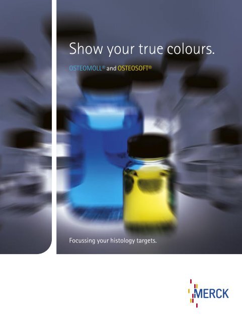
Show your true colours.

You also want an ePaper? Increase the reach of your titles
YUMPU automatically turns print PDFs into web optimized ePapers that Google loves.
<strong>Show</strong> <strong>your</strong> <strong>true</strong> <strong>colours</strong>.<br />
OSTEOMOLL® and OSTEOSOFT®<br />
Focussing <strong>your</strong> histology targets.
OSTEOMOLL®<br />
and<br />
OSTEOSOFT®<br />
Decalcifying solutions<br />
for routinely used hard tissue<br />
and sensitve hard tissue<br />
Hard tissue like bones or teeth and all speci-<br />
mens of calcified tissue like bone marrow<br />
punches require special pretreatment before<br />
routine processing by a histological laboratory.<br />
Such tissue can either be embedded in plastic<br />
and then sectioned with a microtome<br />
or it must be decalcified for routine paraffin<br />
sectioning.<br />
Decalcification is achieved either by using<br />
solutions containing inorganic acids to<br />
remove the calcium ions or, in the case of<br />
sensitive tissue, using complex-forming<br />
agents like EDTA to bind the calcium ions.<br />
The decalcification method using inorganic acids is fast<br />
and efficient; the process must be monitored as too strong<br />
a decalcification can negatively affect the morphological<br />
structure of the tissue and thus its staining result, in<br />
particular nucleus staining. Immunohistological methods<br />
cannot be employed after this as the antigens can no<br />
longer be detected by antibodies. The decalcifying solution<br />
OSTEOMOLL® is available for this type of decalcification.<br />
OSTEOMOLL® decalcifies effectively, and depending on the<br />
size and density of the tissue it can be used twice provided<br />
that the solution is still clear after the first decalcification<br />
process. OSTEOMOLL® is stained blue to differentiate it<br />
better from other working solutions. The addition of the dye<br />
has no effect on the decalcification process or the tissue<br />
in question.<br />
OSTEOSOFT® is available for gentle decalcification based<br />
on complex-forming agents. OSTEOSOFT® is best used<br />
for the decalcification of bone marrow biopsies as well as<br />
blood vessels, particularly if to be followed by immunohistological<br />
procedures. OSTEOSOFT® preserves antigen,<br />
enzyme and other fine structures, so that also e.g. the<br />
enzyme reaction of specific esterase with naphthol AS-D<br />
chloroacetate or silver impregnation of reticulin fibers<br />
can be carried out successfully. OSTEOSOFT® is used only<br />
once; the quantity required depends on the type and size<br />
of the tissue in question. OSTEOSOFT® is stained yellow for<br />
better differentiation.
Highlights<br />
• OSTEOMOLL® and OSTEOSOFT® – for processing all types of hard tissue<br />
in the histological laboratory<br />
• Stained decalcifying solutions for fast and reliable identification<br />
in the laboratory – OSTEOMOLL® is blue, OSTEOSOFT® is yellow<br />
• OSTEOMOLL® for all types of routinely used hard tissue<br />
• OSTEOMOLL® can be used twice on average<br />
• OSTEOSOFT® for gentle decalcification while preserving tissue structures,<br />
immunohistological procedures and enzyme reactions<br />
• User-friendly package size<br />
• Safe packaging with PE bottles<br />
• Developed, manufactured and tested in accordance with ISO 9001,<br />
CE certified according to IVDD<br />
Colour makes<br />
the difference.
Directions for use<br />
Bone, teeth, hard tissue<br />
The decalcification time and the quantity of OSTEOMOLL® required are<br />
dependent on the size, type, and density of the respective tissue.<br />
A piece of bone 10 x 10 x 3 mm in size, e.g. hip bone, has a decalcification<br />
time of 6 - 8 hours. The decalcification of teeth and other hard tissue<br />
should be monitored carefully.<br />
Slightly calcified tissue<br />
Slightly calcified tissue, e.g. blood vessels, is decalcified after 30 - 60minutes.<br />
Using OSTEOMOLL®<br />
Keratinized tissue<br />
Keratinized tissue, e.g. nails and acuminate warts, can be gently decalcified<br />
by immersing the cut embedded material in OSTEOMOLL® with the<br />
section surface facing downwards for at least 15 - 60 minutes, subsequently<br />
rinsing the block with tap water, and cutting in the usual manner;<br />
the new section should be kept to a minimum (loss of material). Section<br />
thickness is 5 µm.<br />
Determining the end-point of the decalcification process<br />
The end-point of the decalcification process is determined by puncturing the<br />
tissue with a needle at a representative site not of decisive relevance<br />
for the diagnostic procedure.
Using OSTEOSOFT®<br />
Directions for use<br />
Iliac crest and other hard tissue<br />
The decalcification time and the quantity of OSTEOSOFT® required are<br />
dependent on the size, type, and density of the respective tissue.<br />
A piece of bone 15 x 9x 3 mm in size, taken from e.g. the iliac crest,<br />
is immersed in approx. 50 ml of OSTEOSOFT® and has a decalcification<br />
time of 18 - 24 hours.<br />
Fig. 1<br />
Compact, calcified tissue<br />
Compact, calcified tissue 15 x10 x 3 mm in size is decalcified after<br />
48 - 72 hours. If no immunohistological procedures are planned,<br />
decalcification can be carried out more rapidly with OSTEOMOLL®.<br />
Determining the end-point of the decalcification process<br />
In the case of e.g. iliac crest tissue, the end of the decalcification process<br />
is reached when the tissue floats in the solution.<br />
Fig. 2<br />
Result<br />
Decalcified tissue is cartilaginous or rubberlike in its consistency<br />
and exhibits only a weak resistance.<br />
The decalcified tissue can be processed further in normal histology.<br />
Fig. 3<br />
Fig. 1: Iliac crest, decalcification<br />
with OSTEOSOFT®<br />
Reactive changes, detected by<br />
antibody reaction in bone marrow<br />
Fig. 2: lIiac crest, decalcification<br />
with OSTEOSOFT®<br />
Detection of reactive T- cells by<br />
antibody reaction in bone marrow<br />
Fig. 3: Iliac crest, decalcification<br />
with OSTEOSOFT®<br />
Detection of plasmocytom cells by<br />
antibody reaction in bone marrow
OSTEOMOLL®<br />
the fast decalcifying solution for histology Ord. No. 1.01736.1000 1 liter<br />
OSTEOSOFT®<br />
the gentle decalcifying solution for histology Ord. No. 1.01728.1000 1 liter<br />
W 287109 06/06<br />
Merck KGaA<br />
64271 Darmstadt, Germany<br />
Fax: 0049 (0) 61 51 72 - 60 80<br />
E-mail: microscopy@merck.de<br />
www.merck.de<br />
microscopy.merck.de<br />
We provide information and advice to our customers to the best of our knowledge and ability, but without obligation or liability. Existing laws and<br />
regulations are to be observed in all cases by our customers. This also applies in respect to any rights of third parties. Our information and advice do not<br />
relieve our customers of their own responsibility for checking the suitability of our products for the envisaged purpose. OSTEOMOLL® and OSTEOSOFT®<br />
are registered trademarks of Merck KGaA, Darmstadt, Germany.