Enhanced Archaerhodopsin Fluorescent Protein Voltage Indicators
Enhanced Archaerhodopsin Fluorescent Protein Voltage Indicators
Enhanced Archaerhodopsin Fluorescent Protein Voltage Indicators

You also want an ePaper? Increase the reach of your titles
YUMPU automatically turns print PDFs into web optimized ePapers that Google loves.
<strong>Enhanced</strong> Arch <strong>Voltage</strong> Sensors<br />
pulses failed to initiate action potentials, these induced only<br />
sub-threshold voltage depolarizations in the membrane<br />
potential (Methods). The Arch fluorescence trace reported<br />
these sub-threshold events (orange markers), but the GCaMP5<br />
fluorescence trace did not.<br />
Discussion<br />
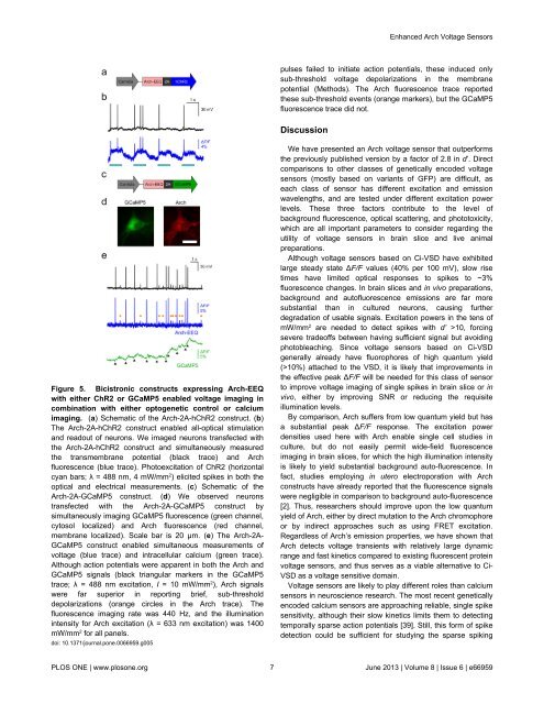
Figure 5. Bicistronic constructs expressing Arch-EEQ<br />
with either ChR2 or GCaMP5 enabled voltage imaging in<br />
combination with either optogenetic control or calcium<br />
imaging. (a) Schematic of the Arch-2A-hChR2 construct. (b)<br />
The Arch-2A-hChR2 construct enabled all-optical stimulation<br />
and readout of neurons. We imaged neurons transfected with<br />
the Arch-2A-hChR2 construct and simultaneously measured<br />
the transmembrane potential (black trace) and Arch<br />
fluorescence (blue trace). Photoexcitation of ChR2 (horizontal<br />
cyan bars; λ = 488 nm, 4 mW/mm 2 ) elicited spikes in both the<br />
optical and electrical measurements. (c) Schematic of the<br />
Arch-2A-GCaMP5 construct. (d) We observed neurons<br />
transfected with the Arch-2A-GCaMP5 construct by<br />
simultaneously imaging GCaMP5 fluorescence (green channel,<br />
cytosol localized) and Arch fluorescence (red channel,<br />
membrane localized). Scale bar is 20 µm. (e) The Arch-2A-<br />
GCaMP5 construct enabled simultaneous measurements of<br />
voltage (blue trace) and intracellular calcium (green trace).<br />
Although action potentials were apparent in both the Arch and<br />
GCaMP5 signals (black triangular markers in the GCaMP5<br />
trace; λ = 488 nm excitation, I = 10 mW/mm 2 ), Arch signals<br />
were far superior in reporting brief, sub-threshold<br />
depolarizations (orange circles in the Arch trace). The<br />
fluorescence imaging rate was 440 Hz, and the illumination<br />
intensity for Arch excitation (λ = 633 nm excitation) was 1400<br />
mW/mm 2 for all panels.<br />
doi: 10.1371/journal.pone.0066959.g005<br />
We have presented an Arch voltage sensor that outperforms<br />
the previously published version by a factor of 2.8 in d’. Direct<br />
comparisons to other classes of genetically encoded voltage<br />
sensors (mostly based on variants of GFP) are difficult, as<br />
each class of sensor has different excitation and emission<br />
wavelengths, and are tested under different excitation power<br />
levels. These three factors contribute to the level of<br />
background fluorescence, optical scattering, and phototoxicity,<br />
which are all important parameters to consider regarding the<br />
utility of voltage sensors in brain slice and live animal<br />
preparations.<br />
Although voltage sensors based on Ci-VSD have exhibited<br />
large steady state ∆F/F values (40% per 100 mV), slow rise<br />
times have limited optical responses to spikes to ~3%<br />
fluorescence changes. In brain slices and in vivo preparations,<br />
background and autofluorescence emissions are far more<br />
substantial than in cultured neurons, causing further<br />
degradation of usable signals. Excitation powers in the tens of<br />
mW/mm 2 are needed to detect spikes with d’ >10, forcing<br />
severe tradeoffs between having sufficient signal but avoiding<br />
photobleaching. Since voltage sensors based on Ci-VSD<br />
generally already have fluorophores of high quantum yield<br />
(>10%) attached to the VSD, it is likely that improvements in<br />
the effective peak ∆F/F will be needed for this class of sensor<br />
to improve voltage imaging of single spikes in brain slice or in<br />
vivo, either by improving SNR or reducing the requisite<br />
illumination levels.<br />
By comparison, Arch suffers from low quantum yield but has<br />
a substantial peak ∆F/F response. The excitation power<br />
densities used here with Arch enable single cell studies in<br />
culture, but do not easily permit wide-field fluorescence<br />
imaging in brain slices, for which the high illumination intensity<br />
is likely to yield substantial background auto-fluorescence. In<br />
fact, studies employing in utero electroporation with Arch<br />
constructs have already reported that the fluorescence signals<br />
were negligible in comparison to background auto-fluorescence<br />
[2]. Thus, researchers should improve upon the low quantum<br />
yield of Arch, either by direct mutation to the Arch chromophore<br />
or by indirect approaches such as using FRET excitation.<br />
Regardless of Arch’s emission properties, we have shown that<br />
Arch detects voltage transients with relatively large dynamic<br />
range and fast kinetics compared to existing fluorescent protein<br />
voltage sensors, and thus serves as a viable alternative to Ci-<br />
VSD as a voltage sensitive domain.<br />
<strong>Voltage</strong> sensors are likely to play different roles than calcium<br />
sensors in neuroscience research. The most recent genetically<br />
encoded calcium sensors are approaching reliable, single spike<br />
sensitivity, although their slow kinetics limits them to detecting<br />
temporally sparse action potentials [39]. Still, this form of spike<br />
detection could be sufficient for studying the sparse spiking<br />
PLOS ONE | www.plosone.org 7 June 2013 | Volume 8 | Issue 6 | e66959