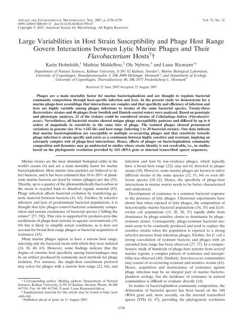
Large Variabilities in Host Strain Susceptibility and Phage Host ...
Large Variabilities in Host Strain Susceptibility and Phage Host ...
Large Variabilities in Host Strain Susceptibility and Phage Host ...

Create successful ePaper yourself
Turn your PDF publications into a flip-book with our unique Google optimized e-Paper software.
APPLIED AND ENVIRONMENTAL MICROBIOLOGY, Nov. 2007, p. 6730–6739 Vol. 73, No. 21<br />
0099-2240/07/$08.000 doi:10.1128/AEM.01399-07<br />
Copyright © 2007, American Society for Microbiology. All Rights Reserved.<br />
<strong>Large</strong> <strong>Variabilities</strong> <strong>in</strong> <strong>Host</strong> Stra<strong>in</strong> <strong>Susceptibility</strong> <strong>and</strong> <strong>Phage</strong> <strong>Host</strong> Range<br />
Govern Interactions between Lytic Mar<strong>in</strong>e <strong>Phage</strong>s <strong>and</strong> Their<br />
Flavobacterium <strong>Host</strong>s †<br />
Kar<strong>in</strong> Holmfeldt, 1 Mathias Middelboe, 2 Ole Nybroe, 3 <strong>and</strong> Lasse Riemann 1 *<br />
Department of Natural Sciences, Kalmar University, S-391 82 Kalmar, Sweden 1 ; Mar<strong>in</strong>e Biological Laboratory,<br />
University of Copenhagen, Str<strong>and</strong>promenaden 5, DK-3000 Hels<strong>in</strong>gør, Denmark 2 ; <strong>and</strong> Department of Ecology,<br />
University of Copenhagen, Thorvaldsensvej 40, DK-1871 Frederiksberg C, Denmark 3<br />
Received 25 June 2007/Accepted 23 August 2007<br />
<strong>Phage</strong>s are a ma<strong>in</strong> mortality factor for mar<strong>in</strong>e bacterioplankton <strong>and</strong> are thought to regulate bacterial<br />
community composition through host-specific <strong>in</strong>fection <strong>and</strong> lysis. In the present study we demonstrate for a<br />
mar<strong>in</strong>e phage-host assemblage that <strong>in</strong>teractions are complex <strong>and</strong> that specificity <strong>and</strong> efficiency of <strong>in</strong>fection <strong>and</strong><br />
lysis are highly variable among phages <strong>in</strong>fectious to stra<strong>in</strong>s of the same bacterial species. Twenty-three<br />
Bacteroidetes stra<strong>in</strong>s <strong>and</strong> 46 phages from Swedish <strong>and</strong> Danish coastal waters were analyzed. Based on genotypic<br />
<strong>and</strong> phenotypic analyses, 21 of the isolates could be considered stra<strong>in</strong>s of Cellulophaga baltica (Flavobacteriaceae).<br />
Nevertheless, all bacterial stra<strong>in</strong>s showed unique phage susceptibility patterns <strong>and</strong> differed by up to 6<br />
orders of magnitude <strong>in</strong> sensitivity to the same titer of phage. The isolated phages showed pronounced<br />
variations <strong>in</strong> genome size (8 to >242 kb) <strong>and</strong> host range (<strong>in</strong>fect<strong>in</strong>g 1 to 20 bacterial stra<strong>in</strong>s). Our data <strong>in</strong>dicate<br />
that mar<strong>in</strong>e bacterioplankton are susceptible to multiple co-occurr<strong>in</strong>g phages <strong>and</strong> that sensitivity towards<br />
phage <strong>in</strong>fection is stra<strong>in</strong> specific <strong>and</strong> exists as a cont<strong>in</strong>uum between highly sensitive <strong>and</strong> resistant, imply<strong>in</strong>g an<br />
extremely complex web of phage-host <strong>in</strong>teractions. Hence, effects of phages on bacterioplankton community<br />
composition <strong>and</strong> dynamics may go undetected <strong>in</strong> studies where stra<strong>in</strong> identity is not resolvable, i.e., <strong>in</strong> studies<br />
based on the phylogenetic resolution provided by 16S rRNA gene or <strong>in</strong>ternal transcribed spacer sequences.<br />
Mar<strong>in</strong>e viruses are the most abundant biological entity <strong>in</strong> the<br />
world’s oceans (4) <strong>and</strong> are a ma<strong>in</strong> mortality factor for mar<strong>in</strong>e<br />
bacterioplankton. Most mar<strong>in</strong>e virus particles are believed to <strong>in</strong>fect<br />
bacteria, <strong>and</strong> it has been estimated that 10 to 20% of planktonic<br />
mar<strong>in</strong>e bacteria are lysed by viruses (phages) per day (55).<br />
Thereby, up to a quarter of the photosynthetically fixed carbon <strong>in</strong><br />
the ocean is recycled back to dissolved organic material (65).<br />
<strong>Phage</strong> <strong>in</strong>fection affects bacterial evolution by transferr<strong>in</strong>g genetic<br />
material between bacteria (41, 62). Further, by selective<br />
<strong>in</strong>fection <strong>and</strong> lysis of predom<strong>in</strong>ant bacterial populations, it is<br />
thought that lytic phages control bacterial community composition<br />
<strong>and</strong> susta<strong>in</strong> coexistence of bacterial species (“kill<strong>in</strong>g the<br />
w<strong>in</strong>ner” [57, 58]). This view is supported by predator-prey-like<br />
oscillations of phage-host systems <strong>in</strong> aquatic environments (19,<br />
69) but is likely to simplify actual conditions, as it does not<br />
account for broad-host-range phages or bacterial acquisition of<br />
resistance (45).<br />
Many mar<strong>in</strong>e phages appear to have a narrow host range,<br />
<strong>in</strong>fect<strong>in</strong>g only the bacterial stra<strong>in</strong> with which they were isolated<br />
(24, 38, 40, 63). However, some f<strong>in</strong>d<strong>in</strong>gs <strong>in</strong>dicate that the<br />
dogma of extreme host specificity among bacteriophages may<br />
be an artifact produced by commonly used methods for phage<br />
isolation. For <strong>in</strong>stance, the s<strong>in</strong>gle-host enrichment protocol<br />
may select for phages with a narrow host range (22, 64), <strong>and</strong><br />
* Correspond<strong>in</strong>g author. Mail<strong>in</strong>g address: Department of Natural<br />
Sciences, Kalmar University, S-391 82 Kalmar, Sweden. Phone: 46 480<br />
447334. Fax: 46 480 447340. E-mail: Lasse.Riemann@hik.se.<br />
† Supplemental material for this article may be found at http://aem<br />
.asm.org/.<br />
Published ahead of pr<strong>in</strong>t on 31 August 2007.<br />
<strong>in</strong>fection <strong>and</strong> lysis by low-virulence phages, which typically<br />
have a broad host range (22), may not be detected <strong>in</strong> plaque<br />
assays (10). However, some mar<strong>in</strong>e phages are known to <strong>in</strong>fect<br />
different stra<strong>in</strong>s of the same species (12, 51, 64) or even different<br />
species (10–12). Hence, the specificity of phage-host<br />
<strong>in</strong>teractions <strong>in</strong> mar<strong>in</strong>e waters needs to be better characterized<br />
<strong>and</strong> understood.<br />
Development of resistance is a common bacterial response<br />
to the presence of lytic phages. Chemostat experiments have<br />
shown that when exposed to lytic phages, the composition of<br />
heterotrophic mar<strong>in</strong>e bacterial populations (34, 35) <strong>and</strong> Escherichia<br />
coli populations (13, 20, 30, 37) rapidly shifts from<br />
dom<strong>in</strong>ance by phage-sensitive clones to dom<strong>in</strong>ance by phageresistant<br />
clones. Consequently, phage-resistant bacterial mutants<br />
seem to be constantly produced <strong>and</strong> tend to replace the<br />
sensitive stra<strong>in</strong>s when the population is exposed to a strong<br />
selective pressure from <strong>in</strong>fectious phages. Further, for E. coli a<br />
strong coevolution of resistant bacteria <strong>and</strong> phages with an<br />
extended host range has been observed (27, 37). In a comprehensive<br />
study of hundreds of phage-host systems from several<br />
mar<strong>in</strong>e regions, a complex pattern of resistance <strong>and</strong> susceptibility<br />
was observed (40). Similarly, Synechococcus communities<br />
may consist of co-occurr<strong>in</strong>g resistant <strong>and</strong> sensitive stra<strong>in</strong>s (61).<br />
Hence, acquisition <strong>and</strong> ma<strong>in</strong>tenance of resistance aga<strong>in</strong>st<br />
phage <strong>in</strong>fection may be an <strong>in</strong>tegral part of mar<strong>in</strong>e bacterioplankton<br />
ecology, but the <strong>in</strong>cidence of resistance <strong>in</strong> natural<br />
communities is difficult to evaluate directly (14).<br />
In studies of bacterioplankton community composition, the<br />
del<strong>in</strong>eation of bacterial species has been based on the 16S<br />
rRNA gene <strong>and</strong>, more recently, on the <strong>in</strong>ternal transcribed<br />
spacer (ITS) (8, 47), provid<strong>in</strong>g the phylogenetic resolution<br />
6730
VOL. 73, 2007 COMPLEXITY OF FLAVOBACTERIUM PHAGE-HOST INTERACTIONS 6731<br />
governed by these particular gene regions. The limitations<br />
associated with such species del<strong>in</strong>eation has recently been discussed<br />
<strong>in</strong> the context of stra<strong>in</strong>-specific niche occupation (21),<br />
where freshwater bacterial isolates with identical or almost-identical<br />
16S rRNA gene <strong>and</strong> ITS sequences were suggested to represent<br />
dist<strong>in</strong>ct populations <strong>and</strong> to probably occupy different ecological<br />
niches (17, 21). These limitations may also apply <strong>in</strong> the<br />
context of stra<strong>in</strong>-specific phage-<strong>in</strong>duced bacterial mortality.<br />
In the present study a collection of 21 Cellulophaga baltica<br />
(Bacteroidetes) stra<strong>in</strong>s, which were highly similar genotypically<br />
<strong>and</strong> phenotypically, allowed us to address phage-host <strong>in</strong>teractions<br />
at the stra<strong>in</strong> level. Our results demonstrate large stra<strong>in</strong>specific<br />
differences <strong>in</strong> phage susceptibility among hosts <strong>and</strong> a<br />
pronounced variability <strong>in</strong> specificity <strong>and</strong> efficiency of <strong>in</strong>fection<br />
for the C. baltica phages.<br />
MATERIALS AND METHODS<br />
Isolation of bacteria <strong>and</strong> phages. Stra<strong>in</strong>s NN014847 to NN016048 were isolated<br />
from various Danish coastal waters <strong>in</strong> 1994 (23). Stra<strong>in</strong>s MM#3 <strong>and</strong> #4 to<br />
#19 were isolated from Øresund surface water (56°2N, 12°37E) <strong>in</strong> February<br />
2000, <strong>and</strong> stra<strong>in</strong> OL12a was isolated from Baltic Sea surface water east off Öl<strong>and</strong><br />
(56°37N, 16°45E) <strong>in</strong> April 2005. The stra<strong>in</strong>s were selected as yellow colonies on<br />
CYT agar plates (1.0 g case<strong>in</strong>, 0.5 g yeast extract, 0.5 g CaCl 2 H 2 O, 0.5 g<br />
MgSO 4 H 2 O, 15 g agar, 1,000 ml deionized water, pH 7.3) with colony morphology<br />
(size, texture, <strong>and</strong> edge appearance) <strong>and</strong> restriction fragment length<br />
polymorphism (RFLP) profiles (16S rRNA gene, HaeIII, <strong>and</strong> NdeII; Roche)<br />
similar to those of the C. baltica type stra<strong>in</strong> (stra<strong>in</strong> NN015840) (23).<br />
<strong>Phage</strong>s were obta<strong>in</strong>ed from Øresund surface water <strong>in</strong> 2000 <strong>and</strong> 2005. After a<br />
0.45-m prefiltration (Millipak; Millipore), phages were concentrated 250-fold<br />
by tangential flow filtration (Millipore) followed by Amicon ultracentrifugation<br />
(Millipore). <strong>Phage</strong>s were isolated us<strong>in</strong>g the top-agar plat<strong>in</strong>g technique (plaque<br />
assay) (48), with all of the isolated bacterial stra<strong>in</strong>s used as hosts. Plaques with<br />
different morphologies were selected from each host <strong>and</strong> purified for three<br />
rounds of <strong>in</strong>fection <strong>and</strong> lysis. Thereafter, 5 ml phage buffer (0.1 M NaCl, 0.008<br />
M MgSO 4 , 0.05 M Tris-HCl, 2 mM CaCl 2 , 0.1% gelat<strong>in</strong>, 0.5 M tryptophan, 5%<br />
glycerol, pH 7.5) was added to a fully lysed plate, <strong>and</strong> the agar overlay was<br />
shredded with a sterile <strong>in</strong>oculation loop. The plate was <strong>in</strong>cubated on a shaker for<br />
20 m<strong>in</strong>, <strong>and</strong> the mixture was then transferred to a sterile tube <strong>and</strong> centrifuged<br />
(5,000 g, 10 m<strong>in</strong>). The supernatant was passed through a 0.22-m filter (Millex<br />
GS; Millipore), <strong>and</strong> the phage stock was stored at 4°C.<br />
In order to exam<strong>in</strong>e potential effects of resistance acquisition on host range,<br />
stra<strong>in</strong>s resistant to two of the isolated phages, S M <strong>and</strong> S T , were developed. These<br />
stra<strong>in</strong>s, #3 r S M <strong>and</strong> #3 r S T , were obta<strong>in</strong>ed after exposure of stra<strong>in</strong> MM#3 to<br />
the phages <strong>in</strong> laboratory experiments <strong>and</strong> isolated from fully lysed plates.<br />
Sequenc<strong>in</strong>g of the 16S rRNA gene <strong>and</strong> the ITS. Bacterial DNA was extracted<br />
us<strong>in</strong>g the DNeasy tissue kit (QIAGEN), <strong>and</strong> the 16S rRNA gene was PCR<br />
amplified us<strong>in</strong>g PuReTaq Ready-To-Go Beads (Amersham Biosciences) <strong>and</strong><br />
primers 27f <strong>and</strong> 1492r (15). The thermal program consisted of 30 cycles of 30 s<br />
of denaturation at 95°C, 30 s of anneal<strong>in</strong>g at 50°C, <strong>and</strong> 45 s of extension at 72°C,<br />
followed by 7 m<strong>in</strong> at 72°C. The ITS was amplified us<strong>in</strong>g primers G1 <strong>and</strong> 23Sr (21)<br />
<strong>and</strong> a thermal program consist<strong>in</strong>g of 35 cycles of 30 s of denaturation at 95°C, 30 s<br />
of anneal<strong>in</strong>g at 54°C, <strong>and</strong> 45 s of extension at 72°C, followed by 72°C for 7 m<strong>in</strong>.<br />
DNA was sequenced bidirectionally (commercially by Macrogen, Korea) us<strong>in</strong>g<br />
primers 27f, 530f, 519r, <strong>and</strong> 907r (26) for the 16S rRNA gene <strong>and</strong> primers G1 <strong>and</strong><br />
23Sr for the ITS. In some cases, the PCR-amplified ITS region could not be<br />
sequenced directly. The PCR products were therefore cloned prior to sequenc<strong>in</strong>g<br />
(TOPO TA clon<strong>in</strong>g kit; Invitrogen). Sequences were aligned <strong>in</strong><br />
Seqman (DNASTAR), <strong>and</strong> phylogenetic trees were constructed us<strong>in</strong>g the<br />
neighbor-jo<strong>in</strong><strong>in</strong>g algorithm <strong>in</strong> Clustal X (59).<br />
Genotyp<strong>in</strong>g by UP-PCR. Universally primed PCR (UP-PCR) is a whole-genome<br />
f<strong>in</strong>gerpr<strong>in</strong>t<strong>in</strong>g technique orig<strong>in</strong>ally developed for fungi (see reference 31<br />
for a review) <strong>and</strong> recently used to discrim<strong>in</strong>ate between genetically similar<br />
bacteria (7, 70). The UP-PCR method uses a s<strong>in</strong>gle primer to prime genomes<br />
arbitrarily, <strong>and</strong> the primer is designed to primarily target less-conserved <strong>in</strong>tergenic<br />
areas (31). Crude DNA templates for PCR (cell lysates) were prepared<br />
from each bacterial isolate as described by Br<strong>and</strong>t et al. (7) <strong>and</strong> stored at 20°C<br />
until use. UP-PCR was performed us<strong>in</strong>g the universal L15/AS19 primer 5-GAG<br />
GGT GGC GGC TAG-3 (32). PCR mixtures (20 l) consisted of molecular<br />
biology reagent-grade water (Sigma), buffer (supplied with DNA polymerase), 2<br />
mM MgCl 2 (supplied with DNA polymerase), 100 M of each deoxynucleoside<br />
triphosphate (AH Diagnostics), 1 ng l 1 of PCR primer (DNA Technology), 1<br />
unit of Dynazyme II DNA polymerase (F<strong>in</strong>nzymes, F<strong>in</strong>l<strong>and</strong>), <strong>and</strong> 1 l template<br />
DNA. PCR was carried out us<strong>in</strong>g the follow<strong>in</strong>g program: <strong>in</strong>itial denaturation at<br />
94°C for 3 m<strong>in</strong> <strong>and</strong> then 32 cycles consist<strong>in</strong>g of denaturation at 94°C for 60 s,<br />
primer anneal<strong>in</strong>g 53°C for 60 s, <strong>and</strong> elongation at 72°C for 60 s. In the first <strong>and</strong><br />
the last cycles, the elongation steps were extended to 90 s <strong>and</strong> 3 m<strong>in</strong>, respectively.<br />
PCR products were subjected to agarose gel electrophoresis (1.5%, 100 V, 40<br />
m<strong>in</strong>) <strong>and</strong> sta<strong>in</strong>ed with ethidium bromide. A 100-bp DNA ladder (Fermenta) was<br />
used as a molecular size marker. UP-PCR analysis was performed at least twice<br />
for each isolate to verify that reproducible b<strong>and</strong> patterns were obta<strong>in</strong>ed. Subsequently,<br />
the isolates were grouped based on identical UP-PCR patterns.<br />
Analysis of lipopolysaccharide (LPS) profiles. Bacterial stra<strong>in</strong>s were grown <strong>in</strong><br />
MLB medium (0.5 g Casam<strong>in</strong>o Acids, 0.5 g peptone, 0.5 g yeast extract, 3 ml<br />
glycerol, 500 ml filtered seawater [Øresund], <strong>and</strong> 500 ml dem<strong>in</strong>eralized water) at<br />
room temperature for 72 h. Pseudomonas fluorescens stra<strong>in</strong> Ag1, which was used<br />
as a reference, was grown to early stationary phase <strong>in</strong> LB medium (10 g tryptone,<br />
5 g yeast extract, 1 g glucose, 10 g NaCl, 1 liter Milli-Q) at 28°C.<br />
For preparation of prote<strong>in</strong>ase K-digested cell lysates, cells were centrifuged<br />
(10,000 g, 5 m<strong>in</strong>), washed once <strong>in</strong> phosphate-buffered sal<strong>in</strong>e, <strong>and</strong> resuspended<br />
<strong>in</strong> lysis buffer (0.5 M Tris-HCl [pH 6.8], 2% sodium dodecyl sulfate (SDS), 10%<br />
glycerol). Cell concentrations were normalized dur<strong>in</strong>g resuspension. Prote<strong>in</strong>ase<br />
K (75 g ml 1 ) was added <strong>and</strong> the lysates <strong>in</strong>cubated at 56°C for 1 h, boiled for<br />
5 m<strong>in</strong>, centrifuged (14,000 g, 30 m<strong>in</strong>), <strong>and</strong> stored at 20°C. Lysates were<br />
analyzed by SDS-polyacrylamide gel electrophoresis us<strong>in</strong>g 14% acrylamide–0.4%<br />
bisacrylamide <strong>and</strong> 4 M urea as described by Sørensen et al. (52), <strong>and</strong> a presta<strong>in</strong>ed<br />
prote<strong>in</strong> ladder (PageRuler Ladder Plus; Fermentas) was <strong>in</strong>cluded on each gel.<br />
After electrophoresis, gels were fixed <strong>in</strong> ethanol-acetic acid overnight <strong>and</strong> sta<strong>in</strong>ed<br />
with a periodic acid-silver sta<strong>in</strong> (60). Specificity of the sta<strong>in</strong><strong>in</strong>g was ensured by<br />
monitor<strong>in</strong>g silver sta<strong>in</strong><strong>in</strong>g of LPS from the <strong>in</strong>ternal control (P. fluorescens Ag1)<br />
without obta<strong>in</strong><strong>in</strong>g sta<strong>in</strong><strong>in</strong>g of the <strong>in</strong>cluded prote<strong>in</strong> ladder. The stra<strong>in</strong>s were<br />
grouped based on similar LPS profiles. The profiles were reproducible between<br />
<strong>in</strong>dependent replications, yet the <strong>in</strong>tensities of <strong>in</strong>dividual b<strong>and</strong>s, especially <strong>in</strong> the<br />
low-molecular-weight region, varied between runs.<br />
Specificity of phage <strong>in</strong>fection. To determ<strong>in</strong>e phage host range <strong>and</strong> the bacterial<br />
susceptibility to specific phages, a cross <strong>in</strong>fectivity test where all bacterial stra<strong>in</strong>s<br />
were exposed to all <strong>in</strong>dividual phage stocks was performed. Three microliters of<br />
phage stock (10 8 to 10 11 PFU ml 1 ) was spotted onto a lawn of host bacteria <strong>in</strong><br />
top agar, <strong>and</strong> plates were exam<strong>in</strong>ed for cell lysis after 1, 3, <strong>and</strong> 5 days. These spot<br />
tests were performed three <strong>in</strong>dependent times. Infection was considered positive<br />
when lysis (i.e., a clear<strong>in</strong>g zone) was seen <strong>in</strong> all three tests. In spot tests, spots may<br />
orig<strong>in</strong>ate from <strong>in</strong>hibition of bacterial growth, often seen as turbid spots, rather<br />
than direct viral lysis (39). To verify the presence of <strong>in</strong>fectious phages, plaque<br />
assays <strong>in</strong> dilution series were performed <strong>in</strong> some cases where turbid spots were<br />
seen. In all cases plaques were obta<strong>in</strong>ed, suggest<strong>in</strong>g that phage lysis caused the<br />
clear<strong>in</strong>g zones. An unweighted-pair group method us<strong>in</strong>g average l<strong>in</strong>kages<br />
(UPGMA) tree was constructed us<strong>in</strong>g the software Quantity One 4.2.1 (Bio-<br />
Rad), where the lysis/no lysis matrix was converted to pairwise distances us<strong>in</strong>g<br />
the Dice similarity coefficient.<br />
S<strong>in</strong>ce several bacterial stra<strong>in</strong>s were susceptible to the same phage, we sought<br />
to determ<strong>in</strong>e whether phage <strong>in</strong>fection was equally efficient <strong>in</strong> different hosts.<br />
Three hosts were exposed to the same phage titer, <strong>and</strong> <strong>in</strong>fectivity was quantified<br />
by plaque assay. PFU were exam<strong>in</strong>ed after 1, 2, <strong>and</strong> 3 days. This was performed<br />
for three different phages.<br />
<strong>Phage</strong> genome sizes. <strong>Phage</strong> genome sizes were determ<strong>in</strong>ed by pulsed-field gel<br />
electrophoresis (PFGE). Freshly made phage stocks (see above) were concentrated<br />
by ultracentrifugation (222,000 g, 2.5 h, 4°C; Beckman). DNA was<br />
obta<strong>in</strong>ed accord<strong>in</strong>g to the protocol for DNA (3) <strong>and</strong> quantified fluorometrically<br />
(PicoGreen; Molecular Probes). Some phage genomes dis<strong>in</strong>tegrated when<br />
extracted us<strong>in</strong>g this protocol. These were therefore lysed <strong>in</strong> agar plugs. Equal<br />
amounts of phage concentrate <strong>and</strong> melted 1.5% low-melt<strong>in</strong>g-po<strong>in</strong>t agarose<br />
(Sigma) <strong>in</strong> MSM buffer without gelat<strong>in</strong> (53) (450 mM NaCl, 50 mM MgSO 4 ,50<br />
mM Tris, pH 8.0) were mixed, transferred to plug molds, <strong>and</strong> left to solidify at<br />
4°C. The plugs were <strong>in</strong>cubated <strong>in</strong> lysis solution (0.25 M EDTA [pH 8], 1% SDS,<br />
1mgml 1 prote<strong>in</strong>ase K) overnight, washed three times <strong>in</strong> TE buffer (10 mM<br />
Tris-HCl, 1 mM EDTA, pH 8), <strong>and</strong> stored <strong>in</strong> TE at 4°C.<br />
For PFGE analysis, 20 ng DNA or 5 10 7 phage particles were loaded <strong>in</strong> each<br />
well. PFGE was performed on a CHEF-DR III system (Bio-Rad) us<strong>in</strong>g 1%<br />
PFGE certified agarose (Sigma). The gel was run for 11 h <strong>in</strong> 0.5 TBE buffer<br />
(1 TBE is 89 M Tris, 2 mM EDTA, <strong>and</strong> 89 mM boric acid, pH 8.3), at a 0.5-<br />
to 5.0-s switch time, 6 V/cm, <strong>and</strong> an <strong>in</strong>cluded angel of 120°. The gel was sta<strong>in</strong>ed<br />
with SYBR gold (Molecular Probes) for 30 m<strong>in</strong>, desta<strong>in</strong>ed <strong>in</strong> 0.5 TBE for 30<br />
m<strong>in</strong>, <strong>and</strong> photographed (Gel Doc XR; Bio-Rad). Genome sizes were determ<strong>in</strong>ed
6732 HOLMFELDT ET AL. APPL. ENVIRON. MICROBIOL.<br />
Taxon<br />
TABLE 1. Genetic <strong>and</strong> morphological characteristics of bacterial stra<strong>in</strong>s used <strong>in</strong> this study<br />
Stra<strong>in</strong><br />
Isolation<br />
yr<br />
Isolation<br />
location<br />
homology b 16S rRNA gene ITS UP-PCR LPS<br />
Colony<br />
morphology<br />
% Similarity c (GenBank accession no.) Group no. accord<strong>in</strong>g to:<br />
% DNA<br />
Cellulophaga NN015840 a 1994 Svaneke 100 100.0 (AJ005972) 100.0 (EF667121) 10 1 7<br />
baltica NN016038 1994 Svaneke 100 100.0 (EF667101) 98.7 (EF667122) 9 1 7<br />
NN015839 1994 Svaneke 100 100.0 (EF667102) 99.1 (EF667123) 10 1 7<br />
NN014845 1994 Svaneke 100 100.0 (EF667103) 99.6 (EF667124) 10 1 6<br />
NN014847 1994 Svaneke 102 100.0 (EF667104) 98.7 (EF667125) 9 1 8<br />
NN014850 1994 Svaneke 100 100.0 (EF667105) 99.9 (EF667126) 10 1 6<br />
NN014873 1994 Svaneke 102 100.0 (EF667106) 99.9 (EF667127) 10 1 6<br />
NN016046 1994 Ellekilde 78 100.0 (EF667107) 98.8 (EF667128) 2 1 10<br />
Hage<br />
NN016048 1994 Ellekilde 71 100.0 (EF667108) 98.1 (EF667129) 3 1 10<br />
Hage<br />
MM#3 2000 Øresund 100.0 (AF497997) 98.4 (EF667130) 2 1 2<br />
#4 2000 Øresund 100.0 (EF667109) 98.5 (EF667131) 4 1 2<br />
#10 2000 Øresund 100.0 (EF667110) 98.7 (EF667132) 5 1 7<br />
#12 2000 Øresund 100.0 (EF667111) 98.5 (EF667133) 6 1 3<br />
#13 2000 Øresund 99.5 (EF667112) 98.8 (EF667134) 1 1 9<br />
#14 2000 Øresund 99.5 (EF667113) 98.5 (EF667135) 2 1 4<br />
#17 2000 Øresund 99.5 (EF667114) 98.1 (EF667136) 7 1 4<br />
#18 2000 Øresund 100.0 (EF667115) 99.0 (EF667137) 8 1 5<br />
#19 2000 Øresund 99.5 (EF667116) 98.2 (EF667138) 2 1 4<br />
OL12a 2005 Off Öl<strong>and</strong> 99.5 (EF667117) 98.9 (EF667139) 10 1 8<br />
#3 r S M Laboratory 100.0 (EF667118) 98.4 (EF667140) 2 1 1<br />
#3 r S T Laboratory 100.0 (EF667119) 98.4 (EF667141) 2 1 2<br />
Cellulophaga<br />
fucicola<br />
Flavobacteriaceae<br />
bacterium IE-7<br />
NN015860 1994 Hirsholm 0 92.3 (AJ005973) 68.4 (EF667142) 11 2 11<br />
#8 2000 Øresund 88.2 (EF667120) 75.1 (EF667143) 12 1 12<br />
a C. baltica type stra<strong>in</strong> (23).<br />
b DNA homology with NN015840 (23).<br />
c Sequence similarity to NN015840.<br />
manually us<strong>in</strong>g an 8- to 48-kb st<strong>and</strong>ard (Bio-Rad) <strong>and</strong> a PFGE marker (Amersham)<br />
as molecular size markers.<br />
Restriction digests of phage genomes. Some phages with identical genome<br />
sizes showed differences <strong>in</strong> host range. To exam<strong>in</strong>e whether these phages were<br />
genetically different, they were compared by RFLP analysis. H<strong>in</strong>dIII was chosen<br />
after test<strong>in</strong>g four different restriction enzymes (NdeII, HaeIII, EcoRI, <strong>and</strong><br />
H<strong>in</strong>dIII; Boehr<strong>in</strong>ger Mannheim), <strong>and</strong> phage DNA (100 ng) was cleaved accord<strong>in</strong>g<br />
to the manufacturer’s recommendation. The restriction digests were analyzed<br />
on an agarose gel (0.7%, 90 V, 2 h), sta<strong>in</strong>ed with ethidium bromide, <strong>and</strong> photographed<br />
as described above. A GeneRuler 1-kb DNA ladder (Fermenta) was<br />
used as a molecular size marker.<br />
TEM. Selected phages represent<strong>in</strong>g a range <strong>in</strong> genome size <strong>and</strong> host range<br />
were exam<strong>in</strong>ed by transmission electron microscopy (TEM). Freshly made lysates<br />
were obta<strong>in</strong>ed from fully lysed plates (see above). Grids were prepared by<br />
plac<strong>in</strong>g 8 l of lysate onto 200-mesh Formvar-coated copper grids (Ted Pella) for<br />
2 m<strong>in</strong>. The phage solution was removed with filter paper, <strong>and</strong> grids were subsequently<br />
sta<strong>in</strong>ed with 8 l 2% sodium phosphotungstate (phages 40:2, 18:4,<br />
18:4, 38:1, 39:1, 4:1, <strong>and</strong> 3:2) or 2% uranyl acetate (phages 13:1, 18:1,<br />
12:1, S M , <strong>and</strong> 10:1) for 2 m<strong>in</strong>. The grids were exam<strong>in</strong>ed us<strong>in</strong>g a Zeiss EM<br />
900 microscope with an accelerat<strong>in</strong>g voltage of 80 kV.<br />
Nucleotide sequence accession numbers. The partial 16S rRNA gene sequences<br />
<strong>and</strong> ITS sequences obta<strong>in</strong>ed <strong>in</strong> this study have been deposited <strong>in</strong><br />
GenBank <strong>and</strong> assigned accession numbers EF667101 to EF667143 (Table 1).<br />
Bacterial characterization. A total of 23 bacterial stra<strong>in</strong>s<br />
obta<strong>in</strong>ed from five localities (Fig. 1) <strong>in</strong> 1994, 2000, <strong>and</strong> 2005<br />
were characterized <strong>and</strong> grouped accord<strong>in</strong>g to the parameters<br />
shown <strong>in</strong> Table 1. The C. baltica stra<strong>in</strong>s formed yellow colonies<br />
on nutrient agar plates <strong>and</strong> could be divided <strong>in</strong>to 10 morphological<br />
groups. Only four of the C. baltica stra<strong>in</strong>s were regarded<br />
as hav<strong>in</strong>g unique colony morphology. The rema<strong>in</strong><strong>in</strong>g isolates<br />
were identical to up to three other isolates. For the phageresistant<br />
stra<strong>in</strong>s, stra<strong>in</strong> #3 r S T had morphology identical to<br />
that of MM#3, while stra<strong>in</strong> #3 r S M differed (Table 1).<br />
RESULTS<br />
FIG. 1. Sampl<strong>in</strong>g sites for bacterial (b) <strong>and</strong> phage (p) isolations.<br />
Exact locations are marked by arrows. b1, Hirsholm; b2, Ellekilde<br />
Hage; b3, Svaneke; b4, Øresund; b5, off Öl<strong>and</strong>. The map was generated<br />
us<strong>in</strong>g the Onl<strong>in</strong>e Map Creation software (http://www.aquarius<br />
.geomar.de/omc/).
VOL. 73, 2007 COMPLEXITY OF FLAVOBACTERIUM PHAGE-HOST INTERACTIONS 6733<br />
FIG. 2. Neighbor-jo<strong>in</strong><strong>in</strong>g trees show<strong>in</strong>g the phylogenetic relationships between bacteria used <strong>in</strong> this study. Trees are based on 16S rRNA gene<br />
sequences (800 bp) (A) <strong>and</strong> ITS gene sequences (675 bp) (B). Phylogenetic relationships were bootstrapped 1,000 times, <strong>and</strong> values greater<br />
than 50% are shown.<br />
Sequenc<strong>in</strong>g of the 16S rRNA gene <strong>and</strong> ITS. Partial sequenc<strong>in</strong>g<br />
of the 16S rRNA gene (800 bp) showed that 15 stra<strong>in</strong>s<br />
were identical to the C. baltica type stra<strong>in</strong> NN015840, while 5<br />
stra<strong>in</strong>s showed a 4-bp difference from this bacterium (Table 1;<br />
Fig. 2A). Stra<strong>in</strong>s NN015860 (Cellulophaga fucicola, accession<br />
no. AJ005973) <strong>and</strong> #8 (98% similar to Flavobacteriaceae bacterium<br />
IE-7, accession no. EF105392) showed only 93% <strong>and</strong><br />
88% 16S rRNA gene similarity to the other stra<strong>in</strong>s (Fig. 2A).<br />
These stra<strong>in</strong>s also had shorter ITS (800 <strong>and</strong> 700 bp, respectively)<br />
than the C. baltica stra<strong>in</strong>s (900 bp). All the C. baltica<br />
stra<strong>in</strong>s showed high ITS sequence similarity (98 to 100%) (Table<br />
1) <strong>and</strong> formed a tight phylogenetic cluster (Fig. 2B).<br />
UP-PCR <strong>and</strong> LPS profil<strong>in</strong>g. Due to the high sequence similarities<br />
of 16S rRNA genes <strong>and</strong> ITS, we sought to dist<strong>in</strong>guish<br />
the C. baltica stra<strong>in</strong>s by means of UP-PCR f<strong>in</strong>gerpr<strong>in</strong>t<strong>in</strong>g. Accord<strong>in</strong>gly,<br />
the C. baltica stra<strong>in</strong>s could be divided <strong>in</strong>to 10 different<br />
UP-PCR groups (Table 1). Still, several of the stra<strong>in</strong>s<br />
had identical UP-PCR f<strong>in</strong>gerpr<strong>in</strong>ts, <strong>and</strong> three of the UP-PCR<br />
groups conta<strong>in</strong>ed two to six identical isolates (Fig. 3).<br />
Acquisition of phage resistance may yield detectable<br />
changes <strong>in</strong> LPS profiles (see, e.g., references 18, 37, <strong>and</strong> 56);<br />
however, we observed no differences <strong>in</strong> LPS profiles among<br />
the C. baltica isolates despite their pronounced variation <strong>in</strong><br />
phage susceptibility (see below). Only isolate NN015860 (C.<br />
fucicola) had a different LPS profile (Table 1; see Fig. S1 <strong>in</strong><br />
the supplemental material).<br />
<strong>Phage</strong> characterization. <strong>Phage</strong>s were obta<strong>in</strong>ed from Øresund<br />
<strong>in</strong> 2000 <strong>and</strong> 2005 us<strong>in</strong>g plaque assays (Fig. 1). Plaques<br />
were isolated for all bacterial host stra<strong>in</strong>s except for stra<strong>in</strong>s #3<br />
r S M , #8, <strong>and</strong> NN014845, <strong>and</strong> a total of 45 phages <strong>in</strong>fect<strong>in</strong>g<br />
the C. baltica stra<strong>in</strong>s <strong>and</strong> 1 phage <strong>in</strong>fect<strong>in</strong>g C. fucicola were<br />
obta<strong>in</strong>ed. From a s<strong>in</strong>gle water sample, up to four different
6734 HOLMFELDT ET AL. APPL. ENVIRON. MICROBIOL.<br />
FIG. 3. Genomic profil<strong>in</strong>g of bacterial isolates. An agarose gel<br />
show<strong>in</strong>g UP-PCR b<strong>and</strong><strong>in</strong>g patterns for 15 of the 23 bacterial isolates<br />
analyzed is shown. Identical stra<strong>in</strong>s (e.g., stra<strong>in</strong>s NN015839 <strong>and</strong><br />
NN015840) <strong>and</strong> stra<strong>in</strong>s with differences (e.g., stra<strong>in</strong>s 10 <strong>and</strong> 12) are<br />
displayed.<br />
plaque morphologies could be dist<strong>in</strong>guished for a s<strong>in</strong>gle bacterial<br />
host (e.g., 48:1 to 48:4 [Table 2]).<br />
<strong>Phage</strong> genome size <strong>and</strong> morphology. To genetically dist<strong>in</strong>guish<br />
the phages, genome sizes were determ<strong>in</strong>ed by PFGE<br />
analysis. <strong>Phage</strong> genome sizes ranged from 8 to 242 kb <strong>and</strong><br />
represented 18 different sizes (Table 2; Fig. 4A). Several<br />
phages had identical genome sizes, <strong>and</strong> only 10 phages were<br />
unique <strong>in</strong> size (Table 2). Some phages showed double b<strong>and</strong>s on<br />
the PFGE with up to a 4-fold difference <strong>in</strong> molecular size.<br />
For example 3:2 had b<strong>and</strong>s of 18 kb <strong>and</strong> 76 kb. Some phages<br />
with very different genome sizes were <strong>in</strong>fectious to the same<br />
bacterial stra<strong>in</strong>. For <strong>in</strong>stance, stra<strong>in</strong> NN015839 could be <strong>in</strong>fected<br />
by both 40:2 ( 242 kb) <strong>and</strong> 39:1 (29 kb).<br />
Selected phages show<strong>in</strong>g a large range <strong>in</strong> genome size (8 to<br />
242 kb) <strong>and</strong> host range (1 to 20 hosts) represented a wide<br />
variety of morphotypes (Fig. 5) <strong>and</strong> belonged to the Myoviridae,<br />
Siphoviridae, <strong>and</strong> Podoviridae families (1) (Table 2).<br />
Broad- <strong>and</strong> narrow-host-range phages were found <strong>in</strong> all three<br />
families. Two phages show<strong>in</strong>g two b<strong>and</strong>s each <strong>in</strong> the PFGE<br />
analysis were exam<strong>in</strong>ed by TEM. For phages 3:2 <strong>and</strong> 18:4,<br />
one <strong>and</strong> two morphotypes could be observed, respectively (Fig.<br />
5). Interest<strong>in</strong>gly, some phages with identical genome sizes, e.g.,<br />
12:1 <strong>and</strong> 18:1, belonged to different families (Table 2;<br />
Fig. 5).<br />
<strong>Host</strong> range <strong>and</strong> diversity of obta<strong>in</strong>ed phages. To exam<strong>in</strong>e<br />
whether the phages were specific for C. baltica, <strong>in</strong>fectivity was<br />
tested on five Bacteroidetes isolates, one alphaproteobacterium,<br />
<strong>and</strong> one gammaproteobacterium. No <strong>in</strong>fections were<br />
observed (data not shown). Also, no cross-<strong>in</strong>fectivity was observed<br />
for C. baltica <strong>and</strong> C. fucicola phages, <strong>and</strong> no phages<br />
<strong>in</strong>fected stra<strong>in</strong> #8 (Fig. 6). With<strong>in</strong> C. baltica, the phages<br />
showed a large variation <strong>in</strong> <strong>in</strong>fectivity (Fig. 6). Six phages<br />
showed narrow host specificity, <strong>in</strong>fect<strong>in</strong>g only the bacterial<br />
stra<strong>in</strong> with which they were orig<strong>in</strong>ally isolated, but a broader<br />
host range was more common. A few phages were able to<br />
<strong>in</strong>fect all bacterial stra<strong>in</strong>s except stra<strong>in</strong>s #3 r S M <strong>and</strong> #3 r<br />
S T . No relationship between host range <strong>and</strong> time of isolation<br />
was found.<br />
In some cases there were no or only small differences <strong>in</strong> host<br />
range between phages with identical genome size. To determ<strong>in</strong>e<br />
whether these phages were genetically different, they<br />
were exam<strong>in</strong>ed by RFLP analysis. The RFLP patterns showed<br />
that phages with even m<strong>in</strong>or differences <strong>in</strong> host range were<br />
genetically different <strong>and</strong> therefore should be regarded as<br />
unique (Fig. 4B). For <strong>in</strong>stance, phages 4:1 <strong>and</strong> 17:2 had a<br />
genome size of 145 kb (Table 2), differed with respect to<br />
<strong>in</strong>fection of a s<strong>in</strong>gle bacterial stra<strong>in</strong> (Fig. 6), <strong>and</strong> showed<br />
slightly different RFLP profiles (Fig. 4B). <strong>Phage</strong>s with identical<br />
genome sizes, host ranges, <strong>and</strong> RFLP patterns were considered<br />
identical (e.g., phages 12:1 <strong>and</strong> 12:3 to 12:5 [Table 2; Fig.<br />
4 <strong>and</strong> 6]). Of the 45 isolated phages, 40 were concluded to be<br />
unique. In all cases, identical phages orig<strong>in</strong>ated from the same<br />
water sample.<br />
<strong>Phage</strong><br />
TABLE 2. <strong>Phage</strong>s isolated <strong>and</strong> analyzed <strong>in</strong> this study<br />
Orig<strong>in</strong>al<br />
host stra<strong>in</strong><br />
Isolation<br />
yr<br />
Genome size<br />
(kb) a<br />
<strong>Phage</strong> family b<br />
40:1 NN015840 2005 68 ND<br />
40:2 NN015840 2005 242 Myoviridae<br />
38:1 NN016038 2005 70 Podoviridae<br />
38:2 NN016038 2005 54 ND<br />
39:1 NN015839 2000 29 Siphoviridae<br />
39:2 NN015839 2005 242 ND<br />
47:1 NN014847 2005 54 ND<br />
50:1 NN014850 2005 242 ND<br />
73:1 NN014873 2005 242 ND<br />
46:1 NN016046 2000 38 ND<br />
46:2 NN016046 2005 21/85 ND<br />
46:3 NN016046 2005 78 ND<br />
46:4 NN016046 2005 145 ND<br />
48:1 NN016048 2005 9/19 ND<br />
48:2 NN016048 2005 18/72 ND<br />
48:3 NN016048 2005 18/72 ND<br />
48:4 NN016048 2005 145 ND<br />
S M MM#3 2000 54 Myoviridae<br />
S T MM#3 2000 97 ND<br />
3:1 MM#3 2000 54 ND<br />
3:2 MM#3 2005 18/76 Myoviridae<br />
4:1 #4 2005 145 Myoviridae?<br />
10:1 #10 2000 54 Myoviridae<br />
12:1 #12 2000 40 Siphoviridae<br />
12:2 #12 2005 8/17 ND<br />
12:3 #12 2000 40 ND<br />
12:4 #12 2000 40 ND<br />
12:5 #12 2000 40 ND<br />
13:1 #13 2000 97 Siphoviridae<br />
13:2 #13 2005 78 ND<br />
14:1 #14 2005 9/19 ND<br />
14:2 #14 2005 110 ND<br />
17:1 #17 2000 42 ND<br />
17:2 #17 2005 145 ND<br />
18:1 #18 2000 40 Podoviridae?<br />
18:2 #18 2000 40 ND<br />
18:3 #18 2005 78 ND<br />
18:4 #18 2005 9/19 Siphoviridae/<br />
Siphoviridae<br />
19:1 #19 2000 60 ND<br />
19:2 #19 2000 97 ND<br />
19:3 #19 2005 70/80 ND<br />
19:4 #19 2005 242 ND<br />
12a:1 OL12a 2005 9/19 ND<br />
3S T :1 #3 r S T 2005 18/76 ND<br />
3S T :2 #3 r S T 2005 54 ND<br />
60:1 NN015860 2000 40 ND<br />
a The presence of two values <strong>in</strong>dicates two b<strong>and</strong>s <strong>in</strong> the PFGE.<br />
b ND, no data.
VOL. 73, 2007 COMPLEXITY OF FLAVOBACTERIUM PHAGE-HOST INTERACTIONS 6735<br />
FIG. 5. TEM of selected C. baltica phages. Bars, 50 nm. (A) 40:2;<br />
(B) 18:4; (C) 18:4; (D) 38:1; (E) 13:1; (F) 39:1; (G) 4:1;<br />
(H) 3:2; (I) 18:1; (J) 12:1; (K) S M ; (L) 10:1. The phage 18:4<br />
lysate <strong>in</strong>cluded two morphotypes (B <strong>and</strong> C). Examples of phages with<br />
identical genome sizes but with different morphotypes are shown (e.g.,<br />
I versus J <strong>and</strong> K versus L). Genome sizes <strong>and</strong> host ranges for the<br />
phages shown are given <strong>in</strong> Table 2 <strong>and</strong> Fig. 6.<br />
FIG. 4. Genomic characterization of phages. (A) PFGE analysis of<br />
genomic DNAs from 9 of the 46 phage isolates used <strong>in</strong> this study. The gel<br />
displays phages with identical (e.g., 12:4 <strong>and</strong> 12:5) <strong>and</strong> different (e.g.,<br />
18:3 <strong>and</strong> 39:1) genomes sizes. (B) H<strong>in</strong>dIII restriction digests of eight<br />
phage genomes. Cleavage patterns for phages with identical (12:4 <strong>and</strong><br />
12:5) <strong>and</strong> different (e.g., 17:1 <strong>and</strong> 18:1) <strong>in</strong>fectivity patterns are shown<br />
along with patterns for phages with almost identical <strong>in</strong>fectivity patterns<br />
(4:1 <strong>and</strong> 17:2; see Fig. 6). Arrowheads <strong>in</strong>dicate differences <strong>in</strong> b<strong>and</strong><strong>in</strong>g<br />
patterns for 4:1 <strong>and</strong> 17:2. See Table 2 for phage genome sizes.<br />
<strong>Susceptibility</strong> <strong>and</strong> resistance to phage <strong>in</strong>fection. The phage<br />
susceptibility of the C. baltica stra<strong>in</strong>s was highly variable. For<br />
<strong>in</strong>stance, the most (stra<strong>in</strong> #13) <strong>and</strong> the least (stra<strong>in</strong> NN014845)<br />
susceptible stra<strong>in</strong>s could be <strong>in</strong>fected by 27 <strong>and</strong> 12 different phages,<br />
respectively (Fig. 6). All stra<strong>in</strong>s showed unique susceptibility patterns.<br />
To facilitate a comparison with other bacterial characteristics,<br />
the phage susceptibility data were analyzed by<br />
UPGMA <strong>and</strong> converted to a dendrogram (Fig. 7). In some<br />
cases the cluster<strong>in</strong>g was consistent with genetic or morphological<br />
characteristics. For <strong>in</strong>stance, for stra<strong>in</strong>s NN015840,<br />
NN014845, NN014850, <strong>and</strong> NN014873, the phage susceptibility<br />
was consistent with high similarity of the 16S rRNA<br />
gene, UP-PCR, <strong>and</strong> colony morphology as well as with a<br />
100% match <strong>in</strong> DNA homology (23). Stra<strong>in</strong>s #13, #14, #17,<br />
<strong>and</strong> #19 grouped accord<strong>in</strong>g to 16S rRNA gene sequences<br />
<strong>and</strong> phage susceptibility but showed small differences <strong>in</strong><br />
UP-PCR pattern <strong>and</strong> colony morphology (Fig. 2A <strong>and</strong> 7).<br />
For most stra<strong>in</strong>s, no consistency was observed between<br />
phage susceptibility <strong>and</strong> any of the analyzed stra<strong>in</strong> characteristics.<br />
The effects of resistance acquisition were exam<strong>in</strong>ed for<br />
stra<strong>in</strong> MM#3. When resistance aga<strong>in</strong>st S M was acquired,<br />
MM#3 became resistant towards 21 phages, while resistance<br />
aga<strong>in</strong>st S T conferred resistance aga<strong>in</strong>st only 3 phages (Fig. 6).<br />
To determ<strong>in</strong>e whether phage <strong>in</strong>fection was equally efficient <strong>in</strong><br />
different hosts, <strong>in</strong>fectivity was exam<strong>in</strong>ed for three phages by expos<strong>in</strong>g<br />
three susceptible hosts to the same phage titer (Table 3).<br />
The number of PFU varied up to 6 orders of magnitude between<br />
host stra<strong>in</strong>s.<br />
DISCUSSION<br />
Similarity of the bacterial stra<strong>in</strong>s. C. baltica stra<strong>in</strong>s are<br />
members of the Flavobacteria class with<strong>in</strong> the Bacteroidetes,<br />
which is one of the dom<strong>in</strong>ant phyla <strong>in</strong> mar<strong>in</strong>e waters (16, 25).<br />
Flavobacteria <strong>in</strong> particular are thought to be important dur<strong>in</strong>g<br />
phytoplankton blooms (42, 46). Therefore, the ecology of the<br />
C. baltica-phage system could be an important part of carbon<br />
cycl<strong>in</strong>g associated with phytoplankton blooms. Accord<strong>in</strong>g to<br />
16S rRNA gene sequences, the C. baltica stra<strong>in</strong>s were closely<br />
related. Therefore, sequenc<strong>in</strong>g of the ITS was performed to<br />
differentiate the stra<strong>in</strong>s. The ITS is known to differ <strong>in</strong> length<br />
<strong>and</strong>/or nucleotide composition with<strong>in</strong> species (47, 49), <strong>and</strong><br />
recently it was shown that bacterial groups show<strong>in</strong>g 99% 16S<br />
rRNA gene similarity may have ITS sequences with 93%<br />
identity (8). Still, the C. baltica stra<strong>in</strong>s showed 98% ITS<br />
sequence similarity, confirm<strong>in</strong>g that there is no overall relationship<br />
between 16S rRNA gene <strong>and</strong> ITS sequence similarity<br />
(8). S<strong>in</strong>ce the different stra<strong>in</strong>s were not widely dist<strong>in</strong>guishable<br />
by 16S rRNA gene <strong>and</strong> ITS sequenc<strong>in</strong>g, a whole-genome PCR<br />
typ<strong>in</strong>g analysis (UP-PCR) was performed. Indeed, us<strong>in</strong>g this<br />
method the stra<strong>in</strong>s could be divided <strong>in</strong>to 10 groups based on<br />
b<strong>and</strong><strong>in</strong>g patterns, but three groups still conta<strong>in</strong>ed two to six<br />
isolates. Comb<strong>in</strong>ed with an earlier analysis show<strong>in</strong>g 100% hybridization-based<br />
DNA-homology for a number of the stra<strong>in</strong>s<br />
(Table 1) (23), our molecular characterizations show that the<br />
C. baltica stra<strong>in</strong>s are highly homologous at the genomic level.<br />
Therefore, these bacterial stra<strong>in</strong>s were considered to be well<br />
suited for this study of phage-host <strong>in</strong>teractions.<br />
<strong>Phage</strong> diversity <strong>and</strong> host range. The C. baltica phages <strong>in</strong>cluded<br />
representatives of Myoviridae, Siphoviridae, <strong>and</strong><br />
Podoviridae <strong>and</strong> ranged <strong>in</strong> genome size from 8 to 242 kb. This<br />
is a significantly wider range than for other isolated aquatic<br />
phages (30 to 125 kb) (12, 51, 67) or for mar<strong>in</strong>e phage<br />
communities (25 to 70 kb), where genomes of 100 kb often
6736 HOLMFELDT ET AL. APPL. ENVIRON. MICROBIOL.<br />
FIG. 6. <strong>Host</strong> ranges of the 46 phages. Gray squares <strong>in</strong>dicate lysis <strong>and</strong> white squares <strong>in</strong>dicate no lysis.<br />
are assumed to orig<strong>in</strong>ate from algal viruses (54, 68). N<strong>in</strong>e of<br />
the 45 C. baltica phages isolated <strong>in</strong> the present study were<br />
100 kb, <strong>and</strong> recently a phage genome of 200 kb from<br />
polluted seawater was characterized (36). Hence, obviously<br />
large genome viruses <strong>in</strong> natural assemblages cannot be assumed<br />
to be phytoplankton viruses.<br />
In some cases the PFGE analysis yielded double b<strong>and</strong>s for a<br />
phage stock. This phenomenon has previously been recognized<br />
as multiple-phage <strong>in</strong>fection accord<strong>in</strong>g to electron microscopy<br />
(12). Accord<strong>in</strong>gly, two phage morphotypes were observed for<br />
18:4. However, for 3:2 only one morphotype was detected.<br />
It rema<strong>in</strong>s unclear whether a second phage was present at low<br />
abundance <strong>in</strong> the 3:2 lysate <strong>and</strong> therefore was not detected or<br />
whether the two b<strong>and</strong>s orig<strong>in</strong>ate from a s<strong>in</strong>gle phage with a<br />
fragmented genome (see, e.g., reference 33). Of the 45 C.<br />
baltica phages, 40 were unique accord<strong>in</strong>g to genome size <strong>and</strong><br />
host range. Selected phages with identical genome sizes were<br />
found to belong to different families accord<strong>in</strong>g to TEM analysis.<br />
Further, RFLP digests demonstrated that even phages<br />
that differed only with respect to <strong>in</strong>fection of a s<strong>in</strong>gle host<br />
stra<strong>in</strong> could be dist<strong>in</strong>guished. Hence, along with other recent<br />
studies (2, 12), our data highlight that whole-genome f<strong>in</strong>ger-
VOL. 73, 2007 COMPLEXITY OF FLAVOBACTERIUM PHAGE-HOST INTERACTIONS 6737<br />
FIG. 7. UPGMA tree based on susceptibility to phages <strong>in</strong>fection. The data shown <strong>in</strong> Fig. 6 were scored <strong>and</strong> converted to pairwise distances<br />
us<strong>in</strong>g the Dice similarity coefficient. Group<strong>in</strong>gs of bacterial stra<strong>in</strong>s accord<strong>in</strong>g to UP-PCR <strong>and</strong> LPS profil<strong>in</strong>g <strong>and</strong> colony morphology are <strong>in</strong>serted<br />
to facilitate comparison.<br />
pr<strong>in</strong>t<strong>in</strong>g analyses of viruses with similar genome sizes may<br />
reveal an otherwise concealed genetic <strong>and</strong> morphological variation.<br />
This <strong>in</strong>dicates that analyses of natural viral assemblages<br />
based on genome size (i.e., PFGE) severely underestimate<br />
genetic diversity <strong>and</strong> viral community dynamics.<br />
Infection by the C. baltica phages appeared to be species<br />
specific, with a wide <strong>in</strong>traspecies host range (Fig. 6) <strong>and</strong> a<br />
tremendous variation <strong>in</strong> <strong>in</strong>fectivity of specific hosts (Table 3).<br />
This illustrates how phage community composition will have a<br />
large impact on the way that phages <strong>in</strong>fluence mortality as well<br />
as the species <strong>and</strong> stra<strong>in</strong> compositions of natural bacterioplankton<br />
communities. Further, s<strong>in</strong>ce phages with up to 10-<br />
fold differences <strong>in</strong> genome size were <strong>in</strong>fectious to the same<br />
host, it is possible that the extent <strong>and</strong>/or efficiency of transduction<br />
events (41) <strong>and</strong> phage conversion (9) <strong>in</strong> the environment<br />
will be highly dependent on <strong>in</strong>fection by specific phages.<br />
Stra<strong>in</strong>-specific susceptibility <strong>and</strong> resistance to phage <strong>in</strong>fection.<br />
While the C. baltica stra<strong>in</strong>s appeared to be genetically<br />
similar, they all showed a unique comb<strong>in</strong>ation of susceptibility<br />
TABLE 3. Efficiency of phage <strong>in</strong>fection <strong>in</strong> three bacterial stra<strong>in</strong>s<br />
Infection efficiency a <strong>in</strong> stra<strong>in</strong>:<br />
<strong>Phage</strong><br />
MM#3 #4 #10<br />
S T 100 14 0.001<br />
38:1 100 0.2 0.003<br />
40:1 100 1 0.0008<br />
a Stra<strong>in</strong>s were exposed to the same phage titer (10 8 PFU). Efficiency is<br />
expressed <strong>in</strong> relative PFU, where the highest PFU was set to 100%.<br />
to the tested phages. This was seen not only as the number of<br />
phages <strong>in</strong>fective to a given bacterial stra<strong>in</strong> but also <strong>in</strong> relative<br />
efficiency of <strong>in</strong>fection when different hosts were exposed to the<br />
same phage <strong>and</strong> titer (Table 3). Similarly, variable <strong>in</strong>fectivity<br />
has been observed for phages <strong>in</strong>fect<strong>in</strong>g different bacterial species<br />
(22). Therefore, the impact of phage <strong>in</strong>fection <strong>and</strong> lysis on<br />
a bacterial population is dependent not only on whether the<br />
phage can <strong>in</strong>fect the host but, just as importantly, on how<br />
susceptible the host is to <strong>in</strong>fection. In E. coli, acquisition of<br />
resistance, often associated with modification or loss of receptor<br />
molecules to which the phage <strong>in</strong>itially b<strong>in</strong>ds (5, 28, 43), may<br />
occur <strong>in</strong> one or two steps lead<strong>in</strong>g to the presence of partially<br />
resistant mutants (29). Hence, the specific mutation <strong>and</strong> receptor<br />
<strong>in</strong> question determ<strong>in</strong>e the extent of resistance. This<br />
complexity is also illustrated by acquisition of resistance <strong>in</strong> the<br />
C. baltica stra<strong>in</strong> MM#3. While resistance aga<strong>in</strong>st S M conferred<br />
resistance aga<strong>in</strong>st a large number of phages, resistance<br />
aga<strong>in</strong>st S T gave resistance aga<strong>in</strong>st only three phages with<br />
similar genome size (Table 2; Fig. 6). This suggests that different<br />
receptors are used by S M <strong>and</strong> S T .<br />
In E. coli the acquisition of resistance is often coupled to<br />
changes <strong>in</strong> the LPS structure, which are sometimes detectable<br />
by SDS-polyacrylamide gel electrophoresis (37, 56). Us<strong>in</strong>g this<br />
method, we found no differences between C. baltica stra<strong>in</strong>s,<br />
despite the fact that all showed unique susceptibility patterns,<br />
or between stra<strong>in</strong> 8 <strong>and</strong> the C. baltica stra<strong>in</strong>s. This calls the<br />
sensitivity of the method <strong>in</strong>to question; however, it cannot be<br />
excluded that susceptibility was determ<strong>in</strong>ed by cellular features<br />
other than LPS (e.g., cell surface prote<strong>in</strong>s) (29).
6738 HOLMFELDT ET AL. APPL. ENVIRON. MICROBIOL.<br />
Temporal <strong>and</strong> spatial co-occurrence of phage-host systems.<br />
In the present study, hosts were isolated from five different<br />
locations dur<strong>in</strong>g a 10-year period, while phages were obta<strong>in</strong>ed<br />
from a s<strong>in</strong>gle location on two occasions with 5 years <strong>in</strong> between<br />
(Fig. 1; Tables 1 <strong>and</strong> 2). The isolated assemblages consisted of<br />
bacterial stra<strong>in</strong>s with variable susceptibility <strong>and</strong> phages with<br />
large host range diversity. Comparable results have been obta<strong>in</strong>ed<br />
for phage-host systems <strong>in</strong> other coastal regions. For<br />
<strong>in</strong>stance, <strong>in</strong> several successive years, Wichels et al. (64) isolated<br />
Pseudoalteromonas phage-host systems from locations <strong>in</strong> the<br />
German Bight <strong>and</strong> observed complex <strong>in</strong>teractions between<br />
phages <strong>and</strong> hosts at the stra<strong>in</strong> level. Also, Comeau et al. (12)<br />
showed that the vibriophage community of the Strait of<br />
Georgia (Canada) consisted of a highly diverse mixture of<br />
phenotypes <strong>and</strong> genotypes.<br />
We obta<strong>in</strong>ed phages <strong>in</strong>fectious to bacterial stra<strong>in</strong>s that were<br />
isolated from localities hundreds of kilometers apart (e.g., see<br />
locations p, b2, <strong>and</strong> b5 <strong>in</strong> Fig. 1) encompass<strong>in</strong>g sal<strong>in</strong>ities from<br />
7to18 practical sal<strong>in</strong>ity units. Interest<strong>in</strong>gly, despite the fact<br />
that one C. baltica stra<strong>in</strong> was isolated from the Baltic Sea<br />
(location b5 <strong>in</strong> Fig. 1), we were not able to isolate phages<br />
<strong>in</strong>fective to any of the C. baltica stra<strong>in</strong>s from this brackish<br />
environment (data not shown). Hence, as also <strong>in</strong>dicated by the<br />
study by Wichels <strong>and</strong> coauthors (64), sal<strong>in</strong>ity may function as a<br />
natural barrier for the distribution of some phage-host systems.<br />
In oceanic waters, genetically related phages may be distributed<br />
over thous<strong>and</strong>s of kilometers (24).<br />
Major f<strong>in</strong>d<strong>in</strong>gs <strong>and</strong> implications. This study shows that there<br />
is a large variation <strong>in</strong> phage susceptibility with<strong>in</strong> a well-def<strong>in</strong>ed<br />
phylogenetic group of mar<strong>in</strong>e bacteria, the C. baltica stra<strong>in</strong>s, as<br />
well as large host range diversity among C. baltica phages. Despite<br />
the fact that only some of the 21 C. baltica stra<strong>in</strong>s were dist<strong>in</strong>guishable<br />
through genomic/membrane profil<strong>in</strong>g <strong>and</strong> DNA sequenc<strong>in</strong>g,<br />
they all showed unique phage susceptibility patterns,<br />
thus <strong>in</strong>dicat<strong>in</strong>g an extremely complex web of phage-host <strong>in</strong>teractions<br />
even with<strong>in</strong> this restricted group of mar<strong>in</strong>e bacteria.<br />
If our data on C. baltica-phage <strong>in</strong>teractions are representative<br />
of mar<strong>in</strong>e bacteria <strong>in</strong> general, the results add completely<br />
new perspectives to the role of phages <strong>in</strong> bacterial diversity <strong>and</strong><br />
function. One potential implication is that phage-host <strong>in</strong>teractions<br />
may have gone undetected <strong>in</strong> studies concern<strong>in</strong>g natural<br />
communities of phages <strong>and</strong> bacteria (see, e.g., references 44<br />
<strong>and</strong> 50) if they were based on the phylogenetic resolution<br />
provided by the 16S rRNA gene or ITS. However, a few studies<br />
based on 16S rRNA gene methodology have been able to<br />
demonstrate viral control of s<strong>in</strong>gle operational taxonomic units<br />
(term<strong>in</strong>al RFLP) (66) or bacterial classes (fluorescent <strong>in</strong> situ<br />
hybridization) (6). The observed variations <strong>in</strong> susceptibility<br />
among our stra<strong>in</strong>s <strong>and</strong> <strong>in</strong> <strong>in</strong>fectivity of <strong>in</strong>dividual phages predict<br />
that the specific stra<strong>in</strong>-level composition of co-occurr<strong>in</strong>g<br />
bacterial <strong>and</strong> phage assemblages determ<strong>in</strong>es to what extent<br />
<strong>in</strong>fection <strong>and</strong> lysis affect bacterial community composition as<br />
analyzed by 16S rRNA gene- or ITS-based methods. Conceivably,<br />
the large diversity at the stra<strong>in</strong> level implies that stra<strong>in</strong>s<br />
exposed to phages are replaced by stra<strong>in</strong>s that are less susceptible<br />
to the co-occurr<strong>in</strong>g phages <strong>and</strong>, consequently, that phage<br />
activity is a driv<strong>in</strong>g force <strong>in</strong> the stra<strong>in</strong>-specific diversity <strong>and</strong><br />
cont<strong>in</strong>uous succession <strong>in</strong> bacterial populations. Indeed, we<br />
found that susceptible bacterial stra<strong>in</strong>s may differ by up to 6<br />
orders of magnitude <strong>in</strong> sensitivity to the same titer of phage.<br />
Hence, we speculate that mar<strong>in</strong>e bacterioplankton species are<br />
susceptible to multiple co-occurr<strong>in</strong>g phages but that the sensitivity<br />
to phage <strong>in</strong>fection exists as a cont<strong>in</strong>uum between two<br />
extremes, highly sensitive <strong>and</strong> resistant. The data presented<br />
contribute to our emerg<strong>in</strong>g view of phage-host <strong>in</strong>teractions <strong>in</strong><br />
mar<strong>in</strong>e waters but also illustrate that the l<strong>in</strong>k between phage<br />
action <strong>and</strong> bacterial diversification is complex <strong>and</strong> poorly understood.<br />
Future studies of basic phage-host <strong>in</strong>teractions <strong>and</strong><br />
dynamics <strong>in</strong> their natural environment are, therefore, fundamental<br />
for improv<strong>in</strong>g exist<strong>in</strong>g conceptual models on the role of<br />
phages <strong>in</strong> bacterial diversity <strong>and</strong> population dynamics.<br />
ACKNOWLEDGMENTS<br />
We thank J. E. Johansen <strong>and</strong> P. Nielsen for provid<strong>in</strong>g stra<strong>in</strong>s<br />
NN014847 to NN016048, B. Olsen for use of PFGE equipment, <strong>and</strong> S.<br />
Iversen for excellent technical assistance with LPS <strong>and</strong> UP-PCR analyses.<br />
We also thank two anonymous reviewers for thoughtful comments<br />
on the manuscript.<br />
This study was supported by Kalmar University (L.R.), the Danish<br />
Natural Sciences Research Council <strong>and</strong> the Directorate for Food Fisheries<br />
<strong>and</strong> Agribus<strong>in</strong>ess (M.M.), <strong>and</strong> the Faculty of Life Sciences, University<br />
of Copenhagen (O.N.).<br />
REFERENCES<br />
1. Ackermann, H. W. 2007. 5500 phages exam<strong>in</strong>ed <strong>in</strong> the electron microscope.<br />
Arch. Virol. 152:227–243.<br />
2. Allen, M. J., J. Mart<strong>in</strong>ez-Mart<strong>in</strong>ez, D. C. Schroeder, P. J. Somerfield, <strong>and</strong><br />
W. H. Wilson. 2007. Use of microarrays to assess viral diversity: from genotype<br />
to phenotype. Environ. Microbiol. 9:971–982.<br />
3. Ausubel, F. M., R. Brent, R. E. K<strong>in</strong>gston, D. D. Moore, J. G. Seidman, J. A.<br />
Smith, <strong>and</strong> K. Struhl (ed.). 1995. Short protocols <strong>in</strong> molecular biology, 3rd<br />
ed. John Wiley & Sons, Inc., New York, NY.<br />
4. Bergh, O., K. Y. Børsheim, G. Bratbak, <strong>and</strong> M. Heldal. 1989. High abundance<br />
of viruses found <strong>in</strong> aquatic environments. Nature 340:467–468.<br />
5. Bohannan, B. J. M., <strong>and</strong> R. E. Lenski. 2000. L<strong>in</strong>k<strong>in</strong>g genetic change to<br />
community evolution: <strong>in</strong>sights from studies of bacteria <strong>and</strong> bacteriophage.<br />
Ecol. Lett. 3:362–377.<br />
6. Bouvier, T., <strong>and</strong> P. A. del Giorgio. 2007. Key role of selective viral-<strong>in</strong>duced<br />
mortality <strong>in</strong> determ<strong>in</strong><strong>in</strong>g mar<strong>in</strong>e bacterial community composition. Environ.<br />
Microbiol. 9:287–297.<br />
7. Br<strong>and</strong>t, K. K., A. Petersen, P. E. Holm, <strong>and</strong> O. Nybroe. 2006. Decreased<br />
abundance <strong>and</strong> diversity of culturable Pseudomonas spp. populations with<br />
<strong>in</strong>creas<strong>in</strong>g copper exposure <strong>in</strong> the sugar beet rhizosphere. FEMS Microbiol.<br />
Ecol. 56:281–291.<br />
8. Brown, M. V., <strong>and</strong> J. A. Fuhrman. 2005. Mar<strong>in</strong>e bacterial microdiversity as<br />
revealed by <strong>in</strong>ternal transcribed spacer analysis. Aquat. Microb. Ecol. 41:<br />
15–23.<br />
9. Brüssow, H., C. Canchaya, <strong>and</strong> W. D. Hardt. 2004. <strong>Phage</strong>s <strong>and</strong> the evolution<br />
of bacterial pathogens: from genomic rearrangements to lysogenic conversion.<br />
Microbiol. Mol. Biol. Rev. 68:560–602.<br />
10. Chiura, H. X. 1997. Generalized gene transfer by virus-like particles from<br />
mar<strong>in</strong>e bacteria. Aquat. Microb. Ecol. 13:75–83.<br />
11. Comeau, A. M., E. Buenaventura, <strong>and</strong> C. A. Suttle. 2005. A persistent,<br />
productive, <strong>and</strong> seasonally dynamic vibriophage population with<strong>in</strong> Pacific<br />
oysters (Crassostrea gigas). Appl. Environ. Microbiol. 71:5324–5331.<br />
12. Comeau, A. M., A. M. Chan, <strong>and</strong> C. A. Suttle. 2006. Genetic richness of<br />
vibriophages isolated <strong>in</strong> a coastal environment. Environ. Microbiol. 8:1164–<br />
1176.<br />
13. Fischer, C. R., M. Yoichi, H. Unno, <strong>and</strong> Y. Tanji. 2004. The coexistence of<br />
Escherichia coli serotype O157:H7 <strong>and</strong> its specific bacteriophage <strong>in</strong> cont<strong>in</strong>uous<br />
culture. FEMS Microbiol. Lett. 241:171–177.<br />
14. Fuhrman, J. A. 1999. Mar<strong>in</strong>e viruses <strong>and</strong> their biogeochemical <strong>and</strong> ecological<br />
effects. Nature 399:541–548.<br />
15. Giovannoni, S. J. 1991. The polymerase cha<strong>in</strong> reaction, p. 177–201. In E.<br />
Stackebr<strong>and</strong>t <strong>and</strong> M. Goodfellow (ed.), Nucleic acid techniques <strong>in</strong> bacterial<br />
systematics. John Wiley & Sons, Inc., New York, NY.<br />
16. Glöckner, F. O., B. M. Fuchs, <strong>and</strong> R. Amann. 1999. Bacterioplankton compositions<br />
of lakes <strong>and</strong> oceans: a first comparison based on fluorescence <strong>in</strong><br />
situ hybridization. Appl. Environ. Microbiol. 65:3721–3726.<br />
17. Hahn, M. W., <strong>and</strong> M. Pockl. 2005. Ecotypes of planktonic act<strong>in</strong>obacteria<br />
with identical 16S rRNA genes adapted to thermal niches <strong>in</strong> temperate,<br />
subtropical, <strong>and</strong> tropical freshwater habitats. Appl. Environ. Microbiol. 71:<br />
766–773.<br />
18. Hancock, R. E., <strong>and</strong> P. Reeves. 1976. Lipopolysaccharide-deficient, bacteriophage-resistant<br />
mutants of Escherichia coli K-12. J. Bacteriol. 127:98–108.
VOL. 73, 2007 COMPLEXITY OF FLAVOBACTERIUM PHAGE-HOST INTERACTIONS 6739<br />
19. Hennes, K. P., C. A. Suttle, <strong>and</strong> A. M. Chan. 1995. Fluorescently labeled<br />
virus probes show that natural virus populations can control the structure of<br />
mar<strong>in</strong>e microbial communities. Appl. Environ. Microbiol. 61:3623–3627.<br />
20. Horne, M. T. 1970. Coevolution of Escherichia coli <strong>and</strong> bacteriophages <strong>in</strong><br />
chemostat culture. Science 168:992–993.<br />
21. Jaspers, E., <strong>and</strong> J. Overmann. 2004. Ecological significance of microdiversity:<br />
identical 16S rRNA gene sequences can be found <strong>in</strong> bacteria with highly<br />
divergent genomes <strong>and</strong> ecophysiologies. Appl. Environ. Microbiol. 70:4831–<br />
4839.<br />
22. Jensen, E. C., H. S. Schrader, B. Riel<strong>and</strong>, T. L. Thompson, K. W. Lee, K. W.<br />
Nickerson, <strong>and</strong> T. A. Kokjohn. 1998. Prevalence of broad-host-range lytic<br />
bacteriophages of Sphaerotilus natans, Escherichia coli, <strong>and</strong> Pseudomonas<br />
aerug<strong>in</strong>osa. Appl. Environ. Microbiol. 64:575–580.<br />
23. Johansen, J. E., P. Nielsen, <strong>and</strong> C. Sjøholm. 1999. Description of Cellulophaga<br />
baltica gen. nov., sp. nov. <strong>and</strong> Cellulophaga fucicola gen. nov., sp.<br />
nov. <strong>and</strong> reclassification of [Cytophaga] lytica to Cellulophaga lytica gen. nov.,<br />
comb. nov. Int. J. Syst. Bacteriol. 49:1231–1240.<br />
24. Kellogg, C. A., J. B. Rose, S. C. Jiang, J. M. Thurmond, <strong>and</strong> J. H. Paul. 1995.<br />
Genetic diversity of related vibrophages isolated from mar<strong>in</strong>e environments<br />
around Florida <strong>and</strong> Hawaii, USA. Mar. Ecol. Prog. Ser. 120:89–98.<br />
25. Kirchman, D. L. 2002. The ecology of Cytophaga-Flavobacteria <strong>in</strong> aquatic<br />
environments. FEMS Microbiol. Ecol. 39:91–100.<br />
26. Lane, D. J., B. Pace, G. J. Olsen, D. A. Stahl, M. L. Sog<strong>in</strong>, <strong>and</strong> N. R. Pace.<br />
1985. Rapid determ<strong>in</strong>ation of 16S ribosomal RNA sequences for phylogenetic<br />
analyses. Proc. Natl. Acad. Sci. USA 82:6955–6959.<br />
27. Lenski, R. E. 1984. Coevolution of bacteria <strong>and</strong> phage: are there endless<br />
cycles of bacterial defenses <strong>and</strong> phage counterdefenses? J. Theor. Biol.<br />
108:319–325.<br />
28. Lenski, R. E. 1988. Experimental studies of pleiotropy <strong>and</strong> epistasis <strong>in</strong><br />
Escherichia coli. 1. Variation <strong>in</strong> competitive fitness among mutants resistant<br />
to vitus T4. Evolution 42:425–432.<br />
29. Lenski, R. E. 1984. Two-step resistance by Escherichia coli B to bacteriophage<br />
T2. Genetics 107:1–7.<br />
30. Lenski, R. E., <strong>and</strong> B. R. Lev<strong>in</strong>. 1985. Constra<strong>in</strong>ts on the coevolution of<br />
bacteria <strong>and</strong> virulent phage: a model, some experiments, <strong>and</strong> predictions for<br />
natural communities. Am. Nat. 125:585–602.<br />
31. Lübeck, M., <strong>and</strong> P. S. Lübeck. 2005. Universally primed PCR (UP-PCR) <strong>and</strong><br />
its application <strong>in</strong> mycology, p. 409–438. In S. K. Deshmukh <strong>and</strong> M. A. Rai<br />
(ed.), The biodiversity of fungi. Their role <strong>in</strong> human life. Science Publishers<br />
Inc., Enfield, NH.<br />
32. Lübeck, P. S., I. A. Alekh<strong>in</strong>a, M. Lübeck, <strong>and</strong> S. A. Bulat. 1998. UP-PCR<br />
genotyp<strong>in</strong>g <strong>and</strong> rDNA analysis of Ascophyta pisi. J. Phytopathol. 146:51–55.<br />
33. Mart<strong>in</strong>, W. J. 1996. Genetic <strong>in</strong>stability <strong>and</strong> fragmentation of a stealth viral<br />
genome. Pathobiology 64:9–17.<br />
34. Middelboe, M. 2000. Bacterial growth rate <strong>and</strong> mar<strong>in</strong>e virus-host dynamics.<br />
Microb. Ecol. 40:114–124.<br />
35. Middelboe, M., Å. Hagström, N. Blackburn, B. S<strong>in</strong>n, U. Fischer, N. H.<br />
Borch, J. P<strong>in</strong>hassi, K. Simu, <strong>and</strong> M. G. Lorenz. 2001. Effects of bacteriophages<br />
on the population dynamics of four stra<strong>in</strong>s of pelagic mar<strong>in</strong>e bacteria.<br />
Microb. Ecol. 42:395–406.<br />
36. Miller, E. S., J. F. Heidelberg, J. A. Eisen, W. C. Nelson, A. S. Durk<strong>in</strong>, A.<br />
Ciecko, T. V. Feldblyum, O. White, I. T. Paulsen, W. C. Nierman, J. Lee, B.<br />
Szczyp<strong>in</strong>ski, <strong>and</strong> C. M. Fraser. 2003. Complete genome sequence of the<br />
broad-host-range vibriophage KVP40: comparative genomics of a T4-related<br />
bacteriophage. J. Bacteriol. 185:5220–5233.<br />
37. Mizoguchi, K., M. Morita, C. R. Fischer, M. Yoichi, Y. Tanji, <strong>and</strong> H. Unno.<br />
2003. Coevolution of bacteriophage PP01 <strong>and</strong> Escherichia coli O157:H7 <strong>in</strong><br />
cont<strong>in</strong>uous culture. Appl. Environ. Microbiol. 69:170–176.<br />
38. Moebus, K. 1992. Further <strong>in</strong>vestigations on the concentration of mar<strong>in</strong>e<br />
bacteriophages <strong>in</strong> the water around Helgol<strong>and</strong>, with reference to the phagehost<br />
systems encountered. Helgoländer. Meeresunters. 46:275–292.<br />
39. Moebus, K. 1983. Lytic <strong>and</strong> <strong>in</strong>hibition responses to bacteriophages among<br />
mar<strong>in</strong>e bacteria, with special reference to the orig<strong>in</strong> of phage-host systems.<br />
Helgoländer. Meeresunters. 36:375–391.<br />
40. Moebus, K., <strong>and</strong> H. Nattkemper. 1981. Bacteriophage sensitivity patterns<br />
among bacteria isolated from mar<strong>in</strong>e waters. Helgoländer. Meeresunters.<br />
34:375–385.<br />
41. Paul, J. H. 1999. Microbial gene transfer: an ecological perspective. J. Mol.<br />
Microbiol. Biotechnol. 1:45–50.<br />
42. P<strong>in</strong>hassi, J., M. M. Sala, H. Havskum, F. Peters, O. Guadayol, A. Malits,<br />
<strong>and</strong> C. Marrase. 2004. Changes <strong>in</strong> bacterioplankton composition under<br />
different phytoplankton regimens. Appl. Environ. Microbiol. 70:6753–6766.<br />
43. Prehm, P., B. Jann, K. Jann, G. Schmidt, <strong>and</strong> S. Stirm. 1976. On a bacteriophage<br />
T3 <strong>and</strong> T4 receptor region with<strong>in</strong> the cell wall lipopolysaccharide of<br />
Escherichia coli B. J. Mol. Biol. 101:277–281.<br />
44. Riemann, L., <strong>and</strong> M. Middelboe. 2002. Stability of bacterial <strong>and</strong> viral community<br />
composition <strong>in</strong> Danish coastal waters as depicted by DNA f<strong>in</strong>gerpr<strong>in</strong>t<strong>in</strong>g<br />
techniques. Aquat. Microb. Ecol. 27:219–232.<br />
45. Riemann, L., <strong>and</strong> M. Middelboe. 2002. Viral lysis of mar<strong>in</strong>e bacterioplankton:<br />
implications for organic matter cycl<strong>in</strong>g <strong>and</strong> bacterial clonal composition.<br />
Ophelia 56:57–68.<br />
46. Riemann, L., G. F. Steward, <strong>and</strong> F. Azam. 2000. Dynamics of bacterial<br />
community composition <strong>and</strong> activity dur<strong>in</strong>g a mesocosm diatom bloom.<br />
Appl. Environ. Microbiol. 66:578–587.<br />
47. Rocap, G., D. L. Distel, J. B. Waterbury, <strong>and</strong> S. W. Chisholm. 2002. Resolution<br />
of Prochlorococcus <strong>and</strong> Synechococcus ecotypes by us<strong>in</strong>g 16S-23S<br />
ribosomal DNA <strong>in</strong>ternal transcribed spacer sequences. Appl. Environ. Microbiol.<br />
68:1180–1191.<br />
48. Sambrook, J., E. F. Fritsch, <strong>and</strong> T. Maniatis. 1989. Molecular clon<strong>in</strong>g: a<br />
laboratory manual, 2nd ed., vol. 1. Cold Spr<strong>in</strong>g Harbor Laboratory Press,<br />
Cold Spr<strong>in</strong>g Harbor, NY.<br />
49. Sche<strong>in</strong>ert, P., R. Krausse, U. Ullmann, R. Söller, <strong>and</strong> G. Krupp. 1996.<br />
Molecular differentation of bacteria by PCR amplification of the 16S-23S<br />
rRNA spacer. J. Microbiol. Methods 26:103–117.<br />
50. Schwalbach, M. S., I. Hewson, <strong>and</strong> J. A. Fuhrman. 2004. Viral effects on<br />
bacterial community composition <strong>in</strong> mar<strong>in</strong>e plankton microcosms. Aquat.<br />
Microb. Ecol. 34:117–127.<br />
51. Shivu, M. M., B. C. Rajeeva, S. K. Girisha, I. Karunasagar, G. Krohne, <strong>and</strong><br />
I. Karunasagar. 2006. Molecular characterisation of Vibrio harveyi bacteriophages<br />
isolated from aquaculture environments along the coast of India.<br />
Environ. Microbiol. 9:322–331.<br />
52. Sørensen, J., J. Skouv, A. Jørgensen, <strong>and</strong> O. Nybroe. 1992. Rapid identification<br />
of environmental isolates of Pseudomonas aerug<strong>in</strong>osa, P. fluorescens<br />
<strong>and</strong> P. putida by SDS-PAGE analysis of whole-cell prote<strong>in</strong> patterns. FEMS<br />
Microbiol. Ecol. 101:41–50.<br />
53. Steward, G. F. 2001. F<strong>in</strong>gerpr<strong>in</strong>t<strong>in</strong>g viral assemblages by pulsed field gel<br />
electrophoresis (PFGE). Methods Microbiol. 30:85–104.<br />
54. Steward, G. F., J. L. Montiel, <strong>and</strong> F. Azam. 2000. Genome size distributions<br />
<strong>in</strong>dicate variability <strong>and</strong> similarities among mar<strong>in</strong>e viral assemblages from<br />
diverse environments. Limnol. Oceanogr. 45:1697–1706.<br />
55. Suttle, C. A. 1994. The significance of viruses to mortality <strong>in</strong> aquatic microbial<br />
communities. Microb. Ecol. 28:237–243.<br />
56. Tanji, Y., T. Shimada, M. Yoichi, K. Miyanaga, K. Hori, <strong>and</strong> H. Unno. 2004.<br />
Toward rational control of Escherichia coli O157:H7 by a phage cocktail.<br />
Appl. Microbiol. Biotechnol. 64:270–274.<br />
57. Th<strong>in</strong>gstad, T. F. 2000. Elements of a theory for the mechanisms controll<strong>in</strong>g<br />
abundance, diversity, <strong>and</strong> biogeochemical role of lytic bacterial viruses <strong>in</strong><br />
aquatic systems. Limnol. Oceanogr. 45:1320–1328.<br />
58. Th<strong>in</strong>gstad, T. F., <strong>and</strong> R. Lignell. 1997. Theoretical models for the control of<br />
bacterial growth rate, abundance, diversity <strong>and</strong> carbon dem<strong>and</strong>. Aquat. Microb.<br />
Ecol. 13:19–27.<br />
59. Thompson, J. D., T. J. Gibson, F. Plewniak, F. Jeanmoug<strong>in</strong>, <strong>and</strong> D. G.<br />
Higg<strong>in</strong>s. 1997. The CLUSTAL_X w<strong>in</strong>dows <strong>in</strong>terface: flexible strategies for<br />
multiple sequence alignment aided by quality analysis tools. Nucleic Acids<br />
Res. 25:4876–4882.<br />
60. Tsai, C. M., <strong>and</strong> C. E. Frasch. 1982. A sensitive silver sta<strong>in</strong> for detect<strong>in</strong>g<br />
lipopolysaccharides <strong>in</strong> polyacrylamide gels. Anal. Biochem. 119:115–119.<br />
61. Waterbury, J. B., <strong>and</strong> F. W. Valois. 1993. Resistance to co-occurr<strong>in</strong>g phages<br />
enables mar<strong>in</strong>e Synechococcus communities to coexist with cyanophages<br />
abundant <strong>in</strong> seawater. Appl. Environ. Microbiol. 59:3393–3399.<br />
62. We<strong>in</strong>bauer, M. G., <strong>and</strong> F. Rassoulzadegan. 2004. Are viruses driv<strong>in</strong>g microbial<br />
diversification <strong>and</strong> diversity? Environ. Microbiol. 6:1–11.<br />
63. Wichels, A., S. S. Biel, H. R. Gelderblom, T. Br<strong>in</strong>khoff, G. Muyzer, <strong>and</strong> C.<br />
Schütt. 1998. Bacteriophage diversity <strong>in</strong> the North Sea. Appl. Environ. Microbiol.<br />
64:4128–4133.<br />
64. Wichels, A., G. Gerdts, <strong>and</strong> C. Schütt. 2002. Pseudoalteromonas spp. phages,<br />
a significant group of mar<strong>in</strong>e bacteriophages <strong>in</strong> the North Sea. Aquat. Microb.<br />
Ecol. 27:233–239.<br />
65. Wilhelm, S. W., <strong>and</strong> C. A. Suttle. 1999. Viruses <strong>and</strong> nutrient cycles <strong>in</strong> the sea.<br />
Bioscience 49:781–788.<br />
66. W<strong>in</strong>ter, C., A. Smit, G. J. Herndl, <strong>and</strong> M. G. We<strong>in</strong>bauer. 2004. Impact of<br />
virioplankton on archaeal <strong>and</strong> bacterial community richness as assessed <strong>in</strong><br />
seawater batch cultures. Appl. Environ. Microbiol. 70:804–813.<br />
67. Wolf, A., J. Wiese, G. Jost, <strong>and</strong> K.-P. Witzel. 2003. Wide geographic distribution<br />
of bacteriophages that lyse the same <strong>in</strong>digenous freshwater isolate<br />
(Sph<strong>in</strong>gomonas sp. stra<strong>in</strong> B18). Appl. Environ. Microbiol. 69:2395–2398.<br />
68. Wommack, E. K., J. Ravel, R. T. Hill, J. Chun, <strong>and</strong> R. R. Colwell. 1999.<br />
Population dynamics of Chesapeake Bay virioplankton: total-community<br />
analysis by pulse-field gel electrophoresis. Appl. Environ. Microbiol. 65:231–<br />
240.<br />
69. Wommack, E. K., J. Ravel, R. T. Hill, <strong>and</strong> R. R. Colwell. 1999. Hybridization<br />
analysis of Chesapeake Bay virioplankton. Appl. Environ. Microbiol. 65:241–<br />
250.<br />
70. Worm, J., <strong>and</strong> O. Nybroe. 2001. Input of prote<strong>in</strong> to lake water microcosms<br />
affects expression of proteolytic enzymes <strong>and</strong> the dynamics of Pseudomonas<br />
spp. Appl. Environ. Microbiol. 67:4955–4962.