Introduction - Kingsnake.com
Introduction - Kingsnake.com
Introduction - Kingsnake.com

Create successful ePaper yourself
Turn your PDF publications into a flip-book with our unique Google optimized e-Paper software.
2004 Course Handbook<br />
Clinical Management of<br />
Snakebite in Papua New Guinea<br />
Editors: David Williams, Dr Simon Jensen & Dr Kenneth D Winkel<br />
INDEPENDENT PUBLISHING
The publication of this Handbook has been made possible through the very<br />
kind financial support and assistance of:<br />
Oil Search Limited<br />
The 2004 Snakebite Management Course has been sponsored by:<br />
INDEPENDENT<br />
Independent Group Limited<br />
Port Moresby<br />
James Cook University<br />
Townsville<br />
CSL Limited<br />
Melbourne<br />
University of Papua New Guinea<br />
Port Moresby<br />
Australian Venom Research Unit<br />
University of Melbourne<br />
Design, Layout and typesetting by David Williams.<br />
Printed by Rapid Print, Port Moresby.<br />
Published in Papua New Guinea by Independent Publishing, Independent Group Limited,<br />
P.O. Box 168, Port Moresby, NCD, Papua New Guinea.<br />
© photographs David Williams (Or as otherwise acknowledged in the text)<br />
ISBN 9980-916-25-7<br />
National Library of Papua New Guinea<br />
Clinical Management of Snakebite in Papua New Guinea<br />
1. Emergency Medicine – Snakebite – Medical Treatment - Papua<br />
New Guinea. I. Williams, David John, 1964-. II. Jensen, Simon. III.<br />
Winkel, Kenneth D.<br />
© 2004. All rights reserved. No part of this work may be reproduced or used in any form or by any means – graphic, electronic or<br />
mechanical, including photocopying, recording, taping or information storage and retrieval systems – without written permission of<br />
the publishers.
About the editors<br />
David J Williams BSc<br />
Mr Williams is a clinical toxinologist and herpetologist who has specialized in the issue of snakebite,<br />
venomous snake systematics and venom research in Papua New Guinea for several years. He is a PNG<br />
National Department of Health-sponsored researcher currently involved in a large epidemiological<br />
study of snakebite in southern Papua New Guinea. David is a postgraduate student in the School of<br />
Public Health and Tropical Medicine at James Cook University in Townsville, Australia.<br />
Dr Simon D Jensen BSc (Hons) MSc (Dist) MBChB FACEM<br />
Dr Jensen is Senior Lecturer in Emergency Medicine at the University of Papua New Guinea’s School<br />
of Medicine and Health Sciences, and is also Honorary Consultant in Emergency Medicine at Port<br />
Moresby General Hospital. Simon sees many victims of snakebite in the Emergency Department at<br />
PMGH and has been interested in issues surrounding the management of snakebite in PNG for some<br />
time.<br />
Dr Kenneth D Winkel MBBS PhD FACTM<br />
Dr Winkel is the director of the Australian Venom Research Unit at the University of Melbourne, and<br />
the President of the Australasian College of Tropical Medicine. The AVRU is involved in research<br />
about envenomation in both Australia and Papua New Guinea, as well as research into the safety and<br />
efficacy of antivenoms, the use of venom detection kits in the diagnosis of snakebite, and<br />
investigations into the venoms and toxins of many dangerous Australasian animals.
Clinical Management of Snakebite in Papua New Guinea<br />
Table of contents<br />
Acknowledgements<br />
<strong>Introduction</strong><br />
1 Snakebite in Papua New Guinea: Facts & Fiction 1.1<br />
2 The snakes of Papua New Guinea 2.1<br />
3 The <strong>com</strong>position and actions of Papua New Guinean snake venoms 3.1<br />
4 Symptoms and signs of snakebite in Papua New Guinea 4.1<br />
5 First aid for snakebite in PNG 5.1<br />
6 Patient Assessment and Diagnosis 6.1<br />
7 Treatment Overview 7.1<br />
8 Clinical assessment and treatment of neurotoxicity 8.1<br />
9 The assessment and treatment of coagulopathy 9.1<br />
10 Treating other effects of envenomation 10.1<br />
11 The role and use of antivenom in Papua New Guinea 11.1<br />
12 The use of anticholinesterase therapy 12.1<br />
13 Managing the respiratory effects of snake envenomation 13.1<br />
14 Management plans for snakebite patients 14.1<br />
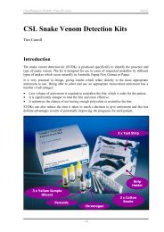
Appendix: CSL snake venom detection kits<br />
Glossary of medical terms<br />
-1-
Clinical Management of Snakebite in Papua New Guinea<br />
Acknowledgements<br />
This course would not have been possible without the co-operation, support, encouragement,<br />
kindness, hard work, perseverance and generosity of many people:<br />
• Sir Philip & Lady Brenda Willmott-Sharp from the Independent Group Limited in Port<br />
Moresby who have provided one of us (DW) with financial, logistical and moral support<br />
for many years while in pursuit of snakes and snakebite research in Papua New Guinea.<br />
• Oil Search Limited (particularly Ross Hutton & Ruth Waram) for their kind and generous<br />
financial support of snakebite research in PNG, including this course and the production<br />
of this Course Handbook.<br />
• CSL Limited (particularly Tim Carroll) for their kind and generous financial support in<br />
funding the snake venom detection kit workshop.<br />
• Dr Timothy Pyakalyia, Deputy Secretary (Technical Health Services) at the National<br />
Department of Health who has supported and encouraged research into snakebite and<br />
facilitated much of what has been achieved.<br />
• Dr Simon Jensen, Senior Lecturer in Emergency Medicine at UPNG who has worked<br />
around the clock to organise the course and to help write and edit the Course Handbook.<br />
• Dr Kenneth Winkel, Director of the Australian Venom Research Unit at the University of<br />
Melbourne, and President of the Australasian College of Tropical Medicine who has<br />
helped enormously in making this course possible.<br />
• The members of the teaching faculty not already mentioned: Dr Antony Chenall from St<br />
Vincent’s Hospital in Melbourne, Dr Gertrude Didei from Modilon Hospital in Madang,<br />
Dr Aaron Limbo from Port Moresby General Hospital, Dr Evelyn Lavu from UPNG, Mr<br />
Benjamin Bal from the Gulf Province Health Department in Kerema, Dr John Vince from<br />
UPNG, Dr Bill Nimorakiotakis from the Australian Venom Research Unit in Melbourne,<br />
and Ronelle Welton from James Cook University in Townsville.<br />
• Dr Mathias Sapuri (Dean), Dr Andrew Masta (Acting Dean), and Dr Harry Aigeeleng<br />
from the School of Medicine & Health Sciences at UPNG, as well as Mrs Claire<br />
Matainaho and her colleagues from UPNG’s MONAHP office who have donated their<br />
time to act as our support staff.<br />
• Dr Andrew Dent from St Vincent’s Hospital in Melbourne for opening important doors.<br />
• Dr Simon Mete from the Port Moresby General Hospital for enabling us to visit the<br />
Emergency Department and Intensive Care Unit facilities.<br />
Finally there has to be a very personal thank you to the many rural health workers across<br />
Papua New Guinea who have helped with the research into snakebite that one of us (DW) has<br />
undertaken over the last four years. The experiences of rural snakebite problems and the many<br />
enlightening conversations we have had together are what lead to the concept of a dedicated<br />
training course in snakebite management being offered to frontline health officers.<br />
David Williams<br />
Course Co-ordinator 2004
Clinical Management of Snakebite in Papua New Guinea<br />
<strong>Introduction</strong><br />
<strong>Introduction</strong><br />
David Williams<br />
Many of us know the feelings of urgency and helplessness that <strong>com</strong>e when a sudden knock at<br />
the door in the early hours of morning brings the news of yet another seriously ill snakebite<br />
patient arriving at our aid posts, health centres and hospitals. Often these patients will already<br />
be in the terminal stages of severe neurotoxic paralysis, and without transport, medicines or<br />
modern hospital equipment, we are left with little to do but stand by and watch as they slowly<br />
slip away to be<strong>com</strong>e yet another victim of the deadly reptiles that live in our midst.<br />
Snakebite is a medical emergency, and we live in a country where venomous snakes, although<br />
few in variety, are many in number. As you will hear during this course, the incidence and<br />
mortality rates after snakebite in Papua New Guinea are among the highest reported in any<br />
part of the world. This is in stark contrast to the experience of our neighbour Australia.<br />
Despite having many of the same species, and much higher populations living in areas with<br />
abundant snake populations, snakebite in Australia is a very minor problem at <strong>com</strong>munity<br />
level. With just one quarter of Australia’s population, and perhaps 200 more deaths from<br />
snakebite each year, PNG’s snakebite mortality rate may be as much as 800 times greater than<br />
Australia. The gap between PNG’s snakebite problem and snakebite in Australia does not end<br />
there.<br />
With a much larger revenue-contributing population, stronger resource base and solid<br />
economic foundation, Australia has some of the best medical facilities in the world. Even in<br />
many of our smaller towns and villages, there are qualified doctors working in hospitals that<br />
are well equipped. In our most remote areas we are able to extend viable emergency medical<br />
services with a long-established privately-funded aeromedical retrieval service, the Royal<br />
Flying Doctor Service. A person bitten by a venomous snake in Australia’s arid heart can be<br />
retrieved using a well-equipped medical airplane, and may find themselves recovering in a<br />
large, modern city hospital in just a matter of hours. Snakebite is, nevertheless, still<br />
considered to be a very serious medical condition, and even in the most modern Australian<br />
intensive care units, snakebite patients may still have a guarded prognosis.<br />
Here in PNG, where the economic base of the country remains in its formative years, the<br />
challenges of basic service delivery, education, transport, health, housing and policing remain<br />
great. With limited resources, and a plethora of important health issues to over<strong>com</strong>e, PNG<br />
faces a much more difficult road to prosperity. Common diseases like measles kill hundreds<br />
every year, malaria and other tropical illnesses are endemic, and the spectre of HIV/AIDS<br />
currently looms like a guillotine above the head of a condemned man. Rural hospitals and<br />
clinics do not usually have qualified doctors on hand, and resources, equipment and essential<br />
drugs are often in short supply. Medical evacuations, if and when they are carried out, <strong>com</strong>e at<br />
a high cost, one beyond the economic means of most grassroots Papua New Guineans.<br />
Relative antivenom costs in PNG are much higher than in Australia, and high prices mean that<br />
even with increases in funding allocations, there simply is not enough money available to<br />
purchase sufficient antivenom for all cases. Without the benefits of modern medical facilities,<br />
a Papua New Guinean snakebite victim faces a struggle to survive that is frequently lost.<br />
-1-
Clinical Management of Snakebite in Papua New Guinea<br />
<strong>Introduction</strong><br />
With so many challenges to over<strong>com</strong>e, one could be forgiven for thinking that survival after<br />
snakebite is a slim possibility, and while the chances of death are certainly higher in PNG<br />
than in Australia, the reality is that many people do survive snakebite.<br />
A good part of that survival is due to the dedication and experience of health personnel who<br />
work hard against numerous obstacles to provide their patients with the best care they can<br />
provide. Well-trained and well-informed health workers are vital to the delivery of health<br />
services, and, for that reason, we have developed this training course to equip rural and urban<br />
health workers with the skills and knowledge needed to improve their snakebite assessment<br />
and management skills even further.<br />
We aim to show you how to work with the resources that are at your disposal in the most<br />
effective and efficient manner. We will teach you the most appropriate, practical snakebite<br />
treatment techniques for your service delivery environments, and we are going to build up<br />
your knowledge base and ability to make the most of the resources that you already have. By<br />
addressing issues concerning the timeliness and use of safe, efficacious first aid treatments,<br />
the assessment, diagnosis and treatment of patients, and the care and support of patients with<br />
respiratory distress, we will try to help you learn to make the most of your resources in order<br />
to improve the prognosis for snakebite patients.<br />
At the <strong>com</strong>pletion of this course we hope that you will return to your <strong>com</strong>munities with the<br />
skills and knowledge to approach the treatment of snakebite with new confidence. We also<br />
hope to stimulate, in all of you, an interest in teaching what you have learned to your<br />
colleagues and staff, and in proactively helping your <strong>com</strong>munities to learn proper snakebite<br />
first aid techniques as a way of improving their chances of survival even further.<br />
Finally, we would like to see some of you take an active interest in snakebite research, and to<br />
perhaps, one day, be able to replace us as the instructors of this course.<br />
-2-
Clinical Management of Snakebite in Papua New Guinea Chapter 1<br />
Snakebite in Papua New Guinea<br />
Facts & Fiction<br />
David Williams<br />
<strong>Introduction</strong><br />
Snakes are widely feared in Papua New Guinea, and with very good reason. In many parts of<br />
PNG snakebite is an almost daily occurrence and venomous snakebite is a serious public<br />
health problem, with localized incidence rates that are among the highest of any tropical<br />
region in the world. Medical and epidemiological studies of snakebite in different parts of the<br />
country have given us detailed snapshots of some of the out<strong>com</strong>es of snakebite, and although<br />
there are still many gaps in our knowledge, there is a significant amount of factual data<br />
available.<br />
Ask anyone about snakebite and they will undoubtedly have a story to tell about someone<br />
they know who was bitten by a snake, or who died of snakebite. It would in fact be very easy<br />
to believe that venomous snakes lie in wait for unsuspecting people at every turn, and that<br />
right across PNG snakebite is claiming dozens of lives every day. If you ask Papua New<br />
Guineans about which snakes are responsible for snakebite one species above all others will<br />
feature at the forefront of every conversation; the fearsome ‘Papuan (Pap) black’.<br />
Of course the challenge for clinicians, health workers and scientists when it <strong>com</strong>es to<br />
snakebite is to separate the finely woven threads of fact and fiction:<br />
• Just how many people really are bitten by snakes, and in what parts of the country do<br />
these bites occur?<br />
• Are there as many deaths as either the scientific data, or the local people would have<br />
us believe?<br />
• Which species are dangerous and bite the most people and which are not?<br />
• What types of antivenom will best suit the needs of certain areas of the country?<br />
We have to be able to answer these and many other questions in order to provide the victims<br />
of snakebite with the most appropriate medical care. Having factual data enables resources<br />
(including antivenoms) to be distributed to the right places, and most importantly of all,<br />
reliable data provides health managers with the information they need to be able to make the<br />
right funding decisions to address <strong>com</strong>munity needs.<br />
At the present time however the extent of our knowledge lacks national clarity; there are<br />
many provinces throughout the country for which we simply have no reliable data. Without<br />
even basic information it is extremely difficult to give clinicians and health workers exact<br />
information about snakebite in their <strong>com</strong>munities, and it is <strong>com</strong>pletely impossible to provide<br />
sound advice to health administrators. As you will learn in this chapter, research into the<br />
epidemiology and clinical consequences of snakebite is taking place, and we have learned<br />
quite a lot about snakebite over the last fifty years. We hope to stimulate your enthusiasm to<br />
assist in ongoing research and to develop our knowledge even further.<br />
- 1.1 -
Clinical Management of Snakebite in Papua New Guinea Chapter 1<br />
Local beliefs & perceptions of snakes and snakebite<br />
Snakes occupy an important role in local culture and tradition, and it is these beliefs and<br />
perceptions that affect the ways in which Papua New Guineans deal with the issue of<br />
snakebite and snakes in general. In many <strong>com</strong>munities across PNG snakes are considered to<br />
be the mortal shells of bush spirits, demons, or deceased ancestors, enemies and sorcerers.<br />
There are numerous legends that tell of fearsome demons which take the natural form of<br />
snakes. The Marind-anim people from south-western PNG and eastern Papua believe, for<br />
example, that a demonic old woman known as the pathogu steals young children while in the<br />
body of a snake. To the Orokolo people living to the west of Kerema in Gulf province, snakes<br />
are the homes of mythical bush spirits – dark and dangerous creatures that can cause untold<br />
trouble.<br />
Many Mekeo people believe that the dreaded ‘Papuan black’ attacks people at the behest of a<br />
sorcerer who may have first stolen something personal from the victim and have placed it into<br />
a heated pot containing the body of a snake. A similar belief is held by the Kiwai people from<br />
the Fly River delta; the ove-devenar sorcerer will collect the faeces of his victim and put it<br />
into the mouth of a snake model made from wood or clay which he then places in an area that<br />
the victim is likely to visit. The model be<strong>com</strong>es a living snake which hunts and kills the ovedevenar’s<br />
target. The Elema people of Gulf province believe in an evil spirit called ove-hahu<br />
who is the cause of accidents, serious sudden illnesses and snakebite. Similar traditional<br />
beliefs about snakes are held by the Arapesh people from the Sepik region, and by many other<br />
clans throughout PNG.<br />
In Central province, the coastal Motu-Koitabu and mountain Koiari peoples often believed<br />
that snakebite, as well as being a tool of the vada (sorcerer), was sometimes a punishment<br />
meted out by good spirits such as the birava for social and traditional crimes like adultery.<br />
The Keakalo people of Marshall Lagoon also believed that snakebite was a <strong>com</strong>mon<br />
punishment for social transgressions such as adultery, theft, spousal abuse or taboo-breaking.<br />
As well as this, their mega mega auri (snake senders) could not only use magic to send out a<br />
snake to bite someone, but could also be sought out and paid to administer a cure. Dr Charles<br />
Campbell, a physician who conducted the first medical studies of snakebite in PNG learned<br />
that this cure usually involved obtaining a variety of tree bark called paia and a rainforest vine<br />
wamela which were chewed by the mega mega auri and then blown onto the face and body of<br />
the victim, who would also be massaged with coconut flesh or milk and kiki leaves. Chewing<br />
the paia ivoa (ginger leaves) and wamela was also considered to be a way of deterring snakes<br />
from biting.<br />
Just as snakes are associated with evil deeds, dark spirits and sudden death, they are also<br />
widely held to play an important role in fertility and the guardianship of important traditional<br />
sites. The Kamea people (known as the Kukukuku by outsiders) live in the remote highlands<br />
of Gulf and Morobe provinces. These small, strongly built people, with an infamous warrior<br />
tradition and a fearsome reputation, have many animistic beliefs and some of them believe<br />
strongly that an enormous snake keeps the world functioning and protects us all. This creature<br />
is guarded by benevolent sorcerers who engage in a constant struggle to protect the snake<br />
from evil sorcerers who would kill it, a calamity that would spell the end of the world.<br />
Kiwai people in Western province believe that the mythical maigidubu snake spirit is crucial<br />
to the success of yam crops, and that if the maigidubu leaves its tracks through the crop<br />
around the time of the yam festival, then success will be assured. Also, their guardian spirits<br />
the etengena, would often take the form of snakes and stand guard over their gardens, biting<br />
intruders to send them away.<br />
- 1.2 -
Clinical Management of Snakebite in Papua New Guinea Chapter 1<br />
Trobriand Islanders believe that some snakes are sacred and either house the spirits of great<br />
chiefs, or are their reincarnations. All the same, finding such as snake in a village was not<br />
considered a good sign, and unless the creature could be tempted to leave with prayers or<br />
other offerings, sudden illness was likely to befall the entire <strong>com</strong>munity. Despite this dreadful<br />
possibility, it was taboo to kill the snake, as this would only make the consequences worse.<br />
The Bariai of West New Britain tell the tale of Moro, a snake-man who started the tradition of<br />
pig exchanges and other ceremonies in honour of firstborn children, and to give praise to the<br />
dead. Legend has it that Moro’s father Kamaia had been killed in a disagreement with his<br />
brothers-in-law, and his liver cut out and cooked. Later during his funeral Moro was tricked<br />
into eating some of the liver by his cousin-in-law Kaukave whereupon his two legs<br />
immediately became fused together and the entire lower half of his body was transformed into<br />
that of a snake. Moro’s transformation is ascribed to the vengeful punishment of his father’s<br />
ghost for the having consumed the liver, albeit unknowingly. Pursued by the ghost of his<br />
father, Moro and his mother escape after Moro uses magic to carve out the Amara River, and<br />
then tricks the ghost into trying to cross, only to be eaten by a large crocodile!<br />
Distinctions between different types of venomous snakes are made by many of the people<br />
living in southern PNG although different names may sometimes be used by different clans of<br />
the same language groups for exactly the same snake, and other groups use just one name to<br />
describe different venomous snakes:<br />
Papuan taipan (Oxyuranus scutellatus canni)<br />
Moveave (Gulf)<br />
Mekeo (Central)<br />
Motu-Koitabu (Central)<br />
Keakalo (Central)<br />
Lavai – ‘black snake with a red stripe that lives in long grasses<br />
Auguma – ‘black snake that bites again’<br />
Kabagi – ‘red snake’ by the people of Barune<br />
Larana Karo – ‘long blacksnake’ by the Barune people<br />
Duba – dark coloured snake by the Kila Kila people<br />
Relena Gamara – ‘big brown snake’<br />
Papuan blacksnake (Pseudechis papuanus)<br />
Moveave (Gulf)<br />
Mekeo (Central)<br />
Motu-Koitabu (Central)<br />
Keakalo (Central)<br />
Mito – ‘black snake that lives in Sago swamps’<br />
Auguma – ‘black snake that bites again’<br />
Larana Karo – ‘long blacksnake’ by the Barune people<br />
Duba – dark coloured snake by the Kila Kila people<br />
Gelema rupa – ‘black snake’<br />
Death adder (Acanthophis spp.)<br />
Mekeo (Central)<br />
Motu-Koitabu (Central)<br />
Keakalo (Central)<br />
Afi – ‘sharp-eyed snake’<br />
Asenamo Api or just Api – ‘short sharp-tailed snake’<br />
Vanaame – ‘short snakes that jumps’<br />
Neither the Mekeo nor Kila Kila Motu people make a significant distinction between taipans<br />
and blacksnakes and use just one name each to describe the two different species. Even the<br />
people of the Moveave region in the east of Gulf province, while using different names to<br />
describe taipans and blacksnakes, identify both as ‘blacksnakes’. The majority of people are<br />
also able to distinguish venomous snakes from non-venomous pythons and other snakes. The<br />
Motu people know pythons as Navara, while their Koitabu cousins use Lavara, and the<br />
Keakalo call large pythons Kapari and may consider some of them sacred spirit animals.<br />
- 1.3 -
Clinical Management of Snakebite in Papua New Guinea Chapter 1<br />
When is a black snake not a blacksnake?<br />
The use of colour to describe and identify snakes is <strong>com</strong>mon all over the world. In Australia<br />
snakes with stripes are called ‘tiger snakes’ (although the reality is that not all of them have<br />
stripes), and even in Papua the small-eyed snake (Micropechis ikaheka) is called a ‘tiger<br />
snake’ because of the striped appearance of some specimens. Brown-coloured snakes in<br />
Australia were given the obvious name ‘brown snake’ while black-coloured snakes are called<br />
‘blacksnakes’; even though at least one member of the ‘blacksnake’ taxonomic group is called<br />
a ‘king brown snake’ (Pseudechis australis) because it is actually a brown-coloured<br />
‘blacksnake’!<br />
In Papua New Guinea people tend to do exactly the same thing. All black-coloured snakes are<br />
called ‘blacksnakes’ based entirely on the colour of their bodies. Whether or not they happen<br />
to really be venomous Papuan blacksnakes (Pseudechis papuanus) is an entirely different<br />
issue. There are in fact many different types of black-coloured snakes in Papua New Guinea,<br />
some of them non-venomous, some moderately venomous and three that are highly venomous<br />
and very dangerous; they include:<br />
Black whipsnakes (Demansia vestigiata)<br />
Moderately venomous<br />
Boelen’s pythons (Morelia boeleni)<br />
Non-venomous<br />
Brown-headed snakes (Furina tristis)<br />
Moderately venomous<br />
Common tree snakes (Dendrelaphis punctulatus) Non-venomous<br />
D’Albert’s pythons (Leiopython albertisi)<br />
Non-venomous<br />
Death adders (Acanthophis spp.)<br />
Highly venomous & dangerous<br />
Forest snakes (Toxicocalamus spp.)<br />
Moderately venomous<br />
Javan file snakes (Acrochordus granulatus)<br />
Non-venomous<br />
Mangrove snakes (Myron richardsoni)<br />
Moderately venomous<br />
Muller’s crowned snakes (Aspidomorphus Muelleri) Moderately venomous<br />
New Guinea ground boas (Candoia aspera)<br />
Non-venomous<br />
Papuan blacksnakes (Pseudechis papuanus)<br />
Highly venomous & dangerous<br />
Papuan taipans (Oxyuranus scutellatus canni)<br />
Highly venomous & dangerous<br />
Slatey-grey snakes (Stegonotus cucullatus)<br />
Non-venomous<br />
Solomon’s coral snakes (Salomonelaps par)<br />
Moderately venomous<br />
Water pythons (Liasis mackloti)<br />
Non-venomous<br />
With so many different ‘Papuan (Pap) blacks’ distributed throughout the country it should be<br />
very obvious why this almost supernatural snake ends up being blamed for virtually every<br />
snakebite that occurs in PNG!<br />
It should also be very clear why some bites by ‘Papuan blacks’ cause absolutely no clinical<br />
illnesses at all, while other may produce minor signs, and still others are lethal.<br />
There is ONE snake in this list that really is a Papuan blacksnake (Pseudechis papuanus) both<br />
in colour and scientific identity. It is a highly venomous species, but the reality is that, of the<br />
venomous snakes in this list of sixteen different types of snakes, only three are highly<br />
venomous, and of these, the one which causes the majority of snakebites is not a ‘Papuan<br />
black’ at all, but the actually the much more dangerous and much more venomous Papuan<br />
taipan (Oxyuranus scutellatus canni). In Central province taipans cause more than 80% of all<br />
snakebites, while Papuan blacksnakes (Pseudechis papuanus) cause less than 5% of the<br />
serious snakebites admitted to PMGH.<br />
- 1.4 -
Clinical Management of Snakebite in Papua New Guinea Chapter 1<br />
Can you pick the REAL Papuan blacksnake?<br />
A<br />
B<br />
C<br />
D<br />
E<br />
F<br />
G<br />
Turn to the last page of this Chapter to see if your attempt at identification was correct or not.<br />
H<br />
- 1.5 -
Clinical Management of Snakebite in Papua New Guinea Chapter 1<br />
Common misconceptions about snakebite<br />
There are several <strong>com</strong>mon misunderstandings and misconceptions about snakes and snakebite<br />
in Papua New Guinea:<br />
Belief: The majority of snakebites are caused by the Papuan blacksnake.<br />
Reality: From a medical perspective, this is a particularly dangerous misconception that<br />
can seriously harm a snakebite patient.<br />
The clinical reality is that there is little evidence to show that the real Papuan<br />
blacksnake (Pseudechis papuanus) causes large numbers of snakebites,<br />
particularly in Central province where investigations using venom<br />
identification assays have shown that only 4.3% of the serious snakebites<br />
admitted to Port Moresby General Hospital were caused by this species.<br />
In Milne Bay, Gulf and Western provinces, it is still possible that a larger<br />
proportion of snakebites are caused by the Papuan blacksnake, but at the<br />
present there is very little reliable evidence available. Papuan blacksnakes do<br />
not occur in any other parts of Papua New Guinea.<br />
In Central province the evidence suggests very strongly that the majority (more<br />
than 80%) of serious snakebites admitted to PMGH are caused by Papuan<br />
taipans (Oxyuranus scutellatus canni). In the past people have died as a result<br />
of the mis-identification of taipans as ‘blacksnakes’.<br />
Belief:<br />
Reality:<br />
Belief:<br />
Reality:<br />
Belief:<br />
Reality:<br />
The death adder uses a poison spine on its tail to harm people.<br />
This is <strong>com</strong>pletely untrue. The soft spine-like projection on the end of a death<br />
adders (Acanthophis spp.) tail contains nothing toxic, and has no role in the<br />
injection of venom.<br />
The ‘spine’ is nothing more than a lure that is wriggled by the snake in order to<br />
attract food – just like dangling a worm on a fishing hook. When a small lizard<br />
or frog tries to eat the lure, the snake bites the animal killing it with venom<br />
injected through fangs in the roof of the mouth.<br />
The forked tongue of a snake is a poisonous sting.<br />
The tongue of a snake is <strong>com</strong>pletely harmless. It is a specialised scent organ<br />
that is used to collect odours from the air and deposit them in a special organ<br />
on the roof of the mouth (Jacobson’s organ) that contains the same types of<br />
olfactory cells that occur in the human nose, and which allow us to smell – the<br />
tongue does nothing more than help the snake detect smells and odours.<br />
All snakes are venomous.<br />
This belief could not be further from the truth; of the 112 species of snakes that<br />
occur in Papua New Guinea and Papua, only a third are venomous, and of 37<br />
species, there are currently only 6 land snake species known to have the ability<br />
to cause human fatality. The two species of sea krait (Laticauda spp.) can also<br />
cause death, as can several species of true sea snakes; but sea snake related<br />
deaths are very rare in PNG.<br />
- 1.6 -
Clinical Management of Snakebite in Papua New Guinea Chapter 1<br />
Epidemiology of snakebite in PNG<br />
Although the first published medical report of snakebite in Papua New Guinea did not appear<br />
in the literature until 1961, the risk to public health presented by venomous snakes was well<br />
known, and snakes generally were (and remain) widely feared.<br />
While the Papua New Guinean perception of snakebite may have revolved around the<br />
traditional beliefs of the various cultural and social groups throughout the country, the<br />
perceptions of colonial medical officers was tempered by ‘western’ attitudes which had by the<br />
start of the 20 th Century largely turned away from belief in supernatural forces towards<br />
acceptance of ‘scientific’ conclusions that were based on rational hypotheses, demonstrable<br />
facts and clinical reality. Significant advances in human understanding of basic physiology<br />
and biology provided a vastly different perspective of the causes of the clinical effects seen<br />
after snakebite. Rather than being considered as the result of sorcery, colonial doctors had a<br />
strong belief that snakebite was the consequence of the physiological changes produced by<br />
organic toxins.<br />
Over the last 5 decades there has been considerable interest in the problems associated with<br />
snakebite in PNG, and a number of epidemiological and clinical studies, aimed at learning<br />
more about the consequences of snakebite and the out<strong>com</strong>es of various treatment strategies,<br />
have been carried out. As a result there exists a considerable body of information to help us in<br />
understanding why snakebite occurs, what the consequences may be, and what we should be<br />
doing to improve the prognosis for snakebite patients.<br />
Incidence and Mortality Rates<br />
Early publications about snakebite in Papua New Guinea give no data on the overall incidence<br />
of morbidity or mortality, and concentrate predominantly on the reporting of case series that<br />
describe clinical syndromes of envenoming and their treatment. Campbell & Young (1961)<br />
reported 15 cases of serious envenoming from Central province between November 1959 and<br />
August 1960. The single death in this series was due to pulmonary oedema. Campbell (1964)<br />
reported an additional 41 cases in addition to further discussion of 11 cases from the earlier<br />
study. All of these cases occurred between mid-February 1960 and September 1962, with 38<br />
cases originating within 80 kilometres of Port Moresby. As with the earlier paper, no data is<br />
presented to explain whether these represent all of the cases treated, or whether they only<br />
represent cases treated by the author.<br />
Campbell also published three papers describing clinical syndromes of envenoming by<br />
presumed death adders Acanthophis antarcticus 1 (1966), Papuan taipans Oxyuranus<br />
scutellatus canni (1967a), and Papuan blacksnakes Pseudechis papuanus (1967c), but again<br />
gives no indications as to whether the cases reported represent all cases during particular<br />
periods, or are simply representative case histories. None of the cases were fatal. In a report<br />
on antivenom use Campbell (1967b) records a case fatality rate of 7.1% from 28 cases treated<br />
over a 21 month period from March 1964 to November 1965. In discussing the management<br />
of 73 cases between 1959 and 1965, Campbell (1969a) records 5 fatalities (CFR of 6.8%).<br />
Campbell (1969b) recorded mean admissions for snakebite in Papuan hospitals as 155.5 cases<br />
per annum for the period 1961-1967, and says that there were 6.3 admissions for snakebite per<br />
1,000 patients (0.63%).<br />
1 Acanthophis antarcticus is not currently believed to occur in Papua New Guinea, and ongoing taxonomic<br />
investigations suggest that the genus is represented by several distinct species including Acanthophis rugosus<br />
and Acanthophis laevis.<br />
- 1.7 -
Clinical Management of Snakebite in Papua New Guinea Chapter 1<br />
Hudson & Pomat (1988) provide data on the incidence of snakebite in Madang province<br />
during the 10-year period from 1977 to 1986, but do not calculate actual incidence rates<br />
despite giving general population data. Using the data, the mean annual incidence can be<br />
calculated as 8.3 cases per 100,000 population. Clinical data for all of the cases is not<br />
available, and consequently any estimate of envenomation incidence would most likely<br />
underestimate the true figure. Only 2 deaths were recorded, giving a mortality rate estimate of<br />
0.09 cases per 100,000 population and a case fatality rate of 1.14%. Brian & Vince (1988)<br />
examined snakebite in children admitted to Port Moresby General Hospital and reported a<br />
case fatality rate of 7.7% among 54 2-16 year olds over 38 months.<br />
Currie et al (1991) produced incidence rates for envenoming snakebite and snakebite<br />
mortality in Papua New Guinea based on antivenom usage data from hospital admissions in<br />
Port Moresby (1987-1989) and Madang (1978-1988), and from CSL antivenom usage<br />
reporting forms (1983-1988). The authors acknowledged that these sources represented<br />
in<strong>com</strong>plete data on antivenom usage in Papua New Guinea. The annual incidence rates<br />
(January 1987-June 1989) of envenomation in rural central Papua and urban Central Papua<br />
were 81.8 and 21.8 cases per 100,000 population respectively. Corresponding annual<br />
mortality rates were given as 4.3 and 2.1 cases per 100,000 population. For the Madang<br />
region (1978-1988) they reported the annual envenomation incidence to be 3.0 cases per<br />
100,000 population, with an annual mortality rate of
Clinical Management of Snakebite in Papua New Guinea Chapter 1<br />
120<br />
100<br />
Number of Admissions<br />
80<br />
60<br />
40<br />
20<br />
0<br />
Bereina<br />
Veifa'a<br />
Kwikila<br />
Kupiano<br />
Moreguina<br />
Iruna<br />
Sogeri<br />
Tapini<br />
Others<br />
Health Centre Locality<br />
FIGURE 1: Average annual admissions for snakebite at major health centres in Central province<br />
1987-1991 (SOURCE: Lalloo et al, 1995)<br />
Not all snakebites resulted in envenomation. The average annual incidence rate for<br />
envenomation was 326.9 cases per 100,000 population; effectively 58.2% of snakebites<br />
resulted in the development of clinical envenoming. Annual mortality rates ranged from 7.5 to<br />
19.3 cases per 100,000 population with an average mortality rate of 13.8 cases per 100,000<br />
population. This is significantly higher than has been demonstrated in the past.<br />
Williams et al (2002) determined case fatality rates for 283 patients admitted to the Intensive<br />
Care Unit at Port Moresby General Hospital for the treatment of snakebite between January<br />
1998 and December 2001. The overall case fatality rate was 9.54% but annual case fatality<br />
rates ranged from 4.4% to as high as 20.6%. Case fatality rates among males (7.7%) were<br />
significantly different (X 2 =24.6, P=
Clinical Management of Snakebite in Papua New Guinea Chapter 1<br />
Hudson & Pomat (1988) examined 129 cases (83 males; 46 females) from Madang province<br />
for which medical records were available. There were 111 adults and 18 children under 12<br />
years of age; however no further data on age distribution is given.<br />
Currie et al (1991) report that the mean age of 347 patients (215 males; 132 females) in their<br />
study was 24.5 years (SD=13.8 yrs), but give no sex-related data. The youngest patient was 2<br />
years old and the oldest was 67 years of age. Ninety patients (25.9%) were under 15 years of<br />
age, and 69% of these were males. The overall male:female sex ratio was 1.6:1. Lalloo et al<br />
(1995) state that the mean age of patients in their study was 25.0 years; but, like Currie et al<br />
(1991), give no sex-related data. Of the admitted patients, 8.5% were children 10 years old or<br />
younger, and children <strong>com</strong>prised 16% of envenomed and 6.9% of non-envenomed patients.<br />
Lalloo et al (1995b) report an overall male:female sex ratio of 1.39:1 based on 1,421<br />
admissions to Central province health centres and Port Moresby General Hospital between<br />
1987 and 1991. At Veifa’a in the north-west of the province the male:female sex ratio was<br />
0.87:1. The lack of sex-specific age data in both of these large recent studies is unfortunate. In<br />
Lalloo et al (1995b) the mean age of all admitted patients (25.0 years) differs from the mean<br />
(21.1 yrs, SD=17.7) and median (15.5 yrs) ages of 20 patients who died from snakebite in<br />
their study. Median ages of male (14 yrs, n=11) and female (16 yrs, n=9) fatalities are at<br />
variance with the mean age given for all admissions. However, it is not possible to <strong>com</strong>pare<br />
this data further as no sex-specific calculations are provided.<br />
Williams et al (2003) do give age-specific data for snakebite in Mekeo, with a mean age for<br />
males of 23.1 years and 22.7 years for females and a median age of 20 years for both sexes.<br />
There were a disproportionate number of 16-30 year old males and females bitten by snakes<br />
in Mekeo; 49.7% of all male snakebite victims and 53.6% of all female snakebite victims in<br />
Mekeo fell into this age range despite that fact that only 27.5% of males and 27.1% of females<br />
in Mekeo were aged between 16 and 30 years. Although 43.5% of the male population, and<br />
41.1% of the female population were aged between 0-15 years old, snakebite victims of these<br />
ages were under-represented; 28.6% of males and 23.4% of females were 0-15 years old.<br />
80<br />
75<br />
70<br />
65<br />
60<br />
55<br />
50<br />
45<br />
40<br />
35<br />
30<br />
25<br />
20<br />
15<br />
10<br />
5<br />
0<br />
0-5 6-10 11-15 16-20 21-25 26-30 31-35 36-40 41-45 46-50 51-55 56-60 61-65<br />
Males<br />
Females<br />
FIGURE 2: Age distribution of 336 male and 261 female patients admitted to health centres in<br />
Mekeo between 1997 and 2001 for whom actual age was recorded (SOURCE: Williams et al, 2003)<br />
- 1.10 -
Clinical Management of Snakebite in Papua New Guinea Chapter 1<br />
Williams et al (2002) give age and sex-specific data for their series of 283 seriously<br />
envenomed patients at Port Moresby General Hospital. Mean age for males was 21.5 years<br />
(n=170, SD=14.9 yrs, median=16 yrs) and 21.1 years (n=77, SD=16.5yrs, median=14 yrs) for<br />
females. Children 15-years old or under <strong>com</strong>prised 39.9% of cases. The overall male:female<br />
sex ratio was 2.2:1. However, in the 15-29 year age group the ratio of males to females was<br />
4.1:1. Males under the age of 30 years <strong>com</strong>prised 46% of cases. McGain et al (2004)<br />
examining fatal snakebite at Port Moresby General Hospital report that 47% (n=41) were<br />
under 15 years of age, and that males were involved in 53% (n=46) of all cases.<br />
Circumstances of Injury<br />
In the first report on snakebite in Papua New Guinea, Campbell & Young (1961) state that all<br />
15 patients were bitten on either the foot or the leg during daytime hours while the victims<br />
were either walking in or near long grass. Campbell (1964) reported that all but 2/52 patients<br />
were bitten in daylight hours; 96% of cases were from Central province and 73.1% came from<br />
within 80 kilometres of Port Moresby. All but one bite occurred on the lower limbs with<br />
17.3% on the toes, 32.7% on the foot, 15.4% on the ankle and 32.7% on the legs themselves.<br />
Bites occurred when the victims were walking on paths near long grass, or through long grass,<br />
and 23% actually trod on the snake itself.<br />
In his series of 15 cases of Acanthophis spp. bites over a six-year period, Campbell (1966)<br />
reported that all but one bite occurred on the lower limbs, typically on either the dorsum of<br />
the foot or the toes. Eight bites happened when the person stood on the snake. In one case a<br />
man laid down on a snake concealed beneath leaf litter and was bitten on the leg, and another<br />
was bitten on the finger by a snake he accidentally picked up in a pile of cut grass. The other<br />
13 bites were on either the feet or ankles. Three cases involved patients bitten between<br />
6.30pm and 7.30pm, while a fourth patient was bitten at 3.00am in the morning. The other<br />
cases occurred between 8.30am and 5.30pm. In the only other study of proven Acanthophis<br />
spp. envenomation in Papua New Guinea, Lalloo et al (1996) found that 22 of the 32 EIAconfirmed<br />
cases occurred in daylight, and that only two bites did not occur on the lower<br />
limbs.<br />
Reporting bites by Oxyuranus scutellatus canni, Campbell (1967a) shows that all bites<br />
occurred on a lower limb (one of these was bitten on the “middle third” of the leg, and another<br />
on the “lower third”), and occurred between 10.00am and 4.00pm. Lalloo et al (1995a)<br />
observed that 85% of proven Oxyuranus scutellatus canni bites occurred during daylight<br />
among people working in gardens or walking along bush tracks, and that 96.4% of all the<br />
bites were to the lower limbs. All nine cases of presumed Oxyuranus scutellatus canni<br />
envenomation reported by Williams & Bal (2003) took place during daylight and involved<br />
bites to the legs, ankles or feet. One patient was bitten at home, two were bitten in the garden<br />
and the other six cases occurred when patients were walking along bush tracks or dirt roads.<br />
Among Campbell’s (1967c) 13 cases of presumed Pseudechis papuanus bites, most (n=10)<br />
were bitten on either the foot or ankle, while 2 were bitten on the hand or arm and 1 was<br />
bitten on the shoulder. Positive identification of the biting snake was only made in one case<br />
(specimen brought to PMGH), and all of the other cases were more likely to have been caused<br />
by Papuan taipans (Oxyuranus scutellatus canni). Only two positive identifications of P.<br />
papuanus snakebite were made during Campbell’s 7 years at PMGH, and his contention that<br />
it was the most <strong>com</strong>mon cause of snakebite in southern PNG is almost certainly erroneous.<br />
All of the bites occurred in daylight. All nine cases of snakebite involving Pseudechis<br />
papuanus discussed in Lalloo et al (1994) involved bites to the lower limb that occurred<br />
during daylight.<br />
- 1.11 -
Clinical Management of Snakebite in Papua New Guinea Chapter 1<br />
150<br />
135<br />
120<br />
Number of Cases<br />
105<br />
90<br />
75<br />
60<br />
45<br />
30<br />
15<br />
0<br />
0001-0559 0600-0859 0900-1159 1200-1459 1500-1759 1800-2059 2100-2400<br />
Time of Day<br />
FIGURE 3: Time of day for snakebites in the Mekeo region of Central province between January<br />
1997 and December 2001; increased frequency (43.9%) during the afternoon (1200-1759 hours)<br />
was statistically significant (p=
Clinical Management of Snakebite in Papua New Guinea Chapter 1<br />
identifications made with the aid of species-specific venom enzyme immunosorbent assay<br />
(EIA) by Lalloo et al (1995b), the snake responsible for the majority of serious snakebites<br />
admitted to PMGH is the Papuan taipan (Oxyuranus scutellatus canni) and not the Papuan<br />
blacksnake (Pseudechis papuanus). Papuan taipans caused an incredible 83.2% of<br />
envenomations <strong>com</strong>pared to just 4.2% that were attributable to Papuan blacksnakes.<br />
Reliable identification of the biting species is a major short<strong>com</strong>ing in most studies of Papua<br />
New Guinean snakebite. In the 1950’s herpetologist Ken Slater believed that the Papuan black<br />
snake Pseudechis papuanus was the <strong>com</strong>monest venomous snake in southern PNG, and even<br />
today, the perception of Papua New Guineans is that this snake is responsible for almost all<br />
snakebites. Slater (1956) wrote that it was most abundant along the coastline to the east of<br />
Port Moresby, and Campbell (1967c) claimed that it was responsible for more hospital<br />
admissions than any other species. In spite of this statement only two cases seen by Campbell<br />
between 1959 and 1967 could be conclusively attributed to this species on the basis of<br />
positive identification of the biting snake at the hospital. Campbell’s (1967c) description of a<br />
syndrome of envenoming attributable to Papuan black snake Pseudechis papuanus bites must<br />
therefore be treated with scepticism.<br />
The much more dangerous Papuan taipan Oxyuranus scutellatus canni often has a distinctive<br />
reddish-orange vertebral stripe as a distinctive feature, and Campbell appears to have relied<br />
upon this colouration as a means of presuming that large dark coloured snakes apparently<br />
lacking it were Pseudechis papuanus.<br />
Although the Papuan taipan is the only large snake in PNG that may have a red or orange<br />
vertebral marking, this is a feature that many snakebite patients simply do not notice in the<br />
heat of the moment. There are no reliable means of visually distinguishing all specimens of<br />
Pseudechis papuanus and Oxyuranus scutellatus canni. One patient in Campbell’s (1967c)<br />
series of 13 victims presented with a dead 1.5 metre snake for identification. Identification in<br />
the other 12 cases appears based on non-reporting of a vertebral stripe and these cannot be<br />
accepted as confirmed bites by Pseudechis papuanus. The only reliable recent description of<br />
envenomation by Pseudechis papuanus was given by Lalloo et al (1994) who detailed the<br />
clinical syndromes of 9 patients bitten between January 1990 and June 1992 identified by<br />
specific venom-antigen enzyme immunosorbent assay.<br />
FIGURE 4: Species responsible for serious snakebites admitted to Port Moresby General Hospital<br />
between January 1990 and June 1992 (SOURCE: Lalloo et al. 1994, 1995)<br />
Papuan taipan<br />
(83.2%)<br />
Death adder<br />
(10.8%)<br />
New Guinea brown<br />
snake (1.8%)<br />
Papuan<br />
blacksnake (4.2%)<br />
- 1.13 -
Clinical Management of Snakebite in Papua New Guinea Chapter 1<br />
Campbell (1964) states that 5 patients in a series of 52 arrived at hospital with a dead snake -<br />
3 with death adders Acanthophis spp, 1 with a Papuan black snake Pseudechis papuanus and<br />
1 with a Papuan taipan Oxyuranus scutellatus canni. Campbell (1966), describing the clinical<br />
syndrome of death adder Acanthophis spp envenoming reports 15 cases. The biting snake was<br />
killed in 11 of these cases and 8 of the victims brought the snake to the hospital where it was<br />
identified. Death adder envenoming is presumed in 5 cases on the basis of statements by<br />
either the patient or a third party, and in the last 2 cases Campbell presumes identification on<br />
the basis of haematological tests. Death adders have a distinctive body plan and appearance<br />
that makes visual identification easier than for other species. Lalloo et al (1995b) used<br />
species-specific venom EIA and found that 10.8% of serious snakebites admitted to PMGH<br />
were caused by death adders. Enzyme immunosorbent assay confirmed 32 cases of<br />
envenomation by Acanthophis spp. that involved a syndrome of progressive neurotoxicity<br />
without coagulopathy (Lalloo et al, 1996), and the 15 cases reported by Campbell (1966)<br />
conform to this profile, lending support to Campbell’s diagnosis. Hudson & Pomat (1988)<br />
give an excellent case report of a death adder bite treated at Madang Hospital where the dead<br />
snake was identified at the hospital.<br />
Campbell (1967a) reports six cases that he believed were attributable to envenoming by<br />
Papuan taipans Oxyuranus scutellatus canni based on positive identification of two dead<br />
snakes brought in with the victims, and a description of an orange or red coloured vertebral<br />
stripe in the remaining four cases. Lalloo et al (1995a) examine the syndrome of envenoming<br />
by Oxyuranus scutellatus canni in detail on the basis of 166 cases in which this species was<br />
implicated by specific venom-antigen EIA. Although they report the specificity for specific<br />
taipan venom-antigen as 87%, only cases in which this was the sole antigen detected were<br />
regarded as cases of definite Oxyuranus scutellatus canni snakebite. Williams & Bal (2003)<br />
reported nine cases of presumed envenoming by Oxyuranus scutellatus canni on the basis of<br />
clinical presentation and/or description of the biting species. In the future it is hoped that the<br />
widespread use of venom detection kits to diagnose snakebite will make it possible to<br />
assemble nationwide data on the species responsible for envenomation.<br />
Hudson & Pomat (1988) reported on 64 cases of envenomation reported in Madang Province<br />
between 1977 and 1986; 15 presented with symptoms suggestive of bites by the small-eyed<br />
snake (Micropechis ikaheka). Warrell et al (1996) reported that in a more recent series, 4/46<br />
(8.7%) cases of snakebite treated at Madang Hospital, and 5/13 (38.5%) treated at Gaubin<br />
Hospital on nearby Karkar Island were identified by EIA as attributable to M. ikaheka. No<br />
cases of M. ikaheka were identified in any of the patients studied using EIA by Lalloo et al<br />
(1994). Brown snake Pseudonaja textilis spp. envenomation has been reported among 1.8%<br />
of EIA-tested snakebite patients admitted to Port Moresby General Hospital between January<br />
1990 and June 1992 (Lalloo et al, 1994).<br />
Climate and Seasonality<br />
There have been few attempts to determine whether bites by certain species are more likely to<br />
occur under specific climatic conditions (temperature, rainfall, etc.) or during particular<br />
seasons throughout the year.<br />
Campbell (1966) reported that for Acanthophis spp., 12 of the 15 cases occurred between<br />
August and December. Campbell (1967a) reports Oxyuranus scutellatus canni bites in June<br />
(2), August (1), December (1), January (1) and March (1). Currie et al (1991) reported that<br />
56.8% of snakebites occurred between the ‘wet season’ months of from November to April.<br />
- 1.14 -
Clinical Management of Snakebite in Papua New Guinea Chapter 1<br />
18<br />
16<br />
Mean Monthly Admissions<br />
14<br />
12<br />
10<br />
8<br />
6<br />
4<br />
2<br />
0<br />
JAN FEB MAR APR MAY JUN JUL AUG SEP OCT NOV DEC<br />
Month<br />
FIGURE 5: Seasonal frequency of admissions for snakebite in the Mekeo region of Central<br />
province between January 1997 and December 2001; increased frequency during the dry season<br />
months of November to April is statistically significant (p=0.04). (SOURCE: Williams et al, 2003).<br />
Lalloo et al (1995b) state that 57.9% of 221 envenomed patients and 51.6% of 128 nonenvenomed<br />
patients were bitten between November-April. Data on monthly admissions for<br />
snakebite were <strong>com</strong>pared to monthly rainfall; however rainfall itself was not shown to be a<br />
significant contributing factor.<br />
Williams et al (2002), reporting cases admitted to the PMGH ICU from January 1997 to<br />
December 2001 from across PNG, found that 54% occurred between November-April, and<br />
Williams et al (2003) found that 53.9% of snakebites in Mekeo occurred during the same<br />
months during this same period. McGain et al (2004) reported that 60% of fatal cases in the<br />
10 years from January 1992 to December 2001 occurred between November and April.<br />
Clinical Epidemiology and Envenomation Syndromes<br />
Local Indications of Envenomation<br />
The clinical syndromes of envenomation after snakebite in Papua New Guinea have been<br />
reported by several authors. Most reports relate to small case series of patients and, in most,<br />
the identification of the biting species is equivocal. Some of the consistently reported<br />
symptoms of snakebite over the last 40 years are shown in Table 1.<br />
Lymph node pain is a consistent feature after snakebite in Papua New Guinea, and tender<br />
enlargement of regional lymph nodes is reported in all published accounts. Venom injected<br />
subcutaneously typically enters the lymphatic system and drains via lymph nodes into the<br />
circulation, and tenderness or swelling of lymph nodes may be an indication of venom<br />
absorption. Trevett et al (1994a) stated that lymphadenitis was a particularly sensitive positive<br />
indication of envenomation and occurred in 93% of envenomed patients.<br />
- 1.15 -
Clinical Management of Snakebite in Papua New Guinea Chapter 1<br />
TABLE 1: Symptoms reported in various studies of snakebite in Papua New Guinea between 1964 and 2004.<br />
Author<br />
Local Pain<br />
Lymph Nodes<br />
Oedema<br />
Headache<br />
Vomiting or<br />
Nausea<br />
Abdominal Pain<br />
Spit\Vomit Blood<br />
Visual<br />
Disturbances<br />
Campbell & Young, 1964 + + + + + + + +<br />
Campbell, 1964 + + + + + + + +<br />
Campbell, 1966 + + + + + - - +<br />
Campbell, 1967a - + - + + + - -<br />
Campbell, 1967c + + + + + + + +<br />
Campbell, 1969a - + - + + + + -<br />
Hudson & Pomat, 1988 + + + + + + - +<br />
Lalloo et al, 1994 + + + + + + + +<br />
Lalloo et al, 1995a - + - + + + + +<br />
Lalloo et al, 1996 - + - + + + - +<br />
Warrell et al, 1996 + + + + + + - +<br />
Williams & Bal, 2002 - + - + + + + +<br />
Williams et al 2003 + + + + + + + +<br />
McGain et al 2004 - + - + + + + +<br />
The extent and severity of local symptoms such as pain or oedema is significantly lower than<br />
in reports of elapid snakebite from Australia. Campbell & Young (1961) reported severe local<br />
pain and oedema in a single patient; however this was most likely caused by prolonged<br />
tourniquet use. Three other patients experienced minor oedema, and local pain was either<br />
absent <strong>com</strong>pletely or mild. Campbell (1964) found severe pain in 3 patients and severe<br />
oedema occurred in two individuals as a result of lengthy tourniquet application.<br />
A third of the patients bitten by death adders Acanthophis spp. in Campbell (1966) had local<br />
pain, and 27% had slight oedema. Neither symptom was present in any of the 6 patients with<br />
Oxyuranus scutellatus canni envenomation reported by Campbell (1967a); however severe<br />
pain, oedema and extensive ecchymoses were seen in a patient bitten on the shoulder<br />
(Campbell, 1967c) and this may have been due to a Papuan blacksnake (P. papuanus) bite.<br />
Pain was a feature in 36% of cases reported by Hudson & Pomat (1988) and 14% of patients<br />
had local oedema and localised muscle pain, most probably associated with rhabdomyolysis<br />
after Micropechis ikaheka envenomation. One patient in a series of eleven bitten by<br />
Micropechis ikaheka reported local pain and two patients had oedema (Warrell et al, 1996).<br />
Lalloo et al (1994) observed slight oedema in one patient bitten by a Papuan black snake<br />
Pseudechis papuanus. In a series of 166 bites by Oxyuranus scutellatus canni (Lalloo et al,<br />
1995a) there were no reports of local pain or swelling. Clinical experience in Australia is that<br />
the bites by members of the blacksnake genus Pseudechis are painful locally and often<br />
associated with local oedema that may be mild to pronounced in severity. Williams et al<br />
(2004) reported severe local pain after a bite by an Australian Collett’s blacksnake<br />
(Pseudechis colletti) and Isbister & Currie (2003) presents a case of mulga snake (Pseudechis<br />
australis) envenomation in a child with persistent bite site pain and swelling. Although<br />
un<strong>com</strong>monly reported in PNG (perhaps because bites are un<strong>com</strong>mon) it should be anticipated<br />
that Papuan blacksnake (Pseudechis papuanus) and Papuan mulga snake (Pseudechis cf.<br />
- 1.16 -
Clinical Management of Snakebite in Papua New Guinea Chapter 1<br />
australis) bites may also produce significant local pain and oedema. Local pain and swelling<br />
caused by snakebite should not be confused with the pain and swelling that can be associated<br />
with ischemic injury due to prolonged tourniquet use.<br />
Non-specific Indications of Envenomation<br />
A number of non-specific features have been reported consistently in patients with snakebite<br />
envenomation. Campbell & Young (1961), and Campbell (1964; 1966; 1967a; 1967c; 1969a)<br />
reported that headache often followed envenomation, and headache has been universally<br />
reported in virtually all published accounts of snakebite in PNG. Headaches are reported to<br />
have last from a few hours to several days. Hudson & Pomat (1988), Lalloo et al (1996) and<br />
Warrell et al (1996) report headache as <strong>com</strong>mon following bites by death adders (Acanthophis<br />
spp.) and small-eyed snakes (Micropechis ikaheka). Herpetologist Ken Slater had persistent<br />
severe headache for two days after a bite from Micropechis ikaheka. Lalloo et al (1994) noted<br />
headache in 2 of 9 patients with confirmed Papuan blacksnake (Pseudechis papuanus) bite,<br />
and Lalloo et al (1995a) reported headache in 54.5% of positively identified Papuan taipan<br />
(Oxyuranus scutellatus canni) victims. In an analysis of referral letters written by rural health<br />
workers during the early 1990’s, Trevett et al (1995) found that headache was reported in<br />
37% of patients at the health centre, and in 58% of the same patients on admission to PMGH.<br />
Headache was reported in 51.9% of 564 patients bitten by snakes in Mekeo (Williams et al<br />
2003). Headache was a documented non-specific symptom in 38 (63.3%) of 60 fatal<br />
snakebites treated at PMGH between 1992 and 2001 (McGain et al, 2004).<br />
Vomiting and abdominal pain or tenderness typically occurs in unison. Campbell & Young<br />
(1961) observed concurrent vomiting and abdominal pain in 3 patients, while another 3<br />
patients had vomiting without abdominal pain. One patient with severe abdominal pain and<br />
vomiting in conjunction with abdominal tenderness was mistakenly diagnosed with acute<br />
appendicitis. Vomiting was reported in 42% and abdominal pain in 34.6% of patients<br />
described in Campbell (1964). Vomiting was a typical non-specific indication in patients of<br />
death adder (Acanthophis spp.) bites reported by Campbell (1966), and was also recorded in<br />
three patients with presumed Papuan taipan (Oxyuranus scutellatus canni) bite (Campbell,<br />
1967a). In the series of 13 bites dubiously ascribed to Papuan blacksnake (Pseudechis<br />
papuanus) envenomation, but most probably really having been caused by Papuan taipans<br />
(Oxyuranus scutellatus canni), 8 patients had early vomiting and 4 had severe abdominal pain<br />
(Campbell, 1967c). Vomiting after bites from the small-eyed snake (Micropechis ikaheka) has<br />
been reported by Blasco & Hornabrook (1972) and Warrell et al (1996). Abdominal pain was<br />
reported in 2 of the 6 patients reported in Warrell et al (1996). Hudson & Pomat (1988)<br />
reported abdominal pain in 56% and vomiting in 25% of 64 envenomed patients treated for<br />
either death adder (Acanthophis spp.) or small-eyed snake (Micropechis ikaheka) bites in<br />
Madang during the 1980’s.<br />
Of 9 patients with confirmed Papuan blacksnake (Pseudechis papuanus) envenomation, 2<br />
vomited and 5 had abdominal pain (Lalloo et al, 1994). In a large series of Papuan taipan<br />
(Oxyuranus scutellatus canni) bite victims, 64.4% vomited and 59.8% had abdominal pain. In<br />
the analysis of referral letters conducted by Trevett et al (1994a), 38% had abdominal pain<br />
and 30% had vomited at the health centre; on admission to PMGH 58% had abdominal pain<br />
and 62% had reported vomiting. Williams & Bal (2003) reported vomiting in 4 of 9 patients<br />
with likely Papuan taipan (Oxyuranus scutellatus canni) envenomation and 3 of these 4<br />
patients had concurrent abdominal pain. Williams et al (2003) reports abdominal pain in<br />
48.9% of 564 patients bitten by snakes in Mekeo; however, nausea or vomiting was only<br />
reported for 6.2%. Abdominal pain was documented in 48 (80%), and vomiting in 40 (66%)<br />
of the 60 fatal snakebites discussed by McGain et al (2004).<br />
- 1.17 -
Clinical Management of Snakebite in Papua New Guinea Chapter 1<br />
Specific Indications of Envenomation: Coagulation defects<br />
Evidence of coagulopathy is an absolute indication for antivenom if observed in conjunction<br />
with a history of snakebite in PNG.<br />
Campbell & Young (1961) reported that 6 of the 15 patients had ‘bleeding tendencies’ and<br />
that 3 patients vomited blood-stained material. Campbell (1964) notes that 11/52 (21.2%)<br />
patients were recorded as either spitting blood or having haematemesis, while 3 patients also<br />
passed ‘blood-stained’ urine. Campbell (1967a) notes bleeding problems in 2 of 6 patients;<br />
one of these had persistent bleeding from a superficial facial wound, while another had<br />
haemoglobinuria and incoagulable blood. Haematemesis was seen in 3 patients and another 2<br />
had haemoptysis among the group of 13 that Campbell (1967c) thought to have been bitten by<br />
Papuan blacksnakes (Pseudechis papuanus). Three of 4 patients in this series had<br />
haemoglobinuria and fibrinogenolysis, and other signs of coagulopathy (spitting blood;<br />
gingival bleeding; bleeding bite wounds) were reported in 8 of the 13 patients. As has been<br />
stated before, Campbell’s presumptive identifications were based on whether or not the<br />
patient reported seeing a red marking on the back of the snake, and while the presence of<br />
reddish-orange marking is a strong indication that the culprit was a Papuan taipan (Oxyuranus<br />
scutellatus canni), it is important to again emphasize that not seeing this type of marking does<br />
not mean the snake was not a Papuan taipan.<br />
Spontaneous bleeding has been reported in two cases of envenomation by the small-eyed<br />
snake (Micropechis ikaheka) that was reported in Warrell et al (1996). Hudson & Pomat<br />
(1988) did not report bleeding disorders in any of the 64 cases of snakebite from Madang<br />
province during the 1980’s. In a study of 32 patients shown to have been bitten by death<br />
adders (Acanthophis spp.) by EIA, there was no clinical evidence of coagulopathy although<br />
elevation in leucocyte counts and slight thrombocytopenia was found on laboratory<br />
investigation (Lalloo et al, 1996). Prothrombin time was significantly prolonged in 13<br />
patients. Broad clinical experience in Australia and PNG indicates that bites by these snakes<br />
do not involve bleeding disorders.<br />
Blood-stained saliva and bleeding from the mouth or nose was reported in 1 patient with<br />
confirmed Papuan blacksnake (Pseudechis papuanus) envenomation, while 2 others had<br />
either epistaxis or gingival bleeding (Lalloo et al, 1994). Laboratory studies on blood from 2<br />
of these patients showed a mild thrombocytopenia (platelet counts: 91-93 × 10 9 /L), and that<br />
three patients had lower than normal levels of several important clotting factors including<br />
protein C, fibrinogen, Factors V, VIII, XIII a , anti-thrombin III, plasminogen and α 2 -<br />
antiplasmin. Prothrombin time (PT) and activated partial thromboplastin times (APTT) were<br />
prolonged briefly and FDP was elevated.<br />
Coagulopathy can be a very prominent feature of bites by Papuan taipans (Oxyuranus<br />
scutellatus canni), and in addition to Campbell’s early reports of bleeding after bites by large<br />
dark coloured snakes, there is a substantial body of evidence from recent studies. Lalloo et al<br />
(1995a) report that 77% of patients with EIA proven bites by Papuan taipans had incoagulable<br />
blood on the basis of 20WBCT > 20 minutes. Systemic bleeding occurred in 43.7% of<br />
patients, and bite site bleeding occurred in 16.3%. Most reported bleeding from either the<br />
gingival sulci (37%) or by haematemesis (12.6%). Three patients had epistaxis; 11% bled<br />
from superficial cuts and scratches; 8.8% bled from venepuncture sites. On laboratory<br />
investigation 52.8% had moderate to severe leucocytosis and 27.5% developed mild<br />
thrombocytopenia.<br />
Brian & Vince (1987) reported that 40 of 54 children bitten by snakes developed evidence of<br />
coagulopathy and that 33 had clotting times greater than 15 minutes. Bleeding problems were<br />
- 1.18 -
Clinical Management of Snakebite in Papua New Guinea Chapter 1<br />
mentioned in 40% of referral letters written by rural health workers, and 60% of the referred<br />
patients were found to have incoagulable blood upon admission to PMGH (Trevett et al,<br />
1994a). According to Trevett et al (1994a), a 20WBCT > 20 minutes had a positive predictive<br />
value (PPV) of 93.5% for a diagnosis of Papuan taipan (Oxyuranus scutellatus canni)<br />
envenomation. Williams et al (2003) reviewed 553 cases of snakebite from the Mekeo region<br />
in which the 20WBCT was used, and found that this simple diagnostic test had a positive<br />
predictive value of 98.4% and a specificity of 99.6% for the subsequent development of<br />
neurotoxicity. The PPV for subsequent coagulopathy was 67.7% with specificity of 95.7%;<br />
however, because the other signs and symptoms of incoagulable blood were often not<br />
reported in the clinical notes, it is probable that the actual PPV for disseminated intravascular<br />
coagulopathy is actually higher. In 60 fatal cases of snakebite reported by McGain et al<br />
(2004), 75% had a 20WBCT > 20 minutes and 35 patients (53.3%) had demonstrable<br />
spontaneous bleeding.<br />
Specific Indications of Envenomation: Myotoxicity<br />
There are few early reports of myotoxicity following snakebite in PNG, although some cases<br />
with ‘haemoglobinuria’ may actually have been patients with myoglobinuria instead. Blasco<br />
& Hornabrook (1972) mention that Ken Slater had ‘weakness’ after a bite from the smalleyed<br />
snake (Micropechis ikaheka), and this is often a sign of rhabdomyolysis. Hudson &<br />
Pomat (1988) report renal failure in 6 patients from Madang province who presented with<br />
dark-coloured urine. Hudson (1988b) reported two cases of presumed small-eyed snake<br />
(Micropechis ikaheka) envenomation. A 13 year-old girl bitten on the finger developed<br />
generalized muscle pain and tenderness and had urine that was dark reddish-brown, but was<br />
erythrocyte-free; and a 50 year-old man bitten on the finger while reaching into a bandicoot<br />
burrow by a ‘black-headed snake with a white belly’ also developed muscle pain and passed<br />
dark urine. This second patient’s muscle pain was mild initially but became more severe over<br />
the next 2 days; at Modilon hospital in Madang his urine was grossly discoloured, but once<br />
again was erythrocyte-free. He received one ampoule of death adder antivenom and three<br />
ampoules of polyvalent antivenom. Both patients were said to be anicteric and without<br />
coagulopathy, and had neurotoxicity; the 13 year old girl subsequently died. Other cases of<br />
presumed Micropechis ikaheka envenoming treated in Madang also showed signs and<br />
symptoms of generalized rhabdomyolysis and myoglobinuria. Warrell et al (1996) describes<br />
classical signs of myolysis (generalised muscle pain and tenderness; trismus; dark urine) in<br />
three patients with EIA proven small-eyed snake envenomation.<br />
The creatine kinase (CK) levels of 8 out of 12 patients bitten by death adders (Acanthophis<br />
spp.) was elevated (164-4,220 IU/L). Similar studies in victims of Papuan taipan (Oxyuranus<br />
scutellatus canni) bites found that creatine kinase levels were raised in 74.7% (43-8,110 IU/L)<br />
and levels of the liver enzyme aspartate transaminase (AST) were >50 IU/L in 52.6% of<br />
patients. Serum myoglobin exceeded 80 ng/ml in 52.2% of patients at the time of their<br />
admission. The serum creatine kinase levels of 2 of the 9 patients with proven Papuan<br />
blacksnake (Pseudechis papuanus) envenomation were slightly elevated; however there were<br />
no indications of rhabdomyolysis in any patient (Lalloo et al, 1994). In Australia, gross<br />
rhabdomyolysis and myoglobinuria after bites by members of the Pseudechis genus are<br />
relatively <strong>com</strong>mon, and creatine kinase levels may exceed 200,000 IU/L. Acute renal failure<br />
is a potential <strong>com</strong>plication of myotoxicity, and McGain et al (2004) reported muscle pain in<br />
41.7% of 60 fatal snakebites treated at PMGH. Renal <strong>com</strong>plications were contributing factors<br />
in the deaths of 16 (26.7%) patients. Testing for creatine kinase, lactate dehydrogenase and<br />
other isoenzymes that quantitatively measure rhabdomyolysis products is not routine in PNG<br />
at present so it is possible that this important syndrome is seriously under-reported.<br />
- 1.19 -
Clinical Management of Snakebite in Papua New Guinea Chapter 1<br />
Specific Indications of Envenomation: Neurotoxicity<br />
The neurotoxic effects of Papua New Guinean snake venoms are well defined in the available<br />
literature, and the balance of evidence demonstrates that snake venom neurotoxins are the<br />
major contributors in the deaths of patients due to the development of progressive flaccid<br />
paralysis that inhibits and <strong>com</strong>promises respiration.<br />
The earliest signs of neurotoxicity often manifest themselves in the craniofacial nerves<br />
producing a sequential syndrome that often starts with diplopia and ptosis before progressing<br />
to ophthalmoplegia, dysarthria, dysphagia and bulbar palsy and eventually involving the<br />
intercostal, diaphragm and peripheral musculature. Ataxia and progressive muscle weakness<br />
results in a loss of lo<strong>com</strong>otor function; patients eventually lose the ability to walk, stand, sit or<br />
even raise their limbs, or control head movements (‘broken neck syndrome’). Deep tendon<br />
reflexes may be lost and failing respiration often leads to ‘abdominal’ breathing.<br />
Campbell & Young (1961) graded the neurotoxic effects in 15 snakebite victims according to<br />
severity. Ptosis was moderate in 10 cases and severe in 4, while ophthalmoplegia was<br />
moderate in 4 patients and severe in 8 others. 10 patients were reported to have had ‘visual<br />
disturbances’; most probably diplopia. 6 patients had moderate to severe facial palsies and<br />
there was moderate to severe paralysis of the tongue and palate in 9 patients. Paralysis of<br />
intercostal muscles was severe in 7 patients with 3 patients experiencing moderate<br />
diaphragmatic paralysis. Paralysis of the diaphragm was severe in only 1 patient. 7 patients<br />
had moderate, and 2 patients had severe, peripheral flaccid paralysis.<br />
Campbell (1964) reports ‘visual disturbances’ in 26.9% of patients, noting both diplopia and<br />
blurred vision. Ptosis was regarded as the earliest sign of neurotoxicity and Campbell<br />
<strong>com</strong>ments that many patients would go to sleep only to awake the next morning with severe<br />
facial palsy. Ptosis was often associated with either partial or <strong>com</strong>plete ophthalmoplegia, and<br />
was observed in 75% of patients in this series. Campbell (1966) says that bites by death<br />
adders (Acanthophis spp.) produced either blurring of vision or an inability to see clearly, and<br />
that ocular and facial paralysis generally followed; however these signs were present in only 3<br />
of the 15 patients. Campbell (1967a) states that ‘muscular paralysis’ was present in only 3 of<br />
6 presumed Papuan taipan (Oxyuranus scutellatus canni) victims yet says that paralysis was<br />
present after 9 of 13 supposed Papuan blacksnake (Pseudechis papuanus) bites. Campbell’s<br />
assertion that during the 1960’s the Papuan blacksnake was the most <strong>com</strong>mon venomous<br />
snake in southern PNG and cause of the most snakebites is a fallacy that persists even today,<br />
and has resulted in many patients being treated incorrectly with inappropriate antivenoms.<br />
Blasco & Hornabrook (1972) describe a fatal case of small-eyed snake (Micropechis ikaheka)<br />
envenoming marked by peripheral limb weakness, shortness of breath and profound<br />
respiratory failure with cyanosis. Warrell et al (1996) reviewed 6 cases of envenomation by<br />
this species (identity confirmed by EIA) and reported ptosis in 4 patients, bulbar paralysis in<br />
3, and peripheral limb weakness in 3 cases. Ptosis was present between 6 and 12 hours postbite.<br />
Two patients died of respiratory paralysis at 19 and 38 hours respectively. A patient from<br />
Papua developed dysarthria, facial paralysis and ataxic gait resulting in an inability to stand<br />
unassisted. Paralysis of the cranial muscles and intercostals was pronounced.<br />
In a study of 32 cases of proven death adder (Acanthophis spp.) bites, Lalloo et al (1996)<br />
found that 18 developed symptoms and signs of envenomation. Ptosis was a symptom in<br />
15/18 (88.2%) and a sign in 17/18 (94.4%). Other symptoms included dysphagia (29.4%),<br />
dysarthria (37.5%), diplopia (18.8%), and respiratory difficulty (20%). On examination there<br />
were signs that these patients had ophthalmoplegia (55.6%), slurring of speech (30.3%), jaw<br />
restriction (28.6%), diminished hand grip (31.3%) and reduced reflexes (12.5%). A total of 5<br />
patients (27.8%) had to be intubated and ventilated, with the median ventilation time being 13<br />
- 1.20 -
Clinical Management of Snakebite in Papua New Guinea Chapter 1<br />
hours. The range of ventilation times was between 2 and 23.5 hours, considerably less than<br />
for victims of Papuan taipans (Oxyuranus scutellatus canni). Lalloo et al (1995a) reported<br />
that the median ventilation time after bites by Papuan taipans was 88 hours with a range of<br />
from 3 to 176 hours.<br />
Hudson & Pomat (1988) reported ptosis as a symptom in 55% and as a sign in 53% of<br />
snakebite envenomations in Madang province. Dysphagia and dysarthria were symptoms in<br />
31%, and 20% reported diplopia. Respiratory distress was a symptom in 9% of bites. Other<br />
signs included bulbar paralysis (17%) and external ophthalmoplegia (13%). Only 5% of<br />
patients subsequently developed respiratory failure. The only highly venomous species in the<br />
province are death adders (Acanthophis spp.) and small-eyed snakes (Micropechis ikaheka).<br />
Hudson (1988a) reports <strong>com</strong>plete bilateral ptosis, ophthalmoplegia and paralysis of tongue,<br />
jaws and pharynx in a man bitten by a death adder. Peripheral limb weakness was present as<br />
well as paralysis of the intercostal muscles. Hudson (1988b) reports bilateral ptosis,<br />
ophthalmoplegia, diplopia, bulbar and peripheral paresis in two patients presumed to have<br />
been bitten by small-eyed snakes (Micropechis ikaheka) in Madang province. Respiratory<br />
distress was evident in both patients, and one of them, a 13 year old girl, eventually died from<br />
cardiorespiratory failure 42 hours port bite. Resolution of ptosis, bulbar paresis and limb<br />
weakness took more than 14 days in the other patient despite the use of a large quantity of<br />
antivenom.<br />
The study of Papuan taipan (Oxyuranus scutellatus canni) envenomation by Lalloo et al<br />
(1995a) remains the most <strong>com</strong>prehensive investigation of neurotoxicity by this large and very<br />
dangerous snake. Of 166 patients with confirmed bites by taipans, 139 (83.7%) developed<br />
signs and symptoms of envenomation. Ptosis was present in 84/131 (64.1%) patients and was<br />
the most frequently reported symptom followed by dysarthria in 39/121 (32.2%) and<br />
dysphagia in 39/124 (31.5%). Diplopia was reported in 26/120 (21.7%) and 17/121 (14%) had<br />
dyspnoea; 41.7% of patients required intubation as paralysis deepened, and ventilation was<br />
required by 36.7% of envenomed patients. Progression to the point where intubation was<br />
required ranged from 3 to 55 hours with a median time point of 13.5 hours.<br />
FIGURE 6: Comparison of clinical signs of neurotoxicity after the EIA-proven bites of three different species of<br />
highly venomous Papua New Guinean snakes. (SOURCES: Lalloo et al, 1994; Lalloo et al, 1995a; Lalloo et al, 1996)<br />
100<br />
90<br />
80<br />
Papuan taipan (n=105-139)<br />
Death adder (n=18)<br />
Papuan blacksnake (n=9)<br />
70<br />
Percentage<br />
60<br />
50<br />
40<br />
30<br />
20<br />
10<br />
0<br />
Ptosis<br />
Ophthalmoplegia<br />
Jaw Restriction<br />
Slurred Speech<br />
Reduced hand<br />
grip<br />
Diminished<br />
reflexes<br />
Intubation<br />
Ventilation<br />
- 1.21 -
Clinical Management of Snakebite in Papua New Guinea Chapter 1<br />
Williams & Bal (2003) reported ptosis and dysphagia in all 9 patients with presumed Papuan<br />
taipan (Oxyuranus scutellatus canni) envenoming, 5 had diplopia, 6 had dysarthria, 5 had<br />
general bulbar paralysis, 4 had dyspnoea and 2 had peripheral limb weakness. Eight of the 9<br />
patients died from respiratory failure. McGain et al (2004) reported neurotoxicity (one or<br />
more of ptosis, dysphagia or dyspnoea) in all 60 patients in their series who died from<br />
snakebite in the PMGH ICU between January 1992 and December 2001.<br />
Brian & Vince (1987) reported ptosis (88%), drowsiness (56%), ophthalmoplegia (48%),<br />
respiratory weakness (42%), difficulty swallowing (40%), slurred speech (38%), limb<br />
weakness (27%) and jaw weakness (23%) in a series of 49 paediatric cases of snakebite<br />
admitted to PMGH between August 1981 and October 1984. Sixteen of these patients<br />
required subsequent intubation, although all 49 had received antivenom. In Trevett et al’s<br />
(1994a) examination of referral letters submitted with patients brought to PMGH from<br />
outlying health centres in Central and Gulf provinces, ptosis (either with or without<br />
concurrent ophthalmoplegia) was identified at the health centre in only 33% of cases, despite<br />
the fact that 82% of the patients had signs of ptosis/ophthalmoplegia on arrival at the hospital.<br />
Other signs and symptoms of neurotoxicity were not provided in the referral letters.<br />
Williams et al (2003) reported symptoms and signs of neurotoxicity in 423 patients admitted<br />
to health centres in the Mekeo region between January 1997 and December 2001. Ptosis was<br />
reported in 68.6% of these cases. Other conditions were less frequently reported:<br />
ophthalmoplegia (2.8%), diplopia (21.7%), dysarthria (22.7%), dysphagia (41.4%), and<br />
dyspnoea (17.7%).<br />
Other clinical effects of envenomation<br />
Many of the victims of snakebite present at rural health centres with symptoms and signs that<br />
do not neatly fit into the three major clinical categories of coagulopathy, myotoxicity and<br />
neurotoxicity.<br />
Campbell & Young (1961) note that transient tachycardia was an occasional feature of<br />
envenomation. Blasco & Hornabrook (1972) reports tachycardia during the terminal stages of<br />
small-eyed snake (Micropechis ikaheka) envenomation. Both tachycardia and bradycardia<br />
were observed in patients with proven envenoming by this species reported by Warrell et al<br />
(1996). One victim of death adder (Acanthophis spp.) envenoming from Western province<br />
presented to the rural health centre with a heart rate of 100 bpm, and subsequently developed<br />
both renal failure and a second-degree atrioventricular block (ventricular rate of 36 bpm).<br />
Lalloo et al (1997) reported a single case of a death adder victim with septal T-wave<br />
inversion, as well as in a single case of New Guinean brown snake (Pseudonaja cf. textilis)<br />
envenomation. Lalloo et al (1995a) found that 24.1% of Papuan taipan (Oxyuranus scutellatus<br />
canni) victims presented to hospital with bradycardia but were rarely hypotensive, and 52.2%<br />
of those tested had electrocardiograms that demonstrated abnormalities, particularly septal T-<br />
wave inversion (31.9%).<br />
Tachycardia was an early feature in 5 fatal cases of presumed Papuan taipan (Oxyuranus<br />
scutellatus canni) envenomation reported by Williams & Bal (2003). Williams et al (2003)<br />
reported tachycardia in 20% of 564 snakebite patients from Mekeo for whom hourly blood<br />
pressure readings were available, while 19.8% had bradycardia, 24.1% were hypertensive and<br />
25.5% were hypotensive. Campbell (1967a) notes that sudden collapse after snakebite is<br />
<strong>com</strong>mon following bites by Australian species. Lalloo et al (1995a) reported sudden early<br />
collapse with transient loss of consciousness as a feature in 23% of Papuan taipan (Oxyuranus<br />
scutellatus canni) bites. McGain et al (2004) reported early collapse in 13.3% of the 60 fatal<br />
cases reported in the ICU at PMGH.<br />
- 1.22 -
Clinical Management of Snakebite in Papua New Guinea Chapter 1<br />
References<br />
BLASCO P, HORNABROOK RW. (1972) A neglected but potentially dangerous New Guinea snake - the small<br />
eyed snake (Micropechis ikaheka). PNG Med J. 15(3):155-156.<br />
BRIAN MJ, VINCE JD. (1987) Treatment and out<strong>com</strong>e of venomous snake bite in children, Port Moresby<br />
General Hospital, Papua New Guinea. Trans R Soc Trop Med & Hyg. 81(5):850-2.<br />
CAMPBELL CH, YOUNG LN. (1961) The symptomatology, clinical course and successful treatment of Papuan<br />
elapine snake envenomation. Med J Aust. 478-486.<br />
CAMPBELL CH. (1966) The death adder (Acanthophis antarcticus): the effect of the bite and its treatment. Med<br />
J Aust. 2:922-925.<br />
CAMPBELL CH, (1967a) The taipan (Oxyuranus scutellatus) and the effect of its bite. Med J Aust. 1(15):735-<br />
739.<br />
CAMPBELL CH. (1967b) Antivenene in the treatment of Australian and Papuan snake bite. Med J Aust.<br />
2(3):106-10.<br />
CAMPBELL CH. (1967c) The Papuan black snake (Pseudechis papuanus) and the effect of its bite. PNG Med J.<br />
10(4):117-121.<br />
CAMPBELL CH. (1969a) Clinical aspects of snake bite in the Pacific area. Toxicon 7(1):25-8.<br />
CAMPBELL CH. (1969b) A clinical study of venomous snakebite in Papua. MD thesis, University of Sydney.<br />
CURRIE BJ, SUTHERLAND SK, HUDSON BJ, SMITH AM (1991) An epidemiological study of snake bite<br />
envenomation in Papua New Guinea. Med J Aust. 154(4): 266-8.<br />
HUDSON BJ, POMAT K. (1988) Ten years of snake bite in Madang Province, Papua New Guinea. Trans R Soc<br />
Trop Med & Hyg. 82(3):506-8.<br />
HUDSON BJ. (1988a) Positive response to edrophonium in death adder (Acanthophis antarcticus) envenomation.<br />
Aust & N Zeal J Med. 18(6):792-4.<br />
HUDSON, BJ. (1988b) The small-eyed snake (Micropechis ikaheka): a review of current knowledge. PNG Med<br />
J. 31(3):173-8.<br />
ISBISTER GK, CURRIE BJ. (2003) Suspected snakebite: one year prospective study of emergency department<br />
presentations. Emerg. Med. 15:160-169.<br />
LALLOO DG, HUTTON R, BLACK J, et al. (1993) Mechanisms of coagulopathy following envenoming by the<br />
Papua New Guinean Taipan. Toxicon. 31(8):937<br />
LALLOO D, TREVETT A, BLACK J, et al. (1994) Neurotoxicity and haemostatic disturbances in patient's<br />
envenomed by the Papuan black snake (Pseudechis papuanus). Toxicon. 12(8):927-936.<br />
LALLOO DG, TREVETT AJ, KORINIHONA A, et al. (1995a) Snake bites by the Papuan taipan (Oxyuranus<br />
scutellatus canni): Paralysis, hemostatic and electrocardiographic abnormalities, and effects of antivenom Am J<br />
Trop Med & Hyg. 52(6): 525-31.<br />
LALLOO DG, TREVETT AJ, SAWERI A, et al. (1995b) The epidemiology of snakebite in Central Province and<br />
National Capital District, Papua New Guinea. Trans Roy Soc Trop Med Hyg. 89:178-182.<br />
LALLOO DG, TREVETT AJ, BLACK J, et al. (1996) Neurotoxicity anticoagulant activity and evidence of<br />
rhabdomyolysis in patients bitten by death adders (Acanthophis sp.) in southern Papua New Guinea. Q J Med.<br />
89:25-35.<br />
LALLOO DG, TREVETT AJ, NWOKOLO N, et al. (1997) Electrocardiographic abnormalities in patients bitten<br />
by taipans (Oxyuranus scutellatus canni) and other elapid snakes in Papua New Guinea Trans Roy Soc Trop Med<br />
Hyg. 91(1):53-6.<br />
MCGAIN F, LIMBO A, WILLIAMS DJ, DIDEI G, WINKEL KD. (2004) Severe Snakebite at Port Moresby<br />
General Hospital, Papua New Guinea 1992-2001. Med J Aust. (In press.)<br />
O’SHEA MT. (1996) A Guide to the Snakes of Papua New Guinea. Independent Publishing Limited, Port<br />
Moresby, PNG.<br />
ROWLANDS JB, MASTAGLIA ML, KAKULAS BA, et al. (1969) Clinical and pathological aspects of a fatal case<br />
of mulga (Pseudechis australis) snakebite. Med.J.Aust. 1:226-230.<br />
SLATER KR (1968) A Guide to the Dangerous Snakes of Papua. Government Printer, Port Moresby, PNG.<br />
SUTHERLAND SK. (1983) Australian Animal Toxins. Oxford University Press. Melbourne.<br />
TREVETT AJ, LALLOO DG, NWOKOLO NC, et al. (1994a) Analysis of referral letters to assess the management<br />
of poisonous snake bite in rural Papua New Guinea. Trans Roy Soc Trop Med Hyg. 88:572-74.<br />
- 1.23 -
Clinical Management of Snakebite in Papua New Guinea Chapter 1<br />
TREVETT AJ, NWOKOLO NC, KEVAU IH, SEATON RA. (1994) Cerebrovascular accident after taipan bite<br />
(letter). Med J Aust. 160(2):94.<br />
TREVETT AJ, LALLOO DG, NWOKOLO NC, et al. (1995a) Electrophysiological findings in patients envenomed<br />
following the bite of a Papuan taipan (Oxyuranus scutellatus canni). Trans R Soc Trop Med Hyg 89(4):415-7.<br />
TREVETT AJ, LALLOO DG, NWOKOLO NC, et al. (1995b) The efficacy of antivenom in the treatment of bites<br />
by the Papuan taipan (Oxyuranus scutellatus canni) Trans Roy Soc Trop Med Hyg. 89:322-25.<br />
WARRELL DA, HUDSON BJ, LALLOO DG, et al. (1996) The emerging syndrome of envenoming by the New<br />
Guinea small-eyed snake Micropechis ikaheka. Q J Med. 89:523-30.<br />
WILLIAMS DJ, KEVAU IH, HIAWALYER GW, LEGGAT PA, MULLER R. (2002) Analysis of Intensive Care<br />
Unit admissions for treatment of serious snakebite at Port Moresby General Hospital. Proceedings of the 11 th<br />
Annual Scientific Meeting of the Australasian College of Tropical Medicine and the 6 th Asia-Pacific Congress of<br />
the International Society of Toxinologists, Cairns, Australia (July 8-12, 2002)<br />
WILLIAMS D, BAL B. (2003) Papuan taipan (Oxyuranus scutellatus canni) envenomation in rural PNG. Annals<br />
ACTM. 4:6-9.<br />
WILLIAMS DJ, KEVAU IH, HIAWALYER GW, et al. (2003) Epidemiology of snakebite in the Mekeo Region,<br />
Central Province, Papua New Guinea. Proceedings 14 th World Congress on Animal, Plant & Microbial Toxins,<br />
Adelaide September 2003.<br />
WILLIAMS DJ, DURRHEIM DN, WINKEL KD, STARKEY BA. (2004) Rhabdomyolysis following<br />
envenomation by an Australian elapid snake, Pseudechis colletti (Collett’s black snake). Submitted to Med J<br />
Aust.<br />
Answers to ‘Can you pick the REAL Papuan blacksnake?’<br />
(A): D’Albert’s python (Leiopython albertisi)<br />
Non-venomous<br />
(B): Papuan taipan (Oxyuranus scutellatus canni) Highly venomous & dangerous<br />
(C): Brown-headed snake (Furina tristis)<br />
Moderately venomous<br />
(D): Papuan blacksnake (Pseudechis papuanus) Highly venomous & dangerous<br />
(E): Solomon’s coral snake (Salomonelaps par) Moderately venomous<br />
(F): Papuan taipan (Oxyuranus scutellatus canni) Highly venomous & dangerous<br />
(G): Papuan blacksnake (Pseudechis papuanus) Highly venomous & dangerous<br />
(H): Slatey-grey snake (Stegonotus cucullatus) Non-venomous<br />
- 1.24 -
Clinical Management of Snakebite in Papua New Guinea Chapter 2<br />
Snakes of Papua New Guinea<br />
David Williams<br />
<strong>Introduction</strong><br />
Papua New Guinea is home to a wide diversity of snake species which have adapted to live in<br />
almost all habitats; from coastal mangroves through to highland forests. Virtually all of them<br />
are openly feared, and many snakes are killed through fear, despite that fact that of more than<br />
112 different varieties, fewer than half a dozen have killed humans, and the majority of<br />
human deaths have been due to just one snake in particular.<br />
The snakes of PNG fall into six taxonomic Families:<br />
NON-VENOMOUS Blind snakes or worm snakes (Family Typhlopidae)<br />
11 species<br />
? Small burrowing snakes with rudimentary eyes (hence the name ‘blind snake’).<br />
? Characteristic small smooth scales with a down turned spine on the tip of the tail that<br />
is used as an anchor (and contains NO poisonous sting).<br />
? Shovel-shaped head for use when burrowing.<br />
? Typically nocturnal, but may <strong>com</strong>e to the surface during rain.<br />
NON-VENOMOUS File snakes (Family Acrochordidae)<br />
2 species<br />
? Aquatic snakes in both brackish water and estuaries.<br />
? Named for their very rough, loose fitting skin; also called ‘wart snakes’ or ‘elephant<br />
trunk snakes’.<br />
? Very agile in the water but have difficulty moving on land.<br />
NON-VENOMOUS Pythons and boas (Family Boidae)<br />
11 species<br />
? The three small (< 1 metre) boa species are viviparous – they produce live young.<br />
? Two harmless species resemble highly venomous death adders in body size, shape<br />
and habits, and are often needlessly killed.<br />
? Pythons range in size from 1 to 6 metres in length and reproduce by laying eggs.<br />
? Nearly all Papuan pythons are nocturnal, and have heat-sensing pits which they use<br />
for tracking prey in total darkness.<br />
? Large pythons are extremely strong and can easily suffocate an adult human being,<br />
although it is very unlikely that they could swallow one. There are anecdotal claims<br />
of children having been eaten by large pythons.<br />
? PNG’s only legally protected snake is Boelen’s python (Morelia boeleni); a large<br />
black and white snake that occurs in cool Highland forests.<br />
- 2.1 -
Clinical Management of Snakebite in Papua New Guinea Chapter 2<br />
NON-VENOMOUS or MILDLY VENOMOUS Colubrid snakes (Family Colubridae) 35 species<br />
? Representatives of the world’s largest group of snakes; there are more than 2,000<br />
species worldwide.<br />
? There are five subfamilies in PNG and the members of two of these are mildly<br />
venomous, but most are unlikely to be a danger to man.<br />
? One species, the brown tree snake (Boiga irregularis) has caused moderate<br />
envenomation in children on the island of Guam, and therefore may be potentially<br />
dangerous.<br />
? The 35 species include a number of tree-climbing species; several aquatic species that<br />
occur in both freshwater as well as in brackish water, muddy estuaries and swamps;<br />
and a number of small ground-dwelling snakes.<br />
? Most of these species grow to no more than 1-1.5 metres in length; the majority<br />
reproduce by laying eggs, although the aquatic species bear live young.<br />
VENOMOUS Front-fanged snakes (Family Elapidae)<br />
30 species<br />
? The Family which includes the species that are known to kill humans; snakes in 6 of<br />
the 14 genera can cause fatalities.<br />
? There are 13 genera of land-dwelling snakes and a single genus containing two<br />
species of marine-dwelling snakes which can return to land.<br />
? Most of the snakes in this Family should be considered potentially dangerous (with<br />
the possible exception of the very small ground-dwelling forest snakes); however<br />
very little is known about the venoms of most species.<br />
? All species, with the exception of death adders (Acanthophis spp.), are oviparous<br />
(reproducing by laying eggs).<br />
? All species produce venom in specialized salivary glands; venom is injected through<br />
the grooves in fangs situated at the front of the maxillary bone in the upper jaw.<br />
? Species may be either diurnal (active by day) or nocturnal (active by night), and some<br />
species are active during both the day and the night.<br />
VENOMOUS True sea snakes (Family Hydrophiinae)<br />
23 species<br />
? True sea snakes are not able to survive for long periods on land, and all have broad,<br />
paddle-shaped tails that have evolved specifically for life in the ocean.<br />
? Some estuarine species have been found long distances from the open ocean in<br />
coastal rivers, and sea snake bites have occurred in rivers in Papua New Guinea.<br />
? Sea snakes have a number of highly specialized adaptations to life in the sea,<br />
including valves on the nostrils that prevent water entering the lungs; the ability to<br />
absorb oxygen from sea water through the skin; salt excreting glands that protect<br />
them from dehydration; and an enlarged lung that can store air for deep diving.<br />
? Some species in the open sea can dive to depths of more than 100 metres.<br />
? Many are highly venomous and some are extremely dangerous to man, especially the<br />
very large fish or crustacean eating species.<br />
? All reproduce by bearing live young.<br />
- 2.2 -
Clinical Management of Snakebite in Papua New Guinea Chapter 2<br />
General biology and ecology of snakes<br />
All snakes share a number of <strong>com</strong>mon biological characteristics that make them unique<br />
among all other members of the animal kingdom. No other animal is as uniquely<br />
handicapped. Despite their lack of limbs; technical deafness; and imprecise long-range vision,<br />
snakes have managed to successfully colonize virtually every corner of the planet with the<br />
exception of the two poles. Snakes reside in the depths of the oceans; the thickest of<br />
rainforests, driest of deserts, and even in sub-arctic tundras where temperatures plummet to<br />
many degrees below freezing. Even though most of us are fearful of snakes, few will not<br />
admit to admiring their incredibly successful occupation of our planet.<br />
Snakes occupy important ecological roles, and in many parts of the world (particularly<br />
Australia and New Guinea) without carnivorous mammals, they are among the highest order<br />
predators and fulfil a very important role in the delicate balance of <strong>com</strong>plex ecosystems. The<br />
removal of snakes from the environment might be a blessing for human safety, however in<br />
ecological terms their loss can be catastrophic; many species play crucial roles in the control<br />
of agricultural pests such as rats and mice, and there are examples from around the world of<br />
huge increases in rodent-related crop losses in the absence of natural predators like snakes.<br />
Biogeographical Radiation<br />
The earliest fossil snakes appeared approximately 50-60 million years ago and were boid<br />
(python-like) snakes of moderate size. In the Australasian region the first front-fanged elapid<br />
snakes appear between 23-34 million years ago. Venomous snakes appear to have evolved<br />
from a <strong>com</strong>mon ancestor and to have radiated across the Australian landmass. During this<br />
time the island of New Guinea was connected to Australia by now-submerged land bridges in<br />
the Arafura Sea and Torres Strait, and this allowed considerable sharing of biodiversity.<br />
While some snake species such as the green tree python (Morelia viridis) and the amethystine<br />
python (Morelia amethistina) appear to have migrated south from New Guinea into north<br />
Queensland, the venomous species have done the opposite; migrating northwards in separate<br />
radiations into New Guinea where they became isolated and diversified into new species.<br />
Our most highly venomous snakes are believed to have reached New Guinea around 6-8<br />
million years ago. At this time two species <strong>com</strong>plexes crossed to New Guinea from northern<br />
Australia; the smooth-scaled death adders (Acanthophis laevis) (6.4-7.5 Mybp) and the mulga<br />
snakes (Pseudechis cf. australis) (6.2-7.4 Mybp). The A. laevis group diversified right<br />
throughout New Guinea and as far west as the Indonesian island of Seram, while P. cf.<br />
australis seems content to have remained living in the open forests and savannah’s of the<br />
Oriomo plateau and Wasur lowlands on the west of the Fly river.<br />
The Papuan black snake (Pseudechis papuanus), whose closest relative can be found in<br />
central-western Queensland, probably arrived here across the trans-Torresian land bridge<br />
around 2.9-3.6 million years ago. The rough-scaled death adders (Acanthophis rugosus) are<br />
only recent colonialists having arrived from Australia sometime in the last 0.6-1.0 million<br />
years. Remarkably genetic studies suggest that the most highly venomous of all New<br />
Guinea’s snakes, the Papuan taipan (Oxyuranus scutellatus canni) only made its way north<br />
during the very late Pleistocene before the loss of the most recent land bridge approximately<br />
8,000 to 10,000 years ago. This snake is such as recent arrival that in genetic terms its status<br />
as a different subspecies to the Australian taipan (Oxyuranus scutellatus scutellatus) is<br />
tenuous.<br />
- 2.3 -
Clinical Management of Snakebite in Papua New Guinea Chapter 2<br />
Physiology<br />
All snakes share a <strong>com</strong>mon physiological structure:<br />
? An elongated backbone that can <strong>com</strong>prise more than 400 individual vertebrae and rib<br />
pairs which can articulate horizontally, vertically and diagonally to give extreme<br />
flexibility and fluidity of motion.<br />
? Dry waterproof skin <strong>com</strong>posed of keratinous material similar to that of human<br />
fingernails; the texture of the skin varies – in some species it is smooth, while in<br />
others it can be slightly to extremely rough to the touch.<br />
? Flexible skulls with loosely joined bones that can separate from each other to enable<br />
the snake to swallow prey items many times larger in diameter; the lower jaw bones<br />
are joined in the middle by elastic ligaments to enable wide separation.<br />
? Ectothermic metabolism which relies on external heating rather than self-generation<br />
of body heat (i.e. as in mammals and birds). The key advantage of this is that snakes<br />
have a metabolic rate that is only 10-15% of that of a ‘warm-blooded’ animal, and<br />
can therefore survive much longer without eating.<br />
? Completely absent external ear openings; despite this they are not deaf – snakes<br />
‘hear’ by detecting sound vibrations through their bodies and these vibrations are<br />
picked up by internal ear structures.<br />
? A bifurcated (‘forked’) tongue which is the primary scent organ; the tongue is flicked<br />
out to collect scent particles in the air which are then transferred to a special groove in<br />
the roof of the mouth (Jacobson’s Organ) that contains chemoreceptors similar to<br />
those present in the noses of other animals.<br />
? Binocular vision of variable acuity and absent eyelids. Snakes track movement rather<br />
than identifying shapes, and some can detect movement at considerable distances.<br />
? With the exception of crocodiles all reptiles have a three-chambered heart. In snakes<br />
the position of the heart largely depends on the behaviour of the species; tree<br />
climbing pythons and colubrids have hearts positioned closer to the head, while<br />
diving sea snakes have a heart that is more centrally located.<br />
? A single functional lung that is elongated and may extend up to two-thirds of the<br />
body length. The second lung is much smaller and is little more than an air sac.<br />
Reproduction<br />
Different species of snakes employ different reproductive strategies, however reproduction is<br />
sexual in nature despite the fact that female snakes of some species can ‘retain’ sperm in their<br />
bodies and produce successive clutches/broods of young without a second mating.<br />
In Papua New Guinea nearly all species mate in late winter and spring (between June and<br />
October) although some may mate all year round in favourable conditions. The males of some<br />
species such as Papuan taipans (Oxyuranus scutellatus canni) engage in ritual fighting prior to<br />
mating. This typically involves two or more <strong>com</strong>batants entwining around each other like<br />
corkscrews and attempting to push each others head to the ground. Biting rarely occurs until<br />
one snake attempts to flee, and then he may be bitten by the pursuing victor. Most snakes are<br />
immune to their own venom, so such bites do not result in death.<br />
A successful mate may mate with a female for many hours over many days. After mating is<br />
<strong>com</strong>plete it is very rare for the two snakes to remain together, as snakes are typically solitary<br />
creatures.<br />
- 2.4 -
Clinical Management of Snakebite in Papua New Guinea Chapter 2<br />
In egg-laying (oviparous) species (such as all pythons and most venomous land species) the<br />
fertilized embryos develop in the female over a period of 6-10 weeks. At the end of this time<br />
the female will find a secluded location such as a deserted animal burrow, or the space under<br />
a large rock or log to use as a laying chamber. Pythons typically coil around their eggs to<br />
protect them from predators and to ensure an even incubation temperature. Female pythons do<br />
not feed while incubating eggs, however many use muscle contractions to generate slight<br />
increases in body heat to warm the eggs, and this added reproductive cost means that they<br />
may loose a considerable amount of body weight during this time.<br />
Most other egg-laying snakes deposit their eggs and then leave them to incubate at ambient<br />
temperature. Many ground-dwelling snakes lay their eggs in rotting vegetation in order to take<br />
advantage of higher temperatures due to de<strong>com</strong>position. These snakes are then able to resume<br />
feeding immediately, and many may subsequently produce additional clutches of eggs in the<br />
same year.<br />
The death adders (Acanthophis spp.) are unique among PNG’s venomous snakes in that they,<br />
like the marine sea snakes of the Family Hydrophiinae, produce live young (viviparous). The<br />
embryos develop inside the body of the female over a lengthy period (from 4 to 6 months in<br />
some species) before being born alive inside a thin membrane from which the young snakes<br />
rapidly break free and disperse. Once the baby snakes hatch, or are born, their parents have no<br />
interest in them, and adult snakes do not protect or guard their young – in fact many species<br />
will eat their own babies if the small snakes do not move away quickly.<br />
Life Expectancy<br />
There is very little information available about how long a snake can live in the wild. Snakes<br />
which are born and raised in captivity may live for more than 15-20 years; however this is not<br />
an accurate estimate of wild life expectancy because captive snakes do not have the same<br />
risks of predation or death through disease.<br />
What is known is that most juvenile snakes do not reach reproductive maturity until they are<br />
between 2 and 3 years of age. Reproduction is the driving force in all organisms and for a<br />
species to be successful it must reproduce at least once and have at least two juveniles survive<br />
long enough to reproduce themselves. The higher the risk to juveniles, the more young a<br />
snake is likely to produce in a single clutch or brood.<br />
Captive pythons have been known to live for more than 25 years, and some captive venomous<br />
snakes have also lived for between 15-20 years, although most live only for 8-10 years. With<br />
this in mind it is likely that the average life expectancy of most wild snakes would be from 5<br />
to 8 years depending on the species.<br />
Aggressive Behaviour<br />
It is very important to realise that snakes are not aggressive animals, although most will<br />
attempt to defend themselves if threatened or approached.<br />
All snakes are secretive. Their absence of limbs despite enabling them to adapt to many<br />
different environments does leave them with vulnerabilities, and most snakes spend the<br />
majority of their time being as elusive and secretive as possible in order to avoid potential<br />
predators. If a snake is given an opportunity to escape from a predator it will always take this<br />
option rather than attacking. At the same time however, different species may be more easily<br />
threatened than others; a death adder (Acanthophis spp.) has to be touched before it will bite,<br />
but a Papuan taipan (Oxyuranus scutellatus canni) may only need to be approached closely<br />
- 2.5 -
Clinical Management of Snakebite in Papua New Guinea Chapter 2<br />
(within 5-6 metres) before feeling sufficiently threatened to bite what it perceives to be a<br />
potential threat. Understanding this sort of behaviour by different species can be important<br />
when teaching people how to avoid snakebite, and it is often useful to use the analogy of the<br />
frightened dog that barks loudly when you are distant, but which may bite if you walk to close<br />
to it. Many snakes are exactly the same – at a distance they may hiss and puff themselves with<br />
air to make themselves look more dangerous, but if you <strong>com</strong>e too close, they be<strong>com</strong>e afraid<br />
and defend themselves in the only way possible – by biting.<br />
There is no truth in the <strong>com</strong>mon misconception that venomous snakes will attack people<br />
during the ‘breeding season’; the only reality is that male snakes are more likely to be<br />
encountered when they are actively looking for females.<br />
If a person sees a snake and either stands <strong>com</strong>pletely still or slowly backs away there is a<br />
99.9% chance that the snake will use the opportunity to escape as quickly as possible. The<br />
only exception to this rule of thumb are the death adders (Acanthophis spp.) which are<br />
ambush hunters and rely on sitting perfectly still to all their prey to <strong>com</strong>e towards them. A<br />
death adder will sit <strong>com</strong>pletely still even if a person walks right up to it, and if the person is<br />
unfortunate enough to touch the snake or stand on it, then the death adder will bite.<br />
Feeding<br />
Most snakes have particular dietary preferences, although some are more specific about what<br />
they eat than others. The small ‘blind snakes’ feed solely in ant and termite eggs, and<br />
consequently spend much of their lives underground in ant and termite nests satisfying this<br />
peculiar appetite. Some species of sea snakes feed solely on fish eggs, while others live upon<br />
crabs and other crustaceans, and still others prefer eels or fish.<br />
Many colubrid snakes live on small fast moving lizards, and have slender streamlined bodies<br />
and great agility to enable them to catch such prey. Some semi-aquatic colubrids live only on<br />
frogs and toads, and in particular the Keelbacks (Tropidonophis spp.) are such successful<br />
amphibian-eaters that they are able to consume poisonous cane toads (Bufo marinus) without<br />
succumbing to the potent cardiotoxins the toad has in its parotid gland secretions.<br />
Small aboreal pythons such as the green tree python (Morelia viridis) have extremely long<br />
front teeth that they use to snatch birds and small bats out of the air as they fly close by.<br />
Ground dwelling giants such as the Papuan python (Apodora papuana) are opportunistic<br />
feeders and will eat almost anything they can overpower including lizards, other snakes,<br />
ground birds and mammals like bandicoots and wallabies. Large Papuan pythons have been<br />
known to eat wallabies up to 23kg in weight – the size of a small child!<br />
Small venomous snakes like death adders (Acanthophis spp.), brown-headed snakes (Furina<br />
tristis), and whipsnakes (Demansia spp.) feed primarily on small lizards and occasionally<br />
small rodents. The New Guinea forest snakes (Toxicocalamus spp.) have been reported<br />
feeding on earthworms and other small invertebrates that live among the leaf litter on the<br />
forest floor. Larger venomous snakes tend to be opportunistic, and this is certainly the case<br />
with species like the New Guinea small-eyed snake (Micropechis ikaheka) which feeds on<br />
lizards, frogs, small rodents and even other snakes. Papuan blacksnakes (Pseudechis<br />
papuanus) are believed to be largely frog-eaters (although they may also eat lizards and<br />
rodents) and this is one of the reasons their numbers appear to have declined rapidly with the<br />
spread of the introduced cane toad (Bufo marinus) which is extremely poisonous if eaten.<br />
Papuan taipans (Oxyuranus scutellatus canni), on the other hand, are highly specialized<br />
mammal feeders, and this is the major reason for their extreme toxicity towards humans; the<br />
venom of these snakes has specifically evolved to target mammalian nervous systems.<br />
- 2.6 -
Clinical Management of Snakebite in Papua New Guinea Chapter 2<br />
Differences between non-venomous and venomous snakes<br />
The accepted means of distinguishing one species of snake from another has for many years<br />
been based upon counting the numbers and arrangements of scales on their bodies (FIGURES 1<br />
& 2). This works very well for zoologists and museum technicians, but unfortunately is<br />
fraught with obvious dangers for everyone else. As a consequence it is very difficult for<br />
untrained people to identify one snake from another, and this has special significance when<br />
dealing with cases of snakebite.<br />
In southern PNG the vast majority of snakebite victims who see a snake identify the offending<br />
animal as a ‘Pap black’; literally taken to mean a Papuan blacksnake (Pseudechis papuanus).<br />
Accepting such an identification at close quarters can, however, have disastrous<br />
consequences, given that:<br />
(a) there are several species of snakes (assuming a snake is actually involved at all) that<br />
may have been responsible for the snakebite; and while,<br />
(b) some of them are <strong>com</strong>pletely non-venomous species, at least two species of blackcoloured<br />
snake are extremely venomous; and<br />
(c) administration of the either the incorrect antivenom, or an antivenom when one is not<br />
required can result in serious medical <strong>com</strong>plications or the death of the patient.<br />
Studies of snakebite in Papua New Guinea have demonstrated that the reality is that less than<br />
5% of serious snakebites admitted to PMGH are caused by Papuan blacksnakes (Pseudechis<br />
papuanus), and that an overwhelming majority (83.2%) of ‘Pap black’ bites are actually<br />
caused by the much more dangerous Papuan taipan (Oxyuranus scutellatus canni).<br />
FIGURE 1: IDENTIFICATION FEATURES OF SNAKES: HEAD SCALATION<br />
Typical arrangement of head scales that are used in the identification of different species<br />
of snakes. KEY: (ACH) anterior chin shield; (AT) anterior temporals; (F) frontal; (GF) gular<br />
fold; (IL) infralabials; (IN) internasal; (LF) lingual fossa; (M) mental; (N) nasal; (P) parietal;<br />
(PCH) posterior chin shield; (PF) prefrontal; (PoO) postoculars; (PrO) preoculars; (PT)<br />
posterior temporals; (R) rostral; (SL) supralabials; (SO) supraocular; (V) ventrals.<br />
SOURCE: A Guide to the Snakes of Papua New Guinea by M T O’Shea (Independent Publishing) (1996)<br />
- 2.7 -
Clinical Management of Snakebite in Papua New Guinea Chapter 2<br />
FIGURE 2: IDENTIFICATION FEATURES OF SNAKES: BODY SCALATION<br />
A key means of differentiating species rapidly is to count the numbers of scale rows across the<br />
dorsal (back) surface at the middle of the body. Scales are counted diagonally. Additional counts of<br />
the total numbers of ventral (belly) scales and subcaudal (beneath tail) scales provide further<br />
information for identification purposes, as does the structure of the larger anal scale covering the<br />
cloaca (it may be either singular or split into two halves). WARNING: Inexperienced people should<br />
never attempt to count the scales of a live snake.<br />
SOURCE: A Guide to the Snakes of Papua New Guinea by M T O’Shea (Independent Publishing) (1996)<br />
On the other side of the island, many people who are bitten by a ‘short, sharp-tailed snake’ or<br />
a‘death adder’ present to health centres in an extremely anxious state, with tachycardia,<br />
shortness of breath and anxiety (to the point of causing nausea or vomiting). Despite the<br />
seemingly clear-cut identification the reality is that the real culprit is not a highly venomous<br />
death adder (Acanthophis spp.), but a <strong>com</strong>pletely non-venomous ground boa (Candoia<br />
aspera) with a bad temper!<br />
So how then can a casual observer tell the difference between a non-venomous and venomous<br />
snake, or for that matter between two different varieties of any snake?<br />
There is no simple answer.<br />
There are however some approximate rules of thumb that may help to distinguish some<br />
species:<br />
(1) A description of either a black or brown-coloured snake ‘with a red stripe on the back’<br />
refers invariably to the highly venomous Papuan taipan (Oxyuranus scutellatus canni).<br />
(2) All snakes can climb, and even small snakes like death adders (Acanthophis spp.) can<br />
climb very well. HOWEVER, as a general rule, the majority of snakes encountered more<br />
than 2 metres above the ground are non-venomous.<br />
(3) A description of a ‘white snake with pink stripes’ typically refers to the highly<br />
venomous New Guinea small-eyed snake (Micropechis ikaheka); ‘white snake’ is the<br />
<strong>com</strong>mon name for this snake in the Madang and Karkar Island districts.<br />
(4) Aquatic snakes with laterally flattened ‘paddle-shaped’ tails are venomous sea snakes.<br />
(5) If a suspected snakebite is involved and you are in doubt – follow the management plan,<br />
perform appropriate diagnostic tests and observe the patient closely over a period of at<br />
least 24 hours.<br />
- 2.8 -
Clinical Management of Snakebite in Papua New Guinea Chapter 2<br />
Venom delivery system<br />
All venomous snakes produce their toxins in modified salivary glands <strong>com</strong>prised of densely<br />
packed epithelial cells which secrete the proteins, carbohydrates and other <strong>com</strong>pounds that<br />
constitute “venom”. Each snake has two separate glands located on either side of the head just<br />
below, and to the rear of, the eyes. The glands are encased in powerful muscles which are<br />
used to <strong>com</strong>press the venom glands and expel venom along narrow ducts which connect to the<br />
tops of thickly grooved fangs at the front of the upper jaws.<br />
Papua New Guinea’s front-fanged land snakes and sea snakes differ from some other<br />
venomous snakes in the degree of evolutionary development present in the highly modified<br />
teeth that are used to inject venom into their prey. The fangs of more primitive venomous<br />
colubrid snakes such as the brown tree snake (Boiga irregularis) are located at the rear of<br />
maxillary bones (hence: ‘rear-fanged’) in the upper jaw, are relatively short, and are only<br />
slightly grooved. This system is relatively inefficient and these snakes need to be able to grasp<br />
prey firmly and ‘chew’ in venom in order to inject venom. In the most highly evolved<br />
venomous snakes, such as the African vipers (i.e.: Bitis spp., Echis spp.) and North American<br />
rattlesnakes (Crotalus spp.), the fangs are extremely large (up to 4.5 centimetres!), are<br />
<strong>com</strong>pletely hollow and fused (like hypodermic needles) and are located at the front of a highly<br />
mobile maxilla; both the fangs and the maxillary bones rotate forward to project the fangs<br />
outward and down when these snakes bite. The elapid and hydrophiid snakes that occur in<br />
PNG and Australia are intermediate between these two groups in terms of evolutionary fang<br />
development (FIGURE 3).<br />
FIGURE 3: VENOM DELIVERY SYSTEM OF ELAPID SNAKES<br />
Elapid snakes are proteroglyphous, meaning that the fangs are grooved rather than <strong>com</strong>pletely hollow, and are<br />
located at the front of the maxillary bone. The fangs of venomous snakes are replaced periodically throughout<br />
their lives, and replacement fangs are always under development. The fangs of most elapid snakes in Papua<br />
New Guinea are extremely short; between 1 and 4 millimetres. The notable exceptions are the Papuan taipan<br />
(Oxyuranus scutellatus canni) which can have fangs more than 1 centimetre long; and the death adders<br />
(Acanthophis spp.) – large specimens can have fangs up to 6-7 millimetres long.<br />
Sheath over fang<br />
Eye<br />
Venom Gland<br />
Muscle<br />
Nostril<br />
Venom Duct<br />
Fang<br />
Maxillary Bone<br />
Epiglottis (on mouth floor)<br />
Active Fang<br />
Emerging Replacement Fang<br />
- 2.9 -
Clinical Management of Snakebite in Papua New Guinea Chapter 2<br />
Venomous Papua New Guinean snakes<br />
There are 37 species of semi-aquatic and land-dwelling venomous snakes in Papua New<br />
Guinea that are currently known to science. The majority of these are not considered to be<br />
dangerous to humans, but this is based more on a lack of information than on specific<br />
scientific evidence.<br />
FIGURE 4: HEAD SCALES<br />
Non-venomous snakes typically have<br />
a loreal scale between the nasal and<br />
preoculars scales; a feature absent in<br />
venomous species. Elapid snakes<br />
have a large temporalabial scale that<br />
wedges between supralabial scales<br />
on the upper lip.<br />
The bites of rear-fanged semi-aquatic species such as the<br />
white-bellied mangrove snake (Fordonia leucobalia) are not<br />
known to be dangerous, and while there have been cases of<br />
envenomation (mainly among children) by the aboreal<br />
brown tree snake (Boiga irregularis) on the island of Guam,<br />
these were unusual cases, and the majority of bites by this<br />
species produce no effects.<br />
Nothing is currently known about the venoms of most of the<br />
30 species of elapid snakes present in Papua New Guinea.<br />
Among nine species of New Guinean forest snakes<br />
(Toxicocalamus spp.) there are several species that grow<br />
large enough to be considered potentially dangerous. The<br />
widely distributed Loria forest snake (Toxicocalamus loriae)<br />
and Preuss’s forest snake (Toxicocalamus preussi) both<br />
reach maximum lengths of between 70-80 centimetres and<br />
should be considered to be of potential medical importance.<br />
Other small elapid snakes that could be dangerous include<br />
the black-striped snake (Rhinoplocephalus nigrostriatus)<br />
from the Oriomo plateau in Western province; the brownheaded<br />
snake (Furina tristis) that occurs right across<br />
southern PNG; and the Solomon’s coral snake<br />
(Salomonelaps par) from Buka, Bougainville, Fauro and<br />
Shortland Islands.<br />
The Papuan whipsnake (Demansia vestigiata) attains a<br />
maximum length of approximately 1.5 metres and is often<br />
mistakenly called a ‘Pap black’. This slender, very fast<br />
snake has a characteristic long whip-like tail and large eyes<br />
that are sometimes surrounded by a yellowish ring of colour<br />
on the ocular scales. Bites by whipsnakes can cause severe<br />
local pain and swelling but there are no current reports of<br />
systemic illness other than allergic reactions in people who<br />
have histories of repeated snakebites.<br />
The bites of seven species are known to be potentially fatal:<br />
? Papuan taipan (Oxyuranus scutellatus canni)<br />
? New Guinea death adders (Acanthophis rugosus and<br />
Acanthophis laevis)<br />
? New Guinea small-eyed snake (Micropechis ikaheka)<br />
? Papuan blacksnake (Pseudechis papuanus)<br />
? Papuan mulga snake (Pseudechis cf. australis)<br />
? New Guinea brown snake (Pseudonaja cf. textilis)<br />
- 2.10 -
Clinical Management of Snakebite in Papua New Guinea Chapter 2<br />
Photographs of some non-venomous PNG snakes<br />
Blind snake (Ramphotyphlops) spp.<br />
Arafuran file snake (Acrochordus arafurae)<br />
Common keelback (Tropidonophis mairii)<br />
Coconut treesnake (Dendrelaphis calligastra)<br />
Slatey-grey snake (Stegnotus cucullatus)<br />
Papuan carpet python (Morelia variegata)<br />
Green tree python (Morelia viridis)<br />
Boelen’s python (Morelia boeleni)<br />
Papuan olive python (Apodora papuana)<br />
D’Albert’s python (Leiopython albertisi)<br />
- 2.11 -
Clinical Management of Snakebite in Papua New Guinea Chapter 2<br />
Papuan taipan (Oxyuranus scutellatus canni)<br />
DESCRIPTION:<br />
SCALATION:<br />
BODY SIZE:<br />
DISTRIBUTION:<br />
A large snake that is typically dark-brown to black above with a broad<br />
orange-red dorso-vertebral stripe extending along most of the back. The<br />
ventral surface can be white to orange in colour. The tip of the nose and<br />
the sides of the lips are usually creamish.<br />
Dorsal scales in 21-23 rows at mid-body; 220-250 ventrals; anal single;<br />
45-80 paired subcaudals.<br />
Average length is from 1.8-2.4 metres; maximum length is 3.36 metres.<br />
Milne Bay, Central, NCD, Gulf and Western Provinces and as far west<br />
as the Wildoman River in West Papua.<br />
HABITAT: Inhabits grasslands and savannah woodland to an altitude of around 400<br />
metres. Adapts well to areas of human habitation and is a frequent<br />
inhabitant of village gardens and residential areas. Specimens have<br />
been collected in Port Moresby on Ela Beach Road.<br />
DIET:<br />
REPRODUCTION:<br />
ACTIVITY:<br />
BEHAVIOUR:<br />
MEDICAL<br />
IMPORTANCE:<br />
ANTIVENOM:<br />
Feeds on warm-blooded prey; primarily rodents and small mammals to<br />
the size of bandicoots, but also known to eat ground-dwelling birds.<br />
Does not appear to have been affected by the introduction of the cane<br />
toad (Bufo marinus), which is hypothesized as a cause of declines in<br />
frog-eating species. If other species continue to decline, the proportion<br />
of snakebites involving this species will only continue to rise.<br />
Oviparous producing 1-2 clutches of 16-22 eggs each year.<br />
Active by day but may be nocturnal just after dark in very hot weather.<br />
Unless approached closely this is a very shy, extremely nervous snake<br />
that endeavours to avoid human contact. Veteran snakeman Ken Slater<br />
(who described this species in 1956) said of the Papuan taipan that he<br />
knew of "no other snake more formidable or adept in defence when at<br />
close quarters nor more capable of clearing the Papuan grasslands of<br />
man if it did adopt truly aggressive behaviour".<br />
If provoked or frightened this very large snake is indeed capable of<br />
unparalleled ferocity and may inflict multiple bites in rapid succession<br />
using a ‘snap and release’ strategy in which larger amounts of venom<br />
are injected with each subsequent bite. Taipans also tend to strike much<br />
higher than other venomous species; bites to the calves or even above<br />
the knee are not un<strong>com</strong>mon. This is the only snake in Papua New<br />
Guinea that will pursue an attack against a perceived threat.<br />
The most venomous snake in Papua New Guinea, with the highest<br />
venom yield and the longest fangs (see CHAPTER 3).<br />
There is considerable evidence that this species is responsible for the<br />
majority of serious snakebites admitted to health centres in Central<br />
Province and to PMGH. In a study by Lalloo et al (1995), it was shown,<br />
using a specific diagnostic test (EIA), that 82.3% of serious snakebites<br />
in Central Province were caused by this species.<br />
CSL monovalent taipan antivenom or CSL polyvalent antivenom.<br />
- 2.12 -
Clinical Management of Snakebite in Papua New Guinea Chapter 2<br />
Death adders (Acanthophis rugosus, Acanthophis laevis)<br />
DESCRIPTION:<br />
SCALATION:<br />
BODY SIZE:<br />
DISTRIBUTION:<br />
HABITAT:<br />
DIET:<br />
REPRODUCTION:<br />
ACTIVITY:<br />
BEHAVIOUR:<br />
MEDICAL<br />
IMPORTANCE:<br />
ANTIVENOM:<br />
Short, thickset snakes with large angular heads and raised supraocular<br />
scales that give the appearance of small horns. The tail is thin and ends<br />
in a soft ‘spine-like’ tip. Colour varies enormously and background<br />
body colour can vary from almost black to red, brown, yellow or light<br />
grey interspersed with alternate light and dark transverse bands which<br />
are most prominent when the snake is threatened. The labial scales are<br />
usually white with dark brown or black streaks. The belly is white,<br />
occasionally with darker spots.<br />
Dorsal scales in 21-23 rows at mid-body; 110-135 ventrals; anal single;<br />
36-60 subcaudals – anteriorly single, paired posteriorly. Dorsal scales<br />
of Acanthophis laevis are usually smooth, while those of Acanthophis<br />
rugosus are strongly keeled and look rough.<br />
Average length is from 0.3-0.6 metre; maximum length is 1.1 metres.<br />
Occurs in all of the mainland PNG Provinces and on closer islands such<br />
as Karkar, Yule, Daru and those in the mouth of the Fly River. Also<br />
found throughout West Papua and on Seram and the Aru islands.<br />
Death adders occur in a wide range of habitats including lowland<br />
grasslands and savannahs, sago swamps, monsoonal forests,<br />
woodlands, rainforest, coffee, tea and cocoa plantations, village<br />
gardens, highland grasslands and other montane environments.<br />
These ground dwelling snakes do well in any area with an abundance of<br />
leaf litter, grass trash or other ground cover.<br />
Predominantly small ground-dwelling lizards, frogs and occasionally<br />
small rodents or ground birds that are attracted to wriggling of the<br />
snake’s grub-like tail. The tail does not contain a poisonous sting.<br />
One of the very few viviparous venomous snakes in PNG which<br />
produce live-born young in litters of 8-12.<br />
Generally nocturnal these snakes usually sit under cover during the day;<br />
often close to pathways along which small animals (and people) often<br />
travel. If disturbed (by the burning of grass for example) they may<br />
move around during the day.<br />
Death adders are unique in their reliance on a ‘sit and wait’ ambush<br />
feeding strategy which means that they will remain motionless on the<br />
ground even when approached very closely. These inoffensive snakes<br />
be<strong>com</strong>e a significant snakebite threat because of this behaviour. While<br />
most snakes will flee from an approaching human, death adders rely on<br />
remaining motionless to avoid detection, but if touched will strike<br />
reflexively.<br />
Although considered responsible for only about 10% of serious<br />
snakebites in Central Province, death adders are the major cause of<br />
snakebites in northern PNG where they are the most <strong>com</strong>monly<br />
encountered venomous snake. (see CHAPTER 3 for venom data).<br />
CSL monovalent death adder antivenom or CSL polyvalent antivenom.<br />
- 2.13 -
Clinical Management of Snakebite in Papua New Guinea Chapter 2<br />
PAPUAN TAIPAN (Oxyuranus scutellatus canni) showing characteristic orange-red dorso-vertebral stripe. The<br />
very dark body colour of some snakes (smaller photograph) often results in this species being mistaken for a<br />
Papuan blacksnake (Pseudechis papuanus)<br />
DEATH ADDERS (Acanthophis spp.) Note large angular head with<br />
raised supraocular scales over eyes. Also note thin tail with soft spinelike<br />
tip that is used as a lure to attract prey – has no poisonous sting.<br />
NEW GUINEA SMALL-EYED SNAKE (Micropechis ikaheka) A fossorial burrowing snake that is often<br />
found living in plantation trash and husk piles on Cocoa plantations in northern Papua New Guinea.<br />
- 2.14 -
Clinical Management of Snakebite in Papua New Guinea Chapter 2<br />
New Guinea small-eyed snake (Micropechis ikaheka)<br />
DESCRIPTION:<br />
This is a thick-bodied snake with an extremely variable colour pattern<br />
based on a greyish head and pale yellow, creamish or salmon coloured<br />
body with dark-tipped scale edges that give rise to broad dark bands<br />
from midbody to the end of the tail. Juvenile snakes are much more<br />
prominently marked than adults (see inset photograph previous page).<br />
In Madang Province the snake is often known as the ‘white snake’ due<br />
to its pale body colour.<br />
SCALATION: Midbody dorsal scale rows are in 15 rows; ventrals number 178-225;<br />
the anal is divided as are 36-55 subcaudals under the tail.<br />
BODY SIZE:<br />
DISTRIBUTION:<br />
HABITAT:<br />
DIET:<br />
REPRODUCTION:<br />
ACTIVITY:<br />
BEHAVIOUR:<br />
MEDICAL<br />
IMPORTANCE:<br />
ANTIVENOM:<br />
Average length is from 1.2-1.4 metres; maximum length is 2.1 metres.<br />
Recorded from northern Western Province (Kiunga to Star Mountains);<br />
and all of the Highland and New Guinea Provinces. Isolated records in<br />
Gulf and Central Provinces. Widely distributed in West Papua and the<br />
Aru Islands.<br />
Lives in wet environments from sea level to over 1,500 metres.<br />
Common in monsoonal forests, lowland swamps and rainforests where<br />
it lives in ground debris. There are abundant regional populations in and<br />
around Cocoa plantations (especially Karkar Island) where the snake<br />
lives in old coconut husk piles.<br />
Believed to be an opportunistic feeder, preying on a wide variety of<br />
small ground dwelling animals including lizards, rodents, frogs and<br />
particularly other snakes, including its own species.<br />
Nothing is currently known although juvenile snakes have been<br />
collected in the wild.<br />
Although generally considered to be a ground dwelling largely<br />
nocturnal species, there is a single report from West Papua of a serious<br />
snakebite having been caused by a large Micropechis ikaheka that was<br />
caught in a bird trap high in a tree.<br />
During the dry season this snake is a <strong>com</strong>mon inhabitant of coconut<br />
husk piles, but appears to disperse during the wet season and may be<br />
encountered at night moving on the ground.<br />
A shy snake until disturbed. If handled this snake is very aggressive and<br />
will bite readily, often chewing down hard and refusing to let go of its<br />
victim. Small specimens are very agile and surprisingly fast.<br />
The small eyes and smooth body scales are specific adaptations for<br />
foraging among ground debris and loose topsoil.<br />
This highly venomous snake is believed to account for only a small<br />
proportion of snakebites in the mainland Madang and Sepik regions,<br />
however one study found that approximately 40% of snakebites on<br />
Karkar Island could be attributed to bites by Micropechis ikaheka (see<br />
CHAPTER 3 for venom data).<br />
CSL polyvalent antivenom.<br />
- 2.15 -
Clinical Management of Snakebite in Papua New Guinea Chapter 2<br />
Papuan blacksnake (Pseudechis papuanus)<br />
DESCRIPTION:<br />
SCALATION:<br />
BODY SIZE:<br />
DISTRIBUTION:<br />
HABITAT:<br />
DIET:<br />
REPRODUCTION:<br />
ACTIVITY:<br />
As the name implies these snakes are typically gunmetal black both on<br />
the dorsal and ventral surfaces. The tip of the nose and the throat may<br />
be cream to yellow in colour. Rare brown-coloured specimens have also<br />
been recorded. This is a large heavy-bodied snake; the head is indistinct<br />
from the neck, unlike the Papuan taipan (Oxyuranus scutellatus canni)<br />
in which the head is clearly distinct from the slender neck.<br />
There are 19 rows of dorsal scales; 220-230 ventrals; a divided anal<br />
scale and from 48-65 subcaudals (single anteriorly, but divided<br />
posteriorly).<br />
Average length is from 1.2-1.7 metres; maximum length is 2.45 metres.<br />
Specimens have been recorded in Milne Bay, Central, Gulf and<br />
Western Province, as well as in neighbouring southern West Papua. The<br />
current status of the species in Milne Bay and Central Provinces is<br />
unknown; very few specimens have been reported over the last 25<br />
years, and no live or dead specimens have been positively identified in<br />
Central Province since 1992. The species is believed to be <strong>com</strong>mon in<br />
coastal areas of Gulf Province, but there are no identified specimens.<br />
Three snakes were collected near Kunini and Weam in Western<br />
Province in 2001, and the species is known to be <strong>com</strong>mon around<br />
Merauke in West Papua.<br />
Coastal swamps and marshland, monsoonal forests, bamboo thickets<br />
and occasionally savannah woodland. Specimens have been found in<br />
rubber tree groves in the Abau district, and in rainforest around Veifa’a.<br />
Opportunistic, but prefers frogs. One hypothesis for the apparent<br />
decline in numbers in Central Province is that the spread of the<br />
poisonous introduced cane toad (Bufo marinus) has resulted in regional<br />
extinction.<br />
Oviparous, producing clutches of 12-18 eggs.<br />
This is a diurnal snake that is more likely to be encountered in daylight<br />
when it <strong>com</strong>es out of hiding to bask or hunt for food. In West Papua<br />
specimens have been collected in the early morning basking close to the<br />
edge of sago palm lined river banks. Specimens have been reported<br />
basking near the edges of forest thickets near Kukipi in Gulf Province.<br />
Ken Slater noted in the 1960’s that these snakes were more likely to be<br />
seen during the late dry season.<br />
BEHAVIOUR: Ken Slater, who collected many Papuan blacksnakes during the 1950’s<br />
and 1960’s for antivenom production, believed that it was a shy snake<br />
which almost always attempted to escape when disturbed, but noted that<br />
if provoked it would bite with minimal provocation.<br />
MEDICAL<br />
IMPORTANCE:<br />
ANTIVENOM:<br />
Lalloo et al (1995) determined by EIA that 4.2% of serious snakebites<br />
admitted to PMGH over a thirty month period were caused by this<br />
snake. (For venom data see CHAPTER 3).<br />
CSL blacksnake antivenom or CSL polyvalent antivenom.<br />
- 2.16 -
Clinical Management of Snakebite in Papua New Guinea Chapter 2<br />
Papuan mulga snake (Pseudechis cf. australis)<br />
DESCRIPTION:<br />
SCALATION:<br />
BODY SIZE:<br />
DISTRIBUTION:<br />
HABITAT:<br />
DIET:<br />
REPRODUCTION:<br />
ACTIVITY:<br />
BEHAVIOUR:<br />
MEDICAL<br />
IMPORTANCE:<br />
ANTIVENOM:<br />
A small slender species that is typically light yellowish-tan to reddishbrown<br />
dorsally, with a creamish ventral surface. The head is short,<br />
broad and indistinct from the neck. When agitated the snake will flatten<br />
the body to make it appear larger to a potential enemy.<br />
Dorsal scales in 17 rows at midbody; 185-225 ventrals; anal divided<br />
with 48-60 subcaudals that are normally all single with last few paired.<br />
Average length is from 0.8-1.0 metre; maximum length is 1.3 metres.<br />
Only recorded near Bensbach and Weam in Western Province but<br />
possibly occurs further east on the Oriomo plateau. Common around the<br />
Etna Bay and Merauke districts in West Papua.<br />
Restricted to dry savannah and savannah woodlands where they hide<br />
among rocks, fallen timber and in animal burrows. Specimens have also<br />
been found near the periphery of wetlands where frogs are abundant.<br />
An opportunistic species that feeds on lizards, frogs, small rodents and<br />
ground birds. Australian specimens are known to be cannibalistic.<br />
Oviparous; however nothing is currently known of clutch sizes or<br />
seasonal reproductive timing in Papuan specimens. In Australia<br />
specimens of Pseudechis australis reproduce in early spring through to<br />
mid-summer, with females depositing clutches of 6-18 eggs.<br />
A diurnal snake that may be<strong>com</strong>e nocturnal in very warm weather.<br />
Mulga snakes are often observed hunting during early to mid-morning<br />
or in the late afternoon. In Australia very closely related forms are<br />
<strong>com</strong>monly nocturnal in the tropics. In north-west Queensland and the<br />
Northern Territory these snakes may be found hunting for food well<br />
into the early hours of the morning; it seems reasonable to anticipate the<br />
same behaviour among Papuan specimens.<br />
The Papuan mulga snakes are much smaller and more slender than their<br />
Australian relatives, and it is possible that they actually represent a<br />
different species. Captive specimens are extremely nervous and flighty<br />
and have been noted to bite with little if any provocation.<br />
There are no current records of bites by this snake in PNG; however,<br />
the venom of mulga snakes is highly toxic (see CHAPTER 3).<br />
CSL blacksnake antivenom or CSL polyvalent antivenom.<br />
- 2.17 -
Clinical Management of Snakebite in Papua New Guinea Chapter 2<br />
New Guinea brown snake (Pseudonaja cf. textilis)<br />
DESCRIPTION:<br />
SCALATION:<br />
BODY SIZE:<br />
DISTRIBUTION:<br />
HABITAT:<br />
DIET:<br />
REPRODUCTION:<br />
ACTIVITY:<br />
BEHAVIOUR:<br />
MEDICAL<br />
IMPORTANCE:<br />
ANTIVENOM:<br />
A slender snake that may be tan to dark brown in colour dorsally with a<br />
cream to yellow belly that is speckled with greyish-brown spots.<br />
Juveniles have a black patch on the top of the head and a black bar<br />
across the nape of the neck; some also have up to 50 black cross bands<br />
that eventually fade with age.<br />
Dorsal scales in 17 rows at midbody; 200-210 ventrals; anal divided;<br />
45-75 paired subcaudals (although a few may be single anteriorly).<br />
Average length is from 1.2-1.8 metres; maximum length is 2.15 metres.<br />
Most <strong>com</strong>mon in Milne Bay and Oro Provinces. A handful of<br />
specimens have been recorded in Central Province. Common in West<br />
Papua near Merauke and also reported in Western Province near Weam.<br />
Grasslands, savannah woodland and coastal heaths. Adapts well to<br />
areas of human settlement.<br />
Very opportunistic, but primarily small lizards and rodents.<br />
Females lay eggs in clutches of up to 22 eggs in October-November.<br />
Almost exclusively diurnal and unlikely to be seen at night. Brown<br />
snakes are very active foraging species that are often encountered<br />
moving around searching for food or shelter.<br />
If approached this is a very aggressive snake that will defend itself with<br />
little encouragement. Brown snakes typically adopt a very characteristic<br />
defensive stance in which they lift the front third or more of the body<br />
high off the ground in a rigid ‘S’ shape and hiss violently while holding<br />
the mouth open. From this position the snake will make repeated lunges<br />
at an antagonist, and will strike several times. This defensive posture is<br />
often well remembered by people who experience it due to the<br />
similarity to the threat display of Asian cobras (Naja spp.)<br />
Lalloo et al (1995) reported that this species was responsible for 1.8%<br />
of the serious snakebites in their study within Central Province, and<br />
reported bites from Tapini, Goldie River, Kapari and 9-Mile. Although<br />
the venom is extremely toxic, both the yield and the fangs used to<br />
deliver it are small (see CHAPTER 3 for venom data).<br />
In Australia the brown snakes (Pseudonaja spp.) are the most <strong>com</strong>mon<br />
cause of serious snakebites, and particularly of fatal cases. The <strong>com</strong>mon<br />
brown snake (Pseudonaja textilis) is distributed throughout eastern<br />
Australia and is abundant even in the suburbs of cities like Melbourne,<br />
Sydney and Adelaide.<br />
CSL brown snake antivenom or CSL polyvalent antivenom.<br />
- 2.18 -
Clinical Management of Snakebite in Papua New Guinea Chapter 2<br />
Sea kraits (Laticauda colubrina and Laticauda laticaudata)<br />
There are two species of banded sea krait that occur in Papua New Guinean waters; the<br />
yellow lipped sea krait (Laticauda colubrina) and the <strong>com</strong>mon sea krait (Laticauda<br />
laticaudata). Both of these snakes have alternating blue-grey and black bands around the<br />
body, and a laterally <strong>com</strong>pressed tail for swimming. The average length is 1 metre with a<br />
maximum length of 1.5 metres. Unlike the true Hydrophiine sea snakes, the sea kraits have<br />
round bodies and are equally at home on land.<br />
Both L. colubrina and L. laticaudata have been reported from Lion, Manubada and other<br />
inshore islands in Central Province, and from Samarai Island in Milne Bay Province. Both<br />
species are also recorded from the seas around Rabaul. L. colubrina has been recorded off<br />
Karkar Island and along the coast north of Madang. It occurs around Manus and Pigeon<br />
Islands, and has been recorded off Aitape.<br />
Sea kraits typically emerge from the water at night and make their way ashore to rest and to<br />
reproduce. Female snakes lay eggs in clutches of 3-6 eggs.<br />
These two species are highly venomous, but generally considered to be of minor medical<br />
importance due to their docile nature and reluctance to bite even when handled.<br />
True Sea Snakes (Hydrophiine species)<br />
There are 20 species of true sea snake in the waters surrounding Papua New Guinea. The<br />
majority of these are inoffensive species and bites are un<strong>com</strong>mon. Several species are,<br />
however, <strong>com</strong>monly caught by fisherman and trawler operators, and these people are at risk<br />
of bites while separating the snakes from their catch. Two large species which are <strong>com</strong>mon in<br />
the waters off southern PNG; the olive sea snake (Aipysurus laevis) and the Stoke’s sea snake<br />
(Astrotia stokesii) have been reported to cause serious envenomation, and have fangs that are<br />
moderately large. A third, smaller species, the spine-bellied sea snake (Lapemis hardwicki) is<br />
also capable of inflicting a serious bite.<br />
Closer inshore, several species of sea snakes inhabit muddy coastal estuaries and sometimes<br />
travel long distances up coastal river systems. The beaked sea snakes (Enhydrina schistosa)<br />
and (Enhydrina zweifeli) have among the most toxic venoms of any snakes in the world, and<br />
one of these species is believed to have been responsible for at least one confirmed death in<br />
Ramu River. Other estuary-dwelling species from PNG including Hydrophis elegans,<br />
Hydrophis ornatus and Lapemis hardwicki have caused fatalities in South-East Asia and<br />
elsewhere.<br />
True sea snakes have tails and bodies that are laterally flattened, and most have great<br />
difficulty moving on dry land. Two exceptions are Hydrelaps darwiniensis and<br />
Parahydrophis mertoni, species that live on mudflats and have learned to move reasonably<br />
well out of the water. H. darwiniensis is recorded from the coast of Western Province. Most<br />
sea snakes average from 0.5-1.0 metre in length, however Aipysurus laevis can reach lengths<br />
of 2.3 metres; Astrotia stokesii grows to 1.8 metres and Hydrophis elegans can attain a<br />
maximum length of almost 2.5 metres. The inshore beaked sea snake Enhydrina schistosa<br />
grows to almost 1.5 metres in length.<br />
- 2.19 -
Clinical Management of Snakebite in Papua New Guinea Chapter 2<br />
PAPUAN BLACKSNAKE<br />
(Pseudechis papuanus)<br />
This species has been disappearing from<br />
the east of its range over the past 25<br />
years. Snake shown has burn scarring.<br />
PAPUAN MULGA SNAKE<br />
(Pseudechis cf. australis)<br />
Known from only a few specimens in<br />
the south-west of Western Province.<br />
The species may occur further east on<br />
the Oriomo plateau towards the mouth<br />
of the Fly River.<br />
NEW GUINEA BROWN SNAKE<br />
(Pseudonaja textilis)<br />
Once considered to be an exotic import<br />
from Australia, it now seems likely that<br />
like other venomous species, the brown<br />
snake invaded PNG during the late<br />
Pleistocene via temporary land bridges.<br />
- 2.20 -
Clinical Management of Snakebite in Papua New Guinea Chapter 2<br />
Venomous Land Snake Distribution by Province<br />
Western<br />
Papuan taipan<br />
Papuan blacksnake<br />
Small-eyed snake<br />
Death adder<br />
Papuan mulga snake<br />
Brown snake<br />
Known only from south of Lake Murray<br />
& Balimo but possible further north<br />
North of Lake Murray & Balimo<br />
Widespread in whole Province<br />
Weam and Bensbach districts<br />
Gulf<br />
Papuan taipan<br />
Papuan blacksnake<br />
Death adder<br />
Small-eyed snake<br />
East of Kerema; but possibly<br />
lowland areas further west<br />
Probable in lowland regions<br />
Widespread in whole Province<br />
Kikori, Purari, Kerema, Omati<br />
also<br />
Central & NCD<br />
Milne Bay<br />
Oro (Northern)<br />
Morobe<br />
Eastern Highlands<br />
Simbu<br />
Southern Highlands<br />
Papuan taipan<br />
Papuan blacksnake<br />
Death adder<br />
Brown snake<br />
Small-eyed snake<br />
Papuan taipan<br />
Papuan blacksnake<br />
Death adder<br />
Brown snake<br />
Small-eyed snake<br />
Death adder<br />
Small-eyed snake<br />
Brown snake<br />
Death adder<br />
Small-eyed snake<br />
Death adder<br />
Small-eyed snake<br />
Death adder<br />
Small-eyed snake<br />
Death adder<br />
Small-eyed snake<br />
Widespread in lowlands but not Yule<br />
Island<br />
Extremely rare but possibly in Mekeo,<br />
Yule Island and Abau<br />
Widespread in whole Province<br />
Un<strong>com</strong>mon but widespread<br />
Rare, but recorded from Dinawa,<br />
Koroko, Sogeri, Musgrave River<br />
Southern coast to the west of Samarai<br />
Island<br />
Few records<br />
Cape Vogel, Dogura, Menapi and<br />
Baiawa<br />
Loani and Kwagira River<br />
Widespread in whole Province<br />
Coastal: Embogo & Popondetta<br />
Widespread in whole Province<br />
Scattered records<br />
Widespread in whole Province<br />
Rare but reported from Aiyura<br />
Common to altitudes of 1800 m<br />
Tive plateau and Karimui<br />
Widespread in whole Province<br />
Mt Sisa and Mt Bosavi<br />
- 2.21 -
Clinical Management of Snakebite in Papua New Guinea Chapter 2<br />
Venomous Land Snake Distribution by Province<br />
Western Highlands Death adder Widespread in whole Province<br />
Enga Death adder Widespread in whole Province<br />
Sandaun<br />
East Sepik<br />
Madang<br />
Manus<br />
West New Britain<br />
East New Britain<br />
New Ireland<br />
North Solomons<br />
Death adder<br />
Small-eyed snake<br />
Death adder<br />
Small-eyed snake<br />
Death adder<br />
Small-eyed snake<br />
Widespread in whole Province; a giant<br />
race occurs in the Sepik valley<br />
Widespread in whole Province<br />
Widespread in whole Province<br />
Widespread in whole Province<br />
Widespread in whole Province and very<br />
<strong>com</strong>mon on Karkar Island<br />
No highly venomous species<br />
No highly venomous species<br />
No highly venomous species<br />
No highly venomous species<br />
No highly venomous species<br />
Important Footnotes to the Distribution Table<br />
The table above is based upon available records of species and records of snakebites in which<br />
the species responsible has been reliably identified.<br />
There are still many gaps in our knowledge of venomous snake distribution in Papua New<br />
Guinea. New distribution records may extend the ranges of some species, and it is also<br />
possible for venomous snakes to be translocated in shipments of freight and other goods.<br />
- 2.22 -
Clinical Management of Snakebite in Papua New Guinea Chapter 2<br />
References<br />
LALLOO DG, TREVETT AJ, BLACK J, et al. (1996) Neurotoxicity anticoagulant activity and<br />
evidence of rhabdomyolysis in patients bitten by death adders (Acanthophis sp.) in southern<br />
Papua New Guinea. Q J Med. 89:25-35.<br />
LALLOO DG, TREVETT AJ, KORINIHONA A, et al. (1995) Snake bites by the Papuan taipan<br />
(Oxyuranus scutellatus canni): Paralysis, hemostatic and electrocardiographic abnormalities,<br />
and effects of antivenom. American Journal of Tropical Medicine & Hygiene 52(6): 525-31.<br />
LALLOO DG, TREVETT AJ, SAWERI A, et al. (1995) The epidemiology of snakebite in<br />
Central Province and National Capital District, Papua New Guinea. Transactions of the Royal<br />
Society of Tropical Medicine & Hygiene. 89:178-182.<br />
O’SHEA MT. (1996) A Guide to the Snakes of Papua New Guinea. Independent Publishing<br />
Limited, Port Moresby, PNG.<br />
SCANLON JD, LEE MSY, ARCHER M. (2003) Mid-Tertiary elapid snakes (Squamata,<br />
Colubroidea) from Riversleigh, northern Australia: early steps in a continent-wide adaptive<br />
radiation. Geobios 36:573–601.<br />
SHINE R (1999) Australian Snakes: A Natural History. Reed New Holland Books, French’s<br />
Forest, Australia.<br />
SLATER KR (1968) A Guide to the Dangerous Snakes of Papua. Government Printer, Port<br />
Moresby, PNG.<br />
WARRELL DA, HUDSON BJ, LALLOO DG, et al. (1996) The emerging syndrome of<br />
envenoming by the New Guinea small-eyed snake Micropechis ikaheka. Q J Med. 89:523-30.<br />
WÜSTER W, DUMBRELL A, HAY C, POOK CE, WILLIAMS DJ. FRY BG. (2004) Snakes<br />
across the Strait: Trans-Torresian Phylogenetic Relationships in Three Genera of Australasian<br />
Snakes (Serpentes: Elapidae: Acanthophis, Oxyuranus and Pseudechis). Molecular<br />
Phylogenetics & Evolution (In Press).<br />
PHOTOGRAPHS:<br />
ILLUSTRATIONS:<br />
David Williams, Mark O’Shea, Richard Mastenbroek, Dr Wolfgang<br />
Wüster and the Queensland Museum.<br />
Adapted from ‘A Guide to the Snakes of Papua New Guinea’ by Mark<br />
O’Shea and from ‘Australian Snakes: A Natural History’ by Richard<br />
Shine.<br />
- 2.23 -
Clinical Management of Snakebite in Papua New Guinea Chapter 3<br />
The <strong>com</strong>position and actions of Papua<br />
New Guinean snake venoms<br />
David Williams and Ronelle Welton<br />
<strong>Introduction</strong><br />
Snake venoms are <strong>com</strong>plex mixtures of bioactive agents with diverse pharmacological<br />
activities against a wide range of physiological targets. Many of these agents are <strong>com</strong>plex<br />
chemicals which have toxic effects upon the cells and cellular mechanisms that they target,<br />
and in some snakes the toxicity is sufficient to be extremely harmful to man. Understanding<br />
the <strong>com</strong>position and activities of different snake venoms forearms clinicians with a<br />
knowledge of the underlying physiological changes responsible for clinical envenomation<br />
syndromes. In some cases this knowledge may enable presumptive identification of the biting<br />
species and early selection of the most appropriate antivenom.<br />
The biological roles of snake venoms<br />
There are three natural uses for snake venom. The most obvious is the role of venom in<br />
subduing or killing other animals that the snake wants to eat. Snakes with toxic venom<br />
evolved in order to limit their exposure to the dangers that their own prey might present to<br />
them. If we consider that snakes already have a natural disadvantage through the lack of limbs<br />
with which to move, or to use for catching food, then the development of a venomous bite is a<br />
logical advantage. By quickly biting and injecting toxins into a prey animal, and then keeping<br />
a safe distance until the prey is immobilized or dead, snakes reduce the possibility that a prey<br />
animal (such as a large bush rat) might turn on them and use its own teeth to damage or kill<br />
the snake.<br />
The second very important role of many types of venom is as an aid to the actual digestion of<br />
the prey that the snake kills and eats. Many of the toxins in snake venom are powerful<br />
hydrolysing agents that break down cellular tissue into more basic, easily absorbed and<br />
utilized nutrients. Snake venom myotoxins that break down muscle cells are one example of<br />
the types of toxin that can assist in the pre-digestion of food.<br />
The role that brings snakes into so much conflict with humans, however, is the use of venom<br />
as a defensive tool against a snake’s perceived enemies, and as a means of ensuring that the<br />
snake does not end up being eaten itself. Most snakes do not like to waste venom on their<br />
enemies, and this is the reason that its use is often reserved only for those occasions when the<br />
snake feels the most threatened or intimidated. Many snakes bite, but actually inject very little<br />
if any venom when they are defending themselves, and this is the reason that many people<br />
who are bitten by snakes do not be<strong>com</strong>e seriously ill.<br />
The amount of venom that a snake uses when it bites is often determined by the reason for its<br />
use, and by other factors such as the temperament of the snake itself – some species are more<br />
- 3.1 -
Clinical Management of Snakebite in Papua New Guinea Chapter 3<br />
easily intimidated than others, and these species are most likely to inject venom when biting a<br />
potential enemy. Snakes that are hunting for food when they bite also tend to inject much<br />
more venom, than a snake that is defensive.<br />
The difference between ‘venoms’ and ‘poisons’<br />
There are no ‘poisonous’ snakes, but many snakes are ‘venomous’. The difference between<br />
being a ‘venomous’ snake as opposed to a ‘poisonous’ snake is related to the nature of<br />
venoms and poisons themselves.<br />
Venoms are produced exclusively by animals, including spiders, scorpions, snakes, stingrays,<br />
stonefish, jellyfish, marine cone snails and invertebrates like ticks and centipedes. Venom is<br />
manufactured in specialized glands in the animal’s body, and each animal has a specific<br />
mechanism for delivering the venom into the body of the animal it is used against. In the case<br />
of snakes, spiders, ticks and centipedes this mechanism is through biting, while stingrays and<br />
stonefish used special spines on their bodies to inject venom as a form of defence. Marine<br />
cone snails produce tiny spear-like teeth that they fire at their prey from their mouths, and<br />
jellyfish have special cells, called nematocysts that evert a long sharp tube into the skin of<br />
prey through which venom is injected.<br />
Poisons on the other hand are passively acquired toxins that are produced by either animals or<br />
plants as a means of defence. These organisms do not have an active mechanism through<br />
which the poison is delivered. Many species of frogs defend themselves against enemies and<br />
predators by producing poison in glands on or under the skin; if they are picked up or eaten,<br />
the poison <strong>com</strong>es in contact with the enemy and may either kill it (preventing it from eating<br />
any more frogs), or taste so unpleasant that the animal spits out the frog and learns never to<br />
try and eat the same type of frog again. Poisonous plants use the same type of principle. The<br />
toxins produce an unpleasant experience for the animal eating them, and results in the plant<br />
being avoided and left alone.<br />
Venoms are typically mixtures of proteins or peptides, while poisons may be much more<br />
diverse types of chemicals including steroids, alkaloids, amines and other substances.<br />
Snake venom evolution and diversity<br />
Not all snake venoms are the same. Although it is believed that the venoms of all venomous<br />
snakes evolved from <strong>com</strong>mon biological ancestors many millions of years ago, the reality<br />
today is that nearly every species of snake has different venom, with different types of<br />
<strong>com</strong>ponents and different types of activity against different types of cellular targets.<br />
This is a very important concept to understand when treating snakebites. Different venoms<br />
produce different clinical effects, and may require different treatments, including different<br />
types of antivenoms.<br />
The vast majority of the world’s 3,000 or so species of snakes belong to an evolutionary<br />
superfamily known as the Colubroidea, which includes all of the venomous species. Some of<br />
these snakes evolved specialized forms of saliva that served as aids to the digestion of food,<br />
and over time the glands producing this saliva diversified and developed further. Through a<br />
process of positive selection, proteins and peptides with specific toxic activity were conserved<br />
and retained. When these early snakes bit their prey the wounds made by their teeth allowed<br />
these toxic chemicals in their saliva to enter the bodies of the prey, speeding death and<br />
assisting in the breakdown of the food. This gave these snakes an important biological<br />
advantage that helped them to survive and colonize different environments.<br />
- 3.2 -
Clinical Management of Snakebite in Papua New Guinea Chapter 3<br />
Evolution also recognised that snakes with larger teeth made larger wounds in their prey,<br />
allowing more of the toxic saliva to enter, resulting in quick effects and even more success at<br />
obtaining food. Over millions of years three different large groups of snakes evolved very<br />
similar (but structurally different) teeth (‘fangs’) and jawbones that aided the injection of<br />
toxins. As we discussed in the previous chapter, the most primitive of these groups evolved<br />
fangs at the rear of the mouth that allowed venom to seep into a wounded prey animal along<br />
slight grooves; more advanced snakes had fangs at the front of the mouth, and the most<br />
advanced evolved very large fangs that folded against the roof of the mouth when not in use –<br />
but which rotated forwards to enable downward stabbing bites that injected venom deep into<br />
the prey for maximum digestive effect.<br />
The process of evolution means that while most snakes started their development of venom<br />
with an arsenal of <strong>com</strong>mon <strong>com</strong>ponents (many toxins can today be used to study the<br />
evolutionary relationships of different snakes), over the millions of years in which the species<br />
have developed and evolved into those that we see today, their venoms have also diversified<br />
and evolved. Studies of the toxic saliva found in some of the species of colubrid snake that we<br />
traditionally assumed to have been non-venomous have actually found that these snakes<br />
possess toxins that are remarkably like the very potent neurotoxins present in some sea snakes<br />
and many of the highly venomous land snakes, and this information tells us a lot about the<br />
origins of venom.<br />
Many factors influence venom evolution and diversity. The diet of a snake is one important<br />
factor. A species that feeds on mammals is likely to have venom that acts more effectively<br />
against mammals than against fish or frogs. Another factor is the ability of the prey to defend<br />
itself against the snake. If the prey animal is potentially dangerous to the snake, there is a<br />
much greater chance of this species having extremely potent venom that acts quickly and<br />
lethally, than would be the case if the prey animal was unable to mount an effective defence.<br />
Activity of Papuan snake venoms<br />
The venoms of the medically important Papua New Guinean snakes contain many different<br />
types of toxins that act against different physiological targets.<br />
Despite this diversity it is possible to broadly classify many <strong>com</strong>ponents on the basis of just a<br />
handful of medically important clinical effects. What is important to remember, however, is<br />
that these effects may be produced by different toxins using different mechanisms of action at<br />
the cellular level. This has very important implications for the treatment of snakebite.<br />
The venom <strong>com</strong>ponents of Papuan snakes include:<br />
(1) Neurotoxins – active against the nervous system<br />
(a) Presynaptic neurotoxins<br />
(b) Postsynaptic neurotoxins<br />
(2) Haemotoxins – affecting normal coagulation and haemostasis<br />
(a) Procoagulants<br />
(b) Anticoagulants<br />
(c) Platelet toxins<br />
(3) Myotoxins – destructive enzymes that cause skeletal muscle degeneration<br />
Snake venoms also contain toxins which produce a variety of less medically obvious effects.<br />
- 3.3 -
Clinical Management of Snakebite in Papua New Guinea Chapter 3<br />
Snake venom neurotoxins<br />
Papua New Guinean snakes produce neurotoxicity using toxins that target both presynaptic<br />
and postsynaptic neuromuscular junction targets. Depending on the species of snake involved,<br />
there may be more than toxin in the venom targeting the neuromuscular junction, and these<br />
may involve more than one mechanism, all with the <strong>com</strong>mon purpose of producing paralysis.<br />
In order to understand how these toxins work, it is important to remember how the interface<br />
between the motor nerves and the skeletal muscle system operates (FIGURE 1):<br />
? Nerve impulses generated by the body in response to either voluntary (i.e.: a conscious<br />
decision to lift an arm) or involuntary (i.e.: the unconscious <strong>com</strong>mands to relax and<br />
contract the diaphragm and other chest muscles that produces respiratory effort)<br />
<strong>com</strong>mands result in the depolarisation of nerve membranes.<br />
? Depolarisation involves sodium (Na + ) and potassium (K + ) ions changing places across the<br />
nerve membrane via ion transport channels.<br />
? At the axolemma of the nerve terminal this depolarisation opens calcium (Ca2 + ) channels<br />
which produce the stimulus for the release of neurotransmitter, typically acetylcholine<br />
(ACh), from internal nerve cell structures known as presynaptic vesicles.<br />
? The vesicles fuse with the nerve cell membrane allowing the acetylcholine inside to<br />
diffuse into the synaptic cleft.<br />
? Acetylcholine binds to special protein <strong>com</strong>plexes in the adjacent muscle cell membrane<br />
that are known as acetylcholine receptors (AChR), and in effect act like a key in a lock,<br />
opening the receptors to allow extracellular Ca 2+ into the cell and K + to leave.<br />
? This in turn activates the internal muscle cell <strong>com</strong>ponents producing activation of the<br />
fibres, resulting in either contraction or relaxation.<br />
? With its job finished, ACh dissociates from the AChR and is bound by another substance<br />
known as acetylcholinesterase (AChE) which breaks ACh down into acetate and choline.<br />
? Acetate and choline are taken back up by the nerve cell, reconstituted to form more ACh<br />
and used to fill more vesicles so that the cycle can be repeated with the next impulse.<br />
Snake venom neurotoxins break this cycle via several different mechanisms that have the<br />
same result: the ability of acetylcholine (ACh) to trigger muscle cell activation is interrupted<br />
and the muscle be<strong>com</strong>es paralysed.<br />
In Papua New Guinean snakes this is achieved by two main types of toxin that can be<br />
categorised simply according to the location of the site upon which they exert their effects:<br />
? Presynaptic neurotoxins that directly target sites on the nerve cell; and,<br />
? Postsynaptic neurotoxins that target the acetylcholine receptor (AChR) to prevent the<br />
binding of acetylcholine (ACh).<br />
Some species such as the Papuan taipan (Oxyuranus scutellatus canni) have both types of<br />
neurotoxin in their venom, while others like death adders (Acanthophis spp.) possess only<br />
postsynaptic neurotoxins.<br />
Although these neurotoxins all aim to produce the same result – paralysis of the victim, the<br />
means by which this occurs differs in different snakes and these different mechanisms mean<br />
that while the visible and detectable effects in the snakebite patient are generally the same, the<br />
type of treatment needed and the risks to patients can be very different. This is just one of the<br />
reasons it is so important to know how the venoms of different species work.<br />
- 3.4 -
Clinical Management of Snakebite in Papua New Guinea Chapter 3<br />
FIGURE 1: Representation of a neuromuscular junction showing normal ion transport and acetylcholine<br />
(neurotransmitter) dispersal, dissociation and reuptake. The target sites of some snake venom presynaptic and<br />
postsynaptic neurotoxins are also shown. Presynaptic: The Papuan taipan (Oxyuranus scutellatus canni) toxin<br />
“taipoxin” is shown bound to presynaptic transmembrane protein <strong>com</strong>plexes in the phospholipid cell wall of the<br />
neuronal axolemma; the consequence of binding is depletion of synaptic vesicles, cessation of acetylcholine<br />
release and physical destruction of the axolemma as a result of damage potentiated by massive Ca 2+ influx into<br />
the neuron. Postsynaptic: Long-chain neurotoxins such as taipan toxin 1 (Papuan taipan; Oxyuranus scutellatus<br />
canni) or acanthophin d (Papuan death adder; Acanthophis spp) <strong>com</strong>pete with acetylcholine for postsynaptic<br />
receptor binding sites; neurotransmission is blocked by <strong>com</strong>petition, but is easily reversed with specific<br />
antivenom and anticholinesterase drugs.<br />
NOTE: These are not the only neurotoxins that act against these targets; there are other toxins in both the same<br />
species and in other snakes that target presynaptic and postsynaptic binding sites.<br />
- 3.5 -
Clinical Management of Snakebite in Papua New Guinea Chapter 3<br />
In Papuan New Guinean snakes with presynaptic neurotoxicity, such as the Papuan taipan<br />
(Oxyuranus scutellatus canni), the toxins belong to a class of very diverse proteins known as<br />
phospholipases A 2 (PLA 2 ) that under normal circumstances are responsible for a number of<br />
cellular activities including signal transduction and phospholipid metabolism through their<br />
ability to catalyse the 2-acyl ester bonds of 3-sn-phosphoglycerides. Not all PLA 2 are toxic,<br />
and many occur naturally in humans. Non-toxic human pancreatic PLA 2 are in fact very<br />
similar to extremely toxic snake venom PLA 2 .<br />
The venom PLA 2 are very small in size <strong>com</strong>pared to some of the toxins that target<br />
haemostasis. In the Australo-papuan elapids these toxins are typically 13-15 kDa and are<br />
polypeptides <strong>com</strong>prised of 118-120 amino acids linked by 7 disulphide bonds that contribute<br />
to their three dimensional shape, and hence to their activity. These PLA 2 possess both toxic<br />
catalytic activity and non-toxic enzymatic activities at different locations within their<br />
structure; this means that they can often have more than one type of action. Some PLA 2 also<br />
bind to <strong>com</strong>mon receptors that may be located on both nerve cells and muscle cells. The result<br />
is that in addition to being neurotoxic, they are also powerful myotoxins which destroy<br />
muscle.<br />
While many of the presynaptic PLA 2 neurotoxins are basic (alkaline) single polypeptide chain<br />
toxins, the most toxic are multi-chain proteins, such as the three subunit PLA 2 ‘taipoxin’ from<br />
the venom of Australian coastal taipans (Oxyuranus scutellatus scutellatus) and Papuan<br />
taipans (Oxyuranus scutellatus canni), and ‘paradoxin’ from the inland taipan (Oxyuranus<br />
microlepidotus).<br />
Postsynaptic neurotoxins that <strong>com</strong>pete with ACh for AChR binding sites are a very diverse<br />
group that include some of the most primitive snake venom toxins. These very small<br />
polypeptides are usually 6-9 kDa in size with between 62-80 amino acids. Although some<br />
may be acidic, the majority are basic proteins. These postsynaptic neurotoxins typically target<br />
binding sites on the two a subunits that form the pentameric acetylcholine receptor (AChR),<br />
and this prevents ACh from binding, thereby inhibiting the activation of the ion channel and<br />
the exchange of Ca 2+ and K + that potentiates muscle contraction and relaxation.<br />
Snake venom haemotoxins<br />
The evolutionary recruitment of toxins that are active against haemostasis and coagulation is<br />
<strong>com</strong>mon in many of the world’s snakes. The integrity of the circulatory system is crucial to<br />
the maintenance of life, and therefore presents itself as a logical target for venom-based<br />
toxins.<br />
Haemostasis is a delicate balance between the two opposing forces of clot formation and<br />
dissolution. Appropriate timely clotting is essential at the site of a wound in order to maintain<br />
hemostasis and to prevent traumatic blood loss; however, the factors that produce clotting<br />
have to remain inactive away from the site of the wound in order to prevent life-threatening<br />
thrombotic events such as stroke. In a healthy person this balance is well maintained by a<br />
series of interconnected sequential reactions involving the proteins that initiate both clot<br />
formation and clot breakdown. Different snake venom toxins interfere with some of the<br />
specific biochemical reactions within coagulation pathways that are designed to ensure this<br />
balance (FIGURE 2), and the result is that they can produce both coagulopathy and thrombosis<br />
depending upon the mechanism that is activated or inhibited by the toxin.<br />
From a Papua New Guinean perspective, the most important coagulation mechanism that is<br />
disrupted by snake venom toxins is the conversion of the zymogen, prothrombin, by activated<br />
Factor X (Factor X a ) to thrombin, which in turn drives the conversion of soluble fibrinogen to<br />
- 3.6 -
Clinical Management of Snakebite in Papua New Guinea Chapter 3<br />
insoluble fibrin – the necessary element, in conjunction with Factor XIII a , for successful clot<br />
formation. Under normal conditions, this reaction is localized to the site of a vascular injury<br />
such as a break in a blood vessel wall, and is enhanced by the presence of activated platelets<br />
and the rate-increasing activated Factor V (Factor V a ). The result is the production of a stable<br />
cross-linked fibrin clot that stops the loss of blood, enabling the underlying vessel wall to heal<br />
and regenerate. As the wound heals, the body’s fibrinolytic system converts plasminogen to<br />
plasmin via a fibrin bound tissue-type plasminogen-activating enzyme. Plasmin dissolves the<br />
fibrin clot, resulting in the production of fibrin degradation products – the clot remnants<br />
which are eventually <strong>com</strong>pletely reassimilated.<br />
FIGURE 2: Simplified representation of coagulation and fibrinolytic pathways indicating the sites of action of<br />
the some important haemotoxic <strong>com</strong>ponents in some Papua New Guinean snake venoms. Other toxins may also<br />
be present in PNG snake venoms that target additional reactions such as the conversion of plasminogen to<br />
plasmin, potentially inhibiting clot dissolution.<br />
Procoagulants<br />
There are four different classes of prothrombin-activating toxins that are defined by their need<br />
for circulating cofactors (i.e.: platelet phospholipids, Ca 2+ and/or Factor V). These are not<br />
present in all snake venoms, however, and are notably absent from the venoms of Papua New<br />
Guinean death adders (Acanthophis spp.). The same is not true for the Papuan taipan<br />
(Oxyuranus scutellatus canni), whose venom contains the most medically significant toxin<br />
affecting coagulation in Papua New Guinean snakebite patients: a potent procoagulant that<br />
- 3.7 -
Clinical Management of Snakebite in Papua New Guinea Chapter 3<br />
converts prothrombin to thrombin in the presence of Ca 2+ and circulating phospholipids<br />
without the need for Factor V (the toxins themselves contain a Factor V-like subunit). The<br />
consequence of abnormal prothrombin activation is a depletion of available fibrinogen<br />
resulting in consumption coagulopathy and incoagulable blood. Disseminated intravascular<br />
coagulopathy is the dominant clinical out<strong>com</strong>e and is the cause of the bleeding from mucosal<br />
membranes and venepuncture sites, prolonged clotting times and haematemesis.<br />
Anticoagulants<br />
The true anticoagulants bind Factor IX and Factor X and produce anticoagulation without<br />
concurrent fibrinolysis. These toxins are typically phospholipases A 2 and may also be<br />
involved in collagen-induced platelet aggregation. Bleeding may be a feature, but is usually<br />
not as significant as that seen following prothrombin activation by procoagulants.<br />
Platelet Inhibitors & Activators<br />
Platelets are abundant in blood and are small, discoid, phospholipid-rich entities with pivotal<br />
roles in maintenance of endothelial integrity and haemostasis. When blood vessel wall<br />
endothelium is damaged, platelets are immediately activated and will adhere and aggregate at<br />
the site of injury, effectively ‘plugging the leak’. Platelets also stimulate circulating<br />
coagulation factors in plasma, and subsequently provide the phospholipid substrate for the<br />
cleavage of thrombin from prothrombin by Factor X a and Factor V a .<br />
There are a number of toxins that act as either inhibitors or activators of platelet aggregation,<br />
a key process in the formation of normal clots. Platelet aggregation inhibition can be the result<br />
of either monophasic or biphasic mechanisms, with the latter involving initial arachidonic<br />
acid-induced platelet aggregation, followed by inhibition. In Papua New Guinean snakes, the<br />
inhibition of platelet aggregation is induced by PLA 2 toxins which also have anticoagulant<br />
effects.<br />
Snake venom-induced myotoxicity<br />
Many neurotoxic phospholipases A 2 are also potent myotoxins that are destructive to skeletal<br />
muscle tissue. Venom-induced myonecrosis, involving disruption of the plasma membrane<br />
and disorganization of the myofibrils, can result in rhabdomyolysis and myoglobinuria arising<br />
from elevated serum myoglobin levels, as tissue damage progresses. Affected muscle tissue<br />
may be<strong>com</strong>e heavily infiltrated with phagocytic cells. Oedema and muscle pain may be<br />
ac<strong>com</strong>panied by gross elevation of serum creatine kinase, lactate dehydrogenase and or<br />
isoenzyme levels.<br />
The destruction, while extensive, falls short of damaging basal lamina and myogenic stem<br />
cells in the muscle, and this enables muscle tissue regeneration, although the process may take<br />
several weeks. Patients affected by myotoxicity are at considerable risk of renal impairment<br />
due to tubular necrosis arising from the infiltration of myoglobin and other myolytic<br />
metabolites into the kidney nephrules.<br />
Patients suffering from rhabdomyolysis frequently develop clinically evident myoglobinuria<br />
and will pass discoloured urine. In severe cases the urine will be deep brown in colour, and if<br />
a sample is allowed to stand it may take on a striated appearance, appearing ‘stringy’. Many<br />
patients with myoglobinuria are mistakenly assumed to have haemoglobinuria, and it is<br />
important to distinguish between these two conditions as early as possible.<br />
- 3.8 -
Clinical Management of Snakebite in Papua New Guinea Chapter 3<br />
Venom Components of Medically Important Species<br />
Papuan taipan (Oxyuranus scutellatus canni)<br />
This species has the third most highly toxic snake venom in the world, with an experimental<br />
LD 50 (lethal dose that kills 50% of test animals) of a mere 0.05 mg/kg body weight.<br />
What makes Papuan taipans even more dangerous to humans is that they also have the most<br />
highly developed venom delivery system of any Australo-papuan snake; the fangs are well<br />
forward on a partially mobile maxilla, are extremely large <strong>com</strong>pared to other elapids (up to<br />
1.2 cm), and the venom yield of up to 600 mg is the highest of any Papuan snake. Taipans use<br />
a lightning-fast ‘snap and release’ biting strategy and may inflict multiple bites in rapid<br />
succession, with increasing quantities of venom being injected with each subsequent bite.<br />
Combined with the relative abundance of the species throughout southern PNG in areas of<br />
high human population, and its nervous, excitable temperament, this large reptile is possibly<br />
the most dangerous snake in the world to encounter. In Central province it has been shown to<br />
cause more than 80% of serious snakebites.<br />
Taipan venom is a <strong>com</strong>plex mixture of many toxins; however, the clinical effects are probably<br />
dominated by just a few, and in particular:<br />
? Taipoxin: a lethal phospholipase A 2 toxin with irreversible presynaptic neurotoxicity and<br />
myotoxicity.<br />
? OS-2: a presynaptic inhibitor of K + transport that contributes to acetylcholine depletion by<br />
impairing vesicle recycling.<br />
? Taipan toxin 1: a postsynaptic neurotoxin that <strong>com</strong>petes with ACh for AChR binding sites<br />
on skeletal muscle cells, as well as several additional postsynaptic neurotoxins.<br />
? Oscutarin: a powerful activator of prothrombin that produces rapid defibrination<br />
coagulopathy.<br />
? Taicatoxin: a potent blocker of voltage-dependent myocardial Ca 2+ channels and L-type<br />
Ca 2+ -dependent neuronal K + channels in the brain.<br />
Taipoxin, OS-2 and taipan-toxin 1<br />
The severe neurotoxicity seen in taipan bite victims is a consequence of the destructive,<br />
irreversible neurotoxicity caused by taipoxin, in conjunction with additional neurotransmitter<br />
inhibition by taipan toxin 1 at the postsynaptic junction and the inhibition of acetylcholine<br />
vesicle recycling by OS-2.<br />
Taipoxin is a multi-subunit neurotoxin that has an LD 50 of a just 2µg/kg (in other words; only<br />
1/500 th of 1 mg for each kg of the victim’s body weight!), and the toxin may <strong>com</strong>prise more<br />
than 17% of the total protein in the venom. Taipoxin has a molecular weight of 45.6 kDa and<br />
binds to the neuronal integral membrane proteins NPR, NP1, NP2 and TCBP49. These are<br />
believed to form a novel pathway for the uptake of taipoxin into the synapse. The toxin<br />
consists of three polypeptide <strong>com</strong>ponents:<br />
? a neurotoxic basic a-subunit of 119 amino acids with an LD 50 of 300 µg/kg;<br />
? a neutral ß-subunit of 118 amino acids that lacks both toxicity and enzymatic activity;<br />
? a 133 amino acid acidic ?-subunit with 133 amino acid that enhances the toxicity of the a-<br />
subunit and is believed to act as both a chaperone and an orientating partner, ensuring that<br />
the toxin is presented properly to its target receptor.<br />
- 3.9 -
Clinical Management of Snakebite in Papua New Guinea Chapter 3<br />
Perhaps the most important difference between taipoxin and other snake venom neurotoxins<br />
in Papua New Guinean species is that taipoxin does not only prevent the transmission of<br />
nerve impulses across the neuromuscular junction, but, more importantly, produces<br />
catastrophic physical destruction of nerve terminals.<br />
The consequence of this is that once taipoxin has bound to the axolemma of the nerve<br />
terminal, the subsequent physical damage cannot be treated with either antivenom or other<br />
drug therapies.<br />
For a patient whose nervous system has been physically damaged in this way the prognosis<br />
for survival is greatly reduced, and while the use of mechanical ventilation is of value in<br />
maintaining respiration while the nerve terminals recover and regenerate, there are numerous<br />
potential problems that can hinder recovery.<br />
FIGURE 3: Damage to nerve terminals caused by exposure to the presynaptic snake venom neurotoxin taipoxin<br />
from the venom of the Papuan taipan (Oxyuranus scutellatus canni). (A) Normal neuromuscular junction on rat<br />
soleus muscle prior to exposure to taipoxin. (B) Rat soleus muscle neuromuscular junction one hour after the<br />
subcutaneous injection of 2 µg of taipoxin into the anterolateral aspect of the rat hind limb. The large arrows<br />
show the loss of cristae and depleted presynaptic vesicles within the axolemma of the nerve terminal.<br />
FIGURE 4: Damage to nerve terminals caused by exposure to the presynaptic snake venom neurotoxin taipoxin<br />
from the venom of the Papuan taipan (Oxyuranus scutellatus canni). (A) Rat soleus muscle neuromuscular<br />
junction 24 hours after exposure to taipoxin. Narrow arrows show the characteristic clathrin-coated ? shaped<br />
indentations on the nerve terminal membrane, while the larger arrows show damaged mitochondria. (B) The<br />
result is <strong>com</strong>plete destruction of the nerve terminal.<br />
SOURCE: HARRIS et al (2000)<br />
- 3.10 -
Clinical Management of Snakebite in Papua New Guinea Chapter 3<br />
FIGURE 5: Nerve terminal damage caused by taipoxin, a presynaptic neurotoxin from Papuan taipan (Oxyuranus<br />
scutellatus canni) venom. Fluorescent antibody-labelled nerve fibre and fluorescein isothiocyanate-conjugated<br />
α-bungarotoxin-labelled acetylcholine receptors (AChR) are shown both (1) before and (2) after exposure to<br />
taipoxin. Loss of the nerve terminals is clearly seen in (2).<br />
SOURCE: HARRIS et al (2000)<br />
Although there is still a lot to be learned about the actual mechanisms that produce the nerve<br />
terminal damage shown in figures 3-5, the sequence of events has been studied by Dr John<br />
Harris and his colleagues from the University of Newcastle in the United Kingdom; they<br />
determined that:<br />
? within 1 hour of injection taipoxin produces depletion of ACh in synaptic vesicles and the<br />
emergence of clathrin-coated ? -shaped indentations at the synapse, as well as<br />
abnormalities in mitochondria and lysosomal structures;<br />
? between 3-6 hours later the neuronal space may be <strong>com</strong>pletely empty of contents with<br />
significant mitochondrial damage, formation of large lysosome-like bodies, and invasion<br />
of junctional clefts with Schwann cell processes;<br />
? at 24 hours more than 70% of muscle fibres are <strong>com</strong>pletely denervated; however<br />
junctional folds and segments of the plasma membrane may still be intact and numerous<br />
phagocytic cells are present;<br />
? 88% of nerve terminals had regenerated and reinnervated within 5 days;<br />
? total regeneration occurred within 28 days, although collateral innervation of the same<br />
fibres is <strong>com</strong>mon and was found to persist for more than 9 months.<br />
It is worth noting that many of the patients who were ventilated in the Intensive Care Unit at<br />
Port Moresby General Hospital between 1992 and 2001 had a median ventilation duration of<br />
4.5-5.0 days. This corresponds well to Dr John Harris’ finding that reinnervation after the loss<br />
of nerve terminals following exposure to taipoxin occurred within 5 days.<br />
In addition to its role as a presynaptic neurotoxin, taipoxin has also been shown to bind to<br />
integral membrane proteins on skeletal muscle cells and to produce clinically significant<br />
rhabdomyolysis. The basic a-subunit of taipoxin produces myonecrosis in both mammal and<br />
bird muscle fibres, and the myotoxicity is enhanced by the acidic taipoxin ?-subunit. The<br />
myotoxicity is the result of the hydrolysis of muscle plasma membranes and disruption of<br />
myofibril structure. Muscle necrosis results and may progress for more than 48 hours before<br />
the damaged tissue begins to recover and regenerate from the surviving basal lamina.<br />
Common clinical features of myonecrosis are oedema, myoglobinuria and elevated serum<br />
levels of myoglobin, creatine kinase, lactate dehydrogenase and other isoenzymes. New<br />
myotubes begin to form within 3 days and small immature fibres are present after 5 days.<br />
Regeneration is usually <strong>com</strong>plete within 21 days.<br />
- 3.11 -
Clinical Management of Snakebite in Papua New Guinea Chapter 3<br />
Among the other neurotoxins that contribute to clinical neurotoxicity is a presynaptic<br />
polypeptide PLA 2 named OS-2 that appears to be involved in the early depletion of ACh from<br />
presynaptic vesicles. The mechanism involves loss of outward K + ions transport from the<br />
nerve terminal due to putative K + channel blockade by the toxin. Like taipoxin, OS-2 was also<br />
found to bind to specific myotubule membrane proteins, and is probably a contributor to<br />
myonecrosis.<br />
Not content with preventing the release of ACh and then physically destroying the nerve<br />
terminals at the neuromuscular junctions, taipan venom also contains several non-PLA 2<br />
postsynaptic neurotoxins that bind to the nicotinic AChR. Only a couple of these toxins have<br />
been described in the scientific literature, including taipan-toxin 1, a ‘short-chain’ neurotoxin<br />
with a molecular weight of 6-7 kDa and a strong similarity to many of the other basic<br />
postsynaptic neurotoxins that are found in other land and sea snake venoms. As previously<br />
explained, the postsynaptic neurotoxins do not produce physical damage to the tissues that<br />
they bind with, and rapidly dissociate from AChR when exposed to specific antivenoms.<br />
Oscutarin<br />
Clinical defibrination coagulopathy seen in victims of taipan envenoming is due to the rapid<br />
cleavage of prothrombin to meizothrombin by Oscutarin, a potent Group III activator of<br />
prothrombin that is dependent on Ca 2+ and phospholipids, but is Factor V independent. This is<br />
one of the largest <strong>com</strong>ponents in taipan venom, being <strong>com</strong>prised of two large (110 kDa and<br />
80 kDa) subunits that closely resemble Factor V a . These are tightly bound to two disulphidebonded<br />
Factor X a -like serine proteinases of 57 kDa that contain the glycine-rich active site.<br />
Oscutarin cleaves prothrombin randomly at either the ARG273-THR274 (thrombin-producing)<br />
site or the ARG322-ILE323 (meizothrombin-producing) site. The presence of phospholipid-rich<br />
platelets accelerates the cleavage of prothrombin. Coagulopathy may develop within less than<br />
1 hour after envenoming and can be detected and monitored readily using the 20WBCT test.<br />
In addition to gross prolongation of the blood clotting time, the clinical features of<br />
coagulopathy may include bleeding of the gingival sulci and other mucous membranes,<br />
haematemesis (‘coffee-ground’ stained vomit), haemoptysis, epistaxis, menorrhagia,<br />
haematuria, venepuncture site bruising and bleeding, and local bite site bleeding.<br />
Disseminated intravascular coagulopathy is a characteristic indication of envenomation by the<br />
Papuan taipan (Oxyuranus scutellatus canni).<br />
Taicatoxin<br />
Taicatoxin is a 52 kDa oligomeric toxin <strong>com</strong>prised of a 16 kDa a-subunit, 8 kDa ß-subunit<br />
and four 7 kDa ?-subunits. The a-subunit is an extremely toxic PLA 2 that is essential to the<br />
activity of the <strong>com</strong>plex. Taicatoxin was originally characterised as a specific voltagedependent<br />
cardiac Ca 2+ channel blocker, but further research demonstrated that it also blocks<br />
apamin-sensitive, small-conductance K + channels on chromaffin cells and in the brain. This<br />
makes taicatoxin unique in being the first small-conductance K + channel blocker to be<br />
described from snake venom.<br />
Although it seems highly probable that this toxin might be one of the causes of some of the<br />
electrocardiographic disturbances (abnormalities in the ‘electrical conductance’ within the<br />
heart) seen in some victims of taipan envenoming, the actual clinical significance of the toxin<br />
remains unclear. Similarly, the potential effects on neurological function as a result of K +<br />
channel blockade in brain tissue are unknown.<br />
- 3.12 -
Clinical Management of Snakebite in Papua New Guinea Chapter 3<br />
New Guinean death adders (Acanthophis spp.)<br />
The death adders are the most widely distributed species-group of highly venomous snakes in<br />
PNG and Papua. As explained in Chapter 2, the group is in urgent need of <strong>com</strong>prehensive<br />
research, not just to determine exactly how many different species do occur, but also to<br />
examine their venoms for specific variations that may result in variable clinical presentations.<br />
Despite their fearsome <strong>com</strong>mon name, death adders are generally inoffensive snakes, and<br />
bites typically only occur when the snake is either stepped upon or accidentally handled by an<br />
unwary victim. These small snakes are largely nocturnal and are responsible for the majority<br />
of serious snakebites that occur at night. In fact, it would be very true to say that the majority<br />
of bites by death adders could be prevented if people used torches or kerosene lamps when<br />
walking around at night and took care to watch where they placed their feet. Death adders<br />
very rarely strike higher than the ankle so wearing shoes is also a very good means of<br />
preventing bites.<br />
Unlike Papuan taipans (Oxyuranus scutellatus canni), the venom of death adders does not<br />
contain activators of prothrombin, and as a consequence incoagulable blood is not a feature of<br />
death adder envenomation. This is a very important distinction between the clinical<br />
presentations of bites by these two very different types of venomous snake.<br />
Death adder venoms are rich in a diversity of ‘short-chain’ and ‘long-chain’ postsynaptic<br />
neurotoxins that bind to nicotinic AChR in skeletal muscle and produce facial and bulbar<br />
paralysis. Among these are the ‘short-chain’ neurotoxins acanthophin a and toxin Aa-c, and<br />
the ‘long-chain’ neurotoxins acanthophin d, toxin Aa-b and toxin Aa-e, all from the<br />
Australian <strong>com</strong>mon death adder (Acanthophis antarcticus). A postsynaptic neurotoxin called<br />
acantoxin IVa from the Seram population of Acanthophis laevis has also been isolated and<br />
characterized. Postsynaptic neurotoxicity can be effectively reversed with antivenom.<br />
One of these neurotoxins (toxin Aa-c) is a basic (alkaline) 6.7 kDa ‘short-chain’ neurotoxin<br />
<strong>com</strong>prised of 62 amino acid residues cross-linked by four structurally supportive disulphide<br />
bridges that shape the toxin into an acceptable ‘key’ for the AChR ‘lock’. Aa-c is very similar<br />
(‘homologous’) to the classic ‘three-fingered erabutoxins’ that are present in the venoms of<br />
the banded sea kraits (Laticauda spp.). The ‘long-chain’ neurotoxins in death adder venoms<br />
have molecular weights of 7.9-8.4 kDa, and <strong>com</strong>prise of 73-79 amino acid residues crosslinked<br />
by five disulphide bridges. Both the ‘short-chain’ and ‘long-chain’ neurotoxins<br />
<strong>com</strong>pete with ACh for AChR binding sites and, once bound, these toxins prevent muscle<br />
contraction initiation, producing paralysis.<br />
Several moderately toxic basic PLA 2 toxins which are presynaptically neurotoxic have<br />
recently been identified in death adder venoms, and were found to possess acceptor molecule<br />
recognition sites that enhance enzymatic activity once bound to their targets. These toxins<br />
may contribute to the potential myotoxicity in some death adder species. Research has shown<br />
that at least one race of New Guinean death adder (Acanthophis rugosus) from Papua<br />
possesses a PLA 2 toxin (Acanmyotoxin 1) capable of inducing in vitro myotoxicity.<br />
While lacking prothrombin activators, death adder venoms have been found to contain several<br />
kunitz-type protease inhibitors of 6.6-7.2 kDa that may be potent inhibitors of coagulation<br />
factors. PLA 2 inhibitors of platelet aggregation known as acanthins are also present; however<br />
their activity is related more on enzyme isoelectric points than to PLA 2 enzymatic activity.<br />
These PLA 2 may be responsible for subclinical anticoagulant activity that prolongs PT and<br />
APTT in vitro without inducing fibrinogenolysis. There is however no clinical evidence of<br />
these activities.<br />
- 3.13 -
Clinical Management of Snakebite in Papua New Guinea Chapter 3<br />
New Guinean small-eyed snake (Micropechis ikaheka)<br />
As is likely to be the case with the Papuan blacksnake (Pseudechis papuanus), the availability<br />
of live small-eyed snakes (Micropechis ikaheka) from wildlife dealers in Papua and Indonesia<br />
has resulted in the recent characterization of several major venom <strong>com</strong>ponents by overseas<br />
researchers. The venom has strong neurotoxic, myotoxic, anticoagulant, platelet aggregation<br />
inhibiting and insulin-secretion stimulating activities. In patients bitten by this species<br />
neurotoxicity and myotoxicity appear to be the major clinical consequences.<br />
Two cases in which patients had incoagulable blood have been attributed to powerful<br />
anticoagulant rather than procoagulant activity; however researchers in Singapore recently<br />
isolated Mikarin, a single-chain metalloproteinase of 47 kDa. Mikarin is unique <strong>com</strong>pared to<br />
all other Australo-papuan snakes in being a Ca 2+ -independent prothrombin activator, and is<br />
the first Group I prothrombin activator to be found in elapid venom.<br />
A novel non-haemolytic, haemoglobinuria-inducing toxin (MiPLA-1), which is a 14 kDa, 124<br />
amino acid residue PLA 2 , has been identified, and shown to also strongly inhibit collageninduced<br />
platelet aggregation, as well as being potently myotoxic and anticoagulant. It has<br />
been suggested that this toxin might produce haemoglobinuria by causing kidney leakage via<br />
either a direct or indirect nephrotoxic mechanism. MiPLA-1 is unique among snake venom<br />
PLA 2 s in that it is one of only a few to possess a ‘pancreatic loop’ region which has a major<br />
role in toxin conformation and hydrolytic activity.<br />
Several ‘short-chain’ and ‘long-chain’ postsynaptic neurotoxins with molecular weights<br />
between 6-8 kDa have also been isolated from Micropechis ikaheka venom. One of these,<br />
Mikatoxin, has been found to produce neuromuscular paralysis through irreversible nicotinic<br />
AChR antagonism. As well as these, an 11 kDa venom fraction containing a ‘long-chain’<br />
neurotoxin also inhibited ADP-induced platelet aggregation. The anticoagulant activity of<br />
Micropechis ikaheka venom is underpinned by the presence of a 17 kDa PLA 2 toxin that<br />
inhibits both endothelial and platelet-induced procoagulation. Three additional PLA 2 toxins<br />
exhibit myotoxicity, anticoagulant activity and stimulate insulin secretion. While the<br />
myotoxicity and anticoagulant activity were induced by the enzymatic actions of the toxins,<br />
the stimulation of insulin secretion was independent of enzymatic activity.<br />
Papuan blacksnake (Pseudechis papuanus)<br />
Until very recently there was only 1 living specimen of this species in captivity anywhere in<br />
the world. As a consequence, very little is known about the actual venom <strong>com</strong>position of<br />
these rare, and possibly endangered, snakes. More specimens have be<strong>com</strong>e available during<br />
the last few years as a result of the wildlife trade in Papua, and a number of snakes are now<br />
held in collections in Europe and North America. Venom from Papuan blacksnakes is<br />
be<strong>com</strong>ing available to researchers and during the next few years our knowledge may improve.<br />
We do know that the main cause of death in experimental animals injected with Papuan<br />
blacksnake venom was cyanosis leading to sudden respiratory arrest, suggesting that<br />
neurotoxins are present. The venoms of other Pseudechis spp. have been shown to contain<br />
abundant PLA 2 toxins with neurotoxic, myotoxic and anticoagulant activity. Weak<br />
presynaptic PLA 2 neurotoxins are present in the venom of the Australian red-bellied black<br />
snake (Pseudechis porphyriacus), and that other species such as Collett’s blacksnake<br />
(Pseudechis colletti) possess a potently myotoxic PLA 2 , as well as a phospholipase B with<br />
haemolytic and erythrolytic activities. This latter species is closely related to the Papuan<br />
blacksnake (Pseudechis papuanus), which has been shown to be haemorrhagic in rats, with a<br />
minimum haemorrhagic dose (MHD) of 46-60 µg/rat, suggesting that Papuan blacksnake<br />
- 3.14 -
Clinical Management of Snakebite in Papua New Guinea Chapter 3<br />
venom may contain a weak non-zinc metalloproteinase haemorrhagin. Papuan blacksnake<br />
venom has a subcutaneous LD 50 in experimental animals of 1.09 mg/kg and this is much less<br />
toxic than the 0.05 mg/kg subcutaneous LD 50 of Papuan taipans (Oxyuranus scutellatus<br />
canni).<br />
A 15 kDa neutral PLA 2 isolated by British researchers from Papuan blacksnake venom is a<br />
potent monophasic platelet aggregation inhibitor with strong anticoagulant activity, but lacks<br />
fibrinolytic activity. Snakebite victims, who were shown to have been bitten by this species<br />
using laboratory tests, typically developed reversible neurotoxicity, and had<br />
thrombocytopenia, mild defibrination with fibrinolysis, spontaneous bleeding and prolonged<br />
prothrombin (PT) and activated partial thromboplastin (APTT) times, suggesting that other<br />
venom <strong>com</strong>ponents affecting coagulation are present in this snake venom. Platelets exposed to<br />
the PLA 2 toxin identified by British researchers lost their normal discoid shape, formed<br />
membranous projections and developed microfilament disruptions that impaired their ability<br />
to aggregate. The same toxin also has moderate myotoxic activity of 546 U/ml. At high<br />
concentrations, whole venom from Papuan blacksnakes does have mild procoagulant<br />
(prothrombin activating) activity, but this was not sufficiently powerful enough to prevent the<br />
clotting of blood within 20 minutes in envenomed animals. The clinical experience with<br />
patients bitten by Papuan blacksnakes is that they respond better to antivenom than victims of<br />
Papuan taipan (Oxyuranus scutellatus canni) bites and appeared to recover much more<br />
quickly.<br />
Papuan mulga snake (Pseudechis cf. australis)<br />
Nothing is currently known about the venom of the mulga snake species that occurs in Papua<br />
and the south-western corner of Western Province, and there are definite records of bites by<br />
these snakes. Reports from herpetologists in Europe who have captive specimens that were<br />
collected in the Merauke district of Papua suggest that this is a pugnacious snake that will bite<br />
with little provocation.<br />
It is probable that the venom is very similar to that of Australian mulga snakes (Pseudechis<br />
australis), and while this snake has venom that contains many different toxins, the dominant<br />
clinical out<strong>com</strong>e is typically massive rhabdomyolysis with myoglobinuria and possible acute<br />
renal failure. There are several potent PLA 2 myotoxins in the venom including mulgatoxin a<br />
and Pa-5. Pa-5 has an LD 50 of 0.25 mg/kg in laboratory mice while mulgatoxin a has an LD 50<br />
of 200 mg/kg in mice when given intraperitoneally. The serum creatine kinase levels of<br />
patient bitten by mulga snakes may rapidly reach levels of over 300,000 IU/L (the normal<br />
range is less then 200 IU/L), and myoglobinuria (dark brown ‘stringy’ urine) may persist for<br />
several days producing a high risk of nephrotic necrosis and renal failure.<br />
Mulga snake venom also contains several presynaptic and postsynaptic neurotoxins, but, from<br />
a clinical perspective, neurotoxicity is usually either minor or non-existent. The neurotoxins<br />
include several 13.5-14.0 kDa PLA 2 toxins and at least one ‘short-chain’ (toxin Pa-a) and one<br />
‘long-chain’ (toxin Pa-1D) postsynaptic neurotoxin. The toxin Pa-1D was found to be nonlethal<br />
in experimental studies despite binding to nicotinic AChR.<br />
There are no known procoagulant toxins in mulga snake venom, but there are a number of<br />
haemolytic PLA 2 and possibly PLB toxins that may produce haemoglobinuria (although this<br />
is very likely to be overshadowed by myoglobinuria). Australian mulga snake venom also<br />
contains true anticoagulant PLA 2 toxins and has been shown to produce prolongation in PT<br />
and APTT, without fibrinolysis. The presence of inhibitors of platelet aggregation is unclear;<br />
however, these are present in other Pseudechis venoms, including Pseudechis papuanus and<br />
may therefore also occur in this snake.<br />
- 3.15 -
Clinical Management of Snakebite in Papua New Guinea Chapter 3<br />
New Guinean brown snake (Pseudonaja cf. textilis)<br />
The <strong>com</strong>mon brown snake (Pseudonaja textilis) from eastern Australia, to which these Papua<br />
New Guinean snakes are the most closely related, is an extremely venomous species that has<br />
been implicated as the cause of most of Australia’s snakebites and snakebite deaths over the<br />
last few decades. Venom from the Papua New Guinean species (Pseudonaja cf. textilis) has<br />
not been specifically studied, but it is very likely that it contains many of the same toxins and<br />
has similar clinical effects.<br />
The most clinically important <strong>com</strong>ponents in brown snake venom appear to be the<br />
prothrombin activator textarin and the plasmin inhibitor textilinin. Disseminated intravascular<br />
coagulopathy is the dominant clinical effect, vastly overshadowing neurotoxicity. The<br />
spontaneous bleeding from gingival sulci, mucous membranes and other sites that is seen after<br />
brown snake (Pseudonaja cf. textilis) envenomation is much more prominent than that seen in<br />
Papuan taipan (Oxyuranus scutellatus canni) envenomation. Textarin is a large molecule with<br />
a molecular weight of approximately 200 kDa and an LD50 of 0.023 mg/kg in laboratory rats.<br />
Defibrination coagulopathy and drops in the levels of Factor V, Factor VIII, plasminogen and<br />
Protein C are reported. In Australia, some patients have had to be treated with as many as 25<br />
ampoules of monovalent brown snake antivenom in order to restore normal coagulation.<br />
Deaths are often due to cerebral haemorrhage.<br />
Despite the fact that neurotoxicity is often considered to be relatively minor after bites by<br />
brown snakes, the <strong>com</strong>mon brown snake (Pseudonaja textilis) produces the most lethal<br />
presynaptic neurotoxin known from any snake venom. This toxin, known as textilon, is a<br />
multi-subunit PLA 2 toxin with some similarity to taipoxin from the venom of the Papuan<br />
taipan (Oxyuranus scutellatus canni). Like taipoxin, it causes irreversible presynaptic<br />
neurotoxicity and structural damage to nerve terminals including mitochondrial and organelle<br />
destruction through phospholipid hydrolysis. Myotoxicity and myoglobinuria have not been<br />
reported in this species, but there is evidence that there may be a directly nephrotoxic<br />
<strong>com</strong>pound, as some Australian patients have experienced acute tubular necrosis in the<br />
kidneys.<br />
A <strong>com</strong>mon occurrence following brown snake bites is early sudden collapse and transient loss<br />
of consciousness that may be due to a brief episode of hypotension brought about by the<br />
presence of vasoactive amines or other <strong>com</strong>ponents.<br />
Sea Kraits (Laticauda spp.) and true sea snakes (Hydrophiinae)<br />
Remarkably, despite there being many different species of sea snakes with diverse dietary<br />
preferences, different evolutionary lineages and widespread distribution, the venoms of these<br />
snakes are unique in their relative simplicity. Sea snake venoms are predominantly <strong>com</strong>prised<br />
of ‘short-chain’ and ‘long-chain’ postsynaptic neurotoxins, most of which share a unique<br />
‘three-fingered’ structural backbone designed to optimise the interaction of the toxin with the<br />
nicotinic AChR. Erabutoxins found in the venoms of sea kraits (Laticauda spp.) are classic<br />
three-fingered toxins that strongly bind to AChR, and this is reversible with antivenom.<br />
Many sea snakes have powerfully myotoxic venom PLA 2 toxins. The beaked sea snake<br />
(Enhydrina schistosa), which is implicated in most sea snake bite fatalities, has a potent<br />
myotoxin and renal failure is a <strong>com</strong>mon out<strong>com</strong>e. In addition to a number of PLA2 toxins,<br />
this venom contains several postsynaptic neurotoxins including three enhydrotoxins. Bites by<br />
Stoke’s sea snake (Astrotia stokesii) also produce extensive rhabdomyolysis and<br />
myoglobinuria. Lecithinases in some sea snake venoms may cause erythrolysis, and some<br />
species are also believed to have weak anticoagulants that may have clinical activity.<br />
- 3.16 -
Clinical Management of Snakebite in Papua New Guinea Chapter 3<br />
Summary Table: Venom <strong>com</strong>ponents, clinical effects and antivenoms<br />
Species<br />
Toxin types<br />
Typical clinical effects<br />
Papuan taipan<br />
(Oxyuranus scutellatus canni)<br />
• Powerful procoagulant toxins<br />
• Irreversible, very destructive<br />
presynaptic neurotoxins<br />
• Postsynaptic neurotoxins<br />
• Myotoxic PLA2<br />
• Ca 2+ channel toxins<br />
• Incoagulable blood (20WBCT > 20 min.)<br />
and spontaneous bleeding<br />
• Irreversible destructive neurotoxicity<br />
leading to sustained facial, bulbar and<br />
respiratory paralysis<br />
• Rhabdomyolysis and myoglobinuria<br />
• Electrocardiographic disturbances<br />
Death adders<br />
(Acanthophis spp.)<br />
• Postsynaptic neurotoxins<br />
• Weak presynaptic neurotoxins<br />
• Weak anticoagulants<br />
• Possible myotoxins<br />
• Platelet aggregation inhibitors<br />
• Reversible postsynaptic neurotoxicity<br />
• Normal 20WBCT (i.e. 20 min.)<br />
• Haemoglobinuria<br />
• Possibly rhabdomyolysis/myoglobinuria<br />
Effective antivenoms<br />
CSL taipan<br />
or<br />
CSL polyvalent<br />
CSL death adder<br />
or<br />
CSL polyvalent<br />
CSL polyvalent<br />
CSL blacksnake<br />
or<br />
CSL polyvalent<br />
- 3.17 -
Clinical Management of Snakebite in Papua New Guinea Chapter 3<br />
Summary Table: Venom <strong>com</strong>ponents, clinical effects and antivenoms (Continued)<br />
Species<br />
Toxin types<br />
Typical clinical effects<br />
Papuan mulga snake<br />
(Pseudechis cf. australis)<br />
• Powerful myotoxins<br />
• Weak presynaptic and postsynaptic<br />
neurotoxins<br />
• Anticoagulant PLA2 toxins<br />
• Local pain, oedema and ecchymosis<br />
• Rhabdomyolysis, myoglobinuria and<br />
subsequent acute renal failure<br />
• Subclinical neurotoxicity<br />
• Possible spontaneous bleeding due to<br />
anticoagulants rather then procoagulants<br />
New Guinean brown snake<br />
(Pseudonaja cf. textilis)<br />
• Dominant procoagulants<br />
• Irreversible and destructive<br />
presynaptic neurotoxin<br />
• Possible nephrotoxins<br />
• Plasmin inhibitors<br />
• Sudden brief loss of consciousness<br />
• Incoagulable blood (20WBCT >20 min.)<br />
• Significant spontaneous bleeding<br />
• Moderate neurotoxicity<br />
• Possible renal failure<br />
Sea kraits (Laticauda spp.)<br />
True sea snakes (Hydrophiinae)<br />
• Postsynaptic neurotoxins<br />
• Powerful myotoxins<br />
• Reversible neurotoxicity<br />
• Massive rhabdomyolysis/myoglobinuria<br />
and subsequent renal failure<br />
Effective antivenoms<br />
CSL blacksnake<br />
or<br />
CSL polyvalent<br />
CSL brown snake<br />
or<br />
CSL polyvalent<br />
CSL seasnake or<br />
CSL polyvalent<br />
- 3.18 -
Clinical Management of Snakebite in Papua New Guinea Chapter 3<br />
References<br />
BELL KL, KEMP BK, MCPHERSON GA, HODGSON WC. (1999) The smooth muscle relaxant effects of venom<br />
from the inland taipan (Oxyuranus microlepidotus). Toxicon 37(1):229-231.<br />
BELL KL, SUTHERLAND SK, HODGSON WC. (1998) Some pharmacological studies of venom from the inland<br />
taipan (Oxyuranus microlepidotus). Toxicon 36(1):63-74.<br />
BERNHEIMER AW, LINDER R, WEINSTEIN SA, et al. (1987) Isolation and characterization of a phospholipase<br />
B from venom of Collett's snake, Pseudechis colletti. Toxicon. 25(5):547-554.<br />
BERNHEIMER AW, WEINSTEIN SA, LINDER R. (1986) Isoelectric analysis of some Australian elapid snake<br />
venoms with special reference to phospholipase B and hemolysis. Toxicon. 24(8):841-849.<br />
BLASCO P, HORNABROOK RW. (1972) A neglected but potentially dangerous New Guinea snake - the small<br />
eyed snake (Micropechis ikaheka). PNG Med J. 15(3):155-156.<br />
BRIGDEN MC, SUTHERLAND SK, (1981). Taipan bite with myoglobinuria. Med.J.Aust. 2:42-43.<br />
BROAD AJ, SUTHERLAND SK, COULTER AR. (1979) The lethality in mice of dangerous Australian and other<br />
snake venom. Toxicon 17:661-664.<br />
BROOK GA, TORRES LF, GOPALAKRISHNAKONE P, et al. (1987) Effects of phospholipase of Enhydrina<br />
schistosa venom on nerve, motor end-plate and muscle of the mouse. Q J Exp Physiol 72(4):571-91.<br />
BROWN AM, YATANI A, LACERDA AE. (1987) Neurotoxins that act selectively on voltage-dependent cardiac<br />
calcium channels. Circ Res 61(4.2):I6-9.<br />
BROWN RL, HALEY TL, WEST, KA, et al. (1999) Pseudechetoxin: a peptide blocker of cyclic nucleotide-gated<br />
ion channels. Proc Natl Acad Sci USA. 96(2):754-9.<br />
CAMPBELL CH, (1967) The taipan (Oxyuranus scutellatus) and the effect of its bite. Med J Aust. 1(15):735-739.<br />
CAMPBELL CH, CHESTERMAN CN. (1972) The effect of the venom of the Papuan Black Snake (Pseudechis<br />
papuanus) on blood coagulation. PNG Med J. 15(3):149-154.<br />
CAMPBELL CH. (1966) The death adder (Acanthophis antarcticus): the effect of the bite and its treatment. Med<br />
J Aust. 2:922-925.<br />
CAMPBELL CH. (1969) Clinical aspects of snake bite in the Pacific area. Toxicon. 7:25-8.<br />
CHANG CC, LEE JD, EAKER D, FOHLMAN J. (1977) Short <strong>com</strong>munications: the presynaptic neuromuscular<br />
blocking action of taipoxin. A <strong>com</strong>parison with beta-bungarotoxin and crotoxin. Toxicon 15(6):571-6.<br />
CHEN SZJ, GOPALAKRISHNAKONE P, GWEE MCE. (1994) Pharmacological effects and pathological changes<br />
induced by the venom of Pseudechis australis in isolated skeletal muscle preparation. Toxicon. 32:303-315.<br />
CHOW G, SUBBURAJU S, KINI RM. (1998) Purification, characterization, and amino acid sequence<br />
determination of acanthins, potent inhibitors of platelet aggregation from Acanthophis antarcticus (<strong>com</strong>mon<br />
death adder) venom. Arch Biochem Biophys. 354:232-8.<br />
CRACHI MT, HAMMER LW, HODGSON WC. (1999) A pharmacological examination of venom from the<br />
Papuan taipan (Oxyuranus scutellatus canni). Toxicon 37:12 1721-34.<br />
DODDS D, OMEIS IA, CUSHMAN SJ, et al. (1997) Neuronal Pentraxin Receptor, a novel putative integral<br />
membrane pentraxin that interacts with neuronal pentraxin 1 and 2 and taipoxin-associated calcium-binding<br />
protein 49. J Bio Chem. 272(34):21488-94.<br />
DODDS D, SCHLIMGEN AK, LU SY, PERIN MS. (1995) Novel reticular calcium binding protein is purified on<br />
taipoxin columns. J Neurochem 64(5):2339-44.<br />
DOORTY KB, BEVAN S, WADSWORTH JD, STRONG PN. (1997) A novel small conductance Ca 2+ -activated K +<br />
channel blocker from Oxyuranus scutellatus taipan venom. Re-evaluation of taicatoxin as a selective Ca 2+<br />
channel probe. J Biol Chem 272(32):19925-30.<br />
FANTINI E, ATHIAS P, TIROSH R, PINSON A. (1996) Effect of TaiCatoxin (TCX) on the electrophysiological,<br />
mechanical and biochemical characteristics of spontaneously beating ventricular cardiomyocytes. Mol Cell<br />
Biochem 160-161:61-6.<br />
FOHLMAN J, EAKER D, DOWDALL MJ, et al. (1979) Chemical modification of taipoxin and the consequences<br />
for phospholipase activity, pathophysiology, and inhibition of high-affinity choline uptake. Eur J Biochem<br />
94(2):531-40.<br />
FOHLMAN J, EAKER D. (1977) Isolation and characterization of a lethal myotoxic phospholipase A from the<br />
venom of the <strong>com</strong>mon sea snake Enhydrina schistosa causing myoglobinuria in mice. Toxicon. 15:385-393.<br />
FOHLMAN J, LIND P, EAKER D. (1997) Taipoxin, an extremely potent presynaptic snake venom neurotoxin.<br />
Elucidation of the primary structure of the acidic carbohydrate-containing taipoxin-subunit, a prophospholipase<br />
homolog. FEBS Lett 84(2):367-71.<br />
- 3.19 -
Clinical Management of Snakebite in Papua New Guinea Chapter 3<br />
FOHLMAN J. (1979) Comparison of two highly toxic Australian snake venoms: the taipan (Oxyuranus s.<br />
scutellatus) and the fierce snake (Parademansia microlepidotus). Toxicon 17(2):170-2.<br />
FRY BG, WICKRAMARATNA JC, HODGSON WC, et al. (2002) Electrospray liquid chromatography/mass<br />
spectrometry fingerprinting of Acanthophis (death adder) venoms: taxonomic and toxinological implications.<br />
Rapid Comm. Mass Spectr. 16:600-608.<br />
FRY BG, WICKRAMARATNA JC, JONES A, et al. (2001) Species and regional variations in the effectiveness of<br />
antivenom against the in vitro neurotoxicity of death adder (Acanthophis) venoms. Toxicol Appl Pharmacol.<br />
175(2):140-8.<br />
FRY BG. (1999) Structure-function properties of venom <strong>com</strong>ponents from Australian elapids. Toxicon 37:11-32.<br />
FURTADO MA, LESTER IA. (1968) Myoglobinuria following snakebite. Med.J.Aust. 1:674.<br />
GAO R, KINI RM, GOPALAKRISHNAKONE P (2002) A novel prothrombin activator from the venom of<br />
Micropechis ikaheka: isolation and characterization. Arch Biochem Biophys. 408(1):87-92.<br />
GAO R, KINI RM, GOPALAKRISHNAKONE P. (1999) Purification, properties, and amino acid sequence of a<br />
hemoglobinuria-inducing phospholipase A(2), MiPLA-1, from Micropechis ikaheka venom. Arch Biochem<br />
Biophys. 369(1):181-92.<br />
GAO R, KINI RM, LI G et al. (2001) Purification and properties of three new phospholipase A 2 isoenzymes from<br />
Micropechis ikaheka venom. Biochimica Et Biophysica Acta 1545(1-2):30-40.<br />
GAWADE SP, GAITONDE BB. (1982) Isolation and characterisation of toxic <strong>com</strong>ponents from the venom of the<br />
<strong>com</strong>mon Indian sea snake (Enhydrina schistosa). Toxicon 20(4):797-801.<br />
GEH SL, VINCENT A, RANG S, et al. (1997) Identification of phospholipase A 2 and neurotoxic activities in the<br />
venom of the New Guinean small-eyed snake (Micropechis ikaheka). Toxicon 35(1):101-9.<br />
HARRIS JB, GRUBB BD, MALTIN CA, DIXON R. (2000) The neurotoxicity of the venom phospholipases A(2),<br />
notexin and taipoxin. Exp Neurol 161(2):517-26.<br />
HARRIS JB, JOHNSON MA, MACDONELL C. (1997) Taipoxin, a presynaptically active neurotoxin, destroys<br />
mammalian skeletal muscle. Br J Pharmacol 61(1):133P.<br />
HUDSON, BJ. (1988) The small-eyed snake (Micropechis ikaheka): a review of current knowledge. PNG Med J<br />
31(3):173-8.<br />
KAMENSKAYA MA, THESLEFF S. (1974) The neuromuscular blocking action of an isolated toxin from the<br />
elapid (Oxyuranus scutellatus). Acta Physiol Scand 90(4):716-24.<br />
KAMIGUTI AS, LAING GD, LOWE GM, et al. (1994) Biological properties of the venom of the Papuan black<br />
snake (Pseudechis papuanus): Presence of a phospholipase A 2 platelet inhibitor. Toxicon. 32(8):915-925.<br />
KAMIGUTI AS, TREWEEKE AT, LOWE GM, et al. (1995) Platelet and neutrophil function inhibition by<br />
Micropechis ikaheka (small-eyed snake) venom: role of phospholipase A 2 . Toxicon. 33(3):275.<br />
KIM HS, TAMIYA N. (1981) Isolation, properties and amino acid sequence of a long-chain neurotoxin,<br />
Acanthophis antarcticus b, from the venom of an Australian snake (the <strong>com</strong>mon death adder, Acanthophis<br />
antarcticus). Biochem J. 193:899-906.<br />
KIM HS, TAMIYA N. (1981) The amino acid sequence and position of the free thiol group of a short-chain<br />
neurotoxin from <strong>com</strong>mon-death-adder (Acanthophis antarcticus) venom. Biochem J. 199:211-218.<br />
KIRKPATRICK LL, MATZUK MM, DODDS DC, PERIN MS. (2000) Biochemical interactions of the neuronal<br />
pentraxins. Neuronal pentraxin (NP) receptor binds to taipoxin and taipoxin-associated calcium-binding protein<br />
49 via NP1 and NP2. J Biol Chem 275(23):17786-92.<br />
LALLOO D, TREVETT A, BLACK J, et al. (1994) Neurotoxicity and haemostatic disturbances in patient's<br />
envenomed by the Papuan black snake (Pseudechis papuanus). Toxicon. 12(8):927-936.<br />
LALLOO DG, HUTTON R, BLACK J, et al. (1993) Mechanisms of coagulopathy following envenoming by the<br />
Papua New Guinean Taipan. Toxicon. 31(8):937.<br />
LALLOO DG, TREVETT AJ, BLACK J, et al. (1996) Neurotoxicity anticoagulant activity and evidence of<br />
rhabdomyolysis in patients bitten by death adders (Acanthophis sp.) in southern Papua New Guinea. Q J Med.<br />
89:25-35.<br />
LALLOO DG, TREVETT AJ, KORINIHONA A, et al. (1995) Snake bites by the Papuan taipan (Oxyuranus<br />
scutellatus canni): Paralysis, hemostatic and electrocardiographic abnormalities, and effects of antivenom<br />
American Journal of Tropical Medicine & Hygiene 52(6): 525-31.<br />
LALLOO DG, TREVETT AJ, NWOKOLO N, et al. (1997) Electrocardiographic abnormalities in patients bitten<br />
by taipans (Oxyuranus scutellatus canni) and other elapid snakes in Papua New Guinea. Trans Roy Soc Trop<br />
Med Hyg. 91(1):53-6.<br />
LAMBEAU G, BARHANIN J, SCHWEITZ H et al. (1989) Identification and properties of very high affinity brain<br />
membrane-binding sites for a neurotoxic phospholipase from the taipan venom. J Biol Chem 264(19):11503-10.<br />
- 3.20 -
Clinical Management of Snakebite in Papua New Guinea Chapter 3<br />
LAMBEAU G, SCHMID-ALLIANA A, LAZDUNSKI M, et al. (1990) Identification and purification of a very high<br />
affinity binding protein for toxic phospholipases A 2 in skeletal muscle. J Biol Chem 265(16):9526-32.<br />
LEONARDI TM, HOWDEN MEH, SPENCE I. (1979) A lethal myotoxin isolated from the venom of the<br />
Australian king brown snake (Pseudechis australis). Toxicon. 17:549-555.<br />
LIND P, EAKER D. (1981) Amino acid sequence of a lethal myotoxic phospholipase A2 from the venom of the<br />
<strong>com</strong>mon sea snake (Enhydrina schistosa). Toxicon 19(1):11-24.<br />
LULLMANN-RAUCH R, THESLEFF S. (1979) Effects of taipoxin on the ultrastructure of cholinergic axon<br />
terminals in the mouse adrenal medulla. Neuroscience 4(6):837-41.<br />
MASCI PP, WHITAKER AN, SPARROW LG, et al. (2000) Textilinins from Pseudonaja textilis textilis.<br />
Characterization of two plasmin inhibitors that reduce bleeding in an animal model. Blood Coagul Fibrinolysis<br />
11(4):385-93.<br />
MEBS D, SAMEJIMA Y, (1980) Purification, from Australian elapid venoms, and properties of phospholipases A<br />
which cause myoglobinuria in mice. Toxicon. 18:443-454.<br />
MORLING AC, MARSHALL LR, HERRMANN RP. (1989) Thrombocytopenia after brown snake envenoming.<br />
Med.J.Aust. 1989; 151:627-628.<br />
MORRISON JJ, PEARN JH, COULTER AR. (1982) The mass of venom injected by two elapidae: the taipan<br />
Oxyuranus scutellatus and the Australian tiger snake Notechis scutatus. Toxicon. 20:739-745.<br />
NAEF W, WASER PG. (1978) The influence of taipoxin on cholinergic synapses in the electric organ of Torpedo<br />
marmorata. J Neural Transm 42(4):263-73.<br />
NIRTHANAN S, GAO R, GOPALAKRISHNAKONE P, et al. (2002) Pharmacological characterization of<br />
mikatoxin, an alpha-neurotoxin isolated from the venom of the New-Guinean small-eyed snake Micropechis<br />
ikaheka. Toxicon 40(7):863-71.<br />
O’SHEA MT. (1996) A Guide to the Snakes of Papua New Guinea. Independent Publishing Limited, Port<br />
Moresby, PNG.<br />
OMEIS IA, HSU YC PERIN MS. (1996) Mouse and human neuronal pentraxin 1 (NPTX1): Conservation,<br />
genomic structure, and chromosomal localization. Genomics. 36:543-45.<br />
POSSANI LD, MARTIN BM, YATANI A, et al. (1992) Isolation and physiological characterization of taicatoxin,<br />
a <strong>com</strong>plex toxin with specific effects on calcium channels. Toxicon 30(11):1343-64.<br />
POSSANI LD, MOCHCA-MORALES J, AMEZCUA J, et al. (1992) Anionic currents of chick sensory neurons are<br />
affected by a phospholipase A 2 purified from the venom of the taipan snake. Biochim Biophys Acta 1134(3):210-<br />
216.<br />
RAMLAU J, BOCK E, FOHLMAN J. (1979) Production of antivenom against detoxified taipoxin and<br />
immunochemical analysis of the alpha, beta and gamma subunits. Toxicon 17(1):43-54.<br />
REID HA. (1961) Myoglobinuria and sea snake poisoning. Br.Med.J. 1:1284-1289.<br />
ROWLANDS JB, MASTAGLIA ML, KAKULAS BA, et al. (1969) Clinical and pathological aspects of a fatal case<br />
of mulga (Pseudechis australis) snakebite. Med.J.Aust. 1:226-230.<br />
SIM KL. (1998) Purification and preliminary characterisation of praelongin phospholipases, antiplatelet agents<br />
from the snake venom of Acanthophis praelongus. Biochim Biophys Acta. 1379:198-206.<br />
SLATER KR (1956) On the New Guinea taipan. Mem Nat Mus Melb. 30(1):201-5.<br />
SU MJ, CHANG CC. (1984) Presynaptic effects of snake venom toxins which have phospholipase A 2 activity<br />
(beta-bungarotoxin, taipoxin, crotoxin). Toxicon 22(4):631-40.<br />
SUNDELL IB, THEAKSTON RD, KAMIGUTI AS, et al. (2001) The inhibition of platelet aggregation and blood<br />
coagulation by Micropechis ikaheka venom. Brit J Haematol 114(4):852-60.<br />
SUTHERLAND SK. (1983) Australian Animal Toxins. Oxford University Press. Melbourne.<br />
SUTHERLAND SK. (1992) Deaths from snake bite in Australia, 1981-1991. Med J Aust. 157:740-745.<br />
TIBBALLS J, KURUPPU S, HODGSON WC, et al. (2003) Cardiovascular, haematological and neurological<br />
effects of the venom of the Papua New Guinean small-eyed snake (Micropechis ikaheka) and their neutralisation<br />
with CSL polyvalent and black snake antivenoms. Toxicon 42(6):647-55.<br />
TREVETT AJ, LALLOO DG, NWOKOLO NC, et al. (1995) Electrophysiological findings in patients envenomed<br />
following the bite of a Papuan taipan (Oxyuranus scutellatus canni). Trans R Soc Trop Med Hyg 89(4):415-7.<br />
TREVETT AJ, LALLOO DG, NWOKOLO NC, et al. (1995) Failure of 3, 4-diaminopyridine and edrophonium to<br />
produce significant clinical benefit in neurotoxicity following the bite of Papuan taipan (Oxyuranus scutellatus<br />
canni). Trans Roy Soc Trop Med & Hyg. 89:444-46.<br />
TREVETT AJ, LALLOO DG, NWOKOLO NC, et al. (1995) The efficacy of antivenom in the treatment of bites<br />
by the Papuan taipan (Oxyuranus scutellatus canni). Trans Roy Soc Trop Med Hyg. 89:322-25.<br />
- 3.21 -
Clinical Management of Snakebite in Papua New Guinea Chapter 3<br />
TREVETT AJ, NWOKOLO NC, KEVAU IH, SEATON RA. (1994) Cerebrovascular accident after taipan bite<br />
(letter). Med J Aust. 160(2):94.<br />
TZENG MC, HSEU MJ, YEN CH. (1989) Taipoxin-binding protein on synaptic membranes: identification by<br />
affinity labeling. Biochem Biophys Res Commun. 165(2):689-94.<br />
VAN DER WEYDEN L, HAINS P, MORRIS M, et al. (1997) Acanthoxin, a toxic phospholipase A 2 from the<br />
venom of the <strong>com</strong>mon death adder (Acanthophis antarcticus). Toxicon 3:1315-25.<br />
VAN DER WEYDEN L, HAINS PG, BROADY KW. (2000) Characterisation of the biochemical and biological<br />
variations from the venom of the death adder species (Acanthophis antarcticus, A. praelongus and A. pyrrhus).<br />
Toxicon 38:1703-1713.<br />
WALKER FJ, OWEN WG, ESMON CT. (1980) Characterization of the prothrombin activator from the venom of<br />
Oxyuranus scutellatus scutellatus (taipan venom). Biochemistry 19(5):1020-3.<br />
WARRELL DA, HUDSON BJ, LALLOO DG, et al. (1996) The emerging syndrome of envenoming by the New<br />
Guinea small-eyed snake Micropechis ikaheka. Q J Med. 89:523-30.<br />
WARRELL DA, LALLOO DG, TREVETT AJ, et al. (1992) Snake bites by taipans in Papua New Guinea: Severe<br />
neurotoxicity and haemostatic dysfunction; limited efficacy of specific antivenom. Toxicon. 31(1):264.<br />
WEINSTEIN SA, BELL RC, BROWN CR, et al. (1992) Pathologic changes induced by phospholipase A 2 isolated<br />
from the venom of Collett's snake, Pseudechis colletti: A light and electron microscopic study. Toxicon.<br />
30(2):171-185.<br />
WHITE J. (1990) Thrombocytopenia after snake envenomation. Med.J.Aust. 152:445-446.<br />
WICKRAMARATNA JC, FRY BG, AGUILAR MI, et al. (2002) Isolation and pharmacological characterisation of<br />
a phospholipase A(2) myotoxin from the venom of the Irian Jayan death adder (Acanthophis rugosus). Brit J<br />
Pharmacol. 138:333-342.<br />
WICKRAMARATNA JC, HODGSON WC. (2001) A pharmacological examination of venoms from three species<br />
of death adder (Acanthophis antarcticus, Acanthophis praelongus and Acanthophis pyrrhus). Toxicon 39:209-<br />
216.<br />
ZAMUDIO F, WOLF KM, MARTIN BM, et al. (1996) Two novel alpha-neurotoxins isolated from the taipan<br />
snake, Oxyuranus scutellatus, exhibit reduced affinity for nicotinic acetylcholine receptors in brain and skeletal<br />
muscle. Biochemistry 35(24):7910-6.<br />
ZIEGLER A. (1978) Effects of taipoxin on cholinergic and adrenergic transmission in the guinea-pig atrium.<br />
Neuroscience 3(4-5):469-72.<br />
- 3.22 -
Clinical Management of Snakebite in Papua New Guinea Chapter 4<br />
Symptoms and signs of snakebite in<br />
Papua New Guinea<br />
Dr Simon Jensen<br />
<strong>Introduction</strong><br />
This first clinical chapter will introduce the key features of bites and envenomation by Papuan<br />
snakes, which can be recognised as typical experienced symptoms and typical detectable<br />
signs.<br />
The chapter will describe the <strong>com</strong>mon features of snakebite, as well as unique features that<br />
ac<strong>com</strong>pany envenomation by specific species of venomous snakes. It will explain what the<br />
signs and symptoms are, and what they mean. It will describe ways to classify symptoms and<br />
signs, and how to elicit different signs. The aim is to provide a clear understanding of the<br />
typical presentation features of the various snakebite syndromes in PNG, covering the various<br />
organ systems and tissues that may be affected.<br />
It is important that you appreciate the difference between snakebite and snakebite with<br />
envenomation. Around 50% of people who are bitten by a snake are either bitten by a<br />
minimally- or non-venomous snake, or, are occasionally bitten, but not significantly<br />
envenomed, by a venomous snake. That is, a bite from a snake does not necessarily mean that<br />
evidence of envenomation will develop.<br />
So, snakebite is simply that, a bite by a snake, with no implication about the presence of toxic<br />
effects, while envenomation (snakebite with envenomation) means that the patient<br />
experiences some direct effects from venom entering their lymphatic system and then into<br />
their bloodstream.<br />
The assessment of a patient with possible or definite snakebite must include a thorough, but<br />
focussed:<br />
• History, including the history of the bite and the subsequent symptoms,<br />
• Examination, determining the vital signs and any signs of envenomation, and<br />
• Investigations (which will depend on what tests are available at your health care facility).<br />
This material is covered in more detail in Chapter 6 (Patient Examination and Diagnosis),<br />
which deals with all aspects of patient assessment, as well as the formation of a diagnosis and<br />
treatment strategy.<br />
When assessing a patient after snakebite, you should be looking for not only symptoms and<br />
signs of envenomation, but also for evidence of:<br />
• Secondary <strong>com</strong>plications of envenomation; including,<br />
• Any injury suffered during a collapse (not discussed in detail here); and,<br />
• Any adverse effects of traditional first aid methods (such as tourniquet use and cutting the<br />
skin near the bite site, which will be discussed here).<br />
- 4.1 -
Clinical Management of Snakebite in Papua New Guinea Chapter 4<br />
What are symptoms? And what are signs?<br />
Symptoms<br />
Signs<br />
Are what the patient experiences and reports. Note that this will include the<br />
history given by relatives and friends, if the patient is unable to <strong>com</strong>municate<br />
with you.<br />
Are what you find on observation of the patient, and can demonstrate through<br />
examination.<br />
Symptoms and signs may overlap. For example, the patient may report swelling, bleeding and<br />
tenderness at the bite site, features which you can also find on examination. They might also<br />
report having coughed up or vomited blood, which you might also observe.<br />
Symptoms and signs of snakebite may be divided into categories such as<br />
• non-specific and specific;<br />
• early and late; or,<br />
• local, regional and systemic.<br />
This chapter will divide symptoms and signs into these categories, as well as looking at the<br />
individual organ systems affected in snakebite, and finally the typical presentation features of<br />
various snakebite syndromes in PNG.<br />
By considering these different categories of symptoms and signs when you assess a snakebitten<br />
patient, you will be able to be more thorough in your assessment, reduce the chance of<br />
omitting significant things, and be better able you to assess the progress of the patient and so<br />
provide better care for them.<br />
You will need to frequently reassess the patient to look for these signs and symptoms as you<br />
monitor their progress and look for the development or progression of signs and symptoms<br />
of envenomation.<br />
Non-specific and specific symptoms & signs<br />
The majority of serious symptoms and signs of snakebite, especially those occurring within<br />
the first 24 hours after a bite, relate to the development of coagulopathy (causing easy<br />
bleeding and bruising) and neurotoxicity (causing muscle weakness). Significant<br />
cardiotoxicity may also occur (causing features such as collapse and cardiac rhythm<br />
disturbances).<br />
Non-specific Symptoms and signs are those which may occur in other medical conditions,<br />
and which are not specific for snakebite, though those listed here do occur very<br />
frequently after snakebite, and strongly imply a degree of envenomation, when<br />
present.<br />
Specific Symptoms and signs are those experienced particularly after snakebite with<br />
envenomation, and should make you think of snakebite when a patient presents<br />
as unwell, even when there is no clear history of a bite.<br />
It is on the basis of such specific signs, that is, on the basis of objective evidence of<br />
envenomation that you will decide to give antivenom, if this is available where you work.<br />
The sooner you can give an appropriate type of antivenom, once a definite indication for its<br />
administration exists, the less likely the patient is to develop life-threatening direct effects and<br />
secondary <strong>com</strong>plications of snakebite.<br />
- 4.2 -
Clinical Management of Snakebite in Papua New Guinea Chapter 4<br />
Some symptoms and signs may occur more frequently, and more quickly, in children than in<br />
adults, and may also be more prominent, or severe. This is due to their smaller size and<br />
weight relative to the venom load from a bite, and the fact that their skin is generally softer<br />
and thinner than an adult’s, and perhaps allows quicker absorption of the venom <strong>com</strong>ponents.<br />
For example, children are more likely to collapse soon after a bite, and with very small<br />
children, not yet old enough to talk, this might initially be the only clue to suggest snakebite.<br />
Children may also experience more abdominal pain and tenderness than adults, and on at least<br />
one documented occasion this has been incorrectly diagnosed as appendicitis, with tragic<br />
consequences.<br />
Non-specific symptoms<br />
1. Early symptoms<br />
• Groin pain (in the case of a lower limb bite), axillary (armpit) pain (in the case of an<br />
upper limb bite – due to lymphadenitis, inflammation of lymph nodes there),<br />
• Abdominal pain (may be due to abdominal lymphadenitis),<br />
• Nausea, and sometimes vomiting (may be due to severe anxiety),<br />
• Backache (when this occurs later it may be due to intramuscular or retroperitoneal<br />
bleeding),<br />
• Headache (beware the early intracranial bleed – see below),<br />
• Palpitations (may also be due to anxiety),<br />
2. Later symptoms<br />
• A changed voice, or slurred speech (dysarthria)<br />
• Generalised weakness,<br />
• Being unable to walk, stand or sit,<br />
• Difficulty breathing (though this may be due to anxiety, in the absence of objective<br />
evidence of neurotoxicity),<br />
• Blurred vision, or<br />
• Dizziness (may also be due to hyperventilation).<br />
These symptoms may occur after envenomation from any of the venomous, and possibly<br />
some minimally-venomous species, and will usually be present within an hour or two of the<br />
bite. They do not represent an indication for antivenom on their own. However when they do<br />
occur you should do a 20WBCT to check for early coagulopathy, which is a clear indication<br />
for antivenom.<br />
Therefore, you must always ask about these symptoms, and monitor their progress as you<br />
frequently reassess the patient.<br />
Note that there may sometimes be an element of symptom exaggeration due to the obvious<br />
anxiety-provoking circumstances in which the patient finds themselves, but you should try not<br />
to let this colour your assessment of the patient. On the other hand, you will be most unwise<br />
not to take a patient’s <strong>com</strong>plaints seriously.<br />
Severe anxiety can cause tachypnoea (rapid breathing), tachycardia, dizziness, nausea and<br />
even vomiting or headache. There is some evidence that these symptoms are more <strong>com</strong>mon<br />
after death adder bites, so there may be some biochemical basis for them.<br />
- 4.3 -
Clinical Management of Snakebite in Papua New Guinea Chapter 4<br />
Specific symptoms<br />
The following specific symptoms are indicative of snakebite envenomation:<br />
• Early dizziness and collapse or “fainting”,<br />
• Bite site symptoms of pain, bleeding, bruising, swelling or tenderness,<br />
• Bruising distant from the bite site, especially at sites of minimal, or no, trauma,<br />
• Bleeding from cuts, or abrasions,<br />
• Bleeding from the gums, or spitting blood,<br />
• Coughing or vomiting up blood,<br />
• Heavy eyelids,<br />
• Diplopia,<br />
• Difficulty opening the mouth,<br />
• A weak voice,<br />
• Difficulty swallowing,<br />
• Progressively increasing weakness, or<br />
• Generalised muscular pain and tenderness, with or without weakness or dark urine.<br />
Non-specific signs<br />
These are signs that while <strong>com</strong>monly reported by those with snakebite envenomations are not<br />
necessarily specific for snakebite, such as:<br />
• Tender, enlarged regional lymphadenopathy; and<br />
• Abdominal tenderness.<br />
In patients with abdominal tenderness there may be voluntary guarding, but this is not usually<br />
associated with rebound tenderness or rigidity (the presence of these two signs should make<br />
you think of causes of an acute abdomen such as appendicitis, ileal perforation, pelvic<br />
inflammatory disease, ectopic pregnancy or pancreatitis).<br />
Specific signs<br />
These are those which are typical of snakebite, and include:<br />
• Obvious abnormal bleeding, such as from the gums, with spitting of blood;<br />
• Ptosis (drooping eyelids), ophthalmoplegia (fixed eyeballs);<br />
• Poor mouth-opening;<br />
• A weak voice (or “thick speech”);<br />
• Pooling of saliva in the mouth (prominent in death adder and taipan bites), or<br />
difficulty swallowing (dysphagia);<br />
• Partial or <strong>com</strong>plete respiratory paralysis, including a weak cough;<br />
• Progressive weakness of the truncal muscles (the back and abdominal muscles)<br />
including an inability to stay sitting up (truncal ataxia);<br />
• Progressive symmetric (i.e.: the same on both sides) limb weakness.<br />
- 4.4 -
Clinical Management of Snakebite in Papua New Guinea Chapter 4<br />
Local, regional and systemic symptoms & signs<br />
Local symptoms and signs are those which occur at the bite site.<br />
Symptoms may include:<br />
• Pain;<br />
• Swelling;<br />
• Bruising and bleeding;<br />
Signs include but are not limited to:<br />
• Fang marks, which may be multiple and give an indication of multiple strikes (you<br />
may see nothing or there may be scratches or even a small laceration; or single or<br />
double puncture marks);<br />
• Swelling, bruising, bleeding;<br />
• Lacerations from scarification, and other effects of deleterious traditional first aid.<br />
Regional symptoms and signs are those which occur in the bitten limb, distant from the actual<br />
bite site. They include the <strong>com</strong>plications of tourniquet use, and include the symptoms of:<br />
• Unilateral limb pain, swelling, and<br />
• Regional lymph node pain,<br />
Common regional signs include:<br />
• Unilateral bruising, swelling or tenderness, mainly of the muscles, and<br />
• Regional lymph node tenderness and enlargement (lymphadenopathy).<br />
Systemic symptoms and signs include all those not listed above, and are an indication of<br />
systemic envenomation. They include symptoms and signs due to:<br />
• Coagulopathy and other haematological effects;<br />
• Neurotoxicity including CNS toxicity;<br />
• Cardiotoxicity;<br />
• Generalised myotoxicity; and,<br />
• Renal effects (direct and secondary).<br />
Organ system and tissue-specific symptoms & signs<br />
(See also Chapter 3 – Composition of PNG snake venoms)<br />
The results of laboratory testing will not be discussed here.<br />
Cardiovascular effects<br />
(See also Chapter 10)<br />
According to Lalloo et al (1995) 23% of people bitten by the Papuan taipan (Oxyuranus<br />
scutellatus canni) experience early collapse with up to 5 minutes of unconsciousness. It is<br />
possible that this is due to a direct cardiotoxic effect of the venom, since this is sometimes<br />
preceded by dizziness. However, transient intravascular thrombosis of coronary arteries, such<br />
as has been suggested as a cause of the early (within the first hour) collapse in Australian<br />
brown snake envenomation is another possibility.<br />
- 4.5 -
Clinical Management of Snakebite in Papua New Guinea Chapter 4<br />
Other effects seen in taipan envenomation include sinus bradycardia (the venom contains<br />
taicatoxin, a calcium channel blocker) and septal T-wave inversion (Lalloo et al, 1995). Rapid<br />
supraventricular tachycardia’s, such as atrial fibrillation has also been observed in a heavily<br />
envenomed patient (Jensen, unpublished). After taipan bites, hypertension, hypotension, and<br />
transient sinus tachycardia have also been reported.<br />
Pulmonary oedema has also been observed, possibly due to either myocardial ischaemia or to<br />
renal failure. Other venom <strong>com</strong>ponents may have cardiac effects, but these don’t appear to be<br />
clinically important.<br />
So you should ask about dizziness, palpitations and periods of collapse.<br />
Additionally, patients who have lost a considerable amount of blood due to coagulopathy may<br />
be shocked, with a high pulse pressure (the difference between the systolic and diastolic<br />
pressures), increased heart rate and hypotension.<br />
A full assessment of the cardiovascular system will involve measuring the heart rate and<br />
blood pressure, and noting peripheral perfusion (blood should return to the nail beds within 2<br />
seconds when these are <strong>com</strong>pressed for 2 seconds, then released), the presence of profuse<br />
haemorrhage, and performing an ECG.<br />
Neurological effects<br />
(See also Chapter 8)<br />
PNG snake venoms contain both presynaptic and postsynaptic neurotoxins (see Chapter 3 for<br />
details). The post-synaptic toxins affect cholinergic receptors of muscle endplates to prevent<br />
acetylcholine binding, and hence the transmission of nervous impulses. This is binding is<br />
reversible, and the weakness due to this binding usually resolves with 24-48 hours, or within a<br />
few hours (1-3) of administration of the appropriate antivenom. This is the case with death<br />
adder neurotoxins, and with a toxin found in taipan venom.<br />
Presynaptic neurotoxins form the main neurologically-active <strong>com</strong>ponent of taipan venom, and<br />
a clinically minor, though very toxic, <strong>com</strong>ponent of brown snake venom. Nerve terminals are<br />
damaged to the extent that they, and the peripheral parts of the nerve, disappear <strong>com</strong>pletely<br />
within 24 hours. As a result of this damage, paralysis can take several days to a few weeks to<br />
recover, which is due to the time taken for the nerve endings to re-grow and for new<br />
connections to form with the muscle end-plates, depending on the timing of any antivenom<br />
therapy and the use of proven first aid measures, i.e.: pressure-immobilisation bandaging,<br />
limb splinting and patient immobilisation.<br />
The first muscles affected are the smallest voluntary muscles, those of the eyelids and the<br />
external ocular muscles. Next affected are the larger facial muscles and bulbar (swallowing<br />
and speaking muscles), then the respiratory muscles (first the intercostals, then the accessory<br />
muscles of respiration – the sternomastoids and trapezius - and finally the diaphragm).<br />
Although voluntary (skeletal) is the most obviously affected muscle type, the urinary<br />
retention that occurs in all paralysed patients suggests some smooth muscle involvement as<br />
well. This has yet to be clarified. Certainly, all require an indwelling urinary catheter, not<br />
only because they are unable to get up to go to the toilet, and because they invariably develop<br />
urinary retention, but also to monitor their renal perfusion and function and the appearance of<br />
gross haematuria or myoglobinuria.<br />
One Papuan taipan toxin, taicatoxin, has an effect on certain brain cells, though the clinical<br />
significance of this is unknown. It may be one of the reasons for early dizziness and collapse<br />
with brief loss of consciousness seen in a significant proportion people bitten by this snake.<br />
- 4.6 -
Clinical Management of Snakebite in Papua New Guinea Chapter 4<br />
It is very important to realise that snake-bitten patients do not develop <strong>com</strong>a unless they have<br />
one of the following:<br />
• Early collapse, and moderate or severe head injury as a result of their collapse,<br />
• Intracerebral haemorrhage,<br />
• Respiratory failure with cerebral hypoxia,<br />
• Or some un-related condition, such as alcohol intoxication, or assault involving head<br />
injury.<br />
Patients have often been left to ‘sleep’ only to be found either dead many hours later, or in<br />
respiratory failure with <strong>com</strong>plete ptosis; so frequent reassessment to avoid missing this<br />
deterioration is vital. It can be very difficult to assess the responsiveness of a paralysed<br />
patient!<br />
When assessing a patient for neurotoxic effects of snake venom, a thorough assessment would<br />
include testing the following (this will be covered more in Chapter 6):<br />
1. All cranial nerves innervating voluntary muscles:<br />
• Nerves III, IV, VI = eye movements, diplopia (external ocular muscles); eye-opening<br />
(eyelid elevation – assess the resting position, and the degree of elevation with<br />
attempted upward gaze = III);<br />
• Nerve V = mouth-opening (masseter);<br />
• Nerves VII = facial expression (upper and lower facial muscles – brow wrinkling,<br />
squeezing the eyes shut, smiling and showing the teeth, puffing out the cheeks);<br />
• Nerves IX, X = speech, swallow, gag reflex, palatal movement (bulbar muscles);<br />
• Nerve XI = shrugging shoulders, neck flexion and head rotation (sternomastoids,<br />
trapezius);<br />
• Nerve XII = tongue protrusion (tongue muscles).<br />
2. Muscles of respiration:<br />
• Intercostal muscles = when these are paralysed there is little actual chest expansion<br />
with respiration and so-called paradoxical breathing is seen; that is, the upward<br />
movement of the abdominal muscles with inspiration;<br />
• Accessory muscles = sternomastoids, trapezius; elevate clavicles to assist chest<br />
expansion; indicates respiratory distress;<br />
• Diaphragm = the last to be paralysed; assess by looking at the degree of abdominal<br />
movement and the degree of air entry on auscultation.<br />
A full respiratory assessment should also include determining the respiratory rate, the<br />
peripheral oxygen saturation, if possible, and the presence of cyanosis – a grave sign<br />
implying airway obstruction, large pulmonary aspiration or advanced respiratory failure,<br />
requiring urgent intubation and ventilatory support to prevent death.<br />
Note that an anaemic patient might not develop cyanosis, since 50g/l of deoxygenated<br />
haemoglobin is required for this sign to be evident.<br />
3. Truncal muscles:<br />
• Can the patient sit up unaided, remain sitting up, or contract their abdominal muscles?<br />
- 4.7 -
Clinical Management of Snakebite in Papua New Guinea Chapter 4<br />
4. Upper and lower limb muscles<br />
• There should be roughly symmetric weakness of the upper limb muscle groups; clear<br />
asymmetry suggests a diagnosis other than snakebite, such as a stroke;<br />
• In the upper limbs, grip strength loss will be evident early on, since it depends partly<br />
on the small muscles of the hand;<br />
• There will be some weakness of the muscles responsible for joint extension before<br />
this is apparent in the joint flexors, since the former are generally the weaker muscles;<br />
• Deep tendon reflexes, such as the knee jerk and biceps jerk, will be<strong>com</strong>e less brisk<br />
and eventually be lost altogether, since they depend on normally-functioning<br />
neuromuscular junctions.<br />
To monitor the progress of neurotoxicity, it is necessary only to follow the progression of a<br />
selection of these signs, which will be included on your snakebite assessment sheet.<br />
Trismus is often reported in referral letters. However, true trismus, that is, spasm of the<br />
muscles of mastication (the chewing muscles), does not occur in snakebite, unless the patient<br />
is fitting due to a severe head injury suffered during a collapse, due to intracerebral bleeding,<br />
or to severe hypoxia secondary to pulmonary aspiration, airway obstruction or respiratory<br />
failure. (Exceptions to this would be when an unimmunised patient actually develops tetanus<br />
– Clostridium tetani infection - with true trismus, or when a patient has an unrelated medical<br />
condition such as severe hypoglycaemia or epilepsy, both resulting in seizures, and is not<br />
actually suffering from snakebite.) Trismus is being confused, most often, with facial muscle<br />
weakness and an inability to open the mouth widely, or with myolysis and painful mouthopening.<br />
Haematologic effects<br />
(See Chapters 3 & 9)<br />
Papua New Guinean snake venoms contain both procoagulants and anticoagulants, and<br />
information about these toxins is presented in Chapters 3 & 9. Haemolysis (erythrocytolysis)<br />
sometimes occurs, as does impairment of platelet function. Collapse soon after snakebite and<br />
especially in the first hour, maybe due to transient coronary or cerebral thrombosis due to the<br />
effects of the procoagulant <strong>com</strong>ponents of the venom.<br />
Extensive peripheral superficial venous thrombosis, without obvious subsequent embolisation<br />
has also been observed after Papuan taipan envenomation (Jensen, unpublished). The result of<br />
consumption of clotting factors, and to a lesser extent, the effect of anticoagulant <strong>com</strong>ponents<br />
of venom and the effect on platelets, is bleeding. This is seen as:<br />
• Bleeding from the bite site and/or bruising and swelling at the bite site;<br />
• Bleeding from scarification sites;<br />
• Bleeding from any subsequent of recent sites of injury (bruises, cuts and abrasions);<br />
• Spontaneous subcutaneous bruising distant from the bite site;<br />
• Bleeding gums and/or spitting of blood;<br />
• Vomiting blood – haematemesis;<br />
• Coughing up blood (haemoptysis) that may be torrential in someone with active<br />
pulmonary TB or a pulmonary tumour;<br />
• Bleeding from i.v. or venepuncture sites: It is important to try to insert an i.v. cannula on<br />
the first attempt, and to take all blood samples from this site; many patients arrive at<br />
PMGH with multiple oozing sites of attempted and failed i.v. insertion;<br />
- 4.8 -
Clinical Management of Snakebite in Papua New Guinea Chapter 4<br />
• Oral or nasal bleeding due to mucosal injury during tracheal intubation, naso- or<br />
orogastric insertion, or suctioning, (take care to perform these procedures as<br />
atraumatically as possible); there may also be altered blood (dark brown) in the<br />
nasogastric drainage;<br />
• Subconjunctival bleeding;<br />
• Subglottic haematoma (under the tongue) may be an indication for intubation to prevent<br />
the development of airway obstruction;<br />
• Microscopic or macroscopic haematuria (care with IDC insertion):<br />
• Excessive menstrual losses;<br />
• Retroplacental bleeding, causing abdominal pain or PV bleeding, in pregnancy;<br />
• Retroperitoneal bleeding, causing back pain and ileus;<br />
• Bloody diarrhoea, rectal bleeding or malaena (black stools);<br />
• Intracranial bleeding, often leading to <strong>com</strong>a and death (mostly avoidable with prompt,<br />
appropriate and adequate antivenom therapy).<br />
Ecchymosis (bruising) is more indicative of coagulation factor defects and deficits, while<br />
petechiae, very small subcutaneous bruises, such as are seen in Henoch-Schonlein purpura,<br />
are more indicative of platelet defects (either low because of DIC – disseminated intravascular<br />
coagulation - or due to functional impairment).<br />
Remember that tetanus vaccination is effective up to 3 days after an injury, though <strong>com</strong>plete<br />
protection is not obtained until after a full course of 3 vaccinations. Therefore, the patient with<br />
obvious coagulopathy should probably not have any intramuscular injections until after<br />
antivenom is given, and all medications that are required should be given intravenously, or<br />
orally.<br />
Myotoxicity<br />
(See Chapter 10)<br />
Myotoxic venom <strong>com</strong>ponents can cause massive lysis of skeletal muscle cells. This causes<br />
release of, most importantly, myoglobin, which can lead to acute renal tubular necrosis and<br />
renal failure (it has a directly toxic effect on the renal tubules). It also releases potassium,<br />
which can lead to cardiac rhythm disturbances. Not only does this lysis cause pain and<br />
swelling and tenderness of the associated muscles, there will be significant muscle weakness,<br />
and even respiratory failure. The muscles will usually repair over the next few weeks,<br />
provided the patient survives.<br />
There may be concern about <strong>com</strong>partment syndrome. This is due to the swelling of muscles<br />
of the limbs, and subsequent increase in the pressure within that muscle <strong>com</strong>partment, which<br />
leads to impairment of sensory function of nerves passing distally, of impaired venous return<br />
from the limb, and eventually to impaired blood flow to the limb, leading to muscle necrosis<br />
and contractures. However, this has been shown, in snakebite, to be best managed<br />
conservatively, that is, without surgery, with a better eventual out<strong>com</strong>e.<br />
Renal effects<br />
(Chapter 10)<br />
These include renal failure (both as a direct effect and as a secondary <strong>com</strong>plication) and<br />
haematuria. Renal failure may be due to a number of different causes, depending on the snake<br />
involved and the clinical circumstances.<br />
- 4.9 -
Clinical Management of Snakebite in Papua New Guinea Chapter 4<br />
Causes of snake venom-related renal failure include:<br />
• Myolysis resulting in myoglobinuria and renal tubular necrosis (myoglobin is directly<br />
toxic to the renal tubules; Papuan blacksnake, sea snakes, mulga and small-eyed snakes),<br />
• Haemolysis, resulting in glomerular and tubular blockage (brown snakes and small-eyed<br />
snakes),<br />
• Micro- and macro-vascular thrombosis (and possibly microangiopathic haemolytic<br />
anaemia (Papuan taipans, brown snakes),<br />
• Direct nephrotoxic effects of venom <strong>com</strong>ponents,<br />
• Glomerular and tubular necrosis due to filtered fibrin degradation products (Papuan<br />
taipans, brown snakes)<br />
• Renal cortical ischaemia due to shock secondary to blood loss,<br />
• Renal cortical ischaemia due to respiratory failure and hypoxia +/- pulmonary aspiration.<br />
Prolonged bladder outlet obstruction due to urinary retention may occasionally contribute to<br />
renal function impairment, in those who are brought to medical attention late, with advanced<br />
muscular weakness and respiratory failure. Coagulopathy is also frequently associated with<br />
microhaematuria, and occasionally gross haematuria. This is to be distinguished from the<br />
effect of extensive rhabdomyolysis, which causes smoky, then dark brown urine (possibly<br />
with white stranding).<br />
Symptoms and signs of <strong>com</strong>plications of snakebite<br />
These are effects that are secondary to the primary effects of the snake venoms on the various<br />
tissues and organ systems.<br />
Cardiac effects such as early intravascular thrombosis, due the procoagulant effects of<br />
venom, may result in early collapse, if the coronary arteries are involved, or if significant<br />
arrhythmias occur. Such early collapse can result in injury, such as cuts and abrasions, or head<br />
injury. Subsequent coagulopathy may result in significant bruising and bleeding, such as<br />
noted by Lalloo et al (1995) in a number of patients bitten by PNG taipans, and where a head<br />
injury has occurred the resultant intracerebral bleeding will be life-threatening.<br />
Coagulopathy usually results in spontaneous haemorrhage, as well at sites of recent injury,<br />
and this may secondarily result in:<br />
• Shock, with myocardial, renal or brain injury;<br />
• Cerebral injury and death from various types of cerebral haemorrhage;<br />
• Partial airway obstruction from haemorrhage in the neck musculature and under the<br />
tongue;<br />
• Acute renal failure due to glomerular and tubular obstruction and necrosis due to fibrin<br />
degradation products (worsened by infusing clotting factors if there is un-neutralised<br />
venom still in the circulation);<br />
• Compartment syndrome due to bleeding within the limb muscles; and<br />
• Anaemia.<br />
Neurotoxicity invariably causes in a degree of bulbar paralysis leading to an inability to<br />
swallow, and respiratory muscle paralysis leading to an inability to cough, both resulting in<br />
pulmonary aspiration of oral secretions, and occasionally of vomitus, or of food or oral fluids<br />
given to snakebite patients.<br />
Paralysis of respiratory muscles results in respiratory failure, meaning that there is inadequate<br />
gas exchange, i.e.: oxygen uptake (oxygenation), and carbon dioxide excretion (ventilation).<br />
- 4.10 -
Clinical Management of Snakebite in Papua New Guinea Chapter 4<br />
This will result in tissue hypoxia and acidosis and eventually cause hypoxic organ damage,<br />
which is particularly serious when it occurs to:<br />
• The brain, causing <strong>com</strong>a and possibly permanent neurological deficits,<br />
• The myocardium, causing infarction, a degree a heart failure and possibly hypotension<br />
(shock);<br />
• The renal cortex, causing acute and possibly chronic renal failure.<br />
The mixed respiratory and metabolic (lactic) acidosis which results will further impair cardiac<br />
function, and CO 2 retention will exacerbate the raised intracranial pressure associated with<br />
any intracerebral bleeding, and possible worsen bleeding, by causing cerebral vasodilatation.<br />
Renal effects of envenomation may result in acute, leading to chronic, renal failure, which<br />
contributes to the late deaths of snakebite patients. This is especially to be anticipated when<br />
marked rhabdomyolysis is observed. Though this is amenable to peritoneal (and haemo-)<br />
dialysis, this treatment is generally not available in PNG at present. The signs of renal failure<br />
are usually apparent after a few days, and will include:<br />
• Oliguria or anuria,<br />
• Pulmonary oedema,<br />
• Cardiac rhythm disturbances due to hyperkalaemia (monitor the QRS interval if you are<br />
unable to measure the serum K + ).<br />
Myotoxic venom <strong>com</strong>ponents can cause massive lysis of muscle cells, as noted above. This<br />
causes release of, most importantly, myoglobin, which can lead to acute renal tubular necrosis<br />
and renal failure. It also releases potassium, which can lead to cardiac rhythm disturbances.<br />
Not only does this lysis cause pain and swelling and tenderness of the associated muscles,<br />
there will be significant muscle weakness, and even respiratory failure. The muscles will<br />
usually repair over the next few weeks, provided the patient survives.<br />
Septicaemia may be the eventual result of:<br />
• Tissue hypoxia (from respiratory failure and shock), leading to bone marrow depression;<br />
• Pulmonary aspiration of oral bacteria leading to aspiration pneumonia; and<br />
• Scarification near the bite site.<br />
Septicaemia may lead to late death.<br />
Symptoms & signs of <strong>com</strong>plications of traditional first aid<br />
(See Chapter 5 for more detail)<br />
The most obvious <strong>com</strong>plications of traditional first aid methods are that:<br />
• They do not prevent the effects of venom<br />
• They defer the use of proven first aid methods<br />
• They often delay the patient seeking medical help, and the definitive treatment with<br />
antivenom.<br />
Scarification (cutting the skin above the bite site) does not prevent the uptake of venom into<br />
the bloodstream, but does result in:<br />
• Pain,<br />
• Bleeding,<br />
• Localised swelling,<br />
• Later skin infection and scarring.<br />
- 4.11 -
Clinical Management of Snakebite in Papua New Guinea Chapter 4<br />
Tourniquet use causes limb pain, swelling, and tenderness distally, due initially to venous<br />
engorgement, and ischaemia later on causing hypoxic damage to the muscles distally, leading<br />
to <strong>com</strong>partment syndrome and even contractures. If the venom contains myotoxins, tourniquet<br />
use may worsen the muscle damage once the tourniquet is released, especially if antivenom is<br />
not available immediately. Herbal and other remedies taken orally may cause poisoning.<br />
Envenomation Syndromes<br />
The following is a summary of the observed effects of envenomation by the various PNG<br />
snakes. It should be noted that the non-specific symptoms of snakebite, especially abdominal<br />
pain +/- nausea and vomiting, regional lymphadenitis, and headache +/- backache, may occur<br />
after envenomation by any of these species.<br />
Snake species<br />
Common & predominant effects<br />
of snake venom<br />
Occasional & minor effects of<br />
venom<br />
Papuan taipan<br />
Brief collapse<br />
Consumption coagulopathy<br />
Persistent destructive presynaptic<br />
neurotoxicity<br />
New Guinea death adders Postsynaptic neurotoxicity Bite site pain<br />
New Guinea small-eyed<br />
snake<br />
Papuan blacksnake<br />
New Guinea brown snake<br />
Papuan mulga snake<br />
True sea snakes<br />
Sea kraits<br />
Myolysis<br />
Postsynaptic neurotoxicity<br />
Acute renal failure<br />
Postsynaptic neurotoxicity<br />
Myolysis<br />
Acute renal failure<br />
Brief loss of consciousness<br />
Consumption coagulopathy<br />
Mild-moderate presynaptic<br />
neurotoxicity<br />
Local pain and swelling, bruising<br />
Myolysis<br />
Acute renal failure<br />
Postsynaptic neurotoxicity<br />
Myolysis<br />
Acute renal failure<br />
Postsynaptic neurotoxicity<br />
Transient cardiotoxicity and ECG<br />
changes<br />
Thrombocytopenia<br />
Haemolysis<br />
Bite site symptoms<br />
Consumption coagulopathy<br />
Platelet effects<br />
Other haemorrhagic effects<br />
Acute renal failure<br />
Anticoagulant effects<br />
Postsynaptic neurotoxicity<br />
Minor coagulation defects<br />
Local swelling<br />
Rubber tourniquet-induced local swelling in the calf of a Brown River man bitten by a Papuan taipan; the<br />
use of tourniquets is not re<strong>com</strong>mended after snakebite (See Chapter 5 for discussion)<br />
- 4.12 -
Clinical Management of Snakebite in Papua New Guinea Chapter 4<br />
Summary<br />
In summary, it is re<strong>com</strong>mended that when you take a history and examine a patient who is<br />
know to have been, or may have been, bitten by a snake, you first determine the need for, then<br />
proceed immediately with, resuscitation of airway, breathing and circulation, if this is<br />
required. Then you should assess the local, then the regional, then the systemic symptoms and<br />
signs, ensuring that all organ systems likely to involved are included.<br />
Next, assess those symptoms and signs elicited to decide if the patient has features typical of<br />
snakebite or not, i.e.: if they have only non-specific symptoms and signs, or if they have some<br />
specific to snakebite. Try to decide if they are representative of a specific envenomation<br />
syndrome. Look for secondary <strong>com</strong>plications of the snakebite.<br />
You will be presented with an identification algorithm later in the handbook (Chapter 14)<br />
which will help you to work through this decision-making process.<br />
- 4.13 -
Clinical Management of Snakebite in Papua New Guinea Chapter 5<br />
First aid for snakebite in PNG<br />
Benjamin Bal and David Williams<br />
<strong>Introduction</strong><br />
Many victims of snakebite are not seen at an aid post or rural health centre until many hours<br />
after they were bitten by the snake. During this time the snake venom has been able to<br />
infiltrate their bodies and spread to reach many of the sites where the toxins exert their deadly<br />
effects. One of the factors that contribute to the poor out<strong>com</strong>es, such as prolonged<br />
hospitalisation on expensive ventilators and high mortality rates that we see after snakebite in<br />
Papua New Guinea, is the fact that, by the time they present for treatment, many patients have<br />
already developed serious signs and symptoms of envenomation.<br />
The majority of snakebite patients make no attempt at any form of first aid to slow the<br />
absorption and spread of the snake venom. Those who do often use potentially dangerous<br />
and/or <strong>com</strong>pletely ineffective first aid treatments that have no clinical benefits. The<br />
consequence is that, by the time the patient presents for treatment, they may already have<br />
developed irreversible neurotoxicity and other problems such as coagulopathy or myotoxicity.<br />
These are medical problems that challenge the abilities of even highly trained emergency<br />
physicians with the benefits of properly equipped high dependency and intensive care<br />
facilities at their disposal. In our rural PNG situations, where the resources and skills are far<br />
less developed, we face very serious difficulties in successfully treating such patients.<br />
The use of correct first aid immediately after snakebite has the potential to significantly delay<br />
the development of systemic envenomation. This can improve the chances of survival a great<br />
deal. Unfortunately, very few snakebite patients arrive at an aid post, health centre or hospital<br />
with appropriately applied, correct first aid having been applied, although this is starting to<br />
change in some districts thanks to public education work by some health workers and<br />
snakebite researchers.<br />
We as health workers must be proactive in the teaching of safe, efficacious snakebite first aid<br />
to the people in the <strong>com</strong>munities that we serve if we want to reduce the burden that this very<br />
serious problem places on our medical and health resources, and more importantly, to save<br />
lives. Going out into our <strong>com</strong>munities, into schools, marketplaces, and other places where<br />
people gather to teach them how to apply proper first aid after a snakebite, and how they<br />
should care for the patient before arrival at the health facility, should be an important part of<br />
our village and <strong>com</strong>munity health promotion efforts.<br />
This chapter of the Handbook discusses some of the very different types of first aid that<br />
Papuan New Guinean people use to treat snakebites and explains why most of them are<br />
ineffective, unsafe or just plain dangerous. It will also explain the rationale and reasoning<br />
behind the current medically endorsed snakebite first aid technique; the use of simple<br />
<strong>com</strong>pression bandages and immobilisation of the patient.<br />
- 5.1 -
Clinical Management of Snakebite in Papua New Guinea Chapter 5<br />
Important principles of first aid for snakebite<br />
The most important things to remember about first aid are that the method used must be able<br />
to be applied with SPEED:<br />
• SAFE It must not be harmful or cause injury to the patient.<br />
• PRACTICAL It should be practical and appropriate to the situations in which it is<br />
most likely to be needed, and the materials should be easily accessible.<br />
• EFFECTIVE It should be effective in reducing the risks of long term illness or death<br />
after snakebite.<br />
• EASY The method should be easy to teach to unskilled users and should be<br />
easy to apply to the patient by unskilled users.<br />
• DISTINCTIVE The method should be easily associated with the treatment of snakebite<br />
so that it is remembered by the people who may need to use it.<br />
Unfortunately, many of the first aid methods that are <strong>com</strong>monly used to treat snakebites in<br />
Papua New Guinea do not meet all five of these important criteria. Some of the first aid<br />
‘treatments’ administered by well-meaning friends and family members are actually more<br />
dangerous to the patient than the snakebite itself, while others based on local traditional<br />
beliefs, or the beliefs of outsiders, simply do not work at all.<br />
Different types of first aid for snakebite<br />
Many different types of first aid have been proposed for either delaying or preventing the<br />
activity of the injected snake venom. First aid treatments tend to be traditional and to have<br />
been handed down as a part of a local pharmacopeia over many generations. Many Papua<br />
New Guinean <strong>com</strong>munities have their own local treatments for snakebite, and with the arrival<br />
in the country of outsiders of European, Asian and African descent, several new treatments, in<br />
the traditions of these new<strong>com</strong>ers, have been introduced.<br />
Most Papua New Guinean snakebite treatments are offered as cures rather than as first aid,<br />
and these may include:<br />
• The use of natural substances such as plants, fungi and animal materials.<br />
• Reversing the effects of sorcery, witchcraft and curses placed on the victim by a living<br />
enemy or the spirit of a relative or other being.<br />
• Bleeding the victim using a sharpened stick or blade, or cauterising with a heated knife.<br />
In traditional Papua New Guinean culture, snakebite is often due to the spirits of dead<br />
ancestors who may have been angered by some wrongdoing of the victim, or it may be caused<br />
by an evil spirit or mythological entity such as the ove-hahu of the Elema people in Gulf<br />
province. The treatment of snakebites caused in these ways is often very spiritually based.<br />
Colonial European settlers brought with them their own traditions for the treatment of<br />
snakebite which, while based more on the principles of medicine, also sometimes had their<br />
own elements of spiritual or supernatural involvement, often gained from colonial experience<br />
elsewhere in the world, such as in Africa or India. The ‘black stone’ brought to Papua New<br />
Guinea by early missionaries for instance, has been used widely in African cultures as a<br />
witchdoctors treatment for snakebites, but gained acceptance by religious workers who spread<br />
its use to other parts of the world. Other exotic treatments for snakebite have also been<br />
brought to PNG by immigrants and expatriate workers from Asia, India and Africa.<br />
- 5.2 -
Clinical Management of Snakebite in Papua New Guinea Chapter 5<br />
‘Imported’ first aid methods and treatments for snakebite include:<br />
• Wound scarification (incising or excising the bite site) to remove venom by bleeding.<br />
• Tourniquet and rope ligature use to delay systemic absorption of venom.<br />
• Suction of venom from a wound either by mouth, vacuum pump, suction device or by<br />
‘cupping’ with a heated glass container.<br />
• The application of electrical current to the bitten area in the attempt to inactivate venom.<br />
• Injecting the bite with potassium permanganate (Condy’s crystals).<br />
• Application of the Belgian ‘black stone’ to attempt to absorb venom from the bite site.<br />
• The use of traditional ‘Chinese’ herbal remedies or ‘snakebite pills’.<br />
• Pressure immobilisation bandaging to delay venom uptake.<br />
The actual efficacy of many of these techniques is extremely questionable. While some<br />
simply do not work, others are actually dangerous to the health and welfare of the snakebite<br />
patient.<br />
Forms of first aid that are potentially dangerous<br />
While any attempt at first aid that does not work, or which delays access to appropriate<br />
medical care, should be considered potentially harmful rather than helpful, there are some<br />
methods of first aid treatment that are <strong>com</strong>monly used which are potentially very dangerous to<br />
the health and safety of the patient:<br />
Wound scarification<br />
Up until the late 1970’s it was <strong>com</strong>monly believed that wound scarification could be helpful<br />
in removing venom from the body of a victim of snakebite. It was believed that by bleeding<br />
the patient venom would be removed and the effects of the snakebite reduced.<br />
Two basic techniques of scarification were in wide use in Australia, and were subsequently<br />
taught in PNG. These were the use of incision (cutting through the bite marks to promote<br />
bleeding) or excision (the pinching up of the skin around the bite and <strong>com</strong>pletely cutting out<br />
the piece of flesh). Although sterile scalpels or razor blades were the <strong>com</strong>monly suggested<br />
tools, the reality is that under actual field conditions people tend to be very ingenious and<br />
everything from broken glass, rusty pieces of tin cans, knives, wood-working chisels and<br />
gardening clippers have been used.<br />
Incising or excising snakebite injuries is contraindicated by the following arguments:<br />
• Many of the snakebites reported in PNG and elsewhere are caused by non-venomous<br />
snakes, and more harm is done by scarification than was caused by the actual snakebite.<br />
• Venom is rarely injected directly into the bloodstream. In most cases it is injected into fat<br />
or muscle tissue and is taken up and spread by the lymphatic system. It is highly unlikely<br />
that venomous is effectively removed as a result of scarification.<br />
• The powerful procoagulants in Papuan taipan (Oxyuranus scutellatus canni) and other<br />
PNG snake venoms produce severe consumption coagulopathy that can result in lifethreatening<br />
haemorrhage. Cutting the bite site can result in serious blood loss that may be<br />
rapidly fatal.<br />
• Serious local trauma to tendons, ligaments, blood vessels and musculature; people bitten<br />
by non-venomous snakes have suffered serious disfiguring injuries by having tendons cut<br />
during first aid. There is also a very high risk of secondary infection, including tetanus.<br />
- 5.3 -
Clinical Management of Snakebite in Papua New Guinea Chapter 5<br />
Tourniquet and ligature use<br />
The use of tourniquets or improvised ligatures in the treatment of snakebite remains <strong>com</strong>mon<br />
throughout the world, despite overwhelming evidence that indicates that these techniques are<br />
both ineffective for practical use, and potentially dangerous.<br />
In Papua New Guinea the use of tourniquets was often advocated in conjunction with wound<br />
scarification by the colonial Australian population who were accustomed to this technique at<br />
home. In rural PNG, tourniquets are still used quite <strong>com</strong>monly and are often fashioned from a<br />
variety of materials including string/rope, wire, strong grass strands, bicycle inner tubes and<br />
packing straps. Many are incorrectly applied to lower limbs where they have even less clinical<br />
benefit than correctly applied upper limb tourniquets.<br />
Tourniquets and ligatures can play an important role in the control of blood loss, and are used<br />
in situations such as accidental amputation where catastrophic haemorrhage may lead to<br />
sudden death. Their use in the treatment of snakebite employs a different strategy; rather than<br />
attempting to control haemorrhage the principle has been to prevent or delay the return of<br />
presumably venom-containing venous blood from the periphery to the heart.<br />
Using a tourniquet or rope ligature to severely restrict venous return is potentially dangerous<br />
and has limited efficacy in the treatment of snakebite. The major factors contraindicating the<br />
use of tourniquets are:<br />
• Limited efficacy; correctly applied, a tourniquet is placed on a single-boned part of a<br />
limb (thigh or upper arm) but must be released briefly every 20-30 minutes in order to<br />
relieve the venous pressure differential – the problem of course is that once released any<br />
venom that has been taken up in the blood by diffusion from tissue will be rapidly<br />
delivered to the systemic circulation.<br />
• Local tissue injury can result from the failure to release a tourniquet or ligature. Harmful<br />
metabolites and waste gases that would normally be removed by kidney filtration or gas<br />
exchange in the lungs be<strong>com</strong>e localised and in conjunction with tissue hypoxia can lead<br />
to ischemic necrosis, gangrene and the loss of limbs through subsequent amputation.<br />
• Prolonged tourniquet use is extremely un<strong>com</strong>fortable and can be very painful.<br />
Injection with potassium permanganate solutions<br />
Potassium permanganate (also called Condy’s crystals) was a <strong>com</strong>mon inclusion in many<br />
snakebite treatment kits during the 20 th century. The injection of a weak solution of the<br />
chemical directly into the snakebite wounds was believed to inactivate the venom based on<br />
laboratory in vitro experiments that suggested this to be the case.<br />
Contraindications to potassium permanganate use include:<br />
• Absence of clinical evidence to demonstrate effective in vivo snake venom inactivation.<br />
• The caustic nature of potassium permanganate and evidence that indicates that it may<br />
have harmful necrotic effects on tissue.<br />
Treatment with electrical current<br />
Although the use of electricity to inactivate snake venom has been widely advocated in the<br />
Americas, its use in Papua New Guinea is un<strong>com</strong>mon. This is extremely fortunate, as in<br />
addition to strong medical and experimental evidence that shows electricity to have absolutely<br />
no effect on the toxicity of snake venoms, there is considerable danger of actual electrocution<br />
and death through the use of excessive electrical current.<br />
- 5.4 -
Clinical Management of Snakebite in Papua New Guinea Chapter 5<br />
Direct suction or the use of suction devices<br />
Anyone who has ever seen an old Western movie on the television will have seen the hero<br />
who <strong>com</strong>es to the rescue of the pretty girl bitten by a rattlesnake by quickly cutting open the<br />
wound with his trusty Bowie knife and then proceeding to use his mouth to suck away the<br />
venom…<br />
We can only hope that our hero has no small abrasions, ulcers or gingival injuries inside his<br />
mouth, because if this were to be the case the chances are that he himself would probably be<br />
the snakes second victim! He might also doom the young lady to potential sepsis by<br />
introducing oral bacteria into the blood stream while he is sucking away at her lacerated arm.<br />
In addition to these potential risks, suction has been shown to remove very little of the<br />
injected venom, even when specially designed suction devices and ‘venom extractors’ are<br />
used. During the Australian administration of PNG it was <strong>com</strong>mon to used heated glass<br />
containers that were placed over the bite site to create a vacuum that was believed to draw<br />
venom out of the body. In reality, all was usually removed was a little blood or serum.<br />
In general, suction is contraindicated because:<br />
• Venom absorption in the oral cavity of the first aid provider may result in there being two<br />
snakebite victims instead of just one.<br />
• Oral bacteria can be transferred into the wound and may cause secondary infection.<br />
• Suction is ineffective at removing all of the injected venom, and the local stimulation<br />
caused by the application of suction may actually enhance the absorption of venom into<br />
the systemic circulation.<br />
• Local burns can be caused by applying very hot glass containers to bite sites without first<br />
cooling the mouth of the bottle or glass.<br />
Forms of first aid that are clinically ineffective<br />
The majority of other first aid techniques <strong>com</strong>monly used in PNG, while not directly<br />
dangerous to the patient, lack clinical efficacy and are likely to delay more appropriate<br />
treatment.<br />
‘Chinese snakebite pills’ and other Asian medicines<br />
Traditional Chinese medicine dates back many thousands of years, and there are many<br />
remedies based on natural Chinese pharmaceuticals that have been proven to have clinical<br />
benefit. This does not apply to all of the treatments offered by Chinese Herbalists and<br />
Pharmacists, and there is scant evidence to justify the use of Chinese snakebite cures. Many of<br />
the Chinese road construction workers who built the Bereina-Malalaua road in southern PNG<br />
were well supplied with these preparations and often gave them to local wantoks.<br />
Among the substances that occasionally find their way into these ‘medicines’ are parts of<br />
certain snakes themselves, arthropods such as the Wugong (centipede), and various types of<br />
tree bark and other plants. Some of the plant materials in these medicines may themselves be<br />
potent toxins. Lobelia which is used in some Asian medicines to treat <strong>com</strong>plaints such as<br />
asthma, kidney disease, cancer and snakebite is also very toxic and even quite small amounts<br />
can cause fatal overdoses. Preparations containing Echinacea, yellow dock, <strong>com</strong>frey, white<br />
oak, valerian, senega snakeroot, garlic and many other herbal materials are often sold as<br />
‘snakebite pills’. Other than costing lots of money most of these ‘medicines’ are probably<br />
quite harmless; the real danger lies in the risk that the user may take them and remain at<br />
home, rather than taking steps to seek urgent medical attention at an aid post or health centre.<br />
- 5.5 -
Clinical Management of Snakebite in Papua New Guinea Chapter 5<br />
The Belgian ‘black stone’<br />
The ‘black stone’ is promoted more as a treatment for snakebite than a means of first aid.<br />
However, because it is often applied by non-medical personnel before actual medical care is<br />
sought or obtained, it is appropriate to discuss its use here.<br />
It is generally accepted that the ‘black stone’ originated in parts of Africa where it was used<br />
widely by local healers and witchdoctors as a traditional cure for venomous snakebite.<br />
European (particularly Belgian) missionaries were the first to encounter its use, and,<br />
impressed by its apparent efficacy, they embraced its use and returned home convinced that<br />
the ‘black stone’ was the answer to all of their colonial snakebite problems.<br />
‘Black stones’ tend to appear everywhere in the world that European missionaries established<br />
colonial Christian outposts. As well as their native Africa, they are <strong>com</strong>mon in South<br />
America, the Caribbean Islands and in parts of Asia and the South Pacific. In north-eastern<br />
Peru the use of the Piedra negra (‘Blackstone’) is even promoted in local medical treatment<br />
books, and instructions on its manufacture are provided to health workers. ‘Stones’ are<br />
actually nothing more than small rectangular pieces of fire-charred and blackened cow bone<br />
that are filed into the right shape and smoothed by rubbing.<br />
Despite the fact that the ‘black stone’ can have very profound placebo effects in true<br />
believers, the reality is that it is little more than a cheap magic trick designed originally by<br />
African witchdoctors to part the snakebite victim from his or her riches in return for its use.<br />
The porous ‘stone’ sticks easily to drying clot (as most things do), and when immersed in<br />
water afterwards produces bubbles as air escapes from the small pores in the bone – giving<br />
the patient an impression that the ‘stone’ has special properties. The reality is that the ‘black<br />
stone’ has absolutely no ability whatsoever to cure the bite of a venomous snake (although it<br />
cures many people bitten by non-venomous snakes!) and it should not be used in preference to<br />
more appropriate first aid or obtaining medical treatment.<br />
FIGURE 1: A traditional ‘black stone’ used in the treatment of snakebites in Papua New Guinea and many other<br />
parts of the developing world. The instructions for use read: “The black stone is used against bloodpoisoning<br />
caused by bites of snakes, scorpions and other venominous (sic) insects. Usage – The bitten spot must be made to<br />
blood. As soon as the stone <strong>com</strong>es in contact with the blood, it sticks to the wound and cannot be detached unless<br />
all poison has been absorbed. After use of the stone, it has to be put in warm water during 30 minutes. As soon<br />
as the bubbling ceases, the stone is to be put in milk for 2 hours and finally to be rinsed in fresh water and dried<br />
in open air. The stone can be used again.”<br />
- 5.6 -
Clinical Management of Snakebite in Papua New Guinea Chapter 5<br />
Traditional PNG herbal treatments for snakebite<br />
There are many different herbal and plant treatments for snakebite in use throughout Papua<br />
New Guinea and in many other traditional <strong>com</strong>munities elsewhere in the world. Surprisingly,<br />
little is known about the efficacy and safety of preparations used in traditional treatments of<br />
snakebite, and without demonstrable clinical and experimental data, it is not appropriate to<br />
endorse the use of these <strong>com</strong>pounds and substances as alternatives to the treatments that have<br />
been shown to work, such as antivenom.<br />
As with the use of the ‘black stone’, the strong local beliefs in the curative effects of plant or<br />
herbal preparations may result in people delaying attempts to obtain proper medical treatment<br />
after snakebite, and such delays can prove fatal in the case of bites by highly venomous<br />
snakes if the traditional remedy is ineffectual.<br />
Only time and careful scientific study will demonstrate whether any of the traditional<br />
snakebite treatments actually prove to be effective. In other parts of the world research has<br />
found that many of the plants used to treatment snakebites do actually contain chemicals with<br />
potentially therapeutic value. The drug atropine which <strong>com</strong>es from the deadly nightshade<br />
(Atropa belladonna) plant is a widely used medical treatment for poisoning that can be used<br />
in conjunction in anticholinesterase drugs (i.e.: neostigmine or edrophonium) in the treatment<br />
of postsynaptic neurotoxicity. Atropine helps to control the excessive oral secretions that can<br />
<strong>com</strong>plicate bites by death adders (Acanthophis spp.) and Papuan taipans (Oxyuranus<br />
scutellatus canni), but does not provide an actual cure to snakebite. Several different species<br />
of plants such as false daisy (Eclipta prostrata) and the African plant Schumanniophyton<br />
magnificum have been shown to inhibit the activity of phospholipases A 2 (present in many<br />
snake venoms) and can also protect mitochondrial membranes in cells. A PLA 2 toxin from<br />
African saw scaled viper (Echis carinatus) venom was shown to react immunologically with<br />
an extract from the seeds of the velvet bean plant (Mucuna pruriens). In Brazil a chemical in<br />
Harpalyce brasiliana a leguminous plant is used to treat snakebite by traditional healers and<br />
has been proven to have a <strong>com</strong>bination of activities against myotoxic and proteolytic toxins<br />
through PLA 2 inhibition.<br />
Not all plant or herb treatments have potential. In northern PNG materials from six different<br />
plant species that were being used locally as either topical or ingested treatments of snakebite<br />
were tested for specific activity but were found to contain no secondary metabolites that<br />
might offer medicinal benefits. A further problem that can arise is that, while some of these<br />
extracts may be able to produce a positive effect when used experimentally, the reality is that<br />
they may themselves be potent toxins that can have serious adverse effects if taken internally.<br />
This has been to be the case with at least one South American traditional cure, which, while<br />
neutralizing the haemorrhagic effects of snake venom, also inhibited the proliferation and<br />
activity of human lymphocytes.<br />
Velvet bean (Mucuna pruriens) – occurs in PNG<br />
Deadly nightshade (Atropa belladonna)<br />
- 5.7 -
Clinical Management of Snakebite in Papua New Guinea Chapter 5<br />
Correct first aid treatment for snakebite in PNG<br />
The most appropriate and effective method for the treatment of snakebites by both terrestrial<br />
and marine species in Papua New Guinea is the PRESSURE IMMOBILISATION BANDAGING<br />
(PIB) technique, developed to treat Australian snakebites more than 25 years ago.<br />
History of development<br />
It is well known that many substances, including hormones and immune system proteins are<br />
transported around the body in the lymphatic system. Snake venom toxins are also transported<br />
from the periphery to the rest of the body via the lymphatic system, and a number of<br />
experiments have shown that venom proteins reach high concentrations in the regional lymph<br />
nodes (lymphadenopathy is a <strong>com</strong>mon early sign of envenomation).<br />
Australian toxinologist and immunologist Professor Struan Sutherland conducted several<br />
important experiments at the Commonwealth Serum Laboratories during the late 1970’s and<br />
was able to demonstrate that snake venom moved from the bite site to the systemic circulation<br />
by lymphatic transport.<br />
Sutherland subsequently proposed that by applying direct <strong>com</strong>pression to a bitten limb the<br />
low-pressure lymphatic vessels be<strong>com</strong>e occluded. In <strong>com</strong>bination with immobilisation of the<br />
limb to prevent movement and remove the ‘muscle pump’ effect, it was shown that snake<br />
venom could be effectively sequestered at or near the site of the bite, effectively preventing<br />
the development of systemic envenomation. In 1978 Professor Sutherland published the<br />
results of his research in the British medical journal ‘The Lancet’. The technique described<br />
was subsequently endorsed by several Australian medical bodies and is now the only<br />
officially approved first aid for Australian snakebites.<br />
Safety and Efficacy<br />
Pressure immobilisation bandaging (PIB) satisfies all of the SPEED criteria for the<br />
appropriate treatment of snake:<br />
• SAFE The use of a broad elastic bandage to <strong>com</strong>press lymphatic vessels is<br />
safe to use for even extended periods of time without causing pain,<br />
dis<strong>com</strong>fort, pain or the risk of ischemic injury. There have been cases<br />
in which correctly applied PIB has been left in situ for many hours on<br />
patients who were evacuated from remote areas without doing harm.<br />
• PRACTICAL PIB is practical and appropriate to all situations in which it may need to<br />
be used. Bandages can be improvised from clothing, bedding or other<br />
easily obtained materials if <strong>com</strong>mercially made elastic bandages are not<br />
available.<br />
• EFFECTIVE There is significant clinical and experimental evidence to show that<br />
correctly applied PIB effectively reduces the risks of long term illness<br />
or death after snakebite, by restricting venom transport and delaying<br />
the<br />
development of systemic envenomation.<br />
• EASY PIB has been successfully taught to millions of Australian men, women<br />
and even young children, and non-medical users can learn to apply PIB<br />
correctly with minimal training.<br />
• DISTINCTIVE Through public education and training, PIB has be<strong>com</strong>e synonymous<br />
with the treatment of snakebite in Australia. Health workers should use<br />
health promotion to achieve the same result in PNG.<br />
- 5.8 -
Clinical Management of Snakebite in Papua New Guinea Chapter 5<br />
Immediate care of a snakebite patient<br />
There are a number of important things to do when a person is bitten by a snake:<br />
RETREAT to a safe distance from the snake.<br />
CALM the patient, sit or lay them down and keep them still – given the reputation that<br />
Papua New Guinean snakes have, anxiety is to be expected, but panic must be avoided.<br />
REMOVE rings, bracelets or other constrictive objects from the bitten limb – if swelling<br />
of the limb should occur these can cause serious injury by constriction.<br />
APPLY a broad pressure bandage to the bitten limb as quickly as possible (see next page<br />
for illustrations) – if proper medical bandages are not available then any sort of flexible<br />
material can be used including clothing, towels or other material.<br />
SPLINT the bitten limb with a stick, long bush knife, axe, shovel or broom handle, and be<br />
sure to bind the splint to the limb thoroughly so that no bending of the joint is possible.<br />
GET HELP from friends, family or colleagues as quickly as possible.<br />
TREAT all snakebites as potential medical emergencies, even if you think the snake<br />
might not be venomous – it is far better to be safe than sorry.<br />
BE AWARE of the potential for sudden collapse and loss of consciousness – if the patient<br />
does be<strong>com</strong>e unconscious lay them over on the side of their body so that they do not<br />
choke or inhale vomit if they be<strong>com</strong>e sick.<br />
GIVE only water if the person be<strong>com</strong>es thirsty.<br />
TRANSPORT the patient to an aid post, health centre or hospital without delay – people<br />
who are treated for snakebite with antivenom within four hours of being bitten have the<br />
best chances of survival.<br />
RESPECT traditional belief within reason – remember the priority is to get the patient to<br />
proper medical care as soon as possible. Be cautious about allowing anyone to give a<br />
patient any herbal medicine or other unknown drug; other practices such as applying<br />
‘black stones’ (which do not work, but may help to calm an anxious patient and their<br />
relatives/friends) may be permissible after medical treatment has been <strong>com</strong>menced.<br />
Things you should not do after snakebite<br />
There are also things that should not be done after snakebite has occurred, and this includes<br />
the following:<br />
· DO NOT attempt to catch, chase or kill the snake – this may result in another bite.<br />
· DO NOT give the patient alcohol, tea, coffee or food.<br />
· DO NOT give the patient any medicines except with the permission of a health worker or<br />
doctor – this includes traditional medicines.<br />
· DO NOT elevate the bitten limb higher than the rest of the body.<br />
· DO NOT wash the wound.<br />
· DO NOT cut the wound by either incising or excising the bite site.<br />
· DO NOT apply a tourniquet or rope ligature.<br />
· DO NOT attempt to either suck the wound or use suction from any device.<br />
· DO NOT apply ice to the bite site.<br />
· DO NOT allow the snakebite patient to move at all – if they have to be taken to a place<br />
where transport can be arranged, make a stretcher from bush materials and carry them.<br />
- 5.9 -
Clinical Management of Snakebite in Papua New Guinea Chapter 5<br />
Application of Pressure Immobilisation Bandaging (PIB)<br />
A broad elastic bandage is firmly applied<br />
directly over the site of the snakebite.<br />
Bandages can be improvised from any type<br />
of flexible material such as clothing or<br />
towels that have been torn into wide strips.<br />
The bandage should be the same tightness as<br />
would be used to support a sprained wrist or<br />
ankle. Keep the patient calm and still. Do<br />
not remove clothing from the limb as the<br />
movement involved helps spread the venom.<br />
Initially bandage from the site of the bite<br />
down to just before the toes or fingertips and<br />
then bandage as high up the limb as<br />
possible. This ensures that the bandage stays<br />
<strong>com</strong>fortable and can be left in place.<br />
Use a rigid object such as part of a shovel<br />
handle, thick stick, rolled up newspaper or<br />
bush knife to make a split that can be bound<br />
to the sides of the limb in order to<br />
<strong>com</strong>pletely immobilise it.<br />
Be sure to bind the split to the full length of<br />
the limb so that movement is <strong>com</strong>pletely<br />
restricted. If the patient is still able to bend<br />
the knee the split is not properly bound and<br />
the bandages will be<strong>com</strong>e ineffective due to<br />
the ‘muscle pump’ effect when the leg is<br />
bent or moved.<br />
For bites to the hand or arm it is sufficient to<br />
splint the limb from the tips of the fingers to<br />
the elbow, and to then place the arm in a<br />
sling across the body in order to achieve<br />
immobilisation. This is be more <strong>com</strong>fortable<br />
than splinting the whole arm, and means the<br />
patient is less likely to try to move the arm.<br />
- 5.10 -
Clinical Management of Snakebite in Papua New Guinea Chapter 5<br />
What to do if the bite is on the body, neck or head<br />
Bites to the torso or to the head and neck are rare. The majority (about 70%) of snakebites<br />
occur on a lower limb, and most other bites (>25%) are on the hands or arms. This means that<br />
the method of pressure immobilisation bandaging (PIB) shown on the preceding page will be<br />
suitable for more than 95% of all snakebite cases.<br />
If a person is unfortunate enough to be bitten on the body, or on the head or the neck, it is not<br />
practical to wrap them in a pressure bandage.<br />
A firm pad of cloth should be used as a substitute for PIB to apply direct pressure over the site<br />
of the snakebite, and this pressure must be maintained until the person reaches medical care.<br />
Is there any value in applying pressure immobilisation to a snakebite<br />
patient who was bitten several hours previously?<br />
This is a potentially contentious issue.<br />
There are strong reasons to believe that applying PIB to a person who presents at an aid post<br />
or health centres long after the actual bite occurred may have very little value, especially if the<br />
person has already walked a long distance and has definite signs and symptoms of<br />
envenomation. More value time may be wasted applying PIB instead of proceeding with the<br />
assessment, diagnosis and treatment of the patient.<br />
The other side of the argument is that very little is known about the rate of absorption of<br />
snake venom in different types of body tissue. The thick calluses on the feet may delay<br />
absorption of some snake venom, and even a delayed pressure bandage might help sequester<br />
this venom until treatment has been <strong>com</strong>menced. In small rural aid posts or health centres that<br />
do not have antivenom stocks, the application of PIB, even after a considerable time has<br />
elapsed, may have some benefit, and if it does not delay the seeking of further treatment, then<br />
there may be no reason not to apply first aid.<br />
Certainly, in the situation where antivenom treatment is not available locally and a medical<br />
transfer to another facility is needed, and where the patient is ac<strong>com</strong>panied by anxious,<br />
frustrated or fearful friends and family, the application of a pressure bandage means that the<br />
health worker is being seen to be doing something positive for the patient. This can diffuse<br />
possibly difficult situations.<br />
Removal of pressure immobilisation bandaging (PIB)<br />
As a general rule it is not advisable to remove PIB from a snakebite patient until after they<br />
have arrived at a health centre or hospital which has supplies of appropriate antivenom, and<br />
they have been assessed and a plan of treatment <strong>com</strong>menced.<br />
In a patient who has clear signs and symptoms of envenomation, or who has been tested using<br />
the 20WBCT or CSL venom detection kit and found to have been bitten by a venomous<br />
species, the ideal time to remove PIB is after the <strong>com</strong>mencement of the antivenom infusion.<br />
Removing the bandages quickly, and especially before antivenom therapy is <strong>com</strong>menced, may<br />
result in sudden deterioration. Bandages should be removed gradually over a 30-60 minute<br />
time frame and if a sudden worsening of the patient’s condition does occur during this process<br />
then it may be advisable to stop removing the bandage any further until the patient has been<br />
reassessed.<br />
- 5.11 -
Clinical Management of Snakebite in Papua New Guinea Chapter 5<br />
The importance of teaching pressure immobilisation bandaging (PIB) and<br />
snakebite education to our <strong>com</strong>munities<br />
Snakebite is a very serious problem in many of our rural <strong>com</strong>munities and even the people<br />
living in urban areas are not safe, because many snakes including the Papuan taipan<br />
(Oxyuranus scutellatus canni) are very good at learning to live close to humans.<br />
The most important thing to remember about the use of pressure immobilisation bandaging is<br />
that if it is not applied until after the snakebite patient has walked for three hours to reach a<br />
health centre, it is most likely to be ineffective!<br />
With this in mind one of the best things we can do in our <strong>com</strong>munities is to conduct positive<br />
health promotion programs and to teach the correct use of pressure immobilisation bandaging<br />
(PIB) to as many people as possible. Such programs can involve visits to schools and<br />
vocational centres, timber camps and mining sites, village markets and other places where<br />
people congregate. Health centres can stock supplies of elastic bandages and sell them to the<br />
members of the public at a small overhead to cover freight costs.<br />
Public education and health promotion programs about snakebite should not only address the<br />
proper use of pressure immobilisation (PIB), but can also be used to teach people about<br />
practical <strong>com</strong>mon sense ways to reduce their risks of snakebite, including:<br />
Keeping long grass away from their homes and the places where children play.<br />
Storing building materials like corrugated iron or timber up off the ground because snakes<br />
like to hide underneath objects on the ground, especially if they are overgrown by weeds<br />
and long grass.<br />
If possible, wearing shoes or gumboots in the bush or when working in the garden.<br />
Teaching their children not to go near snakes, and not to try and catch, chase or kill them.<br />
Using a torch, kerosene light or candle light when walking around at night.<br />
Learning to look down when walking and to watch for snakes on pathways and beside<br />
roads or on trails near water.<br />
Learning to look carefully at where they put their hands when pulling vegetables out of<br />
the garden, when reaching in grass, or when collecting firewood.<br />
We should also teach people that it is not safe to do things such as:<br />
· Delaying seeking medical treatment after snakebite in favour of traditional medicine or<br />
other ‘cures’ that have no scientific basis.<br />
· Put their hands into animal burrows or hollow logs without checking to be sure no snakes<br />
are inside first.<br />
· Run through long grass or along bush paths and roads – most snakes will quickly crawl<br />
away at the approach of a person if they have enough time; a person running often treads<br />
on the snake before it has time to crawl away.<br />
If we as health workers invest the time in providing this vital education to the people in the<br />
<strong>com</strong>munities that we serve, the results will not <strong>com</strong>e overnight. But, if we are consistent and<br />
persist in teaching as many people as possible how to be proactive and avoid snakebite, as<br />
well as how to use proper first aid when bites occur, then we will see change over time.<br />
Papua New Guineans are keen and eager to learn about snakebite and it is our responsibility<br />
to teach them what they need to know to save their lives.<br />
- 5.12 -
Clinical Management of Snakebite in Papua New Guinea Chapter 5<br />
References<br />
DA SILVA AJM, COELHO AL, SIMAS AB, et al. (2004) Synthesis and pharmacological<br />
evaluation of prenylated and benzylated pterocarpans against snake venom. Bioorganic Med<br />
Chem Lett. 14(2):431-435.<br />
DAVIS D, BRANCH K, EGAN NB. (1992) The effect of an electrical current on snake venom<br />
toxicity. J Wild Med. 3:48-53.<br />
FERREIRA LA, HENRIQUES OB, ANDREONI AA, et al. (1992) Antivenom and biological<br />
effects of ar-turmerone isolated from Curcuma longa (Zingiberaceae). Toxicon 30(10):1211-<br />
8.<br />
GUERRANTI R, AGUIYI JC, NERI S, et al. (2002) Proteins from Mucuna pruriens and<br />
enzymes from Echis carinatus venom: characterization and cross-reactions. J Biol Chem<br />
277(19):17072-8.<br />
LI RW, MYERS SP, LEACH DN, et al. (2003) A cross-cultural study: anti-inflammatory<br />
activity of Australian and Chinese plants. J Ethnopharmacology 85(1):25-32.<br />
MARTZ W. (1992) Plants with a reputation against snakebite. Toxicon 30(10):1131-42.<br />
MEBS D. (2000) Notes on the traditional use of plants to treat snake bite in northern Papua<br />
New Guinea. Toxicon 38(2):299-302.<br />
NEWMAN WJ, MORAN NF, THEAKSTON RDG, et al. (1997) Traditional treatments for<br />
snake bite in a rural African <strong>com</strong>munity. Ann Trop Med Parasitol 91(8):967-969.<br />
O’SHEA MT. (1996) A Guide to the Snakes of Papua New Guinea. Independent Publishing<br />
Limited, Port Moresby, PNG.<br />
SUTHERLAND SK, COULTER AR, HARRIS RD. (1979) Rationalisation of first-aid measures<br />
for elapid snakebite. Lancet. 183-186.<br />
SUTHERLAND SK, TIBBALLS J. (2001) Australian animal toxins. Oxford University Press,<br />
Melbourne, Australia.<br />
WHITE J. (2001) CSL Antivenom Handbook. CSL Limited, Parkville, Australia.<br />
ILLUSTRATIONS:<br />
Adapted from ‘A Guide to the Snakes of Papua New Guinea’ by Mark<br />
O’Shea with permission from the publisher.<br />
- 5.13 -
Clinical Management of Snakebite in Papua New Guinea Chapter 6<br />
Patient Assessment and Diagnosis<br />
Dr Bill Nimorakiotakis, Dr Evelyn Lavu and Dr Simon Jensen<br />
<strong>Introduction</strong><br />
The objective of this chapter is to teach the essentials of initial and ongoing patient<br />
assessment, diagnosis and monitoring. A practical standard protocol is presented that will<br />
provide the best possible model for correct diagnosis and hence the implementation of<br />
appropriate early treatment.<br />
Many snakebite case histories lack clear patient histories. The history of the actual snakebite<br />
circumstances and the events occurring subsequently is usually in<strong>com</strong>plete.<br />
There are also often deficiencies in the extent of the initial patient examination, in the<br />
subsequent interpretation of findings, and hence in the diagnosis. There is often confusion<br />
over the interpretation of symptoms and signs and the results of tests. As a result, antivenom<br />
might not be given when indicated, or given late, leading to more severe venom effects and<br />
secondary <strong>com</strong>plications for the patient. For example, a positive 20WBCT (whole blood unclotted<br />
at 20 minutes) is sometimes ignored as a clear indication for antivenom, or the test is<br />
simply not performed at all.<br />
Good, frequent, ongoing assessments and regular monitoring are also not always routine -<br />
though they should be - to monitor the progress of the patient. Patients are sometimes allowed<br />
to “sleep” for hours between examinations, when in fact they may have <strong>com</strong>plete ptosis and<br />
severe neurotoxicity, and even <strong>com</strong>a from cerebral hypoxia. Standard measures such as GCS<br />
(Glasgow Coma Score – developed for use in the assessment and management of head injury)<br />
are not of great value.<br />
As noted in Chapter 4, the assessment of a patient with possible or definite snakebite must<br />
include a thorough, but focussed:<br />
• history, including the history of the bite and the subsequent symptoms,<br />
• examination, determining the vital signs and any signs of envenomation, and<br />
<strong>com</strong>plications of envenomation, and<br />
• investigations (which will depend on what tests are available at your health care facility).<br />
This chapter will look again at these aspects, in more detail than before. The importance of<br />
clear and <strong>com</strong>plete patient histories is emphasised. It will end with a suggested diagnostic<br />
algorithm.<br />
Ongoing assessment, critical to the safe management of snakebite patients, is discussed<br />
briefly here, and in more detail in Chapter 14 (Management plans for snakebite patients).<br />
- 6.1 -
Clinical Management of Snakebite in Papua New Guinea Chapter 6<br />
Assessment<br />
History<br />
The history is vital, as in the medical assessment of all conditions, and will give vital clues as<br />
to the possible, or likely, snake. It will also give you a good idea if the patient has already<br />
experienced symptoms likely to be ascribed to envenomation, and so alert you to look for<br />
specific signs of snakebite when you examine the patient, which would provide you with an<br />
acceptable indication for antivenom (for Indications for Antivenom, see Chapter 11).<br />
The important aspects of the history are the timing of the snakebite, activity performed since<br />
and first aid employed, both traditional and modern, and the presence and absence of specific<br />
symptoms and signs.<br />
Obtaining a history from envenomed patients can be difficult, especially if the patient has<br />
neurotoxic effects of envenomation, though there will usually be family members present to<br />
assist. Small children pose much the same problem when they have been bitten. However,<br />
beware that those present with the patient often guess aspects of the history so as not to<br />
disappoint the person questioning them.<br />
A clear succinct history should include questions listed in the tables below. History-taking<br />
should include open-ended questions so as to not influence answers.<br />
You must be sure to cover all types of symptoms and organ systems likely to be affected in<br />
snakebite. That is, you must ask about:<br />
• local/bite site symptoms,<br />
• regional symptoms<br />
• systemic symptoms<br />
• non-specific and specific symptoms<br />
The follow are examples of the types of information needed.<br />
- 6.2 -
Clinical Management of Snakebite in Papua New Guinea Chapter 6<br />
Sample Snakebite Assessment and Admission Form<br />
History<br />
Patient name:<br />
Age:<br />
Sex:<br />
Snakebite Details:<br />
Village & district:<br />
Date:<br />
Time:<br />
Time of snakebite<br />
Place where snakebite<br />
occurred, eg. home, garden<br />
Location, village, district<br />
Activity being undertaken<br />
when bitten<br />
Description of the snake (length, girth, colouring, head, neck, body, tail)<br />
Body site bitten<br />
Number of strikes/bites<br />
Other snake behaviour, eg. chased patient, moved away slowly, held on when biting<br />
Conditions, ie. wet, dry, swampy, long grass, roadside, village or bush track<br />
Pre-hospital Care<br />
Activity since snakebite, eg. ran, walked, carried, and for how far/how long<br />
Traditional first aid methods used – cutting (scarification), black stone, grass/bark or<br />
fabric tourniquet, other<br />
Modern first aid methods used – pressure bandaging, splinting, patient immobilisation<br />
Past medical history<br />
Previous snakebite history if applicable<br />
• approximate date of previous snakebite<br />
• antivenom given/no. of ampoules<br />
• any history of adverse antivenom reaction<br />
History of atopy – asthma, eczema, hay fever<br />
Medications<br />
Drug and food allergies<br />
Past and present medical problems including heart disease, lung disease, renal disease,<br />
bleeding tendency<br />
Treatment already given elsewhere<br />
Premedication<br />
• adrenaline SC – 0.25mg (0.005 mg/kg)<br />
• other drugs (such as promethazine, hydrocortisone)<br />
• Antivenom (type, amount, time given after bite, time taken to<br />
give the infusion, time when infusion ended<br />
• Tetanus toxoid given<br />
• Other medications given [IV fluids (type, amount), penicillin]<br />
• Resolution of symptoms/signs since antivenom given<br />
• Hospital contacted for advice or referral?<br />
• Mode of transport to the hospital<br />
- 6.3 -
Clinical Management of Snakebite in Papua New Guinea Chapter 6<br />
Examination<br />
Initial attention should be made to the patient’s Airway; Breathing and Circulation. Once you<br />
are happy with the above, a more focused examination should be conducted.<br />
Vital signs: Reading Time Time Time Time Time Time<br />
Heart rate<br />
Blood pressure<br />
Respiratory rate<br />
Peripheral oxygen saturation<br />
Temperature<br />
Urine output<br />
Standard measures such as GCS (Glasgow Coma Score) are of limited value since a<br />
paralysed, but awake, patient may be unable to speak, eye-open or follow <strong>com</strong>mands, but<br />
may be fully conscious. Subtle signs such as the patient <strong>com</strong>plaining of blurred vision, early<br />
collapse, decrease in respiratory function or bleeding of the gums are of much more value.<br />
Other Symptoms and signs<br />
Local<br />
• pain<br />
• swelling<br />
• bleeding, oozing<br />
Bite site<br />
• single or double puncture marks or scratch marks<br />
• multiple punctures, likely to have more severe envenomation<br />
Regional<br />
• limb swelling<br />
• painful/tender lymph nodes<br />
Systemic:<br />
• painful/tender muscles<br />
• headache<br />
• nausea/vomiting<br />
• abdominal pain/tenderness<br />
Neurotoxicity<br />
• early collapse<br />
• blurred vision<br />
• cranial nerves: ptosis (a “sleepy/sleeping” patient); unable to look upwards<br />
ophthalmoplegia (“fixed eyeballs”); facial muscle weakness (expressionless<br />
face); poor mouth-opening (this is NOT trismus); poor tongue protrusion;<br />
pharyngeal muscle weakness (poor swallow, excessive saliva, pooling of<br />
oral secretions); laryngeal muscle weakness (weak or hoarse voice, “thick<br />
speech” ~ bulbar palsy)<br />
- 6.4 -
Clinical Management of Snakebite in Papua New Guinea Chapter 6<br />
Other Symptoms and signs (continued)<br />
Neurotoxicity continued<br />
• peripheral nerves: respiratory muscle weakness (breathless/dyspnoea, poor<br />
expansion, poor cough, weak voice, low SpO 2 ; diaphragm, intercostals)<br />
• limb and truncal muscle function (eg. can’t walk, sit): limb power; limb<br />
reflexes; abdominal muscle tone and power<br />
Coagulopathy<br />
• bleeding (bite site, gums, scarifications/lacerations, haemoptysis,<br />
haematemesis, spitting blood)<br />
• 20WBCT positive (blood unclotted at 20 min.)<br />
Differential diagnoses should be considered when snakebite cannot be confirmed as the cause<br />
of the patient symptoms. These might include:<br />
• infection<br />
• stroke<br />
• myocardial infarction<br />
• allergic reaction<br />
• hypoglycaemia/hyperglycaemia<br />
• drug overdose<br />
• closed head injury<br />
Focal neurological signs should prompt suspicion of either a <strong>com</strong>plication of envenomation<br />
such as head injury, intracranial bleeding, or another diagnosis such as central nervous system<br />
infection, particularly Tb meningitis or cryptococcal abscess, stroke, CNS tumour, or<br />
hyperosmolar non-ketotic diabetic <strong>com</strong>a.<br />
FIGURE 1: Some features of snakebite<br />
Puncture wounds from snakebite on the upper thigh Incoagulable blood Patient with ptosis, and difficult tongue protrusion<br />
- 6.5 -
Clinical Management of Snakebite in Papua New Guinea Chapter 6<br />
Temporal relationship of signs and symptoms of snakebite<br />
LESS THAN 1 HOUR AFTER THE BITE<br />
• Transient hypotension associated with confusion or loss of consciousness<br />
• Transient or persistent blurring of the vision<br />
• Headache<br />
• Nausea, vomiting, abdominal pain<br />
• Coagulopathy (20WBCT or laboratory testing)<br />
• Regional lymphadenitis<br />
1 TO 3 HOURS LATER<br />
• Paresis/paralysis of cranial nerves – ptosis, double vision, (external) ophthalmoplegia,<br />
dysarthria, dysphonia, dysphagia, facial weakness<br />
• Haemorrhage from mucosal surfaces (coughing, spitting or vomiting blood) and needle<br />
punctures<br />
• Tachycardia, or bradycardia, hypotension or hypertension<br />
• Tachypnoea, shallow respirations<br />
MORE THAN 3 HOURS LATER<br />
• Paresis/paralysis of truncal and limb muscles<br />
• Paresis/paralysis of respiratory muscles, bradypnoea and respiratory failure<br />
• Rhabdomyolysis<br />
• Dark urine (due to myoglobinuria or haemoglobinuria)<br />
• Renal failure<br />
• Coma; possibly due to hypoxaemia or shock.<br />
(Adapted from Dr G. Didei, Chapter 8)<br />
Investigations<br />
Bedside tests<br />
There are a variety of fundamental and special diagnostic tests available. Most of these should<br />
also form part of your ongoing assessment and monitoring of patients after snakebite.<br />
Blood tests:<br />
• 20WBCT Test (see Chapter 9)<br />
This is a simple effective test of envenomation, where whole blood is placed in a glass<br />
container/bottle and left untouched for 20 minutes out of direct sunlight, and then<br />
reassessed to see if it has clotted; if the blood has not clotted the patient has a coagulation<br />
problem and the likelihood of envenomation, is high (Positive Predictive Value 98.4%,<br />
Specificity 99.6% for the subsequent development of neurotoxicity).<br />
- 6.6 -
Clinical Management of Snakebite in Papua New Guinea Chapter 6<br />
• Blood Glucose level<br />
This is a vital bedside test (using a hand-held glu<strong>com</strong>eter) for any patient with a possibly<br />
altered conscious state (difficult to assess accurately in the paralysed patient), for<br />
diabetics, and for those who have been fitting; it should also be performed on every<br />
patient who requires resuscitation.<br />
Respiratory function tests:<br />
• A peak flow meter measures the peak expiratory flow rate (monitor regularly to see if<br />
there is a decreasing reading), which is a test of abdominal and intercostal muscle<br />
strength.<br />
• An incentive spirometer, such as is used post-operatively to encourage inspiration, is an<br />
good way to assess diaphragmatic function; the patient sucks through a tube, and sees<br />
how far up a column they can suck each of 3 balls (this will be demonstrated in the<br />
course).<br />
• A simple measure of respiratory function could be as simple as assessing the ability of the<br />
patient to blow up a balloon and noting any deterioration in the ability to do this (this is<br />
untested; it is important to use what you have available to you).<br />
CSL Snake Venom Detection Kit:<br />
• This immunoassay test is important in identifying which monovalent antivenom should be<br />
administered to the patient – which will neutralise the venom. (It must be noted it does<br />
not tell us exactly what species of snake has envenomed the patient.)<br />
• A negative result does not indicate the patient is not envenomed and if there are clear<br />
features of envenomation, antivenom should not be withheld.<br />
Urinalysis:<br />
• Naked eye – haematuria and haemoglobinuria will appear red, while myoglobinuria will<br />
be evident as dark brown urine, which may develop white strands upon standing.<br />
• Bedside assessment of urine is possible with a variety of dipsticks, to supplement the<br />
assessment of the naked eye. These note the presence of blood, haemoglobin (cannot<br />
distinguish this, normally, from myoglobin), protein, glucose, bilirubin, as well as pH<br />
(you should be aiming for a pH of 7 in the treatment of rhabdomyolysis) and specific<br />
gravity (for example, a concentrated urine, with a high SG indicates not only well<br />
functioning kidneys, but inadequate hydration).<br />
Laboratory tests<br />
The relevance and availability of these tests in PNG is very variable, depending on the<br />
institution. Generally, little equipment is available for blood testing, and this is often broken<br />
down, there are no reagents, or there is no-one to run the tests. This is why the bedside tests<br />
are so important. However, when they can be performed, the following tests contribute<br />
significantly to the appropriate and precise management of a snakebite patient:<br />
• blood film for malaria parasites<br />
• renal function tests and electrolytes<br />
• clotting studies INR/PT, APTT, ACT, D-dimer, X-FDP, fibrinogen for details of the<br />
coagulopathy<br />
• creatine kinase for myolysis<br />
• urinalysis for haemoglobin, myoglobin<br />
- 6.7 -
Clinical Management of Snakebite in Papua New Guinea Chapter 6<br />
Blood collection<br />
Principles<br />
Snake venom especially that of the taipan, affects the coagulation pathways and interferes<br />
with the normal clotting process. Disseminated intravascular coagulation (DIC) is also a<br />
<strong>com</strong>plication of snakebites. The overall effects are reduced levels of coagulation factors,<br />
platelets and haemoglobin if there is significant bleeding or haemolysis. Patients may have<br />
established abnormal bleeding and anaemia upon presentation.<br />
Kidney function and liver function may also be altered. The blood glucose should be checked,<br />
especially if the patient is <strong>com</strong>atose and has not being eating or drinking (this is particularly<br />
the case with small children, who have limited reserves of glycogen to make glucose for cells,<br />
and a limited ability to manufacture glucose from protein).<br />
Materials required:<br />
- 20 ml plastic syringe<br />
- 22 or 23 gauge needle<br />
- EDTA bottle (5 ml, pink top) or as provided your laboratory<br />
- Citrate bottle (5 ml)<br />
- 2 plain bottles (10-15 ml)<br />
- Centrifuge (if unavailable, use 1 plain bottle with gel clot activator, though this type<br />
cannot be used for the 20WBCT)<br />
- Blood glucose bottle (3 ml), if you do not have access to a bedside glu<strong>com</strong>eter<br />
Procedure<br />
Blood is ideally collected from the antecubital vein, or through the newly-placed IV cannula,<br />
and the skin must be ‘clean’. Swab this area well with 70% alcohol swab (e.g. isopropanol)<br />
once you have identified a good vein. Collect 10-20 ml of venous blood using a 19-21 gauge<br />
needle (or through the cannula) in adults and 23-25 gauge in young children, attached to a<br />
20ml syringe. Remove the needle to avoid haemolysis before filling the bottles (unless you<br />
are using Vacutainers). Place 4.5 ml into your 5 ml citrate bottle (9:1 ratio) and mix well. Add<br />
3-4 ml into the EDTA bottle and mix thoroughly. Add another 3 ml into the glucose bottle<br />
and mix this as well and the rest should go into the plain bottles. A drop of blood can be used<br />
to measure the venous glucose with a glu<strong>com</strong>eter at the bedside (a cheaper and quicker<br />
method).<br />
If you are in a hospital with a laboratory technician or technologists, all the separation and<br />
preparation is done by them. However if you are alone, the following should be done:<br />
Serum and plasma separation<br />
Serum is obtained from the blood sample in the plain bottle. Placing this sample in a 37 o C<br />
water bath enhances the clotting process. Centrifuge for 15 minutes. Serum is the strawcoloured<br />
fluid which makes up the top layer above the red cells. If you do not have a<br />
centrifuge, a container with gel clot activator should have the same effect or alternatively<br />
leave it in 37 o C water bath for 1-2 hours and then separate the serum.<br />
Plasma, on the other hand, is obtained from centrifuging the citrate sample for 15 minutes at<br />
4000 rpm in a standard bench centrifuge. Unfortunately there is no other alternative for<br />
achieving plasma apart from centrifugation. For the EDTA bottle, make a thin blood film and<br />
send this with the sample without any separation.<br />
- 6.8 -
Clinical Management of Snakebite in Papua New Guinea Chapter 6<br />
Storage and transport of samples<br />
Heat destroys some of the contents of blood that are for analysis. Ideally, the tests should be<br />
done within 1-4 hours of specimen collection. However, in PNG, this is not possible in<br />
isolated health centres. Samples should be stored at 4 o C, or the normal kitchen refrigerator,<br />
and transported in coolers or eskies to the nearest laboratory as soon as is practical, preferably<br />
with the patient.<br />
Interpretation of results<br />
If a patient’s coagulation pathway is affected, the 20 minute whole blood clotting test<br />
(20WBCT), a sensitive and specific screening test for coagulopathy, which can be done at the<br />
bedside, should be positive (See 20WBCT Practical). The PT and APTT should be prolonged.<br />
The PT measures the activity of coagulation factors in the extrinsic pathway and <strong>com</strong>mon<br />
pathway (Factors II, VII, X, Fibrinogen, Fibrin), while the APTT measures the factors in<br />
intrinsic pathway and <strong>com</strong>mon pathway (Factors XII, XI, X, etc). Taipan venom, for example,<br />
contains a prothrombin activator (which is a <strong>com</strong>ponent of the <strong>com</strong>mon pathway).<br />
In DIC, there is both activation of micro-clot formation, and consumption of clotting factors<br />
and platelets (consumptive coagulopathy). Therefore, in this situation, the haemoglobin (Hb)<br />
level and platelet counts may be reduced, and the FDP level should be positive or raised.<br />
These results should guide you in deciding what blood products can be given in such<br />
situations as coagulopathy and DIC, though they may be given only once enough antivenom<br />
has been given to neutralize all the circulating venom, and if the patient is bleeding heavily or<br />
is not making enough new coagulation factors.<br />
Kidney and liver function: monitor these organs in snakebite patients, as they can be directly<br />
and indirectly adversely affected by venom <strong>com</strong>ponents.<br />
Hypoglycaemia should be prevented at all costs, especially in patients who are already<br />
<strong>com</strong>atose for another reason.<br />
Urine collection<br />
Like most urine samples, this should be collected as a midstream specimen, when possible.<br />
Otherwise, a catheter sample is adequate. Analysis should start from the gross appearance of<br />
the colour. The difficulty arises when a patient has red or tea-coloured urine which could be<br />
due to haematuria, haemoglobinuria or myoglobinuria.<br />
Simple dipstick urinalysis can pick up haematuria (and proteinuria), but it cannot distinguish<br />
between red blood cells, haemoglobin (Hb) from lysed red cells, and myoglobin (Mb), from<br />
lysed skeletal muscle cells. Haematuria is confirmed by the presence of red cells on the slide<br />
examined by microscope made from deposits after spinning the urine down by centrifugation.<br />
Mb is released if there is skeletal muscle damage, and can be measured by a sensitive and<br />
specific radio-immune assay. Spectrophotometry can be used to differentiate between Hb and<br />
Mb, although they have absorption spectra which are similar, but not identical. Unfortunately,<br />
in PNG the facilities to do such tests do not currently exist in public hospitals.<br />
Haemoglobin is released into the circulation when there is intravascular haemolysis causing<br />
haemoglobinuria; this is rarely clinically important in envenomed patients.<br />
- 6.9 -
Clinical Management of Snakebite in Papua New Guinea Chapter 6<br />
Patient Monitoring and Reassessment<br />
Patient reassessment and monitoring are critical in the care of snakebite patients. A patient<br />
with confusion or abdominal pain is much more obvious in your Emergency Department<br />
(A&E) than the person with snakebite who quietly develops respiratory paralysis. Relatives<br />
and friends should not be relied upon to alert staff to deterioration in the clinical state of their<br />
relative – they do not have the skills and knowledge to see subtle changes.<br />
Patients who have been bitten by a snake should be kept in a central place in the department,<br />
and should not be left to quietly “sleep” in the corner.<br />
It is critical that the vital signs (HR, BP, RR, SpO2) and urine output (much easier to monitor<br />
with an IDC in place, though an asymptomatic, or minimally-symptomatic, patient does not<br />
need one) are assessed, and recorded, at least hourly for the first 24 hours after a snakebite,<br />
and that any abnormalities are acted upon. It might be unpleasant for a patient to be woken so<br />
frequently, but it is for their own potential benefit, and this must be explained to them.<br />
Symptoms and signs can evolve slowly or rapidly depending on the snake and degree of<br />
envenomation. Repeated re-examination of the patient is required to see if new symptoms or<br />
signs develop. This might not be noted from simply performing hourly observations, so actual<br />
reassessment of clinical signs and investigations, such as cranial nerve function, respiratory<br />
function (eg. with an incentive spirometer), limb power and the 20WBCT will be necessary.<br />
This is the purpose of recording systems such as the Snakebite Observations sheet provided<br />
by the National Department of Health.<br />
- 6.10 -
Clinical Management of Snakebite in Papua New Guinea Chapter 7<br />
Treatment Overview<br />
Dr Simon Jensen<br />
<strong>Introduction</strong><br />
This chapter reviews the general principles and key issues associated with each aspect of the<br />
management of snakebite patients. The focus will be more on the rural setting with minimal<br />
resources, but some detail of advanced management will be mentioned. Further detail is<br />
presented in other chapters. It is hoped that the resources and capabilities of health care<br />
facilities, both in urban and in rural Papua New Guinea, will be developed and improved over<br />
the <strong>com</strong>ing years; in such circumstances, such extra knowledge will <strong>com</strong>e to be useful.<br />
The emphasis will be on presenting an introduction to a practical, resource-appropriate model<br />
treatment strategy and plan for the management of snakebite for rural health centres. It is<br />
hoped that this will help you to develop a practical, resource-appropriate model for your<br />
particular health care facility. This will be discussed in more detail in Chapter 14.<br />
In the 4 decades since the first snakebite research was carried out in PNG, and the first<br />
treatment and management guidelines were introduced by the National Department of Health<br />
of PNG, with the help of Campbell’s work (1961-1969), a lot of new knowledge has been<br />
acquired. A large amount of research, laboratory and clinical, has been carried out in<br />
Australia, in PNG, and around the world, on Australian and Papua New Guinean snakes,<br />
which are clearly, and very closely, related. Much of this has been, and will be, discussed in<br />
other chapters. It is important that Papua New Guineans who are bitten by snakes benefit from<br />
this new knowledge; the course, which this Handbook ac<strong>com</strong>panies, is one way of achieving<br />
this.<br />
The most important achievement in improving the out<strong>com</strong>e for bitten patients throughout the<br />
world has been the development of antivenoms. Australia is one of the first countries to<br />
develop highly effective and safe antivenoms, for Australian snakes. Clinical experience and a<br />
limited amount of research has proven these to be effective in neutralising the venoms of<br />
most, if not all, venomous Papua New Guinean snakes, provided they are administered<br />
appropriately – that is the correct antivenom is given early, in the required dose, and with<br />
the correct supportive care given to the patient.<br />
The second most important advance has been the development of an excellent method of first<br />
aid, pressure immobilisation bandaging, developed by Sutherland (1979). This has led to<br />
vast improvements in the survival of snakebite patients where it is used correctly, in<br />
<strong>com</strong>bination with patient immobilisation, in Australia and for neurotoxic bites by elapid<br />
snakes in Africa. It is also highly appropriate in the mainland and islands of PNG and West<br />
Papua, but is rarely used, or used correctly; the use of traditional methods, or no first aid at<br />
all, is the norm.<br />
This chapter will attempt to address many of the errors and misconceptions that ac<strong>com</strong>pany<br />
the management of bitten patients.<br />
- 7.1 -
Clinical Management of Snakebite in Papua New Guinea Chapter 7<br />
Firstly, priorities and objectives of treatment are discussed, and a general treatment and<br />
nursing strategy is presented. This is discussed in more detail in Chapter 14 (which discusses<br />
management plans for snakebite patients, the importance of having a specific, evidence-based<br />
plan, and the importance of consistency of such plans).<br />
Secondly, a practical overview of treatment is presented. Specific treatments are introduced<br />
for the clinical effects of snakebite, and current controversies are discussed; more detail about<br />
most of these is given in the chapters specific to the particular clinical issues. And finally, the<br />
referral and transportation (disposition) of snakebite patients is discussed, and guidelines for<br />
these processes presented.<br />
Priorities and Objectives<br />
The priorities of treatment are to maintain life and limb, to prevent lasting significant<br />
morbidity (damage to the patient’s organs and tissues), and to do no harm to the patient, by<br />
not administering unnecessary or incorrect treatments, or by delaying the correct treatment<br />
and referral. So, priority must be given to interventions which will make a difference to the<br />
patient’s out<strong>com</strong>e, and in the correct order of priority.<br />
With this in mind, staff caring for snakebite patients must make every effort to ensure that<br />
they are in a position to provide optimal management, within the limits of their clinical<br />
circumstances. This means<br />
• replacing used stocks of consumable items,<br />
• maintaining, and being careful with, vital equipment,<br />
• ensuring their knowledge and skills are kept at a high level,<br />
• knowing when to refer patients to a higher level facility and when to seek advice, and<br />
actually doing these things, when necessary.<br />
Specifically, staff must ensure that they attend to the following needs of the patient:<br />
• the institution of first aid on arrival, if possibly of value,<br />
• appropriate early antivenom, where this is indicated,<br />
• good airway management,<br />
• good respiratory (ventilatory) support,<br />
• dealing appropriately with bleeding and coagulopathy,<br />
• dealing with the effects of myolysis,<br />
• dealing with cardiac effects, and<br />
• supportive care of all vital functions, including hydration, and renal function monitoring.<br />
- 7.2 -
Clinical Management of Snakebite in Papua New Guinea Chapter 7<br />
General Treatment and Nursing Strategy<br />
Essentially, this part of the care of snakebite patients is about basic supportive and nursing<br />
care. It overlaps with the monitoring and frequent reassessment of patients that should be<br />
performed as well. The treatment of a patient with possible or definite snakebite must<br />
include concurrent (at the same time):<br />
• applying pressure-immobilisation bandaging, and splinting, where indicated,<br />
• checking the vital signs:<br />
o heart rate (HR),<br />
o blood pressure (BP),<br />
o respiratory rate (RR),<br />
o peripheral oxygen saturation (SpO2),<br />
o temperature (T),<br />
• instituting monitoring (depending on availability – ECG, oximetry), and frequently<br />
recording and acting on the vital signs,<br />
• resuscitating the patient with respect to:<br />
o Airway,<br />
o Breathing,<br />
o Circulation,<br />
followed by:<br />
• supportive care<br />
o proper patient positioning,<br />
o regular gentle suctioning (whether the patient is intubated or not)<br />
o oxygen as required,<br />
o IV fluids,<br />
o an indwelling urinary catheter, in the patient with respiratory muscle weakness, to<br />
prevent urinary retention, monitor urine output, and watch for a change in urine<br />
colour (suggesting haematuria or myoglobinuria),<br />
o care of potential pressure areas,<br />
o reassurance of patient and relatives (often neglected),<br />
• specific, or definitive, treatments, as outlined below, including<br />
o antivenom,<br />
o airway management and intubation if required,<br />
o possibly the use of atropine to dry secretions if unable to intubate,<br />
o oxygen,<br />
o treatment of shock,<br />
o treatment of coagulopathy,<br />
o treatment of the <strong>com</strong>plications of rhabdomyolysis and managing pain,<br />
o treatment of primary and secondary cardiac effects of envenomation,<br />
o antibiotic treatment and tetanus vaccination, where and when indicated,<br />
o wound care,<br />
• ongoing monitoring and recording (hourly) of<br />
o vital signs,<br />
o secretions,<br />
o respiratory effort,<br />
o urine output (volume and colour),<br />
• frequent (hourly at first) reassessments (re-examination); this is best performed using preformatted<br />
Snakebite Observation sheets such as that provided by the NDoH).<br />
- 7.3 -
Clinical Management of Snakebite in Papua New Guinea Chapter 7<br />
Specific Treatments<br />
This section specifically discusses controversial treatments, and outlines the essentials of<br />
treatment in each of the major areas.<br />
Snakebite happens to children as well as to adults. It is critical that children are given the<br />
correct, weight-calculated dose for all medications, except for antivenom, which is the same<br />
dose as for adults (though it should be diluted in a smaller, weight-based volume of fluid).<br />
Controversial and Unsupported Therapies<br />
The following therapies are controversial. For some, there is no proven benefit and no<br />
research base to support their use.<br />
For others, their use has been traditional, and though they might not have been reported to be<br />
detrimental to patients, there are clear reasons why they can be. For yet others, some recent<br />
research guides us a little about their use and usefulness.<br />
• The routine use of promethazine for premedication – which may be harmful (see below).<br />
• The routine use of hydrocortisone for premedication – often no benefit (see below).<br />
• The routine use of penicillin – benefit in only a few circumstances (see below).<br />
• The use of adrenaline premedication – often omitted or given in an inappropriate way (see<br />
below).<br />
• The routine use of IV fluids (see below).<br />
• The use of sedative drugs in the anxious, bitten patient (see below).<br />
With each of these therapies, and considering the resource-limited environment in which each<br />
of you works, the time, money, syringes, and needles used to administer them, and the drugs<br />
themselves, could often be more usefully be used for other patients with other conditions.<br />
Traditional First Aid Methods<br />
The vast majority of traditional first aid methods are totally ineffective in preventing venom<br />
entering the circulation; in fact, many are harmful, and this is discussed in more detail in<br />
Chapters 4 & 5. You should be looking for <strong>com</strong>plications of these when assessing a snakebite<br />
patient, as outlined in Chapter 5.<br />
Potential benefit<br />
• Little, other than potentially allaying anxiety, which may have a small effect in delaying<br />
the entry of the venom into the circulation.<br />
Potential harm<br />
• Failure to use effective first aid, and hence no impediment to the development of toxic<br />
effects from the venom.<br />
• Delay in seeking definitive medical care (antivenom, in the case of the patient envenomed<br />
by a dangerous snake).<br />
• Muscle ischaemia and necrosis from tourniquet use.<br />
• Burns from hot vessels.<br />
• Toxicity from oral herbal preparations and snakebite pills.<br />
- 7.4 -
Clinical Management of Snakebite in Papua New Guinea Chapter 7<br />
Promethazine<br />
Potential benefit<br />
• There is a quoted benefit in reducing the incidence and severity of antivenom reactions<br />
(but this is unsupported by published data). It may have been appropriate a few decades<br />
ago, when the antivenoms produced were much less pure, and reactions to them were<br />
<strong>com</strong>mon. The current reaction rate in Australia, without premedication, is low. It is,<br />
however, probably of benefit when given in the case of an allergic reaction, once it has<br />
occurred.<br />
• It may reduce airway (oral and bronchial) secretions, due to its mild anticholinergic<br />
effect, and delay the onset pulmonary aspiration (atropine would be more effective).<br />
Potential harm<br />
• Giving it can distract and delay staff from giving life-saving antivenom.<br />
• It may cause hypotension, particularly in a patient who is already<br />
o hypoxic from respiratory failure,<br />
o volume depleted from vomiting or haemorrhage,<br />
o anaemic from haemorrhage,<br />
o suffering adverse cardiac effects from venom <strong>com</strong>ponents, such as tachycardia.<br />
• It will cause sedation in an otherwise unwell patient, by<br />
o its known antihistaminic and anticholinergic effects, (worsening conscious state in a<br />
patient where this is already impaired for some reason),<br />
o worsening respiratory function in a patient with neurotoxicity and causing hypoxia,<br />
o worsening pre-existing hypotension to the point where cerebral circulation is<br />
impaired,<br />
o being given in an excessive dose (particularly when given to children).<br />
Hydrocortisone<br />
Potential benefits<br />
• It is not proven to reduce the incidence of antivenom reactions, particularly if given<br />
immediately before the antivenom, though there is a quoted (but unsupported by<br />
published data) benefit in reducing the incidence and severity of antivenom reactions,<br />
except in special circumstances. It may have been appropriate a few decades ago, when<br />
the antivenoms produced were much less pure, and reactions to them were <strong>com</strong>mon. The<br />
current reaction rate in Australia, without premedication, is low. It is, however, probably<br />
of benefit when given in the case of an allergic reaction, once it has occurred.<br />
• It may be of benefit when given 30-60 minutes before antivenom, in the patient who has<br />
received the same antivenom before (either as a monovalent antivenom or as polyvalent<br />
antivenom). However, health care workers administering antivenom in this, and in all<br />
circumstances, must be alert and ready to treat any allergic reactions, which can still<br />
occur, even with premedication with steroid.<br />
• It may reduce the incidence of allergic reactions in atopic people, i.e.: those with asthma,<br />
eczema, hay fever or multiple food or drug allergies.<br />
Potential harm<br />
• It has no significant adverse effects other than distracting staff from giving necessary<br />
treatments, such as early antivenom.<br />
- 7.5 -
Clinical Management of Snakebite in Papua New Guinea Chapter 7<br />
Penicillin<br />
Potential benefits<br />
• If there is established infection, a wound with a foreign body or that is grossly<br />
contaminated, penicillin will potentially be a suitable antibiotic.<br />
Potential harm<br />
• Allergic reactions to penicillin do occur. Care should be taken to ensure any antibiotic is<br />
given only after a previous history of drug allergy has been sought.<br />
• The administration of an unnecessary drug can delay the administration of antivenom, and<br />
so increase the chance of significant <strong>com</strong>plications such as worsening respiratory failure.<br />
Intravenous Fluids<br />
Potential benefits<br />
• shock<br />
• persistent vomiting<br />
• the administration of antivenom<br />
• for maintenance of hydration when nil by mouth because of respiratory failure or<br />
intubation, or because of an altered conscious state<br />
• treatment of renal impairment and maintenance of urine output in myoglobinuria<br />
Potential Harm<br />
• when administering it delays the administration of antivenom<br />
• the presence of an altered conscious state due to intracerebral haemorrhage – excessive IV<br />
fluid administration can further increase intracranial pressure<br />
• can worsen respiratory function in the patient with severe renal failure pulmonary oedema<br />
(a risk in the severely oliguric and anuric patient)<br />
• multiple failed attempts to insert an IV line at rural health centres is often noted at<br />
PMGH; these patients rarely appear to have needed the fluid, and the many attempts have<br />
caused them dis<strong>com</strong>fort; the sites often subsequently be<strong>com</strong>e multiple sites of bleeding<br />
once a coagulopathy develops.<br />
• may worsen a coagulopathy by further diluting low levels of clotting factors and platelets.<br />
The specific treatments that do potentially provide significant benefit for the patient are listed<br />
below, and most are discussed in detail in subsequent chapters.<br />
First Aid<br />
Health care workers should teach this to the population in the area from which they draw their<br />
patients. It is a way they can help people to help themselves. They should also teach people<br />
how to AVOID snakebite (discussed in Chapter 5). Then they should note the results of their<br />
education efforts: are people presenting more promptly after snakebite, and more often with<br />
correct first aid having been applied? Have they been teaching proper first aid in a way that<br />
people can understand?<br />
The first aid method taught in this course, and in Chapter 5, is proven to be of great benefit for<br />
delaying the onset of toxicity from elapid snakes, such as those found in Australia and Papua<br />
New Guinea. This Pressure Immobilisation Bandaging method, which essentially prevents the<br />
flow of venom <strong>com</strong>ponents through the lymphatic system back to the heart, and thence to the<br />
blood circulation, from where it moves on to the various organs to exert its observed effects.<br />
- 7.6 -
Clinical Management of Snakebite in Papua New Guinea Chapter 7<br />
The following is a summary of the essential aspects of the re<strong>com</strong>mended first aid:<br />
• Move away from the snake; do not attempt to chase it away, to catch it or to kill it, but<br />
note its characteristics, if possible.<br />
• The bitten person should sit or lie down.<br />
• Those with them should try to calm them – they will be very anxious.<br />
• Do not interfere with the bite site at all. Do not apply a tourniquet or other traditional first<br />
aid to the limb.<br />
• Pressure immobilisation bandaging should be applied to the entire limb, ideally with<br />
elastic (“crepe”) bandages as described previously.<br />
• A splint should also be applied to the entire limb to prevent movement at knee and ankle,<br />
or at the elbow and wrist joints.<br />
• If the bitten part is not on a limb, place a large pad over the wound and bandage this<br />
firmly in place.<br />
• Do not allow the patient to walk or move unnecessarily.<br />
• Transport the patient, by carrying or by vehicle, to the nearest health care facility where<br />
either the patient can be assessed and antivenom given, or to a place from where they can<br />
be urgently transported to such a place.<br />
• Do not remove the first aid until the person is in a health care facility where antivenom is<br />
available, the correct antivenom chosen and ready to give.<br />
• If a patient arrives at a health care facility soon after a bite (within 3 hours), the late<br />
application of pressure-immobilisation bandaging may be beneficial.<br />
Antivenom<br />
This is covered in depth in Chapter 11; only the essentials are covered here.<br />
Premedication<br />
Adrenaline<br />
Potential benefits<br />
• There is proven benefit from premedication with adrenaline in reducing the incidence and<br />
severity of antivenom reactions. It must be given by the correct route (subcutaneously,<br />
SC) and in the correct dose, or it will be either less effective or harmful, as with any<br />
medication given for any reason; further adrenaline should be available at the bedside.<br />
• In hospitals, if the patient is in the care of senior staff, it may be appropriate to give no<br />
adrenaline, but to have an adrenaline infusion (6 mg in 1000ml 0.9% saline = 6 µg/ml)<br />
ready to give if needed starting at 1.0 ml/min. (or, alternatively, 6 mg in 100 ml starting at<br />
0.1 ml/min.), i.e.: 6µg/min., and titrate upwards as required monitoring HR, BP & ECG.<br />
Potential Harm<br />
• There is clear evidence that adrenaline given intravenously increases the risk of<br />
hypertension, and therefore of bleeding, particularly intracranial bleeding, in<br />
coagulopathic patients, and deaths have occurred as a result. It is, therefore, no longer<br />
re<strong>com</strong>mended in Australia.<br />
• Adrenaline given intramuscularly can cause hypertension; this route is not re<strong>com</strong>mended<br />
when giving it as a premedicant, though this route is accepted for the treatment of<br />
moderate to severe allergic reactions to antivenom, as is very cautious IV administration<br />
(0.5-1.0 µ/kg boluses initially); it can cause significant intramuscular haematoma, and be<br />
painful, and perhaps erratically absorbed, in patients with extensive myolysis.<br />
- 7.7 -
Clinical Management of Snakebite in Papua New Guinea Chapter 7<br />
Hydrocortisone<br />
Based on the limited available evidence, this course re<strong>com</strong>mends that this is given to the<br />
following people, ideally 30-60 minutes before antivenom, though it should not delay the<br />
administration of the antivenom:<br />
• those with a previous history of antivenom administration,<br />
• those working extensively with horses,<br />
• atopic individuals (asthma, hayfever, eczema, nasal polyps),<br />
• those with multiple food or drug allergies, or a previous life-threatening allergic reaction,<br />
• those receiving multiple doses of antivenom,<br />
• possibly those receiving polyvalent antivenom.<br />
Indications for antivenom<br />
(As in Chapter 12)<br />
The indications for antivenom are specific symptoms or signs such as<br />
• early collapse,<br />
• cranial nerve weakness,<br />
• abnormal bleeding,<br />
• generalised muscular pain and tenderness,<br />
• grossly discoloured urine (red or dark brown), or the pathology test results of<br />
• incoagulable blood, by the whole blood clotting test (20WBCT),<br />
• a grossly elevated serum creatine kinase level,<br />
• haematuria, haemoglobinuria or myoglobinuria, or<br />
• a positive SVDK test on a urine sample in the presence of non-specific or specific<br />
symptoms or signs of envenomation.<br />
Selection of the most appropriate antivenom<br />
This will be discussed elsewhere. (See Chapter 11)<br />
Dosage and administration<br />
In general, all patients require at least one ampoule of antivenom. The use of half ampoules is<br />
strongly discouraged, since the antivenom in one ampoule is apparently equivalent to the<br />
venom yield expressed in an average ‘milking’. Experience in Australia has shown that<br />
people often require more than one, and up to 3 (except in the case of the western brown<br />
snake, where up to 25 ampoules has been needed to fully neutralise the venom effects).<br />
Children receive the same dose as adults, but diluted in a weight-adjusted volume of<br />
crystalloid.<br />
Serum Sickness<br />
This is a syndrome of rash, arthralgia, fever, lymphadenopathy, and a ‘flu-like illness, and<br />
other less <strong>com</strong>mon symptoms such as headache, pleuritis, pericarditis, nephritis. It is due the<br />
deposition of immune <strong>com</strong>plexes (venom-antivenom) in tissues. It occurs predictably 5-10<br />
days after the administration of the antivenom, and may last a week.<br />
Both the incidence and the severity of delayed serum sickness may be reduced by the having<br />
the patient take prednisone, at a dose of 50 mg (adult), or 1 mg/kg (child), once daily for five<br />
days after the administration of the antivenom. This is re<strong>com</strong>mended after polyvalent<br />
antivenom, and after multiple doses of monovalent antivenom.<br />
- 7.8 -
Clinical Management of Snakebite in Papua New Guinea Chapter 7<br />
Treatment of Paralysis and Neurotoxicity<br />
(See also Chapter 8)<br />
The most important point in the care of the patient with neurotoxicity is that people die<br />
because of airway obstruction and aspiration before they would have died because of a lack of<br />
respiratory effort. This is particularly the case for patients with relatively healthy lungs. Those<br />
with co-morbidities will succumb sooner. Either way, they die a slow, hypoxic death that is<br />
both readily preventable, from good first aid, early appropriate antivenom, and good airway<br />
management and ventilatory support; it is not something that they can do anything about<br />
themselves, because all voluntary muscle is paralysed.<br />
The treatment of neurotoxicity includes the following:<br />
• Correct first aid, applied early.<br />
• Rapid transport to the nearest antivenom supply with someone capable of correctly<br />
prescribing it.<br />
• Appropriate patient positioning.<br />
• Gentle oropharyngeal suction.<br />
• Use of regular atropine to reduce oropharyngeal secretions, if intubation not possible.<br />
• Supplemental oxygen to balance the reduced respiratory effort.<br />
• The use of upper airway devices (Guedel, nasopharyngeal airways).<br />
• Endotracheal intubation.<br />
• Assisted or controlled ventilation.<br />
Anticholinesterase drugs<br />
(See Chapter 12)<br />
The use of long-acting anticholinesterase drugs such as neostigmine can reverse the postsynaptic<br />
neurotoxicity of death adder venom. It has been used alone, with prolonged first aid,<br />
and as an adjunct to antivenom when this is in short supply. The unwanted muscarinic effects<br />
(bradycardia, salivation, sweating) are controlled with the use of atropine.<br />
Treatment of Coagulation Disturbances<br />
(See Chapter 9)<br />
This has been fraught with misconceptions in the past. These are dealt with in Chapter 9.<br />
Essentially, the treatment for coagulopathy (a persistently positive 20WBCT or ongoing<br />
bleeding) is more antivenom. Only when adequate antivenom has been given (1-3 ampoules)<br />
and the patient has liver function impairment, or is bleeding heavily, should FFP and other<br />
blood products containing clotting factors be given.<br />
Administering clotting factors when there is un-neutralised venom in the circulation will only<br />
lead to further clot formation, risking worsening organ dysfunction and further depletion of<br />
remaining clotting factors, and hence worsening of bleeding.<br />
Additionally, the patient with frank unilateral pulmonary haemorrhage may need intubation,<br />
with the ETT passed into the other lung (the cuff in the other main bronchus), to secure<br />
oxygenation and prevent contamination of the opposite lung with blood.<br />
- 7.9 -
Clinical Management of Snakebite in Papua New Guinea Chapter 7<br />
Treatment of Rhabdomyolysis<br />
(Chapter 10)<br />
This centres around the maintenance of an alkaline urine, and a high urine output (2 ml/kg/hr),<br />
to reduce the toxic effects of filtered myoglobin on the renal tubules.<br />
The aim is to reduce the severity of renal impairment that results. This is the primary cause of<br />
death in a number of patients admitted with snakebite.<br />
Hyperkalaemia is another potentially dangerous effect of extensive myolysis, often made<br />
worse by renal impairment and subsequent metabolic acidosis. The most significant effects<br />
are cardiac, with ECG changes, bradycardias and ventricular tachycardia’s, the effects of<br />
increasing levels. Aggressive treatment is warranted, as detailed in Chapter 10.<br />
Compartment syndrome, where muscular blood supply is impaired by massive swelling of the<br />
muscles in that <strong>com</strong>partment, can lead to permanent injury, though the normal result of<br />
myolysis is <strong>com</strong>plete recovery. Surgical intervention should be discouraged, since overseas<br />
experience has shown that this worsens the long-term out<strong>com</strong>e.<br />
The final notable effect of extensive myolysis is pain. This should be treated first with<br />
paracetamol and codeine, and the addition of morphine only when maximal doses are being<br />
given regularly. Non-steroidal anti-inflammatory drugs are contraindicated because of the<br />
risks of worsened coagulopathy, and causing gastric bleeding and worsening of renal<br />
impairment.<br />
Treatment of Cardiovascular Effects<br />
(Chapter 10)<br />
These rarely require active treatment. The exceptions are<br />
• Hypotension, without significant blood loss; give IV crystalloid and monitor its effect.<br />
• Severe bradycardia (less than 35 bpm), due to conduction and AV or SA nodal effects,<br />
may respond to the cautious administration of atropine. Note that the BP is not a reliable<br />
indicator of peripheral perfusion in bradycardia, and peripheral warmth and capillary<br />
return may be a better guide. Beware the child with a bradycardia – in children and<br />
infants this immediately precedes cardiac arrest due to hypoxia or volume loss. Also look<br />
for hypoxia due to respiratory failure in adults, as a cause of bradycardia.<br />
• Hyperkalaemia, as above.<br />
Conduction defects, dysrhythmias (tachycardia’s and bradycardia’s), and ST-segment changes<br />
may respond to a further dose of antivenom.<br />
Treatment of Renal Effects<br />
(See Chapter 10)<br />
This is essentially the same as for the treatment of rhabdomyolysis. In the absence of<br />
significant myolysis, a urine output of around 1 ml/kg/hr should be the goal, to ensure that<br />
adequate hydration is being achieved, and to ensure excretion of venom <strong>com</strong>ponents and<br />
venom-antivenom <strong>com</strong>plexes.<br />
- 7.10 -
Clinical Management of Snakebite in Papua New Guinea Chapter 7<br />
Renal function may also be threatened by<br />
• hypoxia, from airway obstruction, pulmonary aspiration, respiratory failure,<br />
• hypotension, from any cause,<br />
• severe anaemia,<br />
• the effect of procoagulant toxins (DIC and intravascular thrombosis),<br />
• direct nephrotoxic effects,<br />
• delayed serum sickness and the glomerular deposition of immune <strong>com</strong>plexes,<br />
and these should be avoided or anticipated, and treated, where possible.<br />
Treatment of Infections<br />
(See Chapter 10)<br />
Prophylactic antibiotics<br />
This has been discussed above.<br />
Antibiotics are only advised in the case of gross wound contamination, imbedded foreign<br />
bodies (remove them!), and established wound infection. Additionally, when pulmonary<br />
aspiration leads to a clinical pneumonia, broad spectrum antibiotic cover, including gram<br />
negative and anaerobic cover, is advisable.<br />
Referral and transport of snakebite patients<br />
Patient referral<br />
Many snakebite patients are managed quite satisfactorily in rural centres, and generally only<br />
those patients who have developed significantly <strong>com</strong>plications are sent to a larger centre for<br />
more specialised care. Generally this will be somewhere where there is a specialist physician,<br />
anaesthetist or paediatrician.<br />
Sometimes, though, patients are referred on because of a lack of antivenom; this is very<br />
unfortunate because of the negative effects this has on patient care:<br />
• greatly delayed antivenom administration,<br />
• secondary increase in <strong>com</strong>plications from envenomation,<br />
• serious dangers of transporting an envenomed patient over long distances, often by road,<br />
often without a nursing or medical escort, and often by PMV,<br />
• potential loss of one staff member who acts as escort,<br />
• inconvenience and cost for the family.<br />
It is vital that staff know when to refer patients to a higher level facility and when to seek<br />
advice, and that they actually do these things, when necessary.<br />
Requests for advice should be sought in the event that the staff dealing with the patient are<br />
unable to decide what to do next for a patient; this advice may be sought from<br />
• the senior medical person at that facility,<br />
• a senior medical officer at the provincial hospital,<br />
• the senior medical officer on duty in the Emergency Department at PMGH, or<br />
• the consultant emergency physician at PMGH.<br />
- 7.11 -
Clinical Management of Snakebite in Papua New Guinea Chapter 7<br />
Patient Transport<br />
The general principle of transporting a patient is that the standard of care should not drop<br />
from referring hospital to transport, transport to receiving hospital. While this is potentially<br />
difficult, the principle is exists for the patient’s best interest, and every effort should be made<br />
to sustain minimum standard of care during patient transfer that doesn’t seriously <strong>com</strong>promise<br />
the safety of the patient.<br />
While it is occasionally possible for aerial retrieval of patients from isolated areas, this is not<br />
at all to be relied upon and it tends only to be available during daylight hours. Therefore, the<br />
only option is land transport, which is also often not possible, due to the state of the roads, or<br />
to the lack, or poor repair, of the local “ambulance”. PMV is often the only option, and people<br />
in smaller <strong>com</strong>munities have walked long distances to reach a PMV stop, only to have none<br />
stop because they already had a full load; patients have died on the side of the road waiting<br />
for a PMV, and clearly this mode of transport cannot be relied upon.<br />
The very unfortunate reality is that many patients die en route to hospital, or soon after arrival<br />
at the facility. There are a number of reasons for this:<br />
• Staff “ac<strong>com</strong>panying” the patient often sit in the front of the pickup, while the patient is<br />
in the back with the relatives, where the staff member cannot check on them.<br />
• Ac<strong>com</strong>panying staff bring no drugs or resuscitation equipment with them on the transport.<br />
• The patient is generally transported without any oxygen, or upper or lower airway device<br />
to maintain the airway; suction is certainly not carried.<br />
• The patient, who often hasn’t received antivenom, frequently develops life-threatening<br />
airway obstruction, pulmonary aspiration or respiratory failure en route.<br />
If a staff member is going to ac<strong>com</strong>pany a patient, they should consider the following issues:<br />
• Patient ready<br />
o family aware and available to also ac<strong>com</strong>pany the patient,<br />
o stable, and any ongoing treatment can be carried out in the transport vehicle (better to<br />
be intubated before transport, than during transport),<br />
o antivenom given, if indicated and available,<br />
o first aid applied, if indicated, and traditional first aid removed,<br />
Any IV line well-secured.<br />
o<br />
• Staff member ready<br />
o<br />
o<br />
capable of dealing with any likely problems with the patient en route,<br />
personal items.<br />
• Drugs and equipment ready<br />
o<br />
o<br />
oxygen (enough for the journey),<br />
resuscitation drugs and equipment (OP and NP airways, portable suction device,<br />
atropine, intubation equipment, Ambu-bag, IV fluid, adrenaline, morphine,<br />
diazepam); the facility that refers a lot of unwell of unstable patients should consider<br />
developing a Transport Bag for just this purpose.<br />
• Communication <strong>com</strong>plete<br />
o<br />
o<br />
o<br />
shift covered,<br />
receiving hospital aware,<br />
referral letter, results and x-rays with patient.<br />
- 7.12 -
Clinical Management of Snakebite in Papua New Guinea Chapter 7<br />
What to teach families if no staff member can ac<strong>com</strong>pany the patient:<br />
• patient positioning<br />
o sitting up if still breathing reasonably well, and clearing secretions;<br />
left or right lateral if there is pooling of secretions;<br />
o<br />
o keep them in this position the entire journey, including when carrying them into the<br />
hospital;<br />
• basic airway manoeuvres – chin lift and jaw thrust;<br />
• insertion of the Guedels airway – if not tolerated or required at the time of patient<br />
departure; consider the nasopharyngeal airway as a good alternative;<br />
• not feeding or giving oral fluid to the patient during the trip if there is any risk that their<br />
ability to protect their airway will be<strong>com</strong>e <strong>com</strong>promised en route;<br />
• IV line care (if the decision is made that the patient needs to continue with this en route).<br />
The family must also be given to take and give to the receiving hospital, a detailed letter, or a<br />
<strong>com</strong>plete copy of the patient’s notes for that admission, detailing the following:<br />
• the bite history,<br />
• initial and subsequent examination findings,<br />
• the results and times of investigations,<br />
• details and timing of any treatment,<br />
• response to treatment,<br />
• other progress,<br />
• ongoing concerns and reasons for referral.<br />
They should also bring any CXR, and the snake, if caught (only to PMGH, where DNA will<br />
be taken for research purposes; otherwise it should be kept in methylated spirits for later<br />
collection, the date and site of collection noted on the container).<br />
The referring professional should note his/her name clearly on the letter, with a contact<br />
telephone number or radio frequency, so that they might receive feedback about their patient.<br />
- 7.13 -
Clinical Management of Snakebite in Papua New Guinea Chapter 8<br />
Clinical assessment and treatment of<br />
neurotoxicity<br />
Dr Gertrude Didei<br />
Overview<br />
Neurotoxicity is the major clinical consequence of all venomous snakebites in Papua New<br />
Guinea and a major contributor to fatal out<strong>com</strong>es.<br />
Mechanism of neurotoxicity & implications for envenomed<br />
patients<br />
Neurotoxins can either be presynaptic or postsynaptic. Some snake venoms contain both<br />
presynaptic and postsynaptic neurotoxins. Presynaptic neurotoxins are difficult to reverse,<br />
whereas postsynaptic neurotoxicity can be reversed with anticholinesterase drugs such as<br />
neostigmine.<br />
The neurotoxins interfere with transmission of nerve impulses from nerve endings to muscles.<br />
This occurs at the neuromuscular junction (NMJ). The presynaptic neurotoxins probably act<br />
by interfering with the release of acetylcholine (ACh), while the postsynaptic neurotoxins<br />
interfere with the action of acetylcholine, as has been explained elsewhere in this Handbook.<br />
Neuromuscular junction (NMJ) and neurotransmission<br />
Acetylcholine is the neurotransmitter at the NMJ which allows normal muscle activity to<br />
occur (see FIGURE 1 – Neuromuscular junction).<br />
The nerve impulse arriving at the end of the motor neuron increases the permeability of the<br />
phospholipid membrane to calcium. This causes an increase in exocytosis of acetylcholine<br />
vesicles in the synaptic cleft. Acetylcholine diffuses across the synaptic cleft to bind with the<br />
acetylcholine receptors on the muscle membrane. Binding of acetylcholine to these receptors<br />
opens up the sodium-potassium pump in the muscle membrane and allows sodium to enter the<br />
muscle cell membrane. The influx of sodium into the cell produces a decrease in potential<br />
outside the muscle membrane and an increase in potential on the inside. As a result, an<br />
electrical impulse known as depolarisation potential is produced. The decrease in the potential<br />
outside also depolarises adjacent muscle membranes to their firing levels.<br />
Postsynaptic neurotoxins <strong>com</strong>petitively bind to acetylcholine receptors on ion channels in the<br />
phospholipid membrane of motor muscle cells, preventing acetylcholine from binding. This<br />
prevents depolarisation and the muscle remains paralysed. Anticholinesterases can be used to<br />
treat postsynaptic neurotoxicity because they inhibit neurotransmitter recycling and enable<br />
acetylcholine to remain in the synaptic cleft longer, increasing neurotransmitter binding to<br />
unblocked receptors, and producing depolarisation.<br />
- 8.1 -
Clinical Management of Snakebite in Papua New Guinea Chapter 8<br />
Presynaptic neurotoxicity is more difficult to treat because the toxins bind to transmembrane<br />
proteins on the motor neurons and not only inhibit acetylcholine release by blocking<br />
exocytosis of the synaptic vesicles, but also cause extensive physical damage to the nerve<br />
ending itself, perhaps through uncontrolled calcium influx into the neuronal cytoplasm, as has<br />
been detailed in Chapter 3. Other presynaptic neurotoxins inhibit outward potassium ion (K + )<br />
transport, which in turn inhibits the actual recycling of the synaptic vesicles.<br />
Impairment of neurotransmission by snake venom toxins predominates in the skeletal<br />
musculature rather than in smooth or cardiac muscle. There is evidence that does suggest,<br />
however, that some snake venom toxins may also exert effects on ion transport in the<br />
myocardium which can lead to arrhythmias and other myocardial conduction problems.<br />
Vasoactive toxins that affect endothelial cells in blood vessel walls may also have a role in<br />
hypotensive syndromes seen after some snakebites.<br />
FIGURE 1: Neuromuscular junction showing the processes of synaptic vesicle exocytosis, acetylcholine (ACh)<br />
binding to receptors on the motor muscle, and dissociation of acetylcholine by acetylcholinesterase (AChE),<br />
followed by re-uptake of acetate and choline by the motor neuron. These processes provide many potential<br />
physiological target sites for snake venom neurotoxins; presynaptic neurotoxins affect the motor nerve ending,<br />
while postsynaptic neurotoxins affect the ability of acetylcholine to bind to the motor muscle receptors (nAChR).<br />
- 8.2 -
Clinical Management of Snakebite in Papua New Guinea Chapter 8<br />
FIGURE 2: Schematic of motor muscle ion channel containing the nicotinic acetylcholine<br />
receptor (nAChR); when acetylcholine (AChR) binds to the nAChR, the ion channel is<br />
opened, allowing ions such as sodium (Na + ) and calcium (Ca 2+ ) into the cell, while<br />
potassium (K + ) is able to leave the cell and enter the extracellular space.<br />
Assessment and Recognition of Neurotoxicity<br />
Bites are relatively painless (with the exception of mulga snakes, and possibly the Papuan<br />
blacksnake) and may be unnoticed. Paired fang marks are usually evident, but sometimes only<br />
scratches or single puncture wounds are found. Papua New Guinean snakes do not cause<br />
extensive damage to local tissues. There may be mild swelling and slight bleeding from the<br />
bite site.<br />
Symptoms and signs of envenomation<br />
Not all possible symptoms or signs occur in a particular case; in some cases, one symptom or<br />
sign may predominate, while in most cases there is a mixture (see TABLE 1).<br />
The earliest symptoms and signs of neurotoxicity typically involve the cranial nerves<br />
affecting the ability to control both the major and minor facial muscles. Ptosis is generally the<br />
first clinical indication of neurotoxicity and involves paralysis of the muscles responsible for<br />
opening the eyelids. Relatives and friends of a snakebite victim, and even health care workers,<br />
may often interpret ptosis as ‘tiredness’ or ‘sleepiness’.<br />
In a patient with a history of suspected snakebite, early ptosis is an important early indication<br />
for the administration of appropriate antivenom and should not be ignored.<br />
Additional symptoms of neurotoxicity may include blurred vision, double vision (diplopia),<br />
slurred speech (dysarthria), difficulty in swallowing (dysphagia) and shortness of breath or<br />
difficulty breathing (dyspnoea).<br />
On examination, the following are important clinical signs of neurotoxicity: inability to open<br />
the eyes (ptosis), fixed gaze and inability to move eyeballs (ophthalmoplegia), dysphonia,<br />
dysphagia, dysarthria, impaired ability to protrude tongue, dyspnoea, tachypnoea, limb<br />
weakness (reduced hand grip is a sign), loss of deep tendon reflexes, abdominal breathing<br />
and, in severe cases, cyanosis caused by insufficient oxygenation of the blood.<br />
- 8.3 -
Clinical Management of Snakebite in Papua New Guinea Chapter 8<br />
TABLE 1: Course of progressive onset of major systemic symptoms and signs of untreated envenomation. In<br />
cases of massive envenomation, or bites in children, a critical illness may develop in minutes rather than in<br />
hours.<br />
LESS THAN 1 HOUR AFTER THE BITE<br />
• Headache<br />
• Nausea, vomiting, abdominal pain<br />
• Transient hypotension associated with confusion or loss of consciousness<br />
• Coagulopathy (20WBCT or laboratory testing)<br />
• Regional lymphadenitis<br />
1 TO 3 HOURS LATER<br />
• Paresis/paralysis of cranial nerves – ptosis, double vision, external ophthalmoplegia,<br />
dysarthria, dysphonia, dysphagia, facial weakness<br />
• Haemorrhage from mucosal surfaces and needle punctures<br />
• Tachycardia, hypotension, tachypnoea, shallow respirations<br />
MORE THAN 3 HOURS LATER<br />
• Paresis/paralysis of truncal and limb muscles<br />
• Paresis/paralysis of respiratory muscles and respiratory failure<br />
• Rhabdomyolysis<br />
• Dark urine (due to either myoglobinuria or haemoglobinuria)<br />
• Renal failure<br />
• Coma; possibly due to hypoxaemia and shock. Shock may result from intracranial<br />
haemorrhage or coagulopathy<br />
FIGURE 3: Ptosis in a patient with suspected Papuan<br />
taipan (Oxyuranus scutellatus canni) envenomation.<br />
Note that despite the brow furrowing caused by the<br />
patient as she attempts to open her eyes, she is<br />
<strong>com</strong>pletely unable to do so.<br />
Ptosis is a very important early sign of developing<br />
systemic envenomation and should be assessed with<br />
care; a <strong>com</strong>mon error is to mistakenly assume that a<br />
person with ptosis is tired or sleepy.<br />
The person should be asked to attempt to open their<br />
eyes as wide as possible, and if unable to do so then<br />
antivenom should be given without delay, especially if<br />
the 20WBCT was positive.<br />
Many patients with ptosis also have a varying degree<br />
of ophthalmoplegia (the paralysis of the muscles which<br />
allow the eyes to move in vertical, horizontal or<br />
diagonal planes), and you can assess this sign by<br />
asking the patient to use their eyes to follow finger<br />
movements through the sphere of vision while they<br />
keep their heads facing forward.<br />
- 8.4 -
Clinical Management of Snakebite in Papua New Guinea Chapter 8<br />
General Treatment Strategy<br />
Before discussing specific treatment strategies for dealing with issues related to neurotoxicity<br />
such as respiratory support and airway protection, a general overview of some other patient<br />
management considerations is helpful:<br />
1. Allay anxiety and fear<br />
• Reassurance.<br />
• Sedation – this can be achieved with use of diazepam i.v, or via an orogastric tube,<br />
but only in the intubated patient, since this may remove what respiratory drive they<br />
have left.<br />
2. Analgesics and antipyretics<br />
• Paracetamol suppositories or crushed tablets via orogastric tube.<br />
• Aspirin, particularly, but also other non-steroidal anti-inflammatory analgesics,<br />
should be avoided in the case of snakebite where coagulopathy may occur, or is<br />
already present.<br />
3. Nutrition<br />
• Insert an orogastric or nasogastric tube for feeds and medication (ideally inserted at<br />
the time of intubation), but take great care not to cause mucosal damage if there is<br />
concurrent coagulopathy.<br />
• Regular 3-4 hourly blood glucose checks and giving glucose-containing iv fluids<br />
when needed.<br />
4. Bowel and bladder care<br />
• Prophylaxis against stress ulcers – cimetidine i.v. or via enteral feeding tube.<br />
• Insert an indwelling urinary catheter for monitoring of renal functions as well as for<br />
checking for blood, blood pigments and myoglobin.<br />
5. General care of an unconscious patient<br />
• Hourly monitoring of vitals – pulse, blood pressure, temperature, respiratory pattern<br />
and rate, level of consciousness, pulse oximetry and urine output.<br />
• Hourly snakebite observations.<br />
• Regular gentle suctioning of secretions in the mouth and the oropharynx, and of the<br />
endotracheal tube if the patient is intubated (don’t pass the catheter beyond the end of<br />
the tube).<br />
• Care of pressure areas by regular changing of position and massaging (gently) of<br />
pressure points; application of soft padding over these areas.<br />
• Limb and chest physiotherapy for those who are heavily envenomed.<br />
6. Oxygen therapy<br />
• Supplementary oxygen should be given to every patient with respiratory failure,<br />
pulmonary aspiration or shock.<br />
7. Antibiotic cover<br />
• This is not required unless the patient has clear skin infection or pneumonia; the use<br />
to “prevent” infection will simply select more resistant organisms to colonise the<br />
patient’s wounds. However, local wound care, after antivenom has been given, is<br />
required.<br />
- 8.5 -
Clinical Management of Snakebite in Papua New Guinea Chapter 8<br />
8. Intravenous fluids<br />
• These are given for shock, to replace fluid losses and for maintenance requirements.<br />
9. Laboratory investigations<br />
• FBE, UEC’s, fibrin degradation products and clotting times.<br />
• Blood grouping and cross-matching (unless there is clearly no coagulopathy).<br />
• Chest x-ray if there are signs of chest infection, or after intubation of the trachea.<br />
• Arterial blood gas analysis – in those centres with this facility; it is vital, as it not only<br />
assists in patient management, but also helps save the cost of oxygen.<br />
Management of patients with respiratory difficulties<br />
Airway maintenance<br />
• Pooling of secretions (saliva), difficulty in swallowing, slurring of speech and hoarseness<br />
of voice are early signs that the airway is <strong>com</strong>promised. Therefore, it is important that<br />
suction apparatus and oxygen delivery equipment are readily available at the bedside.<br />
• Insertion of Guedel airway may not be tolerated at this stage, unless the gag reflex has<br />
been abolished.<br />
• Intubation of the trachea is indicated when there is marked pooling of saliva, the gag<br />
reflex is disappearing, when peripheral oxygen saturations are falling, below 90% despite<br />
oxygen, or when the patient presents with cyanosis or in respiratory arrest. They are likely<br />
to allow fairly easy laryngoscopy and intubation of the trachea.<br />
• The use of diazepam to facilitate intubation should be used with caution, since it does<br />
little to improve intubating conditions (i.e.: make it easier for you to perform), and has a<br />
slow onset of action, but may <strong>com</strong>pletely remove the patient’s respiratory drive before<br />
you have secured the airway. Morphine is a better drug for this, or better still, use the 2<br />
drugs together and be prepared to manually ventilate the patient for a while once<br />
intubated. The use of suxamethonium, as well, will produce the best conditions for<br />
intubation, if you are trained in its use.<br />
• REMEMBER: If you are not confident to intubate, then do not sedate the patient and do<br />
not try to intubate.<br />
• Keep the endotracheal tube sterile at all times; otherwise you risk introducing bacteria<br />
into the lungs and causing pneumonia or tracheitis.<br />
• Do not forcefully perform laryngoscopy or intubation. This causes pain for the patient. It<br />
also causes mucosal injury and the consequent bleeding, if coagulopathy is present, will<br />
further <strong>com</strong>promise the airway and respiration. The stimulation may also cause<br />
bronchospasm, laryngospasm, severe bradycardia (all via vagal nerve stimulation) or<br />
ventricular arrhythmias, which can each kill the patient quite quickly.<br />
• When you intubate, someone should someone should be providing cricoid pressure, to<br />
prevent vomiting, and should monitor the pulse continuously throughout the procedure.<br />
• If the patient resists laryngoscopy, then he or she still has the ability to maintain their own<br />
airway, for now.<br />
• If you are unable to intubate, use the Guedel airway and a non-rebreathing, or a Hudson,<br />
mask with supplementary oxygen.<br />
• If you have managed to intubate the patient, you must always give supplementary oxygen,<br />
since the patient will almost always need it.<br />
- 8.6 -
Clinical Management of Snakebite in Papua New Guinea Chapter 8<br />
• ETT insertion should be followed by very gentle orogastric or nasogastric tube insertion<br />
to empty the stomach, thus making ventilation easier and reducing the risk of reflux of<br />
gastric contents.<br />
Assisted ventilation strategies in rural health centres<br />
• Guedel/nasal airway with Ambu-bag and mask ventilation may be appropriate, but be<br />
aware that this can inflate the stomach with air if not done very carefully, and hence<br />
increase the risk of reflux of gastric contents, or vomiting, and pulmonary aspiration.<br />
• Perform laryngoscopy and intubation (if you are appropriately trained), then assist<br />
ventilation with an Ambu-bag, as discussed above.<br />
• Do not leave the intubated patient breathing through the Ambu-bag with very low flows<br />
of oxygen (less than 4 litres/minute), since their minute volume (respiratory rate times the<br />
volume of each breath) may be higher than this and they will essentially be suffocating. If<br />
you have a limited oxygen supply, it is better to use a T-piece, or to administer oxygen via<br />
a fine suction catheter that has been placed inside the endotracheal tube (ETT) with its tip<br />
not beyond the end of the ETT.<br />
• If you are assisting the patient’s ventilation, or are fully manually ventilating them with<br />
the Ambu-bag, you will need enough oxygen flow to refill the bag after each time you<br />
have squeezed it.<br />
• Do not leave an intubated patient with relatives or untrained staff ventilating, or assistventilating,<br />
a patient for long periods without frequently checking that they are<br />
performing it correctly, that the ETT is still in the correct position, and that the patient’s<br />
vital signs are stable and okay.<br />
• Oxygen must be administered during assisted ventilation.<br />
• Transport of the patient to the nearest hospital with facilities to provide ventilation must<br />
be a high priority.<br />
Assisted ventilation strategies in urban hospitals<br />
• Intubate and assist ventilation with Ambu-bag and mask, or a T-piece breathing circuit.<br />
• Attach to a mechanical ventilator. The mode of ventilation depends on the respiratory<br />
drive/function of the patient. Generally two <strong>com</strong>mon modes are used:<br />
? Synchronized intermittent mandatory ventilation (SIMV)<br />
? Controlled mechanical ventilation (CMV)<br />
• Sometimes patients who have normal respiratory drive, but have pulmonary oedema or<br />
pulmonary aspiration, are ventilated in the continuous positive airway pressure (CPAP)<br />
mode.<br />
- 8.7 -
Clinical Management of Snakebite in Papua New Guinea Chapter 8<br />
Complications of various interventions<br />
A number of different <strong>com</strong>plications can arise during efforts to maintain ventilation,<br />
depending on the particular technique that is being employed:<br />
1. Guedel/nasal airway with bag and mask ventilation<br />
• Increased risk of regurgitation and aspiration.<br />
• Trauma to mucosal surfaces of the oropharynx and nasopharynx.<br />
• Damage to teeth by forceful insertion of a Guedel airway.<br />
2. Bag and mask ventilation via endotracheal tube<br />
• Trauma to soft tissues of the mouth, oropharynx and trachea and to teeth. These are<br />
<strong>com</strong>plications that often occur during laryngoscopy and intubation. Therefore care<br />
must be taken during this procedure. These <strong>com</strong>plications are usually a result of<br />
inexperience and anxiety.<br />
• Spontaneously breathing patients can rebreathe their own expired gases if<br />
inappropriately managed. Increased levels of CO 2 initially cause hypotension due to<br />
vasodilation, then later hypertension, tachycardia and CO 2 narcosis. The CO 2 narcosis<br />
can be mistaken for deepening of neurotoxicity and therefore not treated. The patient<br />
will eventually develop a <strong>com</strong>a.<br />
• Breathing dry gases, since the normal humidifying system (nose) is bypassed. As a<br />
result there may be crust formation in the airways which can cause blockages.<br />
• Over-inflation of the lungs may increase the risk of ruptures of the alveoli and bullae<br />
formation, causing pneumothorax.<br />
• Under-inflation of lungs can precipitate collapse of the alveoli.<br />
FIGURE 4: Patient with presumed Papuan taipan (Oxyuranus scutellatus canni)<br />
envenomation being maintained on a ventilator due to a loss of respiratory effort.<br />
- 8.8 -
Clinical Management of Snakebite in Papua New Guinea Chapter 8<br />
3. Assisted ventilation with mechanical ventilators<br />
• Increased risk of transmission of infection during suctioning and the use of a<br />
breathing circuit.<br />
• Breathing of dry gases.<br />
• Trauma to tracheal mucosa from over inflation of endotracheal tube cuff and<br />
prolonged intubation may result in tracheal mucosal ischaemia and finally fibrosis<br />
and stenosis.<br />
• Unequal distribution of gases and blood flow to the lung tissues (ventilation-perfusion<br />
mismatch). The alveoli on the top (i.e.: proximal in relation to gravity) are better<br />
oxygenated than those distally, which in turn are better perfused. Normally this<br />
mismatch is not so significant, but in ventilated patients this difference is increased.<br />
• Increased risk of rupture or collapse of alveoli with over inflation or under inflation<br />
respectively.<br />
• Increased intrathoracic pressure exerting external pressure on the major vessels such<br />
as the inferior and superior vena cava, thereby reducing venous return to the heart and<br />
eventually reduced cardiac output.<br />
Reversal of neurotoxicity with antivenom<br />
In Papua New Guinea the selection of antivenom has typically been a decision that is made on<br />
the basis of one or more of the following:<br />
1. An identification of the snake responsible by the patient or by those who were with the<br />
patient at the time of the bite.<br />
2. A good description of the snake by the patient or those who were with the patient at the<br />
time of the bite.<br />
3. The availability or otherwise of antivenoms.<br />
4. The clinical presentation of the patient.<br />
Unfortunately, the reality is that identifications made by patients/others are extremely prone to<br />
error, and especially prone to the <strong>com</strong>mon misconception that bites by large snakes are caused<br />
by ‘Papuan (Pap) blacks’, a potentially lethal error if CSL blacksnake antivenom is then given<br />
erroneously to the victim of a much more dangerous Papuan taipan (Oxyuranus scutellatus<br />
canni).<br />
Good descriptions of the snake can be helpful, but always consider them with caution:<br />
• Large ‘dark-coloured snakes with red stripes on the back’ are Papuan taipans (Oxyuranus<br />
scutellatus canni), but REMEMBER the reddish dorsal stripe of the taipan may not<br />
always be seen by the patient, and failure to report it does not mean the snake was not a<br />
Papuan taipan.<br />
• ‘Small snakes with triangle-shaped heads’ and short, ‘sharp’ tails may be death adders<br />
(Acanthophis spp.).<br />
• In northern and highland provinces, a large ‘white snake’ or a ‘pale snake with a dark<br />
head’ is almost certainly a small-eyed snake (Micropechis ikaheka).<br />
• REMEMBER: ‘black snakes’, whether big or small, are not necessarily genuine Papuan<br />
blacksnakes (Pseudechis papuanus) – there are several black or dark-coloured snakes<br />
(including taipans) that are <strong>com</strong>monly mistaken for Papuan blacksnakes.<br />
- 8.9 -
Clinical Management of Snakebite in Papua New Guinea Chapter 8<br />
The most important way of identifying the species responsible for a particular case of<br />
snakebite is to obtain a thorough history from the patient that includes a careful and accurate<br />
assessment of both the reported symptoms and observed signs. The clinical examination will<br />
provide you with reliable information upon which to make management decisions, including<br />
the selection of antivenom. Combined with knowledge of which species of snakes occur in<br />
your region, and what the effects of their venoms may be, it is possible to make sound<br />
presumptive identifications of the species responsible. (See also Chapter 11 page 11.8)<br />
The 20WBCT should be used to determine if a coagulopathy exists, and if blood remains<br />
unclotted at 20 minutes and the bite occurred in Milne Bay, Central, Gulf or Western<br />
province, a diagnosis of envenoming by the Papuan taipan (Oxyuranus scutellatus canni) is<br />
extremely likely.<br />
REMEMBER: The earliest possible administration of antivenom significantly reduces the<br />
risks of airway <strong>com</strong>promise and respiratory failure.<br />
A further consideration is the cost and availability of antivenom. In many cases the high cost<br />
will affect which antivenoms are available, and in Papua New Guinea it is usual to give only a<br />
single ampoule of antivenom because of high costs. Further information on the procedures for<br />
the administration of antivenom is given in detail in Chapter 11 of this Handbook.<br />
Limitations of Antivenom<br />
Antivenom is generally very effective in the reversal of postsynaptic neurotoxicity produced<br />
by species such as death adders (Acanthophis spp.) and can produce remarkable improvement<br />
in the clinical situation within a short period of time for some patients.<br />
The same cannot be said for the reversal of presynaptic neurotoxicity caused by the bites of<br />
snakes like the Papuan taipan (Oxyuranus scutellatus canni). Clinical experience shows that<br />
unless antivenom is given within 4 hours of a bite by this species the majority (70%) of<br />
patients will continue to deteriorate and will require intubation and ventilation. In victims of<br />
taipan bites who present late for treatment and have well established neurotoxicity, the reality<br />
is that while antivenom is of value in correcting coagulation disturbances, it is very unlikely<br />
to reverse the neurotoxicity even if given in multiple doses. Protection of the airway and<br />
planning for the necessity to provide prolonged ventilation should be important priorities.<br />
Ancillary Drug Interventions<br />
Anticholinesterases<br />
Neostigmine is the only anticholinesterase that has been used for snakebite in PNG, although<br />
there are other anticholinesterase drugs available. Neostigmine inactivates the enzyme<br />
acetylcholinesterase (AChE) which is responsible for breaking down acetylcholine (ACh). As<br />
a result, ACh accumulates in the synaptic cleft, producing increased binding to receptors, and<br />
resulting in depolarisation and muscle action.<br />
Dosage: adults 1.25-2.5mg, children 0.05-0.07mg/kg, slow iv, repeated 2-4 hourly if initial<br />
improvement in motor function is observed.<br />
Atropine must be given in conjunction with neostigmine to counteract unwanted side effects<br />
of neostigmine, which can include bradycardia, increased salivation and sweating (See<br />
Chapter 12 for more detailed information on anticholinesterase therapy).<br />
Dosage: adults 0.6-1.2mg, children 0.02mg/kg, slow iv, repeated approximately 6-hourly,<br />
depending on the reappearance of excessive salivation or sweating.<br />
- 8.10 -
Clinical Management of Snakebite in Papua New Guinea Chapter 9<br />
The Assessment and Treatment of<br />
Coagulopathy<br />
Dr Antony Chenhall and David Williams<br />
<strong>Introduction</strong><br />
Many snake venoms contain toxins which interfere with normal haemostasis and the targets<br />
of venom haemotoxins are as diverse as the enzymatic coagulation reactions themselves.<br />
The role of haemostasis is to successfully maintain the integrity of the circulation by ensuring<br />
that the processes of clot formation and clot degradation remain in equilibrium at all times.<br />
Both of these pathways involve step-wise biochemical reactions that activate inactive blood<br />
clotting factors in an ordered sequence that leads to either the production of a viable blood<br />
clot at the site of a breach in the circulatory system such as a cut or scratch, or the breakdown<br />
and removal of that same clot once the underlying tissue has begun to heal.<br />
Snake venoms that interfere with these sequential reaction pathways have the ability to<br />
seriously <strong>com</strong>promise the haemostatic integrity and to cause life-threatening haemorrhage.<br />
An early coagulopathy is an absolute indication for the administration of antivenom in a<br />
patient suspected as having been bitten by a venomous snake in Papua New Guinea.<br />
Coagulation disturbances have been documented following the bites of three <strong>com</strong>mon highly<br />
venomous snakes: Papuan taipans (Oxyuranus scutellatus canni), Papuan blacksnakes<br />
(Pseudechis papuanus) and small-eyed snakes (Micropechis ikaheka). They are also a<br />
prominent feature in bites by brown snakes (Pseudonaja textilis). Slight subclinical changes<br />
in haemostasis have been seen after bites by death adders (Acanthophis spp.), but these are not<br />
likely to be clinically significant.<br />
In southern Papua New Guinea, where the Papuan taipan (Oxyuranus scutellatus canni) may<br />
be responsible for more than 80% of the serious snakebites, coagulopathy is <strong>com</strong>mon and<br />
may be severe. Venom from this snake converts prothrombin to thrombin in the presence of<br />
Ca 2+ and circulating phospholipids in a Factor V-independent reaction. Systemic prothrombin<br />
activation causes depletion of available fibrinogen resulting in consumption coagulopathy and<br />
incoagulable blood. Disseminated intravascular coagulopathy results, and bleeding may occur<br />
from mucosal membranes, bite wounds, minor cuts or grazes, and venepuncture sites.<br />
In the Milne Bay, Central, Gulf and Western provinces it is extremely likely that a person<br />
who presents with incoagulable blood has been bitten by a Papuan taipan (Oxyuranus<br />
scutellatus canni), and the early detection of this abnormality and recognition of its clinical<br />
significance is crucial to successfully treating bites by this extremely venomous snake.<br />
This chapter briefly describes the major mechanisms of snake venom-induced coagulopathy,<br />
outlines the diagnosis and assessment of resultant haemostatic disturbances, and explains the<br />
process of clinical treatment and management.<br />
- 9.1 -
Clinical Management of Snakebite in Papua New Guinea Chapter 9<br />
Mechanisms of snake venom coagulation disturbances<br />
The majority of coagulation disturbances observed in victims of snakebite in Papua New<br />
Guinea are due to either of the following:<br />
Prothrombin activation<br />
The conversion of the zymogen prothrombin to active thrombin enables soluble fibrinogen to<br />
be, in turn, converted into insoluble fibrin which provides the essential matrix for the<br />
formation of a viable blood clot at the site of an injury.<br />
Prothrombin activation is tightly regulated, but may be seriously <strong>com</strong>promised by snake<br />
venoms procoagulant toxins, which are able to catalyse the cleavage of prothrombin to<br />
thrombin in the presence of phospholipid-rich platelets and circulating Ca 2+ ions. Under<br />
normal conditions, this reaction is driven by Factor X a , and another blood clotting enzyme<br />
(Factor V a ) is needed in order to optimise the reaction rate. However, some snakes, including<br />
the Papuan taipan (Oxyuranus scutellatus canni), have developed toxins that mimic both<br />
Factor X a and Factor V a , effectively substituting themselves for the natural factors.<br />
Venom-induced activation of prothrombin results in uncontrolled, excessive thrombin<br />
production, leading to a disseminated intravascular coagulopathy (DIC) that involves:<br />
• Systematic conversion of fibrinogen to fibrin.<br />
• Activation of Factor XIII to Factor XIII a , required for formation of cross-linked fibrin<br />
clots.<br />
• Excessive consumption of fibrinogen, prothrombin, platelets and clotting factors.<br />
• Activation of fibrinolysis by endothelial cell-secreted plasminogen activators.<br />
• Alpha 2 antiplasmin consumption, leading to overabundant plasmin availability and the<br />
degradation of both fibrinogen and fibrin.<br />
• Failure of haemostasis, with overwhelming defibrination, producing incoagulable blood<br />
and a pronounced bleeding diathesis.<br />
Incoagulable blood (hypofibrinogenaemia) leads to bleeding from a variety of sites including:<br />
• Gingival sulci,<br />
• Gastrointestinal mucosa,<br />
• Nasal mucosa,<br />
• Venepuncture sites,<br />
• Minor cuts, scratches or grazes including the bite site,<br />
• Uterine membranes,<br />
• Capillaries in the eyes, lungs, and brain.<br />
It is important to note that consumption coagulopathy results in critical depletion of clotting<br />
factors, through their use as substrates and cofactors for venom-induced thrombin production.<br />
Also that, in the absence of venom neutralisation by antivenom, the replacement of these<br />
depleted clotting factors with FFP, cryoprecipitate or other blood products is very likely to<br />
promote further defibrination and hypofibrinogenaemia. Blood products should not be used<br />
until circulating venom has been neutralised with appropriate antivenom, except in cases of<br />
severe, immediately life-threatening haemorrhage.<br />
- 9.2 -
Clinical Management of Snakebite in Papua New Guinea Chapter 9<br />
Platelet function alteration<br />
Platelets are small, disc-shaped, non-nucleated blood <strong>com</strong>ponents that have phospholipid-rich<br />
plasma membranes that serve as the substrates for several important haemostatic processes,<br />
including the activation of prothrombin by both direct and indirect means.<br />
Platelets are activated in response to haemostatic disruption, and will adhere and aggregate at<br />
injury sites where they act as temporary plugs, and stimulate coagulation factors in plasma to<br />
activate mechanisms in clotting pathways. Inhibitors of platelet aggregation can lead to true<br />
anticoagulation in which the normal pathways to prothrombin activation and fibrin clot<br />
formation are effectively disrupted, resulting in incoagulable blood.<br />
There are inhibitors of platelet aggregation in the venoms of Papua New Guinea snakes that<br />
can produce anticoagulant effects (bleeding tendencies) without defibrination. From a clinical<br />
perspective, the haemostatic effects of these toxins are usually less severe than those produced<br />
by prothrombin activation. The venoms of the Papuan blacksnake (Pseudechis papuanus),<br />
small-eyed snake (Micropechis ikaheka) and death adders (Acanthophis spp.) have all been<br />
found to contain inhibitors of platelet aggregation.<br />
Assessment and recognition of coagulopathy<br />
Coagulation disturbances often develop rapidly after envenomation, and may be pronounced<br />
some time before the first symptoms or signs of neurotoxicity be<strong>com</strong>e apparent.<br />
Recognising coagulopathy at the earliest possible time point can aid in the identification of<br />
the biting species and enable antivenom to be administered without delay. Coagulopathy in a<br />
person with suspected snakebite is an absolute indication for antivenom.<br />
The 20WBCT (20 minute whole blood clotting test)<br />
The 20WBCT (20 minute whole blood clotting test) should be performed on ALL PATIENTS<br />
with suspected snakebite in Papua New Guinea immediately after presentation at an aid post,<br />
health centre or hospital.<br />
Equipment<br />
• Latex gloves and 70% alcohol wipes.<br />
• New, sterile needle and disposable syringe.<br />
• Clean, dry glass bottle or sample vial.<br />
• A watch or clock.<br />
Procedure<br />
1. Wearing gloves, swab the site carefully and perform venepuncture to collect 2 ml of fresh<br />
venous blood from an appropriate venepuncture site, such as the antecubital vein, of from<br />
the IV cannula, if you decide to insert one.<br />
2. Carefully transfer the blood to the glass container and place the sample where it will not<br />
be disturbed and do not touch it for a minimum of 20 minutes.<br />
3. Dispose of used sharps appropriately.<br />
4. After 20 minutes, tilt the glass vial/bottle gently over onto its side and observe whether or<br />
not a functional clot has formed.<br />
5. Record the result on the observation chart clearly as either ‘clotted at 20 minutes’ or<br />
‘unclotted at 20 minutes’<br />
- 9.3 -
Clinical Management of Snakebite in Papua New Guinea Chapter 9<br />
Appearance of the blood at 20 minutes<br />
• If the blood is incoagulable after 20 minutes, it will appear to have a ‘watery’ consistency<br />
and this is regarded as a positive test result.<br />
• Clotted blood will ‘clump’ in the bottom of the glass vial as a ‘jellylike’ consistency and<br />
this is regarded as a negative test result.<br />
Rationale and interpretation of the 20WBCT result<br />
• If the blood is incoagulable after 20 minutes, the patient has a 98.4% chance of<br />
developing neurotoxicity if the bite is not treated with appropriate antivenom<br />
immediately.<br />
• A patient with incoagulable blood after 20 minutes has hypofibrinogenaemia resulting<br />
from a consumption coagulopathy (DIC) and should receive appropriate antivenom as<br />
soon as possible.<br />
• In Milne Bay, Central, Gulf and Western provinces, a positive 20WBCT strongly<br />
indicates a presumptive identification of envenomation by the Papuan taipan (Oxyuranus<br />
scutellatus canni), and either CSL taipan antivenom or CSL polyvalent antivenom should<br />
be administered.<br />
• In other provinces a positive 20WBCT suggests either brown snake (Pseudonaja cf.<br />
textilis) or small-eyed snake (Micropechis ikaheka) envenomation, and CSL polyvalent<br />
antivenom should be administered.<br />
• A negative test (clotting blood at 20 minutes) does not mean that a venomous snakebite<br />
has not occurred. The patient should continue to be observed hourly for a minimum of 24<br />
hours and the 20WBCT should be repeated after six (6) hours.<br />
Repetition of the test<br />
• The 20WBCT can be used to assess the neutralization of circulating venom with<br />
antivenom, and should be repeated six (6) hours after the administration of antivenom.<br />
• A positive 20WBCT (ie. the blood is not clotted after 20 minutes) six hours or more after<br />
antivenom administration indicates that circulating snake venom has not been fully<br />
neutralised and that more antivenom may be required.<br />
Important considerations for proper use of the 20WBCT<br />
• Always take appropriate precautions when drawing blood and handling blood, and take<br />
care with sharps to avoid needle-stick injury.<br />
• Use only clean dry glass tubes: plastic tubes are not suitable and will not give a proper<br />
result. Do not use glass bottles washed with detergent, as their ability to activate clotting<br />
will be reduced.<br />
• Be patient! Do not tilt or shake the tube during the 20 minute ‘clotting interval’:<br />
disturbing the sample will only delay clot formation and this can cause false-positive<br />
results that invalidate the test.<br />
• Do not draw blood from the same limb in which an intravenous line has been established,<br />
unless you have first stopped the infusion for at least 10-15 minutes.<br />
• Take care when drawing blood to minimise the risk of haematoma or bleeding from the<br />
venepuncture site afterwards.<br />
• Record both the time the 20WBCT was carried out and the results of the test itself on the<br />
snakebite observation chart each time that it is performed on the patient.<br />
- 9.4 -
Clinical Management of Snakebite in Papua New Guinea Chapter 9<br />
Other tests of coagulation status<br />
The following specific tests of coagulation status are typically only available in larger<br />
hospitals with functional pathology facilities:<br />
1. Prothrombin Time (PT)<br />
• Measures the function of clotting factors involved in the extrinsic and <strong>com</strong>mon<br />
pathway mechanisms of the coagulation cascade (i.e. Factors II, VII, X, fibrinogen<br />
and fibrin).<br />
• Normal range for is 12-16 seconds.<br />
• In patients with snake venom-induced DIC or anticoagulation, the PT will be<br />
prolonged, and can be greater than 150-180 seconds.<br />
2. Activated Partial Thromboplastin Time (APTT)<br />
• Measures the function of clotting factors involved in the intrinsic and <strong>com</strong>mon<br />
pathway mechanisms of the coagulation cascade (i.e. Factors X, XI, XII, XIII etc).<br />
• Normal range is 25-47 seconds.<br />
• In patients with snake venom-induced DIC or anticoagulation, the APTT will be<br />
prolonged, in some cases grossly so (i.e. >300 sec).<br />
3. Fibrinogen Level<br />
• Normal range is 1.5-4.5 g/L.<br />
• In a patient with a DIC involving defibrination, the fibrinogen level will be critically<br />
below these ranges, and is often undetectable.<br />
• Depletion of fibrinogen is not seen in bites by species with anticoagulant venoms.<br />
4. Fibrin-degradation Products (FDP)<br />
• Normal range is
Clinical Management of Snakebite in Papua New Guinea Chapter 9<br />
FIGURE 1: 20WBCT (20 minute whole blood clotting test) in a severely envenomed patient bitten by a Papuan<br />
taipan (Oxyuranus scutellatus canni). When drawing blood ensure that (a) gloves are worn, (b) precautions<br />
against needle-stick injury are taken (including proper sharps disposal), (c) a clean, dry glass container is<br />
available at the bedside, (d) venepuncture is performed with consideration to minimising subsequent bleeding or<br />
ecchymosis at the site, and (e) if drawing from the same limb that has been used for intravenous line access, turn<br />
off the IV for 10-15 minutes prior to the venepuncture to avoid contaminating or diluting the blood sample. Once<br />
drawn, place 2 ml of blood in the glass container and allow it to stand undisturbed for 20 minutes before<br />
determining the test out<strong>com</strong>e as either (1) ‘clotted at 20 minutes’ or (2) ‘unclotted at 20 minutes’.<br />
Symptoms and clinical signs of coagulation disturbances<br />
The <strong>com</strong>monly reported symptoms of abnormal coagulation are:<br />
• Unabated bleeding from the bite site,<br />
• Bleeding from the sites of minor cuts, scratches or grazes, including those that can occur<br />
if the patient collapses and loses consciousness after the bite,<br />
• Spitting or vomiting of blood,<br />
• Bleeding from the gums and the nose.<br />
Clinical signs of coagulopathy include:<br />
• A positive 20WBCT (the blood is unclotted at 20 minutes),<br />
• Bite site or venepuncture ecchymoses,<br />
• Haematemesis,<br />
• Haemoptysis,<br />
• Epistaxis,<br />
• Haematuria,<br />
• Haemoglobinuria,<br />
• Menorrhagia (at any age) or retroplacental bleeding (pregnant women),<br />
• Retinal or subconjunctival haemorrhage,<br />
• Bloody diarrhoea or per rectal bleeding or malaena,<br />
• Prolonged prothrombin time (PT) and activated partial thromboplastin time (APTT),<br />
• Fibrinogenaemia and grossly elevated FDP levels,<br />
• Thrombocytopenia,<br />
• Intracranial bleeding (may appear like a stroke; leading to <strong>com</strong>a and death).<br />
- 9.6 -
Clinical Management of Snakebite in Papua New Guinea Chapter 9<br />
Monitoring of haemostasis in rural health centres<br />
Rural health centres in Papua New Guinea do not generally have access to the resources of<br />
pathology clinics in urban centres, and cost means that even in urban hospitals many<br />
laboratory tests are either unavailable or uneconomical on the basis of cost.<br />
The most important non-laboratory means of monitoring the haemostatic integrity of a patient<br />
with suspected envenomation in a rural aid post or health centre is to properly perform the<br />
20WBCT to determine whether or not the blood of the patient is coagulable, both upon<br />
admission and over the time that the patient remains under care.<br />
In a rural environment the 20WBCT, therefore, has two critical roles:<br />
• As a specific and absolute indication for administration of appropriate antivenom in<br />
a patient suspected of having been bitten by a venomous snake;<br />
• As a simple, rapid and effective test of the coagulation status of the patient.<br />
An appropriate simple plan for the use of the 20WBCT in the management of snakebite would<br />
be to:<br />
1. Perform a 20WBCT on all suspected snakebite patients immediately upon presentation.<br />
2. If the 20WBCT is positive (blood is unclotted at 20 minutes):<br />
? Administer appropriate antivenom without delay.<br />
? Wait six (6) hours before repeating the 20WBCT to assess coagulability.<br />
? If the patient continues to demonstrate frank bleeding, more antivenom may be<br />
needed within 1-2 hours of the first ampoule (the 20WBCT can be repeated to<br />
confirm the presence of persistent coagulopathy).<br />
? If the 20WBCT is still positive after 6 hours, consider giving more of the appropriate<br />
antivenom.<br />
3. If the 20WBCT is negative (blood is clotted at 20 minutes) and the bite occurred
Clinical Management of Snakebite in Papua New Guinea Chapter 9<br />
• Monitor blood pressure, heart rate, respiratory rate and depth (and peripheral oxygen<br />
saturations, if possible) hourly; temperature can be measured 4-hourly.<br />
• Minimise the risk of bleeding and ecchymoses at venepuncture sites by considering the<br />
use of a second i.v. cannula (inserted in the opposite arm) to draw blood samples.<br />
• Treat shock with IV fluid (normal saline, Hartmanns, or colloid).<br />
Treatment Strategies<br />
Disseminated intravascular coagulopathy (DIC), involving defibrination and consumption of<br />
clotting factors, is the most <strong>com</strong>monly encountered disturbance of haemostasis following<br />
snakebite in Papua New Guinea.<br />
In Milne Bay, Central, Gulf and Western provinces, the most <strong>com</strong>mon cause of snakebiterelated<br />
DIC with defibrination is the Papuan taipan (Oxyuranus scutellatus canni).<br />
Reversal of coagulation disturbances with antivenom<br />
The first and most important step in the treatment of coagulopathy after snakebite is the<br />
earliest possible administration of the most appropriate antivenom.<br />
The selection and use of antivenom is discussed in detail in Chapter 11.<br />
Antivenom is necessary in order to neutralise the toxins that catalyse the enzymatic reactions<br />
of the coagulation pathways that produce catastrophic thrombin overproduction, defibrination<br />
and clotting factor consumption. Until the toxins responsible for coagulopathy are neutralised,<br />
the patient remains at significant risk of haemorrhage.<br />
Use of FFP and other blood products<br />
Blood products such as fresh frozen plasma (FFP), cryoprecipitate and fresh whole blood<br />
should not be used to treat snake venom-induced blood loss until after coagulation is<br />
restored with the administration of appropriate antivenom.<br />
Administration of FFP and other blood products to a patient with un-neutralised circulating<br />
snake venom toxins is potentially dangerous because:<br />
• The addition of new clotting factors provides new substrates and cofactors for use in the<br />
uncontrolled overproduction of thrombin.<br />
• It increases the degree of fibrinolysis and contributes to worsening of the coagulation<br />
defect rather than its repair.<br />
The only circumstances under which FFP, or any other blood product, should be given are:<br />
• After antivenom administration has been shown to have neutralised the circulating<br />
haemostatic toxins and where bleeding has stopped but circulating clotting factor levels<br />
are slow to rise back to normal levels, perhaps as a result of impaired liver function, or<br />
protein malnutrition.<br />
• In cases that involve severe spontaneous bleeding where antivenom has already been<br />
given, but the immediate bleeding tendency is life threatening.<br />
Remember: If you give FFP or other blood products to a patient who has not received<br />
enough antivenom to neutralise the circulating procoagulant toxins, you potentially contribute<br />
to a worsening of the coagulopathy rather than an improvement!<br />
- 9.8 -
Clinical Management of Snakebite in Papua New Guinea Chapter 10<br />
Treating other effects of envenomation<br />
Dr Kenny Limbo Aaron, David Williams and Dr Simon Jensen<br />
<strong>Introduction</strong><br />
Papuan taipans (Oxyuranus scutellatus canni), death adders (Acanthophis spp.) and Papuan<br />
blacksnakes (Pseudechis papuanus) are the most dangerous snakes along the Papuan<br />
coastline. Although the Papuan blacksnake is rare, the Papuan taipan is very <strong>com</strong>mon in parts<br />
of Milne Bay, Central, Gulf and Western provinces, and is the <strong>com</strong>monest venomous snake<br />
implicated in cases of snakebite. Deaths adders are widespread throughout mainland Papua<br />
New Guinea. The small-eyed snake (Micropechis ikaheka), brown snake (Pseudonaja cf<br />
textilis) and mulga snake (Pseudechis cf. australis) are also considered dangerous, but the<br />
incidence of bites by these snakes is not high and we know relatively little about them<br />
because not many studies have been done on these snakes in Papua New Guinea. Therefore,<br />
this section will mostly cover the effects of bites associated with the first three snakes. We<br />
will discuss the following effects:<br />
1. Myocardial effects.<br />
2. Rhabdomyolysis (muscle necrosis).<br />
3. Acute renal failure.<br />
4. Haematological effects.<br />
We know that Papuan taipan (Oxyuranus scutellatus canni) venom contains other toxins apart<br />
from the major <strong>com</strong>ponents, responsible for neurotoxicity and bleeding problems<br />
(coagulopathy). While death adder (Acanthophis spp.) venom is not known to cause any<br />
bleeding abnormalities, it may have other toxins, which may or may not be clinically<br />
significant; because there has been little research on PNG snake venoms, there is nothing<br />
much available in the current literature. The Papuan black (Pseudechis papuanus) has toxins<br />
causing neurotoxicity and a weak coagulopathy, which is associated more with platelet<br />
inhibition and anticoagulation than with thrombin activation and fibrinolysis.<br />
Myocardial Effects<br />
Symptoms and signs of myocardial effects<br />
Rural health centres that lack the ability to monitor cardiac function by ECG monitoring or<br />
recording will not be able to detect some specific myocardial problems. It is important,<br />
however, to recognise some basic changes in cardiac function:<br />
Bradycardia<br />
The normal range for resting heart rate for an adult is 55-75 beats per minute (bpm).<br />
Generally speaking, a resting heart rate of less than 45-50bpm indicates bradycardia. The<br />
resting heart rate of a patient depends on the following factors:<br />
• Age – faster in children and infants; faster again in older adults after middle age.<br />
- 10.1 -
Clinical Management of Snakebite in Papua New Guinea Chapter 10<br />
• Fitness – the resting heart rate is lower in fitter individuals.<br />
• Co-morbidities (as below).<br />
Moderate to severe bradycardia in an adult is indicated by a rate of less than 35 bpm (under<br />
50bpm for a child, 80bpm for an infant). It is not necessarily a problem on its own, unless the<br />
peripheral perfusion is affected (end-organ perfusion), the blood pressure is low, or there is a<br />
clear conduction defect.<br />
Tachycardia<br />
A resting heart rate of more than 80 beats per minute indicates tachycardia in an otherwise<br />
well adult. Sinus tachycardia is <strong>com</strong>mon in snakebite patients, especially after Papuan taipan<br />
(Oxyuranus scutellatus canni) bite, and may be over 120bpm. Other contributing factors are:<br />
• Anxiety.<br />
• Co-morbidities – many acute and chronic medical conditions affect the resting heart rate;<br />
previous myocardial infarction, rheumatic heart disease and other valve disease; severe<br />
pulmonary disease, such as bronchiectasis, extensive pulmonary Tb, chronic obstructive<br />
lung disease; anaemia; dehydration from diarrhoea and vomiting; febrile illness and<br />
infections, eg. malaria, pneumonia; pregnancy.<br />
Hypertension<br />
The normal range for blood pressure for an adult is<br />
• Systolic 100-130<br />
• Diastolic 60-80<br />
The exact pressure will depend on factors such as:<br />
• Co-morbidities (as above).<br />
• Age – lower in younger individuals.<br />
• Sex – lower in women (SBP of 90 is considered normal in a young woman).<br />
• Pregnancy – BP lower, especially in the second trimester.<br />
A systolic blood pressure of more than 140-150/90 implies mild hypertension, 170-180/100<br />
moderate hypertension, 200+/120 severe.<br />
Hypotension<br />
A diastolic pressure below 90 mmHg indicates hypotension, and in more severe cases the<br />
diastolic pressure can be less than 50 mmHg.<br />
Electrocardiographic Findings<br />
The <strong>com</strong>monest reported abnormal ECG findings are septal T-wave inversion, sinus<br />
bradycardia, atrioventricular block and other conduction defects. These are especially<br />
associated with bites by Papuan taipans (Oxyuranus scutellatus canni). Most venomous<br />
snakes in PNG may have myotoxic venom <strong>com</strong>ponents capable of causing myocardial cell<br />
damage, but studies done have been inconclusive. The creatinine phosphokinase (CK) enzyme<br />
levels are high in some patients, but this is more likely to be due to rhabdomyolysis (skeletal<br />
muscle damage) than to myocardial damage. The cardiac enzyme troponin T, however, is a<br />
cardio-specific enzyme, which is released when heart muscle cells are damaged. It would<br />
seem likely that appreciable increases in the blood level of this enzyme would be indicative of<br />
- 10.2 -
Clinical Management of Snakebite in Papua New Guinea Chapter 10<br />
myocardial venom effects. However, serological studies from those patients with ECG<br />
abnormalities have shown no consistent relationship between troponin T and ECG changes.<br />
There are some concepts put forward to explain the ECG changes seen in snakebite patients,<br />
but none of these have been universally accepted:<br />
• Directly acting cardiotoxins (such as the myocardial Ca 2+ channel blocker, taicatoxin) in<br />
venom causing myocardial damage and suppression of myocardial function.<br />
• Myotoxins causing rhabdomyolysis and myocardial damage.<br />
• Coronary vasospasm leading to myocardial ischaemia.<br />
• Coronary artery thrombosis, especially in patients with a consumption coagulopathy,<br />
leading to coronary occlusion.<br />
• Haemorrhagin (a toxin) causing myocardial haemorrhage.<br />
• Electrolytes abnormalities causing electrical changes.<br />
• Severe hypotension and/or respiratory failure causing hypoxia, leading to coronary<br />
insufficiency.<br />
• Cardiac autonomic nervous supply disturbance.<br />
A myotoxin (mulgatoxin) from the Australian mulga snake (Pseudechis australis) is known to<br />
cause both rhabdomyolysis and myocardial damage. A very closely related variant of this<br />
species occurs in Western province and neighbouring Papua. Papuan taipan (Oxyuranus<br />
scutellatus canni) venom does contain a potent voltage-dependent Ca 2+ channel blocker<br />
(taicatoxin) that is specific to myocardial VDCC’s as well as Ca 2+ -dependent K + channels in<br />
brain tissue. Whether or not this toxin is actually responsible for some of the<br />
electrocardiographic changes seen in taipan bite patients is unclear. Death adder (Acanthophis<br />
spp) venoms are unlikely to contain a similar toxin, but phospholipase A2 toxins are present<br />
in death adder venom, and the diversity of targets that these toxins have means that it is<br />
possible some might, one day, be found to cause myocardial effects.<br />
Most experts believe that the ECG changes seen especially in patients bitten by taipans may<br />
be related to the toxin causing myocardial function suppression rather than direct damage. As<br />
explained above it may also be related to the actions of taicatoxin (calcium channel blocker)<br />
in taipan venom or to an abnormal autonomic nervous response to the general<br />
pathophysiology. The current belief is that taipan venom causes no direct myocardial damage.<br />
Jensen (unpublished case) noted that a 35 year old man, who presented in respiratory failure<br />
more than 16 hours after a Papuan taipan (Oxyuranus scutellatus canni) bite, had an irregular,<br />
narrow <strong>com</strong>plex (supraventricular) tachycardia of up to 140bpm, possibly atrial fibrillation,<br />
with a blood pressure in the normal range, and without apparent ST segment changes (no 12-<br />
lead ECG was available). This persisted after intubation, with satisfactory oxygenation and<br />
ventilation, opiate analgesia and sedation, and an initial ampoule of CSL polyvalent<br />
antivenom. A second dose, this time of CSL taipan antivenom, led to a fairly prompt<br />
reduction in the heart rate; within 1 hour he was in sinus rhythm at around 80bpm and<br />
remained so thereafter. No CK results are available because of a reagent shortage.<br />
Treatment considerations<br />
Rural health centres are not equipped to deal with serious electrocardiographic disturbances<br />
and patients with severe detectable rate and rhythm disturbances should be referred to a larger<br />
hospital, such as PMGH in Port Moresby, for treatment and monitoring.<br />
- 10.3 -
Clinical Management of Snakebite in Papua New Guinea Chapter 10<br />
Shock and hypovolaemic disturbances should be treated with appropriate IV fluids, preferably<br />
crystalloid (normal saline or Hartmanns), with urine output monitoring and central venous<br />
pressure monitoring if possible, if there is co-existent renal failure or other serious<br />
<strong>com</strong>plication of envenoming.<br />
Patients with marked bradycardia, especially with associated poor peripheral perfusion (can<br />
occur with a normal blood pressure) or hypotension can treated with atropine (0.02 – 0.05<br />
mg/kg children; 0.3 - 3 mg adults). If persistent after IV fluid resuscitation, inotropic drugs<br />
such as dopamine may be useful for hypotension (also useful for bradycardic patients who do<br />
not respond to atropine), and vasodilators such as hydralazine or GTN might be useful for<br />
treating hypertension (though it is seldom severe, or persistent, enough to warrant<br />
intervention).<br />
Rhabdomyolysis<br />
Mulga snakes (Pseudechis cf. australis) and the small-eyed snake (Micropechis ikaheka) have<br />
specific myotoxins that cause rhabdomyolysis. In other species, phospholipase A 2 toxins have<br />
been long associated with rhabdomyolysis, leading to varying degrees of myoglobinuria,<br />
tubular necrosis of the kidneys and acute renal failure.<br />
Rhabdomyolysis has been reported after bites by Papuan taipans (Oxyuranus scutellatus<br />
canni), and should be expected after bites by Papuan blacksnakes (Pseudechis papuanus).<br />
Although a species of death adder (Acanthophis spp.) from the highlands of Papua has been<br />
shown to produce a myotoxin (Acanmyotoxin 1), clinical myotoxicity has not yet been<br />
reported among patients bitten by death adders in Papua New Guinea.<br />
Muscle cells contain a pigment called myoglobin that is similar to haemoglobin, although not<br />
the same functionally or structurally. This pigment is released from damaged muscle in large<br />
quantities and can cause indirect nephrotoxicity and renal tubular damage by blocking kidney<br />
nephrules, leading to acute renal failure via acute tubular necrosis, as well having direct<br />
tubular toxicity. Acute renal failure is a <strong>com</strong>mon <strong>com</strong>plication in snakebite patients admitted<br />
to PMGH.<br />
Elevated creatinine phosphokinase (another enzyme liberated from inside damaged muscle<br />
cells) is elevated in many snakebite patients and is a clear indication that some muscle<br />
damage is present. There are also variants of this enzyme liberated when the myocardium is<br />
damaged, and without knowing exactly which isoenzyme variant is being released, this can<br />
lead to confusion over whether or not the damage is skeletomuscular or myocardial in nature.<br />
If troponin T levels are normal in the presence of elevated CK, then rhabdomyolysis is the<br />
likely cause.<br />
What is the clinical importance of rhabdomyolysis in snakebite patients?<br />
1. Rhabdomyolysis causes myoglobinaemia and myoglobinuria, resulting in kidney damage<br />
that leads to a degree of acute renal failure – a potentially life-threatening condition.<br />
2. Electrolyte abnormalities also occur and these can have very serious, mainly cardiac<br />
effects.<br />
3. Massive muscle necrosis can occur and may be debilitating, prolonging recovery, which<br />
is however, eventual.<br />
- 10.4 -
Clinical Management of Snakebite in Papua New Guinea Chapter 10<br />
Symptoms of Rhabdomyolysis<br />
• Generalized muscle pain or tenderness (‘aching muscles’) is usually reported by the<br />
patient within 3-4 hours of the snakebite. In the case of the Papuan blacksnake<br />
(Pseudechis papuanus) there may also be bite site pain.<br />
• Weakness of the limbs (care that this might indicate a degree of neurotoxicity, so check<br />
for ptosis as well).<br />
• Complaints of discoloured urine.<br />
• There may also be bilateral renal angle pain.<br />
Signs of Rhabdomyolysis<br />
From a practical perspective, the following may be the only detectable signs:<br />
• Discoloured dark brown urine (often described as being ‘Coca cola coloured’) that may<br />
appear ‘stringy’ if left to stand; will test positive for blood on the urine dipstick, with no<br />
red cells seen in the urine on microscopy (unless there is co-existent coagulopathy).<br />
• Proteinuria (measured by ‘dipstick’).<br />
• Hyperkalaemia (elevated serum potassium level), with ECG changes if the level rises<br />
over 7 mmol/l – tall, peaked T-waves and a prolongation of the QRS duration beyond<br />
0.12 s; progresses to SA node and AV node impairment with bradycardias or lifethreatening<br />
ventricular dysrhythmias as the level rises much further.<br />
• Fluid imbalance: a significant reduction in urine output when there is adequate fluid input<br />
(by IV or by mouth), blood pressure and oxygenation, may be indicative of renal<br />
impairment.<br />
Laboratory findings<br />
Many of the important tests for rhabdomyolysis are unavailable in PNG at the present time,<br />
and certainly in rural health centres, definitive tests are not available. If tests are available, the<br />
following indicate rhabdomyolysis:<br />
• Elevated creatine phosphokinase (CK) levels (levels > 250 IU/L)<br />
• Elevated serum creatinine levels (an increase > 0.2 mmol/L)<br />
• Elevated serum myoglobin (levels > 80 ng/ml)<br />
• Elevated aspartate transaminase (AST) (levels >50 IU/L)<br />
Treatment<br />
The most important treatment of snake venom-induced rhabdomyolysis is the neutralisation<br />
of the circulating toxins with appropriate antivenom.<br />
Very little other treatment is possible in a rural health centres, although it is important to:<br />
• Maintain proper fluid load and hydration, but avoid fluid overload, which can lead to<br />
pulmonary oedema, even without co-existent renal failure (this may be indicated if a<br />
patient receiving high volumes of fluids be<strong>com</strong>es breathless); up to 1000 ml/hr for the<br />
first 4 hours, then reduce to a level that gives the required urine output.<br />
• Maintain a urine pH>7 (this may be on your urine dipsticks), since myoglobin is more<br />
toxic at a lower pH; 50 mmol/hr for the first hour, then 30 mmol/hr; beware that this can<br />
worsen hypocalcaemia, resulting in abdominal pain, skeletal muscle fasciculations,<br />
- 10.5 -
Clinical Management of Snakebite in Papua New Guinea Chapter 10<br />
convulsions, and ECG changes (prolonged QT interval) due to severe hypocalcaemia in<br />
patients whose underlying cause of renal impairment is rhabdomyolysis.<br />
• If fluid replacement (and blood pressure and oxygenation) is adequate, but moderate to<br />
severe renal impairment is present (oliguric urine output < 0.5 ml/kg/hour) give 40 mg<br />
frusemide over 2-3 minutes. If urine output does not improve to at least 40 ml/hour, then<br />
give a second dose of 100 mg over 20 minutes; a third dose of 200 mg over 40 minutes<br />
may be given is there is certainty about the adequacy of the fluid status of the patient, but<br />
persistent oliguria, or the patient is likely to have pulmonary oedema (and not pulmonary<br />
aspiration). Ideally the urine output should be maintained at 2 ml/kg/hour.<br />
• If frusemide does not improve urine output, then a single dose of 200 ml of 20% mannitol<br />
IV over 20 minutes may be given. It is important that you do not give more than one<br />
dose of mannitol, as this can precipitate dangerous fluid electrolyte imbalances once it<br />
causes a diuresis.<br />
Additional considerations<br />
In Papua New Guinea, an observation of dark coloured urine is often interpreted as<br />
haematuria or haemoglobinuria, when myoglobinuria is more likely. There are no dipstick<br />
tests that distinguish directly between haemoglobinuria and myoglobinuria; however, if<br />
muscle weakness, pain or tenderness is reported, then myoglobinuria is the more likely<br />
diagnosis.<br />
In facilities with a microscope, haematuria (red cells in the urine) can be confirmed by<br />
examination of a centrifuged sample under the microscope.<br />
It is important to understand that patients with profound rhabdomyolysis may take many<br />
weeks to recover their muscle strength, and that while physiotherapy during the course of the<br />
myolysis can be extremely painful, physiotherapy during recovery is important to healing.<br />
Acute Renal Failure<br />
Acute renal failure with electrolyte imbalance is a major cause of morbidity and prolonged<br />
hospital stay in snakebite patients. In fact, a high percentage of snakebite patients develop<br />
renal failure, with some requiring peritoneal dialysis. Snake venoms can induce renal failure<br />
in several ways:<br />
• Direct nephrotoxicity: venom can directly damage the renal glomeruli and tubules, and<br />
patients develop acute tubular necrosis and renal failure.<br />
• Myoglobinuria, as discussed above, can also cause acute renal failure.<br />
• Immune-mediated renal damage, especially by the humoral mechanism, is also possible.<br />
• Pre-hospital renal failure is likely in those patients who are kept in the rural health centres<br />
on a nil-by-mouth regime without intravenous fluids, and also in those who develop renal<br />
insufficiency secondary to prolonged hypotension or hypoxia. Snakebite patients need<br />
good hydration in order to maintain an optimal urine output and renal function. Snake<br />
venom toxins are also cleared from the body in urine, so a higher output may help to<br />
remove circulating toxins.<br />
• In those patients with a severe coagulopathy (defibrination syndrome or DIC), fibrin<br />
strands and fibrin degradation product (FDP) deposition in glomeruli, or thrombosis of<br />
renal vessels, can cause renal failure.<br />
Acute renal failure is a serious, potentially life-threatening condition. In the study of snakebite<br />
mortality that was conducted among patients admitted to the PMGH ICU between January<br />
- 10.6 -
Clinical Management of Snakebite in Papua New Guinea Chapter 10<br />
1992 and December 2001, renal failure was a direct cause of death in 10% of the cases, and<br />
was a contributing factor in another 16.7% of fatalities.<br />
Patients with moderate to severe renal failure need urgent medical care in a high dependency<br />
or intensive care facility, and patients who develop signs and symptoms of renal failure in a<br />
rural health centre should be transported to a larger hospital as soon as possible.<br />
Symptoms and signs of developing renal failure<br />
In a rural health centre setting, where there are no laboratory facilities, the diagnosis of renal<br />
impairment is seldom straightforward. The following symptoms may indicate a developing<br />
renal insufficiency:<br />
• A greatly reduced urine output despite adequate hydration, oxygenation and blood<br />
pressure and the use of an IDC. A satisfactory output is >35-45 ml/hour; if the output<br />
drops to < 0.5 ml/kg/hr, this indicates oliguric renal impairment. Anuria is a <strong>com</strong>plete<br />
lack of urine production, and therefore a very serious condition.<br />
• Development of a syndrome of clinical uraemia featuring:<br />
? hiccups;<br />
? nausea and vomiting;<br />
? drowsiness;<br />
? confusion;<br />
? <strong>com</strong>a;<br />
? muscle twitching, ‘flapping’ tremor or convulsions;<br />
? pericardial friction rub or evidence of a pericardial effusion;<br />
? development of fluid overload;<br />
? symptoms suggestive of hyperkalaemia, such as arrhythmias.<br />
• Hypovolaemia: defined by postural hypotension (a reduction in BP upon standing; supine<br />
hypotension <strong>com</strong>es later), cool peripheries, sunken eyeballs (especially children), dryness<br />
of the mucosal tissues, loss of skin turgor and ‘empty’ neck veins.<br />
Treatment in a rural health centre<br />
A patient who is developing renal failure needs to be evacuated to a larger medical facility as<br />
a matter of priority.<br />
There are some steps that can be taken at the health centre that may be helpful:<br />
• Fluid challenge: Give 1-2 litres of isotonic (normal) saline over 1-2 hours with close<br />
observation of their BP, jugular venous pressure and respiratory rate. Stop the challenge if<br />
the vertical height of jugular venous pulsation reaches 6-8 cm above the sternal angle,<br />
with the patient reclining at 45 degrees, or if respiratory distress develops.<br />
• After the fluid challenge, continue to maintain an adequate fluid load and hydration, but<br />
avoid fluid overload, which can lead to pulmonary oedema and other serious<br />
<strong>com</strong>plications (this may be indicated by breathlessness in a patient receiving a large<br />
volume of fluids).<br />
• Correct hyperkalaemia: bradycardia (or rapid tachycardia) and/or shortness of breath –<br />
implies respiratory <strong>com</strong>pensation for a metabolic acidosis, which is usually ac<strong>com</strong>panied<br />
by hyperkalemia; there may also be ac<strong>com</strong>panying pulmonary oedema; the following<br />
medications should be given/<strong>com</strong>menced in this order:<br />
? 10 ml of 10% calcium gluconate IV over 2 minutes, while monitoring heart rate and<br />
rhythm; this dose can be repeated up to 3 times every 30-60 minutes.<br />
- 10.7 -
Clinical Management of Snakebite in Papua New Guinea Chapter 10<br />
? 50 ml of 50% dextrose, with 10 units of soluble insulin (may not be required in the<br />
patient who does not have diabetes), IV over 1 hour; monitor the blood sugar hourly<br />
for the next 4 hours.<br />
? 1 ml/kg (1 mmol/kg) of 8.4% sodium bicarbonate given slowly over 30 minutes, but<br />
be cautious as this may cause abdominal pain, skeletal muscle fasciculations,<br />
convulsions, and ECG changes (prolonged QT interval) due to severe hypocalcaemia<br />
in patients whose underlying cause of renal impairment is rhabdomyolysis.<br />
? Salbutamol, 5-20 mg nebulised (or by Ventolin inhaler).<br />
? Increase the IV fluid load (provided the patient is making SOME urine), ac<strong>com</strong>panied<br />
by IV frusemide (40-80 mg IV over 5-20 minutes).<br />
? Rectal or oral sodium resonium resin will be required (10-15 g tds).<br />
? Stop any K+-containing medications or infusions, and restrict dietary intake of K+<br />
(eg. bananas); stop K+-sparing diuretics.<br />
• If fluid replacement (and blood pressure and oxygenation) is adequate, but moderate to<br />
severe renal impairment is present (oliguric urine output < 0.5 ml/kg/hour) give 40mg<br />
frusemide over 2-3 minutes. If urine output does not improve to at least 40 ml/hour, then<br />
give a second dose of 100mg over 20 minutes; a third dose of 200 mg over 40 minutes<br />
may be given is there is certainty about the adequacy of the fluid status of the patient, but<br />
persistent oliguria, or the patient is likely to have pulmonary oedema (and not pulmonary<br />
aspiration). Ideally the urine output should be maintained at 1-2 ml/kg/hour.<br />
• If frusemide does not improve urine output, then a single dose of 200 ml of 20% mannitol<br />
IV over 20 minutes may be given. It is important that you do not give more than one dose<br />
of mannitol as this can precipitate dangerous fluid electrolyte imbalances once a diuresis<br />
occurs.<br />
Haematological Effects<br />
Coagulopathy and neurotoxicity are the most <strong>com</strong>mon pathophysiological changes seen in<br />
most snakebite patients in Papua New Guinea, and they are covered widely in this Handbook<br />
by other authors. Apart from coagulopathy, snake venoms may also cause other<br />
pathophysiological changes in the blood and blood vessels:<br />
1. Haemolysis (breakdown of red blood cells).<br />
(a) Microangiopathic haemolysis: Destruction of red blood cells in the microvasculature<br />
(capillaries and arterioles) due to a change in their shape and size brought about by<br />
venom <strong>com</strong>ponents. Prothrombin activation and fibrinolysis, <strong>com</strong>mon in Papuan<br />
taipan envenomation, leads to accumulation of fibrin degradation products (FDP).<br />
The deposition of fibrin strands in small vessels can contribute to haemolysis by<br />
catching red cells as they pass.<br />
(b) Direct attack on the red blood cell phospholipid bilayer, leading to cell damage and<br />
haemolysis. Sphero-echinocytic changes in red cell morphology have been observed<br />
after a taipan bite in Australia and may be due to either phospholipase A 2 or<br />
phospholipase B.<br />
Haemolysis is evident by a falling haemoglobin level, increased bilirubin level and<br />
haemoglobinuria.<br />
2. Platelet abnormalities:<br />
(a) Vascular endothelial damage, leading to microthrombosis and platelet consumption,<br />
may occur after the bites by some Papua New Guinean snakes.<br />
- 10.8 -
Clinical Management of Snakebite in Papua New Guinea Chapter 10<br />
(b) Most patients have a normal platelet count, but if they continue to bleed, this may<br />
suggest that some <strong>com</strong>ponent of the venom toxin is suppressing the normal function<br />
of the platelets. Platelet dysfunction is a <strong>com</strong>mon con<strong>com</strong>itant finding in snakebite<br />
patients with coagulopathy.<br />
(c) Thrombocytopenia, due to platelet aggregation inhibition and persistent bleeding,<br />
may be occur in some snakebite patients many hours after a bite, and may be first<br />
noticed when the patient experiences epistaxis.<br />
Treatment issues<br />
The issue of using blood products in the treatment of coagulopathy is dealt with in Chapter 9.<br />
Heparin and antifibrinolytic drugs have no place in the treatment of coagulopathy in Papua<br />
New Guinea. Heparin may precipitate further bleeding and must not be used.<br />
Local effects of snakebites<br />
The majority of patients with snakebite experience few if any local effects from the bite itself.<br />
Local pain<br />
Local pain has been reported after bites by death adders (Acanthophis spp.) and is a particular<br />
feature of bites by blacksnakes (Pseudechis spp.) in Australia, although it is poorly<br />
documented in Papuan blacksnake (Pseudechis papuanus) envenomation. Local pain and<br />
swelling has been reported after envenomation by the small-eyed snake (Micropechis<br />
ikaheka).<br />
If necessary, pain management should be dealt with cautiously. Sedatives or other agents that<br />
affect consciousness should be avoided if possible. If pain medication is considered necessary<br />
then consider:<br />
• Paracetamol suppositories or crushed tablets via an orogastric tube.<br />
• Codeine.<br />
Non-steroidal anti-inflammatory analgesics, especially aspirin, should be avoided in the case<br />
of snakebite where coagulopathy may occur, or is already present.<br />
Local tissue effects<br />
Some degree of swelling, oedema, and ecchymoses may be present after the bites of Papua<br />
New Guinean snakes. Bruising to the bite site may be either the result of the trauma caused by<br />
the bite itself, especially if the snake was large. Small-eyed snakes (Micropechis ikaheka)<br />
may bite and grip the victim for some time before either letting go, or being kicked free. In<br />
these cases the bite site bruising may be distinctive.<br />
Early coagulopathy may also result in local ecchymoses at the bite site, but is often associated<br />
with oozing of blood from the punctures. Mild to moderate localised swelling may cause<br />
dis<strong>com</strong>fort that can be best managed with paracetamol, elevation and cold packs.<br />
Most snake venom related swelling or oedema should subside within 5-7 days without<br />
requiring further treatment. In a few cases more significant local injury may occur, including<br />
the formation of haemorrhagic bullae containing lysed cells and blood. This has been seen<br />
after bites by Australian species of blacksnake (Pseudechis spp.) and may therefore also occur<br />
- 10.9 -
Clinical Management of Snakebite in Papua New Guinea Chapter 10<br />
after bites by either Papuan blacksnakes (Pseudechis papuanus) or New Guinean mulga<br />
snakes (Pseudechis cf. australis).<br />
Local swelling and oedema may also result from ischaemic injury caused by the prolonged<br />
use of homemade tourniquets from materials such as rubber tyre inner-tubes, grass fronds,<br />
rope or electrical wire. Very severe cases of ischaemic injury can lead to amputation.<br />
Bite punctures<br />
FIGURE 1: Local swelling, ecchymoses and haemorrhagic bullae after the bite of a 30 centimetre spotted<br />
blacksnake (Pseudechis guttatus) from Australia. It is possible that similar local injury could result from bites by<br />
Papuan blacksnakes (Pseudechis papuanus) or New Guinean mulga snakes (Pseudechis cf. australis). Treatment<br />
of this case involved surgical debridement of tissue<br />
Secondary infection and sepsis<br />
Clinical infection at a bite site is rarely present by the time a patient presents for medical<br />
attention. However, there is a tradition in PNG of giving every snakebite patient, and, in fact,<br />
every patient with trauma (sometimes even with blunt trauma), penicillin, and sometimes<br />
chloramphenicol as well. This may be in the form of a stat dose, or ongoing treatment for<br />
several days until someone notices and decides to cancel the order.<br />
Various theories have been presented as to why this tradition exists. While there might be<br />
some sympathy for hope that this helps to prevent a subclinical infection developing into a<br />
clinical one (this is an animal bite, after all), there seems to be no research to support this. It is<br />
perhaps more likely is that there will be selective growth of resistant organisms. Certainly the<br />
excessive use of any antibiotic in the past, around the world, has lead to the development of<br />
- 10.10 -
Clinical Management of Snakebite in Papua New Guinea Chapter 10<br />
antibiotic resistance. Similarly, there seems to be little logic, and no research, to support the<br />
idea that a single dose of penicillin will help to prevent the development of tetanus infection,<br />
though a tetanus vaccination soon after the bite might do so. Tetanus infection develops after<br />
germination of infecting spores, which are not sensitive to penicillin. Another adverse result<br />
of giving antibiotics unnecessarily is that the time, syringe, needle and dose of antibiotic are<br />
not being used for another patient who might stand to benefit from them more.<br />
There are some special circumstances in which antibiotic therapy should be considered in<br />
snakebite:<br />
• Established cellulitis around the bite site.<br />
• A retained foreign body, eg. a fang, imbedded in the skin, indicating a deeper<br />
inoculation.<br />
• Early collapse with a <strong>com</strong>pound fracture, or a large laceration.<br />
• Aspiration pneumonia (pulmonary aspiration with a fever).<br />
Ideally, suitable microbiological samples should be obtained for culture before antibiotic<br />
therapy is <strong>com</strong>menced. If antibiotics are given, they should be given intravenously (or orally<br />
or via the NGT), not intramuscularly, especially when the patient is known to have a<br />
coagulopathy.<br />
As with any wound, local wound care is very important in the prevention of infection, which<br />
is due not only to introduced bacteria, but also to local skin flora. This care should probably<br />
be delayed in the snakebite patient until they are at a facility where they can be given, and<br />
have been given, antivenom, where this is indicated. The basis for this assertion is that local<br />
cleaning may release more venom into the circulation; pressure bandaging should certainly<br />
not be removed to clean a wound.<br />
Once it has been established that a patient has not been envenomed, or has been given the<br />
correct antivenom, and any coagulopathy has begun to resolve, the wound may be cleaned<br />
with saline, all foreign material removed, and Betadine applied to the skin of the immediate<br />
area. The wound will generally not need dressing, provided the patient maintains cleanliness<br />
of the area for the next few days. This procedure is very effective in preventing wound<br />
infections in other types of wound, and there is no reason to believe that it would not be<br />
applicable to snakebite as well. However, its efficacy <strong>com</strong>pared to no wound care at all,<br />
which is the norm in PNG, has not yet been formally studied.<br />
If a patient has not had a full tetanus vaccination course, and suffers snakebite, or any other<br />
trauma resulting in a break in the skin, they should receive a booster. If the person has never<br />
received the vaccination, they should be encouraged to have a full 3-dose course, with the<br />
subsequent doses being at 6 weeks and 6 months after the first.<br />
References<br />
MASCI PP, WHITAKER AN, SPARROW LG, et al. (2000). Textilinins from Pseudonaja<br />
textilis textilis. Characterization of two plasmin inhibitors that reduce bleeding in an animal<br />
model. Blood Coag Fibrinol 11(4):385-93.<br />
WARRELL DA. (1999) The clinical management of snakebite in the south-east Asian region.<br />
SE Asian J Trop Med Pub Hlth 30(1):1-85.<br />
WARRELL DA, LALLOO DG. Snake bite and its treatment in Papua New Guinea. In: O’Shea<br />
M A. A guide to the snakes of Papua New Guinea. Independent Publishing Group PO Box<br />
168, Port Moresby, 1996, p23-30.<br />
- 10.11 -
Clinical Management of Snakebite in Papua New Guinea Chapter 11<br />
The role and use of antivenom in Papua<br />
New Guinea<br />
Dr Kenneth D Winkel, Dr Forbes McGain, Dr Bill Nimorakiotakis<br />
and David Williams<br />
<strong>Introduction</strong><br />
Although antivenoms were first developed in France in 1894, Australia only starting using<br />
them in the 1930’s and in PNG they were not available for routine use until the 1960’s.<br />
During the early 1960’s, PNG snakebite patients who developed paralysis often received up to<br />
four ampoules each 1,2 . Since then antivenom use has be<strong>com</strong>e more frugal as antivenom costs<br />
have increased, although in the 1980’s and early 1990’s the majority of patients received at<br />
least one ampoule 3,4 .<br />
Our recent studies at PMGH indicate that the treatment focus has switched from high<br />
antivenom availability and usage with lower rates of ventilation, to one of low antivenom<br />
availability and consequent high rates of ventilation. This is despite clear evidence that<br />
snakebite deaths in PNG may be prevented by more widespread availability of antivenom 5 .<br />
Regrettably, PNG is not alone in the shortage of antivenom supplies, an international problem<br />
that is particularly acute in the Indo-Pacific 6-8 . This chapter will discuss the use of antivenom<br />
and highlight ways in which scarce supplies can be used most effectively.<br />
As explained in Chapter 2 only six of the venomous snakes described in the PNG/Solomon<br />
Island archipelago are considered potentially lethal. Antivenom produced in Australia by CSL<br />
Ltd is the only specific treatment for envenomation by these venomous snakes. The decision<br />
to use antivenom should be based on the patient's history, examination and pathologic<br />
findings, and the type of antivenom used will depend on geographic, clinical and pathologic<br />
factors. Antivenom is only indicated if there is clear evidence of systemic envenomation, and<br />
it should be given by the intravenous route.<br />
Initial re<strong>com</strong>mended doses of antivenom are based on the average venom yields from each of<br />
the snakes concerned. There is evidence that these doses may be insufficient to reverse<br />
coagulopathy associated with the bites of several venomous snakes, notably the brown snake<br />
and the taipan. Larger initial doses should be considered if there is evidence of severe<br />
envenomation (multiple bites, rapidly progressive symptoms, large snakes) and the earlier it is<br />
given the better. Children should receive exactly the same dose as adults.<br />
As antivenoms are animal products, acute and delayed adverse reactions (anaphylaxis and<br />
serum sickness) can occur and premedication should be considered. The patient should be<br />
monitored for their response to antivenom and multiple doses may be required. Other<br />
treatments such as clotting factor replacement, supplemental oxygen and, sometimes,<br />
mechanical ventilation may be indicated. All patients should receive appropriate tetanus<br />
prophylaxis and consideration should be given to antibiotic prophylaxis if the bite wound is<br />
contaminated.<br />
- 11.1 -
Clinical Management of Snakebite in Papua New Guinea Chapter 11<br />
Anticholinesterase inhibitors such as neostigmine and edrophonium might also be useful in<br />
the diagnosis and emergency management of death adder envenomation. When antivenom<br />
supply is limited, there may be some therapeutic value in the prolonged use of pressureimmobilisation<br />
bandaging for death adder bites.<br />
Recent Snakebite Studies at PMGH<br />
Together with Kenny Aaron and Gertrude Didei, we recently examined mortality and<br />
morbidity records at PMGH, and found that snakebite places a greater burden on PMGH ICU<br />
ventilators than all other conditions <strong>com</strong>bined, and that the duration of dependency in cases of<br />
snakebite was significantly longer, undoubtedly adding to treatment costs. So far as the<br />
authors are aware, this represents the highest proportional ventilator load yet reported for<br />
snakebite in any hospital globally.<br />
The second major finding related to the worsening out<strong>com</strong>es for paediatric snakebite<br />
inpatients. A study of snakebite in children during the 1980’s found that only 29.6% of<br />
children admitted to the PMGH ICU with envenomation required intubation; 91% received<br />
antivenom; and the case fatality rate was 7.7% 9 . In contrast 85.4% of children admitted to the<br />
ICU in this study were intubated and ventilated; antivenom use was only documented for 50%<br />
of the paediatric fatalities; and the paediatric case fatality rate was 14.6%. Ventilation was<br />
also a key feature in the management of the adult ICU admissions (See Chapter 13).<br />
Increased reliance on ventilation may be a consequence of inadequate antivenom supplies due<br />
to rising costs influenced partly by the floating of the Kina, and its subsequent devaluation<br />
against the Australian Dollar 6,7,10 (Table 1). The cost issue is even more extreme when<br />
considering direct antivenom purchases from Port Moresby pharmacies by individuals, when<br />
PMGH stocks have been depleted. Purchasing of less expensive monovalent antivenoms, in<br />
<strong>com</strong>bination with appropriate diagnostic use of the simple 20WBCT and CSL’s Venom<br />
Detection Kit (VDK), should not only generate significant unit cost savings, but also result in<br />
increased stock availability. It is hoped that this course will facilitate improved use of scarce<br />
antivenom based on more rational purchasing and prescribing habits.<br />
Previous studies have found that delays in the administration of antivenom significantly<br />
increased the necessity to intubate snakebite patients at PMGH 4,11 . Patients who received<br />
antivenom within four hours post-bite were:<br />
• Three times less likely to require intubation;<br />
• Exhibited more rapid resolution of neurotoxicity; and,<br />
• Had shorter hospital stays.<br />
Unfortunately, despite the fact that many cases in our study arrived at a health facility, either a<br />
peripheral clinic or PMGH itself, within four hours of the bite, very few received antivenom<br />
within that period (Table 2).<br />
Despite clear guidelines for appropriate antivenom administration, their relevance be<strong>com</strong>es<br />
questionable in the absence of adequate antivenom supplies, and consequently antivenom is<br />
often reserved for moribund patients, or only partial ampoules given and its use may be<br />
delayed until significant deterioration has occurred 12,13 . Given the concern about intracranial<br />
haemorrhage in the context of a bleeding diathesis, use of products such as fresh frozen<br />
plasma might appear reasonable. Blood products are, however, scarce, expensive and their<br />
value in the treatment of snakebite coagulopathy is unclear and potentially hazardous.<br />
Moreover, previous studies have demonstrated that systemic bleeding generally resolves<br />
rapidly after administration of appropriate amounts of specific antivenom 4 .<br />
- 11.2 -
Clinical Management of Snakebite in Papua New Guinea Chapter 11<br />
TABLE 1: The cost of antivenom purchased in PNG (1985-2003): <strong>com</strong>parison of prices (per ampoule) for CSL<br />
polyvalent and taipan antivenoms to the PNG Department of Health. Costs are given in PNG Kina (K) with<br />
equivalent Australian Dollar (A$) prices (at effective K/A$ exchange rates).<br />
Type Of Antivenom<br />
CSL Polyvalent<br />
CSL Taipan<br />
Year K A$ K A$<br />
1985 10 212 300 173 245<br />
1987 10 367 580 352 556<br />
1992 803 1180 730 1073<br />
1995 1739 1758 1604 1620<br />
1998 2352 1811 2338 1800<br />
2000 2960 1835 1740 1079<br />
2003 3575 1833 3075 1577<br />
TABLE 2: Time of use of antivenom in the 60 fatal cases of snakebite treated at PMGH 1/1/92 – 31/12/01. This<br />
<strong>com</strong>pares the time of arrival of patients at either a primary health clinic or Port Moresby General Hospital with<br />
the number of each subset who received antivenom at those times. Note that most patients who arrived in either<br />
the hospital or clinic within 4 hours of snakebite did not receive antivenom within those 4 hours.<br />
Clinic Records (n=29)<br />
PMGH Records (n=60)<br />
Time Post-bite<br />
(Hours)<br />
No. of Patients<br />
No. Given<br />
Antivenom<br />
No. of Patients<br />
No. Given<br />
Antivenom<br />
10 1 2 19 16<br />
Total 29 4 60 28<br />
- 11.3 -
Clinical Management of Snakebite in Papua New Guinea Chapter 11<br />
Hospital care of snakebite patients<br />
General/ supportive care<br />
Once the patient reaches hospital, initial management consists of venous cannulation and<br />
resuscitation if required. Where possible, a careful history and examination should be<br />
undertaken with reference to the features of envenomation described above, as well as<br />
previous envenomations and allergies to antivenom or to horse serum. This will assist in<br />
diagnosis and aid decision-making with respect to definitive treatment, and is covered in more<br />
detail elsewhere in these notes. Samples for venom detection and for pathology testing should<br />
be obtained, and an attempt made to identify the genus of snake, if possible (see below).<br />
When an intravenous line is in situ and antivenom and resuscitation facilities, including<br />
adrenaline, are assembled, then first aid measures may be removed. If the patient has not<br />
developed any symptoms or signs of envenomation, nor any indication of coagulopathy or<br />
myolysis on blood taken 4-6 hours after the removal of first aid (or after the bite if no first aid<br />
was used) then the patient has probably not sustained a significant envenomation, although<br />
delayed onset of symptoms (especially neurotoxicity) up to 24 hours after bites have been<br />
described. Overnight observation is desirable, especially if the patient <strong>com</strong>es from a remote<br />
area.<br />
Ideally, envenomed patients should be admitted to hospital and observed for a period of at<br />
least 24 hours, depending on the clinical circumstances. Regular (i.e.: at least hourly)<br />
neurological observations should be performed and basic pathology studies (such as 20WBCT<br />
and Multistix urine tests for blood or protein) repeated regularly to monitor progression of the<br />
illness. The 20WBCT should be repeated as discussed in Chapter 9. After circulating<br />
antivenom has been neutralised, it may be several (4-6) hours before reconstitution of plasma<br />
clotting factors has occurred sufficiently to return clotting times towards normal. A lack of<br />
improvement in clotting times on retesting may therefore represent insufficient antivenom or<br />
insufficient time before re-testing. Improvement in clotting times may represent the efficacy<br />
of antivenom, or the natural history of the disease. Worsening of coagulopathy on testing,<br />
however, is an indication that circulating procoagulants remain un-neutralised, and that<br />
further antivenom is required.<br />
The potential therapeutic value of pressure-immobilisation bandaging<br />
Several studies suggest that there may be some therapeutic value in the prolonged use of<br />
pressure-immobilisation bandaging. Dr Struan Sutherland, who developed the technique in<br />
the late 1970’s, noted that the monkeys who received this type of first aid for experimental<br />
tiger snake envenomation generally developed a less severe coagulopathy. In addition funnelweb<br />
spider venom-induced neurotoxicity was abrogated by pressure-immobilisation firstaid<br />
14 . Building on these observations, Dr John Oakley, an Australian GP working at<br />
Rumginae Hospital in Western Province, examined the out<strong>com</strong>e of patients given prolonged<br />
PIB after death adder bites 15 . His retrospective, uncontrolled study suggested that prolonged<br />
use of PIB, followed by graded cautious release, was effective at managing most cases of<br />
death adder envenomation.<br />
In John Oakley’s study, which was conducted between 1994 and 1999, 44 patients with<br />
unequivocal signs of envenomation were all managed with pressure bandaging, strict bed rest<br />
and close observation. The bandages were kept in place for at least 24 hours and the graded<br />
release <strong>com</strong>menced when there were no longer signs of envenomation. Three cases (7%)<br />
required antivenom for severe central muscle weakness and there were no deaths. It is also<br />
- 11.4 -
Clinical Management of Snakebite in Papua New Guinea Chapter 11<br />
important to note that no significant local <strong>com</strong>plications occurred as a consequence of the<br />
prolonged bandaging. Whilst not strictly <strong>com</strong>parable, from a series of 18 definite death adder<br />
envenomations managed at PMGH between 1990 and 1992, 13 (72.2%) received antivenom<br />
and 5 (27.7%) required intubation and ventilation 16 .<br />
Thus, if antivenom is limited, prolonged use of pressure-immobilisation first-aid may help to<br />
reduce the severity of the neurotoxicity after death adder envenomation. Note that it is<br />
critically important to get pressure applied just right. It has to be firm, but not un<strong>com</strong>fortable<br />
and should cover almost the entire limb – leaving enough space to regularly test capillary<br />
return to the toes or fingers. The place of the prolonged use of this technique after other PNG<br />
snakebites is less clear. This is because, generally speaking, local tissue damage due to the<br />
venom is least likely after death adder bites than after taipan, black snake and small-eyed<br />
snakebite.<br />
Adjunctive Therapies<br />
Other treatments such as analgesia (avoid sedating agents such as morphine if possible),<br />
plasma volume expanders and fresh frozen plasma may be required; but blood products<br />
should only be given after antivenom has been used and the coagulopathy has been arrested.<br />
In severe envenomations resulting in respiratory <strong>com</strong>promise, supplemental oxygen and,<br />
sometimes, mechanical ventilation, are indicated. Incipient renal failure may be treated with<br />
volume replacement and diuretics, but dialysis may occasionally be required, particularly in<br />
cases where treatment has been delayed. Hyperkalaemia secondary to rhabdomyolysis may be<br />
treated with calcium, insulin and glucose, salbutamol, frusemide and IV fluids, and resonium.<br />
All patients should receive appropriate tetanus prophylaxis (after resolution of coagulopathy)<br />
and consideration should be given to antibiotic prophylaxis if the bite wound is contaminated.<br />
Rarely, the snake’s fangs may break and be<strong>com</strong>e embedded in the wound, acting as a foreign<br />
body and a nidus for infection. Crystalline penicillin is the first-line of treatment in such<br />
cases 12 .<br />
Anticholinesterases<br />
(See Chapter 12)<br />
Antivenom<br />
Antivenom is the only specific treatment for effective bites by venomous PNG snakes. In<br />
Australia, prior to the availability of antivenom, death ensued in approximately 90% of taipan<br />
envenomations. The decision to use antivenom should be based on the patient's history,<br />
examination and pathologic findings, and the type of antivenom used will depend on<br />
geographic, clinical and pathologic factors as described above.<br />
Indications for antivenom<br />
Antivenom is indicated if there is specific evidence of systemic envenomation.<br />
Such evidence includes specific symptoms or signs such as collapse, cranial nerve weakness,<br />
abnormal bleeding, generalised muscular pain and tenderness, or grossly discoloured urine<br />
(red or dark brown). Laboratory investigations consistent with systemic envenomation include<br />
incoagulable blood in whole blood clotting test (20WBCT) 21 , a grossly elevated serum<br />
creatine kinase level, haematuria, haemoglobinuria or myoglobinuria, or a positive SVDK test<br />
- 11.5 -
Clinical Management of Snakebite in Papua New Guinea Chapter 11<br />
on a urine sample in the presence of non-specific or specific symptoms or signs of<br />
envenomation.<br />
If pressure-immobilisation first aid is in place, symptoms or signs of envenomation, including<br />
laboratory signs, may only be<strong>com</strong>e apparent when first aid measures are removed.<br />
Puncture marks, and lymphadenopathy and other non-specific symptoms and signs of<br />
envenomation are not indications per se, for antivenom, as these can occur in bites from nonvenomous<br />
snakes, or in cases where little or no venom is injected. Similarly, a positive<br />
SVDK result (see below) from a bite site is not in itself an indication for antivenom, as venom<br />
may be present on the skin or clothing, but not in sufficient quantity in the circulation to cause<br />
systemic envenomation. A positive SVDK result from a urine sample is an indication that<br />
venom is present in the circulation and that antivenom is indicated.<br />
If the blood remains incoagulable (20WBCT positive) 6 hours after the first dose, a second<br />
dose of antivenom is indicated.<br />
TABLE 3: Non-specific and specific signs of probable envenomation in approximate order of occurrence;<br />
these must be present in conjunction with a history of actual or suspected snakebite. *<br />
Type<br />
Neurotoxic signs and<br />
symptoms<br />
Haemostatic abnormality<br />
Cardiovascular abnormality<br />
Depressed consciousness<br />
Generalised rhabdomyolysis<br />
Likely renal failure<br />
Indication<br />
Early – [1-3 hours] cranial nerve signs: such as droopy<br />
eyelids, double vision, reduced or paralysed eye movements,<br />
difficulty with swallowing, talking and tongue protrusion<br />
Late – [>3hrs] limb and respiratory muscle paralysis leading<br />
to respiratory failure<br />
Spontaneous bleeding from wound, gums, mouth, nose,<br />
vomitus, rectum/stool and urinary tract or evidence of nonclotting<br />
blood via the WBCT20<br />
Shock, hypotension, cardiac failure, pulmonary oedema,<br />
arrhythmias and ECG abnormalities<br />
Including confusion and <strong>com</strong>a<br />
Muscle aches and pains, red, brown, black or ‘Coca-cola’<br />
urine<br />
Uraemia, hypercreatininaemia, oliguria, and acidosis<br />
* Adapted from:<br />
Warrell, DA. (1990) Treatment of Snakebite in the Asia-Pacific region: a personal view. In: Gopalakrishnakone, P, Chou, LM. Snakes of<br />
Medical Importance (Asia-Pacific Region), Venom and Toxin Research Group, National University of Singapore,<br />
Warrell DA & Lalloo DG. (1996) Snakebite and its treatment in PNG, In: O’Shea M. 1996. A guide to the snakes of Papua New Guinea.<br />
Independent Publishing, Port Moresby, p23-30.<br />
- 11.6 -
Clinical Management of Snakebite in Papua New Guinea Chapter 11<br />
The indications for antivenom are, thus, specific symptoms or signs such as<br />
• early collapse,<br />
• cranial nerve weakness,<br />
• abnormal bleeding,<br />
• generalised muscular pain and tenderness,<br />
• grossly discoloured urine (red or dark brown),<br />
or the pathology test results of<br />
• incoagulable blood, by the whole blood clotting test (20WBCT) 21 ,<br />
• a grossly elevated serum creatine kinase level,<br />
• haematuria, haemoglobinuria or myoglobinuria, or<br />
• a positive SVDK test on a urine sample in the presence of non-specific or specific<br />
symptoms or signs of envenomation.<br />
Choice of Antivenom<br />
Selecting the correct antivenom is crucial to the successful treatment of envenomation.<br />
Monovalent antivenoms are much less expensive than polyvalent antivenom, and are<br />
associated with a reduced risk of adverse antivenom reactions. Their disadvantage, however,<br />
is that they are only intended for use against the bites of a particular species, rather than all<br />
species (which is the advantage of polyvalent antivenom). Unless the precise identity of snake<br />
responsible for the envenomation can be confirmed, the use of monovalent antivenom can be<br />
hazardous.<br />
If the identity of the snake cannot be determined, then the appropriate antivenom to use, in all<br />
circumstances, is CSL polyvalent antivenom.<br />
There are ways in which the choice of antivenom can be narrowed down and two methods are<br />
discussed here.<br />
Snake Identification<br />
The difficulties of identifying snakes have been discussed in detail in Chapter 2.<br />
Identification of the offending snake will aid in the choice of the appropriate antivenom and<br />
alert clinicians to particular features characteristic of envenomation by that species of snake.<br />
In many cases a snakebite victim will not have seen the snake, or only glimpsed it briefly as it<br />
fled after the bite. An identification of a snake made by a patient, or those ac<strong>com</strong>panying<br />
them, should be treated with scepticism, although a clear description of the offending animal<br />
(such as describing a “blacksnake with a red back”: Papuan taipan) may assist you in<br />
narrowing the possibilities.<br />
Even seemingly experienced snake handlers can misidentify some snakes. Identifications by<br />
the general public or by hospital staff are frequently unreliable, and a formal identification by<br />
a professional herpetologist is the ideal approach, but seldom available in Papua New Guinea.<br />
It is possible to use of some basic information about the snakebite to make a presumptive<br />
identification with enough confidence to make prudent antivenom choices (see algorithm<br />
table on next page). For example, it is possible to split Papua New Guinea into three broad<br />
regions on the basis of the number of highly venomous snakes which occur naturally in<br />
- 11.7 -
Clinical Management of Snakebite in Papua New Guinea Chapter 11<br />
particular provinces, and then <strong>com</strong>bine this knowledge with the clinical signs of envenoming<br />
that are present in order to select appropriate antivenom.<br />
Algorithms, such as the simple one offered here, must to be used with care:<br />
• You must ensure that the 20WBCT was properly performed to ensure a valid test result of<br />
either ‘positive’ or ‘negative’ for incoagulable blood.<br />
• Any description obtained must be offered without prodding or suggesting answers, ie. ask<br />
only open-ended questions. For example, do not ask a patient/relative “Did the snake have<br />
a red back”, just ask “What did the snake look like?”.<br />
• Ensure that the clinical signs have been properly elucidated, and use signs that you have<br />
observed or elicited, rather than less reliable symptoms reported by the patient/relative.<br />
• Record the answers you reach for each question in the algorithm to ensure that the<br />
decision-making process is clear to you and to others who treat the patient.<br />
Use the algorithm cautiously, and if you still have doubts, select the general purpose<br />
CSL polyvalent antivenom.<br />
- 11.8 -
Clinical Management of Snakebite in Papua New Guinea Chapter 11<br />
Presumptive selection of appropriate antivenoms<br />
The following algorithm uses information about the snakebite in order to enable conservative<br />
selection of an antivenom product. This technique of antivenom selection is evidence-based,<br />
but should be used with care. If you are in doubt about the identity of the snake after using<br />
this algorithm, and if it is available, you should use CSL polyvalent antivenom.<br />
1. Where in PNG did the bite occur?<br />
(a) MBAY, ORO, CENT, NCD, GULF, WEST? Go to 2<br />
(b) MOR, SIM, E.HIGH, S.HIGH, W.HIGH, ENGA, SAND, E.SEP, MAD? Go to 4<br />
(c) MAN, WNB, ENB, N.IRE, N.SOL? Go to 6<br />
2. What was the result of the 20WBCT?<br />
(a) Positive test (unclotted at 20 min.) Go to 3<br />
(b) Negative test (clotted at 20 min.) Go to 4<br />
3. Was there a description of the snake that referred to it having “a red stripe on the back” or being a<br />
“blacksnake with a red back”?<br />
(a) Yes<br />
(b) No<br />
CSL taipan antivenom<br />
(Papuan taipan)<br />
CSL polyvalent antivenom<br />
(Papuan blacksnake, brown<br />
snake or small-eyed snake)<br />
4. Are there observed or elicited signs of ptosis, diplopia, or other neurotoxicity?<br />
(a) Yes Go to 5<br />
(b) No<br />
Observe and reassess the<br />
patient hourly for 24 hours<br />
5. Is there generalised muscle pain, muscle tenderness or dark coloured urine?<br />
(a) Yes<br />
(b) No<br />
CSL polyvalent antivenom<br />
(Small-eyed snake)<br />
CSL death adder antivenom<br />
(death adder)<br />
6. Did the bite occur in the ocean, close to the ocean foreshore or in a coastal river?<br />
(a) Yes<br />
CSL seasnake antivenom<br />
(Sea krait or true seasnake)<br />
(b) No Go to 4<br />
- 11.9 -
Clinical Management of Snakebite in Papua New Guinea Chapter 11<br />
Snake Venom Detection Kits<br />
(See also Appendix: CSL Snake Venom Detection Kits)<br />
Australia and Papua New Guinea are the only countries in the world that have potential access<br />
to <strong>com</strong>mercially available snake venom detection kits. At the moment, these kits are not being<br />
used in Papua New Guinea, however this may change soon.<br />
The introduction of snake venom detection kits (SVDK) will provide a major improvement in<br />
the ability to choose the most appropriate antivenoms. This will have several advantages<br />
including:<br />
• Significantly reducing the costs of treating many snakebites.<br />
• Increasing the purchasing power of health authorities so that more vials of appropriate<br />
antivenoms are available without necessarily spending more money.<br />
• Improving the prognosis for patients through greater antivenom availability.<br />
• Greater specificity and potentially reduced risks of adverse serum reactions.<br />
SDVKs consist of a rapid two-step enzyme immunoassay in which wells are coated with<br />
antibodies to the major Australian snake venom groups. Venom from a bite site swab or a<br />
urine sample will react with the antibodies in one specific well, resulting in a colour change<br />
indicating the snake group involved. This information is used to help select the type of snake<br />
antivenom that may be required. Note that the primary purpose of the venom detection kit is<br />
not to decide whether envenomation has occurred (i.e.: whether antivenom is indicated), but<br />
to help to choose the appropriate antivenom if required:<br />
• A positive SVDK result is not, in itself, an indication for antivenom.<br />
• Venom detected at the bite site may be present in insufficient quantity in the circulation to<br />
cause significant illness.<br />
• Venom detected in a urine sample indicates that venom is present in the circulation and<br />
that the indicated antivenom is the correct one to be given.<br />
• A negative SVDK result does not mean that envenomation has not occurred; the venom<br />
may have been washed off or diluted at the bite site, or may not have reached the urine, but<br />
still be present in the circulation.<br />
Bite site swabs are considered to be the most reliable sample for use with venom detection<br />
kits. Venom may also be obtained from clothing, or even from the fangs of the dead snake.<br />
Note that very high concentrations of venom in the sample may cause false positives, with<br />
multiple test wells turning blue, either simultaneously or sequentially. If this occurs, the<br />
sample should be diluted tenfold and the test repeated. Blood and urine samples may also be<br />
used in the venom detection kit, but are less reliable than bite site swabs. Urine, in particular,<br />
may be used if there has been some delay in presentation, if no bite site can be identified, or it<br />
has been washed. Blood (serum) is the least reliable.<br />
The kit has ‘built-in’ positive and negative controls that need to be checked to validate the test<br />
results. In addition, after the final wash, the test wells need to be continuously observed for<br />
the first well to turn blue. Snake Venom Detection Kit results should be used in conjunction<br />
with other information (such as clinical presentation, knowledge of snakes in the geographic<br />
area, identification of snakes brought to hospital with the patient) to determine which<br />
antivenom to use if the patient is significantly envenomed.<br />
If a reliable identification cannot be made, then polyvalent snake antivenom (or the<br />
appropriate <strong>com</strong>bination of monovalent antivenoms for the locality) should be used.<br />
- 11.10 -
Clinical Management of Snakebite in Papua New Guinea Chapter 11<br />
Antivenoms for PNG snakes<br />
At present only 6 of the 25 species of terrestrial elapids described in the PNG/Solomon Island<br />
archipelago are considered potentially lethal 21 – the antivenom requirements for these major<br />
groups, together with sea snakes, are discussed in more detail below. However, it should be<br />
noted that there are a variety of other snake species of potential medical importance.<br />
Firstly, the bite of non-venomous snakes may be confused with those of venomous species.<br />
Non-venomous species (See Chapter 2) include the tree and water pythons, ground and tree<br />
boas and file snakes. As the bitten patient may develop the non-specific symptoms of anxiety,<br />
breathlessness, dizziness, headache, nausea or vomiting, it can be difficult to exclude<br />
envenomation. Hence, even if the bite of a non-venomous snake is suspected, such cases<br />
should be managed as for a venomous snakebite until proved otherwise. Attention should,<br />
therefore, be paid to the specific indications for snake antivenom to avoid unnecessary<br />
antivenom use. Note that any animal (or human!) bite may be <strong>com</strong>plicated by infection. This<br />
is an un<strong>com</strong>mon problem after snakebite in Australia but may be more frequent in PNG. This<br />
issue is discussed further in adjunctive therapies section above.<br />
Secondly, a variety of species, classified as ‘mildly venomous rear-fanged snakes’, may cause<br />
troublesome local effects or pose a significant hazard in very young children. These include<br />
the mangrove, mud, water and cat (tree) snakes. It is unknown whether CSL antivenoms have<br />
any value in treating such cases, but as they are primarily arboreal or semi-aquatic species,<br />
significant bites are unlikely.<br />
The third group, representing potentially dangerous ‘front-fanged terrestrial’ venomous<br />
snakes, includes many very poorly researched species. Some of these are known to be quite<br />
dangerous and others are suspected to be. This includes New Guinea forest snakes, the brown<br />
headed snake, the Solomon’s and Bougainvillean coral snakes, the Solomon’s small-eyed<br />
snake, the Australian small-eyed snake, the New Guinea crowned snake and Papuan whip<br />
snakes. In the absence of information about the VDK reactivity of these venoms and in cases<br />
of definite envenomation from one of these snakes (i.e.: satisfying one of other of the<br />
indications for antivenom described above, it is re<strong>com</strong>mended that CSL polyvalent antivenom<br />
be given.<br />
The following highly venomous snakes (See Chapter 2 for further detail) are capable of<br />
causing human deaths. The bites of any of these snakes can be treated with CSL polyvalent<br />
antivenom, although the monovalent antivenoms shown below are not only effective but<br />
much more cost efficient, and should be used if the identification is reliable and the particular<br />
monovalent antivenom is available:<br />
Papuan Taipan (Oxyuranus scutellatus canni)<br />
This nervous and highly venomous snake is only found in southern PNG from Milne Bay to<br />
Western province (including Daru Island). It also occurs in the Merauke region of Irian Jaya.<br />
It is the longest venomous snake in PNG and has the most efficient bite. The clinical<br />
syndrome includes neurotoxicity, coagulopathy and rhabdomyolysis. Prior to the development<br />
of antivenom, effective bites from taipans were almost invariably fatal. Papuan taipan venom<br />
is neutralised with CSL Australian taipan antivenom if given early after a bite, but<br />
antivenom efficacy declines as neurotoxicity be<strong>com</strong>es established. The reasons for this<br />
reduction in antivenom efficacy have been discussed at length in Chapter 3, and relate to the<br />
physiological nerve damage caused by the toxins, rather than actual inability of the antivenom<br />
to neutralise the venom itself.<br />
- 11.11 -
Clinical Management of Snakebite in Papua New Guinea Chapter 11<br />
Death adders (Genus Acanthophis)<br />
The death adders are found throughout PNG to an altitude of 1800m as well as on Manam and<br />
Karkar islands in Madang Province, the island of Geelvinck Bay and those west of Vogelkop<br />
in Irian Jaya. They are readily identified by their short, squat, viperine appearance. They are<br />
ambush predators, and unlike most snakes, will not necessarily retreat from humans. They<br />
may therefore be trodden on or disturbed by the unwary, and bite defensively. The venom<br />
contains predominantly postsynaptic neurotoxins, with some mild anticoagulant and possibly<br />
myolytic activity (the latter two characteristics are not seen with most Australian death<br />
adders). At least one case of renal failure has been described after a PNG death adder bite.<br />
This venom’s neurotoxicity is readily reversed with CSL death adder antivenom.<br />
The Blacksnakes (Pseudechis papuanus and Pseudechis cf. australis)<br />
Mulga snakes (Pseudechis australis) (also known as king brown snakes) are the largest<br />
venomous snakes in Australia and may reach 3 metres in length; however the New Guinean<br />
race is much smaller and rarely attains 1.3 metres. Large Australian specimens have had the<br />
highest recorded venom outputs of any snake. This species is only known from the far southwestern<br />
corner of Western province and from adjoining southern Papua, but may eventually<br />
be found further east on the Oriomo plateau. The name “king brown” snake may lead to<br />
confusion and to the incorrect use of brown snake antivenom, and is therefore best avoided.<br />
The Papuan black snake (Pseudechis papuanus) is an un<strong>com</strong>mon cause of snakebite, and the<br />
species is rarely encountered. Its conservation status in the eastern two-thirds of southern<br />
PNG is unknown. No live or recently dead specimens have been collected in Central province<br />
since the early 1990’s. Mulga snake and Papuan blacksnake venoms contain neurotoxins,<br />
myotoxins and mild anticoagulants that are neutralised by CSL black snake antivenom.<br />
Brown snakes (Pseudonaja cf. textilis)<br />
The eastern brown snake (Pseudonaja cf. textilis) has been recorded from Western, Central,<br />
Milne Bay and Oro Provinces of PNG, and from southern Papua. Bites are very un<strong>com</strong>mon<br />
and this is in marked contrast to Australia where brown snakes are responsible for the<br />
majority of snakebites and snakebite deaths. Coagulation disturbance is <strong>com</strong>mon in brown<br />
snakebites, but there exists the potential for significant neurotoxicity. Myolysis is not a<br />
feature of brown snake envenomation, although renal failure may ensue putatively as a result<br />
of direct nephrotoxicity. The possibility of direct cardiotoxicity has also been suggested, as<br />
several brown snakebite deaths have been associated with early cardiovascular collapse, but<br />
this is more likely due to the venom procoagulants causing temporary coronary artery<br />
blockage by thrombosis. The venom is neutralised by CSL brown snake antivenom,<br />
although recent Australian experience is that multiple ampoules may be needed in cases of<br />
profound coagulopathy.<br />
The New Guinean small-eyed snake (Micropechis ikaheka)<br />
The PNG small-eyed snake is distributed widely throughout PNG and neighbouring Irian Jaya<br />
at altitudes of 0–1500 m 21,22 . It is also found on Karkar Island in Madang Province and on the<br />
islands of Geelvinck Bay and those west of Vogelkop in Irian Jaya. Although the species has<br />
been implicated in human deaths and serious illness, reports of bites are few 22-25 and, in some<br />
cases, identification of the snake was unverified 23-25 . In one recent series 22 of 11 cases with<br />
two fatalities, reliable attribution of bites to M. ikaheka was made by enzyme immunoassay of<br />
serum, bite site swabs and urine from victims. In these cases, typical symptoms of<br />
envenomation included neurotoxicity, generalised myalgia, spontaneous haemorrhage,<br />
- 11.12 -
Clinical Management of Snakebite in Papua New Guinea Chapter 11<br />
incoagulable blood and passage of dark urine. One of the victims who died had hypotension<br />
associated with untreated respiratory paralysis.<br />
The New Guinean small-eyed snake (Micropechis ikaheka) is not related to the Australian<br />
small-eyed Snake (Rhinoplocephalus nigrescens). Although no specific antivenom exists for<br />
the bite of the New Guinean small-eyed snake Micropechis ikaheka, there are reports of some<br />
patients who appear to have benefited from treatment with CSL polyvalent antivenom, but not<br />
CSL death adder antivenom 22 . These observations are consistent with our recent in vitro and<br />
in vivo neutralisation studies 27 that found that CSL polyvalent antivenom and to a lesser<br />
extent, CSL Blacksnake antivenom, could neutralise this venom’s toxicity. In vitro<br />
immunoreactivity tests suggest that the monovalent blacksnake antivenom <strong>com</strong>ponent is the<br />
most active of the five monovalent <strong>com</strong>ponents of CSL polyvalent antivenom. At this stage,<br />
CSL polyvalent antivenom is re<strong>com</strong>mended as the antivenom of choice for New Guinean<br />
small-eyed snake envenomation.<br />
Sea snakes (Family Hydrophiinae & Laticaudidae)<br />
At least 20 species of sea snakes have been recorded in PNG waters, both coastal and<br />
estuarine. All sea snakes are dangerously venomous, with neurotoxicity and myolysis, leading<br />
to renal failure, being the major features. The bite itself is not particularly painful, and may go<br />
unnoticed, distinguishing it from envenomation by stinging fishes or jellyfish, both of which<br />
cause immediate, and often excruciating, pain. Bites may occur whilst swimming, on the<br />
beach or during fishing activities, as sea snakes may be caught in nets and bite when handled.<br />
It is rarely encountered as a medical problem in PNG and supplies of sea snake antivenom are<br />
very limited. Envenomation should be treated with CSL sea snake antivenom, but if this is<br />
not available, then CSL polyvalent antivenom may be useful. In the case of the latter, two (2)<br />
ampoules should be given initially.<br />
Administration of antivenoms<br />
Where there is clinical evidence of systemic envenomation, the most appropriate available<br />
antivenom should be administered as soon as possible. If the appropriate antivenom is not on<br />
hand, it will be necessary to either:<br />
1. Obtain the appropriate antivenom from the closest source (perhaps a nearby health centre<br />
or hospital) or,<br />
2. Transport the patient without delay to another health centre or to a hospital that does have<br />
stock of the appropriate antivenom.<br />
Giving the wrong antivenom simply because it is the only one available may do more harm to<br />
the patient, and should be avoided. There have been a number of deaths in Papua New Guinea<br />
that were the result of patients having been administered the incorrect antivenom on the basis<br />
of misidentification by the bitten person or their relatives. This <strong>com</strong>monly occurs when health<br />
centre staff accept a claim that the snake responsible was a ‘Papuan black’ and give CSL<br />
Blacksnake antivenom, when in reality the species that caused the bite was a Papuan taipan.<br />
Route of administration<br />
Snake antivenoms are given by the intravenous route. Antivenoms that are given<br />
intravenously should be diluted in at least 100ml of either normal saline, 5% dextrose, or<br />
Hartmann’s solution immediately prior to administration. If giving CSL polyvalent antivenom<br />
it is advisable to dilute 1 in 10 for adults, or 1 in 5 for children (to avoid fluid overload).<br />
- 11.13 -
Clinical Management of Snakebite in Papua New Guinea Chapter 11<br />
Premedication<br />
Adrenaline<br />
In rural health centres patients should be premedicated with subcutaneous adrenaline prior to<br />
the intravenous administration of antivenom.<br />
Dosage: Adults: 0.25 mg subcutaneously<br />
Children: 0.005 mg/kg subcutaneously<br />
Adrenaline premedication should never be given intravenously. This is to avoid hypertension<br />
in the coagulopathic patient with the potential for bleeding, as this has been documented to<br />
cause deaths from intracerebral bleeding. Similarly, it should not be administered<br />
intramuscularly, as this may also lead to hypertension, as well as to haematoma formation in<br />
the presence of coagulopathy.<br />
Additional adrenaline should also be drawn up into a syringe and kept on hand for immediate<br />
use in the event of an early adverse antivenom reaction (see below).<br />
Antihistamines and hydrocortisone<br />
Although both antihistamine (typically Phenergan - promethazine) and hydrocortisone are<br />
traditionally used in the premedication of patients prior to antivenom administration, their use<br />
in Australia has now been widely discontinued.<br />
There is no current proven benefit to giving either antihistamine or hydrocortisone as<br />
premedication before antivenom use. They do have a role in the treatment of allergy and this<br />
is discussed later.<br />
Antivenom dosages<br />
The following table gives the initial monovalent antivenom dosages for the bites of Papua<br />
New Guinean snakes. Additional doses may be required, so use this table as a guide only:<br />
TABLE 4: Initial doses of antivenoms for snake bites. Use this information when the snake has been positively<br />
identified by SVDK, for example. Severe envenomation may require more than one dose of antivenom.<br />
Dosage for children is the same as that for adults. If the snake is unidentified, use CSL polyvalent antivenom.<br />
Snake Appropriate Antivenom Initial Dose<br />
PNG small-eyed snake (Micropechis ikaheka) Polyvalent antivenom 1 vial<br />
Papuan death adder (Acanthophis spp.) Death adder antivenom 6,000 units (1 vial)<br />
Papuan Taipan (Oxyuranus scutellatus canni) Taipan antivenom 12,000 units (1 vial)<br />
Eastern Brown snake (Pseudonaja textilis) Brown snake antivenom 1,000 units (1 vial)<br />
Papuan Black snake (Pseudechis papuanus) Black snake antivenom 18,000 units (1 vial)<br />
King brown (Mulga snake) (Pseudechis australis) Black snake antivenom 18,000 units (1 vial)<br />
Sea snake (multiple species)<br />
Sea snake antivenom, or<br />
Polyvalent antivenom<br />
One ampoule of CSL polyvalent snake antivenom (Australia/Papua New Guinea) contains:<br />
1,000 units (1 vial)<br />
Tiger snake antivenom 3.000 units Taipan antivenom 12,000 units<br />
Brown snake antivenom 1,000 units Black snake antivenom 18,000 units<br />
Death Adder antivenom 6,000 units<br />
Note: If stored at 2-8°C, antivenoms have a shelf life of three years; but they should not be frozen.<br />
- 11.14 -
Clinical Management of Snakebite in Papua New Guinea Chapter 11<br />
The initial doses of antivenom shown in Table 4 are based on the average venom yields from<br />
each of the snakes concerned. There is evidence, however, that these doses may be<br />
insufficient to reverse coagulopathy associated with the bites of several venomous species,<br />
notably the brown snake (Pseudonaja cf. textilis) and the Papuan taipan (Oxyuranus<br />
scutellatus canni). Larger initial doses should be considered if there is evidence of severe<br />
envenomation (multiple bites, rapidly progressive symptoms, description of large snakes).<br />
The dose of antivenom for children should not be reduced according to their weight, since the<br />
amount of venom injected by the snake is independent of patient body size. Patients should be<br />
monitored for their response to antivenom, and additional antivenom may be required (See<br />
Table 3 for indications for repeated doses of antivenom).<br />
Note: The antivenom requirements of patients will vary considerably. Some patients with<br />
minimal envenoming will require no antivenom, while more severely envenomed patients<br />
may require multiple doses of antivenom. Detailed information is packaged with the<br />
individual antivenoms.<br />
Rate of administration<br />
Administer the whole dose of antivenom (the contents of the entire vial) and diluent over a<br />
period of 30-60 minutes.<br />
Early adverse antivenom reactions<br />
Antivenoms are foreign proteins, and it is possible for a patient to experience a reaction to<br />
antivenom administration that can range from mild to severe. According to the manufacturer,<br />
adverse reactions to antivenoms can include:<br />
Type of reaction Frequency Conditions<br />
Hypersensitivity & skin Common Urticaria<br />
Rash<br />
Welts or local swellings<br />
Hypotension<br />
Bronchospasm<br />
Anaphylaxis<br />
Un<strong>com</strong>mon<br />
Angioedema<br />
Neurological Common Headache<br />
Musculoskeletal<br />
Gastrointestinal<br />
Cardiovascular<br />
General<br />
Un<strong>com</strong>mon<br />
Un<strong>com</strong>mon<br />
Un<strong>com</strong>mon<br />
Common<br />
Un<strong>com</strong>mon<br />
Arthralgia<br />
Myalgia<br />
Abdominal pain<br />
Vomiting<br />
Chest pain<br />
Cyanosis<br />
Pyrexia<br />
Pain at the infusion site<br />
- 11.15 -
Clinical Management of Snakebite in Papua New Guinea Chapter 11<br />
If the patient reacts to the antivenom, the rate may need to be slowed or the infusion ceased<br />
temporarily. If the reaction is more severe, then treatment with adrenaline, antihistamines,<br />
corticosteroids, plasma volume expanders and ß-agonists should be undertaken as required.<br />
Skin testing for allergy to antivenom is not re<strong>com</strong>mended, as it is unreliable and may delay<br />
urgent therapy. The decision to re<strong>com</strong>mence antivenom should always be based on the<br />
clinical state of the patient. Special care needs to be taken if the patient has a known allergy to<br />
equine (horse) serum or to antivenom. Antivenom should not be withheld from these people;<br />
however it is important to have adrenaline and other drugs at hand in case they are needed,<br />
and to be in a situation where resuscitation and airway protection are immediately possible.<br />
Issues surrounding the use of premedication<br />
The issue of premedication has been controversial, and until recently, definitive evidence for<br />
its efficacy had been lacking. A randomised, double-blind, placebo controlled trial of the<br />
efficacy of low dose subcutaneous adrenaline to prevent acute adverse reactions to snake<br />
antivenom in Sri Lanka demonstrated a four-fold reduction in such reactions. No adverse<br />
reactions (such as intracranial haemorrhages) were observed in the premedicated patients 28 .<br />
This study strongly suggests the efficacy and safety of low dose subcutaneous adrenaline.<br />
Despite this good evidence, there are conflicting opinions about the role of adrenaline as a<br />
premedication before giving antivenoms. In Australia’s toxinology textbook 29 , Drs Struan<br />
Sutherland and James Tibballs considered the evidence available up to 2001 and concluded<br />
that “premedication with subcutaneous adrenaline is re<strong>com</strong>mended (0.25 mg for an adult,<br />
0.005 mg/kg for a child) before antivenom therapy”. In contrast, in a recent review, Dr Bart<br />
Currie concluded: “with the very low rate of severe reactions to antivenom seen in Australia .<br />
. . and the ability of emergency medicine physicians to adequately manage reactions that may<br />
occur, a policy of withholding premedication but always having adrenaline drawn up and<br />
ready is now re<strong>com</strong>mended by many authorities and is policy in the Northern Territory” 30 . In<br />
the middle is Australia’s antivenom manufacturer, CSL Limited (Product information:<br />
ANG/Polyvalent snake antivenom, 2000): “Some authorities have advocated premedication<br />
with subcutaneous adrenaline and intravenous antihistamine, particularly in those patients<br />
who are known to be at risk, but such use is controversial”. Readers should consult the<br />
references for further details of the respective arguments and the history and evolution of the<br />
manufacturer’s re<strong>com</strong>mendations 31,32 .<br />
Although it is likely that well staffed major hospitals can, if it is recognized early, readily and<br />
rapidly manage antivenom reactions, such events can be severe, progressive and are not<br />
necessarily remediable 33 . Therefore, it is the small rural centres, with more limited staffing<br />
and facilities, that may benefit most from the apparent efficacy of adrenaline<br />
premedication 28,29,32 . If any premedication is to be given, currently the best evidence is that it<br />
should be subcutaneous adrenaline 28 .<br />
Thus, premedication with subcutaneous adrenaline is especially re<strong>com</strong>mended prior to the<br />
intravenous administration of antivenoms in smaller centres. Adults should receive 0.25 mg of<br />
adrenaline by the subcutaneous route (0.005 mg/kg SC for a child).<br />
Adrenaline premedication should never be given intravenously.<br />
This is in order to avoid hypertension in the coagulopathic patient with the potential for<br />
bleeding. Similarly, it should not be administered intramuscularly, as this may also lead to<br />
hypertension, as well as to haematoma formation in the presence of coagulopathy.<br />
Alternatively, a treatment dose of adrenaline should be drawn up in a syringe ready for use (1<br />
in 1000 – see anaphylaxis section below).<br />
- 11.16 -
Clinical Management of Snakebite in Papua New Guinea Chapter 11<br />
The traditional the role of antihistamines in premedication is less clear. On one hand a recent,<br />
well conducted, but underpowered, study from Sri Lanka 34 documented a reduction in mildto-moderate<br />
acute reactions to antivenom with an antihistamine bolus in conjunction with a<br />
hydrocortisone infusion. However, with respect to the clinically important endpoints of<br />
moderate and severe reactions, there was insufficient power in this study to confirm a trend<br />
toward fewer reactions in the hydrocortisone-containing regimens. This study is in contrast to<br />
a previous study from Brazil that failed to demonstrate any difference in early antivenom<br />
reactions with prophylactic promethazine alone 35 .<br />
Adverse reactions to antivenom<br />
Acute adverse reactions and/or anaphylaxis<br />
Snake antivenoms are derived from antibodies of immunized animals; in the case of CSL<br />
snake antivenoms, the animals are horses. The rates of reactions to antivenoms appear to vary<br />
with the species of antibody origin, the extent of enzymatic digestion of the antibody<br />
molecules, the presence of molecular aggregates and the total protein content of the<br />
product 36,37 . It has been presumed that most acute reactions relate to the extent of <strong>com</strong>plement<br />
activation from Fc receptor binding 37,38 , with improvements in quality having largely resulted<br />
from enhancements in antivenom processing 36 .<br />
The antivenoms made in Australia by CSL Ltd, which are also used in Papua New Guinea,<br />
are amongst the safest in the world. The acute reaction rates to these antivenoms are reported<br />
as 10% in the absence of premedication 31 and 4.6% with premedication 32 . Three cases (3.8%)<br />
of delayed serum sickness were reported from 79 adequately followed up patients in one<br />
study 32 . A more recent survey of returned (but not followed up) antivenom usage report forms<br />
found a total of 14 adverse reactions (6%) to antivenom from 232 snakebite cases over 46<br />
months. These figures probably underestimate the true adverse reaction rate to Australian<br />
antivenoms, but nonetheless <strong>com</strong>pare favourably with figures from overseas, where adverse<br />
reactions to antivenoms have been reported in up to 80% of patients.<br />
Many patients, even those with a past history of reaction to equine proteins, such as snake<br />
handlers, have had minimal or no problems with repeat antivenom therapy after<br />
premedication. All antivenoms however, contain foreign proteins (CSL snake antivenoms are<br />
<strong>com</strong>prised of approximately 17% equine IgG), and the possibility of allergic reactions,<br />
including life-threatening anaphylaxis, should always be considered. This is especially so if<br />
the patient has allergic/atopic disease (asthma, hay fever or eczema), if they have a history of<br />
prior exposure to equine serum (eg. anti-tetanus serum or antivenom for a previous snakebite),<br />
or have been given multiple doses of antivenom, especially if this was polyvalent antivenom.<br />
Facilities should be available for dealing with <strong>com</strong>plications such as anaphylaxis before the<br />
administration of antivenoms. The patient should be monitored closely for acute reactions<br />
(most occur within the first 30 minutes after starting antivenom administration). These signs<br />
include generalised feelings of anxiety, warmth, itching, progressive erythematous or<br />
urticarial rash, oedema of face/neck/soft tissues, vomiting, wheeze and shortness of breath,<br />
fever, hypotension, collapse and other evidence of shock. Adrenaline (See next page for<br />
dosages and injection routes) is the immediate treatment of choice for anaphylactic and<br />
anaphylactoid reactions, in conjunction with cessation of antivenom administration, oxygen<br />
by face mask, bronchodilators, H 1 receptor blockers, fluid replacement and corticosteroids.<br />
Once the episode has been treated antivenom administration can be cautiously re<strong>com</strong>menced.<br />
- 11.17 -
Clinical Management of Snakebite in Papua New Guinea Chapter 11<br />
Adrenaline dosages for adverse serum reactions or anaphylaxis<br />
The following initial doses of adrenaline should be given by the intramuscular route (IM).<br />
Adrenaline concentration Patient weight/age Dosage<br />
1 in 1,000<br />
Small adults (< 50 kgs)<br />
Average adults (50-100 kgs)<br />
Large adults (> 100 kgs)<br />
0.25 ml IM<br />
0.5 ml IM<br />
0.75 ml IM<br />
1 in 10,000 Children (< 25 kgs) 10 µg/kg up to 250 µg IM<br />
Only if there is little or no response to the initial dose should the same amount (diluted to 1 in<br />
10,000) be given slowly via an intravenous line. It is important to establish radio contact with<br />
a consultant at PMGH while treatment with adrenaline is being undertaken and to seek<br />
qualified advice specific to the particular reaction that has occurred.<br />
Serum Sickness<br />
Serum sickness, due to the deposition of immune <strong>com</strong>plexes, is a recognized <strong>com</strong>plication of<br />
the administration of foreign protein solutions such as antivenoms. Symptoms include fever,<br />
rash, arthralgia, lymphadenopathy and a flu-like illness. Serum sickness following the<br />
administration of Australian antivenoms was reported in 3 out of a total of 70 cases in one<br />
series, although this is may be an underestimate due to loss to follow up of some patients.<br />
Serum sickness typically occurs between 5-10 days after antivenom has been administered.<br />
The possibility of serum sickness, and the usual symptoms and signs, should be discussed<br />
with the patient prior to discharge, so that it may be recognized and treated early. If a large<br />
amount of antivenom (i.e. polyvalent antivenom or multiple doses of monovalent antivenom)<br />
was given, then corticosteroid treatment should be considered. This may also be advisable if<br />
the patient has a past history of exposure to equine protein.<br />
Both the incidence and severity of delayed serum sickness may be reduced by the having the<br />
patient take prednisone, at a dose of 50 mg (adult), or 1 mg/kg (child) for five days after the<br />
administration of the antivenom.<br />
- 11.18 -
Clinical Management of Snakebite in Papua New Guinea Chapter 11<br />
Quantity of antivenoms to be held by hospitals<br />
The following is a suggested guide to the minimum amounts of antivenoms that should be<br />
held by health centres and hospitals at all times.<br />
Regional Hospitals<br />
1. Adequate antivenom to treat six (6) to eight (8) serious cases of envenomation by the<br />
major venomous snake species found in that province, i.e. a minimum of twelve (12) to<br />
sixteen (16) vials of appropriate relevant monovalent antivenoms; and<br />
2. Eight (8) ampoules of polyvalent antivenom that should be held for the treatment of cases<br />
in which the snake has not been positively identified.<br />
Urban Clinics and Rural Health Centres<br />
The ability of clinics and health centres to maintain stocks of antivenom depends entirely on<br />
whether or not adequate cold chain storage is available and can be maintained. The shelf life<br />
of antivenom is three years when stored protected from light at between 2 0 C to 8 0 C.<br />
Antivenom must not be frozen.<br />
1. Adequate antivenom to treat two (2) serious cases of envenomation by the major<br />
venomous snake species found in that region, i.e. a minimum of four (4) vials of<br />
appropriate relevant monovalent antivenom; and,<br />
2. Four (4) ampoules of polyvalent antivenom that should be held for the treatment of cases<br />
in which the snake has not been positively identified.<br />
Rural health centres that treat large numbers of snakebites each year should stock the same<br />
minimum quantities as large regional hospitals.<br />
In areas where snakebite is infrequent and where there is reasonable timely access to a larger<br />
health centre or hospital, it may not be necessary for all small centres to stock antivenom. In<br />
these circumstances there should, however, be a clear medical evacuation strategy for<br />
transferring a snakebite patient to the larger centre for treatment with minimal delay.<br />
All health centres and hospitals should have clear procedures for replacing antivenom stock as<br />
and when it is used from the appropriate Area Medical Store.<br />
Provincial Antivenom Requirements<br />
The medically important snake species in Papua New Guinea have natural distributions that<br />
vary from one type of snake to another. While some species are widespread and <strong>com</strong>mon<br />
throughout that distribution, other types of snakes may only be found in some parts of the<br />
country, and may not be <strong>com</strong>mon throughout those places (See Chapter 2 for more<br />
information on individual species).<br />
Antivenoms are extremely expensive and it is prudent to conserve resources by planning for<br />
the most appropriate distribution and use of antivenoms, according to which species of<br />
venomous snake occur in particular parts of Papua New Guinea. Until the introduction of<br />
snake venom detection kits across PNG makes it possible to select monovalent antivenoms<br />
for all envenomations, the Provincial antivenom stocking re<strong>com</strong>mendations made on the<br />
following two pages which are based on the current scientific knowledge of the natural<br />
distribution of Papua New Guinea snakes, and should be used as a basis for stocking<br />
antivenom and supplying it to health centres and hospitals in each province.<br />
- 11.19 -
Clinical Management of Snakebite in Papua New Guinea Chapter 11<br />
Re<strong>com</strong>mended antivenoms for snakebites in PNG Provinces<br />
This chart re<strong>com</strong>mends appropriate antivenoms for each of the provinces throughout Papua<br />
New Guinea. The chart can be used in conjunction with the antivenom selection algorithm on<br />
page 11.8 to choice the antivenoms in the absence of SVDK testing.<br />
Province Native Species Antivenoms Needed<br />
Western<br />
Gulf<br />
Central & NCD<br />
Milne Bay<br />
Oro (Northern)<br />
Morobe<br />
Eastern Highlands<br />
Simbu<br />
Southern Highlands<br />
Papuan taipan<br />
Death adder<br />
Papuan blacksnake<br />
Papuan mulga snake<br />
Small-eyed snake<br />
Brown snake<br />
Papuan taipan<br />
Death adder<br />
Papuan blacksnake<br />
Small-eyed snake<br />
Papuan taipan<br />
Death adder<br />
Papuan blacksnake<br />
Brown snake<br />
Small-eyed snake<br />
Papuan taipan<br />
Death adder<br />
Papuan blacksnake<br />
Brown snake<br />
Small-eyed snake<br />
Death adder<br />
Small-eyed snake<br />
Brown snake<br />
Death adder<br />
Small-eyed snake<br />
Death adder<br />
Small-eyed snake<br />
Death adder<br />
Small-eyed snake<br />
Death adder<br />
Small-eyed snake<br />
CSL taipan antivenom<br />
CSL death adder antivenom<br />
CSL polyvalent antivenom<br />
CSL taipan antivenom<br />
CSL death adder antivenom<br />
CSL polyvalent antivenom<br />
CSL taipan antivenom<br />
CSL death adder antivenom<br />
CSL polyvalent antivenom<br />
CSL taipan antivenom<br />
CSL death adder antivenom<br />
CSL polyvalent antivenom<br />
CSL death adder antivenom<br />
CSL polyvalent antivenom<br />
CSL death adder antivenom<br />
CSL polyvalent antivenom<br />
CSL death adder antivenom<br />
CSL polyvalent antivenom<br />
CSL death adder antivenom<br />
CSL polyvalent antivenom<br />
CSL death adder antivenom<br />
CSL polyvalent antivenom<br />
- 11.20 -
Clinical Management of Snakebite in Papua New Guinea Chapter 11<br />
Re<strong>com</strong>mended antivenoms for snakebites in PNG Provinces<br />
This chart re<strong>com</strong>mends appropriate antivenoms for each of the provinces throughout Papua<br />
New Guinea. The chart can be used in conjunction with the antivenom selection algorithm on<br />
page 11.8 to choice the antivenoms in the absence of SVDK testing.<br />
Province Native Species Antivenoms Needed<br />
Western Highlands Death adder CSL death adder antivenom<br />
Enga Death adder CSL death adder antivenom<br />
Sandaun<br />
East Sepik<br />
Madang<br />
Manus<br />
West New Britain<br />
East New Britain<br />
New Ireland<br />
North Solomons<br />
Death adder<br />
Small-eyed snake<br />
Death adder<br />
Small-eyed snake<br />
Death adder<br />
Small-eyed snake<br />
CSL death adder antivenom<br />
CSL polyvalent antivenom<br />
CSL death adder antivenom<br />
CSL polyvalent antivenom<br />
CSL death adder antivenom<br />
CSL polyvalent antivenom<br />
CSL Sea snake antivenom<br />
CSL Sea snake antivenom<br />
CSL Sea snake antivenom<br />
CSL Sea snake antivenom<br />
CSL Sea snake antivenom<br />
- 11.21 -
Clinical Management of Snakebite in Papua New Guinea Chapter 11<br />
Acknowledgements<br />
We gratefully acknowledge the support of the Commonwealth Department of Health and<br />
Ageing, CSL Limited, and Snowy Nominees for the work of the Australian Venom Research<br />
Unit. We also thank David Warrell, Jim Tibballs, Allen Cheng, Gertrude Didei, Kenny Aaron<br />
and the staff of Port Moresby General Hospital for assistance in our work in PNG.<br />
References<br />
1. CAMPBELL CH. Antivenene in the treatment of Australian and Papuan snake bite. Med J<br />
Aust 1967; 2: 106-110. 2.<br />
2. CAMPBELL CH. Clinical aspects of snake bite in the Pacific area. Toxicon 1969; 7: 25-<br />
28.<br />
3. CURRIE BJ, SUTHERLAND SK, HUDSON BJ, SMITH AM. An epidemiological study of<br />
snake bite envenomation in PNG. Med J Aust 1991; 154: 266-268.<br />
4. LALLOO DG, TREVETT AJ, KORINHONA A, et al. Snake bites by the Papuan taipan<br />
(Oxyuranus scutellatus canni): paralysis, hemostatic and electrocardiographic<br />
disturbances, and effects of antivenom. Am J Trop Med Hyg 1995; 52(6): 525-531.<br />
5. CURRIE B. Letter to the editor: the quality and price of snake antivenoms. Med J Aust<br />
1993; 159: 284.<br />
6. CHENG AC, WINKEL K. Snakebite and antivenoms in the Asia-Pacific: wokabaut<br />
wantaim, raka hebou ("walking together"). Med J Aust 2001; 175: 648-651.<br />
7. CHENG AC, WINKEL K. Call for global snake-bite control and procurement funding.<br />
Lancet 2001; 357: 1132.<br />
8. THEAKSTON RD, WARRELL DA. The crisis in snake antivenom supply for Africa.<br />
Lancet 2000 356: 2104.<br />
9. BRIAN MJ, VINCE JD. Treatment and out<strong>com</strong>e of venomous snake bite in children at<br />
Port Moresby General Hospital, PNG. Trans R Soc Trop Med Hyg 1987; 81: 850-852.<br />
10. CURRIE B, VINCE J, NARAQI S. Snake bite in PNG. PNG Med J 1988; 31; 195-198.<br />
11. TREVETT AJ, LALLOO DG, NWOKOLO NC, et al. The efficacy of antivenom in the<br />
treatment of bites by the Papuan taipan (Oxyuranus scutellatus canni). Trans R Soc Trop<br />
Med Hyg 1995; 89: 322-325.<br />
12. Snake bite management, Intensive Care Unit Policy and Procedures, Port Moresby<br />
General Hospital, 2002: 9-11.<br />
13. AITKEN P, ANNERUD C, GALVIN M, et al. Emergency medicine in PNG: Beginning of<br />
a specialty in a true area of need. Emerg Med 2003; 15: 183-187.<br />
14. SUTHERLAND SK, TIBBALLS J. First aid for bites and stings. Chapter 3 In: Australian<br />
Animal Toxins. The creatures, their toxins and care of the poisoned patient. 2 nd Edition,<br />
Oxford University Press, Melbourne. 2001, p34.<br />
15. OAKLEY J. Managing death adder bite with prolonged pressure bandaging. Symposium 8<br />
– paper 3. The Proceedings of the 6 th Asia-Pacific Congress on Animal, Plant and<br />
Microbial Toxins & 11 th Annual Scientific Meeting of the Australasian College of<br />
Tropical Medicine (Cairns Colonial Club, Australia), July 8-12, 2002, p.29.<br />
- 11.22 -
Clinical Management of Snakebite in Papua New Guinea Chapter 11<br />
16. LALLOO DG, TREVETT AJ, BLACK J, et al. Neurotoxicity, anticoagulant activity and<br />
evidence of rhabdomyolysis in patients bitten by death adders (Acanthophis sp.) in<br />
southern Papua New Guinea. QJM. 1996; 89: 25-35.<br />
17. CURRIE B, FITZMAURICE M, OAKLEY J. Resolution of neurotoxicity with<br />
anticholinesterase therapy in death adder envenomation. Med J Aust 1988; 148: 522-525.<br />
18. HUDSON BJ. Positive response to edrophonium in death adder (Acanthophis antarcticus)<br />
envenomation. Aust NZ J Med 1988; 18: 792-794.<br />
19. TREVETT AJ, LALLOO DG, NWOKOLO NC, et al. Failure of 3,4-diaminopyridine and<br />
edrophonium to produce significant clinical benefit in neurotoxicity following the bite of<br />
Papuan taipan (Oxyuranus scutellatus canni). Trans R Soc Trop Med Hyg 1995; 89: 444-<br />
446.<br />
20. CONNOLLY S, TREVETT AJ, NWOKOLO NC, et al. Neuromuscular effects of Papuan<br />
Taipan snake venom. Ann Neurol 1995; 38: 916-920.<br />
21. WARRELL DA, LALLOO DG. Snake bite and its treatment in Papua New Guinea. In:<br />
O’Shea M A. A guide to the snakes of PNG. Independent Publishing Group PO Box 168,<br />
Port Moresby, 1996, p23-30.<br />
22. WARRELL DA, HUDSON BJ, LALLOO DG, et al. The emerging syndrome of<br />
envenoming by the New Guinea small-eyed snake Micropechis ikaheka. Q J Med 1996;<br />
89: 523-530.<br />
23. HUDSON BJ, POMAT K. Ten years of snake bite in Madang Province, Papua New<br />
Guinea. Trans Roy Soc Trop Med & Hyg 1988; 82: 506-508.<br />
24. BLASCO P, HORNABROOK RW. PNG Med J 1972; 15: 155-156.<br />
25. HUDSON BJ. The small-eyed snake (Micropechis ikaheka): a review of current<br />
knowledge. PNG Med J 1988; 31: 173-178.<br />
26. GEH S-L, VINCENT A, RANG S, ABRAHAMS T, et al. Identification of phospholipase<br />
A 2 and neurotoxic activities in the venom of the New Guinean Small-eyed snake<br />
(Micropechis ikaheka). Toxicon 1997; 35: 101-109.<br />
27. TIBBALLS J, KURUPPU S, HODGSON WC, CARROLL T, et al. Cardiovascular,<br />
haematological and neurological effects of the venom of the Papua New Guinean smalleyed<br />
snake (Micropechis ikaheka) and their neutralisation with CSL polyvalent and black<br />
snake antivenoms. Toxicon 2003; 6: 647-655.<br />
28. PREMAWARDHENA AP, DE SILVA CE, FONSEKA MM, et al. Low dose subcutaneous<br />
adrenaline to prevent acute adverse reactions to antivenom serum in people bitten by<br />
snakes: randomised, placebo controlled trial. BMJ 1999; 318: 1041-1043.<br />
29. SUTHERLAND SK, TIBBALLS J. Treatment of snake bite in Australia: Chapter 17. In:<br />
Australian Animal Toxins. The creatures, their toxins and care of the poisoned patient.<br />
2 nd Edition, Oxford University Press, Melbourne. 2001, p312.<br />
30. CURRIE B. Snakebite in tropical Australia, Papua New Guinea and Irian Jaya. Emerg<br />
Med 2000; 12: 285-294.<br />
31. SUTHERLAND SK, LOVERING KE. Antivenoms: use and adverse reactions over a 12-<br />
month period in Australia and Papua New Guinea. Med J Aust 1979; 2: 671-674.<br />
32. SUTHERLAND SK. Antivenom use in Australia. Premedication, adverse reactions and the<br />
use of venom detection kits. Med J Aust 1992; 157: 734-739.<br />
- 11.23 -
Clinical Management of Snakebite in Papua New Guinea Chapter 11<br />
33. DART RC, MCNALLY J. Efficacy, safety, and use of snake antivenoms in the United<br />
States. Ann Emerg Med 2001; 37: 181-188.<br />
34. GAWARAMMANA IB, KULARATNE SAM, DISSANAYAKE WP, et al. Parallel infusion<br />
of hydrocortisone ± chlorpheniramine bolus injection to prevent acute adverse reactions<br />
to antivenom for snake bites. A randomised, double blind, placebo-controlled study. Med<br />
J Aust 2004; 180: 20-23.<br />
35. FAN HW, MARCOPITO LF, CARDOSO JL, et al. Sequential randomized and double<br />
blind trial of promethazine prophylaxis against early anaphylactic reactions to antivenom<br />
for bothrops snake bites. BMJ 1999; 318: 1451-1452.<br />
36. THEAKSTON RD, WARRELL DA, GRIFFITHS E. Report of a WHO workshop on the<br />
standardization and control of antivenoms. Toxicon 2003; 41: 541-557.<br />
37. SUTHERLAND SK. Serum reactions: an analysis of <strong>com</strong>mercial antivenoms and the<br />
possible role of anti<strong>com</strong>plementary activity in de-novo reactions to antivenoms and<br />
antitoxins. Med J Aust 1977; 1: 613-615.<br />
38. MALASIT P, WARRELL DA, CHATHAVANICH P, et al. Prediction, prevention, and<br />
mechanism of early (anaphylactic) antivenom reactions in victims of snake bites. BMJ<br />
1986; 292: 17-20.<br />
- 11.24 -
Clinical Management of Snakebite in Papua New Guinea Chapter 12<br />
The use of anticholinesterase therapy<br />
Dr Kenneth D Winkel, Dr Forbes McGain, Dr Bill Nimorakiotakis<br />
and David Williams<br />
<strong>Introduction</strong><br />
Postsynaptic neurotoxins <strong>com</strong>pete with the acetylcholine (ACh) that is released from nerve<br />
terminals at the neuromuscular junction for available nicotinic ACh receptor sites on adjacent<br />
skeletal muscle cell synaptic clefts. The binding of these types of toxins to the receptor<br />
prevents acetylcholine from depolarising the muscle, producing paralysis. Unlike the<br />
extremely destructive paralysis that is caused by phospholipase A 2 presynaptic neurotoxins,<br />
postsynaptic snake venom neurotoxins tend to be reversible. This means that once bound by<br />
antivenom, these toxins dissociate from the receptor binding site and nerve impulse<br />
conduction resumes. This resumption may be either rapid or gradual, depending upon the<br />
individual toxins and their respective binding affinities. Rapid early responses to antivenom<br />
with varying degrees of persistent residual neurotoxicity have been documented.<br />
Acetylcholine, once discharged from the presynaptic vesicles in the axolemma of the nerve<br />
terminal, is rapidly bound by acetylcholinesterase; this breaks down the acetylcholine into its<br />
<strong>com</strong>ponent parts, choline and acetate. These are then taken back up into the nerve terminal<br />
and recycled to produce new acetylcholine molecules. This process of release, breakdown and<br />
reuptake continues to take place in the presence of postsynaptic neurotoxicity even though<br />
there the blockade of the binding site by the toxin prevents acetylcholine from ac<strong>com</strong>plishing<br />
its job of depolarising the muscle and initiating contraction or relaxation. The result is that<br />
even though postsynaptic paralysis is present, the nerve continues producing neurotransmitter,<br />
which only fails to ac<strong>com</strong>plish the task of initiating movement because of excessive<br />
<strong>com</strong>petition for binding sites caused by the presence of the toxins.<br />
The reality is that, in each synapse, there are large numbers of receptors for acetylcholine, but<br />
the short life of the molecule (before it is split back into acetate and choline by<br />
acetylcholinesterase) prevents it from finding unaffected receptors and activating the muscle.<br />
This opens a unique window for potential non-antivenom intervention in postsynaptic<br />
paralysis induced by snake venom neurotoxins. Anticholinesterases are drugs that inhibit the<br />
process of acetylcholine breakdown and reuptake. These drugs allow acetylcholine to remain<br />
in the synaptic for considerably longer than they would ordinarily, and this prolonged<br />
presence gives acetylcholine greater opportunity to bind to unblocked nicotinic ACh receptor<br />
sites on the muscle cell.<br />
The result can be a limited improvement in neuromuscular function and restoration of normal<br />
physiological function. Patients who have been treated with anticholinesterases after the bites<br />
of death adders (Acanthophis spp.) in Papua New Guinea have experienced improvements<br />
that were measurable and functional. Co-administration of anticholinesterase with antivenom<br />
to death adder victims has been shown to reduce the time to resolution of neurotoxicity, and<br />
even when given without antivenom, anticholinesterases have the potential to improve the<br />
neuromuscular function of the envenomed patient.<br />
- 12.1 -
Clinical Management of Snakebite in Papua New Guinea Chapter 12<br />
Clinical evidence of efficacy<br />
Lalloo et al (1996) observed that in patients treated con<strong>com</strong>itantly with antivenom and the<br />
anticholinesterase drug neostigmine (0.45 mg) (with 0.6 mg atropine) that the recovery of<br />
neuromuscular effort was significant. One patient who had been intubated experienced<br />
recovery of respiratory effort sufficient to enable extubation within 2 hours of administration:<br />
CASE REPORT (from Lalloo et al, 1996)<br />
A seven-year-old girl was bitten on the right instep by a 34-cm-long death adder close to a<br />
stream where she had been drinking. The snake was killed, and she was taken to a local health<br />
centre where a <strong>com</strong>pression bandage was applied. When she arrived at the hospital, 3.5 h after<br />
the bite, she was <strong>com</strong>plaining of pain in the right groin and had been vomiting. Symptoms of<br />
neurotoxicity, heavy eyelids and difficulty in swallowing, had started about an hour and a half<br />
after the bite and she was beginning to have difficulty in breathing. On examination, there was<br />
tenderness but no swelling over two fang marks and she had tender lymph nodes in the groin.<br />
She had moderate ptosis, a partial ophthalmoplegia, and had developed pooling of secretions<br />
because of difficulty in swallowing. Respiratory efforts were weak and involved the diaphragm<br />
only. The whole blood clotting time was normal. She was immediately intubated and ventilated<br />
by hand. One ampoule of death adder antivenom was infused over 20 min, and she was given<br />
0.45 mg neostigmine and 0.6 mg atropine intravenously. Over the following 2 h, there was a<br />
distinct improvement in the level of respiratory effort and the patient was extubated. Mild ptosis<br />
persisted, but 14 h after admission to hospital, all signs of neurotoxicity had disappeared and the<br />
patient made an uneventful recovery.<br />
In another case reported by Hudson (1988) a 20 year-old man bitten by a death adder<br />
(Acanthophis spp.) had rapid resolution of most neurotoxicity within 2 hours after antivenom<br />
therapy, but had persistent ptosis. Treatment with atropine (0.6 mg) and an anticholinesterase<br />
drug, edrophonium (10 mg) resulted in clear improvement.<br />
According to Little et al (2000), a patient bitten by a death adder (Acanthophis antarcticus) in<br />
Australia had significant improvement in lung expansion after treatment with neostigmine (1<br />
mg) and atropine (0.6 mg) that was sustained in duration. Further treatment with 2.5 mg<br />
neostigmine and 1.2 mg atropine effectively resolved neurotoxicity altogether. Currie et al<br />
(1998) treated a Papua New Guinea man who had ptosis, difficulty swallowing and speaking,<br />
and respiratory difficulty after a death adder bite with 2.5 mg neostigmine and 1.2 mg<br />
atropine. The previous symptoms resolved very rapidly (within 5 minutes).<br />
Limitations of acetylcholinesterase therapy<br />
Anticholinesterase drugs have no value in the treatment of presynaptic neurotoxicity, such as<br />
that caused by Papuan taipans (Oxyuranus scutellatus canni). There is also no evidence at the<br />
present time to suggest that they have clinical value in treating the bites of Papuan<br />
blacksnakes (Pseudechis papuanus), mulga snakes (Pseudechis cf. australis), small-eyed<br />
snakes (Micropechis ikaheka) or brown snakes (Pseudonaja cf. textilis).<br />
Anticholinesterases may suffice to improve muscle power in death adder (Acanthophis spp.)<br />
bites, and the effects may only be transient. Obviously, in the context of significant<br />
neuromuscular blockade with respiratory failure, these drugs are no substitute for antivenom<br />
therapy and mechanical ventilation.<br />
The use of anticholinesterases is only justified in the treatment of known death adder bites,<br />
and preferably as an adjunct treatment with antivenom.<br />
- 12.2 -
Clinical Management of Snakebite in Papua New Guinea Chapter 12<br />
Contraindications to use<br />
You should not use anticholinesterase drugs if any of the following apply:<br />
• There is a positive 20WBCT (incoagulable blood).<br />
• The patient has spontaneous bleeding and the 20WBCT has not been done.<br />
• The patient describes a snake larger than 1 metre in length.<br />
• The patient describes a snake with a ‘red stripe on the back’ or a ‘blacksnake with a red<br />
back’.<br />
• The patient describes a ‘white snake’, or a ‘pale snake with a dark head’.<br />
• Previous allergy to the drug.<br />
Assessment of suitability<br />
A useful gauge of whether a patient with neurotoxic envenoming might benefit from<br />
anticholinesterase therapy is to try the “Tensilon test”, using the short-acting<br />
anticholinesterase drug edrophonium chloride:<br />
1. Administer atropine sulphate (0.5-1.0 mg for adults, 20 µg/kg for children) IV, then;<br />
2. Edrophonium chloride (10 mg in adults, 0.10 mg/kg in children) by slow IV injection; or,<br />
by first giving 20% as a test dose, followed by the remainder 1 minute later;<br />
3. Look for signs of improving muscle power after 10 minutes.<br />
If improvement occurs, then administer and maintain neostigmine (much longer-acting):<br />
Dosage: adults 1.25-2.5 mg, children 50-70 µg/kg/dose, by slow IVI, repeated every 2-4 hours<br />
whilst weakness remains, if an initial improvement in motor function is observed.<br />
When moderate to large doses of neostigmine, such as this, are given, atropine must be given<br />
to counteract the unwanted muscarinic side effects of neostigmine, which include bradycardia,<br />
increased salivation and sweating:<br />
Dosage: adults 0.6-1.2mg, children 0.02mg/kg, by slow IVI, repeated approximately 6-hourly,<br />
depending on the reappearance of excessive salivation or sweating and on the continued use<br />
of neostigmine.<br />
Neostigmine is the only anticholinesterase that has been used for snakebite in PNG, although<br />
there are other anticholinesterase drugs available. Clearly, if the patient is already intubated<br />
and ventilated, this test is only helpful in deciding whether to give death adder monovalent<br />
antivenom – in that setting further neostigmine is not required, except on the odd occasion, as<br />
above, when there is persistent ptosis, or other neurological deficit, several hours after dose of<br />
death adder antivenom.<br />
- 12.3 -
Clinical Management of Snakebite in Papua New Guinea Chapter 12<br />
References<br />
1. Snake bite management, Intensive Care Unit Policy and Procedures, Port Moresby<br />
General Hospital, 2002: 9-11.<br />
2. CURRIE B, FITZMAURICE M, OAKLEY J. Resolution of neurotoxicity with<br />
anticholinesterase therapy in death adder envenomation. Med J Aust 1988; 148: 522-525.<br />
3. HUDSON BJ. Positive response to edrophonium in death adder (Acanthophis antarcticus)<br />
envenomation. Aust NZ J Med 1988; 18: 792-794.<br />
4. TREVETT AJ, LALLOO DG, NWOKOLO NC, et al. Failure of 3,4-diaminopyridine and<br />
edrophonium to produce significant clinical benefit in neurotoxicity following the bite of<br />
Papuan taipan (Oxyuranus scutellatus canni). Trans R Soc Trop Med Hyg 1995; 89: 444-<br />
446.<br />
5. CONNOLLY S, TREVETT AJ, NWOKOLO NC, et al. Neuromuscular effects of Papuan<br />
Taipan snake venom. Ann Neurol 1995; 38: 916-920.<br />
6. WARRELL DA, LALLOO DG. Snake bite and its treatment in Papua New Guinea. In:<br />
O’Shea M A. A guide to the snakes of PNG. Independent Publishing Group PO Box 168,<br />
Port Moresby, 1996, p23-30.<br />
- 12.4 -
Clinical Management of Snakebite in Papua New Guinea Chapter 13<br />
Managing the respiratory effects of<br />
snake envenomation<br />
Dr Antony Chenhall<br />
General principles of respiratory management<br />
In general the aim of respiratory management is to maintain adequate gas exchange. That is,<br />
absorption of oxygen and removal of carbon dioxide.<br />
This requires:<br />
• A patent (and protected) airway.<br />
• Ventilation (moving air).<br />
• Functioning alveoli.<br />
• Perfusion of (blood flow to) alveoli.<br />
In the envenomed patient, the respiratory problems are airway and/or ventilation problems:<br />
• Progressive deterioration in conscious state leading to upper airway obstruction.<br />
• Progressive deterioration of muscle strength leading to hypoventilation.<br />
• Pulmonary aspiration of saliva or vomitus (due to not protecting their airway).<br />
In snake envenomed patient, the aim of respiratory management is to maintain adequate<br />
ventilation and to prevent respiratory <strong>com</strong>plications until definitive treatment for the<br />
underlying problem can occur, and their muscle power has returned to near normal.<br />
In managing these patients, it is important to do the basics well.<br />
In this chapter we will cover:<br />
• Non-invasive airway management.<br />
• Manual ventilation.<br />
• Invasive airway management.<br />
• Mechanical ventilation.<br />
- 13.1 -
Clinical Management of Snakebite in Papua New Guinea Chapter 13<br />
Non-invasive airway management<br />
We first need to assess the patient’s airway. Are they maintaining their airway? Are they<br />
protecting their airway?<br />
It is important to continually reassess the airway, as the natural history is for progressive<br />
deterioration. We need to ensure that the airway is both patent and protected.<br />
Is the airway patent?<br />
• Rise and fall of the chest.<br />
• Feel for air movement.<br />
• ‘Misting’ in the oxygen mask.<br />
• A ‘rocking boat’ pattern (alternate elevation of the chest then the abdominal wall) means<br />
attempted ventilation against an upper airway obstruction.<br />
• Lack of air movement may be due to airway obstruction, or hypoventilation.<br />
Airway patency can be maintained using<br />
• Body Posture:<br />
o Left or right lateral.<br />
• Simple airway manoeuvres:<br />
o Jaw thrust.<br />
o Chin lift.<br />
• Simple upper airway devices, eg. Guedel airway, nasopharyngeal airway.<br />
Is the airway protected?<br />
• Absent gag reflex – airway not protected.<br />
• GCS
Clinical Management of Snakebite in Papua New Guinea Chapter 13<br />
• Considerations:<br />
o Bag<br />
~ Rate (breaths/minute) x tidal volume (volume of each breath) = minute<br />
ventilation (minute volume).<br />
~ Rate 12/min. x volume ~500ml = 5-6 litres/min. (average adult).<br />
~ Rate 15–25/min. x 8 ml/kg tidal volume (child or infant).<br />
~ Bag volume varies with brand and size (neonatal, paediatric, adult).<br />
o Mask<br />
~ Size<br />
~ Seal<br />
~ Airway maintenance<br />
Use upper airway devices – Guedel/oropharyngeal.<br />
Simple airway manoeuvres – jaw thrust, chin lift.<br />
Bag valve mask ventilation is a skill that requires hands-on practice.<br />
A very important part of the skill is continuously monitoring the effectiveness of the<br />
ventilation and appropriately modifying the technique. If the way that you are trying to<br />
bag/valve/mask ventilate the patient is not working, then you need to change something; for<br />
example, mask size, hand grip, jaw manoeuvre, or by adding an airway device.<br />
Invasive airway management<br />
This section of the chapter covers techniques where equipment is placed into the<br />
laryngopharynx and lower in the airway. It is not intended as a <strong>com</strong>prehensive course in how<br />
to intubate, but as a discussion of how to use various airway management techniques with<br />
envenomed patients. The practical skills <strong>com</strong>ponent will be covered in the practical session.<br />
1. Endotracheal intubation<br />
• Advantages:<br />
o Both maintains and protects the airway (with the cuff in the trachea).<br />
o Relatively secure.<br />
o Allows for mechanical ventilation.<br />
• Disadvantages:<br />
o Technical skill and equipment is required and is not always available.<br />
o In some cases it may be technically very difficult.<br />
o Patient must be adequately sedated to tolerate the tube.<br />
o The procedure is associated with some <strong>com</strong>plications.<br />
• Indications:<br />
o Patient is not protecting their airway.<br />
o Marked pooling of secretions.<br />
o PaO 2 60mmHg despite best available non-invasive<br />
respiratory support.<br />
o Cyanosis.<br />
o Apnoea (the patient is not breathing).<br />
Envenomed patients will require intubation if they be<strong>com</strong>e too drowsy (due to hypoxia,<br />
intracranial bleeding, or the use of sedative agents) to adequately maintain and protect<br />
their airway, or if they develop respiratory muscle paralysis to the extent that they are<br />
- 13.3 -
Clinical Management of Snakebite in Papua New Guinea Chapter 13<br />
unable to make sufficient respiratory effort. They will not be adequately fasted, and may<br />
be nauseated and vomiting, and so will require a rapid sequence intubation to reduce the<br />
risk of pulmonary aspiration. The respiratory state of envenomed patients will, generally,<br />
deteriorate gradually. Once it be<strong>com</strong>es apparent that the patient is going to require<br />
intubation (before they deteriorate to the point that they need immediate intubation), the<br />
patient can be intubated ‘semi-electively’. Equipment is prepared and checked and<br />
personnel assembled so that the intubation occurs in a planned, orderly and controlled<br />
manner.<br />
Rapid sequence intubation<br />
• Everything checked, worked and at hand.<br />
• Pre-calculated drug doses.<br />
• Fast-acting muscle relaxant (suxamethonium).<br />
• Pre-oxygenation.<br />
• Introducer pre-loaded (if available).<br />
• Suction at hand.<br />
• Cricoid pressure.<br />
• ALWAYS check and confirm ETT tube position.<br />
• Secure tube.<br />
2. Laryngeal mask airways (and <strong>com</strong>bitubes)<br />
These are devices that sit in the oropharynx and provide a maintained and relatively<br />
secure airway but do not protect the airway in the same way as an ETT, especially if<br />
using positive pressure ventilation.<br />
They work by delivering air into the laryngopharynx, which they hold open. They require<br />
relatively normal anatomy to work effectively. Depending on their design, they may have<br />
a ‘cuff’ in the oesophagus to discourage aspiration but as the oesophagus is an easily<br />
distensible tube this will not always be effective. None of these devices have a cuff in the<br />
trachea.<br />
Since none of them protect against aspiration (especially for a patient who is being<br />
positive-pressure ventilated), when it <strong>com</strong>es to the management of envenomed patients,<br />
they are a “second best” alternative to an endotracheal tube.<br />
They may have a limited role where endotracheal intubation is not possible and the<br />
patient’s airway is not able to be maintained by other means; but, it must be remembered<br />
that these patients are still at risk of aspiration, which may be hidden by the airway<br />
device, and they need to be closely monitored for this.<br />
3. Mechanical ventilation<br />
The aim of ventilation in envenomed patients is to provide adequate gas exchange until<br />
the envenomation can be definitively treated and the patient has recovered. Unless they<br />
have co-morbidities (eg. COAD), or have already sustained <strong>com</strong>plications (aspiration),<br />
envenomed patients should have normal lungs and be easy to ventilate, since their<br />
problem is with their weak respiratory and bulbar muscles.<br />
- 13.4 -
Clinical Management of Snakebite in Papua New Guinea Chapter 13<br />
Types of ventilators/modes of ventilation.<br />
There are many different brands and models of ventilators and the different terminology<br />
they use can be<strong>com</strong>e confusing when really they are all doing the same job. There are<br />
only a limited number of parameters that can be manipulated.<br />
All ventilators provide Positive Pressure Ventilation (PPV). They do this by either<br />
pushing a set volume of air into the patient over a set time (volume-driven ventilators) or<br />
by applying a set positive pressure to the patient for a set time (pressure-driven<br />
ventilators). Some transport ventilators (eg. Oxylog 2000) only operate as volume-driven<br />
ventilators, while others can run in volume- or in pressure-driven modes.<br />
With a volume-driven ventilator, the pressures achieved will depend on the patient’s lung<br />
<strong>com</strong>pliance as well as the volumes and times set. In this situation airway pressures (peak<br />
and mean) should be measured to see that they are not too high.<br />
With a pressure-driven ventilator, the tidal and minute volumes will depend on the<br />
patient’s lung <strong>com</strong>pliance as well as on the pressures and times set. In this situation, the<br />
tidal and minute volumes should be measured to see that they are appropriate for the<br />
situation.<br />
Whether using a pressure- or a volume-driven ventilation mode, the aim should be to<br />
ventilate the patient with the lowest pressures that achieve adequate tidal volume (and<br />
minute ventilation).<br />
The breath rate (breaths per minute) must be set. Whether the ‘breaths’ are delivered in a<br />
volume- or a pressure-driven fashion, they can be either ‘mandatory’ or ‘triggered’.<br />
Mandatory breaths are delivered at a set frequency, regardless of the patient’s respiratory<br />
effort. This mode is useful if the patient is not making any respiratory effort. Triggered<br />
breaths are triggered by the patient’s respiratory effort. This mode is good for keeping the<br />
ventilator ‘co-ordinated’ with the patient, and it is very important that the sensitivity of<br />
the trigger is appropriate to the amount of respiratory effort the patient is making. Some<br />
ventilators allow a <strong>com</strong>bination of ‘triggered’ and ‘mandatory’ breaths to be used.<br />
The amount of time that is spent in inspiration and expiration can also be set, and this is<br />
often expressed as the inspired: expired (I:E) ratio.<br />
In addition to the number and way that breaths are delivered, it is important to consider<br />
the ‘resting’ pressure in the circuit between breaths, the end expiratory pressure. This<br />
pressure can be kept above atmospheric pressure either by the ventilator itself, or by using<br />
an expiratory valve in the circuit. This pressure is called PEEP (positive end expiratory<br />
pressure). Increasing the PEEP increases the end expiratory volume of the lung, which<br />
may make more alveoli available for gas exchange, by keeping them inflated.<br />
The following diagrams show pressure changes in the lung with normal breathing,<br />
positive pressure ventilation and the addition of PEEP.<br />
- 13.5 -
Clinical Management of Snakebite in Papua New Guinea Chapter 13<br />
Normal Breathing<br />
Pressure<br />
5<br />
0 Time<br />
-5<br />
Positive Pressure Ventilation<br />
Pressure<br />
10<br />
5<br />
0 Time<br />
-5<br />
PEEP (5cm H2O)<br />
Pressure<br />
15<br />
10<br />
5<br />
0<br />
Time<br />
- 13.6 -
Clinical Management of Snakebite in Papua New Guinea Chapter 13<br />
Initially the ventilation settings need to be chosen based on the patient’s size and clinical<br />
problem. For this discussion it will be assumed that envenomed patients have normal lungs. A<br />
tidal volume (70-80ml/kg) and a rate (12/minute) are chosen, and the minute ventilation<br />
(volume) determined. The I:E ratio should be 1:2. Start with an FiO 2 of 100% until the<br />
patient’s requirements are established. Some envenomed patients may have co-morbidities<br />
that will require more specialised ventilation techniques (eg. COAD).<br />
Then, look at the effect of the ventilator settings chosen for the patient and make appropriate<br />
adjustments, as necessary:<br />
• Check that the pressures (mean and peak) are not too high.<br />
• Check a blood gas.<br />
o Is the PaO 2 adequate (should the FiO 2 be reduced or increased)?<br />
o Is the PaCO 2 acceptable (should the minute ventilation be increased or decreased)?<br />
Close monitoring and continual, appropriate adjustment of ventilator settings is at least as<br />
important as the initial settings.<br />
During mechanical ventilation, it is important that the patient is kept <strong>com</strong>fortable with<br />
adequate analgesia and sedation, and is paralysed if indicated. It is also important to ensure<br />
that all tubing is very secure and that a disconnection alarm is used.<br />
Unless envenomed patients are treated early with appropriate antivenom, they are likely to<br />
require ventilation for several days (median 13 hours for a death adder envenomation, 88<br />
hours for a taipan envenomation; Lalloo et al, 1995).<br />
Consideration of the problems associated with long-term ventilation of patients needs to start<br />
from day one. This includes consideration of pressure care, eye care, fluids and electrolytes,<br />
and nutritional needs.<br />
As the envenomed patient begins to recover, they will gradually be able to take on more of the<br />
respiratory work from the ventilator. The exact weaning method will vary according to the<br />
ventilator modes available.<br />
It is very important to ensure that the patient is not extubated, or is allowed to extubate<br />
themselves (many deaths in hospital have occurred this way; McGain et al, 2004) until they<br />
are able to protect and maintain their airway and exhibit sufficient respiratory effort.<br />
References<br />
LALLOO DG, TREVETT AJ, KORINIHONA A, et al. (1995a) Snake bites by the Papuan<br />
taipan (Oxyuranus scutellatus canni): Paralysis, hemostatic and electrocardiographic<br />
abnormalities, and effects of antivenom Am J Trop Med & Hyg. 52(6): 525-31.<br />
MCGAIN F, LIMBO A, WILLIAMS DJ, DIDEI G, WINKEL KD. (2004) Severe Snakebite at<br />
Port Moresby General Hospital, Papua New Guinea 1992-2001. Med J Aust. (In press.)<br />
- 13.7 -
Clinical Management of Snakebite in Papua New Guinea Chapter 14<br />
Management plans for snakebite<br />
patients<br />
Dr Simon Jensen and David Williams<br />
<strong>Introduction</strong><br />
Snakebite emergencies are usually unexpected sudden events in which critical decisionmaking<br />
at the right time points can have an enormous impact on whether or not the patient<br />
survives or dies. Snakebite can represent a serious challenge to the skills and resources of<br />
even the best staffed and resourced emergency departments in major developed countries. In<br />
an environment such as rural PNG these challenges are hugely magnified by the very basic<br />
conditions of most rural health centres and clinics. The success of treatment depends very<br />
largely on being able to meet each snakebite emergency with a consistent, practical approach<br />
that addresses the important clinical issues as early as possible in a proactive manner.<br />
This chapter therefore deals with the issue of establishing clear plans for the consistent<br />
assessment, treatment and referral of all snakebite patients. In the absence of a clear specific,<br />
evidence-based plan snakebite patients tend to be managed very poorly, and the out<strong>com</strong>es<br />
may be catastrophic. Poor initial clinical assessments are often ac<strong>com</strong>panied by irregular<br />
reassessment, and proper patient monitoring is sometimes absent altogether. This is often<br />
because staff are unsure about what to do, either because of lack of information and<br />
knowledge; because of incorrect information; or because of confusion over the interpretation<br />
and application of standard management guidelines. In the urgency of the moment, staff may<br />
also be distracted by peripheral issues such as having to deal with large numbers of frustrated,<br />
worried relatives and wantoks. Without a plan of management there is no systematic approach<br />
and important clinical events may be overlooked or not recognised until it is too late.<br />
One approach that can be used to over<strong>com</strong>e these problems and to bring order and consistency<br />
to the management of all snakebite patients is to develop and use clinically relevant, evidence<br />
based treatment algorithms. Accurate algorithms for the assessment and diagnosis are<br />
invaluable for maintaining consistency, and chapter will show you two different algorithmic<br />
approaches:<br />
(a) An algorithm for the selection of the most appropriate antivenoms based on clinical<br />
features of envenomation, snake species distribution and species traits; and,<br />
(b) Regional treatment plan algorithms that go slightly beyond that proposed in (a) by<br />
giving direction to the basic process of antivenom therapy, patient re-evaluation and<br />
monitoring.<br />
To get the most out of these algorithmic methods you will need to carefully and thoroughly<br />
examine and assess patients, and ensure that your clinical decisions are always based on the<br />
evidence you obtain from the patient examinations and from the use of diagnostic tests such<br />
as the 20WBCT and the CSL snake venom detection kit (if it is available to you). The<br />
algorithms are intended as the basis of management plans which are both appropriate to the<br />
- 14.1 -
Clinical Management of Snakebite in Papua New Guinea Chapter 14<br />
available resources in your institution, while at the same time ensuring a certain minimum<br />
standard of care, and which address key problem areas with current treatment methods. To be<br />
effective access to algorithms needs to be extended to all of the staff who may be responsible<br />
for the treatment of a patient during any time of the day or night. All of these staff need to be<br />
taught how to understand and use the algorithms, and this implies that all of the staff need to<br />
have a consistent knowledge of the processes of patient assessment and diagnosis, and of the<br />
procedure for instituting treatment.<br />
This implies the concept of operational consistency.<br />
Operational consistency means that all of the staff in a health centre, aid post or hospital (and<br />
even within an organization such as the Department of Health) take the same consistent<br />
approach to a particular issue; in this case the process of assessing, diagnosing and treating<br />
patients with real or suspected snakebite.<br />
Every patient who presents with real or suspected snakebite should be:<br />
• Triaged on arrival according to resuscitation status<br />
o checking vital signs: heart rate (HR), blood pressure (BP), respiratory rate (RR),<br />
peripheral oxygen saturation (SpO 2 ), and temperature (T);<br />
resuscitating the patient with respect to Airway, Breathing and Circulation;<br />
o<br />
o<br />
instituting monitoring (depending on availability – ECG, oximetry).<br />
• supportive care: proper patient positioning, oxygen as required, IV fluids, an indwelling<br />
urinary catheter (in the patient with respiratory muscle weakness).<br />
• Examined for the same signs of envenomation, for example:<br />
o<br />
All patients should have a 20WBCT performed as soon after arrival at the health<br />
centre as possible to determine whether or no blood is clotting normally;<br />
o All patients should be assessed for the presence of signs that indicate neurotoxicity,<br />
such as cranial nerve paralysis (i.e.: ptosis, diplopia, dysphagia and other signs)<br />
• Assessed on the results of the examination and any diagnostic tests in the same manner,<br />
for example:<br />
o<br />
Non-clotting blood should always be interpreted as an indication for antivenom being<br />
given without delay<br />
• Treated appropriately on the basis of clinical assessment:<br />
o<br />
o<br />
appropriate adequate antivenom if indicated;<br />
secondary therapies on the basis of clinical need and justification.<br />
• Monitored and reassessed in the same way.<br />
The priorities of treatment are to maintain life and limb, and to prevent lasting significant<br />
morbidity (damage to the patient’s organs and tissues), and to do no harm to the patient, by<br />
not administering unnecessary or incorrect treatments, or by delaying the correct treatment<br />
and referral. So priority must be given to interventions which will make a difference to the<br />
patient’s out<strong>com</strong>e, and in the correct order of priority.<br />
With this in mind, staff caring for snakebite patients must make every effort to ensure that<br />
they are in a position to provide optimal management within the limits of their clinical<br />
circumstances. This means replacing used stock, maintaining and being careful with, vital<br />
equipment, and ensuring their knowledge is kept at a high level.<br />
Inconsistencies in management protocols frequently contribute to poor out<strong>com</strong>es. Internally<br />
consistent protocols should provide a reliable basis for decision-making, but all staff involved<br />
in the management of snakebite patients must be aware of the locally agreed protocols for the<br />
- 14.2 -
Clinical Management of Snakebite in Papua New Guinea Chapter 14<br />
management of snakebite. This will greatly reduce errors in management which have<br />
previously lead to patient deaths. A consistent approach to snakebite management that is<br />
evidence-based, easy to follow, and easy to implement also has the important function of<br />
helping to protect inexperienced staff from making errors of judgement that might lead to<br />
adverse patient out<strong>com</strong>es and the resultant difficulties with the patient’s wantoks.<br />
The management of snakebite must be based on good evidence and proven scientific facts, for<br />
the safety of the patient. Unproven therapies should not be used in a health care facility.<br />
Standard Management Algorithms<br />
The following algorithms are designed to provide an elementary guide to planned patient<br />
management and should only ever be used to make clinical decisions if the exact conditions of<br />
the algorithm are met. In conjunction with the algorithms you should always:<br />
• Ensure that the clinical signs have been properly elucidated, and use signs that you have<br />
observed or elicited directly yourself rather than less reliable symptoms reported by the<br />
patient/relative.<br />
• Record the answers you reach for each question in the algorithm on the patient record to<br />
ensure that the decision making process is clear to you and to others who treat the patient.<br />
• You must ensure that the 20WBCT was properly performed to ensure a valid test result of<br />
either ‘positive’ or ‘negative’ for incoagulable blood.<br />
• Any description obtained must be offered without prodding or suggesting answers, i.e.:<br />
ask only open-ended questions. For example; do not ask a patient/relative “Did the snake<br />
have a red back”, just ask them “What did the snake look like?” The reason for this is that<br />
people are often very eager to be able to help, and if you ask them a question that suggests<br />
the type of answer you want (i.e.: did the snake have a red back) they will often say ‘yes’<br />
if it they do not know, simply to try and be helpful.<br />
• The algorithm will only work if you follow all of the steps in the process, all of the time,<br />
with all patients who have either real or suspected snakebite.<br />
Use the algorithm cautiously, and if you still have doubts, select the general purpose<br />
CSL polyvalent antivenom.<br />
There are two types of algorithms on the next four pages:<br />
Snake Identification Algorithm<br />
This enables you to ask and answer a series of questions that will give you an indication of the<br />
most likely species responsible for snakebite. This enables you to make an informed choice of<br />
antivenom in the absence of being able to use a CSL snake venom detection kit test.<br />
This algorithm can be used in the case of snakebite in any province of Papua New Guinea.<br />
Regional Snakebite Management Algorithms<br />
There are three regional snakebite management algorithms that are each specific to one<br />
particular group of provinces. These are diagnosis and treatment plan guides.<br />
Be certain that you use the correct algorithm for the province in which the snakebite occurred;<br />
even if the patient is referred to a Central province hospital from a health centre in the Sepik,<br />
you still need to use the algorithm designed for Sepik snakebites<br />
- 14.3 -
Clinical Management of Snakebite in Papua New Guinea Chapter 14<br />
Snake Identification Algorithm<br />
Can be used at any location throughout Papua New Guinea to help select antivenom<br />
1. Whereabouts in PNG did the bite occur?<br />
(a) MBAY, ORO, CENT, NCD, GULF, WEST? Go to 2<br />
(b) MOR, SIM, E.HIGH, S.HIGH, W.HIGH, ENGA, SAND, E.SEP, MAD? Go to 4<br />
(c) MAN, WNB, ENB, N.IRE, N.SOL? Go to 7<br />
2. What was the result of the 20WBCT for incoagulable blood?<br />
(a) Positive test (20WBCT > 20 min.) Go to 3<br />
(b) Negative test (20WBCT < 20 min.) Go to 4<br />
3. Was there a description of the snake that referred to it having “a red stripe on the back” or being a<br />
“blacksnake with a red back”?<br />
(a) Yes<br />
(b) No<br />
Papuan taipan (Oxyuranus scutellatus canni)<br />
CSL taipan or CSL polyvalent antivenom is re<strong>com</strong>mended<br />
Perform a Snake Venom Detection Kit Test (SVDK) or if<br />
unavailable, CSL polyvalent antivenom is re<strong>com</strong>mended<br />
4. Are there observed or elicited signs of ptosis, diplopia, or other neurotoxicity?<br />
(a) Yes Go to 5<br />
(b) No<br />
Observe and reassess the patient hourly for 24 hours<br />
5. Is there generalised muscle pain, muscle tenderness or dark coloured urine?<br />
(a) Yes Go to 6<br />
(b) No<br />
Death adder (Acanthophis spp.)<br />
CSL death adder antivenom is re<strong>com</strong>mended<br />
6. Is there a description of the snake as being:<br />
(a) ‘a white snake’ or a ‘pale snake with a dark<br />
head’?<br />
(b) ‘a black snake’ or a ‘Papuan black’?<br />
(c) There was no description<br />
Small-eyed snake (Micropechis ikaheka)<br />
CSL polyvalent antivenom is re<strong>com</strong>mended<br />
Papuan blacksnake (Pseudechis papuanus)<br />
CSL blacksnake antivenom is re<strong>com</strong>mended<br />
Perform a Snake Venom Detection Kit Test (SVDK) or if<br />
unavailable, CSL polyvalent antivenom is re<strong>com</strong>mended<br />
7. Did the bite occur in the ocean, close to the ocean foreshore or in a coastal river?<br />
(d) Yes<br />
(e) No<br />
CSL seasnake antivenom<br />
(Sea krait or true seasnake)<br />
Perform a Snake Venom Detection Kit Test (SVDK) or if<br />
unavailable, CSL polyvalent antivenom is re<strong>com</strong>mended<br />
This algorithm should be used with caution; if available a SVDK test is preferable.<br />
- 14.4 -
Clinical Management of Snakebite in Papua New Guinea Chapter 14<br />
Snakebite Management Algorithm<br />
For Oro, Milne Bay, Central, NCD, Gulf and Western Provinces<br />
Patient presents at Health Centre with either real or<br />
suspected snakebite<br />
Check “ABC” and resuscitate if necessary<br />
Establish secure i.v. access<br />
Obtain a clear history and examine the patient for signs of<br />
envenoming<br />
Perform 20WBCT<br />
(Is blood incoagulable?)<br />
NO<br />
Are there signs of neurotoxicity?<br />
(Ptosis, diplopia or other signs)<br />
YES YES NO<br />
Was there a description of a<br />
‘blacksnake with a red<br />
striped back’ or similar?<br />
Are there signs of general<br />
muscle pain or tenderness;<br />
or is urine dark coloured?<br />
Observe and reassess the<br />
patient hourly for 24 hours,<br />
repeat 20WBCT after 1 hr<br />
YES<br />
NO<br />
YES<br />
NO<br />
Papuan taipan bite<br />
CSL taipan antivenom or<br />
polyvalent antivenom<br />
should be given without<br />
further delay.<br />
Use an SVDK to select<br />
monovalent antivenom or if<br />
unavailable, give CSL<br />
polyvalent antivenom<br />
without further delay.<br />
Death adder bite<br />
CSL death adder antivenom<br />
or polyvalent antivenom<br />
should be given without<br />
further delay.<br />
Administer one (1) ampoule of the selected antivenom with<br />
adrenaline premedication<br />
Dilute in at least 100 ml of normal saline, 5% dextrose or<br />
Hartmanns solution and infuse over 30-60 minutes<br />
If asymptomatic and no<br />
new signs after 24 hours<br />
then discharge patient home<br />
YES<br />
REPEAT 20WBCT<br />
(Is blood incoagulable)<br />
NO<br />
MONITOR & ASSESS<br />
Observe carefully for six (6) hours and then reassess. If<br />
deterioration occurs then either additional antivenom or<br />
respiratory support may be needed. If improving continue to<br />
monitor closely and reassess for at least 24 hours<br />
Use this algorithm as a guide only: if respiratory distress develops after antivenom has been given<br />
then it is imperative that the airway be protected and steps taken to secure ventilatory support (i.e.:<br />
urgent referral to PMGH or another hospital with ventilation equipment)<br />
- 14.5 -
Clinical Management of Snakebite in Papua New Guinea Chapter 14<br />
Snakebite Management Algorithm<br />
For Morobe, Simbu, Eastern Highlands, Southern Highlands, Western Highlands, Enga,<br />
Sandaun, East Sepik and Madang Provinces<br />
Patient presents at Health Centre with either real or<br />
suspected snakebite<br />
Check “ABC” and resuscitate if necessary<br />
Establish secure i.v. access<br />
Obtain a clear history and examine the patient for signs of<br />
envenoming<br />
Perform 20WBCT<br />
(Is blood incoagulable?)<br />
NO<br />
Are there signs of neurotoxicity?<br />
(Ptosis, diplopia or other signs)<br />
YES YES NO<br />
Are there signs of general muscle<br />
pain or tenderness; or is the urine<br />
darkly discoloured?<br />
Observe and reassess the patient<br />
hourly for 24 hours, and then repeat<br />
the 20WBCT after 1 hr<br />
YES<br />
Small-eyed snake bite<br />
CSL polyvalent antivenom<br />
should be given without<br />
further delay.<br />
NO<br />
Death adder bite<br />
CSL death adder antivenom<br />
or polyvalent antivenom<br />
should be given without<br />
further delay.<br />
Patient Well<br />
If asymptomatic and no new<br />
signs after 24 hours then<br />
discharge patient home<br />
Administer one (1) ampoule of the selected antivenom with<br />
adrenaline premedication<br />
Dilute in at least 100 ml of normal saline, 5% dextrose or<br />
Hartmanns solution and infuse over 30-60 minutes<br />
YES<br />
REPEAT 20WBCT<br />
(Is blood incoagulable)<br />
NO<br />
MONITOR & ASSESS<br />
Observe carefully for six (6) hours and then reassess. If<br />
deterioration occurs then either additional antivenom or<br />
respiratory support may be needed. If improving continue to<br />
monitor closely and reassess for at least 24 hours<br />
Use this algorithm as a guide only: if respiratory distress develops after antivenom has been given<br />
then it is imperative that the airway be protected and steps taken to secure ventilatory support (i.e.:<br />
urgent referral to PMGH or another hospital with ventilation equipment)<br />
- 14.6 -
Clinical Management of Snakebite in Papua New Guinea Chapter 14<br />
Snakebite Management Algorithm<br />
For Manus, West & East New Britain, New Ireland & North Solomons Provinces<br />
Patient presents at Health Centre with either real or<br />
suspected snakebite<br />
Check “ABC” and resuscitate if necessary<br />
Establish secure i.v. access<br />
Obtain a clear history and examine the patient for signs of<br />
envenoming<br />
Perform 20WBCT<br />
(Is blood incoagulable?)<br />
NO<br />
Are there signs of neurotoxicity?<br />
(Ptosis, diplopia or other signs)<br />
YES YES NO<br />
Are there signs of general muscle<br />
pain or tenderness; or is the urine<br />
darkly discoloured?<br />
Observe and reassess the patient<br />
hourly for 24 hours, and then repeat<br />
the 20WBCT after 1 hr<br />
YES<br />
Sea Snake bite<br />
CSL sea snake antivenom<br />
or polyvalent antivenom<br />
should be given without<br />
further delay.<br />
NO<br />
Use an SVDK to select<br />
monovalent antivenom or if<br />
unavailable, give CSL<br />
polyvalent antivenom if<br />
signs are significant<br />
Patient Well<br />
If asymptomatic and no new<br />
signs after 24 hours then<br />
discharge patient home<br />
Administer one (1) ampoule of the selected antivenom with<br />
adrenaline premedication<br />
Dilute in at least 100 ml of normal saline, 5% dextrose or<br />
Hartmanns solution and infuse over 30-60 minutes<br />
YES<br />
REPEAT 20WBCT<br />
(Is blood incoagulable)<br />
NO<br />
MONITOR & ASSESS<br />
Observe carefully for six (6) hours and then reassess. If<br />
deterioration occurs then either additional antivenom or<br />
respiratory support may be needed. If improving continue to<br />
monitor closely and reassess for at least 24 hours<br />
Use this algorithm as a guide only: if respiratory distress develops after antivenom has been given<br />
then it is imperative that the airway be protected and steps taken to secure ventilatory support (i.e.:<br />
urgent referral to PMGH or another hospital with ventilation equipment)<br />
- 14.7 -
Clinical Management of Snakebite in Papua New Guinea Chapter 14<br />
Standard patient administration<br />
In addition to ensuring that patients are clinically managed in a consistent and logical manner<br />
with emphasis on elicited evidence and careful assessment and diagnosis, it is also important<br />
to ensure that the process of patient administration is dealt with consistently.<br />
This involves ensuring that the history and treatment of the patient are correctly recorded in<br />
an appropriate fashion, and that patients are nursed appropriately. It can also be taken to<br />
include having a clear plan in place for what you will do if a patient needs to be referred to<br />
another health centre or to a larger hospital for treatment.<br />
Record keeping<br />
A snakebite observation and assessment chart needs to be maintained for every patient as a<br />
record of the not only the symptoms and signs that are present and the treatment that was<br />
given, but also of the process by which the patients were treated, and their subsequent<br />
progress until an out<strong>com</strong>e was reached.<br />
There are several variations of the standard snakebite observation chart that was first drawn<br />
up over 40 years ago. Some of the things on these charts may no longer be current. For<br />
example: tracheostomy is no longer usual in treating snakebite.<br />
Regardless of what type of form (if any) that you use it is very important to obtain<br />
information about and record the following:<br />
• Name, sex and age of the patient.<br />
• Where they live.<br />
• Who is their next-of-kin (closest relative who may be contacted).<br />
• Where they were referred from (if applicable).<br />
• At what place they were when they were bitten, including the :<br />
o Time of the bite.<br />
o Place name of the location where the bite took place.<br />
o Circumstances through which they came to be bitten (i.e.: what activity were they<br />
engaged in, such as gardening, hunting, walking in the bush etc.).<br />
• What first aid was attempted if any, and in the case of a pressure bandage; whether or not<br />
it was effectively applied (i.e.: is the bandage still firmly in place or is it loose).<br />
• Additional history of the snakebite:<br />
o<br />
o<br />
o<br />
The description of the snake if it was seen.<br />
The number of times the person was bitten.<br />
Symptoms which the patient reports as having occurred since the snakebite.<br />
• What did the person do after being bitten and before <strong>com</strong>ing to the health centre.<br />
• Record the results of the patient examination:<br />
o Record the results of the 20WBCT.<br />
o If it is available record the results of the CSL snake venom detection kit test.<br />
Results of the physical examination.<br />
o<br />
• Record the use of any algorithmic decision-making process – explain how you reached<br />
the decision by listing the steps you have followed in the algorithm.<br />
• Record the diagnosis.<br />
• Record the plan of treatment that you propose to follow as a result of the diagnosis<br />
reached.<br />
- 14.8 -
Clinical Management of Snakebite in Papua New Guinea Chapter 14<br />
• Record the hourly monitoring of the patient and the process of reassessment that takes<br />
place after the initial treatment phase.<br />
• Record patient vital signs and bodily functions (as described elsewhere, including urine<br />
output and toilet).<br />
• Nursing staff should also record their observations of the patient and keep a clear record<br />
of abnormal signs (they should also record when and to whom these are reported).<br />
• A record of all drugs and medicines given to the patient, including the time given, the<br />
route of administration, and dosage.<br />
• The reasons for referral of the patient to another centre should this be<strong>com</strong>e necessary (you<br />
should keep a copy of the referral letter if this is possible, otherwise make a summary of<br />
your reasons in the patient file).<br />
• A summary of the out<strong>com</strong>e of the snakebite:<br />
o The final formal diagnosis at the time of discharge or death<br />
o The date of discharge or death<br />
o A summary of the patient’s condition at the time of discharge<br />
o In the case of a death from snakebite a summary of the reasons that contributed to the<br />
death (i.e.: respiratory failure, intracerebral haemorrhage etc.).<br />
Nursing considerations<br />
After the initial assessment, diagnosis and treatment phase of snakebite management it is<br />
essential that the subsequent nursing of the patient be consistent and that the nursing staff are<br />
appropriately trained and able to identify abnormalities as and when they occur, and then<br />
bring these to the attention of the person responsible for treatment (i.e.: the OIC of the health<br />
centre or aid post, or the treating physician or health extension officer).<br />
There have been cases where a patient who initially responded very well to primary treatment<br />
after snakebite has subsequently deteriorated and died during the nursing phase of care<br />
because the staff were not adequately able to identify the early signs of deterioration, or<br />
neglected to inform the OIC or HEO (sometimes nurses are reluctant to disturb this person at<br />
night, and wait until the following morning before alerting anyone to the problem: by this<br />
time the patient may be in a very serious and possibly terminal condition).<br />
As part of the planning for the management of snakebite it is therefore essential that nursing<br />
staff be properly briefed and taught how to recognise signs of deterioration in a patient. This<br />
can be ac<strong>com</strong>plished by:<br />
• Developing a written set of instructions for nursing officers which clearly indicates what<br />
the potential signs of trouble in a snakebite patient may be;<br />
• Providing a set of written values for measurements such as heart rate, respiration rate and<br />
pattern, blood pressure, temperature etc, that tell them when to be<strong>com</strong>e concerned;<br />
• Give clear instructions regarding the need for immediate notification of deteriorating<br />
condition;<br />
• Providing ongoing training to nursing officers so that their level of knowledge and<br />
awareness is improved;<br />
• Encourage nursing officers to record their patient observations in the patient records.<br />
- 14.9 -
Clinical Management of Snakebite in Papua New Guinea Chapter 14<br />
FIGURE 1: Example of a snakebite observation sheet used for recording history, assessment, diagnosis,<br />
treatment and out<strong>com</strong>e information.<br />
- 14.10 -
Clinical Management of Snakebite in Papua New Guinea Chapter 14<br />
Patient Transport<br />
The general principle of transporting a patient is that the standard of care should not drop<br />
from referring hospital to transport to receiving hospital. While this is potentially difficult, the<br />
principle is for the patient’s best interest, and every effort should be made to sustain minimum<br />
standard of care during patient transfer that doesn’t seriously <strong>com</strong>promise the safety of the<br />
patient.<br />
While it is occasionally possible for aerial retrieval of patients from isolated areas, this is not<br />
at all to be relied upon and it tends only to be available during daylight hours. Therefore, the<br />
only option is land transport, which is often not possible, due to the state of roads, or to the<br />
lack, or poor repair, of the local “ambulance”. PMV transport is often the only option, and<br />
people in smaller <strong>com</strong>munities have walked long distances to reach a PMV stop, only to have<br />
none stop because they already had a full load; patients have died on the side of the road<br />
waiting for a PMV, and clearly this mode of transport cannot be relied upon.<br />
The very unfortunate reality is that many patients die en route to hospital, or soon after arrival<br />
at the facility. There are a number of reasons for this:<br />
• Staff “ac<strong>com</strong>panying” the patient often sit in the front of the utility or 4WD vehicle, while<br />
the patient is in the back with the relatives, where the staff member cannot check on them.<br />
• Ac<strong>com</strong>panying staff bring no drugs or resuscitation equipment with them on the transport.<br />
• The patient is generally transported without any oxygen or upper or lower airway device<br />
to maintain the airway; suction is certainly not carried.<br />
• The patient is often poorly positioned in such a way that airway obstruction is likely to<br />
occur as nervous control of musculature declines.<br />
• The patient, who often hasn’t received antivenom, frequently develops life-threatening<br />
airway obstruction, pulmonary aspiration or respiratory failure en route.<br />
If a staff member is going to ac<strong>com</strong>pany a patient, they should consider the following factors<br />
(already discussed in detail in Chapter 7):<br />
• Patient ready<br />
• Staff member ready<br />
• Drugs and equipment ready<br />
• Communication <strong>com</strong>plete<br />
What to teach families if no staff member can ac<strong>com</strong>pany the patient (See also Chapter 7):<br />
• Patient positioning<br />
• insertion of the Guedels airway – if not tolerated or required at the time of patient<br />
departure; consider the nasopharyngeal airway as a good alternative<br />
• not feeding or giving oral fluid to the patient during the trip if there is any risk that their<br />
ability to protect their airway will be<strong>com</strong>e <strong>com</strong>promised en route.<br />
The family must also be given a detailed letter, or a <strong>com</strong>plete copy of the patient’s notes for<br />
that admission (as per Chapter 7)<br />
- 14.11 -
Clinical Management of Snakebite in Papua New Guinea Chapter 14<br />
Summary<br />
The best chance that a person has for survival after a snakebite is to be admitted to a health<br />
centre or hospital where they are treated in a consistent manner by staff who are well trained<br />
and who adhere to an organised evidence-based management plan, rather than being treated in<br />
a disorganised or disjointed manner as and when their condition worsens.<br />
The key elements of successful management are:<br />
• Attend to resuscitation needs early.<br />
• Properly assess and diagnose each patient in a consistent manner.<br />
• Use the 20WBCT for all patients as soon as they arrive at the health facility.<br />
• Be proactive: know what the specific signs of envenomation are, and institute antivenom<br />
therapy as soon as possible using appropriate antivenom.<br />
• Carefully monitor and reassess all patients according to a systematic management plan.<br />
• Keep careful, thorough records.<br />
• Nurse the patient well.<br />
• Transport in the best manner possible with appropriate precautions if referral is necessary.<br />
- 14.12 -
Clinical Management of Snakebite in Papua New Guinea<br />
Appendix<br />
CSL Snake Venom Detection Kits<br />
Tim Carroll<br />
<strong>Introduction</strong><br />
The snake venom detection kit (SVDK) is produced specifically to identify the presence and<br />
type of snake venom. The kit is designed for use in cases of suspected snakebite by different<br />
types of snakes which occur naturally in Australia, Papua New Guinea or Papua.<br />
It is very practical in design, giving results which relate directly to the most appropriate<br />
antivenom to use. Being able to select and use an appropriate monovalent antivenom has a<br />
number of advantages:<br />
• Less volume of antivenom is required to neutralise the bite, which is safer for the patient.<br />
• It is significantly cheaper to treat the bite and more effective.<br />
• It minimises the chance of not having enough polyvalent to neutralise the bite<br />
SVDKs can also reduce the time it takes to reach a decision to give antivenom and this has<br />
definite advantages in term of potentially improving the prognosis for each patient.<br />
3 x Test Strip<br />
3 x Yellow Sample<br />
Diluent<br />
Peroxide<br />
Chromogen<br />
3 x Cotton<br />
Swabs<br />
Strip<br />
Holder<br />
- A.1-
Clinical Management of Snakebite in Papua New Guinea<br />
Appendix<br />
What is the SVDK? And what is it made with?<br />
The SVDK is a <strong>com</strong>mercially produced diagnostic tool that can help clinicians and health<br />
workers to make the right decisions about the most appropriate type of antivenom to<br />
administer to a person who has been bitten and envenomed by a venomous snake.<br />
The basis of the SVDK is a rapid, freeze dried, sandwich enzyme immunoassay (EIA) that<br />
uses antibodies specific to five different snake venom immunotypes:<br />
• Australian tiger snake group<br />
• Brown snake group<br />
• Blacksnake group<br />
• Death adder group<br />
• Taipan group<br />
Each test in the kit (there are 3 tests per kit) consists of a flat-bottomed plastic, 8 well<br />
microtitre strip that clips into a provided holder. Seven of the eight wells have a dry blue<br />
substance in them (the 8 th is left blank). There are five wells that are each specific to a<br />
particular snake immunotypes, in addition to a positive control and a negative control well<br />
(See below).<br />
Tiger Well Black Well Taipan Well Positive<br />
Control<br />
Brown Well Death Adder Well Negative Control Unused Well<br />
The wells are made of plastic that under the right conditions can bind antibodies to its surface.<br />
For each of the five wells CSL Biosciences manufactures a pair of antibodies specific for each<br />
of the five snake immunotypes. They match the five monovalent antivenoms (although the<br />
test antibodies are produced from rabbits, as the horse antibodies do not work well in this<br />
system). Each of the five wells are coated with a polyclonal antibody against a different<br />
snake, this is unmodified and called the primary antibody. This primary antibody is “glued” to<br />
the plastic.<br />
The conjugate for each of the antibodies has an enzyme (in this case peroxidase) bound to it<br />
and is added to each well. This conjugate antibody is left unattached, and not bound to the<br />
plastic surface. Each well has the appropriate pair of antibodies added, for example Anti-Tiger<br />
to the first well, Anti-Brown to the second well, Anti-Black to the third well, and so on….<br />
The seven wells are then freeze dried, leaving a blue powder in each of the wells.<br />
- A.2-
Clinical Management of Snakebite in Papua New Guinea<br />
Appendix<br />
What does the SVDK test?<br />
SVDKs test for the presence of one of the five snake venom immunotypes. Correctly used<br />
they give a positive result if venom from one of the immunotypes is present in the test sample<br />
and enable an informed choice of appropriate antivenom in the event that the person from<br />
whom the sample was taken develops envenomation.<br />
What types of samples are tested?<br />
The best type of sample for testing in most situations is a swab from the bite site of the<br />
patient. The test is very sensitive to even the smallest nanogram quantities of venom, and even<br />
a bite site that has been washed may still yield a positive result.<br />
If the patient already has symptoms and signs of systemic envenomation, then venom may be<br />
present in the urine, and this is then a useful alternative for sampling, especially if you are not<br />
sure where the actual bite occurred on the body.<br />
Blood is not usually a good sample to test.<br />
Proteins in the plasma can cause non-specific binding which increases the likelihood of an<br />
incorrect result, and for this reason special procedures are necessary in order to test blood.<br />
How are the samples obtained?<br />
There are fours ways in which to obtain a sample for testing in the SVDK:<br />
1. Bite site swabs<br />
• Locate the bite site (if there is first aid in place, cut off the bandage<br />
over the bite site to gain access). Make sure no-one washes the site.<br />
• Take one of the cotton bud swab sticks provided in the kit and an<br />
unused “Yellow Sample Diluent” bottle. Unscrew the cap off the<br />
bottle, revealing the dropper cap. Lever this off, using a fingernail<br />
and put it to one side.<br />
• Put the swab stick into the sample diluent bottle and thoroughly<br />
moisten it.<br />
• Rotate and rub the moistened sample stick vigorously over the bite<br />
site and adjacent skin, to pick up venom on the skin around the bite<br />
and from just beneath the surface of the bite marks.<br />
• Place the swab stick back in the “Yellow Sample Diluent” bottle and<br />
twirl it around, to get the venom off the stick and into the solution,<br />
then remove the swab stick and replace the dropper cap.<br />
2. Urine<br />
• Collect a urine sample from the patient (remember that retention may be a problem in<br />
snakebite patients, so an IDC may need to be inserted in order to obtain a flow of<br />
urine).<br />
• Take an unused yellow-capped “Yellow Sample Diluent” bottle. Unscrew the cap off<br />
the bottle, revealing the dropper cap. Lever this off, using a fingernail and put it to<br />
one side.<br />
• Transfer some of the urine into the “Yellow Sample Diluent” bottle (one or two drops<br />
of urine is sufficient).<br />
• Replace the dropper cap onto the “Yellow Sample Diluent”.<br />
- A.3-
Clinical Management of Snakebite in Papua New Guinea<br />
Appendix<br />
3. Affected Clothing or Bandage Sample<br />
• A sample from the bandage, clothing or bite cover may be used. Snip off a portion<br />
that looks to have blood or tissue exudate and put in a Yellow Sample Diluent bottle.<br />
4. Blood<br />
• Heparinised whole blood may be used and should be mixed into the “Yellow Sample<br />
Diluent.<br />
• This should be considered as a last resort sample in the event that no other sample can<br />
be found.<br />
Note: The SVDK is a very sensitive test and can detect even minute quantities of snake<br />
venom: as little as 10 to 20 ng per ml (nanograms per millilitre).<br />
How is the actual test carried out?<br />
• Open a pack containing a set of test wells (silver-like pack), remove the enclosed set of 8<br />
joined wells and place them in the holder. There is a lug at one end to enable easy<br />
placement in the right orientation.<br />
• Remove the cover from the wells.<br />
• Place two (2) drops of the sample in your “Yellow Sample Diluent” bottle into each<br />
individual well.<br />
• Leave the wells to stand for ten (10) minutes in a safe place where they cannot be<br />
accidentally spilt or thrown away.<br />
• Gently wash all the wells under gently running water seven (7) times * , then invert and<br />
gently tap-out excess water on absorbent paper (don’t try to dry the inside of the wells<br />
with anything!).<br />
• Now add one (1) drop from the “Peroxide” reagent bottle to each of the wells.<br />
• Next add one (1) drop from the “Chromagen” reagent bottle to each of the wells.<br />
• Place the wells on a white background (a sheet of white paper will do) and let them stand<br />
and incubate for ten (10) minutes while you must watch them carefully to see which wells<br />
change colour, and in what order.<br />
• There should be blue colour development in well 7 (positive control well) usually within<br />
only 2-3 minutes.<br />
• There should be no colour change in well 6 (negative control well).<br />
• If venom from one of the five snake venom immunotypes was present in your sample,<br />
then a colour change in one of the well 1 to 5 will indicate the presence of snake venom.<br />
• The number of the well (between 1 and 5) changing colour first indicates the type of<br />
snake venom and corresponding appropriate CSL monovalent antivenom.<br />
• If no venom is detected then there will be no colour change in wells 1 to 5.<br />
• REMEMBER: You have to watch carefully and record which of the five venom wells<br />
changes colour first – this is the correct test result and indicates both the type of venom<br />
present and the best choice of monovalent antivenom.<br />
• Over time other wells will also change colour, ignore these.<br />
• After you have finished the test, put the two reagent tubes, the white plastic well holder,<br />
instructions, unused swab sticks and well packets back in the box and then in the fridge.<br />
* Blood is not a re<strong>com</strong>mended sample to use in an envenomed human patient, but if blood is used then you will<br />
need to wash the wells fifteen (15) times instead of just seven (7) times.<br />
- A.4-
Clinical Management of Snakebite in Papua New Guinea<br />
Appendix<br />
Interpreting the results of the SVDK<br />
Only well 7 positive<br />
No snake venom detected in the sample.<br />
This result does not mean however that a venomous snakebite<br />
has not occurred. Observe the patient hourly for at least 24<br />
hours and retest if symptoms or signs of envenomation occur.<br />
Wells 7 and 1 positive<br />
A colour change in well 1 means that the snake species has a<br />
venom that belongs in the Australian ‘tiger snake’ immunotype.<br />
CSL tiger snake or CSL polyvalent antivenoms would be<br />
appropriate if systemic envenomation is present.<br />
Wells 7 and 2 positive<br />
Brown snake (Pseudonaja cf. textilis) venom is present in the<br />
sample.<br />
CSL brown snake antivenom is re<strong>com</strong>mended.<br />
Wells 7 and 3 positive<br />
Papuan blacksnake (Pseudechis papuanus) or mulga snake<br />
(Pseudechis cf. australis) venom is present in the sample.<br />
CSL blacksnake antivenom is re<strong>com</strong>mended.<br />
- A.5-
Clinical Management of Snakebite in Papua New Guinea<br />
Appendix<br />
Interpreting the results of the SVDK<br />
Wells 7 and 5 positive<br />
Death adder (Acanthophis spp.) venom is present in the sample.<br />
CSL death adder antivenom is re<strong>com</strong>mended.<br />
Wells 7 and 5 positive<br />
Papuan taipan (Oxyuranus scutellatus canni) venom is present<br />
in the sample.<br />
CSL taipan antivenom is re<strong>com</strong>mended.<br />
Wells 1, 3 and 7 positive<br />
This result is sometimes seen after bites by some blacksnakes<br />
(Pseudechis spp.) and by some other Australian species.<br />
CSL blacksnake or CSL polyvalent antivenom re<strong>com</strong>mended.<br />
No wells positive<br />
Indicates that the kit has failed: retest with a new kit.<br />
- A.6-
Clinical Management of Snakebite in Papua New Guinea<br />
Appendix<br />
Interpretation notes and warnings<br />
Always remember that a positive result for venom from the bite site does not mean the patient<br />
has been significantly envenomed.<br />
A positive SVDK from the bite site is not an indication to give antivenom. It is an<br />
indication of the type of antivenom to give if, on clinical or laboratory grounds, the patient<br />
needs antivenom therapy.<br />
A positive SVDK from a urine sample is an indication to give antivenom because it shows<br />
that venom from that type of snake is present in the circulation of the patient: in most cases<br />
the patient will have symptoms and signs of envenomation if urine is positive for venom.<br />
Sea snake venoms are not reliably detected with the SVDK.<br />
There may be some species of non-lethal venomous snakes (i.e.: small species of elapid snake<br />
whose bites are not usually clinically significant) that may sometimes give positive results in<br />
the SVDK because of their venoms contain toxins that correspond to immunotype groups.<br />
Bites from whipsnakes are examples of this phenomenon.<br />
If the SVDK gives a positive result for a non-lethal venomous snake, the indicated antivenom<br />
will have some effect on that particular venom, and can be used if symptoms and signs<br />
be<strong>com</strong>e severe enough to cause concern for the patient.<br />
WARNING: Any sample introduced into the kit must be in the “Yellow Sample Diluent”<br />
• Most useful sample usually bite site, followed by urine<br />
• Ensure that the specimen and the Yellow Sample Diluent are well mixed by inverting<br />
several times<br />
• Blood may cause non-specific results in the assay<br />
WARNING: The most <strong>com</strong>mon technical mistake made in SVDK use is insufficient washing<br />
• Washing is performed a minimum of 7 times and 15 times for blood samples<br />
• A 'flick' is used to remove the washing fluid from the wells<br />
• Tap out the strip on blotting paper or tissue between each wash<br />
• Wash more rather than less<br />
WARNING: The colour reaction observation must be performed as per the instructions<br />
• Colour development must be observed continuously for 10 minutes after addition of<br />
Peroxide and Chromogen<br />
• The first well to show colour development being diagnostic<br />
• Large concentrations of venom in the sample may cause rapid colour development and<br />
more than one blue well at 10 minutes<br />
• Reactions should not be interpreted after 10 minutes<br />
WARNING: Users may be confused when more than one well develops colour<br />
• This is entirely normal and is due to natural cross-reactions in Australian snake venoms<br />
• Many Australian snakes have <strong>com</strong>mon venom <strong>com</strong>ponents detectable with the SVDK<br />
• Example is the well characterised cross-reactivity between King Brown Snake and Tiger<br />
Snake (See Product Insert or Technical Information Booklet for details)<br />
- A.7-
Clinical Management of Snakebite in Papua New Guinea<br />
Appendix<br />
Storage of SVDKs<br />
SVDKs should be stored in refrigerated conditions between 2-8°C and should also be<br />
protected from light. If the kit is frozen or heated the performance of the test strip and the<br />
reagents can not be guaranteed, and may cause erroneous results.<br />
It is strongly re<strong>com</strong>mended by CSL that kits outside of their expiry should not be used; the<br />
performance of the test strip and the reagents can not be guaranteed and may cause erroneous<br />
results.<br />
- A.8-
Clinical Management of Snakebite in Papua New Guinea<br />
Glossary of medical terms<br />
The following is a glossary (list of definitions) of some of the medical terms used in this<br />
Handbook.<br />
Accessory muscles<br />
Anaphylactic<br />
Anaphylactoid<br />
Antecubital<br />
Anuria<br />
Assessment<br />
Bradycardia<br />
Bulbar<br />
Cardiotoxicity<br />
Coagulopathy<br />
Coma<br />
Compartment syndrome<br />
Cranial nerves<br />
Diplopia<br />
Disseminated intravascular<br />
coagulation (DIC)<br />
Dysarthria<br />
Dysphagia<br />
Dysphonia<br />
Dyspnoea<br />
Muscles other than the diaphragm which are involved in<br />
respiration, especially the intercostals and the external neck<br />
muscles<br />
Severe allergic reaction characterised by hypotension and<br />
swelling of upper airway tissues; may also involve wheeze and<br />
rash; mediated by mast cell degranulation and histamine release.<br />
Similar to an anaphylactic reaction but not mediated by mast cell<br />
degranulation; usually less severe.<br />
The anterior surface of the cubital fossae, (ie. The flexor surface<br />
of the elbow).<br />
No output of urine<br />
History, examination and investigations.<br />
Heart rate less than 50 beats per minute.<br />
Related to the cranial nerves 9 and 10 (eg. Bulbar palsy).<br />
Toxicity effecting the tissues of the heart.<br />
Failure of the blood to clot due to an absence or dysfunction of<br />
clotting factors or platelets.<br />
Decreased conscious state to the point of being non-responsive to<br />
stimuli.<br />
Loss of blood supply to a limb when the pressure within a<br />
<strong>com</strong>partment of a limb (<strong>com</strong>partment enclosed by strong facia eg.<br />
Peroneal <strong>com</strong>partment of leg) is greater than the venous blood<br />
pressure. This can occur with any injury involving significant<br />
swelling, such as a burn or a snakebite.<br />
Those nerves that have their origin directly from the brain, rather<br />
than the spinal cord; numbered 1-12.<br />
Double vision; occurs when the eyes are not looking in exactly<br />
the same direction, usually due to nerve or muscle problems with<br />
eye movement.<br />
Coagulopathy due to the consumption of clotting factors and<br />
platelets; a process causing clotting factors to be consumed in a<br />
generalised, disorganised fashion, not involving useful clot<br />
formation.<br />
Difficulty speaking due to problems with the muscles of the<br />
mouth or pharynx (weakness, poor coordination).<br />
Difficulty swallowing<br />
A quiet, altered voice.<br />
Difficulty breathing; respiratory distress.<br />
-1-
Clinical Management of Snakebite in Papua New Guinea<br />
Envenomation<br />
Gag reflex<br />
Haematemesis<br />
Haemoglobinemia<br />
Haemoglobinuria<br />
Haemoptysis<br />
Haemorrhage<br />
Hypertension<br />
Hypotension<br />
Inspiration<br />
Intercostal muscles<br />
Intracranial<br />
Lymphadenitis<br />
Lymphadenopathy<br />
Malaena<br />
Management<br />
Menorrhagia<br />
Minute volume<br />
Myoglobinuria<br />
Myolysis<br />
Myotoxicity<br />
Neurotoxicity<br />
Ocular<br />
Oedema<br />
Oliguria<br />
Ophthalmoplegia (external)<br />
In the setting of snakebite, this means that toxin has entered the<br />
patient’s body and they are showing symptoms or signs.<br />
A reflex whereby stimulus of the pharynx causes the glottis to<br />
close over the laryngeal opening, lost in <strong>com</strong>a and in generalised<br />
paralysis.<br />
Vomiting blood; either bright red, indicating torrential bleeding,<br />
or dark brown (“coffee grounds”) due to alteration of blood in the<br />
stomach by the stomach acid.<br />
Haemoglobin in the blood (in the serum, due to breakdown of red<br />
blood cells).<br />
Haemoglobin in the urine.<br />
Coughing up blood.<br />
Bleeding.<br />
Increased blood pressure.<br />
Low blood pressure.<br />
Breathing in.<br />
The muscles that lie between the ribs, are not used in usual quiet<br />
breathing, but can be used as accessory muscles of inspiration<br />
and expiration<br />
Inside the skull.<br />
Inflammation of lymph nodes.<br />
Enlargement of lymph nodes.<br />
Stools containing, or <strong>com</strong>posed of, altered blood; blood from the<br />
upper GI tract that has been altered by digestive processes (not<br />
fresh blood).<br />
Specific treatments and general supportive treatments aimed at<br />
improving or curing a condition; <strong>com</strong>munication about the<br />
patient (referral) and disposition (where they are sent for care).<br />
Heavy menstrual bleeding.<br />
The volume of air that is breathed (and out) in one minute.<br />
The presence of muscle breakdown products in the urine.<br />
Muscle cell damage/breakdown.<br />
Toxicity to (skeletal) muscle.<br />
Toxicity to nerves/the nervous system<br />
Involving the eye<br />
The collection of fluid within tissues.<br />
A condition of markedly reduced urine output.<br />
Paralysis of the external muscles of the eye.<br />
Oxygenation<br />
Palpitations<br />
Paralysis<br />
The delivery of oxygen, either to a patient, or to their tissues.<br />
Awareness of the heart beat; may be regular or irregular, usually<br />
faster, and possibly stronger, that normal.<br />
A <strong>com</strong>plete loss of the function of a muscle or muscles; can be<br />
either neurological or muscular in origin.<br />
-2-
Clinical Management of Snakebite in Papua New Guinea<br />
Paresis<br />
Procoagulant<br />
Protrusion<br />
Ptosis<br />
Pulmonary<br />
Rectal bleeding<br />
Renal<br />
Respiratory<br />
Rhabdomyolysis<br />
Signs<br />
Sternomastoid<br />
Symptoms<br />
Tachycardia<br />
Tachypnoea<br />
Tidal volume<br />
Trapezius<br />
Trismus<br />
Truncal<br />
Ventilation<br />
Weakness (a partial paralysis).<br />
Factor that increases the propensity of blood to clot.<br />
Sticking out.<br />
Paralysis of the muscles of the eye lid, leading to drooping of the<br />
upper lid(s), and a reduced ability to look upwards.<br />
Related to the lungs or respiratory system.<br />
Bright red (fresh) bleeding from the rectum.<br />
Pertaining to the kidneys.<br />
Related to the lungs or respiratory system.<br />
A clinical syndrome in which there is massive muscle<br />
breakdown; involves myoglobinaemia (myoglobin – the major<br />
muscle protein, land potassium ions leaking into the blood),<br />
myoglobinuria (myoglobin in the urine) and the deposition of<br />
myoglobin in the kidneys leading to acute renal failure.<br />
Objective signs found on physical examination.<br />
An anterior muscle of the neck that can be used as an accessory<br />
muscle of respiration.<br />
Subjective <strong>com</strong>plaints found on history-taking.<br />
An increased heart rate, greater than 100 beats per minute.<br />
An increased respiratory rate, greater than 20 per minute (for<br />
adults).<br />
The volume of air moved in one breath.<br />
A posterior muscle of the neck that can be used as an accessory<br />
muscle of respiration.<br />
Spasm of the muscles of the jaw.<br />
Related to the trunk, i.e.: The truncal muscles are the muscles of<br />
the trunk.<br />
The movement of air in and out of the lungs; allows for the<br />
absorption of oxygen and the exhalation of carbon dioxide; is can<br />
be spontaneous or artificial (eg. Positive pressure ventilation).<br />
-3-