Spatial hemineglect in humans - Cisi
Spatial hemineglect in humans - Cisi
Spatial hemineglect in humans - Cisi

Create successful ePaper yourself
Turn your PDF publications into a flip-book with our unique Google optimized e-Paper software.
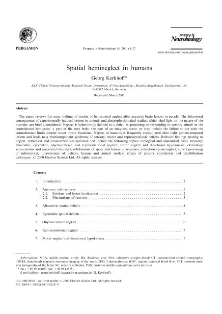
Progress <strong>in</strong> Neurobiology 63 (2001) 1±27<br />
www.elsevier.com/locate/pneurobio<br />
<strong>Spatial</strong> <strong>hem<strong>in</strong>eglect</strong> <strong>in</strong> <strong>humans</strong><br />
Georg Kerkho€*<br />
EKN-Cl<strong>in</strong>ical Neuropsychology Research Group, Department of Neuropsychology, Hospital Bogenhausen, Dachauerstr. 164,<br />
D-80992 Munich, Germany<br />
Received 6 March 2000<br />
Abstract<br />
The paper reviews the ma<strong>in</strong> ®nd<strong>in</strong>gs of studies of hemispatial neglect after acquired bra<strong>in</strong> lesions <strong>in</strong> people. The behavioral<br />
consequences of experimentally <strong>in</strong>duced lesions <strong>in</strong> animals and electrophysiological studies, which shed light on the nature of the<br />
disorder, are brie¯y considered. Neglect is behaviorally de®ned as a de®cit <strong>in</strong> process<strong>in</strong>g or respond<strong>in</strong>g to sensory stimuli <strong>in</strong> the<br />
contralateral hemispace, a part of the own body, the part of an imag<strong>in</strong>ed scene, or may <strong>in</strong>clude the failure to act with the<br />
contralesional limbs despite <strong>in</strong>tact motor functions. Neglect <strong>in</strong> <strong>humans</strong> is frequently encountered after right parieto-temporal<br />
lesions and leads to a multicomponent syndrome of sensory, motor and representational de®cits. Relevant ®nd<strong>in</strong>gs relat<strong>in</strong>g to<br />
neglect, ext<strong>in</strong>ction and unawareness are reviewed and <strong>in</strong>clude the follow<strong>in</strong>g topics: etiological and anatomical basis, recovery;<br />
allocentric, egocentric, object-centered and representational neglect; motor neglect and directional hypok<strong>in</strong>esia; elementary<br />
sensorimotor and associated disorders; subdivisions of space and frames of reference; ext<strong>in</strong>ction versus neglect; covert process<strong>in</strong>g<br />
of <strong>in</strong>formation; unawareness of de®cits; human and animal models; e€ects of sensory stimulation and rehabilitation<br />
techniques. 7 2000 Elsevier Science Ltd. All rights reserved.<br />
Contents<br />
1. Introduction ............................................................ 2<br />
2. Anatomy and recovery. .................................................... 2<br />
2.1. Etiology and lesion localization ......................................... 2<br />
2.2. Mechanisms of recovery. .............................................. 3<br />
3. Allocentric spatial de®cits .................................................. 4<br />
4. Egocentric spatial de®cits ................................................... 5<br />
5. Object-centered neglect .................................................... 6<br />
6. Representational neglect ................................................... 7<br />
7. Motor neglect and directional hypok<strong>in</strong>esia ...................................... 7<br />
Abbreviations: MCA, middle cerebral artery; BA, Brodman area; SSA, subjective straight ahead; CT, computerized cranial tomography;<br />
(f)MRI, (functional) magnetic resonance imag<strong>in</strong>g of the bra<strong>in</strong>; 2DG, 2-desoxyglycose; rCBF, regional cerebral blood ¯ow; PET, positron emission<br />
tomography of the bra<strong>in</strong>; SC, superior colliculus; PmS, posterior middle-suprasylvian cortex (<strong>in</strong> cats).<br />
* Tel.: +49-89-154057; fax: +49-89-156781.<br />
E-mail address: georg.kerkho€@extern.lrz-muenchen.de (G. Kerkho€).<br />
0301-0082/00/$ - see front matter 7 2000 Elsevier Science Ltd. All rights reserved.<br />
PII: S0301-0082(00)00028-9
2<br />
G. Kerkho€ / Progress <strong>in</strong> Neurobiology 63 (2001) 1±27<br />
8. Elementary sensory, motor and associated disorders. ............................... 9<br />
9. Subdivisions of space and di€erent frames of reference. ............................ 10<br />
9.1. Behavioral dissociations <strong>in</strong> di€erent sectors of space <strong>in</strong> neglect .................. 10<br />
9.2. Neurophysiologic and functional imag<strong>in</strong>g evidence for di€erent types of space cod<strong>in</strong>g <strong>in</strong><br />
the bra<strong>in</strong>. ........................................................ 10<br />
10. Ext<strong>in</strong>ction versus neglect .................................................. 11<br />
11. Covert process<strong>in</strong>g of <strong>in</strong>formation ............................................ 13<br />
12. Unawareness of de®cits ................................................... 13<br />
13. Models ............................................................... 14<br />
13.1. Early sensory theories ............................................... 14<br />
13.2. Attentional theories ................................................. 15<br />
13.3. Representational theories ............................................. 17<br />
13.4. Transformational theories ............................................ 17<br />
13.5. Cerebral balance theories ............................................. 18<br />
13.6. One model or a synthesis of models?. .................................... 19<br />
14. Modulation of neglect, ext<strong>in</strong>ction and unawareness by sensory stimulation .............. 19<br />
15. Rehabilitation approaches ................................................. 20<br />
16. Outstand<strong>in</strong>g questions .................................................... 22<br />
Acknowledgements. .......................................................... 22<br />
References ................................................................. 22<br />
1. Introduction<br />
Neglect (synonymous with: hemispatial neglect, hemi<strong>in</strong>attention,<br />
hemisensory neglect, <strong>hem<strong>in</strong>eglect</strong> ) denotes<br />
the impaired or lost ability to react to or process sensory<br />
stimuli (visual, auditory, tactile, olfactory) presented<br />
<strong>in</strong> the hemispace contralateral to a lesion of the<br />
human right or left cerebral hemisphere. Besides the<br />
aforementioned aspects of sensory neglect, motor<br />
neglect may occur and manifest itself as the reduced<br />
use or nonuse of a contralateral extremity (arm, leg)<br />
dur<strong>in</strong>g walk<strong>in</strong>g or bimanual activities. F<strong>in</strong>ally, neglect<br />
may occur <strong>in</strong> the imag<strong>in</strong>ation of spatial scenes (representational<br />
neglect). By de®nition, neglect is not<br />
merely the result of elementary sensory (i.e. hemianopia,<br />
hemisensory loss), motor (i.e. hemiparesis) or cognitive/emotional<br />
disorders (i.e. reduced <strong>in</strong>telligence,<br />
depression). Table 1 summarizes the most frequent<br />
neglect phenomena <strong>in</strong> these di€erent categories.<br />
2. Anatomy and recovery<br />
2.1. Etiology and lesion localization<br />
The most frequent etiological cause of neglect <strong>in</strong><br />
<strong>humans</strong> is (often large) <strong>in</strong>farctions <strong>in</strong> the territory of<br />
the right, less often the left middle cerebral artery<br />
(MCA; Vallar, 1993), caus<strong>in</strong>g lesions which center on<br />
the <strong>in</strong>ferior parietal cortex (Brodman areas, BA 40, 7).<br />
Accompany<strong>in</strong>g damage to adjacent structures such as<br />
the optic radiation, the <strong>in</strong>sula, dorsolateral frontal cortex<br />
(BA 4,6,44,45, 46) and superior temporal cortex<br />
(BA 22, 37) is frequently apparent (Smania et al.,<br />
1998). Furthermore, neglect occurs after posterior thalamic<br />
(Cambier et al., 1980) or basal ganglia lesions<br />
(Damasio et al., 1980), as a result of <strong>in</strong>tracerebral<br />
bleed<strong>in</strong>gs, but has never been described follow<strong>in</strong>g pure<br />
occipital lesions (Smania et al., 1998; Vallar, 1993).<br />
Occasionally, neglect after lateral frontal lesions has<br />
been reported (Husa<strong>in</strong> and Kennard, 1996) where similar<br />
lesions <strong>in</strong> animals regularly lead to contralesional<br />
neglect (Rizzolatti et al., 1983; Deuel and Coll<strong>in</strong>s,<br />
1984). Neglect may also result from tumors or traumatic<br />
<strong>in</strong>juries of the aforementioned areas (Vallar,<br />
1993). In general, severe and multimodal neglect is<br />
caused by very large rightsided lesions which encroach<br />
on parietal, temporal, and regions of occipital or frontal<br />
cortex as well as subcortical structures (Vallar and<br />
Perani, 1986).<br />
Contralesional neglect occurs <strong>in</strong> some 33% of lefthemisphere<br />
and more than 50% of right-hemisphere
G. Kerkho€ / Progress <strong>in</strong> Neurobiology 63 (2001) 1±27 3<br />
Table 1<br />
Summary of sensory, motor and representational neglect phenomena occurr<strong>in</strong>g after unilateral lesions <strong>in</strong> <strong>humans</strong><br />
Type of neglect<br />
Visual<br />
Auditory<br />
Somatosensory<br />
Olfactory<br />
Motor<br />
Representational<br />
De®nition and typical behaviour<br />
Patient searches for stimuli with eye- and head-movements preferentially <strong>in</strong> the ipsilesional hemispace. Omission of<br />
contralesional stimuli dur<strong>in</strong>g read<strong>in</strong>g, writ<strong>in</strong>g, draw<strong>in</strong>g of geometric stimuli, bisect<strong>in</strong>g horizontal l<strong>in</strong>es, or eat<strong>in</strong>g<br />
from a plate. Ipsilesional deviation of the perceived subjective straight ahead.<br />
Patient does not react to sound/speech stimuli from the contralateral hemispace. He/she may turn to the<br />
ipsilesional side when addressed from the contralesional. When several speakers are present the patient responds<br />
preferentially to the most ipsilesional one, irrespective of who has spoken. Ipsilesional deviation of the perceived<br />
auditory midl<strong>in</strong>e position <strong>in</strong> front space.<br />
Ignor<strong>in</strong>g of tactile stimulation (i.e. touch) or pa<strong>in</strong>ful stimuli (cold/hot stimuli, jammed ®ngers <strong>in</strong> wheel or spokes of<br />
the wheelchair) on the contralesional body half. Mislocalization of tactile stimuli <strong>in</strong> this part. Subjective shift of<br />
own body-midl<strong>in</strong>e (i.e. position of the sp<strong>in</strong>e) to the ipsilesional side.<br />
Ignor<strong>in</strong>g smells delivered to one nostril. Rarely observed <strong>in</strong> daily life s<strong>in</strong>ce stimuli are easily detected with the other<br />
nostril.<br />
Reduced use of contralesional arm/leg which is not completely attributable to a sensorimotor loss. Reduced arm<br />
sway dur<strong>in</strong>g walk<strong>in</strong>g; reduced use of contralesional arm dur<strong>in</strong>g bimanual activities (i.e. eat<strong>in</strong>g, carry<strong>in</strong>g loads);<br />
dragg<strong>in</strong>g beh<strong>in</strong>d the contralesional leg/foot dur<strong>in</strong>g walk<strong>in</strong>g.<br />
Patient describes few items located <strong>in</strong> the contralesional part of an imag<strong>in</strong>ed scene (i.e. a famous city place, the<br />
own liv<strong>in</strong>g room or house), but describes much more items when describ<strong>in</strong>g the same scene from a di€erent<br />
perspective (1808 rotated).<br />
lesioned patients 1 (Stone et al., 1991) when tested immediately<br />
(with<strong>in</strong> 7 days) after lesion onset. The absolute<br />
percentage of neglect depends critically on the<br />
criterion or test used but the asymmetry <strong>in</strong> the occurrence<br />
of contralesional neglect has been found across<br />
di€erent samples and methods (Schenkenberg et al.,<br />
1980). Recovery is considerably quicker and more<br />
complete <strong>in</strong> neglect after left-hemisphere lesions as<br />
opposed to right-hemisphere lesions (Stone et al.,<br />
1991). Thus, there is a clear hemispheric asymmetry<br />
show<strong>in</strong>g that neglect is more frequent, more severe and<br />
more permanent follow<strong>in</strong>g right-hemispheric lesions.<br />
However, transient rightsided neglect after left unilateral<br />
lesions occurs <strong>in</strong> some cases (Welman, 1969; Peru<br />
and P<strong>in</strong>na, 1997; Kerkho€ and Zoelch, 1998). More<br />
long-last<strong>in</strong>g rightsided neglect occurs <strong>in</strong> patients with<br />
bilateral cerebral lesions (We<strong>in</strong>traub et al., 1996).<br />
2.2. Mechanisms of recovery<br />
1 S<strong>in</strong>ce leftsided neglect after right cerebral lesions is the most frequent<br />
type of neglect the term ``neglect'' <strong>in</strong> this review refers always<br />
to leftsided neglect <strong>in</strong> <strong>humans</strong> if not <strong>in</strong>dicated otherwise. Of course,<br />
this does not exclude the fact that rightsided neglect after uni- or bilateral<br />
lesions may occur (more rarely) as well.<br />
Recovery from the most obvious signs of neglect<br />
(i.e. the tendency to orient to the ipsilesional side and<br />
the lack of visual exploration <strong>in</strong> the contralesional<br />
hemispace) has been noted <strong>in</strong> the majority of patients<br />
with<strong>in</strong> the ®rst 6 months (Lawson, 1962; Hier et al.,<br />
1983). In the rema<strong>in</strong><strong>in</strong>g 25% neglect may persist for<br />
up to 12 years (Zarit and Kahn, 1974) and performance<br />
<strong>in</strong> tasks which are sensitive to neglect may decl<strong>in</strong>e<br />
aga<strong>in</strong> after cessation of apparently successful rehabilitation<br />
treatment <strong>in</strong> the cl<strong>in</strong>ic (Paolucci et al., 1998;<br />
Hier et al., 1983). Recovery from neglect is more prom<strong>in</strong>ent<br />
after left compared with right-sided cerebral<br />
lesions (Stone et al., 1991). Furthermore, recovery<br />
from ``frontal'' neglect is more rapid and more complete<br />
<strong>in</strong> <strong>humans</strong> than from the classical ``parietal''<br />
neglect syndrome (Matt<strong>in</strong>gley et al., 1994a). Substantial<br />
recovery is less likely after large lesions and <strong>in</strong><br />
those patients with di€use bra<strong>in</strong> atrophy <strong>in</strong> addition to<br />
the focal right hemispheric lesion (Lev<strong>in</strong>e et al., 1986)<br />
Little is known about the mechanisms guid<strong>in</strong>g spontaneous<br />
recovery and/or those enabl<strong>in</strong>g treatmentguided<br />
improvements dur<strong>in</strong>g rehabilitation. Pantano et<br />
al. (1992) reported a concomitant <strong>in</strong>crease <strong>in</strong> regional<br />
cerebral blood ¯ow (rCBF) <strong>in</strong> the posterior areas of<br />
the damaged right hemisphere and the anterior areas<br />
of the <strong>in</strong>tact, left hemisphere probably <strong>in</strong>clud<strong>in</strong>g the<br />
frontal eye ®elds after a speci®c neglect treatment <strong>in</strong><br />
their patients. Only the left frontal activation, <strong>in</strong> the<br />
region of the frontal eye ®elds, covaried with improved<br />
visual scann<strong>in</strong>g behavior after treatment. The authors<br />
concluded that the left (<strong>in</strong>tact) frontal eye ®elds are<br />
crucial for recovery of visual scann<strong>in</strong>g <strong>in</strong> neglect. In a<br />
later PET study with three neglect patients, this ®nd<strong>in</strong>g<br />
was not uniformly replicated (Pizzamiglio et al., 1998).<br />
In this study, behavioral recovery seemed to covary<br />
with improved blood ¯ow <strong>in</strong> surviv<strong>in</strong>g areas of the<br />
lesioned hemisphere.<br />
Similar results have been obta<strong>in</strong>ed <strong>in</strong> primate studies<br />
where neglect was <strong>in</strong>duced by lesion<strong>in</strong>g the frontal<br />
polysensory association cortex. Metabolic mapp<strong>in</strong>g for<br />
local glucose utilization (2-DG) showed a widespread<br />
reduction of glucose <strong>in</strong> ipsilesional striatal and partially<br />
thalamic nuclei connected with the frontal lobe<br />
whereas no reductions were found <strong>in</strong> cortical areas
4<br />
G. Kerkho€ / Progress <strong>in</strong> Neurobiology 63 (2001) 1±27<br />
connected to the frontal lobe (Deuel and Coll<strong>in</strong>s, 1983,<br />
1984). Behavioral recovery of visual somatosensory<br />
and motor neglect was paralleled by a normalization<br />
of glucose metabolism <strong>in</strong> these subcortical circuits of<br />
the lesioned hemisphere (Deuel and Coll<strong>in</strong>s, 1983,<br />
1984). To summarize, as <strong>in</strong> the few human imag<strong>in</strong>g<br />
studies neglect after focal rightsided lesions of the<br />
frontal or parietal cortex is associated with widespread<br />
depressions of metabolic activity <strong>in</strong> the same hemisphere;<br />
behavioral recovery is paralleled (or enabled)<br />
by a normalization of activity <strong>in</strong> this neural network<br />
for spatial attention that <strong>in</strong>cludes cortical and subcortical<br />
areas. This widespread depression of activity<br />
helps to expla<strong>in</strong> why neglect occurs after cortical and<br />
subcortical lesions of di€erent anatomical structures<br />
(see Section 13).<br />
judgments <strong>in</strong> hemianopic patients without neglect<br />
(Axenfeld, 1894; Lohmann, 1911). These patients<br />
bisect a horizontal l<strong>in</strong>e <strong>in</strong> most cases too far to their<br />
bl<strong>in</strong>d hemi®eld (Kerkho€, 1993; Barton et al., 1998)<br />
whereas neglect patients without hemianopia place<br />
their bisection mark too far to their ipsilesional side<br />
thus reproduc<strong>in</strong>g a too large l<strong>in</strong>e segment <strong>in</strong> their contralesional<br />
hemispace (Halligan et al., 1990; Halligan<br />
and Marshall, 1991b; Barton et al., 1998; cf. Fig. 1A).<br />
In a variant of this task termed l<strong>in</strong>e extension (Fig. 1B)<br />
the subject views only the part of the l<strong>in</strong>e displayed <strong>in</strong><br />
the ipsilesional hemispace and subsequently estimates<br />
how long a bar has to be extended to the contralesional<br />
hemispace until both parts appear as equally<br />
long. In this task neglect patients typically oversize the<br />
l<strong>in</strong>e segment <strong>in</strong> their neglected hemispace Ð just as<br />
they do <strong>in</strong> l<strong>in</strong>e bisection (Fig. 1B).<br />
3. Allocentric spatial de®cits<br />
Because damage is <strong>in</strong>variably widespread and compromises<br />
multiple neural systems, pr<strong>in</strong>cipally located<br />
<strong>in</strong> the parieto-temporal cortex (<strong>in</strong> human neglect) as a<br />
result of large lesions a variety of de®cits are encountered<br />
<strong>in</strong> neglect. These may be analyzed with respect to<br />
di€erent aspects of spatial cognition. A dist<strong>in</strong>ction<br />
may be drawn between allocentric, egocentric and<br />
object-centered de®cits. Allocentric space is de®ned <strong>in</strong><br />
this context as concern<strong>in</strong>g the spatial relationship<br />
between two stimuli separated <strong>in</strong> space, i.e. the length<br />
of two l<strong>in</strong>es or the two halves of a bisected l<strong>in</strong>e. In a<br />
broader sense, allocentric relationships de®ne spatial<br />
relations between two or more stimuli <strong>in</strong> three-dimensional<br />
space, <strong>in</strong>clud<strong>in</strong>g those entailed <strong>in</strong> spatial navigation.<br />
In contrast, egocentric de®cits relate to spatial<br />
impairments <strong>in</strong> relation to the patient's own body or<br />
speci®c body parts, i.e. the subjective estimate of her/<br />
his tactile body midl<strong>in</strong>e, the auditory or visual sense of<br />
straight ahead <strong>in</strong> relation to the sagittal midl<strong>in</strong>e of the<br />
body or head. Object-centered (or environment-centered)<br />
de®cits <strong>in</strong>dicate hemispatial de®cits or omissions<br />
<strong>in</strong> the contralesional half of one object, i.e. a face or a<br />
clock face (see below). In contrast to the allocentric<br />
and egocentric de®cits which ®t relatively easy <strong>in</strong>to a<br />
left-right or contralesional/ipsilesional dichotomy of<br />
space (impairments <strong>in</strong> left/contralesional hemispace,<br />
normal or nearly normal performance <strong>in</strong> right/ipsilesional<br />
hemispace) object-centered neglect means that a<br />
patient ignores the left/contralesional part of one<br />
object (i.e. a word, a draw<strong>in</strong>g, or a face), regardless of<br />
the object's location <strong>in</strong> space (Walker, 1995; Olson and<br />
Gettner, 1996).<br />
One of the most frequent allocentric de®cits <strong>in</strong>vestigated<br />
<strong>in</strong> patients with visual neglect is horizontal l<strong>in</strong>e<br />
bisection (Schenkenberg et al., 1980; Burnett-Stuart et<br />
al., 1991), a task orig<strong>in</strong>ally devised to measure spatial<br />
Fig. 1. Survey of de®cits <strong>in</strong> allocentric visuospatial tasks <strong>in</strong> patients<br />
with leftsided visual neglect. The vertical stippled l<strong>in</strong>e represents the<br />
putative border of the contralesional and ipsilesional hemispace<br />
aligned to the objective median plane of the patient. (A) In horizontal<br />
l<strong>in</strong>e bisection the neglect patient transects the bar too far to the<br />
ipsilesional, right side, <strong>in</strong>dicat<strong>in</strong>g a deviated subjective midl<strong>in</strong>e (cf<br />
Halligan et al., 1990). (B) Oversized left l<strong>in</strong>e segment <strong>in</strong> a l<strong>in</strong>e extension<br />
task (Bisiach et al., 1996). In this task, the subject ®rst views<br />
only the black, rightsided bar. He/she is then <strong>in</strong>structed to estimate<br />
how long the leftsided (dotted) bar has to be drawn until it appears<br />
to have the same length as the rightsided black bar. (C) The subject<br />
views a horizontal distance <strong>in</strong> the ipsilesional hemispace which has<br />
to be reproduced with the two black markers <strong>in</strong> the contralesional,<br />
left hemispace. Note the signi®cant overestimation of the leftsided<br />
distance (``space distortion'', <strong>in</strong>dicated by the shaded area; redrawn<br />
after (Kerkho€, 2000). (D) Typical overestimation of horizontal bar<br />
length <strong>in</strong> the contralesional hemispace (``size distortion''; <strong>in</strong>dicated by<br />
the shaded area; redrawn after (Milner and Harvey, 1995). (E,F)<br />
Normal, veridical cod<strong>in</strong>g of vertical size and of symmetric shapes <strong>in</strong><br />
neglect (redrawn after Milner and Harvey, 1995).
G. Kerkho€ / Progress <strong>in</strong> Neurobiology 63 (2001) 1±27 5<br />
Similarly, neglect patients overreproduce a horizontal<br />
distance displayed <strong>in</strong> their contralesional hemispace<br />
when the ``reference'' distance is displayed <strong>in</strong> their ipsilesional<br />
space (Fig. 1C). The same phenomenon is<br />
observed when a horizontal bar has to be reproduced<br />
<strong>in</strong> the left hemispace (Fig. 1D). Here patients reproduce<br />
the bar <strong>in</strong> the contralesional hemispace too long,<br />
which has been termed ``size distortion'' (Harvey et al.,<br />
1995; Milner and Harvey, 1995; Milner et al., 1998).<br />
In contrast, neglect patients show veridical cod<strong>in</strong>g of<br />
vertical bars (Fig. 1E) and of symmetrical, global<br />
shapes like circles (Fig. 1F). This disturbed size cod<strong>in</strong>g<br />
may rema<strong>in</strong> a purely perceptual phenomenon s<strong>in</strong>ce it<br />
does not necessarily imply disturbed adjustment of ®nger<br />
grip aperture when scal<strong>in</strong>g the size of an object <strong>in</strong><br />
grasp<strong>in</strong>g (Pritchard et al., 1997). Together, these<br />
results <strong>in</strong>dicate a non-veridical cod<strong>in</strong>g of with<strong>in</strong>-object<br />
(length of an object) and between-object (distance<br />
between objects) spatial extension <strong>in</strong> the horizontal<br />
plane which is not found when vertical size cues are<br />
apparent. The advanced <strong>in</strong>terpretation for these allocentric<br />
spatial disorders is that the contralesional hemispace<br />
appears ``constricted'' or ``distorted'' <strong>in</strong> relation<br />
to the ipsilesional hemispace (Milner et al., 1998). As a<br />
consequence, elongated objects presented <strong>in</strong> this hemispace<br />
have to be larger <strong>in</strong> order to match the length of<br />
an object <strong>in</strong> ipsilesional hemispace. This has been<br />
found for horizontal object-size, and as well for the<br />
horizontal distance between objects (Kerkho€, 2000;<br />
see also Karnath and Ferber, 1999 for di€erent results<br />
and <strong>in</strong>terpretations). Measures of perceptual distortions<br />
<strong>in</strong> patients with neglect may be quite sensitive<br />
s<strong>in</strong>ce they may reveal size distortions even after apparent<br />
recovery of other signs (l<strong>in</strong>e bisection; Harvey and<br />
Milner, 1999).<br />
4. Egocentric spatial de®cits<br />
Fig. 2. Representative egocentric de®cits <strong>in</strong> neglect. (A) Deviated<br />
visual judgments of neglect patients (without visual ®eld defects)<br />
when <strong>in</strong>dicat<strong>in</strong>g the position of a diode <strong>in</strong> total darkness <strong>in</strong> relation<br />
to their bodily subjective straight ahead position. The judgments<br />
were similarly shifted to the ipsilesional, right side <strong>in</strong> two distances<br />
(1.2 and 3 m), <strong>in</strong>dicat<strong>in</strong>g a rotation of the visual subjective straight<br />
ahead (SSA) <strong>in</strong> front space (redrawn after Ferber and Karnath,<br />
1999). (B) Ipsilesionally shifted visual search area <strong>in</strong> total darkness<br />
(solid l<strong>in</strong>e) and similarly shifted tactile search area (with the ipsilesional<br />
hand, not drawn) with bl<strong>in</strong>dfolded neglect patients (cf. Karnath<br />
and Peren<strong>in</strong>, 1998). Search areas are drawn schematically and<br />
not to scale. (C) Judgments of the subjective visual and tactile vertical,<br />
horizontal and oblique orientation matches <strong>in</strong> a patient with leftsided<br />
neglect (upper two polar plots) and a matched normal subject<br />
(lower two plots). Each th<strong>in</strong> black l<strong>in</strong>es represents the result of one<br />
trial. Visual tests were taken with stimuli displayed on a PC-screen<br />
via software (Kerkho€ and Marquardt, 1995), tactile tests employed<br />
the judgment of a metal bar mounted on a vertical board <strong>in</strong> front of<br />
the patient (Kerkho€, 1999a). Note the counterclockwise tilt of perceptual<br />
judgments <strong>in</strong> both modalities <strong>in</strong> the neglect patient. Together,<br />
these results <strong>in</strong>dicate a subjective rotation of sensory space <strong>in</strong> the<br />
frontal plane go<strong>in</strong>g beyond the shift of the SSA <strong>in</strong> the sagittal plane<br />
(A) <strong>in</strong> neglect patients.<br />
It is a relatively new idea that neglect is not only<br />
found <strong>in</strong> relation to the left half of space but more<br />
precisely <strong>in</strong> relation to what is left of the patient's<br />
trunk midl<strong>in</strong>e, head sagittale or even the position of<br />
the patient's hand <strong>in</strong> space. In one of the early studies<br />
on that topic (Heilman et al., 1983) neglect patients<br />
were required to po<strong>in</strong>t towards their subjective straight<br />
ahead (SSA) <strong>in</strong> relation to their trunk midl<strong>in</strong>e. The<br />
patients' responses were ipsilesionally deviated to the<br />
right side while non-neglect<strong>in</strong>g patients with left hemispheric<br />
lesions showed quite accurate po<strong>in</strong>t<strong>in</strong>g results.<br />
Similar results are obta<strong>in</strong>ed when neglect patients have<br />
to <strong>in</strong>dicate their visual SSA without po<strong>in</strong>t<strong>in</strong>g (Fig. 2A).<br />
If egocentric de®cits are related to the body midl<strong>in</strong>e<br />
or certa<strong>in</strong> body parts modi®cations of their position <strong>in</strong><br />
space might <strong>in</strong>¯uence neglect. In an early study exam<strong>in</strong><strong>in</strong>g<br />
this question (Bisiach et al., 1985), neglect<br />
patients had to tactually explore a honeycomb-shaped<br />
board with their ipsilesional hand to ®nd a marble.<br />
The authors found that neglect covaried with the position<br />
of the trunk-vertical axis and with the head/eyeaxis.<br />
Shift<strong>in</strong>g either the trunk or the head to the left <strong>in</strong><br />
relation to the stationary honeycomb-board reduced<br />
the number of omissions <strong>in</strong> the tactile neglect task.<br />
Karnath (summarized <strong>in</strong> Karnath, 1997) has <strong>in</strong>vestigated<br />
the <strong>in</strong>¯uence of body-and-head position <strong>in</strong> more<br />
detail. In a saccadic reaction-time study, it was found<br />
that leftward trunk-rotation but not head-rotation<br />
improved the delayed reactions to visual targets<br />
appear<strong>in</strong>g <strong>in</strong> contralesional hemispace (Karnath et al.,
6<br />
G. Kerkho€ / Progress <strong>in</strong> Neurobiology 63 (2001) 1±27<br />
1991). This role of the trunk-sagittale as one major<br />
anchor for determ<strong>in</strong><strong>in</strong>g left versus right <strong>in</strong> space <strong>in</strong> relation<br />
to the observer has been replicated <strong>in</strong> several<br />
subsequent studies us<strong>in</strong>g l<strong>in</strong>e bisection and read<strong>in</strong>g<br />
tasks (Chokron and Imbert, 1995; Sch<strong>in</strong>dler and Kerkho€,<br />
1997; Vuilleumier et al., 1999). However, the<br />
trunk midl<strong>in</strong>e is not the only axis that might be important<br />
for spatial orientation: the head sagittale plays<br />
a similar important role as suggested by behavioral<br />
and neurophysiological studies (behavioral: Bisiach et<br />
al., 1985; Kerkho€ and Sch<strong>in</strong>dler, 1997; Vuilleumier et<br />
al., 1999; neurophysiological: Andersen et al., 1989,<br />
1990, 1997; Duhamel et al., 1997). Other types of egocentric<br />
or body-centered de®cits <strong>in</strong> neglect are summarized<br />
<strong>in</strong> Fig. 2. Patients with pure neglect (without<br />
®eld defects) often show an ipsilesionally deviated subjective<br />
estimate of straight ahead (Fig. 2A) which<br />
seems to be anchored to the trunk midl<strong>in</strong>e, although<br />
not every neglect patient shows this de®cit (Bartolomeo<br />
and Chokron, 1999). The visual and tactile ®elds<br />
of search <strong>in</strong> neglect patients (as measured by optoelectronic<br />
devices) are shifted to the ipsilesional, right side<br />
(Fig. 2B), even when no external stimulus is present.<br />
Fig. 3. (A) Examples of object-centered neglect <strong>in</strong> draw<strong>in</strong>g a clock or<br />
¯owers. The arrows <strong>in</strong>dicate the omission of relevant details. Note<br />
that the neglect patient drew two ¯owers laterally beneath each<br />
other, but neglected the leftsided details of each ¯ower. (B) Objector<br />
word-centered neglect dur<strong>in</strong>g read<strong>in</strong>g an <strong>in</strong>dented read<strong>in</strong>g text.<br />
The crossed-out words <strong>in</strong>dicate complete omissions of whole words<br />
(space-based read<strong>in</strong>g errors) while the bold, shadowed, leftsided<br />
parts of words (see arrows) <strong>in</strong>dicate word-based omissions. Note<br />
that <strong>in</strong> most cases the patient read some words correctly which were<br />
located to the left of the word-centered errors, similar to the fact<br />
that both ¯owers were drawn (see A), but with leftsided, object-centered<br />
omissions of details <strong>in</strong> their Gestalt (B based on results from<br />
Kerkho€ et al., 1999b).<br />
However, the deviations of spatial orientation <strong>in</strong> the<br />
horizontal plane are not the only ones encountered <strong>in</strong><br />
neglect. These patients can also show a tilt of their<br />
subjective vertical or horizontal or judgments of<br />
whether two obliquely oriented stimuli are parallel, <strong>in</strong><br />
their frontal plane (Fig. 2C). Hence, a 908-oriented,<br />
vertical bar has to be oriented to 1008 (tilted counterclockwise)<br />
to be perceived as vertical by a neglect<br />
patient. This de®cit occurs both <strong>in</strong> the visual (Kerkho€<br />
and Zoelch, 1998) and tactile (Kerkho€, 1999a) modality<br />
and <strong>in</strong>dicates that the spatial anchor<strong>in</strong>g of subjective,<br />
egocentric space is disturbed <strong>in</strong> neglect patients<br />
<strong>in</strong> at least two spatial planes: the horizontal plane,<br />
lead<strong>in</strong>g to the pathological, ipsilesionally oriented<br />
body orientation (cf. Fig. 2A,B), and the frontal plane,<br />
lead<strong>in</strong>g probably to abnormal posture of the head,<br />
trunk and legs dur<strong>in</strong>g sitt<strong>in</strong>g and stance, as found <strong>in</strong><br />
neglect patients (Rode et al., 1997, 1998; Perennou et<br />
al., 1998, 1999).<br />
5. Object-centered neglect<br />
When we look at or imag<strong>in</strong>e an object we are aware<br />
of its structure. Consider the face of a friend of yours,<br />
your watch or the letter `d'. We know that <strong>in</strong> order to<br />
transform the letter `p' from `d', we have to rotate the<br />
letter `d' <strong>in</strong> the plane of the page. Likewise, you know<br />
if and on which side your friend has a birth mark on<br />
his face, or on which side of your watch the date is<br />
<strong>in</strong>dicated. This k<strong>in</strong>d of structural awareness very likely<br />
depends on a neural system with two features: neurons<br />
selective for locations as de®ned relative to a reference<br />
frame around an object, and an arrangement of several<br />
of such neurons so that their spatial ®elds form a map<br />
of object-centered space (Olson and Gettner, 1995,<br />
1996). Patients with spatial neglect may copy or mark<br />
every feature <strong>in</strong> an array, yet they may fail to reproduce<br />
the left part of an object (leftsided numbers on a<br />
clock face, a ¯ower, or the left part of a word, cf.<br />
Fig. 3).<br />
Hillis and Caramazza (1991) described a patient<br />
with rightsided neglect dyslexia who made read<strong>in</strong>g<br />
errors chie¯y at the end of a word Ð irrespective <strong>in</strong><br />
which visual ®eld the word appeared, or if it were<br />
pr<strong>in</strong>ted from left to right, right to left or vertically<br />
from top to bottom. In all these experimental modi®cations,<br />
the errors occurred at the end of the word.<br />
These results support the idea that mean<strong>in</strong>gful words<br />
form a k<strong>in</strong>d of ``perceptual unit'' (Baylis et al., 1994)<br />
and have an object-centered representation. Likewise,<br />
faces may be coded <strong>in</strong> a similar way which ®ts neatly<br />
the observation of leftsided neglect for the contralesional<br />
half of objects or faces (Walker, 1995).<br />
Which factors do determ<strong>in</strong>e that a patient neglects<br />
the left half of an object? Is the gravitational vertical
G. Kerkho€ / Progress <strong>in</strong> Neurobiology 63 (2001) 1±27 7<br />
or the <strong>in</strong>tr<strong>in</strong>sic vertical of an object the relevant variable?<br />
To decide between these two possibilities, rotated<br />
®gures with critical elements on the left or right side of<br />
the <strong>in</strong>tr<strong>in</strong>sic ®gure axis have been utilized. Some<br />
patients persist <strong>in</strong> show<strong>in</strong>g neglect of the contralesional<br />
part of the rotated object (Driver et al., 1994). This<br />
type of axis-based neglect demonstrates neatly that<br />
neglect might be attached to the object's <strong>in</strong>tr<strong>in</strong>sic axis<br />
and not necessarily to the physical, left side of an<br />
object (but see Farah and Buxbaum, 1997, for an<br />
alternative <strong>in</strong>terpretation).<br />
6. Representational neglect<br />
Neglect is not restricted to perception or action <strong>in</strong><br />
the contralesional part of external space. It may occur<br />
as well when subjects have to scan their <strong>in</strong>ner spatial<br />
representation of a scene. Bisiach and Luzzatti (1978)<br />
reported <strong>in</strong> their famous experiment two patients who<br />
were unable to recall speci®c locations on the left side<br />
of the Piazza de Duomo <strong>in</strong> Milano when they imag<strong>in</strong>ed<br />
fac<strong>in</strong>g it from a particular vantage. When they<br />
had to describe the square from a 1808-rotated perspective<br />
they recalled the omitted details on their left<br />
from the previous condition but neglected now particular<br />
items on their left side which were on the right side<br />
<strong>in</strong> the prior test condition. The authors concluded that<br />
their patients had an unilateral representational de®cit<br />
for one hemispace. In later studies, these results were<br />
con®rmed experimentally us<strong>in</strong>g an elegant technique<br />
(Bisiach et al., 1981). Similar de®cits are found when<br />
patients are required to recall cities from their country<br />
(Bartolomeo et al., 1994; Rode and Peren<strong>in</strong>, 1994 cf.<br />
Fig. 4).<br />
Fig. 4. Example of representational (imag<strong>in</strong>al) neglect <strong>in</strong> a patient<br />
asked to visualize mentally the map of France and to name as many<br />
towns as possible dur<strong>in</strong>g a limited period of time (2 m<strong>in</strong>). The number<br />
at the bottom <strong>in</strong>dicates the number of recalled cities <strong>in</strong> the two<br />
conditions. Temporary remission through caloric (vestibular) stimulation<br />
is seen on the right side of the ®gure. Reproduced from Peren<strong>in</strong><br />
(1997), with permission from Spr<strong>in</strong>ger-Verlag.<br />
Representational neglect seems to be less frequent<br />
than frank visual neglect: only some 25% of the latter<br />
patients show representational neglect when they are<br />
required to recall cities located on the left or right side<br />
of a map of their own country (Besch<strong>in</strong> et al., 1997;<br />
Bartolomeo et al., 1994). Typically, the patients recall<br />
easily cities located on the right side of the map and<br />
fail to report those located on the contralesional, left<br />
side Ð just as <strong>in</strong> the Piazza de Duomo example<br />
described above. Cl<strong>in</strong>ically, similar de®cits can be suspected<br />
when patients have to remember where on the<br />
ward their bedroom is or when they have to recall<br />
details from their home, i.e. the liv<strong>in</strong>g room.<br />
7. Motor neglect and directional hypok<strong>in</strong>esia<br />
Two types of de®cient motor behavior <strong>in</strong> neglect<br />
have to be dist<strong>in</strong>guished clearly: the reduced spontaneous<br />
use or complete nonuse of the contralesional<br />
hand, arm or leg dur<strong>in</strong>g motor activities (termed motor<br />
neglect; cf. Laplane and Degos, 1983; von Giesen et<br />
al., 1994) and the slower execution of arm movements<br />
with the ipsilesional hand or arm towards the contralesional<br />
hemispace (termed directional hypok<strong>in</strong>esia, Heilman<br />
et al., 1985; Bisiach et al., 1990; Matt<strong>in</strong>gley et al.,<br />
1994c; Bisiach et al., 1995). The latter is associated<br />
with a delayed movement <strong>in</strong>itiation and a reduced<br />
movement velocity and is probably a rare form of<br />
neglect (Milner et al., 1993). Ak<strong>in</strong> to the directional<br />
hypok<strong>in</strong>esia for leftward arm movements, neglect<br />
patients show delayed (Sundquist, 1979) and hypometric<br />
(Girotti et al., 1983) saccadic eye movements to<br />
left hemispace and their pattern of visual search is<br />
shifted to the ipsilesional hemispace (Che dru et al.,<br />
1973; Ishiai et al., 1987; Hornak, 1992; Karnath et al.,<br />
1996; as depicted <strong>in</strong> Fig. 2B).<br />
In contrast, motor neglect a€ects only the contralesional<br />
extremity; the use of the neglected hand/arm<br />
often can be prompted by the allocation of attention<br />
to it (Ghika et al., 1998), similar to the cue<strong>in</strong>g e€ects<br />
<strong>in</strong> sensory neglect (see Section 14). This motor neglect<br />
is not a result of a paresis which can be excluded by<br />
the exam<strong>in</strong>ation of magnetic evoked potentials (Von<br />
Giesen et al., 1994). Patients with motor neglect have<br />
often lesions <strong>in</strong>volv<strong>in</strong>g the thalamus, basal ganglia or<br />
the frontal cortex but spare the primary sensorimotor<br />
cortex (Laplane and Degos, 1983). Motor neglect is<br />
also found as a transient phenomenon after unilateral<br />
ventrolateral thalamotomy for the treatment of hemipark<strong>in</strong>sonism<br />
(Vilkki, 1984). Similar to the results of<br />
the 2-DG mapp<strong>in</strong>g studies <strong>in</strong> primate neglect (Deuel<br />
and Coll<strong>in</strong>s, 1984) imag<strong>in</strong>g <strong>in</strong>vestigations <strong>in</strong> <strong>humans</strong><br />
(PET, Von Giesen et al., 1994) <strong>in</strong>dicate a more widespread<br />
metabolic dysfunction <strong>in</strong> the premotor, prefrontal,<br />
c<strong>in</strong>gulate and partially parietal cortex of the
8<br />
G. Kerkho€ / Progress <strong>in</strong> Neurobiology 63 (2001) 1±27<br />
Table 2<br />
Frequently associated de®cits <strong>in</strong> neglect. Percentages are based on studies with neglect patients after right hemisphere lesions, or on right-hemisphere lesioned patients <strong>in</strong> general and may di€er<br />
between studies. In patients with small, circumscribed parietal lesions, other percentages are obta<strong>in</strong>ed (<strong>in</strong>dicated by , see Ghika et al., 1998). In the rightmost column diagnostic procedures are<br />
suggested to di€erentiate between elementary sensory or motor de®cits and neglect-related impairments. If the peripheral sensorimotor pathways are <strong>in</strong>tact VEP, SEP and transcranial magnetic<br />
stimulation should reveal largely normal results<br />
De®cit Frequency<br />
(%)<br />
Source Procedures for di€erential diagnosis<br />
Visual ®eld defects (hemianopia, quadrantanopia) 70±90 Vallar and Perani, 1986; Kerkho€, 1999a Visually evoked potentials (VEP), Vallar et al., 1991;<br />
Sp<strong>in</strong>elli et al., 1994; De Keyser et al., 1990; Special<br />
perimetry, Walker et al., 1991<br />
Hemianaesthesia (impaired position and pa<strong>in</strong> sense <strong>in</strong> contralesional limbs)<br />
63 Vallar et al., 1995b; Vallar et al., 1997; Sterzi et al., 1993; Ghika et al., 1998<br />
Somatosensory evoked potentials (SEP); Cue<strong>in</strong>g of<br />
patient's attention to the neglected limb; Ghika et al., 1998<br />
Hemiplegia/-paresis 100 Sterzi et al., 1993 Transcranial magnetic stimulation of motor cortex; Von<br />
Giesen et al., 1994<br />
Pseudoparesis of the hand 90 Ghika et al., 1998 Transcranial magnetic stimulation (TMS) of motor cortex<br />
Von Giesen et al., 1994<br />
Abnormal ®nger and hand postures 67 Ghika et al., 1998 ±<br />
Ipsilesional deviation of body posture <strong>in</strong> stance 70 Rode et al., 1997; Hesse et al., 1994 Posturography; Rode et al., 1997<br />
Increased pa<strong>in</strong>, avoidance of contralesional limb use 34 Ghika et al., 1998 Measurement of prolonged ``silent periods'' <strong>in</strong> the EMG<br />
after TMS; Classen et al., 1997<br />
Contralateral gaze palsy (tonic ipsilesional eye deviation) 30±50 De Renzi et al., 1982; Tijssen and Gisbergen, 1993 ±<br />
Visuospatial and tactilespatial disorders, impaired time 90 Kerkho€, 1999a; Basso et al., 1996 Evaluation of spatial perception; Kerkho€ and<br />
perception<br />
Marquardt, 1995<br />
Fatigue, reduced susta<strong>in</strong>ed attention ± Robertson et al., 1997 ±
G. Kerkho€ / Progress <strong>in</strong> Neurobiology 63 (2001) 1±27 9<br />
Fig. 5. Highly schematic demonstration of the typical di€erence<br />
between leftsided, visual <strong>hem<strong>in</strong>eglect</strong> and leftsided, homonymous<br />
hemianopia without neglect. In neglect, elementary visual perception<br />
up to the striate cortex may be largely <strong>in</strong>tact <strong>in</strong> the contra- and ipsilesional<br />
visual hemi®eld. In contrast, this is the pr<strong>in</strong>cipal de®cit <strong>in</strong><br />
homonymous, leftsided hemianopia. The opposite is found on a<br />
higher, more abstract level of spatial representation of the environment<br />
and the own body. Here, the left hemispace is less well represented<br />
<strong>in</strong> neglect, but seems <strong>in</strong>tact <strong>in</strong> hemianopia without neglect.<br />
Note, however, that hemianopia or other ®eld defects may be present<br />
due to damage of adjacent cortical structures <strong>in</strong> some 70% of neglect<br />
patients, especially <strong>in</strong> those with large parieto-temporal lesions compromis<strong>in</strong>g<br />
the optic radiation. The ®gure is <strong>in</strong>tended to clarify the<br />
crucial di€erence between a low-level visual disorder for one side of<br />
space (hemianopia) and a high-level hemispatial disorder (neglect).<br />
Note further, that <strong>in</strong> hemianopia a clear-cut, often stable border<br />
between see<strong>in</strong>g and bl<strong>in</strong>d ®eld regions is normally obta<strong>in</strong>ed <strong>in</strong> perimetric<br />
test<strong>in</strong>g. In contrast, the border between neglected and nonneglected<br />
hemispace is more variable and subject to many modulations,<br />
illustrated below <strong>in</strong> Fig. 10.<br />
<strong>in</strong>jured hemisphere <strong>in</strong> motor neglect than the lesion<br />
shown by the structural CT/MRI scans. Classen et al.<br />
(1997) studied the neurophysiological basis of motor<br />
neglect and found an exaggerated ``silent period'' <strong>in</strong><br />
the electromyogram of small hand muscles after transcranial<br />
magnetic stimulation. Accord<strong>in</strong>g to their<br />
results, this ``silent period'' might considerably delay<br />
the <strong>in</strong>itiation of directed motor activities of the contralateral<br />
extremities, and thus lead to the symptoms<br />
of hypok<strong>in</strong>esia, underutilization of the contralateral<br />
extremities as well as delayed movement <strong>in</strong>itiation<br />
(Classen et al., 1997).<br />
8. Elementary sensory, motor and associated disorders<br />
Most patients with neglect have low-level sensory<br />
2 It has been proposed recently that primary visual cortex is also<br />
<strong>in</strong>volved <strong>in</strong> high resolution visual imagery (Kosslyn et al., 1999).<br />
Consequently, striate cortex lesions might cause not only hemianopia<br />
but also a unilateral imagery de®cit similar to that of neglect<br />
patients. However, cl<strong>in</strong>ical experience with purely hemianopic<br />
patients does not support such a hemi-imagery de®cit, at least not<br />
on a coarse level. Possibly, such hemi-imagery de®cits require lesions<br />
upstream area 17 (Goldenberg, 1993).<br />
and motor impairments as well as other associated deficits<br />
(see Table 2). This often creates a diagnostic<br />
dilemma <strong>in</strong> the cl<strong>in</strong>ical exam<strong>in</strong>ation of these patients<br />
s<strong>in</strong>ce it may be dicult to decide whether a patient has<br />
for <strong>in</strong>stance hemianopia or neglect or both. Exam<strong>in</strong>ations<br />
with evoked potentials and magnetic stimulation<br />
of the motor cortex may be used to test the <strong>in</strong>tegrity<br />
of the peripheral sensorimotor pathways up to their<br />
primary cortical representation (Table 2).<br />
The frequent co-occurrence of associated disorders is<br />
expla<strong>in</strong>ed by the characteristically large lesions of<br />
neglect patients which cause damage to many adjacent<br />
structures such as the optic radiation, the pyramidal<br />
tract, temporal, occipital, frontal and other cortical or<br />
subcortical areas. Nevertheless, well-exam<strong>in</strong>ed s<strong>in</strong>gle<br />
cases demonstrate that neglect is not caused by such<br />
associated de®cits, although it is almost certa<strong>in</strong>ly<br />
aggravated by their presence. Despite the phenomenological<br />
similarity of both types of de®cit (i.e. <strong>in</strong> the<br />
sense of a hemivisual de®cit) there is a fundamental<br />
di€erence between the two disorders, illustrated <strong>in</strong><br />
Fig. 5.<br />
To summarize, the hemianopic patient without<br />
neglect has a largely <strong>in</strong>tact, abstract spatial representation<br />
of both hemispaces (left, right) 2 <strong>in</strong>clud<strong>in</strong>g his<br />
Fig. 6. Schematic demonstration of subdivisions <strong>in</strong> subjective space.<br />
Body-space, ultranear space, reach<strong>in</strong>g (or peripersonal) space, far (or<br />
extrapersonal) space and imag<strong>in</strong>ed (or representational) space are<br />
dist<strong>in</strong>guished. While these di€erent space sectors Ð with the exception<br />
of imag<strong>in</strong>ed space Ð are folded like layers around the subject<br />
imag<strong>in</strong>ed space is represented <strong>in</strong> a more abstract fashion and is<br />
therefore drawn apart from the subject. These subdivisions of space<br />
do not relate only to (frequently <strong>in</strong>vestigated) sensory events or<br />
actions <strong>in</strong> the front of a subject (Front space) but also to auditory<br />
and tactile stimuli occurr<strong>in</strong>g from beh<strong>in</strong>d the subject (Back space),<br />
which have only rarely been <strong>in</strong>vestigated <strong>in</strong> neglect patients so far.<br />
In imag<strong>in</strong>ed (or representational) space a neglect patient may omit<br />
the church on the left when recall<strong>in</strong>g the scene depicted <strong>in</strong> the left<br />
lower part of the ®gure when viewed from perspective B. However,<br />
when describ<strong>in</strong>g the same scene from perspective A (1808 rotated,<br />
look<strong>in</strong>g towards B) the patient may now omit the long build<strong>in</strong>g situated<br />
now on his/her left side, which was previously on his/her right<br />
side. Approximate distance <strong>in</strong> meter (m).
10<br />
G. Kerkho€ / Progress <strong>in</strong> Neurobiology 63 (2001) 1±27<br />
own body, while the neglect patient without hemianopia<br />
may have an <strong>in</strong>tact peripheral visual system apparently<br />
sucient to perceive his environment and own<br />
body Ð but fails because this sensory <strong>in</strong>formation is<br />
compromised at a higher level of space representation.<br />
The same reason<strong>in</strong>g holds true for the somatosensory,<br />
auditory or olfactory modality.<br />
9. Subdivisions of space and di€erent frames of<br />
reference<br />
9.1. Behavioral dissociations <strong>in</strong> di€erent sectors of space<br />
<strong>in</strong> neglect<br />
We subjectively experience our surround<strong>in</strong>g space as<br />
well as our body <strong>in</strong> space as a s<strong>in</strong>gle and coherent multisensory<br />
unit, <strong>in</strong>clud<strong>in</strong>g imag<strong>in</strong>ed space and back<br />
space as well. Far extrapersonal space, peripersonal or<br />
reach<strong>in</strong>g space, near and ultranear space (personal<br />
space), as well as imag<strong>in</strong>ed (or representational) spaces<br />
are usually dist<strong>in</strong>guished (see Fig. 6).<br />
Why are these dist<strong>in</strong>ctions? First, there is a behavioral<br />
evidence from neglect patients and those with<br />
other spatial disorders that these di€erent space systems<br />
may partially dissociate. Second, there is an<br />
<strong>in</strong>creas<strong>in</strong>g physiological evidence <strong>in</strong>dicat<strong>in</strong>g that di€erent<br />
bra<strong>in</strong> areas code di€erent aspects of space (see Section<br />
9.2). The behavioral dissocations <strong>in</strong>-patients have<br />
been <strong>in</strong>terpreted <strong>in</strong> terms of this distributed cod<strong>in</strong>g of<br />
space <strong>in</strong> our bra<strong>in</strong>.<br />
Neglect may occur selectively <strong>in</strong> near space (Halligan<br />
and Marshall, 1991a; Mennemeier et al., 1992) or<br />
be more severe <strong>in</strong> far space (Cowey et al., 1994). Such<br />
dissociations are rare (Guariglia and Antonucci, 1992)<br />
and often task-dependent (Keller et al., 1999), and <strong>in</strong><br />
most patients neglect occurs <strong>in</strong> near and far space (Pizzamiglio<br />
et al., 1989). Nevertheless, such dissociations<br />
show that there is probably not one s<strong>in</strong>gle all-purpose<br />
space map <strong>in</strong> our bra<strong>in</strong> Ð as suggested by <strong>in</strong>trospection.<br />
Furthermore, these space maps are probably<br />
dynamically calibrated, for <strong>in</strong>stance by tool use (Berti<br />
and Frass<strong>in</strong>etti, 2000), so that far space may be<br />
remapped <strong>in</strong>to near space when we reach targets with<br />
a stick <strong>in</strong> far space. Similarly, it is very likely that<br />
di€erent sensory space maps <strong>in</strong>teract with each other<br />
<strong>in</strong> creat<strong>in</strong>g a uni®ed space map (Knudsen and Bra<strong>in</strong>ard,<br />
1995). In a recent study concern<strong>in</strong>g the <strong>in</strong>¯uence<br />
of vision on auditory space, we found a rapid recalibration<br />
of auditory space <strong>in</strong> neglect patients by a few<br />
m<strong>in</strong>utes of visual motion stimulation (Kerkho€ et al.,<br />
2000). This suggests plasticity and cont<strong>in</strong>uous reorganization<br />
of space maps by sensory or motor experience,<br />
particularly <strong>in</strong> neglect patients. Apart from the<br />
dissociations <strong>in</strong> external space, neglect may occur<br />
solely <strong>in</strong> the representation of space (Guariglia et al.,<br />
1993; Besch<strong>in</strong> et al., 1997) while spar<strong>in</strong>g external<br />
space.<br />
9.2. Neurophysiologic and functional imag<strong>in</strong>g evidence<br />
for di€erent types of space cod<strong>in</strong>g <strong>in</strong> the bra<strong>in</strong><br />
There are several dist<strong>in</strong>ct space maps cod<strong>in</strong>g di€erent<br />
sectors of space (Graziano and Gross, 1995; Olson<br />
and Gettner, 1995; Rizzolatti et al., 1997; Colby,<br />
1998). Many areas participat<strong>in</strong>g <strong>in</strong> space representation<br />
are motor areas (Rizzolatti et al., 1997) located<br />
<strong>in</strong> frontoparietal cortex. One important area is located<br />
<strong>in</strong> the ventral premotor cortex; neurons <strong>in</strong> this area<br />
discharge when the arm or head of the animal moves.<br />
Many cells are bimodal, thus respond<strong>in</strong>g to visual<br />
and/or tactile stimulation with<strong>in</strong> a particular body or<br />
space sector around the animal. Although these cells<br />
are chie¯y activated through mov<strong>in</strong>g stimuli it has<br />
recently been shown (Graziano et al., 1997) that such<br />
neurons can also tonically signal the presence of an<br />
object <strong>in</strong> darkness follow<strong>in</strong>g its silent removal so as to<br />
deceive the monkey that it is still present. This <strong>in</strong>dicates<br />
that a spatial representation can be generated or<br />
held <strong>in</strong> memory by the monkey on the basis of previous<br />
experience Ð just as when we recall where we<br />
positioned the alarm clock beneath our bed.<br />
Another type of oculocentric space representation<br />
has been shown <strong>in</strong> another cortical area, the monkey's<br />
ventral <strong>in</strong>traparietal cortex (VIP; Colby, 1998). The<br />
bimodal cells <strong>in</strong> this area seem to code ``ultranear''<br />
space s<strong>in</strong>ce they react to cutaneous and/or visual stimuli<br />
<strong>in</strong> a particular sector of the animal's body, pr<strong>in</strong>cipally<br />
around the mouth and extend<strong>in</strong>g up to 30 cm<br />
<strong>in</strong>to surround<strong>in</strong>g space (Colby and Duhamel, 1996).<br />
Two types of neurons were found <strong>in</strong> this area. First,<br />
bimodal neurons, that signaled the presence of stimuli<br />
very close (ultranear) to the animal. The second type<br />
were trajectory neurons, which responded to movements<br />
of stimuli Ð apparently cod<strong>in</strong>g the po<strong>in</strong>t where<br />
a stimulus might contact the animal's body (Colby,<br />
1998). Trimodal cells cod<strong>in</strong>g near space have been<br />
found <strong>in</strong> the ventral premotor cortex react<strong>in</strong>g to tactile,<br />
visual and auditory stimuli <strong>in</strong> front space, and <strong>in</strong><br />
part also to auditory and tactile stimuli presented<br />
beh<strong>in</strong>d the monkey's head (Graziano et al., 1999). This<br />
study shows that there is a physiological correlate of<br />
sensory space beh<strong>in</strong>d the head for cutaneous or acoustic<br />
stimuli. Cells <strong>in</strong> the monkey's supplementary eye<br />
®eld seem to code space <strong>in</strong> yet another, more abstract<br />
spatial manner (Olson and Gettner, 1995). These cells<br />
react to stimuli around an object irrespective of the<br />
position of that object <strong>in</strong> space Ð exactly as <strong>in</strong> the<br />
case of object-centered neglect of a word-part (Hillis<br />
and Caramazza, 1991).<br />
Imag<strong>in</strong>g <strong>in</strong>vestigations support the physiological<br />
results summarized above. F<strong>in</strong>k et al. (1997) <strong>in</strong>vesti-
G. Kerkho€ / Progress <strong>in</strong> Neurobiology 63 (2001) 1±27 11<br />
gated object-based and space-based perceptual judgments<br />
<strong>in</strong> normal subjects with PET. They found partially<br />
overlapp<strong>in</strong>g, <strong>in</strong> addition to diverg<strong>in</strong>g neural<br />
activations <strong>in</strong> the medial parieto-occipital cortex <strong>in</strong>dicat<strong>in</strong>g<br />
separable representations of objects versus the<br />
space between objects. These results were largely replicated<br />
<strong>in</strong> another study (Honda et al., 1999) show<strong>in</strong>g<br />
that object-based spatial judgments activates ventral<br />
stream areas bilaterally. In addition, they found that<br />
the coupl<strong>in</strong>g of object-centered cod<strong>in</strong>g with a motor<br />
response by the subject resulted <strong>in</strong> rightsided posterior<br />
parietal and bilateral frontal activations (ventral premotor,<br />
dorsolateral prefrontal and anterior supplementary<br />
motor areas, Honda et al., 1999). In yet another<br />
imag<strong>in</strong>g study (Galati et al., 2000), it was showed that<br />
egocentric and allocentric cod<strong>in</strong>g of space <strong>in</strong> <strong>humans</strong> is<br />
represented <strong>in</strong> partially overlapp<strong>in</strong>g, but also diverg<strong>in</strong>g<br />
anatomical regions. While egocentric space cod<strong>in</strong>g activated<br />
a large fronto-parietal network with greater activations<br />
<strong>in</strong> the right hemisphere, a small fronto-parietal<br />
subset of the activated areas was activated dur<strong>in</strong>g an<br />
allocentric space task. This ®nd<strong>in</strong>g expla<strong>in</strong>s why egoand<br />
allo-centric signs of neglect often coexist (due to<br />
overlapp<strong>in</strong>g neural circuits), and secondly, why egocentric<br />
signs of neglect are more frequently entailed<br />
than object-centered signs (due to the larger network<br />
for egocentric space cod<strong>in</strong>g).<br />
In summary, there is a grow<strong>in</strong>g evidence that there<br />
are numerous representations of space and objects <strong>in</strong><br />
space <strong>in</strong> our bra<strong>in</strong>. How exactly these di€erent space<br />
maps are <strong>in</strong>tegrated and coord<strong>in</strong>ated rema<strong>in</strong>s to be<br />
established. However, some details concern<strong>in</strong>g the merg<strong>in</strong>g<br />
of sensory space are already known. Bimodal and<br />
trimodal neurons are likely to participate <strong>in</strong> ``b<strong>in</strong>d<strong>in</strong>g''<br />
di€erent sensory space maps together. Furthermore,<br />
subcortical structures (superior colliculus, SC; Meredith<br />
and Ste<strong>in</strong>, 1985, 1986a, 1986b; Ste<strong>in</strong> et al., 1988;<br />
Knudsen and Bra<strong>in</strong>ard, 1995), the pulv<strong>in</strong>ar nuclei<br />
(Grieve et al., 2000) and cortical areas (anterior ectosylvian<br />
cortex of the cat; Ste<strong>in</strong> et al., 1988; Wallace et<br />
al., 1992; Wilk<strong>in</strong>son et al., 1996, the likely homologue<br />
of the temporo-parietal cortex <strong>in</strong> <strong>humans</strong>), are<br />
<strong>in</strong>volved <strong>in</strong> <strong>in</strong>tersensory <strong>in</strong>tegration. Furthermore, parietal<br />
cortex (Lateral <strong>in</strong>traparietal cortex, cf. Batista et<br />
al., 1999) serves as a sensorimotor <strong>in</strong>terface for visually<br />
guided reach<strong>in</strong>g movements. Thus, various cerebral<br />
structures are <strong>in</strong>volved <strong>in</strong> creat<strong>in</strong>g a uni®ed space map<br />
Ð similar to our <strong>in</strong>trospective feel<strong>in</strong>g of a unitary<br />
sense of space.<br />
10. Ext<strong>in</strong>ction versus neglect<br />
In contrast to neglect, ext<strong>in</strong>ction is de®ned as the <strong>in</strong>ability<br />
to process or attend to the more contralesionally<br />
located stimulus when two stimuli are<br />
simultaneously presented, or when two actions have to<br />
be performed with both hands simultaneously (see<br />
Table 3). By de®nition, the process<strong>in</strong>g of a s<strong>in</strong>gle<br />
stimulus or action should be <strong>in</strong>tact or only marg<strong>in</strong>ally<br />
impaired because otherwise an elementary sensory or<br />
motor de®cit should be suspected. In that case, sensory<br />
ext<strong>in</strong>ction may nevertheless be exam<strong>in</strong>ed with uni- and<br />
bi-lateral stimulation <strong>in</strong> the <strong>in</strong>tact visual hemi®eld or<br />
body side (cf. Rapcsak et al., 1987; Ladavas et al.,<br />
1990).<br />
Hence <strong>in</strong> contrast to ext<strong>in</strong>ction, neglect can readily<br />
be observed when only one stimulus (i.e. a laser spot;<br />
Karnath, 1994) or no external stimulus at all is present,<br />
i.e. when describ<strong>in</strong>g a scene from memory<br />
(Bisiach and Luzzatti, 1978) or when search<strong>in</strong>g for a<br />
nonexistent visual target <strong>in</strong> complete darkness (Hornak,<br />
1992). Apart from the <strong>in</strong>herent di€erences<br />
between neglect and ext<strong>in</strong>ction, their dist<strong>in</strong>ction is<br />
further strengthened by the di€erential lesion sites associated<br />
with them. Patients with sensory ext<strong>in</strong>ction<br />
often have subcortical (basal ganglia) lesions (Vallar et<br />
al., 1994), whereas neglect patients most frequently<br />
have parietal lesions (see Section 2). This dist<strong>in</strong>ction<br />
may be less clear <strong>in</strong> light of the recent report by Smania<br />
and colleagues (Smania et al., 1998) who described<br />
visual ext<strong>in</strong>ction <strong>in</strong> four patients with lesion of the anterior<br />
parietal lobe (BA 1,2,3) which extended posteriorly<br />
(BA 39, 40). De Renzi et al. (1984) reported<br />
patients with auditory ext<strong>in</strong>ction whose de®cits were<br />
dissociable from both visual ext<strong>in</strong>ction and as well<br />
from severe visual neglect. Their patients with persistent<br />
auditory ext<strong>in</strong>ction had neither subcortical nor<br />
Table 3<br />
De®nition and description of ext<strong>in</strong>ction phenomena occurr<strong>in</strong>g after unilateral bra<strong>in</strong> lesions <strong>in</strong> <strong>humans</strong><br />
General de®nition<br />
Sensory ext<strong>in</strong>ction<br />
Crossmodal ext<strong>in</strong>ction<br />
Motor ext<strong>in</strong>ction<br />
Inability to perceive two sensory stimuli when presented simultaneously at di€erent locations (one<br />
contralesionally, one more ipsilesionally located). By de®nition, s<strong>in</strong>gle, unilaterally presented stimuli <strong>in</strong> the<br />
contra- and ipsilesional location should <strong>in</strong> most trials be perceived correctly <strong>in</strong> order to rule out an elementary<br />
sensory defect.<br />
When two visual, auditory or tactile stimuli are presented simultaneously the patient frequently does not report<br />
(``ext<strong>in</strong>guishes'') the more contralesional stimulus.<br />
The more contralesional stimulus of two stimuli presented <strong>in</strong> di€erent modalities is ext<strong>in</strong>guished (i.e. a visual<br />
stimulus on the left and a tactile stimulus on the right side).<br />
When bimanual actions are required the patient frequently ``forgets'' to use the contralesional hand/arm.
12<br />
G. Kerkho€ / Progress <strong>in</strong> Neurobiology 63 (2001) 1±27<br />
parietal lesions but had susta<strong>in</strong>ed largely superior-temporal<br />
lesions which <strong>in</strong>volved the auditory pathways.<br />
S<strong>in</strong>ce ext<strong>in</strong>ction may occur with diverse lesion sites<br />
Ð just as neglect Ð the search for di€erential mechanisms<br />
might be more reward<strong>in</strong>g. Exactly such a criterion<br />
for di€erentiation was recently reported by Smania et<br />
al. (1998) <strong>in</strong> reaction-time experiments for visual stimuli<br />
delivered <strong>in</strong> contra- and ip-silesional hemispace (see<br />
Fig. 7).<br />
This study shows that neglect patients when<br />
respond<strong>in</strong>g to a s<strong>in</strong>gle visual stimulus have a gradient<br />
of reaction time <strong>in</strong>crease <strong>in</strong> the contralesional hemispace<br />
and a paradoxical reaction time decrease <strong>in</strong> the<br />
ipsilesional hemispace around 108 eccentricity. In contrast,<br />
ext<strong>in</strong>ction patients and the normal control subjects<br />
respond more slowly to more eccentric, compared<br />
with more central visual targets. A similar reactiontime<br />
advantage for the more ipsilesional stimulus over<br />
Fig. 7. Contrast<strong>in</strong>g results <strong>in</strong> a visual reaction-time task <strong>in</strong> patients<br />
with spatial neglect versus ext<strong>in</strong>ction (redrawn after results from<br />
(Smania et al., 1998). Note the progressive speed<strong>in</strong>g up of reaction<br />
times (lower ®gure) and <strong>in</strong>crease of correct detections (upper ®gure)<br />
of a stimulus <strong>in</strong> ipsilesional hemispace of patients with left neglect<br />
while patients with leftsided ext<strong>in</strong>ction do not show this pattern <strong>in</strong><br />
their ipsilesional hemi®eld.The arrow <strong>in</strong> the upper ®gure <strong>in</strong>dicates<br />
the paradoxical speed<strong>in</strong>g of reaction times <strong>in</strong> the more peripheral<br />
208-position as contrasted to the 108- position <strong>in</strong> the ipsilesional ®eld<br />
<strong>in</strong> the neglect group. Note that neither the ext<strong>in</strong>ction patients nor<br />
the normal controls subjects show this pattern. Instead their reaction<br />
times are progressively slower for more eccentric visual targets.<br />
the more contralesional one has been reported by<br />
Ladavas et al. (1990).<br />
Visual ext<strong>in</strong>ction can be reduced by us<strong>in</strong>g stimuli<br />
that are perceptually ``grouped'' (Ward et al., 1994)<br />
<strong>in</strong>to a coherent ``Gestalt'' or when the two stimuli<br />
show the same, (e.g. horizontal) orientation and<br />
appeared <strong>in</strong> a paracentral area of 2 88 <strong>in</strong> the visual<br />
®eld (Pavlovskaya et al., 1997). The bene®cial e€ect of<br />
co-oriented stimuli on visual ext<strong>in</strong>ction seems to only<br />
hold true for the central visual ®eld (Pavlovskaya et<br />
al., 1997). This may be expla<strong>in</strong>ed by the known longrange<br />
horizontal <strong>in</strong>teractions <strong>in</strong> visual cortex which<br />
evidently may rema<strong>in</strong> <strong>in</strong>tact <strong>in</strong> patients with ext<strong>in</strong>ction.<br />
In a unique s<strong>in</strong>gle-case study, Matt<strong>in</strong>gley et al. (1997a,<br />
1997b) showed that ext<strong>in</strong>ction no longer occurred<br />
when two visual stimuli form one perceptual object.<br />
The authors concluded from this that preattentive<br />
mechanisms occurr<strong>in</strong>g presumably <strong>in</strong> ``early'' visual<br />
cortical process<strong>in</strong>g stages are <strong>in</strong>tact <strong>in</strong> visual ext<strong>in</strong>ction.<br />
Tactile ext<strong>in</strong>ction is usually tested with light touches<br />
on the back of the patient's left and right hand<br />
(Bender, 1952, 1977), or more quantitatively with same<br />
or di€erent tactual surfaces delivered to either or both<br />
hands (Schwartz et al., 1977) and the patient has to<br />
identify both tactile surfaces beyond the mere detection<br />
of a touch (Schwartz et al., 1977). In some cases, tactile<br />
ext<strong>in</strong>ction may be so severe that real objects held<br />
<strong>in</strong> the contralesional hand are ext<strong>in</strong>guished when<br />
another object is held <strong>in</strong> the ipsilesional hand simultaneously<br />
(Berti et al., 1999). However, general test<strong>in</strong>g<br />
is accomplished when the hands are placed adjacent to<br />
each other (left hand left of body midl<strong>in</strong>e, right hand<br />
right of body midl<strong>in</strong>e). Cross<strong>in</strong>g the position of the<br />
hands, so that the left hand is positioned <strong>in</strong> right hemispace<br />
and vice versa, signi®cantly reduces the tactile<br />
ext<strong>in</strong>ction rate on the contralesional left hand signi®cantly<br />
(apparently 30%, cf. Smania and Aglioti, 1995).<br />
This <strong>in</strong>dicates Ð as <strong>in</strong> egocentric neglect phenomena<br />
Ð a modulat<strong>in</strong>g <strong>in</strong>¯uence of hand position <strong>in</strong> relation<br />
to the midl<strong>in</strong>e of the body or head. Moscovitch and<br />
Behrmann (1994) <strong>in</strong>vestigated tactile ext<strong>in</strong>ction with<br />
touches on the patients' left and right wrist. They<br />
reported that tactile ext<strong>in</strong>ction always occurred for the<br />
more contralesional, leftsided stimulus, <strong>in</strong>dependent of<br />
whether the hand was placed palm up or palm down.<br />
This <strong>in</strong>dicates that spatial <strong>in</strong>formation <strong>in</strong> the somatosensory<br />
system is not coded <strong>in</strong> somatotopic coord<strong>in</strong>ates<br />
but <strong>in</strong> a more abstract, body-centric frame of<br />
reference. Remy et al. (1999) found <strong>in</strong> a PET-study<br />
that tactile ext<strong>in</strong>ction is associated with reduced activity<br />
<strong>in</strong> the secondary somatosensory cortex, but not<br />
<strong>in</strong> the primary somatosensory cortex, suggest<strong>in</strong>g that<br />
process<strong>in</strong>g of bilateral tactile stimuli requires a<br />
``higher'' stage <strong>in</strong> somotosensory process<strong>in</strong>g.<br />
Apart from unimodal ext<strong>in</strong>ction, crossmodal ext<strong>in</strong>c-
G. Kerkho€ / Progress <strong>in</strong> Neurobiology 63 (2001) 1±27 13<br />
tion may also occur. Matt<strong>in</strong>gley et al. (1997b) showed<br />
<strong>in</strong> three patients that stimuli <strong>in</strong> di€erent modalities<br />
(i.e. touch and vision) may ext<strong>in</strong>guish each other. Likewise<br />
crossmodal ext<strong>in</strong>ction of visual and tactile stimuli<br />
near the face region was recently shown (LaÁ davas et<br />
al., 1998) <strong>in</strong>dicat<strong>in</strong>g an <strong>in</strong>tegrated visuo-cutaneous<br />
space map around the subject's head (see Section 9.2).<br />
F<strong>in</strong>ally, motor ext<strong>in</strong>ction has been reported when a<br />
patient has to act with both hands simultaneously<br />
(Robertson and North, 1994).<br />
To summarize, ext<strong>in</strong>ction phenomena are nearly as<br />
diverse and dissociable as neglect phenomena. They<br />
may occur <strong>in</strong> any modality, dissociate from each other<br />
and from neglect as well although they may often<br />
occur <strong>in</strong> conjunction, possibly due to damage of overlapp<strong>in</strong>g<br />
neural circuits. They are modulated <strong>in</strong> part by<br />
similar spatial pr<strong>in</strong>ciples as neglect (i.e. egocentric<br />
manipulations). As neglect, ext<strong>in</strong>ction phenomena are<br />
strongly modulated by attentional and expectational<br />
factors, as well as fatigue (Bender and Teuber, 1946;<br />
De Renzi et al., 1984).<br />
11. Covert process<strong>in</strong>g of <strong>in</strong>formation<br />
S<strong>in</strong>ce the neglect of sensory stimuli <strong>in</strong> neglect<br />
patients is not per se caused by an elementary de®cit<br />
(see Section 8), it is most likely that the patients have<br />
seen, heard or felt the stimuli but deny their presence.<br />
This leads one to question on which level of process<strong>in</strong>g<br />
the contralesional de®cit arises, or up to which level<br />
the <strong>in</strong>com<strong>in</strong>g <strong>in</strong>formation concern<strong>in</strong>g the neglected<br />
stimulus is still preserved, albeit partially. Volpe et al.<br />
(1979) exam<strong>in</strong>ed four patients with right parietal<br />
lesions and visual ext<strong>in</strong>ction. Dur<strong>in</strong>g unilateral tachistoscopic<br />
presentation of draw<strong>in</strong>gs or words, all performed<br />
nearly perfectly <strong>in</strong> both hemi®elds. However,<br />
dur<strong>in</strong>g bilateral stimulus presentation two failed to<br />
identify any stimulus <strong>in</strong> the left contralesional hemi-<br />
®eld, while the other two correctly identi®ed 23% and<br />
46% of items. In contrast to their <strong>in</strong>ability to identify<br />
the leftsided stimulus explicitly, all four patients were<br />
able to decide if both stimuli presented <strong>in</strong> bilateral<br />
stimulation were identical or not. Though two patients<br />
claimed that they did not perceive anyth<strong>in</strong>g <strong>in</strong> the contralesional<br />
hemi®eld, and that the task was senseless<br />
for them they performed correctly (88%, 100%) when<br />
forced to guess if both stimuli were identical or not. In<br />
a subsequent <strong>in</strong>vestigation (Berti and Rizzolatti, 1992),<br />
this ®nd<strong>in</strong>g was replicated and extended show<strong>in</strong>g that<br />
ext<strong>in</strong>ction patients were also able to decide if both<br />
stimuli belonged to the same category of objects (i.e.<br />
fruits, animals).<br />
In another type of experiment Marshall and Halligan<br />
(1988) showed draw<strong>in</strong>gs of two houses to a neglect<br />
patient. The houses were arranged vertically over each<br />
other and were identical except that ®re emerged from<br />
the left side of one house. Their patient <strong>in</strong>dicated that<br />
both houses were identical, thus ignor<strong>in</strong>g the ®re on<br />
the left, but when asked <strong>in</strong> which house she preferred<br />
to live she always chose the house without ¯ames.<br />
Bisiach and Rusconi (1990) replicated these results<br />
only partially <strong>in</strong> four neglect patients; two preferred<br />
the house with the ¯ames on the left side, thus <strong>in</strong>dicat<strong>in</strong>g<br />
no covert process<strong>in</strong>g.<br />
Residual, but unconscious process<strong>in</strong>g of somatosensory<br />
<strong>in</strong>formation <strong>in</strong> tactile ext<strong>in</strong>ction has also been<br />
reported as well (Maravita, 1997). In another study<br />
(Peru et al., 1997) covert process<strong>in</strong>g was <strong>in</strong>vestigated<br />
with chimeric stimuli. Chimerics are ®gures constructed<br />
of two halves that normally do not belong<br />
together (i.e. left half of a cow comb<strong>in</strong>ed with the right<br />
half of a horse). The two halves were either semantically<br />
(same category) or perceptually (similar form) related<br />
to each other or were completely <strong>in</strong>compatible to<br />
each other. Interest<strong>in</strong>gly, the number of leftsided<br />
ext<strong>in</strong>ction errors was m<strong>in</strong>imal when the perceptual discrepancy<br />
between the chimerics was maximal. Thus,<br />
perceptual con¯icts (based on object shape) were more<br />
e€ective <strong>in</strong> reduc<strong>in</strong>g the ext<strong>in</strong>ction errors than semantic<br />
con¯icts (Peru et al., 1997). Electrophysiological<br />
evidence shows that nonext<strong>in</strong>guished visual stimuli <strong>in</strong><br />
the contralateral visual ®eld (dur<strong>in</strong>g bilateral stimulation)<br />
are associated with activations of the P1 and<br />
N1 components which were absent dur<strong>in</strong>g ext<strong>in</strong>guished<br />
trials (Marzi et al., 2000).<br />
Together, these results <strong>in</strong>dicate that visual and tactile<br />
<strong>in</strong>formation may be well represented at a preattentive<br />
level (Peru et al., 1997) and sometimes even up to<br />
a categorical (Berti and Rizzolatti, 1992) or semantic<br />
(McGl<strong>in</strong>chey-Berroth et al., 1993) level of stimulus<br />
process<strong>in</strong>g <strong>in</strong> neglect and ext<strong>in</strong>ction. Nevertheless, it<br />
seems unlikely that every patient shows this type of<br />
<strong>in</strong>tact, covert process<strong>in</strong>g. Fe<strong>in</strong>berg and colleagues<br />
(Fe<strong>in</strong>berg et al., 1994) also found confabulations<br />
regard<strong>in</strong>g the identity of stimuli ¯ashed <strong>in</strong> their contralesional<br />
hemi®eld which were <strong>in</strong> no way related to<br />
the shown target. These <strong>in</strong>terest<strong>in</strong>g studies show that<br />
covert or unconscious process<strong>in</strong>g of visual <strong>in</strong>formation<br />
is not only found <strong>in</strong> hemianopic patients with h<strong>in</strong>dsight<br />
follow<strong>in</strong>g occipital lesions (Stoerig and Cowey,<br />
1997) but also to an even higher level of process<strong>in</strong>g <strong>in</strong><br />
parietal neglect and ext<strong>in</strong>ction (see Driver and Matt<strong>in</strong>gley,<br />
1998 for review).<br />
12. Unawareness of de®cits<br />
Unawareness of disease (also termed anosognosia) is<br />
frequent <strong>in</strong> cerebral diseases (McGlynn and Schacter,<br />
1997) but <strong>in</strong> no way exclusively coupled with neglect,<br />
s<strong>in</strong>ce it occurs also with Wernicke's aphasia (Lebrun,
14<br />
G. Kerkho€ / Progress <strong>in</strong> Neurobiology 63 (2001) 1±27<br />
1987), severe amnesia, dysexecutive (problem-solv<strong>in</strong>g)<br />
de®cits after frontal lobe lesions (McGlynn and Schacter,<br />
1990), or psychiatric disease (McGlynn and Schacter,<br />
1997). Historically, unawareness for hemiplegia<br />
(Bab<strong>in</strong>ski, 1914) and cerebral bl<strong>in</strong>dness (Redlich and<br />
Dorsey, 1945) was described several decades ago. S<strong>in</strong>ce<br />
both types of unawareness are frequent <strong>in</strong> neglect<br />
(Bisiach et al., 1986) unawareness of hemiplegia and/or<br />
hemianopia is viewed as a frequently associated, but<br />
dissociable disorder (Fe<strong>in</strong>berg et al., 1994) <strong>in</strong> neglect.<br />
In the acute phase of neglect, the patient is mostly<br />
unaware of his de®cits and their cause, and can not<br />
imag<strong>in</strong>e further consequences. This is <strong>in</strong> contrast to<br />
patients with elementary hemisensory de®cits, i.e. leftsided,<br />
homonymous hemianopia without neglect, who<br />
often show awareness of their impairments and can<br />
therefore describe them <strong>in</strong> detail (see Table 4 for two<br />
contrast<strong>in</strong>g case examples).<br />
Table 4<br />
Case examples of a patient with leftsided, homonymous hemianopia<br />
(HH ), without neglect, and a patient with leftsided neglect (N ), without<br />
hemianopia. E: Exam<strong>in</strong>er. Both patients had objective de®cits <strong>in</strong><br />
read<strong>in</strong>g (omissions on the left side of the text; slowness of read<strong>in</strong>g,<br />
omissions of s<strong>in</strong>gle l<strong>in</strong>es) as well as <strong>in</strong> visual exploration of the environment<br />
(caus<strong>in</strong>g bump<strong>in</strong>g <strong>in</strong>to objects on their left side, or fail<strong>in</strong>g<br />
to perceive them <strong>in</strong> time). Based on Kerkho€ (1999c)<br />
Anamnesis with a left hemianopic patient without neglect<br />
E: Did you experience any signi®cant changes <strong>in</strong> vision s<strong>in</strong>ce your<br />
illness (bra<strong>in</strong> <strong>in</strong>farction)?<br />
HH: Yes, I have problems perceiv<strong>in</strong>g th<strong>in</strong>gs on my left; and read<strong>in</strong>g<br />
is a problem.<br />
E: Why is read<strong>in</strong>g a problem?<br />
HH: It is slower than before my illness and more exhaust<strong>in</strong>g.<br />
Sometimes I omit words on my very left.... or I omit a whole l<strong>in</strong>e. I<br />
only realize it at the end of a sentence when it doesn't make sense...<br />
E: Do you have any other visual impairments?<br />
HH: Yes, sometimes I bump <strong>in</strong>to th<strong>in</strong>gs or persons on my left, or<br />
detect them rather late... E: What about your orientation outside the<br />
cl<strong>in</strong>ic, can you ®nd your way?<br />
HH: It's dicult, especially when many people are around, on<br />
places... or when I have to ®nd one particular th<strong>in</strong>g, i.e. <strong>in</strong> a<br />
supermarket,... when it is on the left..<br />
Anamnesis with a left <strong>hem<strong>in</strong>eglect</strong> patient without hemianopia<br />
E: Did you experience any signi®cant changes s<strong>in</strong>ce your illness<br />
(bra<strong>in</strong> <strong>in</strong>farction)?<br />
N: No, I haven't realized any changes. Except... the spectacles don't<br />
®t.<br />
E: Do you have problems with read<strong>in</strong>g?<br />
N: No, not really.<br />
E: Do you omit words or syllables on your left side?<br />
N: No, I don't th<strong>in</strong>k so.<br />
E: Have you noticed that your vision is impaired on your left side?<br />
N: The left eye is ®ne, no problem.<br />
E: Do you bump sometimes <strong>in</strong>to th<strong>in</strong>gs or persons walk<strong>in</strong>g on your<br />
left side?<br />
N: Rarely! Well, sometimes it occurs, but that is because there are so<br />
many people <strong>in</strong> this hospital, and they don't care...<br />
E: Can you ®nd your way <strong>in</strong>side the cl<strong>in</strong>ic, and outside?<br />
N: I ®nd everyth<strong>in</strong>g that I want to ®nd.<br />
As <strong>in</strong> neglect, multiple dissociations can also occur<br />
<strong>in</strong> (un)awareness for certa<strong>in</strong> de®cits. Hence, a patient<br />
may deny the paresis of her left arm but acknowledge<br />
read<strong>in</strong>g problems or cognitive problems. Likewise,<br />
unawareness for hemiplegia is dissociable from neglect<br />
for the left side of the body (personal neglect, cf.<br />
Adair et al., 1995).<br />
Recent analyses of the behavior and lesions <strong>in</strong><br />
patients with unawareness of hemiplegia (Ellis and<br />
Small, 1997) revealed that it is dissociable from visuospatial<br />
neglect, and that most patients (87%) show deep<br />
white matter or basal ganglia lesions. Although transient<br />
unawareness for hemianopia is found <strong>in</strong> the ®rst<br />
week after various lesion sites <strong>in</strong> pure hemianopia<br />
(Celesia et al., 1997) more chronic unawareness of ®eld<br />
defects is most often found after right parietal lesions<br />
(Koehler et al., 1986). In contrast to many neglect<br />
patients, hemianopic patients with temporal or occipital<br />
lesions (and thus probably without neglect) rapidly<br />
ga<strong>in</strong> <strong>in</strong>sight <strong>in</strong>to their visual problems <strong>in</strong> daily life and<br />
learn from their daily experience (i.e. omissions <strong>in</strong><br />
read<strong>in</strong>g, bump<strong>in</strong>g <strong>in</strong>to objects, Kerkho€, 1999b).<br />
To summarize, unawareness of the disorder is frequently<br />
associated with, but not causally l<strong>in</strong>ked to,<br />
neglect and ext<strong>in</strong>ction, and is caused by separable<br />
white matter lesions (for hemiplegia) and parietal<br />
lesions (for hemianopia). It is likely that other types of<br />
unawareness (i.e. for visuospatial disorders like the<br />
tilted visual vertical and horizontal <strong>in</strong> Fig. 2C) are associated<br />
with slightly di€erent lesion sites. This would<br />
suggest a distributed, patchwork-like cerebral network<br />
for awareness and consciousness of de®cits <strong>in</strong> sensory,<br />
motor, cognitive or emotional areas (see McGlynn and<br />
Schacter, 1997). Focal lesions to this distributed network<br />
would then lead to a variety of dist<strong>in</strong>ct and dissociable<br />
phenomena of unawareness. Milner (1995) has<br />
suggested that elaborate awareness may require the<br />
ventral stream and its connections to memory structures<br />
while the dorsal visual stream is more engaged <strong>in</strong><br />
unconscious, rapid visuomotor behavior.<br />
13. Models<br />
13.1. Early sensory theories<br />
Early models of neglect and ext<strong>in</strong>ction have stressed<br />
the importance of low-level sensory impairments as<br />
well as a generalized deterioration of cognitive and<br />
<strong>in</strong>tellectual functions (Bender, 1952; Battersby et al.,<br />
1956). Although such disorders certa<strong>in</strong>ly have an<br />
aggravat<strong>in</strong>g e€ect they cannot be the core de®cit s<strong>in</strong>ce<br />
both neglect and ext<strong>in</strong>ction occur without elementary<br />
sensorimotor impairments (cf. Section 8). Current<br />
models can be grouped <strong>in</strong> four ma<strong>in</strong> categories: atten-
G. Kerkho€ / Progress <strong>in</strong> Neurobiology 63 (2001) 1±27 15<br />
tional, representational, transformational, and cerebral<br />
balance theories (cf. Table 5).<br />
13.2. Attentional theories<br />
Accord<strong>in</strong>g to the orient<strong>in</strong>g vector model, proposed<br />
by K<strong>in</strong>sbourne (K<strong>in</strong>sbourne, 1987, 1993), both hemispheres<br />
conta<strong>in</strong> a k<strong>in</strong>d of orient<strong>in</strong>g vector that directs<br />
attention to the contralateral hemispace. These orient<strong>in</strong>g<br />
tendencies are not only active <strong>in</strong> the exploration of<br />
external space but also of <strong>in</strong>ternal spatial representations<br />
(K<strong>in</strong>sbourne, 1993). Lesion to this putative vector<br />
<strong>in</strong> the right hemisphere therefore leads to a<br />
hypoexploration of the left hemispace and a hyperattention<br />
for stimuli located <strong>in</strong> the ipsilesional, right<br />
hemispace due to the <strong>in</strong>tact attentional vector <strong>in</strong> the<br />
healthy left hemisphere which operates <strong>in</strong> right hemispace.<br />
Accord<strong>in</strong>g to this theory, every unilateral hemispheric<br />
lesion should produce contralateral neglect Ð<br />
which obviously is not the case (see Section 2).<br />
Another critical po<strong>in</strong>t of the theory is that it fails to<br />
expla<strong>in</strong> why there is this strik<strong>in</strong>g hemispheric asymmetry<br />
<strong>in</strong> the frequency and severity of spatial <strong>hem<strong>in</strong>eglect</strong>.<br />
This would require the assumption of a stronger<br />
right-hemispheric attentional vector and a weaker lefthemisphere<br />
vector.<br />
This is exactly what two other theories of neglect by<br />
Heilman and coworkers (1995) and Mesulam (1998)<br />
have proposed. Heilman and Van Den Abell (1980)<br />
proposed some 20 years ago that the right hemisphere<br />
might be dom<strong>in</strong>ant for attention <strong>in</strong> both hemispaces<br />
while the left is only specialized for the right hemispace.<br />
Mesulam later restated this idea <strong>in</strong> anatomical<br />
terms. His theory states that the right hemisphere conta<strong>in</strong>s<br />
a neural network for directed visuo- or tactilespatial<br />
attention which is specialized for both the left<br />
and right hemispace, while the comparable system <strong>in</strong><br />
the left hemisphere subserves only the right hemispace.<br />
The neural network <strong>in</strong>cludes the lateral premotor cortex<br />
(frontal eye ®elds), posterior parietal cortex, c<strong>in</strong>gulate<br />
cortex and subcortically the basal ganglia and<br />
thalamus (Mesulam, 1998). While the more anterior<br />
areas <strong>in</strong> each hemisphere are relevant for shift<strong>in</strong>g the<br />
focus and guid<strong>in</strong>g exploration posterior areas <strong>in</strong> the<br />
parietal cortex deal with visual salience of stimuli <strong>in</strong><br />
external space (see Fig. 8A). Lesions to the right hemisphere<br />
would produce left neglect while left hemisphere<br />
lesions lead only rarely to contralesional neglect s<strong>in</strong>ce<br />
the stronger right-hemispheric attentional system may<br />
compensate for the de®cit as a result of its bilateral<br />
attentional focus. This model is supported by imag<strong>in</strong>g<br />
studies for tactile-spatial (Gitelman et al., 1996) and<br />
visuospatial attention (Nobre et al., 1997; Gitelman et<br />
al., 1999) <strong>in</strong> healthy subjects. In both studies, a stronger<br />
right parietal activation was found <strong>in</strong> most but not<br />
all subjects that would neatly expla<strong>in</strong> how right neglect<br />
may occur sometimes after leftsided lesions, but is<br />
nevertheless rare. Furthermore, the di€erent stations of<br />
Table 5<br />
Summary of current neglect theories (see text for further details)<br />
Attentional theories: Neglect is viewed as<br />
An exaggeration of the orient<strong>in</strong>g of attention towards the ipsilesional side (ipsilesional hyperattention, K<strong>in</strong>sbourne (1987).<br />
A de®cit <strong>in</strong> disengag<strong>in</strong>g attention from an ipsilesional focus towards a new stimulus on the contralesional side (Posner and Driver, 1992).<br />
As a result of damage to a neuronal network preferentially elaborated <strong>in</strong> the right hemisphere for the direct<strong>in</strong>g of attention towards the ipsiand<br />
contralesional hemispace (Heilman and Van Den Abell 1980; Nobre et al., 1997).<br />
The result of damage to a visual salience map located predom<strong>in</strong>antly <strong>in</strong> the human right hemisphere (Anderson, 1996).<br />
Representational theories<br />
Perception of sensory events requires their mental representation. Neglect results from an impairment <strong>in</strong> the representation of the contralesional<br />
space and body (Bisiach and Luzzatti 1978; Bisiach et al., 1981).<br />
Each hemisphere conta<strong>in</strong>s a topographically organized memory map of the contralateral visual world (Ga€an and Hornak, 1997), which is<br />
disrupted <strong>in</strong> neglect.<br />
Di€erent parts of extrapersonal and body space are motorically coded or represented by di€erent neural structures. A lesion of these<br />
structures causes contralesional neglect (Rizzolatti et al., 1997).<br />
Transformational theories<br />
Act<strong>in</strong>g <strong>in</strong> space requires a transformation of the various sensory <strong>in</strong>put <strong>in</strong>formations from sensory (visual, auditory, tactile, olfactory) <strong>in</strong>to<br />
motor coord<strong>in</strong>ates which operate <strong>in</strong> body-part-centered coord<strong>in</strong>ates (eye-,hand-, arm-, head-centered, Vallar, 1997; Karnath, 1997; Colby,<br />
1998). This coord<strong>in</strong>ate transformation is thought to be impaired <strong>in</strong> neglect.<br />
Cerebral balance theories<br />
Neglect studies <strong>in</strong> cats <strong>in</strong>dicate that is not the absolute level of neural activity with<strong>in</strong> each cerebral hemisphere that determ<strong>in</strong>es neglect but the<br />
relative (<strong>in</strong>)balance between cortical and subcortical structures <strong>in</strong> the lesioned and <strong>in</strong>tact hemisphere (Lomber and Payne, 1996; Payne et al.,<br />
1996). Complex, excitatory and <strong>in</strong>bibitory <strong>in</strong>teractions occur on a cortical and subcortial level between the lesioned and the nonlesioned<br />
hemisphere, probably via callosal ®bres (Payne et al., 1991).<br />
Dorsal versus ventral contributions to neglect<br />
Without <strong>in</strong>tend<strong>in</strong>g to o€er a complete model of neglect it is suggested (Milner, 1995, 1998) that neglect results from a dysfunction of the<br />
ventral stream while visuomotor functions are thought to be largely spared because of the <strong>in</strong>tegrity of the visuomotor functions <strong>in</strong> the dorsal<br />
stream.
16<br />
G. Kerkho€ / Progress <strong>in</strong> Neurobiology 63 (2001) 1±27<br />
the neural network would expla<strong>in</strong> why neglect occurs<br />
after lesions to structures as divergent as the thalamus,<br />
basal ganglia, the dorsolateral frontal lobe and posterior<br />
parietal cortex.<br />
Posner and Driver (1992) have argued that the disengagement<br />
of attention from a current ipsilesional<br />
focus to a contralesional stimulus is the core de®cit <strong>in</strong><br />
neglect patients. In support of their ``spotlight-ofattention''<br />
theory, it was found that patients with superior<br />
parietal lesions show such disengagement de®cits<br />
and that valid cue<strong>in</strong>g of their attention towards a<br />
target appear<strong>in</strong>g later <strong>in</strong> the contralesional hemispace<br />
reduced this de®cit signi®cantly (Posner et al., 1984).<br />
Similarly, Baynes et al. (1986) found that both left and<br />
right hemisphere lesions cause a slow<strong>in</strong>g of reaction<br />
several times but only right parietal lesions cause a<br />
selective de®cit <strong>in</strong> the orient<strong>in</strong>g of attention towards<br />
the contralesional left hemispace. In support of this<br />
theory, Ste<strong>in</strong>metz and colleagues (1994; Ste<strong>in</strong>metz,<br />
1998) found that parietal neurons of macaques are<br />
engaged <strong>in</strong> covert attentional shifts towards the contralateral<br />
hemispace.<br />
While Posner's theory could expla<strong>in</strong> why neglect<br />
patients have diculties <strong>in</strong> direct<strong>in</strong>g their attention<br />
to left hemispace, it would probably not expla<strong>in</strong><br />
why they search preferentially <strong>in</strong> their ipsilesional<br />
hemispace <strong>in</strong> total darkness and <strong>in</strong> the absence of<br />
sensory stimuli (cf. Hornak, 1992; Karnath et al.,<br />
1996), or why the visual or auditory SSA is ipsilesionally<br />
deviated <strong>in</strong> neglect.<br />
Recently, an elaborated mathematical model of<br />
neglect has been proposed by Anderson (1996) which<br />
makes rather explicit assumptions about the strength<br />
and spatial distribution of the left- and right-hemispheric<br />
hypothesized attentional vectors (see Fig. 8B).<br />
The model assumes a weaker and more contralaterally<br />
(around 10±208 eccentricity towards the right side)<br />
shifted salience function of the healthy left hemisphere<br />
and a stronger salience function operat<strong>in</strong>g <strong>in</strong> both<br />
hemi®elds Ð similar to the model of Heilman and<br />
Watson (1995), and Mesulam (1998). Furthermore,<br />
Anderson's model assumes that the salience functions<br />
of the two hemispheres add (cf. Fig. 8B, lower plot).<br />
Disruption of the stronger right hemisphere salience<br />
function would lead to left neglect while damage to<br />
the left hemisphere salience system would be compensated<br />
by the bilaterally operat<strong>in</strong>g right hemisphere system.<br />
Simulations of this model can e€ectively expla<strong>in</strong><br />
the typical l<strong>in</strong>e bisection e€ects (Burnett-Stuart et al.,<br />
Fig. 8. Schematic illustration of the neglect model by Mesulam (A) and the salience model by (Anderson (B). (A) The circles represent visual targets<br />
<strong>in</strong> the left and right hemispace, the arrows attached to the circles represent the attentional vectors (short arrows: left-hemisphere vectors;<br />
large arrows: right hemisphere attentional vectors). Below the ®gure, the two hemispheres of a bra<strong>in</strong> are drawn schematically. Anterior areas are<br />
thought to be responsible for active exploration and shift<strong>in</strong>g of the attentional focus, while posterior areas conta<strong>in</strong> a salience map for visual targets<br />
<strong>in</strong> the two hemispaces. (B) The salience functions of the two cerebral hemispheres are drawn <strong>in</strong> the top ®gure (small curve: left hemisphere<br />
salience function; large curve: right hemisphere salience function). The lower ®gure shows the comb<strong>in</strong>ed function.
G. Kerkho€ / Progress <strong>in</strong> Neurobiology 63 (2001) 1±27 17<br />
1991; Harvey et al., 1995; Barton et al., 1998) <strong>in</strong> leftsided<br />
visual neglect.<br />
13.3. Representational theories<br />
Bisiach and colleagues formulated a model of topological<br />
space representation (Bisiach and Luzzatti,<br />
1978; Bisiach et al., 1981). This model assumes that<br />
every sensory event has a representation. This mental<br />
representation can be activated either through sensory<br />
a€erences or by memory engrams. More particularly,<br />
the model assumes that this topological space is not<br />
coded veridically <strong>in</strong> neglect patients. Their left side of<br />
representational space is enlarged whereas the right<br />
side is constricted compared with normal subjects<br />
(Bisiach et al., 1996). In contrast to the premotor theory<br />
by Rizzolatti (see below), this model does not<br />
detail where and how this topological space representation<br />
is created. This model is compatible with the<br />
recent evidence <strong>in</strong> patients with a unilateral temporal<br />
lobe removal (Hornak et al., 1997) and experimentally<br />
<strong>in</strong>duced neglect <strong>in</strong> monkeys (Ga€an and Hornak,<br />
1997; see review <strong>in</strong> Ga€an and Hornak, 1999). Accord<strong>in</strong>g<br />
to these ®nd<strong>in</strong>gs, widespread cortical areas <strong>in</strong> each<br />
cerebral hemisphere <strong>in</strong>teract with each other <strong>in</strong> memory<br />
retrieval to produce a ret<strong>in</strong>otopically organized<br />
representation of the contralateral visual world.<br />
Lesion<strong>in</strong>g of the temporal cortex Ð which is often but<br />
probably not always <strong>in</strong>cluded <strong>in</strong> neglect patients Ð<br />
would then produce a hemi-amnesia for th<strong>in</strong>gs occurr<strong>in</strong>g<br />
<strong>in</strong> the contralateral hemispace. This is compatible<br />
with Bisiach's theory. One possible problem with this<br />
theory is the explanation of the hemispheric asymmetry<br />
<strong>in</strong> neglect.<br />
A more physiologically oriented theory of neglect<br />
states that space representation is enabled by the activity<br />
of premotor cortical structures (Rizzolatti et al.,<br />
1997) which are l<strong>in</strong>ked closely to attention (Sheliga et<br />
al., 1995). Accord<strong>in</strong>g to this view, space is motorically<br />
coded by actions towards positions <strong>in</strong> certa<strong>in</strong> space<br />
sectors (cf. Section 9.2). A lesion to these specialized<br />
premotor structures would then cause a neural representational<br />
de®cit lead<strong>in</strong>g to neglect <strong>in</strong> a certa<strong>in</strong><br />
space sector. S<strong>in</strong>ce attention and movement towards<br />
targets <strong>in</strong> contralateral space are <strong>in</strong>tricately connected,<br />
this theory would encompass attentional and motor<br />
neglect phenomena as well. Moreover, it is not <strong>in</strong>compatible<br />
with the more cognitive representational<br />
account of Bisiach, although it di€ers from the latter<br />
<strong>in</strong> that it does not hypothesize one topological spatial<br />
map but rather a number of decentrally organized<br />
(Rizzolatti et al., 1983) space maps, which relate to the<br />
di€erent motor e€ectors (Rizzolatti et al., 1997).<br />
13.4. Transformational theories<br />
These theories assume that the transformation of<br />
sensory (<strong>in</strong>put) <strong>in</strong>formation <strong>in</strong>to motor (output)<br />
action, which is necessary due to the di€erent reference<br />
frames or `format' <strong>in</strong> which sensory and motor <strong>in</strong>formations<br />
are coded <strong>in</strong> the bra<strong>in</strong> is impaired <strong>in</strong> spatial<br />
neglect (Jeannerod and Biguer, 1987, 1989). S<strong>in</strong>ce such<br />
coord<strong>in</strong>ate transformations are likely to be computed<br />
<strong>in</strong> parietal cortex (Andersen, 1995; Andersen et al.,<br />
1997) a parietal lesion caus<strong>in</strong>g neglect might impair<br />
such transformations. In a variant of this orig<strong>in</strong>al<br />
account both Vallar (1997) and Karnath (1997) have<br />
proposed that this coord<strong>in</strong>ate transformation seems to<br />
work with a consistent error, i.e. the straight ahead<br />
direction is not coded as 08 but as +158 to +208 ipsilesionally,<br />
thus caus<strong>in</strong>g the observed shift <strong>in</strong> the subjective<br />
straight ahead and the deviated visual and<br />
tactile exploratory patterns (Fig. 2A, B). The two theories<br />
di€er <strong>in</strong> their assumptions regard<strong>in</strong>g the type of<br />
spatial error. Vallar postulates a translation of the egocentric<br />
midsagittal representation <strong>in</strong> relation to the<br />
trunk midl<strong>in</strong>e while Karnath assumes a rotation<br />
around the subject's body midl<strong>in</strong>e (see Fig. 9).<br />
While the assumptions of both models (at least <strong>in</strong><br />
front space) would ®t the observed data on egocentric<br />
de®cits <strong>in</strong> neglect, it rema<strong>in</strong>s unclear exactly how this<br />
``error term'' <strong>in</strong> the coord<strong>in</strong>ate transformation models<br />
is created and on which physiological grounds it is<br />
based. The transformational theories do not deal with<br />
allocentric and object-centered neglect and do not<br />
attempt to expla<strong>in</strong> these phenomena <strong>in</strong> their framework.<br />
Fig. 9. Schematic demonstration of translation of the auditory subjective<br />
straight ahead (SSA, left ®gure) and rotation of the auditory<br />
SSA (right ®gure) <strong>in</strong> leftsided neglect. The translational results<br />
(redrawn based on data by Vallar et al., 1995a) <strong>in</strong>dicate a similar<br />
shift of the SSA <strong>in</strong> front and back space towards the right, ipsilesional<br />
side by 15±208. In contrast, the rotational hypothesis is based<br />
on a rightsided shift of the SSA <strong>in</strong> front space and a correspond<strong>in</strong>g<br />
leftward shift of the SSA <strong>in</strong> back space. Note that these results are<br />
not due to peripheral hear<strong>in</strong>g de®cits s<strong>in</strong>ce pure tone audiometry<br />
reveals normal peripheral hear<strong>in</strong>g functions <strong>in</strong> these patients.
18<br />
G. Kerkho€ / Progress <strong>in</strong> Neurobiology 63 (2001) 1±27<br />
13.5. Cerebral balance theories<br />
Although some of the attentional theories <strong>in</strong> a way<br />
imply an imbalance of psychological attentional mechanisms<br />
<strong>in</strong> the two hemispheres <strong>in</strong> neglect, there are<br />
more direct experiments and theories favor<strong>in</strong>g this<br />
account on a physiological level. Sprague (1966)<br />
showed, <strong>in</strong> a pioneer<strong>in</strong>g experiment <strong>in</strong> cats, that hemianopia<br />
caused by a cortical lesion can be improved by<br />
ablat<strong>in</strong>g the SC on the opposite side to the cortical<br />
damage or by cutt<strong>in</strong>g the <strong>in</strong>tertectal commissure. In an<br />
extension of this experimental philosophy, Payne,<br />
Lomber and coworkers (Lomber and Payne, 1996;<br />
Payne et al., 1996) by reversible deactivation studies<br />
through local cool<strong>in</strong>g of the posterior middle-suprasylvian<br />
cortex (PmS) or SC postulated that there are<br />
dynamic <strong>in</strong>teractions <strong>in</strong> spatial attention based on the<br />
level of neural activity <strong>in</strong> these structures of both<br />
hemispheres. As reported earlier with unilateral SC<br />
lesions (Sprague, 1966) and small unilateral perisylvian<br />
lesions <strong>in</strong> cats (Hardy and Ste<strong>in</strong>, 1988), cool<strong>in</strong>g of<br />
these same structures causes a profound visual <strong>hem<strong>in</strong>eglect</strong><br />
of the contracooled hemispace (Payne et al.,<br />
1996). Subsequent cool<strong>in</strong>g of the homologue area <strong>in</strong><br />
the other hemisphere completely abolished the neglect<br />
behavior, or created now a neglect contralateral to the<br />
second cooled hemisphere if the cool<strong>in</strong>g temperature<br />
was slightly lower than that <strong>in</strong> the orig<strong>in</strong>ally cooled<br />
hemisphere (Geraerts and Vandenbussche, 1999).<br />
Together these results <strong>in</strong>dicate that the PmS <strong>in</strong> cats<br />
Ð like the SC Ð is engaged <strong>in</strong> direct<strong>in</strong>g visuospatial<br />
attention to the contralesional hemispace. More importantly<br />
for human neglect models, it is not the absolute<br />
level of activity with<strong>in</strong> a cortico-subcortical spatialattentional<br />
network but the ratio of neural activity <strong>in</strong><br />
these structures with<strong>in</strong> cerebral structures of one hemisphere<br />
and between the two hemispheres. This cerebral<br />
(<strong>in</strong>)balance theory has implications for our th<strong>in</strong>k<strong>in</strong>g of<br />
neglect and for the development of treatment techniques.<br />
Although cool<strong>in</strong>g of the healthy hemisphere <strong>in</strong><br />
a patient with left neglect from a right parietal lesion<br />
is of course impossible for ethical reasons alternative<br />
methods might be developed to either strengthen the<br />
lesioned hemisphere or weaken the healthy hemisphere<br />
by speci®c sensory stimulation maneuvers. For<br />
<strong>in</strong>stance, stimulat<strong>in</strong>g the right hemisphere via leftsided<br />
somatosensory magnetic stimulation might <strong>in</strong>crease<br />
neural activity <strong>in</strong> the lesioned hemisphere. Transcranial<br />
magnetic stimulation of the parietal cortex <strong>in</strong> the<br />
healthy hemisphere would lead to a transient decrease<br />
of activity <strong>in</strong> this hemisphere and might consequently<br />
reduce leftsided neglect by chang<strong>in</strong>g the cerebral<br />
(<strong>in</strong>)balance. Moreover, optok<strong>in</strong>etic stimulation of one<br />
visual hemi®eld might speci®cally activate cortical and/<br />
or subcortical structures <strong>in</strong> the contralateral hemisphere<br />
(Pizzamiglio et al., 1990; Brandt et al., 1998;<br />
Fig. 10. E€ects of sensory stimulation on di€erent neglect phenomena.<br />
(A) L<strong>in</strong>e cancellation performance before (basel<strong>in</strong>e), immediately<br />
after caloric (ice-water) stimulation of the contralesional<br />
horizontal ear canal, and 5 m<strong>in</strong> after stimulation. Note the signi®cant<br />
reduction of omissions (uncrossed l<strong>in</strong>es) <strong>in</strong> the left part of the<br />
display dur<strong>in</strong>g stimulation (after Rubens, 1985). (B) E€ect of l<strong>in</strong>ear<br />
visual background motion dur<strong>in</strong>g a horizontal, visual size judgment<br />
test <strong>in</strong> neglect. When the background stimuli are stationary (no<br />
motion) the left bar is reproduced too large as compared to the right<br />
bar on the screen. Left motion alleviates this size distortion while<br />
rightsided background motion (velocity: 7.58/s) produces similar<br />
e€ects as no motion (based on Kerkho€, 2000; Kerkho€ et al.,<br />
1999c). (C) Cue<strong>in</strong>g of attention to the left, contralesional side. Hav<strong>in</strong>g<br />
the patient identify the letter on the left of the bisection l<strong>in</strong>e (z)<br />
prior to bisection reduces the ipsilesional deviation <strong>in</strong> l<strong>in</strong>e bisection<br />
(cf. Riddoch and Humphreys, 1983). Display of apparent movement<br />
of a light at the contralesional end of the l<strong>in</strong>e (squares with arrows)<br />
has a similar bene®cial e€ect on l<strong>in</strong>e bisection, even if this phi-movement<br />
is not consciously perceived due to an additional leftsided ®eld<br />
defect (cf. Butter et al., 1990). (D) E€ect of head orientation (circle,<br />
black triangle <strong>in</strong>dicat<strong>in</strong>g nose, and thus head direction) and trunk rotation<br />
to the left (L, 208 rotation) on l<strong>in</strong>e bisection judgments. Leftward<br />
head orientation (with the trunk straight ahead) and leftward<br />
trunk orientation (with the head straight ahead) both lead to a normalization<br />
of the l<strong>in</strong>e bisection error (cf. Sch<strong>in</strong>dler and Kerkho€,<br />
1997; Fujii et al., 1996). E€ect of vibrat<strong>in</strong>g the left posterior neck<br />
muscles <strong>in</strong> leftsided visual neglect leads to an ocular exploration pattern<br />
<strong>in</strong> total darkness that is organized more symmetrically around<br />
the body midl<strong>in</strong>e (very right plot). Note, <strong>in</strong> contrast, the ipsilesionally<br />
(to the right) deviated exploration pattern <strong>in</strong> the condition without<br />
vibration (cf. Karnath et al., 1996). (E) E€ects of head-on-trunkrotation<br />
(circles, black arrows <strong>in</strong>dicat<strong>in</strong>g head orientation) and visual<br />
background motion displayed on a computer screen while neglect<br />
patients read a text displayed on the same screen. The shaded area<br />
<strong>in</strong>dicates approximately the omitted words (= neglect dyslexia).<br />
Note that leftsided head rotation (208, without visual background<br />
motion) as well as left visual background motion (with the head and<br />
trunk rema<strong>in</strong><strong>in</strong>g straight) both reduce neglect dyslexia; the strongest<br />
reduction of omissions is seen when both conditions are comb<strong>in</strong>ed,<br />
<strong>in</strong>dicat<strong>in</strong>g a summation of both e€ects (cf. Kerkho€ et al., 1999b).
G. Kerkho€ / Progress <strong>in</strong> Neurobiology 63 (2001) 1±27 19<br />
Dieterich et al., 1998) thus modulat<strong>in</strong>g neural activity<br />
<strong>in</strong> a way that reduces neglect (see Section 14).<br />
13.6. One model or a synthesis of models?<br />
Given the multiple and dissociable phenomena of<br />
spatial neglect and the characteristically large lesions<br />
<strong>in</strong> neglect patients, it seems unlikely that any one<br />
model can expla<strong>in</strong> all features of neglect. Rather, every<br />
model is able to expla<strong>in</strong> some features but not other,<br />
and vice versa. Furthermore, some theories are compatible<br />
with each other although this po<strong>in</strong>t is rarely<br />
admitted by their proponents. For <strong>in</strong>stance, the shift<br />
of the SSA and visual exploration towards the ipsilesional<br />
hemispace by about 15±208 <strong>in</strong> left neglect is<br />
equally well expla<strong>in</strong>ed by the transformational theory<br />
as well as the salience model. Likewise, the reactiontime<br />
data by Smania et al. (1998), with the pathological<br />
reaction-time decrease <strong>in</strong> the neglect patients<br />
around 15±208 ipsilesionally is consistent with both<br />
accounts. F<strong>in</strong>ally, it would be possible to test the hypothesis<br />
whether the de®cit <strong>in</strong> the disengagement of<br />
attention (Posner et al., 1984) is modulated by egocentric<br />
parameters, i.e. the position of the head, hand<br />
or trunk <strong>in</strong> space, thus comb<strong>in</strong><strong>in</strong>g attentional and<br />
transformational approaches. F<strong>in</strong>ally, many of the sensory<br />
stimulation experiments described below are compatible<br />
with the idea of a cerebral (<strong>in</strong>)balance between<br />
various neural relay stations of a network distributed<br />
with<strong>in</strong> and across both hemispheres.<br />
To conclude, mutual <strong>in</strong>¯uences between the theories<br />
and research paradigms <strong>in</strong> the study of neglect <strong>in</strong> particular,<br />
and space cod<strong>in</strong>g <strong>in</strong> general, might be more<br />
fruitful than strict demarcations between the various<br />
`claims'. F<strong>in</strong>ally, the recent animal work on neglect<br />
(Lomber and Payne, 1996; Payne et al., 1996) deserves<br />
more attention.<br />
14. Modulation of neglect, ext<strong>in</strong>ction and unawareness<br />
by sensory stimulation<br />
A variety of di€erent techniques (see Vallar et al.,<br />
1997, for a detailed review) has been used to <strong>in</strong>¯uence<br />
neglect behavior (Fig. 10). Caloric (ice±water) stimulation<br />
of the contralesional ear (usually the left) or<br />
warm water stimulation of the ipsilesional ear (the<br />
right <strong>in</strong> patients with left neglect) stimulates the horizontal<br />
ear canal of the vestibular system and leads to a<br />
tonic deviation of the eyes towards the contralesional<br />
hemispace thereby reduc<strong>in</strong>g sensory neglect for some<br />
10±15 m<strong>in</strong>. This procedure also improves neglect-related<br />
disturbances of the body scheme on the contralesional<br />
side of the body as well (Rode et al., 1998) and<br />
<strong>in</strong> some cases transiently relieves the patient from his/<br />
her anosognosia for the left hemiplegia (10±15 m<strong>in</strong>;<br />
Rode et al., 1992). An easier and more tolerable stimulation<br />
procedure is optok<strong>in</strong>etic or slow visual motion<br />
stimulation with large-®eld visual displays conta<strong>in</strong><strong>in</strong>g<br />
stimuli all mov<strong>in</strong>g to the left or right. Leftward motion<br />
(<strong>in</strong> case of left neglect) temporarily reduces the l<strong>in</strong>ebisection<br />
error (Pizzamiglio et al., 1990; Matt<strong>in</strong>gley et<br />
al., 1994b). Similarly slow motion stimulation towards<br />
the neglected hemispace reduces the ``size'' and<br />
``space'' distortions transiently (cf. Fig. 10B, Kerkho€,<br />
2000) and temporarily reduces tactile ext<strong>in</strong>ction (Nico,<br />
1999). However, negative e€ects have also been<br />
observed with this procedure <strong>in</strong> a l<strong>in</strong>e-extension task<br />
with leftward optok<strong>in</strong>etic stimulation (Bisiach et al.,<br />
1996). It is likely that fast optok<strong>in</strong>etic stimulation<br />
leads to constant changes <strong>in</strong> eye-position which may<br />
render the perceptual task more dicult for a patient.<br />
We have found that slow motion stimulation (velocity:<br />
7.58/s) is completely sucient to observe full normalization<br />
<strong>in</strong> perceptual neglect tasks (Kerkho€, 2000).<br />
This further <strong>in</strong>dicates that the motor component of the<br />
optok<strong>in</strong>etic nystagmus obta<strong>in</strong>ed with high-speed stimulation<br />
is not crucial for the modulatory e€ect on<br />
neglect (Matt<strong>in</strong>gley et al., 1994b).<br />
Another simple stimulation is `cue<strong>in</strong>g' the patient to<br />
attend to stimuli <strong>in</strong> the contralesional hemispace<br />
(Fig. 10C). Cue<strong>in</strong>g may simply be the verbal command<br />
to look further to the left or to require the patient to<br />
read a letter located on the left side of a l<strong>in</strong>e before he<br />
is allowed to attempt bisection (Riddoch and Humphreys,<br />
1983) or to display ¯icker<strong>in</strong>g stimuli <strong>in</strong> the<br />
contralesional hemispace (Butter et al., 1990). L<strong>in</strong> et<br />
al. (1996) have recently elaborated this cue<strong>in</strong>g paradigm.<br />
They showed that circl<strong>in</strong>g of a digit at the left<br />
end of a l<strong>in</strong>e and the trac<strong>in</strong>g of the complete l<strong>in</strong>e with<br />
the right <strong>in</strong>dex ®nger was the most e€ective cue<strong>in</strong>g<br />
procedure: it led to a complete normalization of the<br />
l<strong>in</strong>e bisection error <strong>in</strong> left neglect.<br />
Vibration of the contralesional neck muscles leads to<br />
a temporary shift of the SSA towards the contralesional<br />
hemispace (Karnath et al., 1993) and reduces<br />
neglect-related omissions of visual stimuli <strong>in</strong> this hemispace.<br />
A similar transient e€ect can be achieved<br />
through selective rotation of the head (Sch<strong>in</strong>dler and<br />
Kerkho€, 1997; Kerkho€ et al., 1999b) or trunk (Karnath<br />
et al., 1991; Sch<strong>in</strong>dler and Kerkho€, 1997; Sp<strong>in</strong>elli<br />
and Di Russo, 1996) towards the left hemispace<br />
while ®xation rema<strong>in</strong>s straight ahead (Fig. 10D, E).<br />
Similar e€ects have been reported with di€erent body<br />
positions (upright versus sup<strong>in</strong>e). The activity of the<br />
otoliths of the vestibular system is reduced <strong>in</strong> a sup<strong>in</strong>e<br />
body position. Position<strong>in</strong>g neglect patients <strong>in</strong> a sup<strong>in</strong>e<br />
position reduces neglect (i.e. <strong>in</strong> l<strong>in</strong>e bisection; Mennemeier<br />
et al., 1994; Pizzamiglio et al., 1997).<br />
Apart from passive arm position<strong>in</strong>g active movements<br />
of the contralesional, left arm <strong>in</strong> left hemispace<br />
further leads to a temporary reduction of sensory
20<br />
G. Kerkho€ / Progress <strong>in</strong> Neurobiology 63 (2001) 1±27<br />
neglect phenomena (Robertson and North, 1993).<br />
Another <strong>in</strong>¯uential factor is cue<strong>in</strong>g with alert<strong>in</strong>g stimuli,<br />
i.e. a sudden beep. This leads to an immediate<br />
alertness reaction and temporarily improves visual<br />
neglect (Robertson, 1999). Transcutaneous electroneural<br />
stimulation (TENS) is widely used <strong>in</strong> physiotherapy,<br />
i.e. to treat stays or to stimulate muscles.<br />
TENS of the left hand has a small e€ect on perceptual<br />
neglect (Vallar et al., 1995c) but this e€ect is much<br />
weeker than vibration of the left neck (Karnath, 1995).<br />
Scherder et al. (1995) obta<strong>in</strong>ed similar bene®cial e€ects<br />
<strong>in</strong> Alzheimer's disease suggest<strong>in</strong>g that TENS probably<br />
<strong>in</strong>creases the arousal level. In contrast, Guariglia et al.<br />
(1998) reported small, but signi®cant improvements <strong>in</strong><br />
representational neglect and draw<strong>in</strong>g after leftsided<br />
and deteriorations after rightsided stimulation of the<br />
neck with TENS, thus suggest<strong>in</strong>g a more speci®c<br />
e€ect.<br />
All these di€erent methods show that neglect is considerably<br />
<strong>in</strong>¯uenced by sensory, attentional or gravitational<br />
factors. This elucidates mechanisms crucial to<br />
the genesis of neglect and might <strong>in</strong> the future help to<br />
®nd e€ective, systematic rehabilitation techniques.<br />
15. Rehabilitation approaches<br />
Despite recovery of the most obvious signs of <strong>hem<strong>in</strong>eglect</strong><br />
a considerable portion of neglect patients Ð<br />
especially with large right-hemispheric lesions Ð is<br />
severely impaired <strong>in</strong> functional activities of daily liv<strong>in</strong>g<br />
(ADL), i.e. dress<strong>in</strong>g, eat<strong>in</strong>g, transfer from bed to the<br />
wheelchair, wheelchair navigation or read<strong>in</strong>g. Neglect<br />
patients have a delayed recovery from hemiplegia<br />
(Paolucci et al., 1996), postural problems (Rode et al.,<br />
1997) and require further ambulant treatment after discharge<br />
from the hospital (Paolucci et al., 1998). Few<br />
neglect patients recover <strong>in</strong> a way that allows them to<br />
live <strong>in</strong>dependently or even return to their premorbid<br />
job. Neglect is therefore a major negative predictor of<br />
recovery from bra<strong>in</strong> lesions (Denes et al., 1982; Paolucci<br />
et al. 1996; Katz et al., 1999).<br />
Early treatment approaches for neglect, which began<br />
<strong>in</strong> the 1970s (Diller and We<strong>in</strong>berg, 1977), were ma<strong>in</strong>ly<br />
based on cl<strong>in</strong>ical experience and were less theory-driven<br />
than more recent approaches to the syndrome<br />
(Robertson, 1999). S<strong>in</strong>ce disturbed visual exploration<br />
of space is one of the most obvious de®cits <strong>in</strong> neglect<br />
patients, this was one that received most attention <strong>in</strong><br />
Table 6<br />
Summary of treatment techniques with permanent improvements (unshaded part of table) and experimental procedures yield<strong>in</strong>g transient<br />
improvements (lightly shaded part of table) <strong>in</strong> patients with hemispatial neglect<br />
Treatment Basic therapeutic/modulat<strong>in</strong>g pr<strong>in</strong>ciple Practicability/outcome/transfer/source<br />
Visual<br />
exploration<br />
Limb activation<br />
Susta<strong>in</strong>ed<br />
attention<br />
Neck muscle<br />
vibration<br />
<strong>Spatial</strong><br />
perception<br />
TENS<br />
Prisms<br />
Optok<strong>in</strong>etics<br />
Vestibular<br />
stimulation<br />
Cue<strong>in</strong>g<br />
Improvement of search strategy and speed us<strong>in</strong>g paper-pencil<br />
and wide-®eld projection devices. Reduction of critical<br />
omissions by systematic strategy<br />
Improvement of visual neglect by act<strong>in</strong>g with the<br />
contralesional arm (usually the left) <strong>in</strong> the contra-lesional<br />
hemispace. Such activities are believed to activate attentional<br />
circuits <strong>in</strong> the lesioned hemisphere<br />
Amelioration of neglect phenomena by activat<strong>in</strong>g susta<strong>in</strong>ed,<br />
nonlateralized attentional capacities by stimuli which have an<br />
alert<strong>in</strong>g quality (i.e. brisk tone)<br />
Vibration of left dorsal neck muscles leads to a shift of<br />
subjective space towards the contralesional hemispace<br />
Easy to realize <strong>in</strong> a rehabilitation sett<strong>in</strong>g; Limited transfer to<br />
daily activities; Needs numerous tra<strong>in</strong><strong>in</strong>g sessions (i.e. 40)<br />
(Antonucci et al., 1995; Kerkho€, 1998)<br />
Easy to use <strong>in</strong> cl<strong>in</strong>ic and at home; Demonstrated transfer to<br />
daily activities; Often not applicable due to severe<br />
contralesional sensorimotor de®cits (arm/hand) (Robertson and<br />
North, 1993; Robertson, 1999)<br />
Easy to use <strong>in</strong> cl<strong>in</strong>ic and at home; Transfer to non-tra<strong>in</strong>ed<br />
activities Ð Stability of improvements questionable (Robertson<br />
et al., 1995)<br />
Easy to use <strong>in</strong> di€erent daily situations (i.e. self-care)); Transfer<br />
to non-tra<strong>in</strong>ed activities and to the tactile modality (Sch<strong>in</strong>dler<br />
et al., 1999)<br />
Feedback-related tra<strong>in</strong><strong>in</strong>g of visuospatial de®cits <strong>in</strong> neglect;<br />
reduction of uncerta<strong>in</strong>ty <strong>in</strong> space perception<br />
Transfer to daily activities; Requires relatively few tra<strong>in</strong><strong>in</strong>g<br />
sessions (
G. Kerkho€ / Progress <strong>in</strong> Neurobiology 63 (2001) 1±27 21<br />
treatment studies (Diller and We<strong>in</strong>berg, 1977; We<strong>in</strong>berg<br />
et al., 1979). Later studies have implemented a<br />
similar approach and compared the tra<strong>in</strong><strong>in</strong>g of ecient<br />
scann<strong>in</strong>g <strong>in</strong> neglect patients with nonspeci®c cognitive<br />
tra<strong>in</strong><strong>in</strong>g (Antonucci et al., 1995) or comb<strong>in</strong>ed it with<br />
speci®c visuospatial tra<strong>in</strong><strong>in</strong>g (We<strong>in</strong>berg et al., 1979)<br />
patients. Cognitive tra<strong>in</strong><strong>in</strong>g has only m<strong>in</strong>or e€ects on<br />
neglect, while speci®c visuospatial tra<strong>in</strong><strong>in</strong>g ameliorates<br />
the associated spatial problems, but does not <strong>in</strong>¯uence<br />
visual scann<strong>in</strong>g. Table 6 summarizes evaluated treatment<br />
approaches as well as stimulation techniques<br />
with transient e€ects that might eventually become<br />
e€ective techniques of treatment.<br />
As <strong>in</strong> other areas of neurorehabilitation, pharmacological<br />
treatments have been suggested for di€erent<br />
types of disorders (Feeney and Hovda, 1985; Kertzman<br />
et al., 1990; McDowell, 1991; MuÈ ller and von<br />
Cramon, 1994; Huber et al., 1997). Treatment of<br />
neglect with dopam<strong>in</strong>agonists was prompted by the<br />
observation that experimental reduction of dopam<strong>in</strong>e<br />
unilaterally <strong>in</strong> monkeys (Apicella et al., 1991) and rats<br />
(Carli et al., 1985) causes neglect-like de®cits <strong>in</strong> these<br />
animals. Fleet et al. (1987) reported improved scann<strong>in</strong>g<br />
<strong>in</strong> two neglect patients follow<strong>in</strong>g application of a<br />
dopam<strong>in</strong>e agonist. However, the opposite e€ect was<br />
reported <strong>in</strong> a recent study (Grujic et al., 1998) Evidently<br />
no ®rm conclusion is at present possible concern<strong>in</strong>g<br />
pharmacological stimulation <strong>in</strong> the<br />
rehabilitation of neglect.<br />
In contrast to the variety of stimulation and rehabilitation<br />
procedures that are be<strong>in</strong>g proposed for neglect,<br />
very little is known about how ext<strong>in</strong>ction can be treated.<br />
S<strong>in</strong>ce it persists even when the most overt neglect<br />
signs have recovered (Karnath, 1988) treatments of<br />
ext<strong>in</strong>ction clearly would be of practical importance.<br />
Goldman (1966) described impressive and stable<br />
improvements <strong>in</strong> 20 bra<strong>in</strong> damaged patients with tactile<br />
ext<strong>in</strong>ction follow<strong>in</strong>g a teach<strong>in</strong>g strategy with the<br />
aim to direct the patient's attention to the ext<strong>in</strong>guished<br />
side and improve his/her awareness for the contralateral<br />
stimulus. This shows that the allocation of attention<br />
(cue<strong>in</strong>g) is also helpful <strong>in</strong> ext<strong>in</strong>ction. Another,<br />
more physiologically oriented promis<strong>in</strong>g treatment<br />
approach is somatosensory magnetic stimulation of the<br />
contralesional hand. This procedure led to an <strong>in</strong>crease<br />
<strong>in</strong> the identi®cation rate <strong>in</strong> bilateral tactile stimulation<br />
by some 40%, <strong>in</strong> one patient (Heldmann et al., 2000).<br />
S<strong>in</strong>ce the test<strong>in</strong>g took place some 30 m<strong>in</strong> after the end<br />
of the magnetic stimulation, this <strong>in</strong>dicates that the<br />
e€ect is not merely a short-lived ``on/o€'' stimulation<br />
e€ect but obviously has endur<strong>in</strong>g e€ects overlast<strong>in</strong>g<br />
the stimulation period. Possibly, repetitive stimulation<br />
of this type might help to rehabilitate tactile ext<strong>in</strong>ction<br />
more permanently than the short-lived sensory stimulation<br />
maneuvers reported above.<br />
Despite the improvements that have been made <strong>in</strong><br />
the last decade <strong>in</strong> the development of theory-based<br />
treatment techniques for neglect and ext<strong>in</strong>ction, the<br />
patients often rema<strong>in</strong> impaired at the end of cl<strong>in</strong>ical<br />
rehabilitation. This shows that the e€orts so far are<br />
only partially e€ective. Therefore, the scienti®c development<br />
of e€ective treatment techniques <strong>in</strong> this ®eld is<br />
urgently required. The <strong>in</strong>creas<strong>in</strong>g physiological, anatomical,<br />
neuropsychological and cognitive knowledge<br />
concern<strong>in</strong>g sensory, motor and representational aspects<br />
of space cod<strong>in</strong>g <strong>in</strong> health and pathology will hopefully<br />
help to develop such treatments for the severely disabled<br />
patients with neglect. In exchange, the results<br />
obta<strong>in</strong>ed with such new methods will <strong>in</strong>¯uence our<br />
current th<strong>in</strong>k<strong>in</strong>g of normal spatial process<strong>in</strong>g <strong>in</strong> the<br />
human bra<strong>in</strong>.<br />
Table 7<br />
Outstand<strong>in</strong>g questions and topics for further research<br />
. Neglect is often multimodal. What is the exact <strong>in</strong>terplay between these di€erent modalities and which pr<strong>in</strong>ciples guide this type of multisensory<br />
<strong>in</strong>teraction and <strong>in</strong>tegration?<br />
. Models of human neglect have payed little attention to recent contributions of animal experimentation <strong>in</strong> the doma<strong>in</strong> of neglect. Integrat<strong>in</strong>g<br />
these ®nd<strong>in</strong>gs will probably lead to another, novel view of the mechanisms and possible modulations of neglect.<br />
. Auditory and somatosensory neglect are not well understood. The more sophisticated technical exam<strong>in</strong>ation of space <strong>in</strong> these two modalities<br />
might improve our understand<strong>in</strong>g of the cortical organisation of auditory and somesthetic space.<br />
. Are representational neglect and unilateral, scene-based memory de®cits related to each other? The study of this relationship might help to<br />
understand how short- and long-term memory feed <strong>in</strong>to spatial imagery <strong>in</strong> neglect.<br />
. How do ext<strong>in</strong>ction and neglect relate to each other? Is ext<strong>in</strong>ction a completely di€erent disorder from neglect, or does it simply re¯ect a less<br />
severe, residual form of it?<br />
. Which treatment methods for the rehabilitation of neglect are e€ective? How can di€erent treatment approaches be comb<strong>in</strong>ed e€ectively to<br />
reach a maximal outcome for the patient?<br />
. Which mechanisms guide spontaneous recovery and enable improvements dur<strong>in</strong>g systematic rehabilitation? Functional imag<strong>in</strong>g may be useful<br />
to evaluate which cortical and subcortical regions <strong>in</strong> the damaged and healthy hemisphere contribute to these two processes. Such studies<br />
could compliment the physiological basis for current behavioral rehabilitation studies.
22<br />
G. Kerkho€ / Progress <strong>in</strong> Neurobiology 63 (2001) 1±27<br />
16. Outstand<strong>in</strong>g questions<br />
It has become obvious that <strong>hem<strong>in</strong>eglect</strong> is a multicomponent<br />
syndrome result<strong>in</strong>g from damage to a<br />
number of cerebral structures, and hence neural circuits<br />
relevant for sensory, motor and cognitive function<strong>in</strong>g<br />
<strong>in</strong> the bra<strong>in</strong>. Although much progress has been<br />
made <strong>in</strong> the last two decades, many issues are presently<br />
still unresolved or controversial (summarized <strong>in</strong><br />
Table 7).<br />
Most of the open questions center around the issue<br />
of how our subjective, <strong>in</strong>trospective feel<strong>in</strong>g of one perceptual,<br />
motor and cognitive space (<strong>in</strong>clud<strong>in</strong>g our own<br />
body) is accomplished although so many di€erent egocentric,<br />
allocentric and object-centered neural representations<br />
of di€erent spatial aspects exist <strong>in</strong> our bra<strong>in</strong>.<br />
How these are <strong>in</strong>terwoven (Driver and Spence, 1998)<br />
and how they relate to awareness is a fasc<strong>in</strong>at<strong>in</strong>g<br />
research task for the next years to come.<br />
Acknowledgements<br />
I am grateful to Professor Charles Heywood, Durham,<br />
and Igor Sch<strong>in</strong>dler, Ph.D., Lyon, for helpful suggestions<br />
on a previous version of the manuscript.<br />
References<br />
Adair, J.C., Na, D.L., Schwartz, R.L., Fennell, E.M., Gilmore, R.L.,<br />
Heilman, K.M., 1995. Anosognosia for hemiplegia: test of the<br />
personal neglect hypothesis. Neurology 45, 2195±2199.<br />
Anderson, B., 1996. A mathematical model of l<strong>in</strong>e bisection behaviour<br />
<strong>in</strong> neglect. Bra<strong>in</strong> 119, 841±850.<br />
Andersen, R.A., 1995. Encod<strong>in</strong>g of <strong>in</strong>tention and spatial location <strong>in</strong><br />
the posterior parietal cortex. Cerebral Cortex 5, 457±469.<br />
Andersen, R.A., Bracewell, R.M., Barash, S., Gnadt, J.W., Fogassi,<br />
L., 1990. Eye position e€ects on visual, memory, and saccade-related<br />
activity <strong>in</strong> areas LIP and 7a of macaque. J. Neuroscience<br />
10, 1176±1196.<br />
Andersen, R.A., Snyder, L.H., Bradley, D.C., X<strong>in</strong>g, J., 1997.<br />
Multimodal representation of space <strong>in</strong> the posterior parietal cortex<br />
and its use <strong>in</strong> plann<strong>in</strong>g movements. Ann. Rev. Neurosci. 20,<br />
303±330.<br />
Antonucci, G., Guariglia, C., Judica, A., Magnotti, L., Paolucci, S.,<br />
Pizzamiglio, L., Zoccolotti, P., 1995. E€ectiveness of neglect rehabilitation<br />
<strong>in</strong> a randomized group study. J. Cl<strong>in</strong>. and Exp.<br />
Neuropsych. 17, 383±389.<br />
Apicella, P., Legallet, E., Nieoullon, A., Trouche, E., 1991. Neglect<br />
of contralateral visual stimuli <strong>in</strong> monkeys with unilateral striatal<br />
dopam<strong>in</strong>e depletion. Behav. Bra<strong>in</strong> Res. 46, 187±195.<br />
Axenfeld, D., 1894. E<strong>in</strong>e e<strong>in</strong>fache methode hemianopsie zu constatiren.<br />
Neurol. Centralbl. 13, 437±438.<br />
Bab<strong>in</strong>ski, M.J., 1914. Contribution l'tude des troubles mentaux dans<br />
l'he miple gie organique ce re brale (Anosognosie). Rev. Neurol. 22,<br />
845±848.<br />
Bartolomeo, P., Chokron, S., 1999. Egocentric frame of reference: its<br />
role <strong>in</strong> spatial bias after right hemisphere lesions.<br />
Neuropsychologia 37, 881±894.<br />
Bartolomeo, P., D'Erme, P., Ga<strong>in</strong>otti, G., 1994. Relationship<br />
between visuospatial and representational neglect. Neurology 44,<br />
1710±1714.<br />
Barton, J.S., Behrmann, M., Black, S., 1998. Ocular search dur<strong>in</strong>g<br />
l<strong>in</strong>e bisection. The e€ects of hemi-neglect and hemianopia. Bra<strong>in</strong><br />
121, 1117±1131.<br />
Basso, G., Nichelli, P., Frass<strong>in</strong>etti, F., Dipellegr<strong>in</strong>o, G., 1996. Time<br />
perception <strong>in</strong> a neglected space. NeuroReport 7, 2111±2114.<br />
Batista, A.P., Buneo, C.A., Snyder, L.H., Andersen, R.A., 1999.<br />
Reach plans <strong>in</strong> eye-centered coord<strong>in</strong>ates. Science 285, 257±260.<br />
Battersby, W.S., Bender, M.B., Pollack, M., Kahn, R.L., 1956.<br />
Unilateral ``spatial agnosia'' (``<strong>in</strong>attention'') <strong>in</strong> patients with cerebral<br />
lesions. Bra<strong>in</strong> 79, 68±93.<br />
Baylis, G.C., Driver, J., Baylis, L.L., Rafal, R.D., 1994. Read<strong>in</strong>g of<br />
letters and words <strong>in</strong> a patient with Bal<strong>in</strong>t's syndrome.<br />
Neuropsychologia 32, 1273±1286.<br />
Baynes, K., Holtzman, J.D., Volpe, B.T., 1986. Components of<br />
visual attention. Alterations <strong>in</strong> response pattern to visual stimuli<br />
follow<strong>in</strong>g parietal lobe <strong>in</strong>farction. Bra<strong>in</strong> 109, 99±114.<br />
Bender, M.B., 1952. Disorders <strong>in</strong> Perception. Academic Press,<br />
Spr<strong>in</strong>g®eld, Ill<strong>in</strong>ois, pp. 1±109.<br />
Bender, M.B., 1977. Ext<strong>in</strong>ction and other patterns of sensory <strong>in</strong>teraction.<br />
In: We<strong>in</strong>ste<strong>in</strong>, E.A., Friedland, R.P. (Eds.), Advances <strong>in</strong><br />
Neurology. Raven Press, New York, pp. 107±110.<br />
Bender, M.B., Teuber, H.L., 1946. Phenomena of ¯uctuation, ext<strong>in</strong>ction<br />
and completion <strong>in</strong> visual perception. Arch. Neurol.<br />
Psychiatr. 55, 627±658.<br />
Berti, A., Oxbury, S., Oxbury, J., A€anni, P., Umilt, C., Orlandi, L.,<br />
1999. Somatosensory ext<strong>in</strong>ction for mean<strong>in</strong>gful objects <strong>in</strong> a<br />
patient with right hemisphere stroke. Neuropsychologia 37, 333±<br />
343.<br />
Berti A., Frass<strong>in</strong>etti F., 2000. When far becomes near: re-mapp<strong>in</strong>g of<br />
space by tool use. J. Cogn. Neurosci. <strong>in</strong> press.<br />
Berti, A., Rizzolatti, G., 1992. Visual process<strong>in</strong>g without awareness:<br />
evidence from unilateral neglect. J. Cogn. Neurosci. 4, 345±351.<br />
Besch<strong>in</strong>, N., Cocch<strong>in</strong>i, G., Dellasala, S., Logie, R.H., 1997. What the<br />
eyes perceive, the bra<strong>in</strong> ignores Ð a case of pure unilateral representational<br />
neglect. Cortex 33, 3±26.<br />
Bisiach, E., Rusconi, M.L., 1990. Breakdown of perceptual awareness<br />
<strong>in</strong> neglect. Cortex 26, 643±649.<br />
Bisiach, E., Vallar, G., Perani, D., Papagno, C., Berti, A., 1986.<br />
Unawareness of disease follow<strong>in</strong>g lesions of the right hemisphere:<br />
anosognosia for hemiplegia and anosognosia for hemianopia.<br />
Neuropsychologia 24, 471±482.<br />
Bisiach, E., Capitani, E., Luzzatti, C., Perani, D., 1981. Bra<strong>in</strong> and<br />
conscious representation of outside reality. Neuropsychologia 19,<br />
543±551.<br />
Bisiach, E., Capitani, E., Porta, E., 1985. Two basic properties of<br />
space representation <strong>in</strong> the bra<strong>in</strong>: evidence from unilateral<br />
neglect. JNNP 48, 141±144.<br />
Bisiach, E., Gemianiani, G., Berti, A., Rusconi, M.L., 1990.<br />
Perceptual and premotor factors of unilateral neglect. Neurology<br />
40, 1278±1281.<br />
Bisiach, E., Luzzatti, C., 1978. Unilateral neglect of representational<br />
space. Cortex 14, 129±133.<br />
Bisiach, E., Pizzamiglio, L., Nico, D., Antonucci, G., 1996. Beyond<br />
unilateral neglect. Bra<strong>in</strong> 119, 851±857.<br />
Bisiach, E., Tgner, R., Ladavas, E., Rusconi, M.L., Mijovic, D.,<br />
Hjaltason, H., 1995. Dissocation of ophthalmok<strong>in</strong>etic and melok<strong>in</strong>etic<br />
attention <strong>in</strong> unilateral neglect. Cerebral Cortex 5, 439±447.<br />
Brandt, Th., Bucher, S.F., Seelos, K.C., Dieterich, M., 1998.<br />
Bilateral functional MRI activations of the basal ganglia and<br />
middle temporal/medial superior temporal motion-sensitive areas<br />
Ð optok<strong>in</strong>etic stimulation <strong>in</strong> homonymous hemianopia. Arch.<br />
Neurol. 55, 1126±1131.<br />
Burnett-Stuart, G., Halligan, P., Marshall, J.C., 1991. A newtonion<br />
model of perceptual distortion <strong>in</strong> visuo-spatial neglect.<br />
NeuroReport 2, 255±257.
G. Kerkho€ / Progress <strong>in</strong> Neurobiology 63 (2001) 1±27 23<br />
Butter, C.M., Kirsch, N., 1995. E€ect of lateralized k<strong>in</strong>etic visual<br />
cues on visual search <strong>in</strong> patients with unilateral spatial neglect. J.<br />
Cl<strong>in</strong>. and Exp. Neuropsychol. 17, 856±867.<br />
Butter, C.M., Kirsch, N.L., Reeves, G., 1990. The e€ect of lateralized<br />
dynamic stimuli on unilateral spatial neglect follow<strong>in</strong>g right<br />
hemisphere lesions. Rest. Neurol. Neurosci. 2, 39±46.<br />
Cambier, J., Elghozi, D., Strube, E., 1980. Le sions du thalamus droit<br />
avec syndrome de l'he misphe re m<strong>in</strong>eur. Discussion du concept de<br />
ne gligence thalamique. Rev. Neurol. 136, 105±116.<br />
Carli, M., Evenden, J.L., Robb<strong>in</strong>s, T.W., 1985. Depletion of unilateral<br />
striatal dopam<strong>in</strong>e impairs <strong>in</strong>itiation of contralateral actions<br />
and not sensory attention. Nature 313, 679±682.<br />
Celesia, G.G., Brigell, M., Vaphiades, M.S., 1997. Hemianopic anosognosia.<br />
Neurology 49, 88±97.<br />
Che dru, F., Le blanc, M., Lhermitte, F., 1973. Visual search<strong>in</strong>g <strong>in</strong><br />
normal and bra<strong>in</strong> damaged subjects: contributions to the study of<br />
unilateral <strong>in</strong>attention. Cortex 9, 94±111.<br />
Chokron, S., Imbert, M., 1995. Variations of the egocentric reference<br />
among normal subjects and a patient with unilateral neglect.<br />
Neuropsychologia 33, 703±711.<br />
Classen, J., Schnitzler, A., B<strong>in</strong>kofski, F., Werhahn, K.J., Kim, Y.S.,<br />
Kessler, K.R., Benecke, R., 1997. The motor syndrome associated<br />
with exaggerated <strong>in</strong>hibition with<strong>in</strong> the primary motor cortex of<br />
patients with hemiparetic stroke. Bra<strong>in</strong> 120, 605±619.<br />
Colby, C.L., 1998. Action-oriented spatial reference frames <strong>in</strong> cortex.<br />
Neuron 20, 15±24.<br />
Colby, C.L., Duhamel, J.R., 1996. <strong>Spatial</strong> representations for action<br />
<strong>in</strong> parietal cortex. Cogn. Bra<strong>in</strong> Res. 5, 105±115.<br />
Cowey, A., Small, M., Ellis, S., 1994. Left visuo-spatial neglect can<br />
be worse <strong>in</strong> far than <strong>in</strong> near space. Neuropsychologia 32, 1059±<br />
1066.<br />
Damasio, A.R., Damasio, H., Chang Chui, H., 1980. Neglect follow<strong>in</strong>g<br />
damage to frontal lobe or basal ganglia. Neuropsychologia<br />
18, 123±132.<br />
De Keyser, M., Neetens, A., Vissenberg, I., 1990. Objective assessment<br />
and detection of hemianopia and <strong>hem<strong>in</strong>eglect</strong> with visual<br />
evoked potentials. Neuro-Ophthalmology 11, 49±58.<br />
De Renzi, E., Colombo, A., Faglioni, P., Gibertoni, M., 1982.<br />
Conjugate gaze paresis <strong>in</strong> stroke patients with unilateral damage.<br />
An unexpected <strong>in</strong>stance of hemispheric asymmetry. Archives of<br />
Neurology 39, 482±486.<br />
De Renzi, E., Gentil<strong>in</strong>i, M., Pattac<strong>in</strong>i, F., 1984. Auditory ext<strong>in</strong>ction<br />
follow<strong>in</strong>g hemisphere damage. Neuropsychologia 22, 733±744.<br />
Denes, G., Semenza, C., Stoppa, E., Lis, A., 1982. Unilateral spatial<br />
neglect and recovery from hemiplegia. A follow-up study. Bra<strong>in</strong><br />
105, 543±552.<br />
Deuel, R.K., Coll<strong>in</strong>s, R.C., 1983. Recovery from unilateral neglect.<br />
Exp. Neurol. 81, 733±748.<br />
Deuel, R.K., Coll<strong>in</strong>s, R.C., 1984. The functional anatomy of frontal<br />
lobe neglect <strong>in</strong> the monkey: behavioral and quantitative 2-deoxyglucose<br />
studies. Ann. Neurol. 15, 521±529.<br />
Dieterich, M., Bucher, S.F., Seelos, K., Brandt, T., 1998. Horizontal<br />
or vertical optok<strong>in</strong>etic stimulation activates visual motion-sensitive,<br />
ocular motor and vestibular cortex areas with right hemispheric<br />
dom<strong>in</strong>ance. An fMRI study. Bra<strong>in</strong> 121, 1479±1495.<br />
Diller, L., We<strong>in</strong>berg, J., 1977. Hemi-<strong>in</strong>attention <strong>in</strong> rehabilitation: the<br />
evolution of a rational remediation program. Adv. Neurol 18,<br />
63±82.<br />
Driver, J., Baylis, G.C., Goodrich, S.J., Rafal, R.D., 1994. Axisbased<br />
neglect of visual shapes. Neuropsychologia 32, 1353±1365.<br />
Driver, J., Matt<strong>in</strong>gley, J.B., 1998. Parietal neglect and visual awareness.<br />
Nature Neuroscience 1, 17±22.<br />
Driver, J., Spence, C., 1998. Attention and the crossmodal construction<br />
of space. Trends Cogn. Sci. 2, 254±262.<br />
Duhamel, J.R., Bremmer, F., Benhamed, S., Graf, W., 1997. <strong>Spatial</strong><br />
<strong>in</strong>variance of visual receptive ®elds <strong>in</strong> parietal cortex neurons.<br />
Nature 389, 845±848.<br />
Ellis, S., Small, M., 1997. Localization of lesion <strong>in</strong> denial of hemiplegia<br />
after acute stroke. Stroke 28, 67±71.<br />
Farah, M.J., Buxbaum, L.J., 1997. Object-based attention <strong>in</strong> visual<br />
neglect: conceptual and empirical dist<strong>in</strong>ctions. In: Thier, P.,<br />
Karnath, H.-O. (Eds.), Parietal Lobe Contributions to<br />
Orientation <strong>in</strong> 3D Space. Spr<strong>in</strong>ger, Heidelberg, pp. 385±400.<br />
Feeney, D.M., Hovda, D.A., 1985. Re<strong>in</strong>statement of b<strong>in</strong>ocular depth<br />
perception by amphetam<strong>in</strong>e and visual experience after visual cortex<br />
ablation. Bra<strong>in</strong> Res. 342, 352±356.<br />
Fe<strong>in</strong>berg, T.E., Doane, D.M., Kwan, P.C., Sch<strong>in</strong>dler, R.J., Haber,<br />
L.D., 1994. Anosognosia and visuoverbal confabulation. Arch<br />
Neurol. 51, 468±473.<br />
Ferber, S., Karnath, H.-O., 1999. Parietal and occipital lobe contributions<br />
to perception of straight ahead orientation. JNNP 67,<br />
572±578.<br />
F<strong>in</strong>k, G.R., Dolan, R.J., Halligan, P.W., Marshall, J.C., Frith, C.D.,<br />
1997. Space-based and object-based visual attention: shared and<br />
speci®c neural doma<strong>in</strong>s. Bra<strong>in</strong> 120, 2013±2028.<br />
Fleet, W.S., Valenste<strong>in</strong>, E., Watson, R.T., Heilman, K.M., 1987.<br />
Dopam<strong>in</strong>e agonist therapy for neglect <strong>in</strong> <strong>humans</strong>. Neurology 37,<br />
1765±1770.<br />
Fujii, T., Fukatsu, R., Suzuki, K., Yamadori, A., 1996. E€ect of<br />
head-centered and body-centered hemispace <strong>in</strong> unilateral neglect.<br />
J. Cl<strong>in</strong>. and Exp. Neuropsychol. 18, 777±783.<br />
Ga€an, D., Hornak, J., 1997. Visual neglect <strong>in</strong> the monkey.<br />
Representation and disconnection. Bra<strong>in</strong> 120, 1647±1657.<br />
Ga€an, D., Hornak, J., 1999. Amnesia and Neglect. In: Burgess, N.,<br />
Je€ery, K.J., O'Keefe, J. (Eds.), Beyond the Delay-Brion System<br />
and the Hebb Synapse. Oxford University Press, Oxford, pp.<br />
345±358.<br />
Galati G., Lobel E., Vallar G., Berthoz A., Pizzamiglio L., Le Bihan<br />
D. 2000. The neural basis of egocentric and allocentric cod<strong>in</strong>g of<br />
space <strong>in</strong> <strong>humans</strong>: a functional magnetic resonance study. Exp.<br />
Bra<strong>in</strong>. Res. <strong>in</strong> press.<br />
Geraerts S., Vandenbussche E., 1999. E€ect of cool<strong>in</strong>g on reversible<br />
<strong>hem<strong>in</strong>eglect</strong> <strong>in</strong> cats. Rest. Neurol. Neurosci. 14, 237±250.<br />
Ghika, J., Ghika-Schmid, F., Bogousslavsky, J., 1998. Parietal motor<br />
syndrome: a cl<strong>in</strong>ical description <strong>in</strong> 32 patients <strong>in</strong> the acute phase<br />
of pure parietal strokes studied prospectively. Cl<strong>in</strong>. Neurol.<br />
Neurosurg. 100, 271±282.<br />
Girotti, F., Casazza, M., Musicco, M., Avanz<strong>in</strong>i, G., 1983.<br />
Oculomotors disorders <strong>in</strong> cortical lesions <strong>in</strong> man: the role of unilateral<br />
neglect. Neuropsychologia 21, 543±553.<br />
Gitelman, D.R., Alpert, N.M., Kosslyn, S., Da€ner, K., Sc<strong>in</strong>to, L.,<br />
Thompson, W., Mesulam, M.M., 1996. Functional imag<strong>in</strong>g of<br />
human right hemispheric activation for exploratory movements.<br />
Ann. Neurol. 39, 174±179.<br />
Gitelman, D.R., Nobre, A.C., Parrisch, T.B., LaBar, K.S., Kim, Y.-<br />
H., Meyer, J.R., Mesulam, M.-M., 1999. A large-scale distributed<br />
network for covert spatial attention. Bra<strong>in</strong> 122, 1093±1106.<br />
Goldenberg, G., 1993. The neural basis of mental imagery. Baillire's<br />
Cl<strong>in</strong>ical Neurology 2, 265±286.<br />
Goldman, H., 1966. Improvement of double simultaneous stimulation<br />
perception <strong>in</strong> hemiplegic patients. Arch. Phys. Med.<br />
Rehabil. 63, 681±687.<br />
Graziano, M.S.A., Gross, C.G., 1995. The representation of extrapersonal<br />
space: a possible role for bimodal, visual-tactile neurons.<br />
In: Gazzaniga, M.S. (Ed.), The Cognitive Neurosciences. The<br />
MIT Press, Cambridge, Massachusetts, pp. 1021±1034.<br />
Graziano, M.S.A., Reiss, L.A.J., Gross, C.G., 1999. A neuronal representation<br />
of the location of nearby sounds. Nature 397, 428±<br />
430.<br />
Graziano, M.S.A., Tian Hu, X., Gross, C.G., 1997. Cod<strong>in</strong>g the locations<br />
of objects <strong>in</strong> the dark. Science 277, 239±241.<br />
Grieve, K.L., Acua, C., Cudeiro, J., 2000. The primate pulv<strong>in</strong>ar<br />
nuclei: vision and action. TINS 23, 35±39.<br />
Grujic, Z., Mapstone, M., Gitelman, D.R., 1998. Dopam<strong>in</strong>e agonists
24<br />
G. Kerkho€ / Progress <strong>in</strong> Neurobiology 63 (2001) 1±27<br />
reorient visual exploration away from neglected hemispace.<br />
Neurology 51, 1395±1398.<br />
Guariglia, C., Antonucci, G., 1992. Personal and extrapersonal<br />
space: a case of neglect dissociation. Neuropsychologia 30, 1001±<br />
1009.<br />
Guariglia, C., Lippolis, G., Pizzamiglio, L., 1998. Somatosensory<br />
stimulation improves imagery disorders <strong>in</strong> neglect. Cortex 34,<br />
233±241.<br />
Guariglia, C., Padovani, A., Pantano, P., Pizzamiglio, L., 1993.<br />
Unilateral neglect restricted to visual imagery. Nature 364, 235±<br />
237.<br />
Halligan, P.W., Mann<strong>in</strong>g, L., Marshall, J.C., 1990. Individual variation<br />
<strong>in</strong> l<strong>in</strong>e bisection: a study of four patients with right hemisphere<br />
damage and normal controls. Neuropsychologia 28, 1043±<br />
1051.<br />
Halligan, P.W., Marshall, J.C., 1991a. Left neglect for near but not<br />
far space <strong>in</strong> man. Nature 350, 498±500.<br />
Halligan, P.W., Marshall, J.C., 1991b. <strong>Spatial</strong> compression <strong>in</strong> visual<br />
neglect: a case study. Cortex 27, 623±629.<br />
Hardy, S.C., Ste<strong>in</strong>, B.E., 1988. Small lateral suprasylvian cortex<br />
lesions produce visual neglect and decreased visual activity <strong>in</strong> the<br />
superior colliculus. J. Comp. Neurol. 273, 527±542.<br />
Harvey, M., Milner, A.D., 1999. Residual perceptual distortion <strong>in</strong><br />
`recovered' hemispatial neglect. Neuropsychologia 37, 745±750.<br />
Harvey, M., Milner, A.D., Roberts, R.C., 1995. An <strong>in</strong>vestigation of<br />
hemispatial neglect us<strong>in</strong>g the landmark task. Bra<strong>in</strong> and Cognition<br />
27, 59±78.<br />
Heilman, K.M., Bowers, D., Coslett, H.B., Whelan, H., Watson,<br />
R.T., 1985. Directional hypok<strong>in</strong>esia: prolonged reaction times for<br />
leftward movements <strong>in</strong> patients with right hemisphere lesions and<br />
neglect. Neurology 35, 855±859.<br />
Heilman, K.M., Bowers, D., Watson, R.T., 1983. Performance on<br />
hemispatial po<strong>in</strong>t<strong>in</strong>g task by patients with neglect syndrome.<br />
Neurology 33, 661±664.<br />
Heilman, K.M., Van Den Abell, T., 1980. Right hemisphere dom<strong>in</strong>ance<br />
for attention: the mechanism underly<strong>in</strong>g hemispheric asymmetries<br />
of <strong>in</strong>attention (neglect). Neurology 30, 327±330.<br />
Heilman K.M. and Watson R.T. (1995). The neglect syndrome Ð a<br />
unilateral defect of the orient<strong>in</strong>g response. Handbook of<br />
Neuropsychology (Boller, F., Grafman, eds.); pp. 285±302.<br />
Heldmann B., Kerkho€ G., Struppler A., Jahn Th., 2000. Repetitive<br />
peripheral magnetic stimulation alleviates tactile ext<strong>in</strong>ction.<br />
NeuroReport, submitted.<br />
Hesse, S., Schauer, M., Malezic, M., Jahnke, M., Mauritz, K.-H.,<br />
1994. Quantitative analysis of ris<strong>in</strong>g from a chair <strong>in</strong> healthy and<br />
hemiparetic subjects. Scand. J. Rehab. Med. 26, 161±166.<br />
Hier, D.B., Mondlock, J., Caplan, L.R., 1983. Recovery of behavioral<br />
abnormalities after right hemisphere stroke. Neurology 33,<br />
345±350.<br />
Hillis, A.E., Caramazza, A., 1991. De®cit to stimulus-centered, letter<br />
shape representations <strong>in</strong> a case of ``unilateral neglect''.<br />
Neuropsychologia 29, 1223±1240.<br />
Honda, M., Wise, S.P., Weeks, R.A., Deiber, M.-P., Hallett, M.,<br />
1999. Cortical areas with enhanced activation dur<strong>in</strong>g objectcentred<br />
spatial <strong>in</strong>formation process<strong>in</strong>g. A PET study. Bra<strong>in</strong> 121,<br />
2145±2158.<br />
Hornak, J., 1992. Ocular exploration <strong>in</strong> the dark by patients with<br />
visual neglect. Neuropsychologia 30, 547±552.<br />
Hornak, J., Oxbury, S., Oxbury, J., Iversen, S.D., Ga€an, D., 1997.<br />
Hemi®eld-speci®c visual recognition memory impairments <strong>in</strong><br />
patients with unilateral temporal lobe removals.<br />
Neuropsychologia 35, 1311±1315.<br />
Huber, W., Willmes, K., Poeck, K., Van Vleymen, B., Deberdt, W.,<br />
1997. Piracetam as an adjuvant to language therapy for aphasia:<br />
a randomized double-bl<strong>in</strong>d placebo-controlled pilot study.<br />
Archives of Physical Medic<strong>in</strong>e and Rehabilitation 78, 245±250.<br />
Husa<strong>in</strong>, M., Kennard, C., 1996. Visual neglect associated with frontal<br />
lobe <strong>in</strong>farction. Journal of Neurology 243, 652±657.<br />
Ishiai, S., Furukawa, T., Tsukagoshi, H., 1987. Eye-®xation patterns<br />
<strong>in</strong> homonymous hemianopia and unilateral neglect.<br />
Neuropsychologia 25, 675±679.<br />
Jeannerod, M., Biguer, B., 1987. The directional cod<strong>in</strong>g of reach<strong>in</strong>g<br />
movements. A visuomotor conception of spatial neglect. In:<br />
Jeannerod, M. (Ed.), Neurophysiological and Neuropsychological<br />
Aspects of <strong>Spatial</strong> Neglect (The Directional). North-Holland,<br />
Amsterdam, pp. 87±113.<br />
Jeannerod, M., Biguer, B., 1989. Re ference e gocentrique et espace<br />
repre sente . Rev. Neurol. 145, 635±639.<br />
Karnath, H.-O., 1988. De®cits of attention <strong>in</strong> acute and recovered<br />
visual hemi-neglect. Neuropsychologia 26, 27±43.<br />
Karnath, H.-O., 1994. Subjective body orientation <strong>in</strong> neglect and the<br />
<strong>in</strong>teractive contribution of neck muscle proprioceptive and vestibular<br />
stimulation. Bra<strong>in</strong> 117, 1001±1012.<br />
Karnath, H.-O., 1995. Transcutaneous electrical stimulation and vibration<br />
of neck muscles <strong>in</strong> neglect. Experimental Bra<strong>in</strong> Research<br />
105, 321±324.<br />
Karnath, H.-O., 1997. <strong>Spatial</strong> orientation and the representation of<br />
space with parietal lobe lesions. Philosophical Transactions of the<br />
Royal Society, London B, B352.<br />
Karnath, H.-O., Christ, W., Hartje, W., 1993. Decrease of contralateral<br />
neglect by neck muscle vibration and spatial orientation of<br />
trunk midl<strong>in</strong>e. Bra<strong>in</strong> 116, 383±396.<br />
Karnath, H.-O., Ferber, S., 1999. Is space representation distorted <strong>in</strong><br />
neglect? Neuropsychologia 37, 7±15.<br />
Karnath, H.-O., Fetter, M., Dichgans, J., 1996. Ocular exploration<br />
of space as a function of neck proprioceptive and vestibular <strong>in</strong>put<br />
Ð observations <strong>in</strong> normal subjects and patients with spatial<br />
neglect after parietal lesions. Experimental Bra<strong>in</strong> Research 109,<br />
333±342.<br />
Karnath, H.O., Peren<strong>in</strong>, M.-T., 1998. Tactile exploration of peripersonal<br />
space <strong>in</strong> patients with neglect. NeuroReport 9, 2273±2277.<br />
Karnath, H.O., Schenkel, P., Fischer, B., 1991. Trunk orientation as<br />
the determ<strong>in</strong><strong>in</strong>g factor of the `contralateral' de®cit <strong>in</strong> the neglect<br />
syndrome and as the physical anchor of the <strong>in</strong>ternal representation<br />
of body orientation <strong>in</strong> space. Bra<strong>in</strong> 114, 1997±2014.<br />
Katz, N., Hartman-Maeir, A., R<strong>in</strong>g, H., Soroker, N., 1999.<br />
Functional disability and rehabilitation outcome <strong>in</strong> right hemisphere<br />
damaged patients with and without unilateral spatial<br />
neglect. Arch. Phys. Med. Rehabil. 80, 379±384.<br />
Keller, I., Sch<strong>in</strong>dler, I., Golz, D., 1999. The <strong>in</strong>¯uence of distance and<br />
motor response on visual <strong>hem<strong>in</strong>eglect</strong>. Neural Plasticity Suppl 1,<br />
156±157.<br />
Kerkho€, G., 1993. Displacement of the egocentric visual midl<strong>in</strong>e <strong>in</strong><br />
altitud<strong>in</strong>al postchiasmatic scotomata. Neuropsychologia 31, 261±<br />
265.<br />
Kerkho€, G., 1998. Rehabilitation of visuospatial cognition and<br />
visual exploration <strong>in</strong> neglect: a cross-over study. Rest. Neurol.<br />
Neurosci. 12, 27±40.<br />
Kerkho€, G., 1999a. Multimodal spatial orientation de®cits <strong>in</strong> leftsided<br />
visual neglect. Neuropsychologia 37, 1387±1405.<br />
Kerkho€, G., 1999b. Restorative and compensatory therapy<br />
approaches <strong>in</strong> cerebral bl<strong>in</strong>dness Ð a review. Rest. Neurol.<br />
Neurosci. 15, 255±271.<br />
Kerkho€, G., 1999c. Neglect. In: GoÈ tze, R., Hfer, B. (Eds.),<br />
Alltagsorientierte Therapie. Thieme, Stuttgart, pp. 102±110.<br />
Kerkho€ G., 2000. Multiple perceptual distortions and their modulation<br />
<strong>in</strong> patients with left visual neglect. Neuropsychologia <strong>in</strong><br />
press.<br />
Kerkho€, G., Art<strong>in</strong>ger, F., Ziegler, W., 1999a. Contrast<strong>in</strong>g spatial<br />
hear<strong>in</strong>g de®cits <strong>in</strong> hemianopia and spatial neglect. NeuroReport<br />
10, 3555±3560.<br />
Kerkho€, G., Kriz, G., Keller, I., Marquardt, C., 1999b. Head direc-
G. Kerkho€ / Progress <strong>in</strong> Neurobiology 63 (2001) 1±27 25<br />
tion and optok<strong>in</strong>etic stimulation modulate space-based but not<br />
word-based neglect dyslexia. Neural Plasticity Suppl 1, 155±156.<br />
Kerkho€, G., Sch<strong>in</strong>dler, I., Keller, I., Marquardt, C., 1999c. Visual<br />
background motion reduces size distortion <strong>in</strong> spatial neglect.<br />
NeuroReport 10, 319±323.<br />
Kerkho€, G., Marquardt, C., 1995. VS Ð a new computer program<br />
for detailed o‚<strong>in</strong>e analysis of visual-spatial perception. J.<br />
Neurosci. Meth. 63, 75±84.<br />
Kerkho€ G., Ziegler W., Art<strong>in</strong>ger F., 2000. Coherent visual motion<br />
recalibrates auditory space. Evidence from audiospatial neglect.<br />
Submitted.<br />
Kerkho€, G., Zoelch, Ch., 1998. Disorders of visuospatial orientation<br />
<strong>in</strong> the frontal plane <strong>in</strong> patients with neglect follow<strong>in</strong>g right<br />
or left parietal lesions. Exp. Bra<strong>in</strong> Res. 122, 108±120.<br />
Kertzman, C., Rob<strong>in</strong>son, D.L., Litvan, I., 1990. E€ects of physiostigm<strong>in</strong>e<br />
on spatial attention <strong>in</strong> patients with progressive supranuclear<br />
palsy. Arch. Neurol. 47, 1346±1350.<br />
K<strong>in</strong>sbourne, M., 1987. Mechanisms of unilateral neglect. In:<br />
Jeannerod, M. (Ed.), Neurophysiological and Neuropsychological<br />
Aspects of <strong>Spatial</strong> Neglect. North-Holland, Amsterdam, pp. 69±<br />
86.<br />
K<strong>in</strong>sbourne, M., 1993. Orientational bias model of unilateral neglect:<br />
evidence form attentional gradients with<strong>in</strong> hemispace. In:<br />
Robertson, I.H., Marshall, J. (Eds.), Unilateral Neglect: Cl<strong>in</strong>ical<br />
and Experimental Studies. LEA publishers, Hove, pp. 63±86.<br />
Knudsen, E.I., Bra<strong>in</strong>ard, M.S., 1995. Creat<strong>in</strong>g a uni®ed representation<br />
of visual and auditory space <strong>in</strong> the bra<strong>in</strong>. Annual Review<br />
of Neuroscience 18, 19±43.<br />
Koehler, P.J., Endtz, L.J., Te Velde, J., Hekster, R.E.M., 1986.<br />
Aware or non-aware. On the signi®cance of awareness for the<br />
localizaion of the lesion responsible for homonymous hemianopia.<br />
J. Neurol. Sci. 75, 255±262.<br />
Kosslyn, S.M., Pascual-Leone, A., Felician, O., Camposano, S.,<br />
Keenan, J.P., Thompson, W.L., Ganis, G., Sukel, K.E., Alpert,<br />
N.M., 1999. The role of area 17 <strong>in</strong> visual imagery: convergent evidence<br />
from PET and rTMS. Science 284, 167±170.<br />
Ladavas, E., Petronio, A., UmiltaÁ , C., 1990. The development of<br />
visual attention <strong>in</strong> the <strong>in</strong>tact ®eld of <strong>hem<strong>in</strong>eglect</strong> patients. Cortex<br />
26, 307±317.<br />
LaÁ davas, E., Zeloni, G., FarneÁ , A., 1998. Visual peripersonal space<br />
centred on the face <strong>in</strong> <strong>humans</strong>. Bra<strong>in</strong> 121, 2317±2326.<br />
Laplane, D., Degos, J.D., 1983. Motor neglect. Journal of<br />
Neurology, Neurosurgery, and Psychiatry 46, 152±158.<br />
Lawson, I.R., 1962. Visual-spatial neglect <strong>in</strong> lesions of the right cerebral<br />
hemisphere. Neurology 12, 23±33.<br />
Lebrun, Y., 1987. Anosognosia <strong>in</strong> aphasics. Cortex 23, 251±263.<br />
Lev<strong>in</strong>e, D.N., Warach, J.D., Benowitz, L., Calvanio, R., 1986. Left<br />
spatial neglect: e€ects of lesion size and premorbid bra<strong>in</strong> atrophy<br />
on severity and recovery follow<strong>in</strong>g right cerebral <strong>in</strong>farction.<br />
Neurology 36, 362±366.<br />
L<strong>in</strong>, K.C., Cermak, S.A., K<strong>in</strong>sbourne, M., Trombly, C.A., 1996.<br />
E€ects of left-sided movements on l<strong>in</strong>e bisection <strong>in</strong> unilateral<br />
neglect. JINS 2, 404±411.<br />
Lohmann, W., 1911. Zur sehstoÈ rung der hemianopiker. Arch. f.<br />
Ophthalmol 80, 270±279.<br />
Lomber, S.G., Payne, B.R., 1996. Removal of two halves restores<br />
the whole: reversal of visual <strong>hem<strong>in</strong>eglect</strong> dur<strong>in</strong>g bilateral cortical<br />
or collicular <strong>in</strong>activation <strong>in</strong> the cat. Vis. Neurosci 13, 1143±1156.<br />
Maravita, A., 1997. Implicit process<strong>in</strong>g of somatosensory stimuli disclosed<br />
by a perceptual after-e€ect. NeuroReport 8, 1671±1674.<br />
Marshall, J., Halligan, P., 1988. Bl<strong>in</strong>dsight and <strong>in</strong>sight <strong>in</strong> visuospatial<br />
neglect. Nature 336, 766±767.<br />
Marzi C., Girelli M., M<strong>in</strong>iussi C., Smania N., Maravita A., 2000.<br />
Electrophysiological correlates of conscious vision: evidence from<br />
unilateral ext<strong>in</strong>ction. J. Cogn. Neurosci. <strong>in</strong> press.<br />
Matt<strong>in</strong>gley, J.B., Bradshaw, J.A., Bradshaw, N.C., 1994a. Recovery<br />
from directional hypok<strong>in</strong>esia and bradyk<strong>in</strong>esia <strong>in</strong> unilateral<br />
neglect. J. Cl<strong>in</strong>. and Exp. Neuropsychol. 16, 861±876.<br />
Matt<strong>in</strong>gley, J.B., Bradshaw, J.L., Bradshaw, J.A., 1994b. Horizontal<br />
visual motion modulates focal attention <strong>in</strong> left unilateral spatial<br />
neglect. JNNP 57, 1228±1235.<br />
Matt<strong>in</strong>gley, J.B., Philips, J.G., Bradshaw, J.L., 1994c. Impairments<br />
of movement execution <strong>in</strong> unilateral neglect: a k<strong>in</strong>ematic analysis<br />
of directional bradyk<strong>in</strong>esia. Neuropsychologia 32, 1111±1134.<br />
Matt<strong>in</strong>gley, J.B., Davis, G., Driver, J., 1997a. Preattentive ®ll<strong>in</strong>g-<strong>in</strong><br />
of visual surfaces <strong>in</strong> parietal ext<strong>in</strong>ction. Science 275, 671±674.<br />
Matt<strong>in</strong>gley, J.B., Driver, J., Besch<strong>in</strong>, N., Robertson, I.H., 1997b.<br />
Attentional competition between modalities: ext<strong>in</strong>ction between<br />
touch and vision after right hemisphere damage.<br />
Neuropsychologia 35, 867±880.<br />
McDowell, F.H., 1991. Activation of rehabilitation.<br />
Arzeimittelforschung/Drug Research 41, 355±359.<br />
McGl<strong>in</strong>chey-Berroth, R., Milberg, W.P., Verfaellie, M., Alexander,<br />
M., Kildu€, P.T., 1993. Semantic process<strong>in</strong>g <strong>in</strong> the neglected<br />
visual ®eld: evidence from a lexical decision task. Cogn.<br />
Neuropsych. 10, 79±108.<br />
McGlynn, S.M., Schacter, D.L., 1990. Unawarenss of de®cits <strong>in</strong> neuropsychological<br />
syndromes. J. Cl<strong>in</strong>. and Exp. Neuropsychol. 11,<br />
143±150.<br />
McGlynn, S.M., Schacter, D.L., 1997. The neuropsychology of<br />
<strong>in</strong>sight Ð impaired awareness of de®cits <strong>in</strong> a psychiatric context.<br />
Psych. Ann. 27, 806±811.<br />
Mennemeier, M., Wertman, E., Heilman, K.M., 1992. Neglect of<br />
near peripersonal space. Bra<strong>in</strong> 115, 37±50.<br />
Mennemeier, M., Chatterjee, A., Heilman, K.M., 1994. A comparison<br />
of the <strong>in</strong>¯uences of body and environment centred reference<br />
frames on neglect. Bra<strong>in</strong> 117, 1013±1021.<br />
Meredith, M.A., Ste<strong>in</strong>, B.E., 1985. Descend<strong>in</strong>g e€erents from the superior<br />
colliculus relay <strong>in</strong>tegrated multisensory <strong>in</strong>formation.<br />
Science 227, 657±659.<br />
Meredith, M.A., Ste<strong>in</strong>, B.E., 1986a. <strong>Spatial</strong> factors determ<strong>in</strong>e the activity<br />
of multisensory neurons <strong>in</strong> cat superior colliculus. Bra<strong>in</strong><br />
Research 365, 350±354.<br />
Meredith, M.A., Ste<strong>in</strong>, B.E., 1986b. Visual, auditory, and somatosensory<br />
convergence on cells <strong>in</strong> superior colliculus results <strong>in</strong> multisensory<br />
<strong>in</strong>tegration. J. Neurophysiol. 56, 640±662.<br />
Mesulam, M.-M., 1998. From sensation to cognition. Bra<strong>in</strong> 121,<br />
1013±1052.<br />
Milner, A.D., 1995. Cerebral correlates of visual awareness.<br />
Neuropsychologia 33, 1117±1130.<br />
Milner, A.D., 1998. Neuropsychological studies of perception and<br />
visuomotor control. Phil. Trans. Royal Soc. London B 353,<br />
1375±1384.<br />
Milner, A.D., Harvey, M., 1995. Distortion of size perception <strong>in</strong><br />
visuospatial neglect. Curr. Biol. 5, 85±89.<br />
Milner, A.D., Harvey, M., Pritchard, C.L., 1998. Visual size process<strong>in</strong>g<br />
<strong>in</strong> spatial neglect. Exp. Bra<strong>in</strong> Res. 123, 192±200.<br />
Milner, A.D., Harvey, M., Roberts, R.C., Forster, S.V., 1993. L<strong>in</strong>e<br />
bisection errors <strong>in</strong> visual neglect: misguided action or size distortion.<br />
Neuropsychologia 31, 39±49.<br />
Moscovitch, M., Behrmann, M., 1994. Cod<strong>in</strong>g of spatial <strong>in</strong>formation<br />
<strong>in</strong> the somatosensory system: evidence from patients with neglect<br />
follow<strong>in</strong>g parietal lobe damage. J. Cogn. Neurosci. 6, 151±155.<br />
MuÈ ller, U., Von Cramon, D.Y., 1994. The therapeutic potential of<br />
bromocript<strong>in</strong>e <strong>in</strong> neuropsychological rehabilitation of patients<br />
with acquired bra<strong>in</strong> damage. Prog.Neuro-Psychopharmacol. and<br />
Biol.Psychiat. 18, 1103±1120.<br />
Nico, D., 1999. E€ectiveness of sensory stimulation on tactile ext<strong>in</strong>ction.<br />
Exp. Bra<strong>in</strong> Res. 127, 75±82.<br />
Nobre, A.C., Sebestyen, G.N., Gitelman, D.R., Mesulam, M.M.,<br />
Frackowiak, R.S.J., Frith, C.D., 1997. Functional localization of<br />
the system for visuospatial attention us<strong>in</strong>g positron emission tomography.<br />
Bra<strong>in</strong> 120, 515±533.
26<br />
G. Kerkho€ / Progress <strong>in</strong> Neurobiology 63 (2001) 1±27<br />
Olson, C.R., Gettner, S.N., 1995. Object-centered direction selectivity<br />
<strong>in</strong> the macaque supplementary eye ®eld. Science 269, 985±988.<br />
Olson, C.R., Gettner, S.N., 1996. Bra<strong>in</strong> representation of object-centered<br />
space. Curr. Op<strong>in</strong>. Neurobiol. 6, 165±170.<br />
Pantano, P., Di Piero, V., Fieschi, C., Judica, A., Guariglia, C.,<br />
Pizzamiglio, L., 1992. Pattern of CBF <strong>in</strong> the rehabilitation of<br />
visuospatial neglect. Intern. J. Neurosci. 66, 153±161.<br />
Paolucci, S., Antonucci, G., Guariglia, C., Magnotti, L., Pizzamiglio,<br />
L., Zoccolotti, P., 1996. Facilitatory e€ect of neglect rehabilitation<br />
on the recovery of left hemiplegic stroke patients Ð a crossover<br />
study. J. Neurol. 243, 308±314.<br />
Paolucci, S., Traballesi, M., Giallloretti, L.E., Pratesi, L., Lubich, S.,<br />
Antonucci, G., Caltagirone, C., 1998. Changes <strong>in</strong> functional outcome<br />
<strong>in</strong> <strong>in</strong>patient stroke rehabilitation result<strong>in</strong>g from new health<br />
policy regulations <strong>in</strong> Italy. European Journal of Neurology 5, 17±<br />
22.<br />
Pavlovskaya, M., Sagi, D., Soroker, N., R<strong>in</strong>g, H., 1997. Visual<br />
ext<strong>in</strong>ction and cortical connectivity <strong>in</strong> human vision. Cogn. Bra<strong>in</strong><br />
Res. 6, 159±162.<br />
Payne, B.R., Lomber, S.G., Geeraerts, S., Van Der Gucht, E., 1996.<br />
Vandenbussche E. Reversible visual <strong>hem<strong>in</strong>eglect</strong>. Proc. Natl.<br />
Acad. Sci. USA 93, 290±294.<br />
Payne, B.R., Siwek, D.F., Lomber, S.G., 1991. Complex transcallosal<br />
<strong>in</strong>teraction <strong>in</strong> visual cortex. Vis. Neurosci. 6, 283±289.<br />
Peren<strong>in</strong>, M.-T., 1997. Optic ataxia and unilateral neglect: cl<strong>in</strong>ical evidence<br />
for dissociable spatial function <strong>in</strong> posterior parietal cortex.<br />
In: Thier, P., Karnath, H.-O. (Eds.), Parietal Lobe Contributions<br />
to Orientation <strong>in</strong> 3D Space. Spr<strong>in</strong>ger, Heidelberg, pp. 289±308.<br />
Perennou, D.A., Amblard, B., Leblond, C., Plissier, J., 1998. Biased<br />
postural vertical <strong>in</strong> <strong>humans</strong> with hemispheric cerebral lesions.<br />
Neurosci Lett. 252, 75±78.<br />
Perennou, D., Benaim, C., Rouget, E., Rousseaux, M., Blard, J.M.,<br />
Pelissier, J., 1999. Postural balance follow<strong>in</strong>g stroke Ð towards a<br />
disadvantage of the right bra<strong>in</strong>-damaged hemisphere. Rev.<br />
Neurol. (Paris) 155, 281±290.<br />
Peru, A., Moro, V., Avesani, R., Aglioti, S., 1997. In¯uence of perceptual<br />
and semantic con¯icts between the two halves of chimeric<br />
stimuli on the expression of visuo-spatial neglect.<br />
Neuropsychologia 35, 583±589.<br />
Peru, A., P<strong>in</strong>na, G., 1997. Right personal neglect follow<strong>in</strong>g a left<br />
hemisphere stroke: a case report. Cortex 33, 585±590.<br />
Pizzamiglio, L., Cappa, S., Vallar, G., Zoccolotti, P., Bott<strong>in</strong>i, G.,<br />
Ciurli, P., Guariglia, C., Antonucci, G., 1989. Visual neglect for<br />
far and near extra-personal space <strong>in</strong> <strong>humans</strong>. Cortex 25, 471±477.<br />
Pizzamiglio, L., Frasca, R., Guariglia, C., Incoccia, C., Antonucci,<br />
G., 1990. E€ect of optok<strong>in</strong>etic stimulation <strong>in</strong> patients with visual<br />
neglect. Cortex 26, 535±540.<br />
Pizzamiglio, L., Perani, D., Cappa, S.F., Vallar, G., Paolucci, S.,<br />
Grassi, F., Paulesu, E., Fazio, F., 1998. Recovery of neglect after<br />
right hemispheric damage - (h2o)-o- 15 positron emission tomographic<br />
activation study. Arch Neurol. 55, 561±568.<br />
Pizzamiglio, L., Vallar, G., Doricchi, F., 1997. Gravitational <strong>in</strong>puts<br />
modulate visuospatial neglect. Experimental Bra<strong>in</strong> Research 117,<br />
341±345.<br />
Pizzamiglio, L., Vallar, G., Magnotti, L., 1996. Transcutaneous electrical-stimulation<br />
of the neck muscles and <strong>hem<strong>in</strong>eglect</strong> rehabilitation.<br />
Rest. Neurol. Neurosci. 10, 197±203.<br />
Posner, M.I., Driver, J., 1992. The neurobiology of selective attention.<br />
Curr. Biol. 2, 165±169.<br />
Posner, M.I., Walker, J.A., Friedrich, F.A., Rafal, R.D., 1984.<br />
E€ects of parietal <strong>in</strong>jury on covert orient<strong>in</strong>g of attention. J.<br />
Neurosci. 4, 1863±1874.<br />
Pritchard, C.L., Milner, A.D., Dijkerman, H.C., MacWalter, R.S.,<br />
1997. Visuospatial neglect: veridical cod<strong>in</strong>g of size for grasp<strong>in</strong>g<br />
but not for perception. Neurocase 3, 437±443.<br />
Rapcsak, S.Z., Watson, R.T., Heilman, K.M., 1987. Hemispacevisual<br />
®eld <strong>in</strong>teractions <strong>in</strong> visual ext<strong>in</strong>ction. JNNP 50, 1117±1124.<br />
Redlich, F.C., Dorsey, J.F., 1945. Denial of bl<strong>in</strong>dness by patients<br />
with cerebral disease. Arch. Neurol. Psychiatr 53, 407±417.<br />
Remy, P., Zilbovicius, M., Degos, J.-D., Bachoudlevi-Le vi, A.-C.,<br />
Rancurel, G., Cesaro, P., Samson, Y., 1999. Somatosensory cortical<br />
activations are suppressed <strong>in</strong> patients with tactile ext<strong>in</strong>ction.<br />
A PET study. Neurology 52, 571±577.<br />
Riddoch, M.J., Humphreys, G.W., 1983. The e€ect of cue<strong>in</strong>g on unilateral<br />
neglect. Neuropsychologia 21, 589±599.<br />
Rizzolatti, G., Fadiga, L., Fogassi, L., Gallese, V., 1997. The space<br />
around us. Science 277, 190±191.<br />
Rizzolatti, G., Matelli, M., Pavesi, G., 1983. De®cits <strong>in</strong> attention<br />
and movement follow<strong>in</strong>g the removal of postarcuate (area 6) and<br />
prearcuate (area 8) cortex <strong>in</strong> macaque monkeys. Bra<strong>in</strong> 106, 655±<br />
673.<br />
Robertson, I.H., 1999. Cognitive rehabilitation: attention and<br />
neglect. Trends Cogn. Sci. 3, 385±393.<br />
Robertson, I.H., North, N., 1993. Active and passive activation of<br />
left limbs: <strong>in</strong>¯uence on visual and sensory neglect.<br />
Neuropsychologia 31, 293±300.<br />
Robertson, I.H., North, N., 1994. One hand is better than two:<br />
motor ext<strong>in</strong>ction of left hand advantage <strong>in</strong> unilateral neglect.<br />
Neuropsychologia 32, 1±11.<br />
Robertson, I.H., Ridgeway, V., Green®eld, E., Parr, A., 1997. Motor<br />
recovery after stroke depends on <strong>in</strong>tact susta<strong>in</strong>ed attention Ð a<br />
two-year follow-up-study. Neuropsychology 11, 290±295.<br />
Robertson, I.H., Tegne r, R., Tham, K., Lo, A., Nimmo-Smith, I.,<br />
1995. Susta<strong>in</strong>ed attention tra<strong>in</strong><strong>in</strong>g for unilateral neglect: theoretical<br />
and rehabilitation implications. J. Cl<strong>in</strong>. and Exp.<br />
Neuropsychol 17, 416±430.<br />
Rode, G., Charles, N., Peren<strong>in</strong>, M.-T., Vighetto, A., Trillet, M.,<br />
Aimard, G., 1992. Partial remission of hemiplegia and somatoparaphrenia<br />
through vestibular stimulation <strong>in</strong> a case of unilateral<br />
neglect. Cortex 28, 203±208.<br />
Rode, G., Peren<strong>in</strong>, M.-T., 1994. Temporary remission of representational<br />
<strong>hem<strong>in</strong>eglect</strong> through vestibular stimulation. NeuroReport<br />
5, 869±872.<br />
Rode, G., Tiliket, C., Boisson, D., 1997. Predom<strong>in</strong>ance of postural<br />
imbalance <strong>in</strong> left hemiparetic patients. Scand. J. Rehab. Med. 29,<br />
11±16.<br />
Rode, G., Tiliket, C., Charlopa<strong>in</strong>, P., Boisson, D., 1998. Postural<br />
asymmetry reduction by vestibular caloric stimulation <strong>in</strong> left<br />
hemiparetic patients. Scand. J. Rehab. Med. 30, 9±14.<br />
Rossetti, Y., Rode, G., Pisella, L., Farne , A., Boisson, D., Peren<strong>in</strong>,<br />
M.-T., 1998. Prism adaptation to a rightward optical deviation<br />
rehabilitates left hemispatial neglect. Nature 98, 166±169.<br />
Rubens, A.B., 1985. Caloric stimulation and unilateral visual neglect.<br />
Neurology 35, 1019±1024.<br />
Schenkenberg, T., Bradford, D.C., Ajax, E.T., 1980. L<strong>in</strong>e bisection<br />
and unilateral visual neglect <strong>in</strong> patients with neurologic impairment.<br />
Neurology 30, 509±517.<br />
Scherder, E.J.A., Bouma, A., Steen, A.M., 1995. E€ects of shortterm<br />
transcutaneous electrical nerve stimulation on memory and<br />
a€ective behavior <strong>in</strong> patients with probable Alzheimer's disease.<br />
Beh. Bra<strong>in</strong> Res. 67, 211±219.<br />
Sch<strong>in</strong>dler, I., Kerkho€, G., 1997. Head and trunk orientation modulate<br />
visual neglect. NeuroReport 8, 2681±2685.<br />
Sch<strong>in</strong>dler, I., Kerkho€, G., Karnath, H.-O., Keller, I., Goldenberg,<br />
G., 1999. Neck muscle vibration and visual exploration tra<strong>in</strong><strong>in</strong>g:<br />
visual and crossmodal e€ects on neglect rehabilitation. Neural<br />
Plasticity Suppl 1, 160±160.<br />
Schwartz, A.S., Marchok, P.L., Flynn, R.E., 1977. A sensitive test<br />
for tactile ext<strong>in</strong>ction: results <strong>in</strong> patients with parietal and frontal<br />
lobe disease. JNNP 40, 228±233.<br />
Sheliga, B.M., Riggio, L., Rizzolatti, G., 1995. <strong>Spatial</strong> attention and<br />
eye movements. Exp. Bra<strong>in</strong> Res. 105, 261±275.<br />
Smania, N., Aglioti, S., 1995. Sensory and spatial components of
G. Kerkho€ / Progress <strong>in</strong> Neurobiology 63 (2001) 1±27 27<br />
somethetic de®cits follow<strong>in</strong>g right bra<strong>in</strong> damage. Neurology 45,<br />
1725±1730.<br />
Smania, N., Mart<strong>in</strong>i, M.C., Gamb<strong>in</strong>a, G., Tomelleri, G., Palamara,<br />
A., Natale, E., Marzi, C.A., 1998. The spatial distribution of<br />
visual attention <strong>in</strong> <strong>hem<strong>in</strong>eglect</strong> and ext<strong>in</strong>ction patients. Bra<strong>in</strong> 121,<br />
1759±1770.<br />
Sp<strong>in</strong>elli, D., Burr, D.C., Morrone, M.C., 1994. <strong>Spatial</strong> neglect is associated<br />
with <strong>in</strong>creased latencies of visual evoked potentials. Vis.<br />
Neurosci. 11, 909±918.<br />
Sp<strong>in</strong>elli, D., Di Russo, F., 1996. Visual evoked potentials are a€ected<br />
by trunk rotation <strong>in</strong> neglect patients. NeuroReport 7, 553±556.<br />
Sprague, J.M., 1966. Interaction of cortex and superior colliculus <strong>in</strong><br />
mediation of visually guided behaviour <strong>in</strong> the cat. Science 153,<br />
1544±1547.<br />
Ste<strong>in</strong>, B.E., Huneycutt, W.S., Meredith, M.A., 1988. Neurons and<br />
behavior: the same rules of multisensory <strong>in</strong>tegration apply. Bra<strong>in</strong><br />
Res 448, 355±358.<br />
Ste<strong>in</strong>metz, M.A., 1998. Contributions of posterior parietal cortex to<br />
cognitive functions <strong>in</strong> primates. Psychobiology 26, 109±118.<br />
Ste<strong>in</strong>metz, M.A., Connor, C.E., Constant<strong>in</strong>ides, C., McLaughl<strong>in</strong>,<br />
J.R., 1994. Covert attention suppresses neuronal responses <strong>in</strong><br />
area 7a of the posterior parietal cortex. J. Neurophysiol. 72,<br />
1020±1023.<br />
Sterzi, R., Bott<strong>in</strong>i, G., Celani, M.G., Righetti, E., Lamassa, M.,<br />
Ricci, S., Vallar, G., 1993. Hemianopia, hemianaestesia, and<br />
hemiplegia after right and left hemisphere damage. A hemispheric<br />
di€erence. JNNP 56, 308±310.<br />
Stoerig, P., Cowey, A., 1997. Bl<strong>in</strong>dsight <strong>in</strong> man and monkey. Bra<strong>in</strong><br />
120, 535±559.<br />
Stone, S.P., Wilson, B., Wroot, A., Halligan, P.W., Lange, L.S.,<br />
Marshall, J.C., 1991. The assessment of visuo-spatial neglect after<br />
acute stroke. JNNP 54, 345±350.<br />
Sundquist, A., 1979. Saccadic reaction-time <strong>in</strong> parietal-lobe dysfunction.<br />
The Lancet 870±870.<br />
Tijssen, C.C., Gisbergen, J.A.M., 1993. van. Conjugate eye deviation<br />
after hemispheric stroke. A contralateral saccadic palsy? Neuro-<br />
Ophthalmology 13, 107±118.<br />
Vallar, G., 1993. The anatomical basis of spatial neglect <strong>in</strong> <strong>humans</strong>.<br />
In: Robertson, I.H., Marshall, J. (Eds.), Unilateral Neglect:<br />
Cl<strong>in</strong>ical and Experimental Studies. Lawrence Erlbaum Associates,<br />
Hove, UK, pp. 27±53.<br />
Vallar, G., 1997. <strong>Spatial</strong> frames of reference and somatosensory process<strong>in</strong>g:<br />
a neuropsychological perspective. Phil. Trans.<br />
Roy.Soc.,London, B B 352, 1401±1409.<br />
Vallar, G., Guariglia, C., Nico, D., Bisiach, E., 1995a. <strong>Spatial</strong> <strong>hem<strong>in</strong>eglect</strong><br />
<strong>in</strong> back space. Bra<strong>in</strong> 118, 467±472.<br />
Vallar, G., Guariglia, C., Magnotti, L., Pizzamiglio, L., 1995b.<br />
Optok<strong>in</strong>etic stimulation a€ects both vertical and horizontal de®cits<br />
of position sense <strong>in</strong> unilateral neglect. Cortex 31, 669±683.<br />
Vallar, G., Rusconi, M.L., Barozzi, S., Bernad<strong>in</strong>i, B., Ovadia, D.,<br />
Papagno, C., Cesar<strong>in</strong>i, A., 1995c. Improvement of left visuospatial<br />
<strong>hem<strong>in</strong>eglect</strong> by left-sided transcutaneous electrical stimulation.<br />
Neuropsychologia 33, 73±82.<br />
Vallar, G., Guariglia, C., Rusconi, M.L., 1997. Modulation of the<br />
neglect syndrome by sensory stimulation. In: Thier, P., Karnath,<br />
H.-O. (Eds.), Parietal Lobe Contributions to Orientation <strong>in</strong> 3D<br />
Space. Spr<strong>in</strong>ger, Berl<strong>in</strong>, pp. 555±578.<br />
Vallar, G., Perani, D., 1986. The anatomy of unilateral neglect after<br />
right hemisphere stroke lesions. A cl<strong>in</strong>ical/CT-scan correlation<br />
study <strong>in</strong> man. Neuropsychologia 24, 609±622.<br />
Vallar, G., Rusconi, M.L., Bignam<strong>in</strong>i, G., Gem<strong>in</strong>iani, G., 1994.<br />
Anatomical correlates of visual and tactile ext<strong>in</strong>ction <strong>in</strong> <strong>humans</strong>:<br />
a cl<strong>in</strong>ical CT scan study. Journal of Neurology, Neurosurgery,<br />
and Psychiatry 57, 464±470.<br />
Vallar, G., Sandroni, P.R.M.L., Bignam<strong>in</strong>i, L.G.G., Perani, D.,<br />
1991. Hemianopia, hemianesthesia and spatial neglect: a study<br />
with evoked potentials. Neurology 41, 1918±1922.<br />
Vilkki, J., 1984. Visual hemi-<strong>in</strong>attention after ventrolateral thalamothomy.<br />
Neuropsychologia 22, 399±408.<br />
Volpe, B.T., Ledoux, J.E., Gazzaniga, M.S., 1979. Information process<strong>in</strong>g<br />
of visual stimuli <strong>in</strong> an `ext<strong>in</strong>guished' ®eld. Nature 282,<br />
722±724.<br />
Von Giesen, H.J., Schlaug, G., Ste<strong>in</strong>metz, H., Benecke, R., Freund,<br />
H.J., Seitz, R.J., 1994. Cerebral network underly<strong>in</strong>g unilateral<br />
motor neglect: evidence from positron emission tomography. J.<br />
Neurol. Sci. 125, 29±38.<br />
Vuilleumier, P., Valenza, N., Mayer, E., Perrig, S., Landis, T., 1999.<br />
To see better to the left when look<strong>in</strong>g more to the right: e€ects of<br />
gaze direction and frames of spatial coord<strong>in</strong>ates <strong>in</strong> unilateral<br />
neglect. JINS 5, 75±82.<br />
Walker, R., F<strong>in</strong>dlay, J.M., Young, A.W., Welch, J., 1991.<br />
Disentangl<strong>in</strong>g neglect and hemianopia. Neuropsychologia 29,<br />
1019±1027.<br />
Walker, R., 1995. Key issues: spatial and object-based neglect.<br />
Neurocase 1, 371±383.<br />
Wallace, M.T., Meredith, M.A., Ste<strong>in</strong>, B.E., 1992. Integration of<br />
multiple sensory modalities <strong>in</strong> cat cortex. Exp Bra<strong>in</strong> Res. 91, 484±<br />
488.<br />
Ward, R., Goodrich, S., Driver, J., 1994. Group<strong>in</strong>g reduces visual<br />
ext<strong>in</strong>ction: neuropsychological evidence for weight-l<strong>in</strong>kage <strong>in</strong><br />
visual selection. Vis. Cogn 1, 101±129.<br />
We<strong>in</strong>berg, J., Diller, L., Gordon, W.A., Gerstman, L.J., Lieberman,<br />
A., Lak<strong>in</strong>, P., Hodges, G., Ezrachi, O., 1979. Tra<strong>in</strong><strong>in</strong>g sensory<br />
awareness and spatial organization <strong>in</strong> people with right bra<strong>in</strong><br />
damage. Arch. Phys. Med. Rehabil. 60, 491±496.<br />
We<strong>in</strong>traub, S., Da€ner, K.R., Ahern, G.L., Price, B.H., Mesulum,<br />
M.-M., 1996. Right sided hemispatial neglect and bilateral cerebral<br />
lesions. JNNP 60, 342±344.<br />
Welman, A.J., 1969. Right-sided unilateral visual spatial agnosia,<br />
asomatognosia and anosognosia with left hemisphere lesions.<br />
Bra<strong>in</strong> 92, 571±580.<br />
Wilk<strong>in</strong>son, L.K., Meredith, M.A., Ste<strong>in</strong>, B.E., 1996. The role of anterior<br />
ectosylvian cortex <strong>in</strong> cross-modality orientation and<br />
approach behavior. Exp. Bra<strong>in</strong>. Res. 112, 1±10.<br />
Zarit, S.H., Kahn, R.L., 1974. Impairment and adaptation <strong>in</strong> chronic<br />
disabilities: spatial <strong>in</strong>attention. J. Nerv. Ment. Dis. 159, 63±72.