Mycology Dermatophytes Basic Characteristics Clinical ... - UNMC
Mycology Dermatophytes Basic Characteristics Clinical ... - UNMC
Mycology Dermatophytes Basic Characteristics Clinical ... - UNMC

Create successful ePaper yourself
Turn your PDF publications into a flip-book with our unique Google optimized e-Paper software.
Click icon<br />
for audio<br />
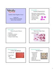
<strong>Mycology</strong><br />
<strong>Dermatophytes</strong><br />
Please click audio icon<br />
to hear Carol’s narration<br />
Division of Medical Technology<br />
Carol Larson MSEd, MT(ASCP)<br />
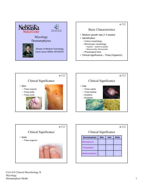
<strong>Basic</strong> <strong>Characteristics</strong><br />
• Medium growth rate (1-3 weeks)<br />
• Identification<br />
– Colony morphology<br />
– Microscopic morphology<br />
• Hyphae – hyaline & septate<br />
• Macroconidia, Microconidia<br />
– Physiological tests<br />
• <strong>Clinical</strong> significance – Tinea (ringworm)<br />
<strong>Clinical</strong> Significance<br />
•Skin<br />
– Tinea corporis<br />
– Tinea pedis<br />
– Tinea cruris<br />
Click icon<br />
for audio<br />
<strong>Clinical</strong> Significance<br />
•Hair<br />
– Tinea capitis<br />
– Tinea barbae<br />
– Ectothrix<br />
– Endothrix<br />
Click icon<br />
for audio<br />
<strong>Clinical</strong> Significance<br />
Click icon<br />
for audio<br />
<strong>Clinical</strong> Significance<br />
Click icon<br />
for audio<br />
•Nails<br />
– Tinea unguium<br />
Dermatophyte<br />
Microsporum<br />
Skin<br />
X<br />
Hair<br />
X<br />
Nails<br />
Trichophyton<br />
X<br />
X<br />
X<br />
Epidermophyton<br />
X<br />
X<br />
CLS 419 <strong>Clinical</strong> Microbiology II<br />
<strong>Mycology</strong><br />
Dermatophyte Molds 1
Click icon<br />
for audio<br />
Epidemiology<br />
• Anthropophilic<br />
–Man<br />
• Zoophilic<br />
–Animals<br />
• Geophilic<br />
–Soil<br />
What three body sites do the<br />
dermatophytes infect?<br />
Hair, skin and nails<br />
What is the difference between<br />
endothrix and ectothrix?<br />
What infection do the dermatophytes<br />
cause?<br />
Endothrix means the mold has conidia inside the hair<br />
shaft, whereas Ectothrix means the conidia are only on<br />
the outside of the hair shaft.<br />
Tinea (also referred to as “ringworm”). Another term<br />
that can be used is dermatophytosis.<br />
Laboratory Diagnosis<br />
• Specimen collection<br />
• Direct examination<br />
•Culture<br />
• Identification<br />
Click icon<br />
for audio<br />
Laboratory Diagnosis<br />
Specimen Collection<br />
•Hair<br />
– Plucked, not cut, from edge of lesion<br />
•Skin<br />
– Wash, scrape from margin of lesion<br />
•Nails<br />
– Scrapings from nail bed or infected area<br />
• Transport in sterile petri dish<br />
Click icon<br />
for audio<br />
CLS 419 <strong>Clinical</strong> Microbiology II<br />
<strong>Mycology</strong><br />
Dermatophyte Molds 2
Laboratory Diagnosis<br />
Click icon<br />
for audio<br />
Laboratory Diagnosis<br />
Click icon<br />
for audio<br />
Direct Examination<br />
Direct Examination<br />
• Examine hair for fluorescence<br />
– Wood’s lamp<br />
• Examine specimen for fungal elements<br />
– 10% KOH<br />
preparation<br />
– Calcofluor<br />
white stain<br />
– Yellow green<br />
fluorescence = positive<br />
Laboratory Diagnosis<br />
Click icon<br />
for audio<br />
Laboratory Diagnosis<br />
Click icon<br />
for audio<br />
Specimen processing<br />
Culture Media<br />
•Hair<br />
– Cut into short segments<br />
•Nails<br />
– Mince into small pieces<br />
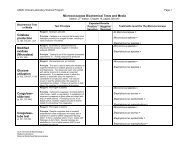
• Select two media types<br />
– General purpose – Sabouraud’s agar<br />
– Selective – Mycosel agar<br />
• Antibiotics<br />
– Gentamicin: inhibits normal bacterial flora<br />
– Cycloheximide: inhibits saprophytic fungi<br />
Laboratory Diagnosis<br />
Culture Growth Requirements<br />
• Place specimen pieces on culture media<br />
• Can streak for isolation<br />
• Incubate at 30°C in ambient (room) air<br />
• Growth at 3 days to 3 weeks<br />
• Examine plates frequently for 4 weeks<br />
Click icon<br />
for audio<br />
Laboratory Diagnosis<br />
Identification<br />
• Colony morphology<br />
• Microscopic morphology<br />
– Scotch tape preparation<br />
– Tease prep<br />
– Slide culture<br />
Click icon<br />
for audio<br />
CLS 419 <strong>Clinical</strong> Microbiology II<br />
<strong>Mycology</strong><br />
Dermatophyte Molds 3
Laboratory Diagnosis<br />
Identification<br />
Click icon<br />
for audio<br />
• Physiologic tests<br />
– Urea hydrolysis<br />
– Hair perforation<br />
– Rice grain media<br />
– Vitamin requirements<br />
How can hair, skin and nails be<br />
evaluated directly for fungal<br />
elements?<br />
Wood’s lamp fluorescence (hair only), 10% KOH<br />
preparation, and Calcofluor white fluorescent stain.<br />
What are the incubation<br />
requirements when suspecting a<br />
dermatophyte infection?<br />
Fungal media is incubated at 30°C in ambient air for 4<br />
weeks. There is one exception and that is<br />
Trichophyton verrucosum that requires 35ºC.<br />
What primary procedures are<br />
performed to identify the<br />
dermatophytes?<br />
Colony morphology, microscopic morphology (Scotch<br />
tape prep, tease prep, or slide culture), and physiologic<br />
tests such as urea hydrolysis and hair perforation.<br />
Etiologic Agents<br />
• Microsporum species<br />
• Epidermophyton species<br />
• Trichophyton species<br />
Click icon<br />
for audio<br />
Microsporum canis<br />
• Colony morphology:<br />
Click icon<br />
for audio<br />
Dermatophyte<br />
Microsporum<br />
Trichophyton<br />
Epidermophyton<br />
Skin<br />
X<br />
X<br />
X<br />
Hair<br />
X<br />
X<br />
Nails<br />
X<br />
X<br />
CLS 419 <strong>Clinical</strong> Microbiology II<br />
<strong>Mycology</strong><br />
Dermatophyte Molds 4
Click icon<br />
for audio<br />
Click icon<br />
for audio<br />
• Microscopic<br />
morphology:<br />
Microsporum canis<br />
Microsporum gypseum<br />
• Colony morphology:<br />
Click icon<br />
for audio<br />
Click icon<br />
for audio<br />
Microsporum gypseum<br />
• Microscopic morphology:<br />
Microsporum audouinii<br />
• Colony morphology:<br />
Click icon<br />
for audio<br />
Microsporum audouinii<br />
• Microscopic morphology:<br />
How can Microsporum species be<br />
differentiated from each other<br />
microscopically?<br />
Characteristic appearance of the macroconidia, and the<br />
general appearance of the hyphae (such as pectinate<br />
bodies). As a group, Microsporum have few to absent<br />
microconidia.<br />
CLS 419 <strong>Clinical</strong> Microbiology II<br />
<strong>Mycology</strong><br />
Dermatophyte Molds 5
Click icon<br />
for audio<br />
Click icon<br />
for audio<br />
Epidermophyton floccosum<br />
• Colony morphology:<br />
Epidermophyton floccosum<br />
• Microscopic morphology:<br />
Click icon<br />
for audio<br />
Click icon<br />
for audio<br />
Trichophyton rubrum<br />
• Colony morphology:<br />
Trichophyton rubrum<br />
• Microscopic morphology:<br />
Click icon<br />
for audio<br />
Click icon<br />
for audio<br />
Trichophyton rubrum<br />
• Physiological tests<br />
– Urea: negative<br />
– Hair perforation: negative<br />
Trichophyton mentagrophytes<br />
• Colony morphology:<br />
Downy Granular Velvet<br />
CLS 419 <strong>Clinical</strong> Microbiology II<br />
<strong>Mycology</strong><br />
Dermatophyte Molds 6
Click icon<br />
for audio<br />
Click icon<br />
for audio<br />
Trichophyton mentagrophytes<br />
• Microscopic<br />
morphology:<br />
Trichophyton mentagrophytes<br />
• Physiologic tests:<br />
– Urea: positive<br />
– Hair perforation:<br />
positive<br />
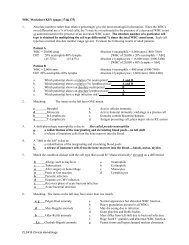
How can Trichophyton<br />
mentagrophytes be differentiated<br />
from Trichophyton rubrum?<br />
Urea hydrolysis and hair perforation tests.<br />
T. mentagrophytes is positive for both, and<br />
T. rubrum is negative for both.<br />
How can Microsporum,<br />
Epidermophyton, and<br />
Trichophyton species be<br />
differentiated microscopically?<br />
Microsporum has numerous thick-walled macroconidia<br />
with RARE microconidia, Epidermophyton has numerous<br />
club-shaped macroconidia hanging out in groups of 2-3<br />
with NO microconidia, and Trichophyton has thin-walled<br />
macroconidia and MANY microconidia.<br />
<strong>Dermatophytes</strong><br />
In Summary …<br />
• Causes Tinea (ringworm)<br />
• Medium growth rate = 1-3 weeks<br />
• Grows on Mycosel agar<br />
• Identification<br />
– Colony morphology, microscopic exam,<br />
and physiologic tests<br />
• Etiologic agents<br />
– Microsporum, Epidermophyton,<br />
Trichophyton species<br />
Click icon<br />
for audio<br />
Who am I?<br />
Potato Dextrose Agar Reverse LPCB Stain of Slide Culture<br />
Microsporum canis<br />
CLS 419 <strong>Clinical</strong> Microbiology II<br />
<strong>Mycology</strong><br />
Dermatophyte Molds 7
Who am I?<br />
Who am I?<br />
Potato Dextrose Agar<br />
LPCB Stain of Slide Culture<br />
Potato Dextrose Agar<br />
LPCB Stain of Slide Culture<br />
Hair<br />
Perforation<br />
Epidermophyton floccosum<br />
Trichophyton mentagrophytes<br />
Who am I?<br />
Potato Dextrose Agar Reverse LPCB Stain of Slide Culture<br />
Microsporum gypseum<br />
CLS 419 <strong>Clinical</strong> Microbiology II<br />
<strong>Mycology</strong><br />
Dermatophyte Molds 8