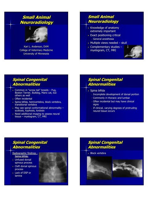
Small Animal Neuroradiology Small Animal Neuroradiology
Small Animal Neuroradiology Small Animal Neuroradiology
Small Animal Neuroradiology Small Animal Neuroradiology

Create successful ePaper yourself
Turn your PDF publications into a flip-book with our unique Google optimized e-Paper software.
<strong>Small</strong> <strong>Animal</strong><br />
<strong>Neuroradiology</strong><br />
Kari L. Anderson, DVM<br />
College of Veterinary Medicine<br />
University of Minnesota<br />
<strong>Small</strong> <strong>Animal</strong><br />
<strong>Neuroradiology</strong><br />
• Knowledge of anatomy<br />
extremely important<br />
• Exact positioning critical<br />
– General anesthesia<br />
• Multiple views needed – skull<br />
• Complementary studies –<br />
myelogram, , CT, MRI<br />
Spinal Congenital<br />
Abnormalities<br />
• Common in “screw tail” breeds – Pug,<br />
Boston Terrier, Bulldog, Manx cat, but<br />
others as well<br />
• Often incidental<br />
• Spina bifida, hemivertebra, , block vertebra,<br />
transitional vertebra<br />
• May see spinal conformational abnormality –<br />
scoliosis, kyphosis, lordosis<br />
• Need additional imaging to assess neural<br />
tissue – myelogram, , CT, MRI<br />
Spinal Congenital<br />
Abnormalities<br />
• Spina bifida<br />
– Incomplete development of dorsal portion<br />
– Commonly in thoracic and lumbar<br />
– Often incidental but may have clinical<br />
signs<br />
– If clinical, varying degrees of protruding<br />
neural tissue occurs<br />
Spinal Congenital<br />
Abnormalities<br />
Radiographic findings -<br />
Spina bifida:<br />
• Unfused dorsal<br />
spinous process<br />
• Cleft dorsal spinous<br />
process<br />
• Lack of DSP or<br />
lamina<br />
Spinal Congenital<br />
Abnormalities<br />
• Block vertebra
Spinal Congenital<br />
Abnormalities<br />
• Hemivertebra<br />
Spinal Congenital<br />
Abnormalities<br />
• Transitional<br />
vertebra<br />
Atlantoaxial Subluxation<br />
• Miniature and toy breeds – Yorkshire<br />
terrier, Chihuahua, Toy Poodle,<br />
Pekinese<br />
• Young dogs<br />
• Developmental anomaly<br />
– Hypoplasia of dens<br />
– Cord compression caused by abnormal<br />
rotation of C2 into spinal canal<br />
Atlantoaxial Subluxation<br />
Radiographic findings:<br />
• Hypoplastic or<br />
absent dens<br />
• Widened space<br />
between C1 and C2<br />
• Abnormal<br />
angulation of C2<br />
Spondylosis Deformans<br />
• Degenerative change – related to<br />
instability and degeneration – may be<br />
secondary to many abnormalities<br />
• Common<br />
• Rarely clinically significant<br />
Intervertebral Disk<br />
Protrusion<br />
• Chondrodystrophoid breeds – Hansen Type<br />
1 herniation associated with chondroid<br />
metaplasia and disk degeneration<br />
• Nonchondrodystrophoid breeds – Hansen<br />
• Type 2 herniation associated with fibroid<br />
metaplasia and disk degeneration<br />
• Intervertebral disk degeneration:<br />
mineralized disk material in the disk space<br />
• Uncommon in cranial thoracic spine – WHY?
Intervertebral Disk<br />
Protrusion<br />
Intervertebral Disk<br />
Protrusion<br />
Radiographic findings:<br />
• Narrowed or wedged IVD<br />
space<br />
• Decreased size of IV foramen<br />
• Increased opacity of IV<br />
foramen<br />
• Narrowed articular facet space<br />
• Endplate sclerosis and<br />
spondylosis<br />
• Extradural compression on<br />
myelogram<br />
Cervical<br />
Spondylomyelopathy<br />
• AKA: Wobbler syndrome, cervical<br />
vertebral malformation-<br />
malarticulation syndrome, cervical<br />
vertebral instabilitiy<br />
• Common in middle age to older<br />
Dobes and younger Great Danes<br />
• Multiple underlying spinal<br />
abnormalities<br />
Cervical<br />
Spondylomyelopathy<br />
Radiographic findings:<br />
• Malformed<br />
vertebrae<br />
• Coning or stenosis<br />
of canal<br />
• Dorsal tipping of<br />
vertebrae<br />
• Facet proliferation<br />
and/or spondylosis<br />
Degenerative<br />
Lumbosacral Stenosis<br />
• Multifactorial disease:<br />
– Stenotic spinal canal<br />
– Lumbosacral malalignment<br />
– Lumbosacral instability<br />
– Herniated IVD<br />
– Spondylosis – may impinge on nerve<br />
roots<br />
• Large breed dogs – GSD
Degenerative<br />
Lumbosacral Stenosis<br />
Radiographic findings:<br />
• Best evaluated with CT<br />
or MRI<br />
• Endplate sclerosis and<br />
spondylosis<br />
• Narrowing and<br />
wedging of LS disk<br />
space<br />
• LS subluxation<br />
• Stenotic canal<br />
Discospondylitis<br />
• Infection of disk space with extension<br />
to adjacent vertebrae<br />
• Often multiple spaces affected<br />
• Generally hematogenous – commonly<br />
associated with urogenital infections<br />
• Staph spp, E. coli, Brucella canis,<br />
mycoses, mycobacterium<br />
Discospondylitis<br />
Spinal Neoplasia<br />
Radiographic findings:<br />
• Irregular endplate lysis<br />
• Bony sclerosis and<br />
proliferation<br />
• Widening or collapse of<br />
disk space<br />
• Spondylosis deformans<br />
Spinal Trauma<br />
Hydrocephalus<br />
• Congenital form – small, toy, and<br />
brachycephalic breeds (Yorkshire<br />
terrier, Chihuahua, Maltese, Toy<br />
Poodle, etc)<br />
• Radiographic abnormalities seen in<br />
congenital form<br />
• Better evaluated with US, CT, MRI
Hydrocephalus<br />
Radiographic findings:<br />
• Domed cranium<br />
• Fontanelles and<br />
open sutures<br />
• Cortical thinning<br />
• Homogenous<br />
appearance to brain<br />
Craniomandibular Osteopathy<br />
(CMO)<br />
• Young dogs 3-83<br />
8 months of age<br />
• Terrier breeds, Labs, Dobes, , GSD,<br />
Boxers<br />
• Osteoproliferative disorder that is self-<br />
limiting and slows at 7-87<br />
8 months of<br />
age<br />
• Secondary TMJ fusion can occur<br />
Craniomandibular Osteopathy<br />
(CMO)<br />
Radiographic findings:<br />
• Irregular, opaque<br />
new bone<br />
• Generally bilateral<br />
• Ankylosis of TMJ<br />
may occur<br />
• Ceases with<br />
maturity<br />
Otitis Externa and Media<br />
• Radiographs very helpful in evaluation<br />
– even better with CT<br />
• Otitis interna is clinical diagnosis!<br />
• Rule-out trauma, tumor,<br />
nasopharyngeal polyps<br />
Otitis Externa and Media<br />
Radiographic findings:<br />
• No abnormalities<br />
• Narrowing, obliteration,<br />
mineralization of<br />
external ear canals<br />
(otitisotitis externa)<br />
• Otitis media<br />
– Soft tissue/fluid opacity<br />
in tympanic bulla<br />
– Bony changes of bulla
Nondestructive Rhinitis<br />
Etiologies:<br />
• Infection<br />
• Foreign bodies<br />
• Nasopharyngeal polyps<br />
• Allergies<br />
• Coagulopathy<br />
Nondestructive Rhinitis<br />
Radiographic findings:<br />
• Increased soft<br />
tissue/fluid opacity of<br />
nasal passages and<br />
frontal sinuses<br />
• May see radiopaque<br />
foreign body<br />
• Should not see lysis<br />
unless very chronic<br />
infection or foreign<br />
body<br />
Destructive Rhinitis<br />
• Etiologies:<br />
– Nasal tumor<br />
– Fungal infection<br />
– Chronic infection or foreign bodies<br />
• Most common clinical sign is epistaxis<br />
• CT or MRI offers better evaluation<br />
Destructive Rhinitis<br />
Radiographic findings:<br />
• Early may mimic<br />
nondestructive<br />
• Irregular opacities in nasal<br />
passages and frontal<br />
sinuses<br />
• Bony lysis of ethmoid<br />
turbinates or nasal septum<br />
• May see extranasal<br />
extension
Hyperparathyroidism<br />
• Primary vs<br />
secondary<br />
Radiographic findings:<br />
• Loss of lamina dura<br />
• Demineralization of<br />
maxilla and<br />
mandible<br />
• Floating teeth