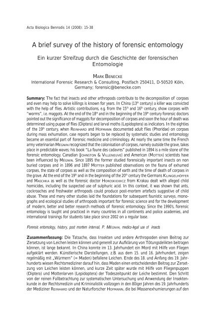
A brief survey of the history of forensic entomology - Wiki2.benecke ...
A brief survey of the history of forensic entomology - Wiki2.benecke ...
A brief survey of the history of forensic entomology - Wiki2.benecke ...

Create successful ePaper yourself
Turn your PDF publications into a flip-book with our unique Google optimized e-Paper software.
A <strong>brief</strong> <strong>survey</strong> <strong>of</strong> <strong>the</strong> <strong>history</strong> <strong>of</strong> <strong>forensic</strong> <strong>entomology</strong> 15<br />
Acta Biologica Benrodis 14 (2008): 15-38<br />
A <strong>brief</strong> <strong>survey</strong> <strong>of</strong> <strong>the</strong> <strong>history</strong> <strong>of</strong> <strong>forensic</strong> <strong>entomology</strong><br />
Ein kurzer Streifzug durch die Geschichte der forensischen<br />
Entomologie<br />
MARK BENECKE<br />
International Forensic Research & Consulting, Postfach 250411, D-50520 Köln,<br />
Germany; <strong>forensic</strong>@benecke.com<br />
Summary: The fact that insects and o<strong>the</strong>r arthropods contribute to <strong>the</strong> decomposition <strong>of</strong> corpses<br />
and even may help to solve killings is known for years. In China (13 th century) a killer was convicted<br />
with <strong>the</strong> help <strong>of</strong> flies. Artistic contributions, e.g. from <strong>the</strong> 15 th and 16 th century, show corpses with<br />
“worms”, i.e. maggots. At <strong>the</strong> end <strong>of</strong> <strong>the</strong> 18 th and in <strong>the</strong> beginning <strong>of</strong> <strong>the</strong> 19 th century <strong>forensic</strong> doctors<br />
pointed out <strong>the</strong> significance <strong>of</strong> maggots for decomposition <strong>of</strong> corpses and soon <strong>the</strong> hour <strong>of</strong> death was<br />
determined using pupae <strong>of</strong> flies (Diptera) and larval moths (Lepidoptera) as indicators. In <strong>the</strong> eighties<br />
<strong>of</strong> <strong>the</strong> 19 th century, when REINHARD and HOFMANN documented adult flies (Phoridae) on corpses<br />
during mass exhumation, case reports began to be replaced by systematic studies and <strong>entomology</strong><br />
became an essential part <strong>of</strong> <strong>forensic</strong> medicine and criminology. At nearly <strong>the</strong> same time <strong>the</strong> French<br />
army veterinarian MÉGNIN recognized that <strong>the</strong> colonisation <strong>of</strong> corpses, namely outside <strong>the</strong> grave, takes<br />
place in predictable waves; his book “La faune des cadavres” published in 1894 is a mile stone <strong>of</strong> <strong>the</strong><br />
<strong>forensic</strong> <strong>entomology</strong>. Canadian (JOHNSTON & VILLENEUVE) and American (MOTTER) scientists have<br />
been influenced by MÉGNIN. Since 1895 <strong>the</strong> former studied <strong>forensic</strong>ally important insects on non<br />
buried corpses and in 1896 and 1897 MOTTER published observations on <strong>the</strong> fauna <strong>of</strong> exhumed<br />
corpses, <strong>the</strong> state <strong>of</strong> corpses as well as <strong>the</strong> composition <strong>of</strong> earth and <strong>the</strong> time <strong>of</strong> death <strong>of</strong> corpses in<br />
<strong>the</strong> grave. At <strong>the</strong> end <strong>of</strong> <strong>the</strong> 19 th and in <strong>the</strong> beginning <strong>of</strong> <strong>the</strong> 20 th century <strong>the</strong> Germans KLINGELHÖFFER<br />
and MASCHKA as well as <strong>the</strong> <strong>forensic</strong> doctor HOROSKIEWICZ from Krakau dealt with alleged child<br />
homicides, including <strong>the</strong> suspected use <strong>of</strong> sulphuric acid. In this context, it was shown that ants,<br />
cockroaches and freshwater arthropods could produce post-mortem artefacts suggestive <strong>of</strong> child<br />
abuse. These and many o<strong>the</strong>r studies laid <strong>the</strong> foundations for subsequent faunistic <strong>survey</strong>s, monographs<br />
and ecological studies <strong>of</strong> arthropods important for <strong>forensic</strong> science and for <strong>the</strong> development<br />
<strong>of</strong> modern, better and better research methods <strong>of</strong> <strong>forensic</strong> <strong>entomology</strong>. Since <strong>the</strong> 1990’s, <strong>forensic</strong><br />
<strong>entomology</strong> is taught and practiced in many countries in all continents and police academies, and<br />
international trainings for students take place since 2002 on a regular base.<br />
Forensic <strong>entomology</strong>, <strong>history</strong>, post mortem interval, P. MÉGNIN, medico-legal use <strong>of</strong> insects<br />
Zusammenfassung: Die Tatsache, dass Insekten und andere Arthropoden einen Beitrag zur<br />
Zersetzung von Leichen leisten können und generell zur Aufklärung von Tötungsdelikten beitragen<br />
können, ist lange bekannt. In China konnte im 13. Jahrhundert ein Mord mit Hilfe von Fliegen<br />
aufgeklärt werden. Künstlerische Darstellungen, z.B. aus dem 15. und 16. Jahrhundert, zeigen<br />
regelmäßig mit „Würmern” (= Maden) befallene Leichen. Ende des 18. und Anfang des 19. Jahrhunderts<br />
wiesen Rechtsmediziner darauf hin, dass Maden einen entscheidenden Beitrag zur Zersetzung<br />
von Leichen leisten können, und kurze Zeit später wurde mit Hilfe von Fliegenpuppen<br />
(Diptera) und Mottenlarven (Lepidoptera) der Todeszeitpunkt der Leiche bestimmt. Den Schritt<br />
von der reinen Fallbetrachtung zur systematischen Untersuchung und Anwendung der Insektenkunde<br />
in der Rechtsmedizin und Kriminalistik vollzogen in den 80iger Jahren des 19. Jahrhunderts<br />
der Mediziner REINHARD und der Naturforscher HOFMANN, die bei Massenexhumierungen auf den<br />
Acta Biologica Benrodis 14 (2007)
16 MARK BENECKE<br />
Leichen erwachsene Buckelfliegen (Phoridae) entdeckten. Etwa zur selben Zeit erkannte MÉGNIN<br />
in Frankreich, dass die Besiedlung von Leichen – besonders auch außerhalb des Grabes – in<br />
vorhersagbaren Wellen abläuft. Sein 1894 erschienenes Buch „La faune des cadavres“ gilt als<br />
Meilenstein der forensischen Entomologie. Seine Konzepte beeinflussten die kanadischen Forscher<br />
JOHNSTON & VILLENEUVE, die seit 1895 umfangreiche forensisch-entomologische Untersuchungen<br />
an freiliegenden Menschenleichen durchführten, sowie MOTTER in den USA, der 1896 und 1897 die<br />
Fauna exhumierter Leichen bestimmte sowie Leichen- und Erdbeschaffenheit und Liegezeit im<br />
Grab bestimmte. Gegen Ende des 19. und Anfang des 20. Jahrhunderts befassten sich die deutschen<br />
Ärzte KLINGELHÖFFER and MASCHKA sowie der Rechtsmediziner HOROSKIEWICZ aus Krakau mit<br />
angeblichen Kindstötungen mit Hilfe von Schwefelsäure. In diesem Zusammenhang konnte gezeigt<br />
werden, dass Ameisen, Schaben und Süßwasser-Arthropoden postmortale Schäden verursachen<br />
können, die wie Kindesmissbrauch aussehen. Diese und zahlreiche andere Untersuchungen<br />
legten den Grundstein für nachfolgende Monographien über forensisch wichtige Arthropoden, für<br />
diesbezügliche faunistische und ökologische Studien sowie für die Entwicklung moderner und<br />
immer präziserer Methoden der forensischen Entomologie. Seit den 90iger Jahren des vorigen<br />
Jahrhunderts wird forensische Entomologie in zahlreichen Ländern auf nahezu allen Kontinenten<br />
gelehrt und praktiziert. Außerdem gibt es seit 2002 regelmäßig internationale Schulungen.<br />
Forensische Entomologie, Geschichte, Liegezeitbestimmung, P. MÉGNIN, kriminalbiologischer Nutzen von<br />
Insekten<br />
1. Introduction<br />
Hundreds <strong>of</strong> arthropod species are attracted<br />
by corpses, primarily flies (Diptera), beetles<br />
(Coleoptera) and <strong>the</strong>ir larvae, respectively, but<br />
also mites, isopods, opiliones and nematodes<br />
can be found. These animals feed, live or<br />
breed in and on <strong>the</strong> corpse, depending on<br />
<strong>the</strong>ir biological preferences and on <strong>the</strong> state<br />
<strong>of</strong> decomposition. Since arthropods are by<br />
far <strong>the</strong> largest and most important biological<br />
group on earth (<strong>the</strong>y outnumber even plants),<br />
<strong>the</strong>y can be found in a wide variety <strong>of</strong> locations<br />
including crime scenes. This opens a<br />
wide range <strong>of</strong> applications for <strong>forensic</strong> <strong>entomology</strong>,<br />
<strong>the</strong> investigation <strong>of</strong> insects recovered<br />
from crime scenes and corpses.<br />
The following article gives a <strong>brief</strong> <strong>survey</strong><br />
<strong>of</strong> <strong>the</strong> historic sources describing <strong>the</strong> development<br />
that led to <strong>the</strong> present state <strong>of</strong> <strong>the</strong> art<br />
with a major focus on work done between<br />
1850 and 1950 (BENECKE 1998, BENECKE<br />
2001a, BENECKE & LECLERCQ 1999).<br />
2. Medieval China to 19 th century<br />
The first documented <strong>forensic</strong> <strong>entomology</strong><br />
case is reported by <strong>the</strong> Chinese lawyer and<br />
death investigator SÒNG CÍ in <strong>the</strong> 13 th century<br />
in <strong>the</strong> medico-legal text book<br />
(Xiyuan jílù; one possible translation: “collected<br />
writings on <strong>the</strong> washing away <strong>of</strong><br />
wrongs”). He describes <strong>the</strong> case <strong>of</strong> a stabbing<br />
near a rice field. The day after <strong>the</strong> murder,<br />
<strong>the</strong> investigator told all workers to lay down<br />
<strong>the</strong>ir working tools (i.e., sickles) on <strong>the</strong> floor.<br />
Invisible traces <strong>of</strong> blood drew blow flies to a<br />
single sickle. So confronted, <strong>the</strong> tool’s owner<br />
confessed to his crime and “knocked his head<br />
on <strong>the</strong> floor” (Fig. 1; see SÒNG CÍ/SUNG TZ’U<br />
1924; MCKNIGHT 1981). It took nearly eight<br />
hundred years until <strong>the</strong> next Chinese book<br />
on <strong>forensic</strong> <strong>entomology</strong> was published (HU<br />
2000).<br />
In addition to medical and legal experts,<br />
sculptors, painters and poets have closely observed<br />
<strong>the</strong> decomposition <strong>of</strong> human bodies,<br />
noting, in particular, <strong>the</strong> effects <strong>of</strong> feeding<br />
maggots. Early documents illustrating maggots<br />
on corpses date to <strong>the</strong> Middle Ages,<br />
including woodcuts from “Dances <strong>of</strong> <strong>the</strong><br />
Death”, oil paintings (both 15 th century), engravings<br />
in tombstones (19 th century), and<br />
<strong>the</strong> intricately cut ivory carving, “Skeleton in<br />
<strong>the</strong> Tumba” (16 th century, Figs. 2-4) (BENE-<br />
CKE 1999). Detailed observations were pos-
A <strong>brief</strong> <strong>survey</strong> <strong>of</strong> <strong>the</strong> <strong>history</strong> <strong>of</strong> <strong>forensic</strong> <strong>entomology</strong> 17<br />
Fig. 1: First page <strong>of</strong><br />
chapter 5 <strong>of</strong> SÒNG CÍS<br />
book on <strong>forensic</strong> medicine<br />
deals with a case<br />
<strong>of</strong> stabbing solved by<br />
use <strong>of</strong> insects. Adult<br />
flies detected blood on<br />
<strong>the</strong> killer’s sickle (from<br />
BENECKE 1999).<br />
Abb. 1: Erste Seite des<br />
Kapitels 5 aus dem<br />
Buch über forensische<br />
Medizin von SÒNG CÍ<br />
mit einem Fall, der mit<br />
Hilfe von Insekten geklärt<br />
werden konnte.<br />
Adulte Fliegen ließen<br />
sich nur auf der mit<br />
Blut kontaminierten<br />
Tatwaffe, einer Sichel,<br />
nieder (aus BENECKE<br />
1999).<br />
sible not only from victims <strong>of</strong> <strong>the</strong> frequent<br />
and violent wars but also exposure to decomposing<br />
bodies during plague outbreaks<br />
and similar diseases; <strong>the</strong> plague alone killed<br />
1/3 <strong>of</strong> all Europeans in <strong>the</strong> years 1346-1351.<br />
Such artwork accurately depicts <strong>the</strong> insectmediated<br />
pattern <strong>of</strong> body mass reduction,<br />
particularly <strong>the</strong> skeletonization <strong>of</strong> <strong>the</strong> skull<br />
and <strong>the</strong> reduction <strong>of</strong> internal organs, with<br />
large parts <strong>of</strong> <strong>the</strong> skin left intact (LANGLOIS<br />
1852; STAMMLER 1948); however, a metaphoric<br />
connotation is sometimes also present, e.g.<br />
<strong>the</strong> maggots as “snakes” (Fig. 3), and <strong>the</strong><br />
blowfly replacing <strong>the</strong> heart (Fig. 4). Also, <strong>the</strong><br />
poem “Une charogne” by <strong>the</strong> French poet C.<br />
BAUDELAIRE (1821-1867) must be mentioned<br />
in this context, too, since it contains very good<br />
observations on <strong>the</strong> decay <strong>of</strong> human bodies,<br />
including an accurate reference to <strong>the</strong> sound<br />
<strong>of</strong> maggot masses on corpse (see BAUDELAIRE<br />
1857):<br />
“Et ce monde rendait une étrange musique,<br />
Comme l’eau courante et le vent,<br />
Ou le grain qu’un vanneur d’un mouvement<br />
rhythmique<br />
Agite et tourne dans son van.”<br />
(BAUDELAIRE 1955)<br />
A century earlier, in 1767, <strong>the</strong> natural scientist<br />
C. VON LINNÉ made <strong>the</strong> observation that<br />
three flies would destroy a horse as fast as a<br />
Acta Biologica Benrodis 14 (2007)
18 MARK BENECKE<br />
Fig. 2: Close observation <strong>of</strong> decomposition <strong>of</strong> human corpses built <strong>the</strong> basis for <strong>the</strong>se figures.<br />
a: Grave <strong>of</strong> ROBERT TOUSE who “experts <strong>the</strong> resurrection <strong>of</strong> <strong>the</strong> dead” (exact time unknown, from<br />
LANGLOIS 1852); b, c: “Dances <strong>of</strong> <strong>the</strong> Death” (ca. 1460, from STAMMLER 1948). Note that maggots<br />
are displayed similar to snakes or worms, and that all heads are already skeletonized due to <strong>the</strong> fact<br />
that adults preferably deposit <strong>the</strong>ir eggs on eyes, nose, ears and mouth.<br />
Abb. 2: Diese Abbildungen sind nicht nur symbolisch zu verstehen, sondern beruhen auf Beobachtungen<br />
von Zersetzungsstadien echter Leichen. a: Grabplatte von ROBERT TOUSE, der auf die<br />
Auferstehung wartet (genaue Zeit unbekannt, aus LANGLOIS 1852); b, c: Totentänze (etwa 1460,<br />
aus STAMMLER 1948). Man beachte das schlangen- oder wurmartige Aussehen der Fliegenlarven,<br />
aber auch die Tatsache, dass Maden zuerst die Schädel skelettieren, weil Fliegen ihre Eier bevorzugt<br />
an Augen, Nase, Ohren und Mund ablegen.<br />
lion would (in <strong>the</strong> sense <strong>of</strong> <strong>the</strong>m producing<br />
large masses <strong>of</strong> maggots) (LINNÉ 1775) (Fig. 6).<br />
3. Early cases from France<br />
During mass exhumations in France and<br />
Germany in <strong>the</strong> 18 th and 19 th centuries, medico-legal<br />
doctors observed that buried bodies<br />
are colonized by arthropods <strong>of</strong> many kinds.<br />
In 1831, <strong>the</strong> famous French medical doctors<br />
ORFILA & LESUEUR (1831, 1835) observed a<br />
large number <strong>of</strong> exhumations. They understood<br />
that maggots play an important role<br />
in <strong>the</strong> decomposition <strong>of</strong> corpses (Fig. 7).<br />
The first modern <strong>forensic</strong> <strong>entomology</strong> case<br />
report to include an estimation <strong>of</strong> postmor-
A <strong>brief</strong> <strong>survey</strong> <strong>of</strong> <strong>the</strong> <strong>history</strong> <strong>of</strong> <strong>forensic</strong> <strong>entomology</strong> 19<br />
Fig. 3: Painting “Les amants trépassés”<br />
from <strong>the</strong> Musée de l‘Œvre Notre-Dame<br />
(Frauenhausmuseum, Strasbourg) from ca.<br />
1470. The bodies are mummified; <strong>the</strong> animals<br />
do <strong>the</strong>refore not correlate to actual<br />
feeding patterns but seem to have a mostly<br />
symbolic meaning.<br />
Abb. 3: Gemälde “Les amants trépassés”<br />
aus dem Museum de l‘Œvre Notre-Dame<br />
(Frauenhausmuseum, Straßburg) von<br />
1470. Die Körper sind mumifiziert. Die<br />
Auswahl der Tiere, auch der Insekten, ist<br />
hier eher symbolisch zu verstehen; demgemäß<br />
passen auch die Fraßmuster nicht<br />
zu einer Madenbesiedlung.<br />
Fig. 4: Ivory “Little Dead” (Tödlein) as a<br />
Memento Mori. Note <strong>the</strong> very precise and<br />
realistic artistic depiction <strong>of</strong> a corpse in advanced<br />
decomposition under <strong>the</strong> influence<br />
<strong>of</strong> maggots: head fully skeletonized, rib case<br />
only partially skeletonized with patches <strong>of</strong><br />
intact skin (from Western Switzerland, ca.<br />
1520; length <strong>of</strong> skeleton 36 cm; SCHNÜT-<br />
GEN-Museum Cologne, Germany).<br />
Abb. 4: Tödlein aus Elfenbein als Memento<br />
Mori. Man beachte die sehr präzise und<br />
realistische Darstellung eines Körpers in<br />
fortgeschrittener Zersetzung unter Einfluss<br />
von Maden: der Kopf ist vollständig skelettiert,<br />
der Brustkorb nur zum Teil (aus<br />
der Westschweiz, ca. 1520; Länge 36 cm;<br />
SCHNÜTGEN-Museum Köln, Germany).<br />
Acta Biologica Benrodis 14 (2007)
20 MARK BENECKE<br />
Fig. 5: Illustration from<br />
<strong>the</strong> <strong>forensic</strong> entomologist<br />
A. OLIVA (Museum<br />
<strong>of</strong> Natural History, Buenos<br />
Aires, Argentina)<br />
relating to BAUDELAIRE’s<br />
poem “Une chargone”<br />
that precisely describes<br />
states <strong>of</strong> decomposition<br />
including maggot activity<br />
(Frontispiz in Baudelaire<br />
1955).<br />
Abb. 5: Von der Entomologin<br />
A. OLIVA (Naturkundemuseum<br />
Buenos<br />
Aires) angefertigte<br />
Illustration zu BAUDE-<br />
LAIREs Gedicht „Ein<br />
Aas“ („Une chargone“),<br />
das präzise die Zersetzung<br />
einer Leiche einschließlich<br />
der Aktivität<br />
von Maden beschreibt.<br />
Abb. 6: Beschreibung<br />
einer Schmeißfliege und<br />
ihrer Zerstörungskraft<br />
(aus LINNÉ 1775).<br />
Fig. 6: Description <strong>of</strong> a<br />
fly and tissue loss due to<br />
maggots (from LINNÉ<br />
1775).<br />
tem interval (PMI) was given by <strong>the</strong> French<br />
doctor BERGERET (1855). The case dealt with<br />
blow fly pupae and larval moths. Though<br />
BERGERET was by pr<strong>of</strong>ession a hospital physician<br />
(at <strong>the</strong> Hopital Civil d’Arbois), his interest<br />
in cadaver study is clear, as he states that<br />
a corpse dealt with by him resembled those<br />
he had observed found in o<strong>the</strong>r locations<br />
(i.e., “in <strong>the</strong> hot and dry lands (“pays chauds”)<br />
on <strong>the</strong> cemetery <strong>of</strong> <strong>the</strong> Capucins <strong>of</strong> Palerme”,<br />
or “in Toulouse”). His original report<br />
to <strong>the</strong> court was dated March 28, 1850. In his
A <strong>brief</strong> <strong>survey</strong> <strong>of</strong> <strong>the</strong> <strong>history</strong> <strong>of</strong> <strong>forensic</strong> <strong>entomology</strong> 21<br />
Fig. 7: Early account <strong>of</strong> insects on<br />
corpses already determined to <strong>the</strong><br />
species (from ORFILA & LESUEUR<br />
1831).<br />
Abb. 7: Frühe Darstellung von bereits<br />
bis zur Art bestimmt Insekten<br />
auf Leichen (aus ORFILA & LESU-<br />
EUR 1831).<br />
journal article, next to a long description <strong>of</strong><br />
<strong>the</strong> criminal impact in <strong>the</strong> trial, he also describes<br />
<strong>the</strong> court proceedings:<br />
“Within three years, four different tenants<br />
[i.e., families] lived in <strong>the</strong> flat. The first <strong>of</strong><br />
<strong>the</strong>m left in December 1848, and <strong>the</strong> person<br />
examined started to live <strong>the</strong>re at <strong>the</strong> end <strong>of</strong><br />
1844. I [i.e., BERGERET] was brought to <strong>the</strong><br />
house <strong>of</strong> Mme. Saillard [i.e., <strong>the</strong> landlady] in<br />
rue du Citoyen, 4, (...) to examine <strong>the</strong> corpse<br />
<strong>of</strong> a child. (...) Length <strong>of</strong> corpse: [46 cm].<br />
[Then details on measurements <strong>of</strong> bones,<br />
state <strong>of</strong> internal organs, etc. follow.]. The<br />
questions we had to deal with now were: 1.<br />
Was <strong>the</strong> child born regularly/at <strong>the</strong> right time,<br />
2. Was it alive when it was born, 3. How long<br />
did it live, 4. How did it die?, 5. What was <strong>the</strong><br />
time interval between birth and death?” Questions<br />
1 to 4 were answered with classical <strong>forensic</strong><br />
pathology. Question 5 was commented<br />
in <strong>the</strong> following way: “To answer this question,<br />
legal medicine must check with ano<strong>the</strong>r<br />
science, <strong>the</strong> natural sciences.” However, BER-<br />
GERET (1855) does not state whe<strong>the</strong>r he<br />
Acta Biologica Benrodis 14 (2007)
22 MARK BENECKE<br />
worked toge<strong>the</strong>r with ano<strong>the</strong>r person.<br />
In his paper, BERGERET (1855) gives a <strong>brief</strong><br />
overview on <strong>the</strong> life cycle <strong>of</strong> insects in general.<br />
He mistakenly assumes, however, that<br />
metamorphosis would generally require a full<br />
year. Fur<strong>the</strong>rmore, he assumes that females<br />
generally lay eggs in summer and that <strong>the</strong><br />
larvae would transform to pupae (he calls<br />
<strong>the</strong>m nymphs) <strong>the</strong> following spring and hatch<br />
in summer. Some details <strong>of</strong> BERGERET’s calculation:<br />
“The eggs <strong>of</strong> <strong>the</strong> larvae we found on <strong>the</strong><br />
corpse in March 1850 must have been deposited<br />
<strong>the</strong>re in <strong>the</strong> middle <strong>of</strong> 1849. Therefore,<br />
<strong>the</strong> corpse must have been deposited<br />
before this time interval. Next to <strong>the</strong> many<br />
living larvae <strong>the</strong>re were numerous pupae present,<br />
and <strong>the</strong>y must come from eggs that have<br />
been laid earlier, i.e., in 1848. (...) Could it be<br />
that <strong>the</strong> corpse was deposited even before<br />
that time [i.e., 1848]? The fly that emerges<br />
from <strong>the</strong> pupae that we found in <strong>the</strong> body<br />
cavities, is Musca carnaria L. that lays its eggs<br />
before <strong>the</strong> body dries out. We found o<strong>the</strong>r<br />
pupae <strong>of</strong> little butterflies <strong>of</strong> <strong>the</strong> night [moth],<br />
too, that attack bodies that are already dried<br />
out. If <strong>the</strong> body was deposited, say, in 1846<br />
or 1847, we would not have found those larvae<br />
[since <strong>the</strong>y would have hatched]. In conclusion,<br />
two generations <strong>of</strong> insects were<br />
found on <strong>the</strong> corpse, representing two years<br />
postmortem: on <strong>the</strong> fresh corpse, <strong>the</strong> flesh<br />
fly deposited its eggs in 1848, on <strong>the</strong> dried<br />
out corpse, <strong>the</strong> moth laid <strong>the</strong>ir eggs in 1849.”<br />
In retrospect, one should understand that<br />
BERGERET did not focus on <strong>forensic</strong> <strong>entomology</strong><br />
in his report but used <strong>the</strong> method as<br />
one <strong>forensic</strong> tool among o<strong>the</strong>rs. Indeed, <strong>the</strong><br />
mummification <strong>of</strong> <strong>the</strong> cadaver appears to be<br />
his overriding issue <strong>of</strong> interest in this case.<br />
BERGERET references ORFILA & LESUEUR (1831,<br />
1835) in <strong>the</strong> matters <strong>of</strong> both mummification<br />
and <strong>forensic</strong> <strong>entomology</strong>. He also clearly<br />
notes <strong>the</strong> lack <strong>of</strong> knowledge concerning insect<br />
succession on corpses in his day.<br />
In 1879, <strong>the</strong> president <strong>of</strong> <strong>the</strong> French Society<br />
<strong>of</strong> Forensic Medicine, BROUARDEL reported<br />
ano<strong>the</strong>r early case. P.C.H. BROUARDEL, born in<br />
Saint-Quentin on 13 February 1837, became<br />
a member <strong>of</strong> <strong>the</strong> French Academy <strong>of</strong> Medicine<br />
in 1880. He worked on tuberculosis, vaccination<br />
and legal medicine. His numerous<br />
medico-legal accounts include practical guidelines<br />
for his colleagues in <strong>the</strong> morgue. A contemporary<br />
said that “his work is conscientious,<br />
clear, methodic, and serves as a model”<br />
(quote from encyclopedia <strong>of</strong> that time;<br />
retrieved in 1998 by MB in Manhattan (New<br />
York University (NYU) Hospital Library);<br />
exact title and year unknown.)<br />
In his report, after referencing <strong>the</strong> work <strong>of</strong><br />
BERGERET (l.c.), BROUARDEL (1879) describes<br />
<strong>the</strong> case <strong>of</strong> a newborn child that was autopsied<br />
by him on January 15, 1878. The mummified<br />
body was inhabited by several arthropods,<br />
including butterfly larvae and mites,<br />
which led to a request for assistance from<br />
Monsieur PERIER, pr<strong>of</strong>essor at <strong>the</strong> Museum<br />
<strong>of</strong> Natural History in Paris, and army veterinarian<br />
J.P. MÉGNIN. PERIER reported that <strong>the</strong><br />
body was most likely dried out before it was<br />
abandoned. The determination <strong>of</strong> mites was<br />
left to MÉGNIN whereas PERIER determined<br />
<strong>the</strong> butterfly larvae as “chenilles d’aglosses”,<br />
i.e., larvae from <strong>the</strong> genus Aglossa (small<br />
moth, family Pyralidae). From <strong>the</strong> state <strong>of</strong><br />
preservation and from <strong>the</strong> larvae found,<br />
PERIER stated that <strong>the</strong> baby may have been<br />
born and died <strong>the</strong> summer before (“de l’été<br />
dernier probablement”), i.e., around six to<br />
seven month before <strong>the</strong> corpse was autopsied.<br />
MÉGNIN (1894) reported that <strong>the</strong> whole<br />
body was covered with a brownish layer composed<br />
exclusively <strong>of</strong> mite skins and mite<br />
feces, but not living mites. Inside <strong>the</strong> cranium<br />
he found large numbers <strong>of</strong> a single mite<br />
species. Initially, a few larval mites must have<br />
been carried to <strong>the</strong> corpse by o<strong>the</strong>r arthropods.<br />
MÉGNIN (1894) calculated that on <strong>the</strong><br />
whole body 2.4 million dead or living mites<br />
were present. He also calculated that after 15<br />
days <strong>the</strong> first generation with 10 females and<br />
5 males had developed; after 30 days, 100 fe-
A <strong>brief</strong> <strong>survey</strong> <strong>of</strong> <strong>the</strong> <strong>history</strong> <strong>of</strong> <strong>forensic</strong> <strong>entomology</strong> 23<br />
males and 50 males; after 45 days, 1,000 females<br />
and 500 males. Finally, after 90 days,<br />
1 million females and 500.000 males were present.<br />
Since this was <strong>the</strong> number <strong>of</strong> individual<br />
he estimated being on <strong>the</strong> corpse, he made<br />
a conservative guess and reported that <strong>the</strong><br />
corpse must have been abandoned for at least<br />
five months (three months <strong>of</strong> mite development,<br />
preceded by two months for desiccation)<br />
but more likely seven to eight months.<br />
This is <strong>the</strong> same case as Case No. 12 in<br />
MÉGNIN’s “La faune des cadavres” (see below<br />
for details; Fig. 8), and MÉGNIN states that<br />
this is his “prémiere étude médico-légale”<br />
(first medico-legal study). In a period <strong>of</strong> nine<br />
years, he published four articles on <strong>the</strong> topic<br />
(MÉGNIN 1887, 1889, 1894, 1896).<br />
Fig. 8: Front page <strong>of</strong> MÉGNIN’s “La Faune des<br />
cadavres” (1894). This book popularized <strong>the</strong><br />
subject <strong>of</strong> <strong>forensic</strong> <strong>entomology</strong> until today.<br />
Abb. 8: Titelseite von MÉGNINs „Die Fauna der<br />
Leichen“ (1894). Dieses allgemein verständliche<br />
Buch machte die forensische Entomologie der<br />
Öffentlichkeit bekannt.<br />
This case also illustrates nicely how early<br />
researchers in <strong>the</strong> field investigated <strong>the</strong> use <strong>of</strong><br />
molds, slime fungi, crustaceans, mites, and<br />
plants in addition to insects.<br />
4. Fur<strong>the</strong>r mass exhumations<br />
On April 6, 1881, <strong>the</strong> German medical doctor<br />
REINHARD, born in Dresden on November<br />
15th, 1816, reported <strong>the</strong> first systematic<br />
study in <strong>forensic</strong> <strong>entomology</strong> (REINHARD<br />
1882). Dealing with exhumed bodies from<br />
Saxonia, he collected mainly phorid flies taxonomically<br />
identified by <strong>the</strong> entomologist<br />
BRAUER in Vienna. He also described beetles<br />
in graves older than 15 years. In some instances,<br />
he found <strong>the</strong> insects breeding within<br />
cracks <strong>of</strong> adipocire. But REINHARD concluded<br />
that <strong>the</strong>ir presence may have more to do with<br />
<strong>the</strong>ir feeding on plant roots protruding into<br />
<strong>the</strong> graves ra<strong>the</strong>r than any direct association<br />
with <strong>the</strong> corpses. REINHARD’s work remained<br />
well known for a long time, and in 1928 an<br />
extensive citation <strong>of</strong> his paper appeared in<br />
<strong>the</strong> work <strong>of</strong> <strong>the</strong> phorid fly expert (see SCHMITZ<br />
1928) and in o<strong>the</strong>r scientific articles.<br />
Ano<strong>the</strong>r entomological report <strong>of</strong> exhumations,<br />
this time from Franconia, was given by<br />
HOFMANN in 1886. HOFMANN found phorids,<br />
too, and identified <strong>the</strong>m as Conicera tibialis<br />
Schmitz, 1925, today known as <strong>the</strong> “c<strong>of</strong>fin<br />
fly”.<br />
Around <strong>the</strong> same time, <strong>the</strong> 60 year old doctor<br />
J.P. MÉGNIN started to develop his <strong>the</strong>ory<br />
<strong>of</strong> predictable, ecological waves <strong>of</strong> insect life<br />
on corpses. MÉGNIN, born in Herimoncourt<br />
(Doubs) on January 18, 1928, went to school<br />
at <strong>the</strong> Ecole d’Alfort from 1849 till his graduation<br />
in 1853. In 1855, he became an army<br />
veterinarian (quote from encyclopedia <strong>of</strong> that<br />
time; retrieved in 1998 by MB in Manhattan<br />
(New York University (NYU) Hospital Library);<br />
exact title and year unknown.). His<br />
books include “Maladies de la Peau des Animaux”<br />
(1867-1882), and “Maladies parasitaires”<br />
(1880). MÉGNIN likewise worked on<br />
Acari (publications in this matter date between<br />
Acta Biologica Benrodis 14 (2007)
24 MARK BENECKE<br />
1876 and 1879) and reported some <strong>of</strong> his<br />
results in his book “Faune des Tombeaux”<br />
(Fauna <strong>of</strong> <strong>the</strong> Tombs, 1887). No affiliation<br />
to a university or a Museum <strong>of</strong> Natural History<br />
was mentioned in his articles, and because<br />
he became a member <strong>of</strong> <strong>the</strong> French<br />
Academy <strong>of</strong> Medicine in 1893, one might<br />
conclude that he considered himself primarily<br />
a medical doctor.<br />
MÉGNIN drew on his 15 years <strong>of</strong> medicolegal<br />
experience with corpses in publishing<br />
14, mostly <strong>brief</strong>, papers between 1883 (see<br />
ANONYMUS 1883) and 1896. He found fault<br />
in <strong>the</strong> dissertation <strong>of</strong> his younger French colleague<br />
G.P. YOVANOVITCH, <strong>of</strong> <strong>the</strong> Faculty <strong>of</strong><br />
Medicine, Paris, on <strong>the</strong> same subject (YOVA-<br />
NOVITCH 1888). MÉGNIN was under <strong>the</strong> impression<br />
that YOVANOVITCH’s data were not<br />
sufficiently precise. Previously, <strong>the</strong> two researchers<br />
had co-operated in <strong>the</strong> sense that<br />
YOVANOVITCH was allowed to use MÉGNIN’s<br />
data, including tables <strong>of</strong> mites and <strong>the</strong> succession<br />
table <strong>of</strong> five cadaverous fauna waves<br />
that YOVANOVITCH titled ”Toilers on <strong>the</strong><br />
Dead” (“Les travailleurs de la mort”, obviously<br />
being a pun related to <strong>the</strong> book “Toilers <strong>of</strong><br />
<strong>the</strong> Sea” (“Les travailleurs de la mer” by <strong>the</strong><br />
French author V. HUGO, 1866).<br />
Finally, in 1894, MÉGNIN published his<br />
book “La faune des cadavres” (Fig. 8). In it,<br />
he expanded his former <strong>the</strong>ory <strong>of</strong> four insect<br />
waves for freely exposed corpses to eight successive<br />
waves. For buried corpses, he reported<br />
two waves. The book dealt with larval and<br />
adult forms <strong>of</strong> a number <strong>of</strong> families, and its<br />
drawings focused on wing venation, posterior<br />
spiracles, and overall anatomy <strong>of</strong> <strong>the</strong> insects<br />
for identification (Fig. 9). MÉGNIN also describes<br />
19 case reports, including his own<br />
cases between 1879 and 1888. (Some <strong>of</strong> <strong>the</strong><br />
cases were in co-operation with BROUARDEL.)<br />
He cites his original statements given in court<br />
as well as <strong>the</strong> basic questions asked <strong>of</strong> him as<br />
an expert witness.<br />
In addition to advancing <strong>the</strong> science <strong>of</strong> <strong>forensic</strong><br />
<strong>entomology</strong> MÉGNIN’s work greatly<br />
popularized <strong>the</strong> subject. His contributions<br />
to our knowledge <strong>of</strong> <strong>the</strong> arthropod fauna <strong>of</strong><br />
graves and <strong>the</strong> general fauna and flora <strong>of</strong><br />
mummified, or o<strong>the</strong>rwise decayed, corpses<br />
was later honored in <strong>the</strong> naming <strong>of</strong> <strong>the</strong> mold<br />
Endoconidium megnini.<br />
In 1897, inspired by MÉGNIN, <strong>the</strong> Canadian<br />
researches W. JOHNSTON and G. VILLE-<br />
NEUVE, <strong>of</strong> Montreal, started a number <strong>of</strong><br />
systematic entomological studies on human<br />
corpses. The two scientists write <strong>of</strong> MÉGNIN:<br />
“(...) in no single instance did <strong>the</strong> results <strong>of</strong><br />
<strong>the</strong> inquiry go to show that MÉGNIN’s deductions<br />
were erroneous. (...) The chief danger<br />
to be feared from MÉGNIN’s imitators is<br />
that <strong>the</strong>y might tend to indulge in guesses<br />
having no very solid basis and to apply rules<br />
to countries and climates where <strong>the</strong>y were<br />
inapplicable.” They aimed to refine <strong>the</strong> work<br />
<strong>of</strong> MÉGNIN and to adapt it to <strong>the</strong>ir local<br />
faunas.<br />
Ano<strong>the</strong>r study on this subject had already<br />
been set up by M.G. MOTTER, “Volunteer in<br />
<strong>the</strong> United States Bureau for Animal Industry”,<br />
and his co-workers a few years previous.<br />
Shortly after, in <strong>the</strong> summers <strong>of</strong> 1896<br />
and 1897, MOTTER‘s group systematically<br />
and critically checked more than 150 exhumed<br />
corpses from Washington, D.C. In<br />
his report, MOTTER (1898) provides <strong>brief</strong><br />
descriptions <strong>of</strong> <strong>the</strong> entomological findings<br />
as well as <strong>brief</strong> comments on soil type,<br />
grave depth, etc. A speech he “read before<br />
<strong>the</strong> public section <strong>of</strong> <strong>the</strong> British Medical Association”<br />
in 1897 carried <strong>the</strong> title “Underground<br />
Zoology and Legal Medicine” (MOT-<br />
TER 1897).<br />
Ano<strong>the</strong>r report in 1895 came from Sweden<br />
where SCHÖYEN gave an overview <strong>of</strong><br />
work that could be applied to <strong>the</strong> investigation<br />
<strong>of</strong> “graveness fauna”, or <strong>the</strong> fauna <strong>of</strong><br />
graves. (SCHÖYEN 1895). However, he refers<br />
primarily to species already mentioned in<br />
REINHARD’s and MÉGNIN’s publications. The<br />
only <strong>forensic</strong> <strong>entomology</strong> studies <strong>of</strong> that<br />
time no longer available are those performed<br />
by HOUGH in New Bedford from 1894 to<br />
1897, as he never published his data.
A <strong>brief</strong> <strong>survey</strong> <strong>of</strong> <strong>the</strong> <strong>history</strong> <strong>of</strong> <strong>forensic</strong> <strong>entomology</strong> 25<br />
Fig. 9: Figures <strong>of</strong> flies from MÉGNIN’s “Faune des cadavres” (1894). a “Sarcophaga carnaria” (Sarcophaga<br />
carnaria); b “Pyophila petasionis.” (Piophila casei); c “Lucilia caesar” (maggot); d from left to<br />
right: “Silpha obscura” (adult and larva), “Saprinus rotondatus” (adult and larva), “Hister cadaverinus”<br />
(adult), and “Tenebrio obscurens” (adult and larva). Determination features were given quite <strong>the</strong><br />
same way as today, e.g. by wing venation, antennae, posterior spiracles and characteristics <strong>of</strong> pupae.<br />
Abb. 9: Abbildungen aus MÉGNINs „Fauna der Leichen“ (1894). a „Sarcophaga carnaria.” (Sarcophaga<br />
carnaria); b „Pyophila petasionis” (Piophila casei); c „Lucilia caesar “ (maggot); d von links nach rechts:<br />
„Silpha obscura” (adult und larval), „Saprinus rotondatus” (adult und larval), „Hister cadaverinus”<br />
(adult) und „Tenebrio obscurens” (adult und larval). Die Bestimmungsmerkmale waren wie heute<br />
noch u.a. durch Flügeläderung, die Antennen, Stigmen und Merkmale der Puppen.<br />
Acta Biologica Benrodis 14 (2007)
26 MARK BENECKE<br />
5. Turn <strong>of</strong> <strong>the</strong> century<br />
Previous <strong>forensic</strong> insect studies by <strong>the</strong> German<br />
doctors KLINGELHÖFFER and MASCHKA,<br />
and <strong>the</strong> <strong>forensic</strong> pathologist S. VON HOROSZ-<br />
KIEWICZ from Krakow University (<strong>the</strong>n<br />
Austria, now Poland), had focused on <strong>the</strong><br />
bite patterns <strong>of</strong> cockroaches and ants. KLIN-<br />
GELHÖFFER (1898), a district medical doctor<br />
responsible for <strong>the</strong> Frankfurt area, relates <strong>the</strong><br />
case <strong>of</strong> a poor family, whose nine month old,<br />
sickly baby died on May 26, 1889, and was<br />
autopsied three days later, on May 29. In <strong>the</strong><br />
meantime, <strong>the</strong> local “doctor responsible for<br />
<strong>the</strong> poor” had filed a report to <strong>the</strong> police because<br />
he had observed patches in <strong>the</strong> face <strong>of</strong><br />
<strong>the</strong> child, leading to <strong>the</strong> fa<strong>the</strong>r’s arrest. During<br />
<strong>the</strong> resulting autopsy, <strong>the</strong> “patches” were<br />
noted on <strong>the</strong> nose and lips and to proceed<br />
downwards from <strong>the</strong> child’s mouth. The<br />
tongue was not discolored, but bleeding on<br />
<strong>the</strong> tip. Of particular interest to <strong>the</strong> police<br />
was confirmation <strong>of</strong> <strong>the</strong>ir suspicions that <strong>the</strong><br />
fa<strong>the</strong>r had tried to make <strong>the</strong> child drink sulphuric<br />
acid, a common method <strong>of</strong> poisoning<br />
at that time. However, KLINGELHÖFFER<br />
found no signs <strong>of</strong> poisoning, and concluded<br />
that <strong>the</strong> abrasion-like patterns had most<br />
likely been caused by cockroaches. The fa<strong>the</strong>r<br />
was released after three weeks in prison (KLIN-<br />
GELHÖFFER 1898).<br />
HOROSZKIEWICZ (1902) dealt with a similar<br />
case, in which a child was autopsied in April,<br />
1899. The autopsy found no internal signs<br />
<strong>of</strong> violent death, however, numerous abrasions<br />
could be seen on <strong>the</strong> nose, cheeks,<br />
lips and chin, with more obvious marks on<br />
<strong>the</strong> surface <strong>of</strong> <strong>the</strong> neck and backside <strong>of</strong> <strong>the</strong><br />
left hand, fingers, genitals and <strong>the</strong> inner thighs.<br />
When questioned by HOROSZKIEWICZ, <strong>the</strong><br />
mo<strong>the</strong>r stated that when she came home<br />
from preparing for <strong>the</strong> funeral, <strong>the</strong> body <strong>of</strong><br />
her child had looked as if it was covered with<br />
a black shroud (“mit einem schwarzen Leichentuche<br />
bedeckt”, p. 236) <strong>of</strong> cockroaches,<br />
but she did not see any abrasions at that time.<br />
To verify whe<strong>the</strong>r <strong>the</strong> cockroaches could be<br />
<strong>the</strong> sole cause for <strong>the</strong> abrasions, HOROSZ-<br />
KIEWICZ put pieces <strong>of</strong> fresh tissue from human<br />
corpses in glasses filled with cockroaches.<br />
While no obvious signs <strong>of</strong> cockroach feeding<br />
were apparent immediately after <strong>the</strong> feeding<br />
activity, <strong>the</strong>y became visible when <strong>the</strong> skin<br />
dried, explaining why <strong>the</strong> mo<strong>the</strong>r had not<br />
seen <strong>the</strong> abrasions but <strong>the</strong> medical examiners<br />
had.<br />
Similar cases were reported by medical examiner<br />
MASCHKA (1881) from Austria who became<br />
involved in high pr<strong>of</strong>ile cases in <strong>the</strong><br />
modern sense <strong>of</strong> <strong>the</strong> phrase. In one case, he<br />
found abrasions on a child whose body was<br />
discovered in a well. It was believed that a<br />
sexual <strong>of</strong>fender may have abused <strong>the</strong> child<br />
and <strong>the</strong>n strangled, or throttled, it before<br />
throwing it in <strong>the</strong> well. MASCHKA, however,<br />
concluded that <strong>the</strong> lesions must have been<br />
caused by arthropods. In ano<strong>the</strong>r case, it was<br />
thought that a fa<strong>the</strong>r may have killed his threeday-old<br />
child by forcing it to drink sulphuric<br />
acid. The fa<strong>the</strong>r, however, stated that he had<br />
put <strong>the</strong> child, after it had died <strong>of</strong> natural<br />
causes, near <strong>the</strong> window at 22:00 hrs on April<br />
14, 1880. He reported that on 04:00 hrs <strong>the</strong><br />
next day, <strong>the</strong> child’s head, located under a blanket,<br />
was already covered with ants. MASCHKA’s<br />
findings at autopsy were consistent with <strong>the</strong><br />
fa<strong>the</strong>r’s account.<br />
Ano<strong>the</strong>r experimental account was given<br />
by E. RITTER VON NIEZABITOWSKI (1902), also<br />
a medical examiner at <strong>the</strong> Medico-Legal Institute<br />
<strong>of</strong> Krakow University. His experiments<br />
were performed from May, 1899, to September,<br />
1900, using aborted fetuses and cat, fox,<br />
rat, mole and calf cadavers that he put on <strong>the</strong><br />
windowsill in <strong>the</strong> institute as well as in a nearby<br />
vegetable garden. His observations dealt primarily<br />
with flies: calliphorids, Lucilia caesar,<br />
Sarcophaga carnaria, and “Pyophila nigriceps”<br />
(most likely, cheese skippers Piophila casei); but it<br />
also included beetles, mostly Silpha, Necrophorus<br />
or Dermestes. His important contribution to <strong>the</strong><br />
field was <strong>the</strong> experimental pro<strong>of</strong> that human<br />
corpses share <strong>the</strong> same fauna with animal<br />
corpses, both vertebrate and invertebrate.
A <strong>brief</strong> <strong>survey</strong> <strong>of</strong> <strong>the</strong> <strong>history</strong> <strong>of</strong> <strong>forensic</strong> <strong>entomology</strong> 27<br />
Fig. 10: The fauna <strong>of</strong> corpses was a popular subject at <strong>the</strong> end <strong>of</strong> <strong>the</strong> 19 th century. Figures from one<br />
<strong>of</strong> <strong>the</strong> best known books <strong>of</strong> its time in Germany, “BREHMs Thierleben”. Top: Animals on fresh corpses:<br />
Dead mole with correct selection <strong>of</strong> clearly identifiable insects that are attracted to early decomposition,<br />
e.g. silphid, histerid and staphylinid beetles, blowflies (Calliphoridae), flesh flies (Sarcophagidae)<br />
and o<strong>the</strong>rs. Bottom row: Animals on dried out corpses: 1, 2 Anthrenus sp. (“museum beetle”) with<br />
larvae; 3-5 Ptinus sp. and larvae; 6, 7 Attagenus pellio with larvae; 8, 9 Dermestes lardarius and larvae<br />
(from TASCHENBERG 1877).<br />
Abb. 10 : Leichenfaunen waren gegen Ende des 19. Jahrhunderts in Deutschland sehr populär. Abbildungen<br />
finden sich in einem der am besten bekannten Bücher dieser Zeit „BREHM’s Thierleben”. Oben:<br />
Tiere auf frischen Leichen: Toter Maulwurf mit einer korrekten Auswahl von deutlich zu identifizierenden<br />
angelockten Insekten, u.a. Silphiden, Histeriden, Staphyliniden, Calliphoriden und Sarcophagiden.<br />
Unten: Tiere an ausgetrockneten Leichen: 1, 2 Anthrenus sp. („Museumskäfer”) mit Larven; 3-5 Ptinus sp.<br />
mit Larven; 6, 7 Attagenus pellio mit Larven; 8, 9 Dermestes lardarius mit Larven (aus TASCHENBERG 1877).<br />
Acta Biologica Benrodis 14 (2007)
28 MARK BENECKE<br />
Meanwhile, turn-<strong>of</strong>-<strong>the</strong>-century France and<br />
Germany enjoyed a general increase in interest<br />
in zoological studies including invertebrate<br />
life. As evidence, we see <strong>the</strong> great success <strong>of</strong><br />
two popular book series from that time, “A.<br />
BREHMs Thierleben“ (BREHM 1876-1879, Fig.<br />
10), and even more “J. H. FABRE’s Souvenirs<br />
entomologiques” (FABRE 1879-1909) with its<br />
German edition (FABRE 1908-1910; see Fig.<br />
11), among o<strong>the</strong>r topics specifically dealing<br />
with carrion beetles and blow flies. These<br />
books, still well-known to <strong>the</strong> public in Central<br />
Europe, inspired an interest in <strong>entomology</strong><br />
in large numbers <strong>of</strong> people. Among <strong>the</strong><br />
lasting benefits <strong>of</strong> this popularity are numerous<br />
ecological studies that continue to be<br />
drawn upon in <strong>forensic</strong> case studies today.<br />
In 1907, C. MORLEY published an article in<br />
England dealing with <strong>the</strong> question <strong>of</strong> what<br />
species should be classified as carrion beetles.<br />
He stated that during ten years <strong>of</strong> collecting<br />
he found that winter was “almost <strong>the</strong> best<br />
time” for carrion beetles and that <strong>the</strong>re are<br />
(so-called) carrion beetles that are not carnivorous<br />
but “act as final dissolvers to <strong>the</strong> ancient<br />
carcasses. (...) it is still a mystery to me what<br />
N[ecrophorus] vespillo feeds upon.” Papers like<br />
this were <strong>the</strong> early basis for <strong>the</strong> systematic<br />
ecological studies that have influenced <strong>forensic</strong><br />
<strong>entomology</strong> since <strong>the</strong> 1920’s. In 1912,<br />
<strong>the</strong>re was even a paper presented at <strong>the</strong> German<br />
Society for Forensic Medicine (ANONY-<br />
MUS 1912), and in 1919, <strong>the</strong>re was even a report<br />
by HUNZIKER dealing with <strong>the</strong> fauna and<br />
flora found in graves in Basel (Switzerland).<br />
6. Circa <strong>the</strong> World Wars<br />
Beginning in <strong>the</strong> 1920s, species lists and<br />
monographs on <strong>forensic</strong>ally important insects<br />
Fig. 11: HENRI FABRE popularized <strong>the</strong> life <strong>of</strong> arthropods in his “Souvenirs entomologiques“. Here a<br />
German edition <strong>of</strong> his books. Note pictures <strong>of</strong> carrion-feeding insects on <strong>the</strong> covers (from FABRE<br />
1908-1910).<br />
Abb. 11: HENRI FABRE popularisierte die Biologie von Arthropoden mit seinen „Souvenirs entomologiques“.<br />
Hier eine deutsche Ausgabe; auf den Titelseiten finden sich sogar Abbildungen von Aas<br />
fressenden Insekten (aus FABRE 1908-1910).
A <strong>brief</strong> <strong>survey</strong> <strong>of</strong> <strong>the</strong> <strong>history</strong> <strong>of</strong> <strong>forensic</strong> <strong>entomology</strong> 29<br />
were finally published, with a focus on ecology,<br />
metabolism or anatomy. Pest control,<br />
and “maggot <strong>the</strong>rapy” were both <strong>of</strong> growing<br />
interest during this period, and many contributions<br />
stemmed from <strong>the</strong>se fields, creating<br />
a major scientific source for interpretation <strong>of</strong><br />
<strong>forensic</strong> insect evidence. In <strong>the</strong> context <strong>of</strong> pest<br />
control, for instance, it was found that adult<br />
flies may be present near dying persons or<br />
animals before <strong>the</strong>ir actual death. It also became<br />
popular to investigate <strong>the</strong> entomological<br />
status <strong>of</strong> ancient mummies.<br />
The interest in maggots on corpses remained<br />
high in 1922, when K. MEIXNER, pr<strong>of</strong>essor<br />
at <strong>the</strong> Institute for Legal Medicine in<br />
Vienna and Innsbruck, reported cases in involving<br />
bodies that quickly disintegrated while<br />
being put into storage in <strong>the</strong> institute’s basement<br />
(MEIXNER 1922). This rapid disintegration<br />
was most dramatic with juvenile<br />
corpses. Apart from references to ORFILA and<br />
MÉGNIN, no fur<strong>the</strong>r data were collected by<br />
MEIXNER.<br />
A few years later, H. MERKEL, pr<strong>of</strong>essor at<br />
<strong>the</strong> Institute for Legal Medicine in Munich,<br />
extended MEIXNER’s observations with case<br />
reports that demonstrated that <strong>the</strong> circumstances<br />
<strong>of</strong> death could influence <strong>the</strong> course<br />
<strong>of</strong> insect succession. In a case from summer<br />
1919, a son had killed his parents and stored<br />
<strong>the</strong> bodies next to each o<strong>the</strong>r for three weeks.<br />
At autopsy, <strong>the</strong> bodies were found to be in<br />
different states <strong>of</strong> decomposition: The obese<br />
body <strong>of</strong> <strong>the</strong> mo<strong>the</strong>r (shot in <strong>the</strong> heart) was<br />
in full bloated decay, with both eyeballs destroyed<br />
by <strong>the</strong> actions <strong>of</strong> maggots and numerous<br />
maggots already present inside <strong>of</strong><br />
<strong>the</strong> (liquefying) brain tissue. Her internal organs<br />
were comparably intact and no maggots<br />
were present inside <strong>of</strong> <strong>the</strong> fat layers. By contrast,<br />
<strong>the</strong> fa<strong>the</strong>r’s slim body had already been<br />
infested with numerous maggots in all cavities,<br />
with all internal organs destroyed and<br />
pupae already developed. The reason for <strong>the</strong><br />
increased maggot presence in <strong>the</strong> fa<strong>the</strong>r’s<br />
body was that he had not only been shot but<br />
also repeatedly stabbed. This attracted flies to<br />
deposit eggs not only in <strong>the</strong> facial area but<br />
also into <strong>the</strong> wounds. In ano<strong>the</strong>r case, MER-<br />
KEL found <strong>the</strong> mummified body <strong>of</strong> a person<br />
who died at home, with not one single maggot<br />
being present (MERKEL 1925).<br />
In Italy, G. BIANCHINI, director <strong>of</strong> <strong>the</strong> Institute<br />
for Legal Medicine <strong>of</strong> Bari University,<br />
wrote a “contribution to <strong>the</strong> practical and experimental<br />
study <strong>of</strong> <strong>the</strong> fauna <strong>of</strong> corpses” in<br />
1929. BIANCHINI’s case report deals with <strong>the</strong><br />
corpse <strong>of</strong> a four-year-old child that had driedout<br />
lesions <strong>of</strong> <strong>the</strong> skin on <strong>the</strong> ears, arms, <strong>the</strong><br />
abdominal area and <strong>the</strong> upper side <strong>of</strong> <strong>the</strong><br />
thighs. Arthropods collected from <strong>the</strong> body<br />
included mites, “very small scorpions”, small<br />
beetles and ants. Identification <strong>of</strong> <strong>the</strong> ants<br />
was performed by C. MINOZZI, and after fur<strong>the</strong>r<br />
experimentation, BIANCHINI concluded<br />
that <strong>the</strong> lesions must have been caused by<br />
ants <strong>of</strong> <strong>the</strong> same species as found on <strong>the</strong><br />
corpse within a period <strong>of</strong> around 24 hours.<br />
A former case report <strong>of</strong> RAIMONDI & ROSSI<br />
(1888) dealt with <strong>the</strong> influence <strong>of</strong> Gammarus<br />
pulex, a freshwater crustacean, on corpses. The<br />
authors found that Gammarus can produce<br />
large numbers <strong>of</strong> small needle-like lesions.<br />
In <strong>the</strong>ir case report, it was concluded that a<br />
body had been stored in a freshwater containment.<br />
The only case report during <strong>the</strong> 1930’s<br />
seems to come from F.J. HOLZER, medical<br />
examiner at <strong>the</strong> Institute for Legal Medicine<br />
in Innsbruck, Austria. HOLZER (1939) investigated<br />
<strong>the</strong> type <strong>of</strong> destruction caused by<br />
caddis flies feeding on corpses submerged in<br />
freshwater. In an actual case from April 1937,<br />
he found that caddis flies had destroyed all<br />
skin layers <strong>of</strong> <strong>the</strong> thighs up to <strong>the</strong> lower border<br />
<strong>of</strong> a pair <strong>of</strong> shorts as well as larger parts<br />
<strong>of</strong> <strong>the</strong> facial skin (Fig. 12). It was late winter/<br />
early spring with low temperatures, and <strong>the</strong>re<br />
had clearly been no blowfly maggots present.<br />
HOLZER had never observed such patterns<br />
<strong>of</strong> destruction, even in cases where caddis fly<br />
casings had actually been present on corpses.<br />
Therefore, he collected caddis flies from <strong>the</strong><br />
body <strong>of</strong> water in which <strong>the</strong> corpse had been<br />
Acta Biologica Benrodis 14 (2007)
30 MARK BENECKE<br />
Fig. 12: Influence <strong>of</strong> caddis flies on a child’s body; case investigated by HOLZER (1939).<br />
Abb. 12: Einwirkungen von Köcherfliegen auf den Körper eines Kindes; Fallbeispiel von HOLZER<br />
(1939).
A <strong>brief</strong> <strong>survey</strong> <strong>of</strong> <strong>the</strong> <strong>history</strong> <strong>of</strong> <strong>forensic</strong> <strong>entomology</strong> 31<br />
found and put <strong>the</strong>m in three aquariums containing<br />
an aborted fetus, a rat and a guinea<br />
pig, respectively. In doing so, he demonstrated<br />
that caddis flies were <strong>the</strong> cause <strong>of</strong> <strong>the</strong> lesions<br />
observed on <strong>the</strong> child.<br />
In 1933, K. WALCHER from <strong>the</strong> Institute<br />
for Legal Medicine in Munich reports that he<br />
found maggots entering <strong>the</strong> spongiosa <strong>of</strong><br />
long bones to reach <strong>the</strong> bone marrow (circumstances:<br />
suicide, post mortem interval 100<br />
days outside). Since <strong>the</strong> skeleton was intact,<br />
WALCHER (1933) suggested that <strong>the</strong> animals<br />
crept through foramina nutritia, tiny gaps in<br />
<strong>the</strong> bones that allow blood vessels, and nerves<br />
to enter <strong>the</strong> bones. (I observed similar<br />
but always and only with cheese skipper larvae,<br />
Piophila casei, which are much smaller than<br />
blowfly larvae (Fig. 13)).<br />
7. After <strong>the</strong> World Wars<br />
During <strong>the</strong> 1940’s, only a note <strong>of</strong> J. BEQUAERT<br />
(ANONYMUS 1945) seems to deal with <strong>the</strong> use<br />
<strong>of</strong> insects to determine <strong>the</strong> postmortem interval.<br />
In <strong>the</strong> 1950’s, H. CASPERS from <strong>the</strong><br />
Zoological Institute and Museum <strong>of</strong> <strong>the</strong> State<br />
Hamburg introduced <strong>the</strong> use <strong>of</strong> caddis fly<br />
casings as a tool for <strong>forensic</strong> investigation (Fig.<br />
14; CASPERS 1952). The body <strong>of</strong> a dead woman,<br />
naked except <strong>of</strong> a pair <strong>of</strong> red socks and<br />
wrapped in a sack, had been found in 1948 in<br />
a moat <strong>of</strong> a windmill. The question was if<br />
<strong>the</strong> body was disposed <strong>the</strong>re immediately after<br />
<strong>the</strong> killing or if it was stored elsewhere<br />
before it was dumped. In a caddis fly casing<br />
(most likely <strong>of</strong> Limnophilus flavicornis) that was<br />
found on one sock, fibers <strong>of</strong> <strong>the</strong> red socks<br />
Fig. 13: Confirmation <strong>of</strong> an exclusive observation <strong>of</strong> WALCHER (1933): live larval Piophila casei in<br />
a fully intact bone <strong>of</strong> a human corpse entered through tiny holes (foramina nutritia) before (Cologne,<br />
October 2001).<br />
Abb. 13: Bestätigung der sonst nie berichteten Beobachtung von WALCHER (1933): lebende Larven<br />
von Piophila casei in einem soeben aufgesägten, zuvor intakten Langknochen einer menschlichen<br />
Leiche (Köln, Oktober 2001). Die Tiere sind durch foramina nutritia ins Innere der Knochen<br />
eingedrungen.<br />
Acta Biologica Benrodis 14 (2007)
32 MARK BENECKE<br />
Fig. 14: A case reported by CASPERS (1952). A caddis-fly larvae have built in <strong>the</strong>ir shelters <strong>the</strong> red<br />
fibers <strong>of</strong> <strong>the</strong> socks <strong>of</strong> a dead woman, thus, giving evidence for <strong>the</strong> period <strong>the</strong> corps was in <strong>the</strong><br />
water.<br />
Abb. 14: Ein von CASPERS (1952) berichteter Fall, in dem von Trichopteren-Larven in ihre Wohnröhren<br />
eingearbeiteten rote Sockenfasern Anhaltspunkte für die Liegezeit der Leiche im Wasser<br />
gaben.<br />
had clearly been used to build <strong>the</strong> casing.<br />
However, <strong>the</strong> fibers were only found at <strong>the</strong><br />
very top, and <strong>the</strong> very bottom <strong>of</strong> <strong>the</strong> casing<br />
which meant that <strong>the</strong> fly had already built her<br />
case before she entered <strong>the</strong> sack. She <strong>the</strong>n finished<br />
<strong>the</strong> casing (fibers on top) and attached<br />
it to <strong>the</strong> sock (fibers on bottom). Since <strong>the</strong><br />
attachment procedure lasts at least some days,<br />
it was estimated that <strong>the</strong> body was lying in<br />
<strong>the</strong> water for at least one week. Fur<strong>the</strong>r criminal<br />
evidence led to <strong>the</strong> conclusion that <strong>the</strong><br />
entomological result indicated that <strong>the</strong> body<br />
had been stored elsewhere before it was<br />
dumped. With <strong>the</strong> description <strong>of</strong> CASPER’s<br />
case, our historic <strong>survey</strong> on <strong>forensic</strong> <strong>entomology</strong><br />
ends.<br />
8. Recent History<br />
Between <strong>the</strong> 1960’s and 1980’s, <strong>forensic</strong> <strong>entomology</strong><br />
was maintained primarily by LE-<br />
CLERCQ (1968), LECLERCQ & QUINET (1949),<br />
LECLERCQ & BRAHY (1990) (Belgium) and<br />
pr<strong>of</strong>essor <strong>of</strong> biology P. NUORTEVA ET AL.<br />
(1967, 1974) (first, Helsinki Zoological Museum,<br />
later, pr<strong>of</strong>essor at <strong>the</strong> Department <strong>of</strong><br />
Environmental Protection and Conservation,<br />
University <strong>of</strong> Helsinki, Finland, Fig. 15), with<br />
a focus on case work, German doctors with a<br />
specialization in <strong>forensic</strong> medicine POLLAK &<br />
REITER (1988), REITER (1984, 1985), REITER<br />
& WOLLENEK (1982, 1983, 1985) and REITER<br />
& HAJEK (1984), and in <strong>the</strong> United States, by<br />
GOFF (2000), GREENBERG & KUNICH (2002),<br />
LORD et al. (1986, 1992, 1994, 1998) and LORD<br />
& BURGER (1983), amongst o<strong>the</strong>rs (Fig. 16).<br />
NUORTEVA (1977) and NUORTEVA ET AL.<br />
(1974) wrote an important and quite lengthy<br />
handbook article about <strong>forensic</strong> <strong>entomology</strong><br />
that inspired many researchers (e.g. NUORTEVA<br />
1977). K. SMITH, <strong>the</strong>n head <strong>of</strong> <strong>the</strong> fly collection<br />
in <strong>the</strong> British Museum (London),<br />
published <strong>the</strong> very influential “Manual <strong>of</strong><br />
Forensic Entomology” (1985, 1986) that, in<br />
his own words, holds mostly historical value<br />
but in spite <strong>of</strong> this is still in good use in<br />
some laboratories.<br />
Since <strong>the</strong>n, basic research and advanced application<br />
<strong>of</strong> <strong>forensic</strong> <strong>entomology</strong> opened <strong>the</strong><br />
way to routine casework. It seems that recent
A <strong>brief</strong> <strong>survey</strong> <strong>of</strong> <strong>the</strong> <strong>history</strong> <strong>of</strong> <strong>forensic</strong> <strong>entomology</strong> 33<br />
Fig. 15: Case report by PEKKA NUORTEVA. The importance <strong>of</strong> case reports in <strong>forensic</strong> literature<br />
cannot be overestimated.<br />
Abb. 15: Fallstudie von PEKKA NUORTEVA. Einzelfallberichte sollten in der forensischen Literatur<br />
nicht unterbewertet werden.<br />
Acta Biologica Benrodis 14 (2007)
34 MARK BENECKE<br />
Fig. 16: Selection <strong>of</strong> current<br />
textbooks about <strong>the</strong><br />
topic.<br />
Abb. 16: Auswahl von<br />
Lehrbüchern zum Thema.<br />
Fig. 17: A jet ant (wood<br />
ant) Lasius fuliginosus became<br />
crucial evidence in a<br />
conviction for manslaughter<br />
performed by a<br />
German priest. The ant<br />
was found under <strong>the</strong> boot<br />
<strong>of</strong> <strong>the</strong> <strong>of</strong>fender (Photo: A.<br />
NIVAGGIOLI; case details in<br />
BENECKE & SEIFERT 1999).<br />
Abb. 17: Eine Holzameise<br />
Lasius fuliginosus gab<br />
den entscheidenden Hinweis<br />
für die Verurteilung<br />
eines Priesers wegen Totschlags.<br />
Die Ameise wurde<br />
unter dem Steifel des<br />
Angeklagten gefunden<br />
(Foto: A. NIVAGGIOLI; Einzelheiten<br />
des Falles bei<br />
BENECKE & SEIFERT 1999).<br />
high pr<strong>of</strong>ile cases (Fig. 17), popular science<br />
accounts and textbooks (Figs. 8, 16) as well<br />
as international trainings (e.g., at <strong>the</strong> FBI Academy<br />
(Quantico) at <strong>the</strong> Anthropological Research<br />
Facility (Univ. Tennessee) and at <strong>the</strong> University<br />
<strong>of</strong> Cologne) helped most to establish<br />
<strong>the</strong> discipline in current routine police and<br />
<strong>forensic</strong> practice.<br />
By now, researchers worldwide use <strong>entomology</strong><br />
in criminal investigations including
A <strong>brief</strong> <strong>survey</strong> <strong>of</strong> <strong>the</strong> <strong>history</strong> <strong>of</strong> <strong>forensic</strong> <strong>entomology</strong> 35<br />
murder, child neglect and crimes against<br />
animals. The French State Police (Institute<br />
de Recherche Criminelle de la Gendarmerie<br />
Nationale) is one <strong>of</strong> <strong>the</strong> few police institutions<br />
with a fully equipped and operational<br />
laboratory; most <strong>of</strong> <strong>the</strong> o<strong>the</strong>r <strong>forensic</strong><br />
<strong>entomology</strong> laboratories are based at universities<br />
with very unstable funding. There<br />
is still a lot <strong>of</strong> room for research, expansion<br />
and improvement, not only because<br />
<strong>of</strong> climatic changes that strongly affect insect<br />
communities but also because <strong>of</strong> a lack<br />
<strong>of</strong> proper job <strong>of</strong>fers for <strong>the</strong> past and for<br />
<strong>the</strong> next generation <strong>of</strong> <strong>forensic</strong> entomologists<br />
(INTRONA & CAMPOBASSO 1998; ER-<br />
ZINÇLIOGLU 2000; BYRD & CASTNER 2001;<br />
BENECKE 2001b, 2004)<br />
Acknowledgements<br />
Burkhard MADEA for bringing to my attention<br />
<strong>the</strong> painting “Les aments trespasses” and<br />
to <strong>the</strong> Frauenhausmuseum, Strasbourg, for<br />
providing extra information. Hartmut GRE-<br />
VEN and Kristina BAUMJOHANN meticulously<br />
edited <strong>the</strong> script. Corinna BREMER helped with<br />
<strong>the</strong> complicated Chinese translations and<br />
phonetic transcriptions.<br />
References<br />
ANONYMUS, 1883: Report on P. MÉGNIN, Préparations,<br />
en partie microscopiques, de tous les<br />
restes d’insects trouvès sur ou dans un<br />
cadavre. – Revue d’Hygiène et de Police Sanitaire<br />
5, 203-204.<br />
ANONYMUS, 1912: Report on C. STRAUCH, Die<br />
Fauna der Leichen. In Verhandlungen der VII.<br />
Tagung der Deutschen Gesellschaft für Gerichtliche<br />
Medizin. – Vierteljahresschrift für<br />
gerichtliche Medizin und öffentliches Sanitätswesen<br />
(Suppl. 2) 43, 44-49.<br />
ANONYMUS, 1945: [Report on] J.C. BEQUAERT,<br />
Some observations on <strong>the</strong> fauna <strong>of</strong> putrefecation<br />
and its potential value in establishing<br />
<strong>the</strong> time <strong>of</strong> death. – The New England<br />
Journal <strong>of</strong> Medicine 227, 856.<br />
BAUDELAIRE, C., 1857: Les Fleurs du Mal. –<br />
Poulet-Malassis et de Broise, Paris.<br />
BAUDELAIRE, C., 1955: The Flowers <strong>of</strong> Evil (M.<br />
MATHEWS & J. MATHEWS, eds.). English and<br />
French. – New Directions Publishing, New<br />
York.<br />
BENECKE, M. (ed.) 1998: Rechtsmedizinisch angewandte<br />
kerb- und spinnentierkundliche<br />
Begutachtungen in Europa: eine kurze Übersicht<br />
über Ursprünge und den aktuellen Stand<br />
der Forschung. – Rechtsmedizin 8, 153-155.<br />
BENECKE, M. (ed.), 2001a: A <strong>brief</strong> <strong>history</strong> <strong>of</strong><br />
<strong>forensic</strong> <strong>entomology</strong>. – Forensic Science International<br />
120, 2-14.<br />
BENECKE, M. (ed.), 2001b: Forensic Entomology<br />
Special Issue. – Forensic Science International<br />
120, 1-160.<br />
BENECKE, M. (ed.), 2004: Forensic Entomology<br />
Special Issue. – Anil Aggrawal’s Internet<br />
Journal for Forensic Medicine & Toxicology<br />
5, 1-59.<br />
BENECKE, M., LECLERCQ, M., 1999: Ursprünge der<br />
modern angewandten rechtsmedizinisch-kriminalistischen<br />
Gliedertierkunde bis zur Wende<br />
zum 20. Jahrhundert. – Rechtsmedizin 9,<br />
41-45.<br />
BENECKE, M., SEIFERT, B., 1999: Forensische Entomologie<br />
am Beispiel eines Tötungsdeliktes.<br />
Eine kombinierte Spuren- und Liegezeitanalyse.<br />
– Archiv für Kriminologie 204,<br />
52-60.<br />
BERGERET, M., 1855: Infanticide. Momification<br />
naturelle du cadavre. Découverte du cadavre<br />
d’un enfant nouveau-né dans une cheminée<br />
où il s’était momifié. Détermination de<br />
l’epoque de la naissance par la présence de<br />
nymphes et de larves d’insectes dans le<br />
cadavre, et par l’étude de leurs métamorphoses.<br />
– Annales d’hygiène publique et de<br />
médecine légale 4, 442-452.<br />
BIANCHINI, G., 1929: Contributio practico e sperimentale<br />
allo studio della fauna cadaverica.<br />
– Bernardinno, Siena.<br />
BREHM, A., 1876-1879: Brehms Thierleben. –<br />
Bibliographisches Institut, Leipzig.<br />
BROUARDEL, P., 1879: De la détermination de<br />
l’époque de la naissance et de la mort d’un<br />
nouveau-né, faite a l’aide de la présence des<br />
acares et des chenilles d’aglosses dans un<br />
cadavre momifié. – Annales d’hygiène publique<br />
et de médecine légale 2, 153-158.<br />
BYRD, J., CASTNER, J., 2001: Forensic Entomology:<br />
The Utility <strong>of</strong> Arthropods in Legal Investigations.<br />
– CRC Press, Boca Raton (USA).<br />
Acta Biologica Benrodis 14 (2007)
36 MARK BENECKE<br />
CASPERS, H., 1952: Ein Köcherfliegen-Gehäuse<br />
im Dienste der Kriminalistik. – Archiv für<br />
Hydrobiologie 46, 125-127.<br />
ERZINÇLIOGLU, Z., 2000: Maggots, murder and<br />
men: memories and reflections <strong>of</strong> a <strong>forensic</strong><br />
entomologist. – Harley Books, Great Horkesley<br />
(Colchester, UK).<br />
FABRE, J.H., (1879-1909): Souvenirs entomologiques.<br />
Etudes sur l’instinct et les moeurs des<br />
insectes (10 vols). – Delagrave, Paris.<br />
FABRE, J.H., (1908-1910): „Bilder aus der Insektenwelt”<br />
(1st and 2nd Series). – Kosmos,<br />
Stuttgart.<br />
GOFF, M.L., 2000: A Fly for <strong>the</strong> Prosecution. –<br />
Harvard University Press, Cambridge (USA),<br />
London (UK).<br />
GREENBERG, B., KUNICH, J.C., 2002: Entomology<br />
and <strong>the</strong> Law. Flies as Forensic Indicators. –<br />
Cambridge University Press, Cambridge (UK).<br />
HOFMANN, O., 1886: Observations de larves de<br />
Diptères sur des cadavres exhumés. –<br />
Comptes-Rendus des Séances de la Société<br />
Entomologique de Belgique 74, 131-132.<br />
HOLZER, F.J., 1939: Zerstörung an Wasserleichen<br />
durch Larven der Köcherfliege. – Zeitschrift<br />
für die gesamte gerichtliche Medizin 31, 223-<br />
228.<br />
HU, C., MIN, J., CHEN, Y., DING, H., LI, J., MA, Y.,<br />
WANG, J., XU, C., YE, G., ZHANG, Z., 2000:<br />
(Forensic Entomology) – Chongqing Publishing,<br />
China.<br />
HUNZIKER, H., 1919: Über die Befunde bei Leichenausgrabungen<br />
auf den Kirchhöfen Basels.<br />
Unter besonderer Berücksichtigung der<br />
Fauna und Flora der Gräber. – Frankfurter<br />
Zeitung für Pathologie 22, 147-207.<br />
HOROSZKIEWICZ, VON S., 1902: Casuistischer Beitrag<br />
zur Lehre von der Benagung der Leichen<br />
durch Insecten. – Vierteljahresschrift der<br />
Gerichtlichen Medizin (3. Folge) 23, 235-<br />
239.<br />
HUGO, V., 1866: Les travailleurs de la mer. –<br />
Nelson, Paris, 1963.<br />
INTRONA, F., CAMPOBASSO, C., 1998: Entomologia<br />
forense, il ruolo dei ditteri nelle indagini medico<br />
legali. – Essebiemme eds., Noceto, Italy.<br />
JOHNSTON, W., VILLENEUEVE, G., 1897: Original<br />
Communications. On <strong>the</strong> Medico-Legal Application<br />
<strong>of</strong> Entomology. – The Montreal<br />
Medical Journal 26, 81-90.<br />
KLINGELHÖFFER, D., 1898: Zweifelhafte Leichenbefunde<br />
durch Benagung von Insekten. – Vierteljahresschrift<br />
der Gerichtlichen Medizin 25,<br />
58-63.<br />
LANGLOIS, E.H., 1852: Essai historique, philosophique<br />
et pittoresque sur les danses des<br />
morts. – Lebrument, Rouen.<br />
LECLERCQ, M., 1968: Entomologie en Gerechtelijke<br />
Geneeskunde. – Tijdschrift voor Geneeskunde<br />
22, 1193-1198.<br />
LECLERCQ, M., QUINET, L., 1949: Quelques cas<br />
d´application de l’entomologie a la détermination<br />
de l´époque de mort. – Annales<br />
d’hygiene publique et de medicine legale 29,<br />
324-326.<br />
LECLERCQ, M., BRAHY, G., 1990: Entomologie et<br />
médicine légale: origines, evolution, actualisation.<br />
– Revue Médicale de Liège 45, 348-<br />
58.<br />
LINNÉ, VON, CARL, 1775: Des Ritters Carl von<br />
Linné, königlich Schwedischen Leibartzes<br />
vollständiges Natursystem nach der zwölften<br />
lateinischen Ausgabe und nach Anleitung des<br />
holländischen Houttuynischen Werks, mit<br />
einer ausführlichen Erklärung. Ausgefertigt<br />
von Philipp Ludwig Statius Müller, Pr<strong>of</strong>essor<br />
der Naturgeschichte zu Erlangen, 5. Theil,<br />
2. Band: Von den Insecten. – Gabriel Nikolas<br />
Raspe, Nürnberg.<br />
LORD, W.D., BURGER, J.F., 1983: Collection and<br />
preservation <strong>of</strong> <strong>forensic</strong>ally important entomological<br />
materials. – Journal <strong>of</strong> Forensic<br />
Sciences 28, 936-944.<br />
LORD, W.D., CATTS, E.P., SCARBORO, D.A., HAD-<br />
FIELD, D.B., 1986: The green bottle fly, Lucilia<br />
illustris (Meigen), as an indicator <strong>of</strong> human<br />
post-mortem interval: a case <strong>of</strong> homicide<br />
from Fort Lewis, Washington – Bulletin <strong>of</strong><br />
<strong>the</strong> Society for Vector Ecology 11, 271-275.<br />
LORD, W.D., ADKINS, T.R., CATTS, E.P., 1992: The<br />
use <strong>of</strong> Syn<strong>the</strong>siomyia nudesita (van der Wulp)<br />
(Diptera: Muscidae) and Calliphora vicina (Robineau-Desvoidy)<br />
(Diptera: Calliphoridae) to<br />
estimate <strong>the</strong> time <strong>of</strong> death <strong>of</strong> a body buried<br />
under a house. – Journal <strong>of</strong> Agricultural Entomology<br />
9, 227-235.<br />
LORD, W.D., GOFF, M.L., ADKINS, T.R., HASKELL,<br />
N.H., 1994: The black soldier fly Hermetia<br />
illuscens (Diptera: Stratiomyidae) as a potential<br />
measure <strong>of</strong> human postmortem interval:<br />
observations and case histories. – Journal <strong>of</strong><br />
Forensic Sciences 39, 215-222.<br />
LORD, W.D., DIZINNO, J.A., WILSON, M.R., BU-<br />
DOWLE, B., TAPLIN, D., MEINKING, T.L., 1998:
A <strong>brief</strong> <strong>survey</strong> <strong>of</strong> <strong>the</strong> <strong>history</strong> <strong>of</strong> <strong>forensic</strong> <strong>entomology</strong> 37<br />
Isolation, amplification, and sequencing <strong>of</strong><br />
human mitochondrial DNA obtained from<br />
human crab louse, Pthirus pubis (L.) blood<br />
meals. – Journal <strong>of</strong> Forensic Sciences 43,<br />
1097-1100.<br />
MASCHKA, J., 1881: Angeblicher Tod eines Kindes<br />
infolge von Verletzungen. - Natürliche<br />
Todesart. - Entstehung der Verletzung nach<br />
dem Tod durch Ameisenbisse. – Vierteljahresschrift<br />
der Gerichtlichen Medizin (Neue<br />
Folge) 34, 193-197.<br />
MÉGNIN, P., 1887: La faune des tombeaux. –<br />
Comptes-rendus hebdomadaires des séances<br />
de l’Académie des Sciences 105, 948-951.<br />
MÉGNIN, P., 1889: Entomologie appliquée a la<br />
médicine légale à propos de la thèse de M.<br />
Georges Yovanovitch. – Bulletin de la société<br />
de Medecine légale de France 21, 249-251.<br />
MÉGNIN, P., 1894: La faune de cadavres. Application<br />
de l’entomologie a la médicine légale. –<br />
Encyclopdie scientifique des Aides-Mémoire,<br />
Masson, Paris, Gauthier-Villars, Paris.<br />
MÉGNIN, P., 1896: Note sur une collection d’insectes<br />
des cadavres intéressants à connaître<br />
au point de vue médico-légal, <strong>of</strong>ferte au Muséum.<br />
– Bulletin du Muséum D’Histoire naturelle<br />
2, 187-190.<br />
MEIXNER, K., 1922: Leichenzerstörung durch<br />
Fliegenmaden. – Zeitschrift für Medizinalbeamte<br />
35, 407-413.<br />
MERKEL, H., 1925: Die Bedeutung der Art der<br />
Tötung für die Leichenzerstörung durch Madenfrass.<br />
– Deutsche Zeitschrift für die gesamte<br />
Gerichtliche Medizin 5, 34-44.<br />
MORLEY, C., 1907: Ten years’ work among vertebrate<br />
carrion. – Entomologists Monthly Magazine<br />
43, 45-51.<br />
MOTTER, M.G., 1897: Underground zoology and<br />
legal medicine. – Journal <strong>of</strong> <strong>the</strong> American<br />
Medical Association 29, 646; 810.<br />
MOTTER, M.G., 1898: A contribution to <strong>the</strong> study<br />
<strong>of</strong> <strong>the</strong> fauna <strong>of</strong> <strong>the</strong> grave. A study <strong>of</strong> on<br />
hundred and fifty disinterments, with some<br />
additional experimental observations. – Journal<br />
<strong>of</strong> <strong>the</strong> New York Entomological Society<br />
6, 201-233.<br />
NIEZABITOWSKI, VON E., 1902: Experimentelle<br />
Beiträge zur Lehre von der Leichenfauna. –<br />
Vierteljahresschrift für gerichtliche Medizin<br />
und öffentliches Sanitätswesen 1, 44-50.<br />
NUORTEVA, P., ISOKOSKI, M., LAIHO, K., 1967:<br />
Studies on <strong>the</strong> possibilities <strong>of</strong> using blowflies<br />
(Dipt.) as medicolegal indicators in Finland.<br />
1. Report <strong>of</strong> four indoor cases from <strong>the</strong> city<br />
<strong>of</strong> Helsinki. – Annales entomologici Fennici<br />
33, 217-225.<br />
NUORTEVA P., SCHUMANN, H., ISOKOSKI, M., LAIHO,<br />
K., 1974: Studies on <strong>the</strong> possibilities <strong>of</strong> using<br />
blowflies (Dipt., Calliphoridae) as medicolegal<br />
indicators in Finland. 2. Four cases<br />
where species identification was performed<br />
from larvae. – Annales entomologici Fennici<br />
40, 70-74.<br />
NUORTEVA, P., 1977: Sacrophagous insects as <strong>forensic</strong><br />
indicators, pp. 1072-1095. In: TEDE-<br />
SCHI, C.G., ECKERT, W.G., TEDESCHI, L.G. (eds):<br />
Forensic Medicine (Bol. 2). – Saunders, Philadelphia.<br />
ORFILA, M.J.B., LESUEUR, C. A., 1831: Traité des<br />
exhumations juridiques, et considérations sur<br />
les changements physiques que les cadavres<br />
éprouvent en se pourrissant dans la terre, dans<br />
l´eau, dans les fosses d´aisance et dans le fumier.<br />
– Béchet Jeune, Paris.<br />
ORFILA, M.J.B., LESUEUR, C. A., 1835: Handbuch<br />
zum Gebrauche bei gerichtlichen Ausgrabungen<br />
und Aufhebungen menschlicher Leichname<br />
jeden Alters in freier Luft, aus dem<br />
Wasser, den Abtrittsgruben und Düngerstätten<br />
(Übersetzt von E.W. Güntz.). – Barth,<br />
Leipzig.<br />
POLLAK, S., REITER, C., 1988: Vortäuschung von<br />
Schußverletzungen durch postmortalen Madenfraß.<br />
– Archiv für Kriminologie 181, 146-154.<br />
RAIMONDI C., ROSSI, U., 1888: Un’applicazione<br />
della carcinologia alla Medicina legale. – Rivista<br />
sperimentale di freniatria e medicina legale<br />
delle alienazioni mentali 14, 79-85.<br />
REINHARD, H., 1882: Beiträge zur Gräberfauna. –<br />
Verhandlungen der Kaiserlich-Königlichen<br />
Zoologische-Botanischen Gesellschaft in<br />
Wien 31, 207-210.<br />
REITER, C., 1984: Zum Wachstumsverhalten der<br />
Maden der blauen Schmeißfliege Calliphora<br />
vicina. – Zeitschrift für Rechtsmedizin 91,<br />
295-308.<br />
REITER, C., 1985: Verwitterungsbeständigkeit und<br />
forensischer Informationsgehalt leerer Fliegenpuppenhülsen.<br />
– Archiv für Kriminologie<br />
175, 47-55.<br />
REITER, C., WOLLENEK, G., 1982: Bemerkungen<br />
zur Morphologie forensisch bedeutsamer<br />
Schmeißfliegen. – Zeitschrift für Rechtsmedizin<br />
89, 197-206.<br />
Acta Biologica Benrodis 14 (2007)
38 MARK BENECKE<br />
REITER, C., WOLLENEK, G., 1983: Zur Artbestimmung<br />
der Puparien forensisch bedeutsamer<br />
Schmeißfliegen. – Zeitschrift für Rechtsmedizin<br />
90, 309-316.<br />
REITER, C., HAJEK, P., 1984: Zum altersabhängigen<br />
Wandel der Darmtraktfüllung bei<br />
Schmeißfliegenlarven - eine Untersuchungsmethode<br />
im Rahmen der forensischen Todeszeitbestimmung.<br />
– Zeitschrift für Rechtsmedizin<br />
92, 39-45.<br />
REITER, C., WOLLENEK, G., 1985: Verwitterungsbeständigkeit<br />
und forensischer Informationsgehalt<br />
leerer Fliegenpuppenhülsen. – Archiv<br />
für Kriminologie 175, 47-55.<br />
SCHMITZ, S.J., 1928: Phoriden in doodkisten. –<br />
Natuurhistorisch Maandblad 17, 150-153.<br />
SCHÖYEN, W.M., 1895: Et bidrag til “gravenes<br />
fauna”. – Entomologisk Tidskrift 5, 121-<br />
124.<br />
*SÒNG C. (veraltet: SUNG T.), 1924: The “Hsi<br />
Yüan Lu” [Xiyuan jilu, Anmerkung MB, 25.<br />
April 2008] or “Instructions to Coroners”.<br />
After <strong>the</strong> book version from 1843. Compiled<br />
by T’ung Lien (translated by H.A. Giles). –<br />
Proceedings <strong>of</strong> <strong>the</strong> Royal Society <strong>of</strong> Medicine<br />
17, 59-107.<br />
SMITH, K. G. V., 1985: A manual <strong>of</strong> <strong>forensic</strong> <strong>entomology</strong>.<br />
– Comstock Publishing Associates,<br />
Cornell University Press, Ithaca, New York.<br />
SMITH, K. G. V., 1986: A manual <strong>of</strong> <strong>forensic</strong><br />
<strong>entomology</strong>. Trustees <strong>of</strong> <strong>the</strong> British Museum<br />
(Natural History). – London, Cornell University<br />
Press, Ithaca.<br />
STAMMLER, W., 1948: Der Totentanz. – Holbein<br />
Verlag, München.<br />
SUNG T., 1235: The washing away <strong>of</strong> wrongs.<br />
(Englisch von B. McKnight.) Originaltitel:<br />
Hsi yüan chi lu; here: 2. book, 5. chapter. –<br />
Center for Chinese Studies, University <strong>of</strong><br />
Michigan, Ann Arbor, 1981.<br />
TASCHENBERG, E. L., 1877: Die Insekten, Tausendfüßer<br />
und Spinnen. In: BREHMS Thierleben,<br />
Große Ausgabe, Band 9, 2. Auflage. 4.<br />
Ab<strong>the</strong>ilung: Wirbellose Thiere (1.Band). –<br />
Bibliographisches Institut, Leipzig.<br />
WALCHER; K., 1933: Das Eindringen von Maden<br />
in die Spongiosa der großen Röhrenknochen.<br />
– Deutsche Zeitschrift für die gesamte gerichtliche<br />
Medizin 20, 469-471.<br />
YOVANOVITCH, G.P., 1888: Entomologie appliquée<br />
à la médicine légale. – Ollier-Henry, Paris.<br />
Received: 29.08.2007<br />
Accepted: 15.09.2007<br />
* Die korrekte transkribierte Schreibweise lautet Sòng Cí, wie MB in China verifizieren konnte. In<br />
den Biblio<strong>the</strong>ken ist der Autor jedoch in der falschen Schreibweise Sung Ti zu finden.