Parastenheliidae (Copepoda: Harpacticoida) from ... - Luciopesce.net
Parastenheliidae (Copepoda: Harpacticoida) from ... - Luciopesce.net
Parastenheliidae (Copepoda: Harpacticoida) from ... - Luciopesce.net

Create successful ePaper yourself
Turn your PDF publications into a flip-book with our unique Google optimized e-Paper software.
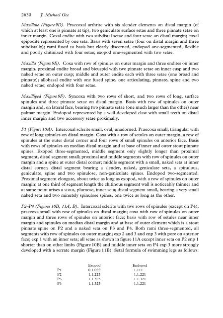
2630 J. Michael Gee<br />
Maxillule (Figure 9D). Praecoxal arthrite with six slender elements on distal margin (of<br />
which at least one is pinnate at tip), two geniculate surface setae and three pinnate setae on<br />
inner margin. Coxal endite with two subdistal setae and four setae on distal margin; coxal<br />
epipodite represented by one seta. Basis with seven setae (four on distal margin and three<br />
subdistally); rami fused to basis but clearly discerned, endopod one-segmented, flexible<br />
and poorly chitinized with four setae; exopod one-segmented with two setae.<br />
Maxilla (Figure 9E). Coxa with row of spinules on outer margin and three endites on inner<br />
margin, proximal endite broad and bicuspid with two pinnate setae on inner cusp and two<br />
naked setae on outer cusp; middle and outer endite each with three setae (one broad and<br />
pinnate); allobasal endite with one fused spine, one articulating, pinnate, spine and two<br />
naked setae; endopod with four setae.<br />
Maxilliped (Figure 9F). Syncoxa with two rows of short, and two rows of long, surface<br />
spinules and three pinnate setae on distal margin. Basis with row of spinules on outer<br />
margin and, on lateral face, bearing two pinnate setae (one much larger than the other) near<br />
palmar margin. Endopod represented by a well-developed claw with small teeth on distal<br />
inner margin and two accessory setae proximally.<br />
P1 (Figure 10A). Intercoxal sclerite small, oval, unadorned. Praecoxa small, triangular with<br />
row of long spinules on distal margin. Coxa with a row of setules on outer margin, a row of<br />
spinules at the outer distal corner and four rows of small spinules on anterior face. Basis<br />
with rows of spinules on median distal margin and at base of inner and outer stout pinnate<br />
spines. Exopod three-segmented, middle segment only slightly longer than proximal<br />
segment, distal segment small; proximal and middle segments with row of spinules on outer<br />
margin and a spine at outer distal corner; middle segment with a small, naked seta at inner<br />
distal corner; distal segment bearing a slender, naked, geniculate seta, a spinulous,<br />
geniculate, spine and two spinulose, non-geniculate spines. Endopod two-segmented.<br />
Proximal segment elongate, about twice as long as exopod, with a row of spinules on outer<br />
margin; at one third of segment length the chitinous segment wall is noticeably thinner and<br />
at same point arises a stout, plumose, inner seta; distal segment small, bearing a very small<br />
naked seta and two minutely spinulose spines, one twice as long as the other.<br />
P2–P4 (Figures 10B, 11A, B). Intercoxal sclerite with two rows of spinules (except on P4);<br />
praecoxa small with row of spinules on distal margin; coxa with row of spinules on outer<br />
margin and three rows of spinules on anterior face; basis with row of setules near inner<br />
margin and spinules on median distal margin and at base of outer element which is a stout<br />
pinnate spine on P2 and a naked seta on P3 and P4. Both rami three-segmented, all<br />
segments with row of spinules on outer margin; exp 2 and 3 and enp 3 with pore on anterior<br />
face; exp 1 with an inner seta; all setae as shown in figure 11A except inner seta on P2 enp 1<br />
shorter than on other limbs (Figure 10B) and middle inner seta on P4 exp 3 more strongly<br />
developed with a serrate margin (Figure 11B). Setal formula of swimming legs as follows:<br />
Exopod<br />
Endopod<br />
P1 0.1.022 1.111<br />
P2 1.1.223 1.1.221<br />
P3 1.1.323 1.1.321<br />
P4 1.1.323 1.1.221