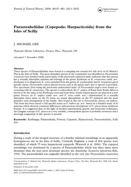
Parastenheliidae (Copepoda: Harpacticoida) from ... - Luciopesce.net
Parastenheliidae (Copepoda: Harpacticoida) from ... - Luciopesce.net
Parastenheliidae (Copepoda: Harpacticoida) from ... - Luciopesce.net

You also want an ePaper? Increase the reach of your titles
YUMPU automatically turns print PDFs into web optimized ePapers that Google loves.
Journal of Natural History, 2006; 40(47–48): 2611–2652<br />
<strong>Parastenheliidae</strong> (<strong>Copepoda</strong>: <strong>Harpacticoida</strong>) <strong>from</strong> the<br />
Isles of Scilly<br />
J. MICHAEL GEE<br />
Plymouth Marine Laboratory, Prospect Place, Plymouth, UK<br />
(Accepted 7 November 2006)<br />
Abstract<br />
Three species of <strong>Parastenheliidae</strong> were found at a sampling site around low tide level on St Martin’s<br />
Flat in the Isles of Scilly. The most abundant species in the community was identified as Parastenhelia<br />
ornatissima but detailed study, particularly of the previously unknown male, indicates that this species<br />
has a sexually dimorphic antenna and belongs in the genus Karllangia as K. ornatissima comb. nov.<br />
Karllangia is re-diagnosed, K. tertia excluded <strong>from</strong> the genus; K. psammophila and K. bengalensis given<br />
specific status; Parastenhelia reducta placed incertae sedis in the genus and a key to species is provided.<br />
Five specimens (four being the previously undescribed male) of Parastenhelia anglica were found cooccurring<br />
with K. ornatissima. The species is redescribed; the P. anglica of Kunz <strong>from</strong> South Africa is<br />
shown to be the same as his Karllangia tertia and both these closely related species are placed in a new<br />
genus Foweya (as F. anglica comb. nov. and F. tertia comb. nov.) characterized by a sexually<br />
dimorphic inner spine on the P1 basis, no sexual dimorphism on the P3 endopod and the most<br />
primitive setal arrangement in the family. Also found at this site is Parastenhelia spinosa var bulbosa.<br />
This form has been raised to full specific status as P. bulbosa sp. nov. based on a detailed study of its<br />
anatomy which showed consistent differences with previous descriptions of P. spinosa <strong>from</strong> northwest<br />
Europe. It is suggested that, in the light of modern experimental, ge<strong>net</strong>ic and molecular techniques,<br />
the Langian concept of P. spinosa as a very variable, cosmopolitan species is probably erroneous and a<br />
thorough reappraisal of this species is needed.<br />
Keywords: Karllangia, Parastenhelia, Foweya, <strong>Copepoda</strong>, <strong>Harpacticoida</strong>, <strong>Parastenheliidae</strong>, Scilly<br />
Isles<br />
Introduction<br />
During a study of the integral structure of a benthic infaunal assemblage at an apparently<br />
homogeneous site in the Isles of Scilly, Cornwall, England, a total of 464 species were<br />
identified, of which 75 were harpacticoid copepods (Warwick et al. 2006). The copepod<br />
assemblage was dominated by a species of <strong>Parastenheliidae</strong> which was three times more<br />
abundant than the next most dominant species, the Ameiridae Sicameira leptoderma Klie,<br />
1950. The parastenheliid was originally identified as the very rare Parastenhelia ornatissima<br />
Correspondence: J. Michael Gee, Plymouth Marine Laboratory, Prospect Place, The Hoe, Plymouth PL1 3DH, UK. Email:<br />
jmge@pml.ac.uk<br />
Published 30 December 2006<br />
ISSN 0022-2933 print/ISSN 1464-5262 online # 2006 Taylor & Francis<br />
DOI: 10.1080/00222930601108194
2612 J. Michael Gee<br />
(Monard) Lang, 1948 which was previously known only <strong>from</strong> a single female found in the<br />
Roscoff area of northern France. Two very rare species in the St Martins community also<br />
belonged in the <strong>Parastenheliidae</strong>, one identified as Parastenhelia anglica Norman and T.<br />
Scott, 1905 known only <strong>from</strong> two female specimens found off the Cornish coast and<br />
possibly a few specimens <strong>from</strong> South Africa. The other species was identified as<br />
Parastenhelia spinosa var. bulbosa (Bozic) Wells, 1963 known only <strong>from</strong> Roscoff, the Exe<br />
Estuary in Devon and the Scilly Isles.<br />
Briefly, Thompson and A. Scott (1903) created the genus Parastenhelia Thompson and<br />
A. Scott, 1903 to accommodate a new species P. hornelli Thompson and A. Scott, 1903<br />
<strong>from</strong> Ceylon. Previous to this Claus (1863) had described Thalestris forficula Claus, 1863<br />
<strong>from</strong> the Mediterranean, whilst later Sars (1911) described Microthalestris littoralis Sars,<br />
1911 and transferred Claus’s species to the same genus. Lang (1934) made M. littoralis a<br />
subspecies of M. forficula, transferred both to the genus Parastenhelia along with P.<br />
hornelli, P. anglica and P. gracilis Brady, 1910 and placed the genus in the Thalestridae.<br />
Lang (1936) created a new sub-family, the Parastenheliinae, within the Thalestridae to<br />
accommodate Parastenhelia. Lang (1944) raised the sub-family to full family status,<br />
recognized that Harpacticus spinosus Fischer, 1860 belonged in Parastenhelia, so making P.<br />
spinosa (Fischer) Lang, 1944 the type species of the genus. Lang (1948) gave a full list of<br />
synonyms for P. spinosa and designated three forms of the species, forma typica (for P.<br />
forficula), forma littoralis, and forma penicillata (for the Microthalestris littoralis var.<br />
penicillata of Willey, 1935). Finally, Lang (1948) moved Thalestrella ornatissima Monard,<br />
1935 into the genus as P. ornatissima. Since Lang’s (1948) monograph, two new forms of<br />
P. spinosa and six new species have been added to the genus (see Bodin, 1997). Most<br />
recently, Willen (2000) moved the genus Karllangia Noodt, 1964 <strong>from</strong> the Ameiridae into<br />
the <strong>Parastenheliidae</strong>.<br />
In this paper I give a detailed reappraisal of the three species of Parastenhelia <strong>from</strong> the<br />
Scilly Isles and conclude that P. ornatissima should be moved to the genus Karllangia; that<br />
P. anglica should be removed to a new genus, along with another species of Karllangia; that<br />
P. spinosa var. bulbosa should be raised to specific status and that the Langian concept of P.<br />
spinosa as a variable, cosmopolitan species should be questioned.<br />
Materials and methods<br />
Material <strong>from</strong> the Isles of Scilly was obtained <strong>from</strong> cores in clean coarse sand at LWST on<br />
St. Martin’s Flats between the islands of St. Martin and Tresco. All sediments were sieved<br />
through a 63 mm sieve and retained animals were fixed in 10%, and preserved in 4%,<br />
formalin. Before dissection, measurements of body length were made <strong>from</strong> whole<br />
specimens temporarily mounted in lactophenol. Specimens were dissected in lactophenol,<br />
the parts individually mounted in lactophenol under coverslips subsequently sealed with<br />
clear nail varnish. All drawings were prepared using a camera lucida on a Nikon Optiphot<br />
20 differential interference contrast microscope. The terminology of the body and<br />
appendage morphology follows that of Huys and Boxshall (1991). Abbreviations used in<br />
the text and figures are P1–P6 for thoracopods 1–6; exp(enp) 1(2, 3) to denote the<br />
proximal (middle, distal) segment of a ramus; benp denotes the baseoendopod of P5; and a<br />
for aesthetasc. Body length was measured <strong>from</strong> the base of the rostrum to the median<br />
posterior border of the anal somite. All material has been deposited in the Natural History<br />
Museum, London.
<strong>Parastenheliidae</strong> <strong>from</strong> the Isles of Scilly 2613<br />
Taxonomy<br />
Family <strong>Parastenheliidae</strong> Lang, 1944<br />
Genus Karllangia Noodt, 1964<br />
Diagnosis<br />
<strong>Parastenheliidae</strong>. Body small, usually with deeply divided ornamental hyaline frills. Rostrum,<br />
defined at base, triangular, reaching at most to end of second segment of antennule.<br />
Operculum semi-circular, variously ornamented. Caudal ramus with row(s) of spinules across<br />
dorsal surface and with seven elements. Female antennule eight- or indistinctly ninesegmented<br />
but with distal four or five segments small, combined length only slightly longer<br />
than segment 4. Male antennule eight-segmented, haplocer with three segments distal to<br />
geniculation, segment 5 moderately swollen. Female antenna with partially divided allobasis<br />
with a small pinnate seta on abexopodal margin; exopod two-segmented (or segments partially<br />
fused), proximal segment with two setae, distal segment with one lateral and two terminal<br />
setae; endopod with two subdistal spines and on distal margin one spine, four geniculate setae<br />
and two normal setae. Male antenna with enlarged, highly plumose seta on allobasis and<br />
exopod segments of different size with only one seta on proximal segment and all lateral setae<br />
enlarged and highly plumose. Mandibular basis with three setae, endopod one-segmented<br />
with eight setae, exopod usually present as a very small segment with two setae. Maxillule with<br />
slender elements on distal margin of praecoxal arthrite, two surface setae and two setae on<br />
inner margin; coxal endite with two to four setae and usually a seta representing the epipodite;<br />
basis and rami poorly chitinized, endopod present, exopod represented by one seta. Maxilla<br />
with three syncoxal endites with 3.2-3.2-3 elements, allobasal endite with three elements,<br />
endopod with four or five setae. Maxilliped syncoxa with one seta, basis oval with two setae on<br />
palmar margin, endopod a claw with one accessory seta. P1 exopod of three equal segments,<br />
exopod 2 with or without a small inner seta, distal segment with four elements (a naked seta, a<br />
geniculate spine and two outer spines, all spines strongly spinulous); endopod two-segmented,<br />
proximal segment about twice as long as exopod with inner seta implanted medially in a region<br />
of reduced chitinization of segment wall, distal segment small with one naked seta, a<br />
geniculate spine and a normal spine. P2–P4 rami three-segmented, exopod 1 without inner<br />
seta, setae on other segments well developed. Usually no sexual dimorphism in swimming legs<br />
but occasionally outer spine on P3 enp 3 in male reduced in size. Setal formula of exp/enp as<br />
follows: P2 0.1.223/1.1.121; P3 0.1.2-323/1.1.221; P4 0.1.323/1.1.221. Female P5 with<br />
triangular endopodal lobe with five setae and exopod two to five times longer than wide with<br />
five or six setae. Male P5 endopodal lobe with two setae, exopod with four setae.<br />
Type species: Karllangia arenicola Noodt, 1964.<br />
Other species: K. psammophila Wells, 1967, K. bengalensis Wells and Rao, 1987, K.<br />
pulchra Meilke, 1994, K. obscura Mielke, 1994, K. ornatissima (Monard) comb. nov.<br />
Karllangia ornatissima (Monard) comb. nov.<br />
(Figures 1–6)<br />
Synonyms<br />
Thalestrella ornatissima Monard 1935; Parastenhelia ornatissima (Monard) Lang 1948;<br />
Parastenhelia anglica Norman and T. Scott in Wells (1961).
2614 J. Michael Gee<br />
Figure 1. Karllangia ornatissima comb. nov. R habitus: (A) lateral view; (B) dorsal view; (C) ventral view of<br />
urosome, omitting P5-bearing somite.
<strong>Parastenheliidae</strong> <strong>from</strong> the Isles of Scilly 2615<br />
Figure 2. Karllangia ornatissima comb. nov. R operculum and caudal ramus: (A) dorsal view; (B) lateral view; (C)<br />
ventral view. R: (D) antenna; (E) maxillule; (F) maxilla.
2616 J. Michael Gee<br />
Figure 3. Karllangia ornatissima comb. nov. R: (A) antennule segmentation; (B) antennule armature; (C)<br />
mandible; (D) maxilliped; (E) P5.
<strong>Parastenheliidae</strong> <strong>from</strong> the Isles of Scilly 2617<br />
Figure 4. Karllangia ornatissima comb. nov. R: (A) P1; (B) P2.
2618 J. Michael Gee<br />
Figure 5. Karllangia ornatissima comb. nov. R: (A) P3; (B) P4. „: (C) P3 endopod 3.
<strong>Parastenheliidae</strong> <strong>from</strong> the Isles of Scilly 2619<br />
Figure 6. Karllangia ornatissima comb. nov. „: (A) urosome, lateral view; (B) antennule segmentation; (C)<br />
antenna; (D) P5 and P6.
2620 J. Michael Gee<br />
Material examined<br />
Neotype, 1 adult R, spirit preserved, NHM Reg. No. 2006.170. Paratypes, 2R and 1„ (each<br />
dissected onto three slides), NHM Reg. Nos 2006.1971–1973 and 50R and 50„ spirit<br />
preserved, NHM Reg. Nos 2006.1974–1983<br />
Description of female<br />
Body (Figure 1). Length 0.315–0.46 mm (mean 0.37 mm, n510) semi-cylindrical, widest<br />
at posterior margin of cephalothorax, tapering gradually posteriorly and without clear<br />
distinction between prosome and urosome. Cephalothorax tapering anteriorly with welldeveloped<br />
pleural area and ornamented with pores and sensilla as in Figure 1. All<br />
prosomites with pronounced, deeply divided, serrate hyaline frill on dorsal and lateral<br />
posterior border. Urosomite 1 (P5-bearing somite) with two short rows of small spinules<br />
dorsally and a deeply divided hyaline frill on posterior border. Genital double somite with<br />
fusion line marked by dorsal and lateral sub-cuticular rib bearing a row of spinules dorsally;<br />
posterior margin of double somite with two short ventro-lateral spinule rows and a deeply<br />
divided hyaline frill. Genital apparatus (Figure 1C) consisting of median ventral copulatory<br />
pore and separate anterior gonopores each covered by a vestigial P6 bearing three setae.<br />
Urosomites 4 and 5 with deeply divided hyaline frill, former also with complete row of<br />
spinules on ventral posterior margin, latter without spinular ornamentation. Anal somite<br />
short, partially divided on posterior margin which bears a row of spinules ventrally at base<br />
of caudal rami. Anal operculum semi-circular, ornamented with six to eight coarse teeth<br />
(Figures 1B, 2A). Caudal rami (Figure 2A–C) about as long as broad with one or two<br />
diagonal rows of spinules on dorsal face and a row of spinules and a tube pore (arrowed in<br />
Figure 2C) on ventral posterior margin: bearing seven setae, antero-lateral seta I minute;<br />
setae II and III broad, spinulose with terminal flagellum; setae IV and V well developed,<br />
seta VI small and naked; triarticulate seta VII arising <strong>from</strong> dorsal outer distal corner of<br />
ramus.<br />
Rostrum (Figure 1A,B). Well developed, reaching middle of segment 2 of antennule,<br />
defined at base, triangular with rounded tip and a pair of sub-apical sensilla.<br />
Antennule (Figure 3A,B). Indistinctly nine-segmented (distal two segments partially fused),<br />
distal five segments small, combined length only slightly longer than segment 4. Segment 1<br />
with patch of spinules, segment 2 with a short pinnate seta; aesthetascs on segments 4 and<br />
9. Setal formula as follows: 1–[1], 2-[9], 3–[6], 4-[1+(1+a)], 5-[2], 6-[2?], 7-[2?], 8-[3], 9-<br />
[5?+(2+a)].<br />
Antenna (Figure 2D). With well-developed coxa. Allobasis partially divided with a pinnate<br />
seta and a row of spinules on abexopodal margin. Exopod of two approximately equal sized<br />
segments, proximal segment with two setae (a weak, naked proximal seta and a strong<br />
pinnate distal seta); distal segment with a distal dentate frill and three setae (strong pinnate<br />
setae on lateral and distal margin and a weak naked seta at outer distal corner). Endopod<br />
with row of spinules and two sub-distal spines on outer margin; distal margin with two rows<br />
of spinules and seven elements: a pinnate spine, four geniculate setae (inner with large<br />
pinnules at geniculation) and two plain setae, one of which is fused to base of inner<br />
geniculate seta.
<strong>Parastenheliidae</strong> <strong>from</strong> the Isles of Scilly 2621<br />
Mandible (Figure 3C). Coxal gnathobase well developed with bicuspid and unicuspid teeth<br />
and a seta at distal corner. Palp biramus, consisting of basis bearing three pinnate setae, an<br />
elongate one-segmented endopod with two lateral and six terminal setae and a small onesegmented<br />
exopod bearing two terminal setae.<br />
Maxillule (Figure 2E). Small, poorly chitinized, particularly in region of basis and endopod,<br />
with setae closely bunched up and difficult to discern. Praecoxal arthrite with a proximal<br />
row of spinules, with three pairs of slender elements on distal margin, two geniculate<br />
surface setae and two setae on inner margin. Coxal endite with three (or four) setae on<br />
distal margin (and possibly an epipodite represented by one seta which could not be<br />
discerned in these dissected specimens). Basis with five setae; endopod one-segmented with<br />
two setae; exopod represented by one seta.<br />
Maxilla (Figure 2F). Small, difficult to make out exact setation. Bears three coxal endites,<br />
proximal endite broad and bicuspid with two setae (one pinnate) on inner cusp and one or<br />
two seta(e) on outer cusp; middle endite with two setae, outer endite with three setae (one<br />
broad and pinnate); allobasal endite with one fused spine, one articulating, pinnate, spine<br />
and one or two naked setae; endopod with four setae.<br />
Maxilliped (Figure 3D). Syncoxa with row of spinules and one pinnate seta on distal<br />
margin. Basis with row of spinules on outer margin and on each lateral face, bearing two<br />
small setae near palmar margin. Endopod represented by a well-developed claw with small<br />
teeth on distal inner margin and one accessory seta proximally.<br />
P1 (Figure 4A). Intercoxal sclerite (not illustrated) small, oval, unadorned. Coxa almost<br />
square with a row of setules on outer margin, a row of spinules and a pore at the outer distal<br />
corner and four rows of small spinules on anterior face. Basis with rows of spinules on<br />
median distal margin and at base of inner and outer stout pinnate spines, a pore also at base<br />
of inner spine. Exopod of three more or less equal-sized segments; proximal and middle<br />
segments with row of spinules on outer margin and a spine at outer distal corner; middle<br />
segment with a very small, weak seta which in most cases lies along the posterior face of the<br />
ramus and is only visible under x100 oil immersion; distal segment bearing a slender naked<br />
seta and a spinulous geniculate spine on distal margin and two spinulose, non-geniculate<br />
spines on outer margin. Endopod two-segmented; proximal segment elongate, about twice<br />
as long as exopod, with a few spinules on outer distal margin; at mid-point of segment the<br />
chitinous segment wall is noticeably thinner and at same point arises a stout inner seta with<br />
a plumose distal portion; distal segment small, bearing a very small naked seta, a geniculate<br />
spine and a spinulose spine.<br />
P2–P4 (Figures 4B, 5A, B). Intercoxal sclerite unadorned; praecoxa small; coxa with row of<br />
spinules on outer margin and two or three rows of spinules on anterior face; basis with row<br />
of spinules on median distal margin and at base of outer element which is a naked spine on<br />
P2 and a naked seta on P3 and P4. Both rami three-segmented, all segments with row of<br />
spinules on outer margin; exp 2 and 3, enp 1 in P2 and enp 3 in P3–P4 with pore on<br />
anterior face; exp 1 without an inner seta but all setae on other segments well developed,<br />
distal outer element on exp 3 a pinnate spine and middle inner seta on P4 exp 3 strongly<br />
developed with a serrate margin. Setal formula of swimming legs as follows:
2622 J. Michael Gee<br />
Exopod<br />
Endopod<br />
P1 0.1.022 1.111<br />
P2 0.1.223 1.1.121<br />
P3 0.1.223 1.1.221<br />
P4 0.1.323 1.1.221<br />
P5 (Figure 3E). Benps of each side not fused medially and exopods also separate. Benp<br />
with well-developed inner expansion triangular in shape with row of spinules on median<br />
inner margin and distal outer margin and bearing five elements, two short pinnate spines on<br />
inner margin and three naked setae on distal margin. Outer basal peduncle of benp with a<br />
few spinules and a naked seta. Exopod slender, five times longer than wide, bearing six<br />
naked setae, two on distal margin and four on outer margin.<br />
Description of male<br />
As in female except for urosome, antennule, antenna, P5 and P6.<br />
Body. Slightly smaller than female, length 0.258–0.4 mm (mean 0.302 mm, n510) and<br />
urosomites 2 and 3 not fused (Figure 6A). Body ornamentation as in female except that<br />
ventral row of spinules on urosomite 3 complete (Figure 6D).<br />
Antennule (Figure 6B). Eight-segmented, haplocer, with moderately swollen segment 5<br />
with row of spinules and aesthetasc, acrothek of two setae and small aesthetasc on distal<br />
segment. All setae naked except a small pinnate seta on segment 2. Setal formula tentatively<br />
given as follows although some setae may be missing:- 1-[1], 2-[11], 3[4?], 4{2?], 5-<br />
[8?+(1+a)], 6[1+2modified spines], 7[4], 8-[7?+(2+a)].<br />
Antenna (Figure 6C). Allobasis with enlarged and highly plumose seta on abexopodal<br />
margin. Exopod two-segmented but proximal segment reduced in size and bearing only one<br />
element, a large highly plumose seta; distal segment swollen, with proximal seta swollen<br />
and highly plumose and two setae on distal margin more strongly developed. Subdistal<br />
spines on outer margin of endopod more strongly spinulose.<br />
P5 (Figure 6D). Benps of each side fused medially, inner expansions more rounded and<br />
bearing two elements, a small serrate spine and a smaller naked spine. Exopod only twice as<br />
long as wide, ovoid with short row of spinules on outer margin and four naked setae (note<br />
figured specimen shows an aberrant setal arrangement on one baseoendopodal lobe).<br />
P6 (Figure 6D). A single plate with one pinnate and two naked setae on a small extension<br />
on each side.<br />
Genus Foweya gen. nov.<br />
Synonyms<br />
Parastenhelia (part); Karllangia (part).
<strong>Parastenheliidae</strong> <strong>from</strong> the Isles of Scilly 2623<br />
Diagnosis<br />
<strong>Parastenheliidae</strong>. Body with faint, deeply divided, hyaline frills. Rostrum, defined at base,<br />
triangular, reaching at most to end of second segment of antennule. Operculum semicircular,<br />
variously ornamented. Caudal ramus with row(s) of spinules across dorsal surface<br />
and with seven elements. Female antennule nine-segmented, distal five segments with a<br />
combined length greater than that of segment 4. Male antennule 10-segmented, haplocer<br />
with three segments distal to geniculation, segments 5–7 moderately swollen. Antenna with<br />
partially divided allobasis with a pinnate seta on abexopodal margin; exopod twosegmented,<br />
proximal segment with two setae, distal segment with two lateral and three<br />
terminal setae; endopod with two subdistal spines and on distal margin one spine, four<br />
geniculate setae and two normal setae. Mandibular basis with three setae, endopod onesegmented<br />
with eight setae, exopod one-segmented with four setae. Maxillule with slender<br />
elements on distal margin of praecoxal arthrite, two surface setae and three setae on inner<br />
margin; coxal endite with six setae and an epipodite represented by one seta; endopod<br />
poorly chitinized, exopod present bearing two setae. Maxilla with three syncoxal endites<br />
with 4.3.3 elements, allobasal endite with four elements, endopod with four or five setae.<br />
Maxillipedal syncoxa with three setae, basis oval with two setae on palmar margin, endopod<br />
a claw with two accessory setae. P1 basis inner seta sexually dimorphic (bifid tip in male),<br />
exopod three-segmented, exp 2 only slightly longer than exp 1, with an inner seta, distal<br />
segment small, with a naked seta, a geniculate spine and two outer spines, all spines<br />
strongly spinulous; endopod two-segmented, proximal segment about twice as long as<br />
exopod with inner seta implanted at 35% of ramus length in a region of reduced<br />
chitinization of segment wall, distal segment small with one naked seta and two pinnate<br />
spines. P2–P4 rami three-segmented, except male P2 endopod sometimes two-segmented<br />
as result of fusion of middle and distal segments, no sexual dimorphism in P3 endopod:<br />
setal formula of exp/enp as follows: P2 1.1.223/1.1.221; P3 1.1.323/1.1.321; P4 1.1.323/<br />
1.1.221. Female P5 endopodal lobe with five setae, exopod about three times longer than<br />
wide with six setae. Male P5 endopodal lobe with two setae, exopod with five or six setae.<br />
Type species: Foweya anglica (Norman and T. Scott) comb. nov.<br />
Other species: Foweya tertia (Kunz) comb. nov.<br />
Etymology<br />
Fowey (pronounced Foy) is the Cornish port that is the type locality of the genus.<br />
Foweya anglica (Norman and T. Scott) comb. nov.<br />
(Figures 7–11)<br />
Synonyms<br />
Parastenhelia anglica Norman and T. Scott, 1905.<br />
Material examined<br />
Holotype, 1 R, dissected onto one slide, collected by Norman at Fowey, Cornwall, NHM<br />
Reg. No. 1911.11.8.M.2423.371. Paratype, 1R (spirit preserved) in Norman Collection<br />
NHM Reg. No. 1911.11.8.43325. Other material <strong>from</strong> St. Martins Flat, Isles of Scilly, 1R
2624 J. Michael Gee<br />
Figure 7. Foweya anglica comb. nov. R habitus: (A) lateral view. R urosome, omitting P5 bearing somite: (B) dorsal<br />
view; (C) ventral view.
<strong>Parastenheliidae</strong> <strong>from</strong> the Isles of Scilly 2625<br />
Figure 8. Foweya anglica comb. nov. „ urosome (excluding P5 bearing somite): (A) dorsal view; (B) lateral view;<br />
(C) ventral view; (D) Antennule segmentation. R operculum and caudal ramus: (E) dorsal view; (F) ventral view.
2626 J. Michael Gee<br />
Figure 9. Foweya anglica comb. nov. R: (A) Antennule segmentation; (B) antenna; (C) mandible; (D) maxillule;<br />
(E) maxilla; (F) maxilliped.
<strong>Parastenheliidae</strong> <strong>from</strong> the Isles of Scilly 2627<br />
Figure 10. Foweya anglica comb. nov. R: (A) P1; (B) P2 protopod and endopod 1; (C) P5. „: (D) P1 basis.
2628 J. Michael Gee<br />
Figure 11. Foweya anglica comb. nov. R: (A) P3; (B) P4 exopod 3. „: (C) P5.
<strong>Parastenheliidae</strong> <strong>from</strong> the Isles of Scilly 2629<br />
and 1„ (each dissected onto three slides), NHM Reg. Nos 2006.1984–1985 and 3„ (spirit<br />
preserved), NHM Reg. Nos 2006.2003–2005.<br />
Description of female<br />
Body (Figure 7). Length 0.53 mm, semi-cylindrical, widest at posterior margin of<br />
cephalothorax, tapering posteriorly, without clear distinction between prosome and<br />
urosome. Cephalothorax tapering anteriorly and with pores and sensilla as in Figure 7A.<br />
This and free prosomites with plain hyaline frills. Urosomite-1 (P5-bearing somite) with<br />
two short rows of small spinules dorsally. Genital double somite with fusion line marked by<br />
lateral sub-cuticular rib and a row of spinules dorsally; posterior margin of double somite<br />
with lateral row of short spinules and a ventro-lateral row of longer spinules. Genital<br />
apparatus (Figure 7C) consisting of median ventral copulatory pore and separate anterior<br />
gonopores armed internally with a row of teeth each covered by a vestigial P6 bearing three<br />
setae. Urosomite 4 with lateral row of small spinules and ventral row of larger spinules.<br />
Urosomite 5 unadorned. Hyaline frills on urosomites delicate but deeply divided and with<br />
minutely dentate distal margin. Anal somite with a row of spinules near ventral anterior<br />
margin; partially divided on posterior margin which bears a row of spinules ventrally at base<br />
of caudal rami. Anal operculum semi-circular, ornamented with approximately 30 small,<br />
closely set denticles (Figures 7B, 8E). Caudal rami (Figure 8E, F) about as long as broad<br />
with a diagonal rows of spinules on dorsal face and a row of spinules and a tube pore on<br />
ventral posterior margin: bearing seven setae, antero-lateral seta I minute; setae II and III<br />
broad, spinulose with terminal flagellum; setae IV and V well developed, seta VI long and<br />
naked; triarticulate seta VII arising <strong>from</strong> dorsal distal part of ramus.<br />
Rostrum (Figure 9A). Well developed, reaching almost to end of segment 2 of antennule,<br />
defined at base, triangular with rounded tip and a pair of sub-apical sensilla.<br />
Antennule (Figure 9A). Distinctly nine-segmented, combined length of distal five segments<br />
markedly longer than segment 4. All segments without pinnate setae; aesthetascs on<br />
segments 4 and 9. Setal formula as follows: 1–[1], 2-[8], 3–[4], 4-[2+(1+a)], 5-[2], 6-[2], 7-<br />
[2], 8-[2], 9-[5+(2+a)] but some setae probably missing on segments 2 and 3.<br />
Antenna (Figure 9B). Allobasis partially divided, with a pinnate seta and a row of spinules<br />
on abexopodal margin. Exopod two-segmented, proximal segment with two setae (a naked<br />
proximal seta and a strong pinnate distal seta); distal segment with a distal dentate frill and<br />
five setae (two strong pinnate setae on lateral margin and, on distal margin, a strong pinnate<br />
seta, a well-developed naked seta and a very small naked seta). Endopod with row of<br />
spinules and two sub-distal spines on outer margin; distal margin with two rows of spinules<br />
and seven elements, a pinnate spine, four geniculate setae (inner with large pinnules at<br />
geniculation) and two plain setae, one of which is fused to base of inner geniculate seta.<br />
Mandible (Figure 9C). Coxal gnathobase well developed with bicuspid and unicuspid teeth<br />
and a seta at distal corner. Basis with row of spinules on outer and inner margin and three<br />
pinnate setae on distal margin; endopod elongate, one-segmented, with two lateral and six<br />
terminal setae; exopod small, one-segmented, with two lateral and two terminal setae.
2630 J. Michael Gee<br />
Maxillule (Figure 9D). Praecoxal arthrite with six slender elements on distal margin (of<br />
which at least one is pinnate at tip), two geniculate surface setae and three pinnate setae on<br />
inner margin. Coxal endite with two subdistal setae and four setae on distal margin; coxal<br />
epipodite represented by one seta. Basis with seven setae (four on distal margin and three<br />
subdistally); rami fused to basis but clearly discerned, endopod one-segmented, flexible<br />
and poorly chitinized with four setae; exopod one-segmented with two setae.<br />
Maxilla (Figure 9E). Coxa with row of spinules on outer margin and three endites on inner<br />
margin, proximal endite broad and bicuspid with two pinnate setae on inner cusp and two<br />
naked setae on outer cusp; middle and outer endite each with three setae (one broad and<br />
pinnate); allobasal endite with one fused spine, one articulating, pinnate, spine and two<br />
naked setae; endopod with four setae.<br />
Maxilliped (Figure 9F). Syncoxa with two rows of short, and two rows of long, surface<br />
spinules and three pinnate setae on distal margin. Basis with row of spinules on outer<br />
margin and, on lateral face, bearing two pinnate setae (one much larger than the other) near<br />
palmar margin. Endopod represented by a well-developed claw with small teeth on distal<br />
inner margin and two accessory setae proximally.<br />
P1 (Figure 10A). Intercoxal sclerite small, oval, unadorned. Praecoxa small, triangular with<br />
row of long spinules on distal margin. Coxa with a row of setules on outer margin, a row of<br />
spinules at the outer distal corner and four rows of small spinules on anterior face. Basis<br />
with rows of spinules on median distal margin and at base of inner and outer stout pinnate<br />
spines. Exopod three-segmented, middle segment only slightly longer than proximal<br />
segment, distal segment small; proximal and middle segments with row of spinules on outer<br />
margin and a spine at outer distal corner; middle segment with a small, naked seta at inner<br />
distal corner; distal segment bearing a slender, naked, geniculate seta, a spinulous,<br />
geniculate, spine and two spinulose, non-geniculate spines. Endopod two-segmented.<br />
Proximal segment elongate, about twice as long as exopod, with a row of spinules on outer<br />
margin; at one third of segment length the chitinous segment wall is noticeably thinner and<br />
at same point arises a stout, plumose, inner seta; distal segment small, bearing a very small<br />
naked seta and two minutely spinulose spines, one twice as long as the other.<br />
P2–P4 (Figures 10B, 11A, B). Intercoxal sclerite with two rows of spinules (except on P4);<br />
praecoxa small with row of spinules on distal margin; coxa with row of spinules on outer<br />
margin and three rows of spinules on anterior face; basis with row of setules near inner<br />
margin and spinules on median distal margin and at base of outer element which is a stout<br />
pinnate spine on P2 and a naked seta on P3 and P4. Both rami three-segmented, all<br />
segments with row of spinules on outer margin; exp 2 and 3 and enp 3 with pore on anterior<br />
face; exp 1 with an inner seta; all setae as shown in figure 11A except inner seta on P2 enp 1<br />
shorter than on other limbs (Figure 10B) and middle inner seta on P4 exp 3 more strongly<br />
developed with a serrate margin (Figure 11B). Setal formula of swimming legs as follows:<br />
Exopod<br />
Endopod<br />
P1 0.1.022 1.111<br />
P2 1.1.223 1.1.221<br />
P3 1.1.323 1.1.321<br />
P4 1.1.323 1.1.221
<strong>Parastenheliidae</strong> <strong>from</strong> the Isles of Scilly 2631<br />
P5 (Figure 10C). Benps of each side not fused medially and exopods also separate. Benp<br />
with well-developed inner expansion triangular in shape with row of setules on inner<br />
margin, row of spinules on outer margin and bearing five pinnate setae (four more or less<br />
equal in length, inner distal seta, missing in Figure 10C, somewhat longer). Outer basal<br />
peduncle of benp with a few spinules and a naked seta. Exopod slender, almost three times<br />
longer than wide, with row of pinnules on outer margin and spinules on inner margin, and<br />
bearing six setae (one pinnate seta on inner margin, two naked setae on distal margin and<br />
three minutely pinnate setae on outer margin).<br />
Description of male<br />
As in female except for urosome, antennule, P1 basis, P5 and P6.<br />
Body. Slightly smaller than female, length 0.32–0.443 mm (mean 0.37 mm, n54) and<br />
urosomites 2 and 3 not fused (Figure 8A–C). Body ornamentation as in female except that<br />
row of spinules on urosomite 3 complete both dorsally and ventrally.<br />
Antennule (Figure 8D). Ten-segmented, haplocer, with moderately swollen segments 5–7<br />
and major articulation between segments 7 and 8; with row of spinules and aesthetasc on<br />
segment 5, acrothek of two setae and small aesthetasc on distal segment. All setae naked.<br />
Tentative setal formula:- 1 [1], 2 [10], 3 [8?], 4 [2], 5–7 [11+(1+a)], 8 [2sp+1], 9[4], 10<br />
[5+(2+a)].<br />
P1 basis (Figure 10D). As in female except that inner spine modified to have a bifid tip<br />
bearing a minute setule.<br />
P5 (figure 11C). Benps of each side fused medially, inner expansions more rounded than in<br />
female, with row of spinules on inner and outer margin and bearing two pinnate setae,<br />
outer twice as long as inner. Exopod a single segment, oval, not quite twice as long as wide,<br />
with short row of spinules on inner and outer margin and bearing six setae as shown in<br />
Figure 11C.<br />
P6 (Figure 8C). A single plate with three setae on a small extension on each side.<br />
Foweya tertia (Kunz) comb. nov.<br />
(Figures 12, 13)<br />
Synonyms<br />
Parastenhelia anglica in Kunz (1963), Karllangia tertia Kunz, 1975.<br />
Material examined<br />
From paratypes held in Zoologisches Museum Hamburg Reg. No. K 30368 (collected<br />
<strong>from</strong> shell gravel in tidal pools on a reef off East London, South Africa, Kunz, 1975), 1R<br />
and 1„ (each dissected onto three slides) NMH Reg Nos 2006.1986–1987 and 1R spirit<br />
preserved, NHM Reg. No. 2006.1988.
2632 J. Michael Gee<br />
Figure 12. Foweya tertia comb. nov. R urosome: (A) dorsal view; (B) lateral view; (C) ventral view; (D) operculum<br />
and caudal ramus, dorsal view. „: (E) urosome, excluding P5 bearing somite, ventral view.
<strong>Parastenheliidae</strong> <strong>from</strong> the Isles of Scilly 2633<br />
Figure 13. Foweya tertia comb. nov. „: (A) P1 basis; (B) P2; (C) P5.<br />
Description of female<br />
As described by Kunz (1975) with following additions or corrections. Body length of two<br />
specimens measured 0.39 mm. Operculum (Figure 12D) semi-circular, ornamented with<br />
numerous very fine spinules, only visible under x100 oil immersion. Otherwise body<br />
(Figure 12A–C), mouthparts, swimming legs and P5 as for F. anglica. Note that this implies<br />
that there is some deviation <strong>from</strong> the original description in the setation of some of the<br />
mouthparts and of the P3 enp 3 given in Table 5 in Kunz (1975).
2634 J. Michael Gee<br />
Description of male<br />
Slightly smaller than female. As for male of F. anglica in body ornamentation (Figure 12E)<br />
and sexually dimorphic inner spine on P1 basis (Figure 13A). Differs <strong>from</strong> K. anglica in that<br />
P2 endopod (Figure 13B) is only two-segmented but still carries the same number of setae;<br />
the P5 exopod (Figure 13C) bears only five setae (the median inner short seta in F. anglica<br />
is missing) and the two setae on the baseoendopod are equal in length.<br />
Genus Parastenhelia Thompson and A. Scott, 1903<br />
Parastenhelia bulbosa sp. nov.<br />
(Figures 14–19)<br />
Synonyms<br />
Parastenhelia spinosa in Bozic (1955). Parastenhelia spinosa forma bulbosa in Wells (1963).<br />
Parastenhelia spinosa in Wells (1970) <strong>from</strong> Porth Hellick.<br />
Material examined<br />
Holotype; 1R (dissected onto four slides) <strong>from</strong> St. Martins Flat, Isles of Scilly, NHM Reg.<br />
No. 2006.1989. Paratypes, <strong>from</strong> the same locality as the holotype, 1R (dissected onto three<br />
slides) and 2„ (each dissected onto three or four slides) NHM Reg Nos 2006.1990–1992;<br />
4R, 4„ and 3 copepodites (spirit preserved), NHM Reg. No. 2006.1993–2002. Other<br />
material, 1„ (spirit preserved) collected by University of London Sub-Aqua Club in gravel<br />
at low water at Porth Hellick, St Marys, Isles of Scilly, NHM Reg. No. 1967.10.31.50.<br />
Description of female<br />
Body. Length (of contracted specimens) 0.41–0.49 mm (mean50.46 mm, n54), semicylindrical,<br />
widest at posterior margin of cephalothorax, tapering posteriorly without clear<br />
distinction between prosome and urosome. Urosomites (Figure 14A–C) with wide,<br />
palisade hyaline frills finely dentate on posterior margin (Figure 15A). Genital doublesomite<br />
(Figure 14A–C) completely fused without trace of sub-cuticular rib, ornamented<br />
only with a short ventro-lateral row of spinules on posterior margin. Genital apparatus<br />
(Figure 14C) with median ventral copulatory pore and separate anterior gonopores, each<br />
armed internally with a row of teeth and covered by a vestigial P6 bearing three setae.<br />
Urosomites 4 and 5 with short lateral row of spinules on posterior margin. Anal somite with<br />
median ventral row of short spinules and, on posterior margin, a ventral and lateral row of<br />
stronger spinules and setules on dorsal margin; anal operculum (Figure 15A) semi-circular<br />
with a sub-marginal row of minute spinules and a marginal row of about 35 larger spinules.<br />
Caudal rami (Figure 15A, B) broader than long, with rows of spinules on inner and outer<br />
margin, diagonally across dorsal surface and around ventral posterior margin; minute seta I<br />
and larger seta II implanted anteriorly on lateral margin; robust seta III implanted at distal<br />
outer corner; terminal seta IV moderately slender but with characteristic swollen bulbose<br />
base; terminal seta V robust, large without noticeably swollen base; terminal, inner seta VI<br />
small and slender and triarticulated seta VII implanted on dorsal surface.
<strong>Parastenheliidae</strong> <strong>from</strong> the Isles of Scilly 2635<br />
Figure 14. Parastenhelia bulbosa sp. nov. R: urosome, excluding P5 bearing somite (A) dorsal view; (B) lateral view;<br />
(C) ventral view: (D) mandible; (E) maxilliped.
2636 J. Michael Gee<br />
Figure 15. Parastenhelia bulbosa sp. nov. R: (A) anal somite and caudal ramus, dorsal view; (B) caudal ramus,<br />
ventral view; (D) antenna. „: (C) caudal ramus, ventral view; (E) maxillule; (F) maxilla.
<strong>Parastenheliidae</strong> <strong>from</strong> the Isles of Scilly 2637<br />
Figure 16. Parastenhelia bulbosa sp. nov. R: (A) P1; (B) P2.
2638 J. Michael Gee<br />
Figure 17. Parastenhelia bulbosa sp. nov. P3: (A) R; (B) „.
<strong>Parastenheliidae</strong> <strong>from</strong> the Isles of Scilly 2639<br />
Figure 18. Parastenhelia bulbosa sp. nov. P4: (A) „; (B) R endopod 3. P5: (C) R (D) „.
2640 J. Michael Gee<br />
Figure 19. Parastenhelia bulbosa sp. nov. „: (A) urosome, excluding P5 bearing somite, ventral view, (C) rostrum<br />
and antennule segmentation. R: (B) rostrum and antennule segmentation.<br />
Rostrum (Figure 19B). Moderately small, only reaching a short way past proximal margin of<br />
second antennular segment, defined at base, triangular with rounded tip and a pair of subapical<br />
sensilla.<br />
Antennule (Figure 19B). Distinctly nine-segmented, combined length of distal five<br />
segments markedly longer than segment 4; all segments without pinnate setae,<br />
aesthetascs on segments 4 and 9. Tentative setal formula as follows 1-[1], 2-[10], 3-[8],<br />
4–[2+(1+a)], 5–[2], 6-[4?], 7-[2?], 8-[2?], 9-[5+(2+a)].<br />
Antenna (Figure 15D). Allobasis partially divided, abexopodal margin with a proximal<br />
transverse row of spinules and a more distal vertical row of spinules below a pinnate seta.<br />
Exopod two-segmented, proximal segment with two setae, distal segment with five setae<br />
(two proximal, lateral setae and one large and two small setae on distal margin) and a
<strong>Parastenheliidae</strong> <strong>from</strong> the Isles of Scilly 2641<br />
subdistal row of spinules. Endopod with row of spinules and two minutely pinnate subdistal<br />
spines on outer margin; distal margin with two rows of spinules and seven elements, a<br />
pinnate spine, four geniculate setae (inner with large pinnules at geniculation) and two<br />
plain setae, one of which is fused to base of inner geniculate seta.<br />
Mandible (Figure 14D). Coxal gnathobase well developed with bicuspid and unicuspid<br />
teeth and a seta at distal corner. Basis with rows of spinules on outer and inner margin and<br />
across anterior and posterior face and with three pinnate setae on distal margin; endopod<br />
elongate, one-segmented, with two lateral and five terminal setae (two pairs fused at base);<br />
exopod small, one-segmented, with four setae.<br />
Maxillule (Figure 15E). Praecoxal arthrite with six slender elements on distal margin, two<br />
geniculate surface setae and two pinnate setae on inner margin. Coxal endite with four<br />
setae on distal margin; coxal epipodite represented by one seta. Basis with seven setae (five<br />
on distal margin and two subdistally); rami fused to basis but clearly discerned, endopod<br />
one-segmented, with four setae; exopod one-segmented with four setae.<br />
Maxilla (Figure 15F). Coxa with three endites on inner margin, proximal endite broad and<br />
bicuspid with two pinnate setae on inner cusp and two naked setae on outer cusp; middle<br />
and outer endite each with three setae; allobasal endite with one fused spine, one<br />
articulating, pinnate, spine and two naked setae; endopod with four setae.<br />
Maxilliped (Figure 14E). Syncoxa with two rows of short surface spinules and three pinnate<br />
setae on distal margin. Basis with row of spinules on outer margin, on lateral face and on<br />
palmar margin, which also bears two setae. Endopod represented by a well-developed claw<br />
with small teeth on distal inner margin and three accessory setae proximally.<br />
P1 (Figure 16A). Intercoxal sclerite small, unadorned. Praecoxa, small, triangular with row<br />
of spinules on distal margin. Coxa almost square with row of setules on outer margin, rows<br />
of strong spinules on inner and outer distal margin and three rows of spinules on anterior<br />
face. Basis also almost square, with row of strong spinules at base of inner and outer<br />
pinnate spine and on distal margin at base of endopod. Exopod three-segmented, proximal<br />
segment with row of spinules on outer margin and a spine at outer distal corner; middle<br />
segment markedly elongate, three times longer than proximal segment, with row of spinules<br />
on outer margin, a small spine at outer distal corner and a small seta at inner distal corner;<br />
distal segment very small, bearing a short, slender geniculate seta and one geniculate and<br />
two non-geniculate, recurved, short, strong spines. Endopod two-segmented, proximal<br />
segment longer than entire exopod with a small, pinnate seta implanted within proximal<br />
third of segment; distal segment small with a few spinules, one minute seta and two short,<br />
recurved, dentate spines.<br />
P2–P4 (Figures 16B, 17A, 18B). Intercoxal sclerites unadorned; praecoxa small triangular<br />
with spinule row on distal margin; coxa broader than long with row of setules on outer<br />
margin and row of spinules on anterior face; basis with row of setules near proximal inner<br />
margin, row of fine spinules distally at base of exopod and row of strong spinules near base<br />
of endopod, with pinnate outer spine on P2 and slender naked outer seta on P3 and P4.<br />
Rami three-segmented, segments with variable row of spinules on outer margin, exp 2 and<br />
3 and enp 3 with pore on anterior face; exp 1 with inner seta on P2 and P3. The inner seta
2642 J. Michael Gee<br />
on exp 1 and exp 2 and the distal inner seta on exp 3 are extremely small or slender, are<br />
inserted on the posterior face and often lie along the posterior face of the succeeding<br />
segment. They are therefore, sometimes very difficult to see even with x100 oil immersion<br />
objective. The setal formula is as follows:<br />
Exopod<br />
Endopod<br />
P1 0.1.022 1.111<br />
P2 1.1.123 0.1.021<br />
P3 1.1.323 0.1.221 „ 0.1.12+apophysis<br />
P4 0.1.323 1.1.221 „ 1.1.121<br />
P5 (Figure 18C). Benps of each side not fused medially and exopods also separate. Benp<br />
with well-developed inner expansion triangular in shape with row of setules on inner<br />
margin and bearing five pinnate setae, lengths as shown in Figure 18C. Outer basal<br />
peduncle of benp with a naked seta. Exopod slender, three times longer than wide, with<br />
row of pinnules on outer margin and spinules on inner margin, and bearing six setae (one<br />
pinnate seta on inner margin, two naked setae on distal margin and one pinnate and two<br />
naked setae on outer margin).<br />
Description of male<br />
As in female except for urosome, caudal ramus, antennule, P3, P4, P5 and P6.<br />
Body. Slightly smaller than female, contracted length 0.34–0.36 mm (mean50.35 mm,<br />
n55) and urosomites 2 and 3 not fused. Body ornamentation (Figure 19A) as in female<br />
except urosomite 3 with row of small spinules near ventral anterior margin and urosomites<br />
3 and 4 with complete row of spinules on ventral posterior margin. Caudal ramus seta IV<br />
not swollen at base (Figure 19A).<br />
Antennule (Figure 19C). Eleven-segmented, haplocer with moderately swollen segments 5–<br />
7, with major articulation between segments 7 and 8 and with four segments distal to<br />
articulation; aesthetascs on segments 5 and 11 and all setae naked; tentative setal formula<br />
as follows: 1-[1], 2-[10], 3-[8], 4-[4?], 5-[2+(1+a)], 6-[4?], 7-[2], 8[2 modified spines],<br />
9,[1], 10-[4], 11-[5+(2+a)].<br />
P3 (Figure 17B). Exopod as in female. Enp 2 more slender than female with a shorter,<br />
stouter inner seta; enp 3 shorter than female, with one small seta on inner margin, two setae<br />
on distal margin and the outer spine fused to the segment to form an apophysis at outer<br />
distal corner.<br />
P4 (Figure 18A). As in female except that enp 3 has only one inner naked seta.<br />
P5 (Figure 18D). Benps of each side fused medially, inner expansions more rounded than<br />
in female, with row of spinules on outer margin and bearing two pinnate setae separated by<br />
a pore, inner twice as long as outer; outer peduncle bearing long naked seta (lost in<br />
Figure 18D). Exopod three-segmented, oval, overall not quite twice as long as wide;<br />
proximal segment with row of spinules on outer margin and a naked seta at outer distal<br />
corner; middle segment with long naked seta on inner margin and short pinnate seta at
outer distal corner; distal segment with spinule row on inner and outer margin, and with<br />
three pinnate and one naked seta.<br />
P6 (Figure 19A). A single plate with three setae on each side.<br />
Variability<br />
In one dissected male the P2 enp 2 lacked the inner terminal seta on one side and the outer<br />
seta on the benp of the P5 on one side was implanted close to the inner seta and was twice<br />
as long as shown in Figure 18D. In one dissected female the P3 exp 1 lacked the inner seta<br />
on both sides.<br />
Discussion<br />
Karllangia<br />
<strong>Parastenheliidae</strong> <strong>from</strong> the Isles of Scilly 2643<br />
Monard (1935) described Thalestrella ornatissima <strong>from</strong> a single female (0.4 mm long) found<br />
in sublittoral sand near Roc’h Iliévec in the region of Roscoff. He was unable to place it<br />
with any certainty in a family because he thought it was like the Diosaccidae in the rostrum,<br />
the Thalestridae in the antenna, mouthparts and P1, and the Ameiridae in general<br />
appearance, the P2–P4 and the operculum. Subsequently, Lang (1948) transferred<br />
Thalestrella to the genus Parastenhelia. Since Monard’s (1935) description, there have been<br />
only three references to this species being found elsewhere, firstly by Por (1964) who found<br />
seven females in eulittoral detrital sand at Achzib, north of Haifa on the Mediterranean<br />
coast of Israel, ‘‘which agreed with Monard’s description in all respects except that the<br />
operculum had only seven large teeth and the baseoendopodal setae of the P5 were a little<br />
shorter’’. Secondly, Bodin (1968) reported finding seven females and nine males in a<br />
midlittoral sand sample <strong>from</strong> Banc du Bûcheron (Ile de Ré) near La Rochelle, but gave no<br />
morphological details of any of the specimens. Finally, Thistle (1980) found two specimens<br />
of a Parastenhelia c.f. ornatissima in sublittoral fine sand off seagrass beds in St. George’s<br />
Sound, Florida.<br />
Unfortunately there is no type material of P. ornatissima but the description and figures in<br />
Monard (1935) are reasonably good. In Monard’s single specimen and the present material<br />
<strong>from</strong> the Scilly Isles, the general body facies of prominent hyaline frills, body<br />
ornamentation and coarsely toothed operculum agree, although in his Figure 89 Monard<br />
shows the operculum with about 12 teeth. The proportions of the segments of the female<br />
antennule are the same although Monard does not show (Figure 90) a partial suture on the<br />
distal segment. Monard twice illustrates, in Figure 91, a two-segmented antennal exopod<br />
but the number of setae on the proximal segment and the arrangement of the four setae on<br />
the distal segment are different in each drawing. The Scilly Isles material has only three<br />
setae on the distal segment of the A2. Monard also illustrates the maxilliped (Figure 92)<br />
and shows only one seta on the syncoxa (as in my material) but a large seta on the inner<br />
margin of the basis (instead of two small ones). The proportions and setation of the P1<br />
agree, as do the segmentation and general setation of the P2–P4. However, whilst there is<br />
no seta on P2–P4 exp-1, Monard shows (Figures 94–96) only a small weak seta on P2–P4<br />
exp 2 (instead of a normally developed one as in Figure 5B) and only two inner setae on P4<br />
exp 3 (the seta distal to the large medial seta in Figure 5B is the one missing). In his<br />
Figure 86, Monard illustrates the female P5 exopod as only about three times longer than<br />
wide (but states it is four times longer than wide in the text) rather than the five times in my<br />
material. Finally, Monard illustrates and describes (Figure 88) the caudal ramus with a
2644 J. Michael Gee<br />
transverse row of setules on the ventral face. This latter point is clearly incorrect as all<br />
<strong>Parastenheliidae</strong> have this transverse setule row on the dorsal surface of the caudal ramus.<br />
Monard’s imprecision with respect to the setation of the exopod of the antenna suggests<br />
that he did not pay too much attention to the setation of the mouthparts so, in my opinion,<br />
no great significance should be attached to these differences. The setation of P4 exopod 3 is<br />
probably the only significant difference between Monard’s specimen and the Scilly Isles<br />
material, especially as Por (1964) indicated that he found specimens with the same setation<br />
of the swimming legs. However, it is perfectly possible that Monard’s specimen had<br />
accidentally lost this seta, either in dissection or in capture and as the Scilly Isles are only a<br />
short way <strong>from</strong> Roscoff compared to Por’s site on the eastern shore of the Mediterranean, I<br />
feel justified in synonymizing my material with Parastenhelia ornatissima and suggesting that<br />
Por’s material could be a different species.<br />
With the discovery of the male of P. ornatissima it was clear that this species has<br />
characteristics not found in other species of Parastenhelia but which are diagnostic of the<br />
genus Karllangia. Noodt (1964) placed his genus in the Ameiridae but both Mielke (1994)<br />
and Huys et al (1996) suggested that it should probably belong in the <strong>Parastenheliidae</strong>.<br />
This was confirmed by the phyloge<strong>net</strong>ic analysis of Willen (2000) who formally placed<br />
Karllangia in the <strong>Parastenheliidae</strong>. The principal apomorphy which distinguishes Karllangia<br />
<strong>from</strong> other members of this family is the sexual dimorphism of the antenna (allobasal seta<br />
much more plumose in the male and the exopod proximal segment smaller and with only<br />
one strong, plumose seta and distal segment broader with more strongly plumose setae than<br />
in the female). Sexual dimorphism of the antenna is rare within the <strong>Harpacticoida</strong> but has<br />
arisen independently a number of times. Apart <strong>from</strong> Karllangia, the most pronounced<br />
modifications in segmentation and/or setation of the antenna between the sexes are to be<br />
found in Aegisthus Giesbrecht, 1891 (Aegisthidae), Tigriopus Norman, 1868<br />
(Harpacticidae) and Diarthrodella Klie, 1949 (Paramesochridae) but slight modifications<br />
in segmentation or spinular ornamentation are to be found in a number of other genera or<br />
families (see review in Huys 1988). Other apomorphies for the genus Karllangia include the<br />
compressed appearance of the distal segments of the female antennule; the presence of only<br />
three setae on exp 2 of the antenna, a mandibular exopod with only two setae; a<br />
maxillipedal syncoxa with only one seta, P2–P4 exp 1 without an inner seta and a male P5<br />
exopod with only four setae.<br />
The other species at present in the genus Karllangia are the type species, K. arenicola and<br />
K. tertia, K. psammophila, K. bengalensis, K. pulchra and K. obscura. As will be shown below<br />
K. tertia, described by Kunz (1975) <strong>from</strong> shell gravel in tidal pools near East London,<br />
South Africa, does not belong in Karlangia as it exhibits none of the above mentioned<br />
apomorphies for that genus but is very closely related to P. anglica. Wells (1967) originally<br />
described K. psammophila (<strong>from</strong> Inhaca Island) as a separate species but later Wells and<br />
Rao (1987) reduced it to a subspecies of K. arenicola along with another form (K. arenicola<br />
bengalensis) which they found in the Andaman and Nicobar Islands. This species they stated<br />
was identical to K. psammophila in all respects except for the lack of an attenuated outer<br />
distal border on the first segment of the antennule of both sexes. However, in my opinion<br />
there are additional significant differences in the setation of the swimming legs and the<br />
ornamentation of the operculum to warrant specific status for both these forms. I have<br />
examined paratype specimens of K. psammophila (NHM Reg. No, 1967.8.4.62) and found<br />
the following corrections to the description in Wells (1967): a seta is present on the<br />
abexopodal margin of the allobasis of the antenna; the female antennal exopod has two<br />
setae on the proximal segment and one lateral seta and two distal setae on the distal
<strong>Parastenheliidae</strong> <strong>from</strong> the Isles of Scilly 2645<br />
segment although the boundary between the segments is indistinct; the mandibular basis<br />
bears three setae and a small exopod bearing two setae is present; the maxillipedal syncoxa<br />
bears one seta and the basis has two setae on the palmar margin. I have not been able to<br />
examine specimens of K. bengalensis but assume the same corrections apply to that species.<br />
An outstanding problem within this genus is the fact that the original description by Noodt<br />
(1964) of the type species K. arenicola <strong>from</strong> coralline sands on the Egyptian coast of the Red<br />
Sea does not mention any sexual dimorphism in the male antenna. As far as I am aware K.<br />
arenicola has not been found again and unfortunately I have been unable to locate Noodt’s<br />
material, despite extensive enquiries and searches by Dr Ruth Bottger-Schnack. It is quite<br />
possible that this feature was overlooked by Noodt because all the species of Karllangia are<br />
very small and, as stated above, sexual dimorphism in this limb is a very unusual feature. It<br />
was certainly overlooked by Wells (1961) when he identified specimens <strong>from</strong> St Martins in<br />
the Scilly Isles as P. anglica which were, in fact, the species under consideration here.<br />
Apostolov (1975) described a new species, Parastenhelia reducta Apostolov, 1975 based<br />
on one female and one male <strong>from</strong> sandy sediments at 5 m depth on the Bulgarian coast of<br />
the Black Sea. He suggested that the reduced limb setation of the P2–P4 of this species (no<br />
inner seta on exp 1 and only two inner setae on exp 3) indicated a close relationship with P.<br />
ornatissima. Indeed, in addition to the reduced setation of P2–P4 there are other features of<br />
P. reducta which would suggest it might belong to Karllangia; namely the female antennule<br />
has markedly compressed terminal segments; the maxillipedal syncoxa bears only one seta,<br />
and there is no sexual dimorphism in the swimming legs. However, the male P5 exopod has<br />
five setae (instead of the four in Karllangia) and, according to Apostolov (1975), the female<br />
antenna is as described by Monard for P. ornatissima (with one seta on the proximal<br />
segment and four on the distal segment) and there is no sexual dimorphism in the male<br />
antenna. If this latter statement is true, it would definitely exclude P. reducta <strong>from</strong><br />
Karllangia. As stated above however, it is posssible for this character to be overlooked and<br />
in Apostolov’s Figure 3h he has drawn (but not commented upon in the text) a peculiar<br />
structure attached to, or underlying, the second segment of the male antennule which has<br />
not been seen in any other parastenheliid but which could be part of a modified male<br />
antenna. There is, therefore, a need for this species to be re-examined but until that is<br />
possible, I suggest that P. reducta should be placed incertae sedis in Karllangia.<br />
Key to species of Karllangia<br />
1. P3 exp 3 with two inner setae . . . . . . . . . . . . . . . 2<br />
P3 exp 3 with three inner setae . . . . . . . . . . . . . . . 3<br />
2. P1 exp 2 without inner seta, operculum with ca 16 teeth . . . . K. arenicola<br />
P1 exp 2 with inner seta, operculum with 6-8 teeth . . . . . K. ornatissima<br />
3. R P5 exp with five setae, operculum with approx, 40 setules, no sexual dimorphism<br />
in male P3 enp 3 . . . . . . . . . . . . . . . . . . . 4<br />
R P5 exp with six setae, operculum with 12–25 spinules, outer spine in „ P3 enp 3<br />
very reduced . . . . . . . . . . . . . . . . . . . . 5<br />
4. Outer corner of proximal segment of A1 attenuated . . . . . K. psammophila<br />
Outer corner of proximal segment of A1 normal . . . . . . K. bengalensis<br />
5. P1 exp 2 with inner seta, hyaline frills lobate . . . . . . . . K. pulchra<br />
P1 exp 2 without inner seta, hyaline frills plain . . . . . . . K. obscura
2646 J. Michael Gee<br />
Foweya<br />
Parastenhelia anglica was first described by Norman and T. Scott (1905) and first figured in<br />
Norman and T. Scott (1906) <strong>from</strong> one or two female specimens dredged <strong>from</strong> about three<br />
fathoms outside Fowey harbour, Cornwall. They placed it in Parastenhelia because it was<br />
almost identical to Parastenhelia hornelli, except that Norman and T. Scott thought that the<br />
P1 endopod in their specimen was three-segmented instead of two-segmented. This,<br />
however, was a misinterpretation as the chitin of the segment wall is very thin at the point of<br />
insertion of the inner seta and in mounted specimens sometimes kinks to give the<br />
appearance of a segment boundary. Norman and T. Scott (1906) did not mention the<br />
setation of P2 and P3 and the slide of the type specimen in the Natural History Museum is<br />
all but dried out and only the P5 is discernible with any degree of clarity.<br />
Parastenhelia anglica was not mentioned again until Wells (1961) reported finding large<br />
numbers of both sexes at around mid- to low water on the sand flats of Tresco and St.<br />
Martins in the Scilly Isles. He briefly described the antennule and P5 of the male, stating<br />
that the latter limb had four setae on the exopod. The last description of P. anglica was<br />
given by Kunz (1963) based on three females and one male <strong>from</strong> intertidal sands in<br />
Southwest Africa. He described the P1 with a narrowing of the chitin wall of the proximal<br />
endopod segment, gave the full setal formula of the swimming legs, described a twosegmented<br />
endopod of the male P2 and disagreed with Wells (1961) in that his male<br />
specimen had five setae on the P5 exopod.<br />
It is clear, based on the abundance and location data along with the description of the<br />
male P5 given in Wells (1961), that the specimens which he identified as P. anglica were, in<br />
fact, those that I have described above as K. ornatissima. Similarly, the specimens <strong>from</strong><br />
Southwest Africa that Kunz (1963) described as P. anglica are identical to specimens he<br />
later described (Kunz 1975) <strong>from</strong> southeast Africa as K. tertia.<br />
Although the female specimen described here is a little smaller (0.53 mm) than that in<br />
the original description (0.65 mm) and there are some discrepancies in the mouthparts<br />
(which I have noted above were not always reliably described by early authors) I am<br />
confident that my specimens can be assigned to the P. anglica of Norman and T. Scott<br />
(1905, 1906). The Scilly Isles are only a few miles <strong>from</strong> the type locality of the species; the<br />
P1 is the same as shown in Norman and T. Scott (1906) (plate XVI, Figure 4) with an area<br />
of narrowing of the chitin wall in the region of the inner seta on endopod 1; the<br />
ornamentation of the female urosome, including the distinctive row of setules near the<br />
anterior ventral border of the anal somite is the same as that illustrated by Norman and T.<br />
Scott (1906) (plate XIII, Figure 4) except that the latter authors also show an antero-lateral<br />
row of setules on urosomite 4; the female P5 exopod is of the same dimensions and<br />
armature as that shown by Norman and T. Scott (1906) (plate XVII, Figure 4). The true<br />
characteristics of the male of this species are therefore those described in this paper; most<br />
noticeably a 10-segmented antennule with three segments distal to the geniculation; a<br />
sexually dimorphic, bifid, inner basal spine on the P1; no sexual dimorphism on any of the<br />
other swimming legs and a P5 exopod of one segment with six setae.<br />
As noted above, K. tertia does not belong in Karllangia as the male does not have a<br />
sexually dimorphic antenna and the P5 has five setae; in the female, the distal segments of<br />
the antennule are not compressed; in both sexes the antennal exopod has five setae on the<br />
distal segment, the mandibular exopod has four setae, the maxillipedal syncoxa has three<br />
setae and the P2–P4 exopod 1 has an inner seta. However, it is clear that K. tertia is very<br />
closely related to P. anglica. The pre- and post-oral appendages are identical in both<br />
species; the structure of the P1 (exopod 2 only slightly longer than exopod 1; endopod 1
<strong>Parastenheliidae</strong> <strong>from</strong> the Isles of Scilly 2647<br />
with thin-walled region; male inner basal seta with bifid tip) is the same and the setal<br />
formula of the swimming legs is identical and unique within the family. The two species can<br />
be distinguished, in both sexes, by the ornamentation of the operculum and, in the male, by<br />
the number of setae on the P5 exopod and the segmentation of the P2 endopod (twosegmented<br />
in K. tertia as a result of the failure of the second and third segments to<br />
separate).<br />
These two species have been placed in a new genus Foweya on the basis of the following<br />
characters:<br />
(1) The principle apomorphy is the bifid, sexually dimorphic inner spine on the P1 basis<br />
which has not been reported in any other species in the family.<br />
(2) The narrowing of the chitin wall on the P1 endopod 1 is a character shared with<br />
Karllangia but has not been reported for any other species of Parastenhelia.<br />
(3) The 10-segmented male antennule with only three segments distal to the articulation<br />
is probably an apomorphy. It is shown earlier that Karllangia also has three segments<br />
distal to the articulation but the segments 5–7 making up the swollen section of the<br />
limb in Foweya are fused into a single segment in Karllangia. Unfortunately details of<br />
the male antennule in this family have rarely been reported in the literature but as far<br />
as I can ascertain in other Parastenhelia species, the antennule is probably 11-<br />
segmented with four segments distal to the geniculation (see Figure 19B in the<br />
present paper and Figure 2F in Song et al. 2003).<br />
(4) The other distinctive characteristics of Foweya are probably the plesiomorphic<br />
condition for the family. One of these is the absence of sexual dimorphism in the<br />
endopod of P3, another character shared only with Karllangia. The two-segmented<br />
condition of the P2 endopod in F. tertia (but retaining the female armature<br />
complement) is found in four species of Parastenhelia (see Figure 20).<br />
(5) Foweya also has the most primitive setal formula within the family. The two species<br />
are the only ones to have two inner setae on the terminal segment of each ramus of the<br />
P2 and to have three inner setae on P3 enp 3. Pallares (1982) described Parastenhelia<br />
costata Pallares, 1982 with three inner setae on this segment but Mielke (1990) found<br />
only one or two setae in this position in specimens he tentatively identified as P. costata.<br />
Parastenhelia<br />
Bozic (1955) described Parastenhelia spinosa <strong>from</strong> Roscoff in which the female outer<br />
terminal seta (seta IV) was distinctly swollen and bulb-shaped while the inner terminal seta<br />
(seta V) was more or less normal at the base. Wells (1961) reported finding many similar<br />
specimens in the Scilly Isles and Wells (1963) also found them at two sites in the estuary of<br />
the River Exe and in the latter publication formally proposed to distinguish them as P.<br />
spinosa var. bulbosa.<br />
Parastenhelia spinosa was first described as Harpacticus spinosus by Fischer (1860) <strong>from</strong><br />
the island of Madeira but the description is lacking in any real detail (eight-segmented<br />
antennule and P5 with four spines on exopod and baseoendopod) and only the drawing of<br />
the P1 could identify it as a Parastenhelia. Lang (1948) gives the full synonymy of the genus<br />
and in his review suggests that P. forficula, P. littoralis and P. littoralis f. penicillata should be<br />
regarded as three forms (forma typica, forma littoralis and forma penicillata respectively) of a<br />
single species, P. spinosa. He maintained that ‘‘it is impossible to keep the three forms apart<br />
because a number of intermediate forms are present and the combination of characters vary
2648 J. Michael Gee<br />
Figure 20. Sexual dimorphism of limb segmentation and armature within the <strong>Parastenheliidae</strong>. In the P3 endopod<br />
there is no inner seta on enp 1 in P. bulbosa and P. oligochaeta and no inner setae on enp 3 in the latter species: only<br />
P. bulbosa has one inner seta on „ enp 3.
<strong>Parastenheliidae</strong> <strong>from</strong> the Isles of Scilly 2649<br />
in every conceivable manner’’ (my translation). Since that time, Apostolov (1968, 1973)<br />
has described another form, P. spinosa forma bulgarica, and Wells and Rao (1987) mention<br />
that their P. spinosa is most similar to the form described by Sewell (1940) as P. littoralis<br />
forma scotti. Thus there appears to be five or six recognizable forms of P. spinosa with the<br />
result that it has come to be regarded as a highly variable, cosmopolitan species. Whilst<br />
there is evidence that some Parastenhelia can exhibit an unusual degree of intra-individual<br />
(Mielke 1974) and intra-population (Mielke 1990) variability in setal numbers, it has been<br />
shown here that setae can often be very small and/or delicate and could easily have been<br />
overlooked in many of the earlier works. Furthermore, there is much recent evidence <strong>from</strong><br />
cross-breeding experiments, e.g. Glatzel and Königshoff (2005) on Phyllognathopus viguieri<br />
(Maupas) and ge<strong>net</strong>ic and molecular analyses, e.g. Rocha-Olivares et al. (2001) on<br />
Cletocamptus deitersi (Richard) and Staton et al. (2005) on Nannopus palustris Brady, 1880,<br />
that species which were previously thought to be cosmopolitan and variable are, in fact,<br />
separate species for which the morphological manifestations may be very small and subtle<br />
often involving features, such as mouthpart structure and body ornamentation (Castro-<br />
Longoria et al. 2003), to which many previous descriptions have paid little attention.<br />
With this in mind, I have compared the structures exhibited by P. spinosa f. bulbosa <strong>from</strong><br />
the Scilly Isles with the generally accepted descriptions of P. spinosa f. typica and P. spinosa<br />
f. littoralis given by Sars (1911), Lang (1948) and Mielke (1974) based on material <strong>from</strong><br />
north-west Europe. Sars (1911) stated that the only real difference between f. typica and f.<br />
littoralis was in the number of setae on the exopod of the female P5 (eight in the former<br />
compared to six in the latter) although he did state that in the former seta V of the caudal<br />
ramus was slightly broader than normal at the base and the body and antennule of the latter<br />
was slightly more robust than the former. The males of the two forms have the same P5<br />
structure with a three-segmented exopod bearing seven setae. Lang (1948) pointed out that<br />
both the number of segments in the female antennule varied between eight and nine even<br />
within one individual, and that the extent of the broadening of the base of seta V varied<br />
between individuals. Mielke (1974) described f. typica with three inner setae on exp 3 of P3<br />
and P4 (compared with only two in Sars 1911) but the distal inner seta was a very small,<br />
fine, outwardly curving seta that could easily have been missed in the earlier descriptions.<br />
Unfortunately in no descriptions are the complete structure of the mouthparts or<br />
ornamentation of the body somites and operculum detailed enough to be able to compare<br />
with the present material. However, the following are the points of difference between the<br />
present material and previous descriptions of P. spinosa <strong>from</strong> northwest Europe on which I<br />
have based my opinion that P. bulbosa should be regarded as a separate species:<br />
(1) In all female specimens of P. bulbosa, seta IV (the outer principal terminal seta) has a<br />
distinctly bulbous basal swelling (Figure 15A) whereas in P. spinosa, seta V, the inner<br />
principal terminal seta, has a variable amount of broadening at the base which even in<br />
plate LXXVI in Sars (1911) is barely noticeable.<br />
(2) In both sexes of P. bulbosa the spines on the terminal segment of both rami of the P1<br />
are short, robust and claw-shaped, whereas in P. spinosa they are much more slender<br />
and spine-like (cf. Figure. 16A in the present paper with Figure 9B in Mielke 1974).<br />
(3) Consistently, in both sexes of P. bulbosa there is no inner seta on either the proximal<br />
or distal segment of the endopod of P2 (setal formula 0:1:021), whereas in all<br />
previous descriptions of P. spinosa an inner seta is present on both segments (setal<br />
formula 1:1: 121).<br />
(4) Similarly, in P. bulbosa the proximal segment of the endopod of P3 in both sexes has<br />
no inner seta whereas it is present in all previous descriptions of P. spinosa.
2650 J. Michael Gee<br />
(5) The male of P. bulbosa is unique within the genus in that the terminal segment of the<br />
sexually dimorphic P3 endopod has an inner seta (endopod setal formula<br />
0:1:12+apophysis) which is lost in all other species (setal formula 1:1:02+apophysis).<br />
(6) The male of P bulbosa has sexual dimorphism on the P4 enp 3 in that it carries one<br />
fewer inner setae (enp. setal formula 1:1:121) than in the female (setal formula<br />
1:1:221). Willey (1935) reported the same sexual dimorphism on P4 endopod for P.<br />
littoralis f. penicillata. Pallares (1982) reported similar dimorphism on the P4 enp 3 in<br />
Parastenhelia minuta Pallares, 1982 but in this species both inner setae are lost in the<br />
male. There are no reports of sexual dimorphism on the P4 of any other forms of P.<br />
spinosa <strong>from</strong> northwest Europe.<br />
(7) Sars (1911) reports that the the urosomites in f. typica are ‘‘densely spinulose at the<br />
hind edge both ventrally and laterally’’ whereas in P. bulbosa (at least <strong>from</strong> the Scilly<br />
Isles) there is only a weak short ventro-lateral row of spinules on the urosomites<br />
(Figure 14A–C).<br />
Whilst it is outside the scope of this paper, in my opinion there is a need for a more<br />
thorough and much more critical review of P. spinosa-like material <strong>from</strong> around the world<br />
than that given in Lang (1948). For instance, Lang (1948) states that ‘‘if you compare this<br />
[Willey’s (1935) description of M. littoralis var. penicillata] and Fischer’s (1860) description<br />
of Harpacticus spinosus with one another, you will find that the two forms must be without<br />
doubt identical’’ (my translation). However, as stated above, not only is Fischer’s<br />
description so bad that it is only recognisable as a Parastenhelia by a figure of the P1 but also<br />
Willey’s description has enough noticeable differences <strong>from</strong> other descriptions of P. spinosa<br />
to suggest that it might in fact be a separate species. Willey (1935) noted that the distinctive<br />
features of M. littoralis f. penicillata are:<br />
(1) the presence of two spines more coarsely penicillate that in Tisbe on the distal margin<br />
to the antennal endopod and on the distal margin of both rami of the P1;<br />
(2) there is the P. bulbosa type sexual dimorphism on the male P4 enp-3;<br />
(3) the male P5 exopod is one-segmented (but with seven setae);<br />
(4) both seta IV and seta V of the caudal ramus are distinctively shaped at the base;<br />
(5) in the endopod of P2–P4 the middle and terminal segments are usually fused.<br />
Lang (1948) dismisses most of these points because he states that Brian (1921) (reference<br />
not seen by the present author) found similar spines on the antennal endopod in specimens<br />
near Messina in the Mediterranean, without considering the possibility that there might be<br />
a group of species with these characteristic spines.<br />
Finally, I have shown that the absence of sexual dimorphism on the endopod of the male<br />
P3 (other than the reduction in size of the outer spine in two species) is a characteristic<br />
(although probably the plesiomorhic condition) of both Karllangia and Foweya. This<br />
reinforces the view of Mielke (1990) that the major autapomorphy for the genus<br />
Parastenhelia is the form of sexual dimorphism shown in Figure 20 whereby the outer spine<br />
on P3 enp 3 is transformed into a fused apophysis and there is a reduction in the number of<br />
inner setae (to one in P. bulbosa and none in all other species). If this is the case, then both<br />
Parastenhelia megarostrum Wells, Hicks and Coull, 1982 and Parastenhelia pyriformis Song,<br />
Kim and Chang, 2003 do not belong in Parastenhelia but should be assigned to one or more<br />
new genera.
<strong>Parastenheliidae</strong> <strong>from</strong> the Isles of Scilly 2651<br />
Acknowledgements<br />
My thanks to the staff of the Plymouth Marine Laboratory for collecting the specimens; to<br />
Dr. Ruth Bottger-Schnack for her efforts to trace material of the type species of Karllangia;<br />
to Mr. R. Summers for translation of German texts and to Dr. Rony Huys for useful<br />
discussions and suggestions. This work was carried out whilst the author held an Honorary<br />
Fellowship at the Plymouth Marine Laboratory.<br />
References<br />
Apostolov A. 1968. Neue und bemerkenswerte harpacticoide Ruderfußkrebse (Crustacea <strong>Copepoda</strong>) aus dem<br />
Küstengrundwasser Bulgariens. Zoologische Anzeiger 180:395–402.<br />
Apostolov A. 1973. Apport vers l’études d’harpacticoides pontiques habitant les algues marines. Zoologische<br />
Anzeiger 191:263–281.<br />
Apostolov A. 1975. Les harpacticoides marins de la Mer Noire. Description de quelques formes nouvelles. Vie et<br />
Milieu 25A:165–178.<br />
Bodin P. 1968. Copepodes Harpacticoides marins des substrats meubles des environs de La Rochelle. Recueil des<br />
Travaux de la Station Marine d’Endoume 59:279–283.<br />
Bodin P. 1997. Catalogue of the new marine harpacticoid copepods (1997 edition). In: Fiers F, editor.<br />
Studiedocumenten van het Koninklijk Belgisch Instituut voor Natuurwetenschappen 89:304 pp.<br />
Bozic B. 1955. Travaux de la station biologique de Roscoff, XXV. Archives de Zoologie Expérimental et Générale,<br />
Notes et Revue, 92:1–12.<br />
Castro-Longoria E, Alvarez-Borrego J, Rocha-Olivares A, Gomez S, Kober V. 2003. Power of a multidisciplinary<br />
approach: use of morphological, molecular and digital methods in the study of harpacticoid cryptic species.<br />
Marine Ecology Progress Series 249:297–303.<br />
Claus C. 1863. Die frei lebenden Copepoden. Leipzig.<br />
Fischer S. 1860. Beiträge zur Kenntniss der Entomostraceen. Abhandlungen der Mathemat-Physikalischen classe<br />
der Koeniglich Bayerischen Akademie der Wissenschaften 31:649–682.<br />
Glatzel T, Königshoff D. 2005. Cross-breeding experiments among different populations of the ‘cosmopolitan’<br />
species Phyllognathopus viguieri (<strong>Copepoda</strong>, <strong>Harpacticoida</strong>). Hydrobiologia 534:141–149.<br />
Huys R. 1988. Sexual dimorphism in Aegisthid cephalosomic appendages (<strong>Copepoda</strong>, <strong>Harpacticoida</strong>): a<br />
reappraisal. Bijdragen tot de Dierkunde 58:114–136.<br />
Huys R, Boxshall GA. 1991. Copepod Evolution. London: The Ray Society. 468 pp.<br />
Huys R, Gee JM, Moore C, Hammond R. 1996. Marine and brackish water harpacticoid copepods., Barnes<br />
RSK, Crothers JH, editors. Linnean Society of London, Synopses of the British Fauna (New Series), No 51,<br />
355 pp.<br />
Kunz H. 1963. Weitere Harpacticoiden (Crustacea <strong>Copepoda</strong>) von der süd-westafrikanischen Küste. Zoologische<br />
Anzeiger 171:33–51.<br />
Kunz H. 1975. <strong>Copepoda</strong> Harpacticoidea aus dem litoral des sudlichen Afrika. 1 Teil. Kieler Meeresforschungen<br />
31:179–212.<br />
Lang K. 1934. Marine Harpacticiden von der Campbell-Insel und einigen anderen südlichen Inseln. Lunds. Univ.<br />
Årsskr. N. F., Adv. 2. 30(14).<br />
Lang K. 1936. <strong>Copepoda</strong>, <strong>Harpacticoida</strong>. In: Nordenskjöld O, editor. Further Zoological Results of the Swedish<br />
Antarctic Expedition 1901–1903. Vol III, No. 3, Stockholm: P.A. Norstedt and Söner. 68 pp.<br />
Lang K. 1944. Monographie der Harpacticiden (Vorlaufige Mitteilung). (Uppsala: Almqvist and Wiksells<br />
Boktryckeri Ab), 39 pp.<br />
Lang K. 1948. Monographie der Harpacticiden. 2 vols. Lund: Håkan Ohlsson’s Bøktryckeri: Stockholm: Nordiska<br />
Bøkhandeln. 1682 pp.<br />
Mielke W. 1974. Eulittorale Harpacticoidea (<strong>Copepoda</strong>) von Spitzbergen. Microfauna des Meeresbodens<br />
37:1–52.<br />
Mielke W. 1990. A Parastenhelia species <strong>from</strong> Bahĩa Lapataia, Ushuaia, Tierra del Fuego (Argentina). Microfauna<br />
Marina 6:157–167.<br />
Mielke W. 1994. Two co-occurring new Karllangia species (<strong>Copepoda</strong>: Ameiridae) <strong>from</strong> the Caribbean coast of<br />
Costa Rica. Revista de Biologia Tropical 42:141–153.<br />
Monard A. 1935. Ètude sur la fauna des harpacticoides marins de Roscoff. Travaux de la Station Biologique de<br />
Roscoff 13:1–88.
2652 J. Michael Gee<br />
Noodt W. 1964. <strong>Copepoda</strong> Harpacticoidea aus dem litoral des Roten Meeres. Keiler Meeresforschungen<br />
20:128–154.<br />
Norman AM, Scott T. 1905. Crustacea, <strong>Copepoda</strong> new to science <strong>from</strong> Devon and Cornwall. Annals and<br />
Magazine of Natural History, Series 7 15:284–300.<br />
Norman AM, Scott T. 1906. The Crustacea of Devon and Cornwall. London: W. Wesley and Son. 232 pp., 24 pl.<br />
Pallares RE. 1982. Copepodos Harpacticoides marinos de Tierra Del Fuego (Argentina). IV. Bahia Thetis.<br />
Contribuciõn Cientĩfica del Centro de Investigaciõnde Biologĩa Marina (CICIMA) Buenos Aires, Argentina<br />
186:1–39.<br />
Por FD. 1964. A study of the Levantine and Pontic <strong>Harpacticoida</strong> (Crustacea, <strong>Copepoda</strong>). Zoologische<br />
Verhandelingen uitgegeven door het Rijksmuseum van Natuurlijke Historie te Leiden 64:1–128.<br />
Rocha-Olivares A, Fleeger JW, Foltz DW. 2001. Decoupling of molecular and morphological evolution in deep<br />
lineages of a meiobenthic harpacticoid copepod. Molecular Biology and Evolution 18:1088–1102.<br />
Sars GO. 1911. An account of the Crustacea of Norway. Vol. 5 <strong>Copepoda</strong>, <strong>Harpacticoida</strong>, Bergen: Bergen<br />
Museum. 449 pp., 180pl.<br />
Sewell RBS. 1940. <strong>Copepoda</strong>, <strong>Harpacticoida</strong>. British Museum of Natural History, Scientific Reports of the John<br />
Murray Expedition 1933–1934, 7:117–382.<br />
Song SJ, Kim W, Chang C-Y. 2003. A new species of Parastenhelia (<strong>Copepoda</strong>: <strong>Harpacticoida</strong>: <strong>Parastenheliidae</strong>)<br />
<strong>from</strong> Korea. Zoological Science 20:221–228.<br />
Staton JL, Wickliffe LC, Garlitska L, Villanueva SM, Coull BC. 2005. Ge<strong>net</strong>ic isolation discovered among<br />
previously described sympatric morphs of a meiobenthic copepod. Journal of Crustacean Biology<br />
25:551–557.<br />
Thistle D. 1980. The response of a harpacticoid copepod community to a small-scale natural disturbance. Journal<br />
of Marine Research 38:381–395.<br />
Thompson IC, Scott A. 1903. Report on the <strong>Copepoda</strong> collected by Prof. Herdman at Ceylon in 1902. Report to<br />
the Government on Celyon Pearl Oyster Fishery, Gulf of Manaar 1:227–307.<br />
Warwick RM, Dashfield SL, Somerfield PJ. 2006. The integral structure of a benthic infaunal assemblage. Journal<br />
of Experimental Marine Biology and Ecology 330:12–18.<br />
Wells JBJ. 1961. Interstitial copepods <strong>from</strong> the Isles of Scilly. Crustaceana 2:262–274.<br />
Wells JBJ. 1963. <strong>Copepoda</strong> <strong>from</strong> the littoral region of the estuary of the River Exe (Devon, England). Crustaceana<br />
5:10–26.<br />
Wells JBJ. 1967. The littoral <strong>Copepoda</strong> of Inhaca Island, Mozambique. Transactions of the Royal Society of<br />
Edinburgh 67:189–358.<br />
Wells JBJ. 1970. The marine flora and fauna of the Isles of Scilly Crustacea: <strong>Copepoda</strong>: <strong>Harpacticoida</strong>. Journal of<br />
Natural History 4:255–268.<br />
Wells JBJ, Rao GC. 1987. Littoral <strong>Harpacticoida</strong> (Crustacea: <strong>Copepoda</strong>) <strong>from</strong> Andaman and Nicobar Islands.<br />
Memoirs of the Zoological Survey of India 16:1–385.<br />
Willen E. 2000. Phylogeny of the Thalestridomorpha Lang, 1944 (Crustacea: <strong>Copepoda</strong>). Göttingen: Cuvillier Verlag.<br />
233 pp.<br />
Willey A. 1935. Harpacticoid <strong>Copepoda</strong> <strong>from</strong> Bermuda – Part II. Annals and Magazine of Natural History, Series<br />
10 15:50–100.