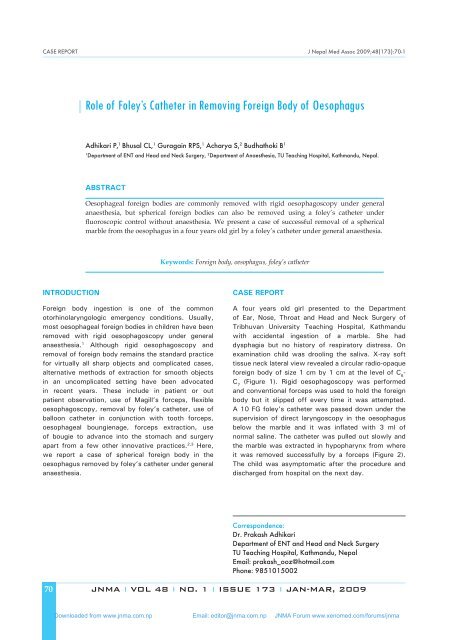
Role of Foley's Catheter in Removing Foreign Body of Oesophagus
Role of Foley's Catheter in Removing Foreign Body of Oesophagus
Role of Foley's Catheter in Removing Foreign Body of Oesophagus

Create successful ePaper yourself
Turn your PDF publications into a flip-book with our unique Google optimized e-Paper software.
CASE REPORT J Nepal Med Assoc 2009;48(173):70-1<br />
<strong>Role</strong> <strong>of</strong> Foley’s <strong>Catheter</strong> <strong>in</strong> Remov<strong>in</strong>g <strong>Foreign</strong> <strong>Body</strong> <strong>of</strong> <strong>Oesophagus</strong><br />
Adhikari P, 1 Bhusal CL, 1 Guraga<strong>in</strong> RPS, 1 Acharya S, 2 Budhathoki B 1<br />
1<br />
Department <strong>of</strong> ENT and Head and Neck Surgery, 2 Department <strong>of</strong> Anaesthesia, TU Teach<strong>in</strong>g Hospital, Kathmandu, Nepal.<br />
Abstract<br />
Oesophageal foreign bodies are commonly removed with rigid oesophagoscopy under general<br />
anaesthesia, but spherical foreign bodies can also be removed us<strong>in</strong>g a foley’s catheter under<br />
fluoroscopic control without anaesthesia. We present a case <strong>of</strong> successful removal <strong>of</strong> a spherical<br />
marble from the oesophagus <strong>in</strong> a four years old girl by a foley’s catheter under general anaesthesia.<br />
Keywords: <strong>Foreign</strong> body, oesophagus, foley’s catheter<br />
INTRODUCTION<br />
<strong>Foreign</strong> body <strong>in</strong>gestion is one <strong>of</strong> the common<br />
otorh<strong>in</strong>olaryngologic emergency conditions. Usually,<br />
most oesophageal foreign bodies <strong>in</strong> children have been<br />
removed with rigid oesophagoscopy under general<br />
anaesthesia. 1 Although rigid oesophagoscopy and<br />
removal <strong>of</strong> foreign body rema<strong>in</strong>s the standard practice<br />
for virtually all sharp objects and complicated cases,<br />
alternative methods <strong>of</strong> extraction for smooth objects<br />
<strong>in</strong> an uncomplicated sett<strong>in</strong>g have been advocated<br />
<strong>in</strong> recent years. These <strong>in</strong>clude <strong>in</strong> patient or out<br />
patient observation, use <strong>of</strong> Magill’s forceps, flexible<br />
oesophagoscopy, removal by foley’s catheter, use <strong>of</strong><br />
balloon catheter <strong>in</strong> conjunction with tooth forceps,<br />
oesophageal boungienage, forceps extraction, use<br />
<strong>of</strong> bougie to advance <strong>in</strong>to the stomach and surgery<br />
apart from a few other <strong>in</strong>novative practices. 2,3 Here,<br />
we report a case <strong>of</strong> spherical foreign body <strong>in</strong> the<br />
oesophagus removed by foley’s catheter under general<br />
anaesthesia.<br />
CASE REPORT<br />
A four years old girl presented to the Department<br />
<strong>of</strong> Ear, Nose, Throat and Head and Neck Surgery <strong>of</strong><br />
Tribhuvan University Teach<strong>in</strong>g Hospital, Kathmandu<br />
with accidental <strong>in</strong>gestion <strong>of</strong> a marble. She had<br />
dysphagia but no history <strong>of</strong> respiratory distress. On<br />
exam<strong>in</strong>ation child was drool<strong>in</strong>g the saliva. X-ray s<strong>of</strong>t<br />
tissue neck lateral view revealed a circular radio-opaque<br />
foreign body <strong>of</strong> size 1 cm by 1 cm at the level <strong>of</strong> C 6<br />
-<br />
C 7<br />
(Figure 1). Rigid oesophagoscopy was performed<br />
and conventional forceps was used to hold the foreign<br />
body but it slipped <strong>of</strong>f every time it was attempted.<br />
A 10 FG foley’s catheter was passed down under the<br />
supervision <strong>of</strong> direct laryngoscopy <strong>in</strong> the oesophagus<br />
below the marble and it was <strong>in</strong>flated with 3 ml <strong>of</strong><br />
normal sal<strong>in</strong>e. The catheter was pulled out slowly and<br />
the marble was extracted <strong>in</strong> hypopharynx from where<br />
it was removed successfully by a forceps (Figure 2).<br />
The child was asymptomatic after the procedure and<br />
discharged from hospital on the next day.<br />
Correspondence:<br />
Dr. Prakash Adhikari<br />
Department <strong>of</strong> ENT and Head and Neck Surgery<br />
TU Teach<strong>in</strong>g Hospital, Kathmandu, Nepal<br />
Email: prakash_ooz@hotmail.com<br />
Phone: 9851015002<br />
70<br />
JNMA I VOL 48 I NO. 1 I ISSUE 173 I JAN-MAR, 2009<br />
Downloaded from www.jnma.com.np Email: editor@jnma.com.np JNMA Forum www.xenomed.com/forums/jnma
Adhikari et al. <strong>Role</strong> <strong>of</strong> Foley’s <strong>Catheter</strong> <strong>in</strong> Remov<strong>in</strong>g <strong>Foreign</strong> <strong>Body</strong> <strong>of</strong> <strong>Oesophagus</strong><br />
DISCUSSION<br />
Oesophageal foreign bodies <strong>in</strong> children are one <strong>of</strong><br />
the most common emergencies that are frequently<br />
encountered <strong>in</strong> ENT practice. Co<strong>in</strong>s are the most<br />
common foreign bodies <strong>in</strong> children. 4 <strong>Foreign</strong> bodies <strong>in</strong><br />
children mostly present at the level <strong>of</strong> cricopharyngeal<br />
junction. 4<br />
Spherical foreign bodies like marbles <strong>in</strong> the oesophagus<br />
are uncommon <strong>in</strong> our hospital too. Such foreign bodies<br />
are difficult to remove by rigid oesophagoscopy and<br />
conventional forceps. They require different techniques<br />
and <strong>in</strong>struments.<br />
Figure 1. X-ray S<strong>of</strong>t tissue neck show<strong>in</strong>g radio opaque<br />
foreign body at C6-7 level<br />
Figure 2. Marble and foley’s catheter after extraction<br />
Foley’s catheter has been used to remove such spherical<br />
foreign bodies <strong>in</strong> the west. Removal <strong>of</strong> foreign body by<br />
foley’s catheter is usually conducted under fluoroscopic<br />
guidance and has been establish as a relatively safe and<br />
cost effective procedure. 5,6 But <strong>in</strong> our case, as we did not<br />
have fluoroscopic control, we did direct laryngoscopy<br />
and <strong>in</strong>sert foley’s catheter us<strong>in</strong>g stone hold<strong>in</strong>g forceps.<br />
Foley’s catheter is generally known for its usefulness<br />
<strong>in</strong> remov<strong>in</strong>g recently <strong>in</strong>gested and proximally located<br />
blunt objects. Foley’s catheter extractions <strong>in</strong>volve<br />
pass<strong>in</strong>g a balloon catheter distally to the <strong>in</strong>gested<br />
object, <strong>in</strong>flat<strong>in</strong>g the balloon and withdraw<strong>in</strong>g the<br />
catheter and the <strong>in</strong>gested object proximally. It has been<br />
shown to be effective even without the usual sedation<br />
and fluoroscopic guidance and is recommended as the<br />
treatment <strong>of</strong> choice for reta<strong>in</strong>ed co<strong>in</strong>s <strong>in</strong> children who<br />
do not show signs <strong>of</strong> significant oesophgeal oedema<br />
that would caus<strong>in</strong>g tracheal impairment. However,<br />
there are certa<strong>in</strong> contra<strong>in</strong>dications for the use <strong>of</strong><br />
foley’s catheter technique, which <strong>in</strong>clude foreign body<br />
<strong>in</strong>gestion more than 24 hours before <strong>in</strong>tervention or at<br />
an unknown earlier time, prior oesophageal stricture or<br />
surgery, signs and symptoms <strong>of</strong> marked oesophageal<br />
obstruction, stridor or compromised respiration. 7<br />
Besides foley’s catheter, angiographic catheter has also<br />
been used by different authors. 8 Foley’s catheter seems<br />
to be an alternative technique to remove spherical and<br />
proximal foreign bodies from the oesophagus.<br />
REFERENCES<br />
1. Marrow SE, Bickler SW, Kennedy AP, Syndler CL. Baloon<br />
extraction <strong>of</strong> esophageal foreign bodies <strong>in</strong> children. J Pediatr<br />
Surg 1998;33:266-70.<br />
2. Dunlap LB. Removal <strong>of</strong> an esophageal foreign body us<strong>in</strong>g a<br />
foley catheter. Am Emerg Med 1981;10:101-3.<br />
3. Calk<strong>in</strong>s CM, Christians KK, Sell LL. Cost analysis <strong>in</strong> the<br />
management <strong>of</strong> esophageal co<strong>in</strong>s: endoscopy versus<br />
bougienage. J Pediatr Surg 1999;34:412-4.<br />
4. Adhikari P, Shrestha BL, Baskota DK, S<strong>in</strong>ha BK. Accidental<br />
foreign body <strong>in</strong>gestion: analysis <strong>of</strong> 163 cases. Intl Arch<br />
Otorh<strong>in</strong>olaryngol 2007;267-70.<br />
5. Conners GP. A literature based comparison <strong>of</strong> three methods<br />
<strong>of</strong> pediatric esophageal co<strong>in</strong> removal. Pediatr Emerg Care<br />
1997;13:154-7.<br />
6. Kelley JE, Leech MH, Carr MG. A safe and cost effective<br />
protocol for the management <strong>of</strong> esophageal co<strong>in</strong>s <strong>in</strong> children.<br />
J Pediatr Surg 1993;28:898-900.<br />
7. Campbell JB, Foley LC. A safe alternative to endoscopic<br />
removal <strong>of</strong> blunt esophageal foreign bodies. Arch Otolaryngol<br />
1983;109:323-5.<br />
8. Reddy NV, Bhatt C, Vaughan-Jones RH, Reddy TN. Spherical<br />
foreign bodies <strong>in</strong> the oesophagus removed by balloon<br />
angiographic catheter. J Laryngol Otol 2002;116:208-10.<br />
JNMA I VOL 48 I NO. 1 I ISSUE 173 I JAN-MAR, 2009 71<br />
Downloaded from www.jnma.com.np Email: editor@jnma.com.np JNMA Forum www.xenomed.com/forums/jnma