Effect of pulse duration on plasmonic enhanced ultrafast laser-induced
Effect of pulse duration on plasmonic enhanced ultrafast laser-induced
Effect of pulse duration on plasmonic enhanced ultrafast laser-induced

Create successful ePaper yourself
Turn your PDF publications into a flip-book with our unique Google optimized e-Paper software.
Appl Phys A<br />
DOI 10.1007/s00339-012-7210-1<br />
<str<strong>on</strong>g>Effect</str<strong>on</strong>g> <str<strong>on</strong>g>of</str<strong>on</strong>g> <str<strong>on</strong>g>pulse</str<strong>on</strong>g> <str<strong>on</strong>g>durati<strong>on</strong></str<strong>on</strong>g> <strong>on</strong> plasm<strong>on</strong>ic <strong>enhanced</strong> <strong>ultrafast</strong><br />
<strong>laser</strong>-<strong>induced</strong> bubble generati<strong>on</strong> in water<br />
R. Lachaine · E. Boulais · E. Bourbeau · M. Meunier<br />
Received: 21 January 2011<br />
© Springer-Verlag 2012<br />
Abstract Bubbles generated in water by focusing femtosec<strong>on</strong>d<br />
and picosec<strong>on</strong>d <strong>laser</strong> <str<strong>on</strong>g>pulse</str<strong>on</strong>g>s in the presence <str<strong>on</strong>g>of</str<strong>on</strong>g><br />
100 nm gold nanoparticles have been investigated in the fluence<br />
range usually used for efficient cell transfecti<strong>on</strong> (100–<br />
200 mJ/cm 2 ). Since resulting bubbles are at the nanoscale,<br />
direct observati<strong>on</strong> using optical microscopy is not possible.<br />
An optical in-situ method has been developed to m<strong>on</strong>itor the<br />
time-resolved variati<strong>on</strong> in the extincti<strong>on</strong> cross-secti<strong>on</strong> <str<strong>on</strong>g>of</str<strong>on</strong>g> an<br />
irradiated nanoparticle soluti<strong>on</strong> sample. This method is used<br />
to measure the bubbles lifetime and deduce their average diameter.<br />
We show that bubbles generated with femtosec<strong>on</strong>d<br />
<str<strong>on</strong>g>pulse</str<strong>on</strong>g>s (40–500 fs) last two times l<strong>on</strong>ger and are larger in<br />
average than those generated with picosec<strong>on</strong>d <str<strong>on</strong>g>pulse</str<strong>on</strong>g>s (0.5–<br />
5 ps). C<strong>on</strong>trolling those bubble properties is necessary for<br />
optimizing <str<strong>on</strong>g>of</str<strong>on</strong>g>f-res<strong>on</strong>ance plasm<strong>on</strong>ic <strong>enhanced</strong> <strong>ultrafast</strong> <strong>laser</strong><br />
cell transfecti<strong>on</strong>.<br />
1 Introducti<strong>on</strong><br />
Cell transfecti<strong>on</strong> is a technique widely used by biologists<br />
to modify cells behavior [1–8]. While many methods, including<br />
viral vectors [1], electroporati<strong>on</strong> [2–4], and lip<str<strong>on</strong>g>of</str<strong>on</strong>g>ecti<strong>on</strong><br />
[5–8] are currently available, efficient transfecti<strong>on</strong><br />
still faces major challenges [9]. A promising way to achieve<br />
cell transfecti<strong>on</strong> is the <strong>ultrafast</strong> <strong>laser</strong>-<strong>induced</strong> cell perforati<strong>on</strong><br />
[10–15]. In this method, a femtosec<strong>on</strong>d <strong>laser</strong> <str<strong>on</strong>g>pulse</str<strong>on</strong>g> is<br />
R. Lachaine (�) · E. Boulais · E. Bourbeau · M. Meunier<br />
Laser Processing and Plasm<strong>on</strong>ics Laboratory, Ecole<br />
Polytechnique <str<strong>on</strong>g>of</str<strong>on</strong>g> M<strong>on</strong>treal, M<strong>on</strong>treal, Quebec H3C 3A7, Canada<br />
e-mail: remi.lachaine@polymtl.ca<br />
M. Meunier<br />
e-mail: michel.meunier@polymtl.ca<br />
tightly focused <strong>on</strong>to the cell membrane to induce membrane<br />
permeabilizati<strong>on</strong>. However, tight focusing limits the<br />
throughput and makes the method inappropriate for most biological<br />
and medical applicati<strong>on</strong> where large populati<strong>on</strong> <str<strong>on</strong>g>of</str<strong>on</strong>g><br />
cells must be transfected. To recover a high throughput, it<br />
is possible to amplify locally the <strong>laser</strong> electromagnetic field<br />
using plasm<strong>on</strong>ic nanostructures deposited <strong>on</strong> the cells, such<br />
as gold spherical nanoparticles (NPs) [16–19]. Nanoparticles,<br />
through the plasm<strong>on</strong>ic effect, amplify and absorb<br />
the <strong>laser</strong> near-field and generate cavitati<strong>on</strong> bubbles that are<br />
thought to permeabilize the cell membrane, enabling cell<br />
perforati<strong>on</strong> and transfecti<strong>on</strong> with high efficiency [20, 21].<br />
Measurement <str<strong>on</strong>g>of</str<strong>on</strong>g> the bubble average dimensi<strong>on</strong> and lifetime<br />
as a functi<strong>on</strong> <str<strong>on</strong>g>of</str<strong>on</strong>g> <strong>laser</strong> parameters is important to get<br />
a better understanding <str<strong>on</strong>g>of</str<strong>on</strong>g> the basic mechanisms underlying<br />
nanocavitati<strong>on</strong> and to optimize the plasm<strong>on</strong>ic <strong>enhanced</strong><br />
<strong>laser</strong> transfecti<strong>on</strong> process. Major difficulty comes from the<br />
reduced dimensi<strong>on</strong>s <str<strong>on</strong>g>of</str<strong>on</strong>g> the bubbles that are generated, preventing<br />
the use <str<strong>on</strong>g>of</str<strong>on</strong>g> optical microscopy to properly measure<br />
their diameter. In this paper, we present an in-situ optical<br />
method to measure the variati<strong>on</strong> <str<strong>on</strong>g>of</str<strong>on</strong>g> the extincti<strong>on</strong> crosssecti<strong>on</strong><br />
<str<strong>on</strong>g>of</str<strong>on</strong>g> an irradiated colloidal nanosphere suspensi<strong>on</strong><br />
sample and then determine both the lifetime and the average<br />
dimensi<strong>on</strong> <str<strong>on</strong>g>of</str<strong>on</strong>g> the bubbles. This method is then used to<br />
study the effect <str<strong>on</strong>g>of</str<strong>on</strong>g> <strong>laser</strong> <str<strong>on</strong>g>pulse</str<strong>on</strong>g> <str<strong>on</strong>g>durati<strong>on</strong></str<strong>on</strong>g> <strong>on</strong> the bubble characteristics.<br />
2 In-situ nanobubble optical detecti<strong>on</strong> setup<br />
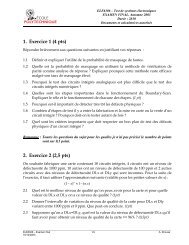
Figure 1 presents the in-situ optical setup used to characterize<br />
the generated bubbles. A first <strong>laser</strong>, called pump<br />
(Ti:Sapphire, 6 mJ/<str<strong>on</strong>g>pulse</str<strong>on</strong>g>, τ<str<strong>on</strong>g>pulse</str<strong>on</strong>g> =[40 fs–5 ps], 800 nm), is<br />
focused <strong>on</strong> the sample (gold NPs, 100 nm, 1.4×10 9 part/ml)<br />
using a lens (focal = 10 cm) and generates the bubbles.
Fig. 1 In-situ nanobubble detecti<strong>on</strong> setup. Fs pump <strong>laser</strong> <str<strong>on</strong>g>pulse</str<strong>on</strong>g> <str<strong>on</strong>g>durati<strong>on</strong></str<strong>on</strong>g><br />
can be changed from 40 fs up to 5 ps. The sample is composed<br />
<str<strong>on</strong>g>of</str<strong>on</strong>g> 100 nm gold NPs in water soluti<strong>on</strong>. Bubbles are detected using the<br />
variati<strong>on</strong> <str<strong>on</strong>g>of</str<strong>on</strong>g> the extincti<strong>on</strong> cross-secti<strong>on</strong><br />
A sec<strong>on</strong>d <strong>laser</strong>, called probe (He:Ne, CW, 2 mW), is<br />
collinear with the pump <strong>laser</strong> and focused in the sample.<br />
A dichroic mirror separates the two <strong>laser</strong> beams after the<br />
sample. The probe beam is then spatially filtered using a<br />
10 μm pinhole. This spatial filter blocks light that is scattered<br />
by cavitati<strong>on</strong> bubbles. When this occurs, a drop in the<br />
intensity <str<strong>on</strong>g>of</str<strong>on</strong>g> the probe signal is detected by the photodiode<br />
(Si 1-GHz photoreceiver, trise = 0.6 ns) located behind the<br />
pinhole. Note that this intensity loss is related to the total extincti<strong>on</strong>,<br />
since it comes from both absorpti<strong>on</strong> and scattering,<br />
which is blocked by the spatial filter.<br />
3 Results and discussi<strong>on</strong><br />
Up<strong>on</strong> irradiati<strong>on</strong>, plasm<strong>on</strong>ic nanostructures enhance the<br />
near-field and create a plasma in the medium. This plasma<br />
eventually relaxes, leading to a local energy depositi<strong>on</strong><br />
into the water surrounding the NPs [21]. This generates a<br />
fast, quasi-isochoric, temperature rise al<strong>on</strong>g with a str<strong>on</strong>g<br />
stress c<strong>on</strong>finement. A pressure wave is then released in the<br />
medium and, if the energy is sufficient, a cavitati<strong>on</strong> bubble<br />
is created.<br />
Using the experimental setup described earlier, it is possible<br />
to detect those bubbles. A typical signal obtained at<br />
200 mJ/cm 2 and <str<strong>on</strong>g>pulse</str<strong>on</strong>g> <str<strong>on</strong>g>durati<strong>on</strong></str<strong>on</strong>g> 1 ps is shown in Fig. 2.<br />
If the fluence is increased at values over the transfecti<strong>on</strong><br />
range (which is between 100–200 mJ/cm 2 ), it is possible<br />
to observe the characteristic oscillatory behavior coming<br />
from successive growths and collapses <str<strong>on</strong>g>of</str<strong>on</strong>g> cavitati<strong>on</strong> bubbles<br />
[22],asshownintheinset<str<strong>on</strong>g>of</str<strong>on</strong>g>Fig.2 (the fluence used<br />
was 450 mJ/cm 2 ). This observati<strong>on</strong> str<strong>on</strong>gly suggests that<br />
R. Lachaine et al.<br />
Fig. 2 Typical signal acquired with the experimental setup. Fluence<br />
used was 200 mJ/cm 2 and <str<strong>on</strong>g>pulse</str<strong>on</strong>g> <str<strong>on</strong>g>durati<strong>on</strong></str<strong>on</strong>g> was 1 ps. The inset shows the<br />
oscillatory behavior observed at higher fluences (450 mJ/cm 2 )<br />
the measured signal comes from cavitati<strong>on</strong> bubbles and not<br />
from any other phenomen<strong>on</strong>. At fluences usable for the<br />
transfecti<strong>on</strong> experiment, this oscillatory behavior is however<br />
not observed, either because the bubbles are too small to initiate<br />
oscillati<strong>on</strong> or because the sec<strong>on</strong>dary bubble is bey<strong>on</strong>d<br />
the resoluti<strong>on</strong> <str<strong>on</strong>g>of</str<strong>on</strong>g> the optical method.<br />
3.1 Bubble lifetime<br />
The bubble lifetime measurement is quite direct. As shown<br />
in Fig. 2, the bubble lifetime is defined as the time between<br />
the beginning <str<strong>on</strong>g>of</str<strong>on</strong>g> the intensity drop and the moment where<br />
the intensity comes back to zero for the first time. Note that<br />
the asymmetry between rise and collapse times is due to both<br />
the presence <str<strong>on</strong>g>of</str<strong>on</strong>g> bubbles <str<strong>on</strong>g>of</str<strong>on</strong>g> different lifetimes within the focal<br />
volume and the <str<strong>on</strong>g>of</str<strong>on</strong>g>f-res<strong>on</strong>ance irradiati<strong>on</strong> cavitati<strong>on</strong> mechanism<br />
itself. Details are explained elsewhere [21].<br />
Bubble lifetime was measured as a functi<strong>on</strong> <str<strong>on</strong>g>of</str<strong>on</strong>g> average<br />
fluences for different <str<strong>on</strong>g>pulse</str<strong>on</strong>g> <str<strong>on</strong>g>durati<strong>on</strong></str<strong>on</strong>g>s (Fig 3a). For fluences<br />
below 300 mJ/cm 2 , which include the transfecti<strong>on</strong> fluence<br />
range (100–200 mJ/cm 2 ), results show that bubble lifetimes<br />
are l<strong>on</strong>ger for femtosec<strong>on</strong>d (fs) <str<strong>on</strong>g>pulse</str<strong>on</strong>g>s (below 500 fs) than<br />
for picosec<strong>on</strong>d (ps) <str<strong>on</strong>g>pulse</str<strong>on</strong>g>s (above 500 fs). For example, at<br />
200 mJ/cm 2 , bubble lifetime is 600 ns when irradiating with<br />
a40fs<str<strong>on</strong>g>pulse</str<strong>on</strong>g>comparedto350nswitha1ps<str<strong>on</strong>g>pulse</str<strong>on</strong>g>.<br />
For all <str<strong>on</strong>g>pulse</str<strong>on</strong>g> <str<strong>on</strong>g>durati<strong>on</strong></str<strong>on</strong>g>s, results show that bubble lifetimes<br />
range from a few hundreds <str<strong>on</strong>g>of</str<strong>on</strong>g> nanosec<strong>on</strong>ds to about 1 μs.<br />
However, permeabilizati<strong>on</strong> <str<strong>on</strong>g>of</str<strong>on</strong>g> the membrane remains for a<br />
few microsec<strong>on</strong>ds, as shown by patch-clamp experiments<br />
[12], allowing the foreign DNA to enter the cell. In that<br />
regard, bubble lifetime is probably less important than the<br />
bubble dimensi<strong>on</strong> in the perforati<strong>on</strong> process.
<str<strong>on</strong>g>Effect</str<strong>on</strong>g> <str<strong>on</strong>g>of</str<strong>on</strong>g> <str<strong>on</strong>g>pulse</str<strong>on</strong>g> <str<strong>on</strong>g>durati<strong>on</strong></str<strong>on</strong>g> <strong>on</strong> plasm<strong>on</strong>ic <strong>enhanced</strong> <strong>ultrafast</strong> <strong>laser</strong>-<strong>induced</strong> bubble generati<strong>on</strong> in water<br />
Fig. 3 (a) Bubble lifetime and<br />
(b) average diameter for<br />
different average fluences at<br />
different <str<strong>on</strong>g>pulse</str<strong>on</strong>g> <str<strong>on</strong>g>durati<strong>on</strong></str<strong>on</strong>g><br />
Fig. 4 Cth ext comparis<strong>on</strong> with and without a NP in the center <str<strong>on</strong>g>of</str<strong>on</strong>g> the<br />
bubble<br />
3.2 Bubble dimensi<strong>on</strong><br />
It is possible to deduce an approximate average bubble diameter<br />
from the signal amplitude A defined as the maximal<br />
loss in transmissi<strong>on</strong> (see Fig. 2). First, a Mie code was used<br />
to calculate the theoretical extincti<strong>on</strong> coefficient Cth ext <str<strong>on</strong>g>of</str<strong>on</strong>g> a<br />
vapor bubble <str<strong>on</strong>g>of</str<strong>on</strong>g> different diameters at the probe wavelength<br />
(633 nm). Of course, for small bubbles, Cth ext is affected by<br />
the presence <str<strong>on</strong>g>of</str<strong>on</strong>g> the NP in the center <str<strong>on</strong>g>of</str<strong>on</strong>g> the bubble. A Mie<br />
coated code [23] has thus been used to calculate the extincti<strong>on</strong><br />
coefficient <str<strong>on</strong>g>of</str<strong>on</strong>g> a NP in water coated with a vapor layer <str<strong>on</strong>g>of</str<strong>on</strong>g><br />
different thickness in order to evaluate the importance <str<strong>on</strong>g>of</str<strong>on</strong>g> the<br />
NP. Figure 4 shows that the presence <str<strong>on</strong>g>of</str<strong>on</strong>g> the NP is not negligible<br />
for bubble diameters under 140 nm (20 nm vapor layer<br />
thickness). As it will be shown later, the smallest bubbles<br />
that have been detected have a diameter <str<strong>on</strong>g>of</str<strong>on</strong>g> about 240 nm.<br />
Therefore, the presence <str<strong>on</strong>g>of</str<strong>on</strong>g> the NP at the center <str<strong>on</strong>g>of</str<strong>on</strong>g> the bubble<br />
has been neglected for simplicity reas<strong>on</strong>.<br />
Two main approximati<strong>on</strong>s were made to evaluate the bubbles<br />
diameter: (i) there is <strong>on</strong>e and <strong>on</strong>ly <strong>on</strong>e bubble created<br />
around each NP and those bubbles remain disjoint; (ii) all<br />
bubbles have the same diameter within the focal volume,<br />
thus an average bubble diameter could be found. Knowing<br />
the normalized signal amplitude A, the NPs c<strong>on</strong>centrati<strong>on</strong> C<br />
(1.4 × 10⁹ part/ml) as well as the focal depth L (755 μm),<br />
Beer–Lambert law was applied to deduce the experimental<br />
extincti<strong>on</strong> coefficient C exp<br />
ext . The ratio between the pump<br />
and probe <strong>laser</strong> focal spots was also c<strong>on</strong>sidered because the<br />
pump spot was smaller than the probe spot.<br />
C exp<br />
ext =<br />
ln (1 − A) r<br />
CL<br />
2 probe<br />
r2 pump<br />
The average bubble diameter is then deduced by finding<br />
the bubble diameter matching Cth ext and Cexp ext .Usingthis<br />
method, we were able to measure average bubble diameter<br />
down to 240 nm.<br />
Fluences useful for cell transfecti<strong>on</strong> usually range from<br />
100 to 200 mJ/cm2 [20]. Results presented in Fig. 3b show<br />
that fs <str<strong>on</strong>g>pulse</str<strong>on</strong>g>s produce larger average bubbles than ps <str<strong>on</strong>g>pulse</str<strong>on</strong>g>s<br />
in that range . For example, at 200 mJ/cm2 , fs <str<strong>on</strong>g>pulse</str<strong>on</strong>g>s (40 fs)<br />
create 750 nm average diameter bubbles while ps <str<strong>on</strong>g>pulse</str<strong>on</strong>g>s<br />
(1 ps) generate a smaller 380 nm average diameter bubble.<br />
This is probably because l<strong>on</strong>ger <str<strong>on</strong>g>pulse</str<strong>on</strong>g>s are less efficient<br />
to photoi<strong>on</strong>ize the water around the particle. Indeed, simulati<strong>on</strong>s<br />
were performed to calculate the amount <str<strong>on</strong>g>of</str<strong>on</strong>g> energy<br />
deposited into the water for a 40 fs <str<strong>on</strong>g>pulse</str<strong>on</strong>g> in comparis<strong>on</strong><br />
with a 1 ps <str<strong>on</strong>g>pulse</str<strong>on</strong>g> when irradiating a 100 nm gold NP and<br />
results show that the deposited energy is approximately 3<br />
times larger for fs <str<strong>on</strong>g>pulse</str<strong>on</strong>g>s than for ps <str<strong>on</strong>g>pulse</str<strong>on</strong>g>s. Those results<br />
are shown in Fig. 5. Classical rate equati<strong>on</strong>s in 0D, including<br />
electr<strong>on</strong> generati<strong>on</strong> with Keldysh theory [24, 25], collisi<strong>on</strong><br />
term [26, 27], and recombinati<strong>on</strong> term [24], have been<br />
solved to determine the plasma density and the temperature.<br />
Further details are given elsewhere [21].<br />
Fs <str<strong>on</strong>g>pulse</str<strong>on</strong>g>s seem thus more effective to generate larger average<br />
bubbles than ps <str<strong>on</strong>g>pulse</str<strong>on</strong>g>s below 200 mJ/cm2 . However,<br />
the fact that the average bubbles are measured to be larger<br />
does not necessarily imply that the maximum bubbles are<br />
also larger. Indeed, NPs located near the center <str<strong>on</strong>g>of</str<strong>on</strong>g> the focal<br />
spot are irradiated with a fluence higher than the average<br />
(1)
Fig. 5 Simulati<strong>on</strong> results for the deposited energy into water when<br />
irradiating a 100 nm gold NP with a 800 nm, 40 fs, and 1 ps <strong>laser</strong><br />
<str<strong>on</strong>g>pulse</str<strong>on</strong>g>. The deposited energy is about 3 times larger for fs <str<strong>on</strong>g>pulse</str<strong>on</strong>g>s than<br />
for ps <str<strong>on</strong>g>pulse</str<strong>on</strong>g>s<br />
fluence, thus creating maximum bubbles larger than the average<br />
bubbles. Further investigati<strong>on</strong>s would be required to<br />
make a statement c<strong>on</strong>cerning the maximum bubble dimensi<strong>on</strong>s.<br />
4 C<strong>on</strong>clusi<strong>on</strong><br />
In c<strong>on</strong>clusi<strong>on</strong>, the effect <str<strong>on</strong>g>of</str<strong>on</strong>g> <str<strong>on</strong>g>pulse</str<strong>on</strong>g> <str<strong>on</strong>g>durati<strong>on</strong></str<strong>on</strong>g> <strong>on</strong> <strong>laser</strong> <strong>induced</strong><br />
bubble generati<strong>on</strong> in the presence <str<strong>on</strong>g>of</str<strong>on</strong>g> gold nanoparticles was<br />
investigated. Using the bubbles scattering signal and a relatively<br />
simple model, we were able to measure the bubble<br />
lifetime and average diameter. Those characteristics were<br />
measured for different pump <str<strong>on</strong>g>pulse</str<strong>on</strong>g> <str<strong>on</strong>g>durati<strong>on</strong></str<strong>on</strong>g>s, from 40 fs<br />
up to 5 ps. It has been shown that fs <str<strong>on</strong>g>pulse</str<strong>on</strong>g>s produce larger<br />
and l<strong>on</strong>ger average bubbles than ps <str<strong>on</strong>g>pulse</str<strong>on</strong>g>s for <strong>laser</strong> fluences<br />
used for cell transfecti<strong>on</strong>. Works are underway to relate the<br />
bubble characteristics and dynamics to the efficiency <str<strong>on</strong>g>of</str<strong>on</strong>g> <str<strong>on</strong>g>of</str<strong>on</strong>g>fres<strong>on</strong>ance<br />
plasm<strong>on</strong>ic <strong>enhanced</strong> <strong>ultrafast</strong> <strong>laser</strong> cell transfecti<strong>on</strong>.<br />
Acknowledgements The authors would like to thank the Natural<br />
Science and Engineering Research Council (NSERC) and Le F<strong>on</strong>ds<br />
Quebecois de la Recherche sur la Nature et les Technologies (FQRNT)<br />
for financial support. The technical assistance by Yves Drolet is also<br />
acknowledged.<br />
References<br />
R. Lachaine et al.<br />
1. W. Walther, U. Stein, Drugs 60, 2 (2000)<br />
2. T.K. W<strong>on</strong>g, E. Neumann, Biochem. Biophys. Res. Commun. 107,<br />
2 (1982)<br />
3. K. Shigekawa, W.J. Dower, BioTechniques 6, 8 (1988)<br />
4. J. Weaver, Y. Chizmadzhev, Bioelectrochem. Bioenerg. 41, 135<br />
(1996)<br />
5. F.L.Graham,J.Smiley,W.C.Russell,R.Nairn,J.Gen.Virol.36,<br />
1 (1977)<br />
6. T. Wils<strong>on</strong>, D. Papahadjopoulos, R. Taber, Cell 17, 1 (1979)<br />
7. R. Fraley, S. Subramani, P. Berg, D. Papahadjopoulos, J. Biol.<br />
Chem. 255, 21 (1980)<br />
8. P.L. Felgner, Y.J. Tsai, L. Sukhu, C.J. Wheeler, M. Manthorpe,<br />
J. Marshall, S.H. Cheng, Ann. N.Y. Acad. Sci. 772, 126 (1995)<br />
9. J.L.Stilwell,D.M.McCarty,A.Negishi,R.Superfine,R.J.Samulski,<br />
J. Virol. 77, 23 (2003)<br />
10. U.K. Tirlapur, K. K<strong>on</strong>ig, Nature 418, 6895 (2002)<br />
11. D. Stevens<strong>on</strong>, B. Agate, X. Tsampoula, P. Fischer, C.T.A. Brown,<br />
W. Sibbett, A. Riches, F. Gunn-Moore, K. Dholakia, Opt. Express<br />
14, 16 (2006)<br />
12. J. Baumgart, W. Bintig, A. Ngezahayo, S. Willenbrock, H.M. Escobar,<br />
W. Ertmer, H. Lubatschowski, A. Heisterkamp, Opt. Express<br />
16, 5 (2008)<br />
13. V. Kohli, J.P. Acker, A.Y. Elezzabi, Biotechnol. Bioeng. 92, 7<br />
(2005)<br />
14. E. Zeira, A. Manevitch, Z. Manevitch, E. Kedar, M. Gropp,<br />
N. Daudi, R. Barsuk, M. Harati, H. Yotvat, P.J. Troilo, T.G. Griffiths,<br />
S.J. Pacchi<strong>on</strong>e, D.F. Roden, Z. Niu, O. Nussbaum, G. Zamir,<br />
O. Papo, I. Hemo, A. Lewis, E. Galun, FASEB J. 21, 13 (2007)<br />
15. A. Vogel, N. Linz, S. Freidank, G. Paltauf, Phys. Rev. Lett. 100,<br />
038102 (2008)<br />
16. A. Uchug<strong>on</strong>ova, K. K<strong>on</strong>ig, R. Bueckle, A. Isemann, G. Tempea,<br />
Opt. Express 16, 13 (2008)<br />
17. C. Yao, R. Rahmanzadeh, E. Endl, Z. Zhang, J. Gerdes,<br />
G. Huttmann, J. Biomed. Opt. 10, 6 (2005)<br />
18. D. Lapotko, Nanomedicine 4, 7 (2009)<br />
19. V.K. Pustalkov, A.S. Smetannikov, V.P. Zharov, Laser Phys. Lett.<br />
11, 775 (2008)<br />
20. J. Baumgart, L. Humbert, E. Boulais, R. Lachaine, J.J. Lebrun,<br />
M. Meunier, Biomaterials 33, 7 (2012)<br />
21. E. Boulais, R. Lachaine, M. Meunier, Nano Lett. (2012).<br />
doi:10.1021/nl302200w<br />
22. E.M. Glinsky, S.D. Bailey, A.R. L<strong>on</strong>d<strong>on</strong>, A.P. Amendt,<br />
M.A. Rubenchik, M. Strauss, Phys. Fluids 13, 20 (2001)<br />
23. E.C. Le Ru, P.G. Etchegoin, Principles <str<strong>on</strong>g>of</str<strong>on</strong>g> Surface-Enhanced Raman<br />
Spectroscopy (Elsevier, Amsterdam, 2009)<br />
24. A. Vogel, G. Noack, G. Huttmann, G. Paltauf, Appl. Phys. B,<br />
Lasers Opt. 81, 8 (2005)<br />
25. L. Keldysh, Zh. Èksp. Teor. Fiz. 47, 5 (1965)<br />
26. L. Hallo, A. Bourgeade, V. Tikh<strong>on</strong>chuk, C. Mezel, J. Breil, Phys.<br />
Rev. B, C<strong>on</strong>dens. Matter Mater. Phys. 76, 2 (2007)<br />
27. N. Andreev, M. Veisman, V. Efremov, V. Fortov, High Temp. 41,<br />
5 (2003)