ASSESSMENT OF A VENOUS LEG ULCER - Wounds UK
ASSESSMENT OF A VENOUS LEG ULCER - Wounds UK
ASSESSMENT OF A VENOUS LEG ULCER - Wounds UK

Create successful ePaper yourself
Turn your PDF publications into a flip-book with our unique Google optimized e-Paper software.
Review<br />
<strong>ASSESSMENT</strong> <strong>OF</strong><br />
A <strong>VENOUS</strong> <strong>LEG</strong> <strong>ULCER</strong><br />
A thorough and accurate assessment of patients who present with leg ulceration is essential to<br />
ensure timely and appropriate treatment. Assessment should, however, be ongoing as signs and<br />
symptoms can change and nurses need to be able to monitor the impact of their interventions.<br />
Heather Newton is a<br />
Consultant Nurse Tissue<br />
Viability, Royal Cornwall<br />
Hospitals NHS Trust,<br />
Truro, Cornwall<br />
Seventy per cent of all leg ulcers<br />
that develop in Western countries<br />
are caused by venous disease,<br />
yet it has been highlighted that the<br />
knowledge and skills of nurses<br />
often falls short of what is expected<br />
and that there are wide variations<br />
in assessment practices (Browse<br />
and Burnand, 1982; Moffatt and<br />
Franks, 2004).<br />
With the ever-increasing age of the<br />
<strong>UK</strong> population, venous leg ulcer<br />
prevalence could well increase.<br />
Accurate patient assessment is<br />
the key to successful leg ulcer<br />
management and this article will<br />
provide nurses with evidencebased<br />
information to enable the<br />
accurate and thorough assessment<br />
of patients with venous ulcers. It<br />
will explain the reasons why venous<br />
ulcers occur and the specific<br />
factors which influence their<br />
development. Patient assessment<br />
and identification of skin changes<br />
will also be discussed, using<br />
photographs to support learning<br />
and understanding.<br />
Background<br />
Venous leg ulcer management<br />
accounts for a significant<br />
proportion of district and practice<br />
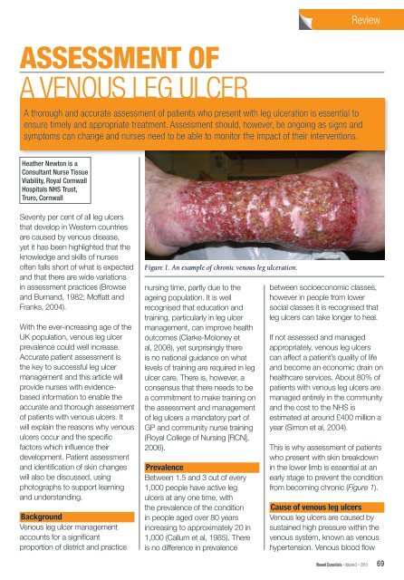
Figure 1. An example of chronic venous leg ulceration.<br />
nursing time, partly due to the<br />
ageing population. It is well<br />
recognised that education and<br />
training, particularly in leg ulcer<br />
management, can improve health<br />
outcomes (Clarke-Moloney et<br />
al, 2008), yet surprisingly there<br />
is no national guidance on what<br />
levels of training are required in leg<br />
ulcer care. There is, however, a<br />
consensus that there needs to be<br />
a commitment to make training on<br />
the assessment and management<br />
of leg ulcers a mandatory part of<br />
GP and community nurse training<br />
(Royal College of Nursing [RCN],<br />
2006).<br />
Prevalence<br />
Between 1.5 and 3 out of every<br />
1,000 people have active leg<br />
ulcers at any one time, with<br />
the prevalence of the condition<br />
in people aged over 80 years<br />
increasing to approximately 20 in<br />
1,000 (Callum et al, 1985). There<br />
is no difference in prevalence<br />
between socioeconomic classes,<br />
however in people from lower<br />
social classes it is recognised that<br />
leg ulcers can take longer to heal.<br />
If not assessed and managed<br />
appropriately, venous leg ulcers<br />
can affect a patient’s quality of life<br />
and become an economic drain on<br />
healthcare services. About 80% of<br />
patients with venous leg ulcers are<br />
managed entirely in the community<br />
and the cost to the NHS is<br />
estimated at around £400 million a<br />
year (Simon et al, 2004).<br />
This is why assessment of patients<br />
who present with skin breakdown<br />
in the lower limb is essential at an<br />
early stage to prevent the condition<br />
from becoming chronic (Figure 1).<br />
Cause of venous leg ulcers<br />
Venous leg ulcers are caused by<br />
sustained high pressure within the<br />
venous system, known as venous<br />
hypertension. Venous blood flow<br />
Wound Essentials • Volume 5 • 2010 69
Review<br />
Figure 2. Pigmentation of the lower leg<br />
with ankle flare.<br />
takes blood back to the heart<br />
through the deep veins of the<br />
leg — namely the femoral vein,<br />
popliteal vein and the anterior and<br />
posterior tibial veins. Superficial<br />
veins, the long saphenous and<br />
short saphenous veins, are found<br />
nearer the surface of the leg and<br />
are connected to the deep veins<br />
by perforator veins, which allow<br />
the blood to flow one way into the<br />
deep veins.<br />
All of the veins in the leg have<br />
valves to make sure that the blood<br />
flows back to the heart, however if<br />
the valves are damaged by surgery,<br />
injury or thrombosis they allow<br />
the blood to flow back into the<br />
deep veins causing an increase in<br />
pressure. As this pressure rises, the<br />
superficial veins become enlarged<br />
and chronic venous hypertension<br />
develops, which is the main cause<br />
of venous leg ulceration. It is<br />
important for nurses to understand<br />
the cause of venous hypertension<br />
and the features that present in<br />
the lower leg in order to accurately<br />
assess and plan appropriate care.<br />
Patient assessment<br />
The RCN guidelines (2006) suggest<br />
that a full clinical history and<br />
physical examination is conducted<br />
for any patient that presents with<br />
either their first or a recurrent<br />
leg ulcer. This is to identify the<br />
underlying cause of the ulceration<br />
and assist in planning the correct<br />
treatment pathway. The RCN<br />
recommendation is based on a<br />
consensus opinion as there is no<br />
research that compares patients’<br />
clinical history and examination with<br />
healing outcomes.<br />
The following information forms<br />
an essential part of the overall<br />
assessment to identify if the leg<br />
ulcer is caused by venous disease.<br />
Many trusts have developed<br />
specific leg ulcer assessment forms<br />
to capture this information and to<br />
guide the nurse as to the relevant<br />
questions to ask patients.<br />
Assessment of venous risk factors<br />
should include:<br />
8 Family history of<br />
venous disease<br />
8 History of varicose veins and<br />
whether they have been treated<br />
or not<br />
8 Proven diagnosis of deep vein<br />
thrombosis (DVT) identifying<br />
which part of the leg was<br />
affected. This is a common<br />
cause of venous ulcers<br />
8 History of inflammation of the<br />
vein (known as phlebitis) and<br />
whether or not it was a single<br />
or recurrent episode<br />
8 History of surgery, fractures or<br />
trauma to the lower leg, which<br />
may have damaged the valves<br />
in the veins<br />
8 Occupation (some jobs<br />
involving prolonged standing or<br />
sitting can increase risk)<br />
8 Obesity and multiple<br />
Figure 3. Gravitational eczema of the lower leg.<br />
70 Wound Essentials • Volume 5 • 2010
Review<br />
pregnancies —this causes<br />
pressure on the venous<br />
system.<br />
Skin assessment<br />
When pressure rises in the veins<br />
of the lower leg over a prolonged<br />
period of time the following skin<br />
changes occur:<br />
8 Pigmentation in the gaiter area<br />
of the leg between the ankle<br />
and the calf (Figure 2)<br />
8 Thickening of the<br />
subcutaneous tissue<br />
8 Oedema<br />
8 Gravitational eczema/stasis<br />
dermatitis (Figure 3)<br />
8 Ankle flare (Figure 2)<br />
8 Atrophie blanche (particular<br />
type of scar arising on the lower<br />
leg).<br />
A collective term for these changes<br />
is lipodermatosclerosis.<br />
The patient may also experience<br />
aching or heaviness in the legs,<br />
tortuous varicose veins in the thigh<br />
or lower leg (Figure 4), itching over<br />
engorged veins and occasional<br />
tenderness and redness in the<br />
lower leg. These features are<br />
described in more detail in (Table 1).<br />
Assessment of leg ulcers<br />
It should be noted that the<br />
following signs and symptoms<br />
are present in some form or other<br />
in the majority of patients who<br />
present with venous disease.<br />
However in others, indications are<br />
not classical and require more indepth<br />
assessment.<br />
Where is the leg ulcer?<br />
Venous leg ulcers are usually<br />
sited in the gaiter area of the<br />
lower leg between the calf<br />
and the ankle. They tend to be<br />
shallow with flat wound margins.<br />
They can, however, occur around<br />
Table 1.<br />
Signs of venous hypertension<br />
Skin changes<br />
Brown/pink discolouration of<br />
the skin between the ankle<br />
and the calf<br />
Thickening of the tissues<br />
beneath the skin. The leg<br />
shape can change and is often<br />
referred to as an inverted<br />
champagne bottle shape<br />
Dry, red itchy eczematous skin<br />
on the lower leg<br />
Dilation of the small blood<br />
vessels around the inner ankle,<br />
known as ankle flare<br />
White, smooth areas of thin<br />
skin which are pale in colour<br />
with visible capillaries, known<br />
as atrophie blanche<br />
Prominent dilated veins<br />
the ankle where they tend to be<br />
multiple, small and painful<br />
(Figure 5).<br />
How did the ulcer start?<br />
It is important that the nurse asks<br />
the patient whether the ulcer just<br />
appeared or if there is a history of<br />
trauma or infection in the leg. It is<br />
also important to ascertain how<br />
long the ulcer has been present.<br />
Many patients describe minor injury<br />
and subsequent failure to heal as<br />
the cause, while others will say that<br />
the ulcer developed in isolation.<br />
A past medical history may also<br />
aid diagnosis. Venous ulcers are<br />
generally slower in growth than<br />
arterial ulcers.<br />
Is there a previous history of<br />
leg ulceration?<br />
If the patient has had ulcers in the<br />
Causative factors<br />
Prolonged high pressure in the veins forces the red blood cells to leak<br />
out into the tissues. Haemoglobin in the red blood cells breaks down<br />
into haemosiderin, which is then permanently deposited into the tissues<br />
If high pressure in the veins persists, fibrous tissue is deposited in the<br />
deep dermis and the fatty layers of the skin. This causes a thickening of<br />
the tissues in the gaiter area<br />
Raised venous pressure causes an increased capillary permeability and<br />
leakage of waste products occurs causing irritation in the tissues of the<br />
lower leg<br />
When the deep and superficial veins become full and under pressure.<br />
the venules (small blood vessels) at the end of the venous system<br />
become enlarged. This is most obvious around the inner ankle area<br />
The causes are not clear, however, it is felt that blockages occur in the<br />
blood vessels in the middle and deep dermal layer of the skin<br />
Defective or damaged valves cause a backflow of blood and a build up<br />
of pressure within the veins<br />
past, this may indicate progressive<br />
venous disease, which may require<br />
surgical intervention to reverse<br />
venous incompetence. It can also<br />
indicate that a patient may not be<br />
wearing their compression hosiery,<br />
if already prescribed, or that the<br />
hosiery is the wrong size.<br />
How large is the ulcer?<br />
It is important that the size is<br />
documented at the first and<br />
subsequent assessments in<br />
order to evaluate the impact of<br />
any treatment plan. A venous leg<br />
ulcer treated with compression<br />
bandages should show good<br />
signs of healing within 12 weeks,<br />
however if the ulcer is enlarging<br />
or remaining static despite the<br />
application of correct evidencebased<br />
treatment, further<br />
investigation such as wound<br />
biopsy needs to be considered.<br />
72 Wound Essentials • Volume 5 • 2010
Review<br />
Figure 5. Venous ulcer sited on the ankle.<br />
What are the characteristics of<br />
the wound bed?<br />
In any assessment of venous<br />
leg ulcers it is important that the<br />
nurse establishes the state of<br />
the wound bed. The following<br />
elements are crucial:<br />
8 How much of the ulcer bed<br />
is viable tissue; how much is<br />
non-viable?<br />
8 Is there any inflammation or<br />
signs of infection?<br />
8 How much exudate<br />
is present?<br />
8 What is the condition of the<br />
wound edges? Are they flat<br />
or rolled?<br />
Venous ulcers can present with<br />
a mixture of granulation tissue<br />
and slough. If very thick slough or<br />
black eschar is present, it usually<br />
indicates arterial insufficiency.<br />
Wound infection is indicated<br />
by changes in the ulcer, the<br />
surrounding skin, the level of<br />
exudate and the amount of pain.<br />
As Collier (2004) states, it<br />
is important to have a clear<br />
understanding of the terms used<br />
for wound infection. The most<br />
commonly used terms include:<br />
8 Wound contamination: where<br />
bacteria are present without<br />
any host reaction<br />
8 Wound colonisation: where<br />
bacteria begin to multiply and<br />
can cause a host reaction<br />
8 Critical colonisation: where<br />
bacteria multiply and cause<br />
early signs of a problem, such<br />
as a delay in healing and<br />
increased pain but no overt<br />
host reaction<br />
8 Infection: where the<br />
multiplication of bacteria<br />
causes a host reaction, which<br />
is present in the ulcer and<br />
the skin.<br />
Some patients with a wound<br />
infection can also develop<br />
systemic symptoms such<br />
as pyrexia (fever), rigors<br />
(exaggerated shivering) and<br />
tachycardia (rapid heartbeat).<br />
Local signs of infection include<br />
redness of the skin around the<br />
ulcer, localised heat and pain,<br />
increase in oedema, spreading<br />
redness of the soft tissues of the<br />
leg (cellulitis) and an increase in<br />
exudate and odour (Figure 6).<br />
Venous ulcers can produce<br />
exudate, which is increased in the<br />
presence of infection. Exudate<br />
can decrease if appropriate<br />
Figure 4. Varicose veins in the groin area.<br />
74 Wound Essentials • Volume 5 • 2010
Review<br />
compression therapy is<br />
introduced.<br />
Generally, the wound edges are<br />
flat in venous leg ulcers, but if<br />
they are rolled and the granulation<br />
tissue looks unhealthy and nonhealing,<br />
malignancy should<br />
be suspected and a biopsy<br />
undertaken.<br />
How much pain is the<br />
patient experiencing?<br />
Venous ulcers were once thought<br />
to be painless, however, many<br />
patients experience a lot of<br />
discomfort and for some the<br />
ulcer can be extremely painful.<br />
It is important to assess the<br />
level of pain so that the patient<br />
can tolerate the treatment and<br />
have an improved quality of life.<br />
Patients with venous disease<br />
also claim that they notice more<br />
discomfort if their legs have been<br />
in a dependent sitting or standing<br />
position for a long time.<br />
Assessment of the lower leg<br />
As part of the assessment the<br />
shape of the limb, the amount<br />
of oedema present, the amount<br />
of movement in the ankle and<br />
the degree of general mobility<br />
should be noted. Limb shape<br />
changes with the degree of<br />
venous insufficiency, and when<br />
chronic disease is present the<br />
limb can look like an inverted<br />
champagne bottle. If the patient<br />
has decreased movement in the<br />
ankle, this affects the calf muscle<br />
pump’s ability to move the blood<br />
from the leg back to the heart.<br />
Patients who sit with their legs<br />
down can develop dependant<br />
oedema and gravitational eczema<br />
as a result of poor venous return.<br />
Assessment of the circulation<br />
In all patients who present with<br />
lower leg ulceration, an assessment<br />
of the blood flow to this area is a<br />
key part of the initial assessment.<br />
If a patient has venous disease<br />
and subsequent leg ulceration,<br />
the mainstay of treatment is<br />
compression bandaging. However,<br />
this is contraindicated in arterial<br />
disease, which should be excluded<br />
before any compression therapy is<br />
started. First, the foot and lower leg<br />
should be observed to determine<br />
the colour and temperature.<br />
Patients with venous disease<br />
generally have warm, well-perfused<br />
feet with palpable foot pulses.<br />
A hand-held Doppler assessment<br />
should be undertaken where<br />
possible. This will not diagnose<br />
venous ulceration, but may be<br />
of value in defining a safe level of<br />
compression bandaging. A Doppler<br />
assessment involves measuring the<br />
amount of blood flow in the arteries<br />
to the lower leg and comparing the<br />
reading with the arterial flow in the<br />
arm. This is known as the anklebrachial<br />
pressure index (ABPI) and<br />
is used in conjunction with a full<br />
clinical assessment.<br />
Recommendations suggest<br />
that as a general rule an ABPI of<br />
0.8 or greater permits the safe<br />
application of high compression<br />
bandages, while an ABPI of less<br />
than 0.8 suggests that there may<br />
be reduced arterial blood flow and<br />
therefore compression bandages<br />
may not be indicated. Reduced<br />
compression can be applied in<br />
Figure 6. An infected venous ulcer.<br />
76 Wound Essentials • Volume 5 • 2010
Review<br />
patients with mixed aetiology<br />
ulcers or a slightly reduced<br />
arterial blood flow, however there<br />
must be accurate assessment<br />
of the vascular status through<br />
Duplex scanning or toe pressure<br />
monitoring beforehand (Vowden<br />
and Vowden, 2001; RCN, 2006).<br />
All nurses should be trained in<br />
hand-held Doppler assessment<br />
and be able to analyse the results.<br />
There should be evidence of<br />
competency and many tissue<br />
viability nurses have written<br />
competency-based educational<br />
programmes to support accurate<br />
leg ulcer assessment. There is<br />
also good evidence to support this<br />
procedure as part of venous ulcer<br />
assessment (RCN, 2006).<br />
Higher performance<br />
less intervention<br />
improved quality of life<br />
In cases where the limb is too<br />
painful or placing a blood pressure<br />
cuff on the lower part of the leg<br />
is not practical, a Duplex scan<br />
(an ultrasound test) should be<br />
requested to exclude the presence<br />
of arterial disease.<br />
In patients with non-healing<br />
venous ulcers, a venous Duplex<br />
scan may also be useful in<br />
determining the condition of the<br />
deep and superficial veins and<br />
whether or not the hypertension<br />
can be corrected with vascular<br />
surgery to the veins. The role<br />
of surgery in the secondary<br />
prevention of venous leg ulcers<br />
is not well established (Scottish<br />
Intercollegiate Guidelines Network<br />
[SIGN], 1998). One randomised<br />
controlled trial suggests that<br />
venous stripping reduces the<br />
recurrence of venous leg ulcers<br />
and should be offered over<br />
compression alone (Barwell et<br />
al, 2004). However, the role of<br />
surgery in the healing of venous<br />
ulcers requires more research.<br />
3M TM Tegaderm TM Foam Adhesive<br />
High Performance Foam Adhesive Dressing<br />
Coming<br />
soon . . .<br />
www.3mhealthcare.co.uk<br />
or call 0800 616 066<br />
3M and Tegaderm are trademarks of the 3M Company.<br />
© 3M Health Care 2010<br />
Wound Essentials • Volume 5 • 2010 77
Review<br />
In the author’s trust only those<br />
patients with non-healing venous<br />
ulcers who have had compression<br />
therapy and have no deep vein<br />
incompetence are recommended<br />
for superficial venous surgery (e.g.<br />
endovenous ablation, which uses<br />
lasers to burn the damaged vein),<br />
foam sclerotherapy (where ‘foamed<br />
sclerosant drugs’ are injected into<br />
a blood vessel), and stripping or<br />
ligation of the affected vein.<br />
Other factors to consider<br />
Another factor to consider as part<br />
of venous ulcer assessment, is<br />
the patient’s nutritional status. In<br />
one study, Wipke-Tevis and Stotts<br />
(1998) uncovered moderate to high<br />
nutritional risk in 84% of patients<br />
who had at least one venous leg<br />
ulcer. This study also found that<br />
calorific and protein intake in 15 out<br />
of 20 patients was inadequate to<br />
enable the ulcers to heal (Wipke-<br />
Tevis and Stotts, 1998).<br />
Psychosocial factors also need<br />
to be assessed. Elderly patients<br />
may be isolated with their only<br />
social interaction being with<br />
the healthcare professional<br />
who redresses their wounds.<br />
Assessment needs to include<br />
an understanding of how the leg<br />
ulcer affects the patient’s quality<br />
of life and what support they<br />
already have or require. Clinical<br />
investigations such as blood tests<br />
can exclude other underlying<br />
conditions, e.g. anaemia and<br />
diabetes and can also detect<br />
protein malnutrition through serum<br />
albumin levels. Blood pressure and<br />
urinalysis also need to be recorded<br />
on initial assessment.<br />
Conclusion<br />
A thorough and accurate<br />
assessment of patients who present<br />
with leg ulceration is essential to<br />
ensure that timely and appropriate<br />
treatment is started. Assessment<br />
should, be ongoing, as signs and<br />
symptoms can change and nurses<br />
need to monitor the impact of their<br />
interventions and evaluate progress.<br />
Good quality patient assessment<br />
can save time and costs through<br />
reducing inappropriate treatment<br />
regimens. WE<br />
Barwell JR, Davies CE, Deacon<br />
J et al (2004) Comparison of<br />
surgery and compression with<br />
compression alone in chronic<br />
venous ulceration (ESCHAR<br />
study): randomised controlled<br />
trial. Lancet 363: 1854–9<br />
Browse NI, Burnand KG (1982)<br />
The cause of venous ulceration.<br />
Lancet 2(8292): 243–45<br />
Callum MJ, RuckleyCV, Harper<br />
DR, Dale JJ (1985) Chronic<br />
ulceration of the leg: extent of the<br />
problem and provision of care. Br<br />
Med J 290(6485): 1855–6<br />
Collier M (2004) Recognition and<br />
management of wound infections.<br />
World Wide <strong>Wounds</strong>. Available at:<br />
http://www.worldwidewounds.<br />
com/2004/january/Collier/<br />
Management-of-Wound-infections.<br />
html (accessed 15 May, 2010)<br />
Clarke-Moloney M, Keane N,<br />
Kavanagh E (2008) Changes in<br />
leg ulcer management practice<br />
following training in an Irish<br />
community setting. J Wound<br />
Care 17(3): 116–21<br />
Moffatt CJ, Franks PJ (2004)<br />
Epidemiology and health services<br />
research. Implementation of a leg<br />
ulcer strategy. J Dermatol 151(4):<br />
857–67<br />
Key points<br />
8 A thorough and accurate<br />
assessment of patients who<br />
present with leg ulceration<br />
is essential to ensure that timely<br />
and appropriate treatment<br />
is started.<br />
8 Assessment should be regular<br />
and ongoing.<br />
8 Signs and symptoms can change<br />
and nurses need to monitor the<br />
impact of their interventions and<br />
evaluate progress.<br />
8 Good quality patient assessment<br />
can save time and costs<br />
through reducing inappropriate<br />
treatment regimens.<br />
RCN (2006) Clinical Practice<br />
Guidelines. RCN, London<br />
SIGN (1998) The Care of Patients<br />
with Chronic Leg Ulcer. Scottish<br />
Intercollegiate Guidelines<br />
Network, Edinburgh<br />
Simon DA, Dix FP, McCollum CN<br />
(2004) Management of venous<br />
leg ulcers. Br Med J 328(7452):<br />
1358–62<br />
Vowden P, Vowden K (2001)<br />
Doppler assessment and ABPI:<br />
Interpretation in the management<br />
of leg ulceration. Available at:<br />
http://www.worldwidewounds.<br />
com/2001/march/Vowden/<br />
Doppler-assessment-and-ABPI.<br />
html (accessed 15 May, 2010)<br />
Wipke-Tevis DD, Stotts NA,<br />
(1998) Nutritional, tissue<br />
oxygenation and healing of<br />
venous leg ulcers. J Vasc Nurs<br />
16(3): 48–56<br />
78 Wound Essentials • Volume 5 • 2010