2009 ABSTRACTS - Universität Leipzig
2009 ABSTRACTS - Universität Leipzig
2009 ABSTRACTS - Universität Leipzig

You also want an ePaper? Increase the reach of your titles
YUMPU automatically turns print PDFs into web optimized ePapers that Google loves.
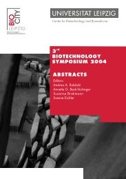
Saxon<br />
Biotechnology Symposium<br />
May 26, <strong>2009</strong><br />
<strong>2009</strong><br />
<strong>ABSTRACTS</strong><br />
Editors:<br />
Andrea A. Robitzki<br />
Manfred Blessing<br />
Thole Züchner<br />
Michael Brand<br />
Francis Stewart<br />
Andreas Beyer<br />
Erik Schäffer
IMPRINT<br />
Editors<br />
Prof. Dr. Andrea A. Robitzki<br />
Director<br />
Center for Biotechnology and Biomedicine, Universität <strong>Leipzig</strong><br />
Prof. Dr. Manfred Blessing<br />
Professorship for Molecular Pathogenesis<br />
Center for Biotechnology and Biomedicine, Universität <strong>Leipzig</strong><br />
Dr. Thole Züchner<br />
Junior Group Leader<br />
Center for Biotechnology and Biomedicine, Universität <strong>Leipzig</strong><br />
Prof. Dr. Michael Brand<br />
Scientific Director<br />
Biotechnology Center, Technische Universität Dresden<br />
Prof. Dr. Francis Stewart<br />
Professor of Genomics<br />
Biotechnology Center, Technische Universität Dresden<br />
Dr. Andreas Beyer<br />
Junior Group Leader<br />
Biotechnology Center, Technische Universität Dresden<br />
Dr. Erik Schäffer<br />
Junior Group Leader<br />
Biotechnology Center, Technische Universität Dresden<br />
Compilation<br />
Dr. Svenne Eichler<br />
Chief Executive Officer<br />
Center for Biotechnology and Biomedicine, Universität <strong>Leipzig</strong><br />
Dr. Sabine Matthiä<br />
Head of Administration<br />
Biotechnology Center, Technische Universität Dresden<br />
Anja Landsmann<br />
Coordinator of the PbF3, Universität <strong>Leipzig</strong><br />
Layout Katrin Giersch, M. A.<br />
Center for Biotechnology and Biomedicine, Universität <strong>Leipzig</strong><br />
Jana Schulze, M. A.<br />
Center for Biotechnology and Biomedicine, Universität <strong>Leipzig</strong><br />
Print<br />
Merkur Druck- und Kopierzentrum GmbH<br />
Editorial Deadline March 15, <strong>2009</strong><br />
ISBN 978-3-00-027884-6
Contents<br />
Page<br />
Foreword 31<br />
Program 33<br />
Presentations<br />
1. Non-coding RNAs as Regulators and Tools 37<br />
1 STAT3 regulated non-coding RNAs in tumor cells<br />
Friedemann Horn 39<br />
2 Predicting and modeling microRNA regulation with an<br />
application to stem cell development<br />
Fabian J. Theis, Dominik Lutter, Carsten Marr, Lena Uetzmann,<br />
Heiko Lickert 40<br />
3 Exploring chemical space with aptamers<br />
Michael Famulok 41<br />
4 From RNAi Screens to Molecular Function: A Systematic<br />
Pipeline for Gene Function in Mammalian Cells<br />
Frank Buchholz 42<br />
2. Stem cell therapies for neurological disorders 45<br />
5 Stem Cell Therapy for Parkinson’s Disease<br />
Patrick Brundin 47<br />
6 Biomaterial assisted cellular self-organization and tissue<br />
program: a new paradigm in regenerative medicine<br />
Carlos E. Semino 48
7 Regenerating the brain with endogenous stem cells<br />
Verdon Taylor, Onur Basak, Claudio Giachino, Philip Knuckles 49<br />
8 Cell Transplantation into the Retina: A Treatment Option<br />
for Photoreceptor Loss?<br />
Marius Ader 50<br />
3. Biophotonics – molecular motors and optical<br />
tweezers 53<br />
9 Single Molecule Biophysics: Soft Matter, Weak Interactions<br />
and Complex Mechanisms<br />
Dario Anselmetti 55<br />
10 Optical trapping and 3D particle tracking: from concept to<br />
versatile applications<br />
Anna Wozniak, Joost van Mameren, Gerd Behme,<br />
Sebastian Roth 56<br />
11 Bioanalytics at the nanometer and attoliter scale<br />
Horst Vogel, Ralf Schmauder 57<br />
12 Single molecule studies in live and reconstituted cellular<br />
systems<br />
Petra Schwille 58<br />
Posters<br />
4. Molecular Medicine 61<br />
13 PDE10A inhibitors for the treatment of schizophrenia and<br />
psychosis<br />
Ghadir Barbar Asskar, Sabine Mann, Karen Nieber, Norbert<br />
Sträter, Peter Brust, Detlef Briel 63
14 Aminosulfonate-Modulated pH-Induced Closure of Cx26<br />
Hemichannels Observed by High-Resolution Atomic Force<br />
Microscopy<br />
Christian A. Bippes, Jinshu Yu, Galen M. Hand, Daniel J.<br />
Müller, Gina E. Sosinsky 64<br />
15 THE P53 TRANSCRIPTOME – Discovery of Regulated noncoding<br />
RNA Genes<br />
Levin Böhlig, Antje Kretzschmar, Kristin Reiche, Jörg<br />
Hackermüller, Friedemann Horn, Kurt Engeland 65<br />
16 Interaction between PIP2 and Ceramide in Drosophila<br />
Phototransduction<br />
Salvatore Chiantia, Ujjaini Dasgupta, Acharia Usha,<br />
Petra Schwille 66<br />
17 Wnt signaling in osteogenic specification of embryonic<br />
stem cells<br />
Huawen Ding, Beatrice Kuske, Nicole I. zur Nieden 67<br />
18 Role of FGD6 in Actin Ring Formation and Organelle<br />
Dynamics in Resorbing Osteoclasts<br />
Ana Isabel Espírito Santo, Tobias Heckel, Bernard Hoflack 68<br />
19 Dual role of the EGF-receptor in regulation of glutamate<br />
transporter expression<br />
Darko Glisic, Claudia Lehmann, Jürgen Engele 69<br />
20 Two-dimensional difference in gel electrophoresis (DIGE)<br />
for analyzing ischemic Cardiomyocytes<br />
Sina Haas, Martin von Bergen, Andrea A. Robitzki 70<br />
21 Comprehensive (Molecular) Cytogenetic Characterization<br />
of rare intracranial tumors<br />
Heidrun Holland, Helene Hantmann, Wolfgang Krupp,<br />
Ronald Koschny, Michela Livrea, Ralf Schober, Jürgen<br />
Meixensberger, Peter Ahnert 71
22 The effects of thrombin on RPE cells are mediated by<br />
transactivation of growth factor receptors<br />
Margrit Hollborn, Carola Petto, Peter Wiedemann, Leon<br />
Kohen, Andreas Bringmann 72<br />
23 Copper overload leads to fragmentation of mitochondrial<br />
membrane lipids: implications for the pathogenesis of liver<br />
toxicity in Wilson disease<br />
Dominik Huster, Irina Yurkova, Jürgen Arnhold 73<br />
24 Loss of pluripotency and acquisition of an osteoblast fate<br />
in embryonic stem cells is accompanied by modulated<br />
microRNA expression<br />
Dorota Karniowska, Kristin Reiche, Antje K. Kretzschmar,<br />
Joerg Hackermueller, Nicole I. zur Nieden 74<br />
25 Synthesis of a new class of N1,N3- adamantylated uraciles<br />
and their biological activity as future non-nucleoside<br />
Inhibitors (NNIs)<br />
Matthias Klemm, Kurt Eger, Rudolf Fahrig, Detlef Briel 75<br />
26 Determination of the structure of F 1<br />
F 0<br />
ATP synthase rotor<br />
from Acetobacterium woodii<br />
Adriana Klyszejko, Michael Fritz, Thomas Meier, Volker<br />
Müller, Daniel J. Müller 76<br />
27 The importance of osteoblasts in the stump of the<br />
regenerating tail fin<br />
Franziska Knopf, Christina Hammond, Stefan Schulte-Merker,<br />
Gilbert Weidinger 77<br />
28 Adiponectin Receptor 1 Dimerization is Inhibited by<br />
Adiponectin<br />
David Kosel, John T. Heiker, Cornelia M. Wottawah, Matthias<br />
Blüher, Karin Mörl, Annette G. Beck-Sickinger 78
29 Novel Electroactive Nanovalves for a Implantable<br />
Controlled Drug Delivery Device<br />
Randy Kurz, Anselm Sickinger, Andrea A. Robitzki 79<br />
30 Inhibition of dedicated Wnt/β-catenin pathway-associated<br />
kinases by natural and chemical modified polyphenoles<br />
with peculiar features<br />
Katja Steffi Lerche, Robert Günther, Hans-Jörg Hofmann, Rolf<br />
Gebhardt 80<br />
31 The influence of phosphatidylserine content in lipidlayers of<br />
biopolymer-coated CaCO 3<br />
-particles on phorbol myristate<br />
acetate differentiated U937<br />
Jacqueline Lessig, Uta Reibetanz, Björn Neu, Hans-Jürgen<br />
Glander, Jürgen Arnhold 81<br />
32 Critical Role of Granulocyte-Macrophage Colony-<br />
Stimulating-Factor in Ultraviolet B Radiation-Induced Murine<br />
Skin Cancer<br />
Amrit Mann, Kerstin Niekisch, Thorsten Maass, Peter<br />
Schirmacher, Manfred Blessing 82<br />
33 A gene-dosage effect for IL-4Ralpha expression has an<br />
impact on Th2-mediated allergic inflammation during<br />
bronchopulmonary mycosis<br />
Uwe Müller, Werner Stenzel, Gabriele Köhler, Tobias<br />
Polte, Manfred Blessing, Amrit Mann, Daniel Piehler, Frank<br />
Brombacher, Gottfried Alber 83<br />
34 IL-4/IL-13-dependent alternative activation of macrophages<br />
but not microglial cells is associated with uncontrolled<br />
cerebral cryptococcosis<br />
Uwe Müller, Werner Stenzel, Gabriele Köhler, Frank L.<br />
Heppner, Manfred Blessing, Andrew N.J. McKenzie, Frank<br />
Brombacher, Gottfried Alber 84
35 The transcription factor Elf3 in the gastrointestinal tract:<br />
pathomorphological changes in the transgenic mouse<br />
model<br />
Martina Protschka, Manfred Blessing 85<br />
36 Metabolites of flavones – observations and results of<br />
synthesis and tests<br />
Benjamin Reissig, Steffen Rodewald, Detlef Briel, Rolf Gebhardt 86<br />
37 Osteoclasts Control Osteoblast Chemotaxis via PDGF-BB/<br />
PDGF receptor beta Signaling in vitro<br />
María Arántzazu Sánchez Fernández, Bernard Hoflack 87<br />
38 Peptide mediated DNA import into mitochondria<br />
Ingo Schäfer, Christian Kukat, Alexandra Kukat, Peter Seibel 88<br />
39 Distribution of mitofusin 2 (Mfn2) after the formation of<br />
megamitochondria<br />
Susanna Schubert, Christian Kukat, Ingo Schäfer, Alexandra<br />
Kukat, Peter Seibel 89<br />
40 In the absence of IL-12 protective immunity to infection with<br />
Salmonella Enteritidis depends on Il-23 and is associated<br />
with IL-22 but not IL-17<br />
Silke Mara Schulz, Gabriele Köhler, Alissa A. Chackerian,<br />
Ellen Witte, Kerstin Wolk, Robert Sabat, Yoichiro Iwakura,<br />
Robert A. Kastelein, Gottfried Alber, Christoph Holscher,<br />
Uwe Müller 90<br />
41 Non-protein coding RNAs as highly specific biomarkers<br />
for cancer<br />
Katharina Schutt, Friedemann Horn, Kerstin Ullmann, Antje<br />
K. Kretzschmar, Jörg Hackermüller 91<br />
42 Synthesis of novel PDE10A-Inhibitors<br />
Gregor Schwan, Lenka Kubicova, Karen Nieber, Norbert<br />
Sträter, Peter Brust, Detlef Briel 92
43 Effect of purine analogues on macrophage function during<br />
in vitro ischemia<br />
Fritzi Siegert, Karen Nieber 93<br />
44 Cymantrene-peptide bioconjugates: a promising approach<br />
to generate cytostatic compounds<br />
Katrin Splith, Harmel Peindy N’dongo, Ulrich Schatzschneider,<br />
Ines Neundorf 94<br />
45 Single-cell force spectroscopy reveals ß1 -integrin as<br />
central molecule mediating /ABL expression of 32D-BCR/<br />
ABL cells to bone marrow stromal cells<br />
Anna Taubenberger, Fernando A. Fierro, Pierre-Henri Puech,<br />
Gerhard Ehninger, Martin Bornhauser, Daniel J. Müller,<br />
Thomas Illmer 95<br />
46 MicroRNAs lost during prostate carcinoma pathogenesis<br />
cooperatively regulate mRNAs involved in Androgen<br />
Receptor signalling<br />
Kerstin Ullmann, Antje Kretzschmar, Friedemann Horn, Nora<br />
Mörbt, Martin von Bergen, Gerald Verhaegh, Jack Schalken,<br />
Katharina Schutt, Jörg Hackermüller 96<br />
47 The Role of extra- and intracellular domains for Y Receptor<br />
Internalisation<br />
Cornelia Walther, Diana Lindner, Ilka Böhme, Annette G.<br />
Beck-Sickinger 97<br />
48 Characterization of substrates and inhibitors for the in<br />
vitro assessment of BCRP mediated drug transport in the<br />
lactating mammary gland of dairy cattle<br />
Luise Waßermann, Stefan Lindner, Kerstin Honscha, Walther<br />
Honscha 98
49 Reconstitution of the Interleukin-4 dependent JAK/STAT<br />
signalling pathway for a detailed analysis by single<br />
molecule fluorescence detection methods in living cells<br />
Thomas Weidemann, Remigiusz Worch, Tibor Szekeres,<br />
Petra Schwille 99<br />
50 Inhibition of matrix metalloproteinases (MMPs) by selected<br />
anthraquinones and synthetic peptides<br />
Claudia Wierzchacz, Rolf Gebhardt 100<br />
51 Identification of the receptor for Pigment Epithelium Derived<br />
Factor on retinal endothelial cells<br />
Xiu Mei Yang, Wolfram Eichler, Johannes Lange, Andreas<br />
Reichenbach, Peter Wiedemann 101<br />
52 Refolding of ecto-nucleotidases<br />
Karen Yates, Norbert Sträter 102<br />
53 Structure-based design of inhibitors of human cAMP<br />
specific phosphodiesterase 4A<br />
Michael Zahn, Roy Eylenstein, E. Bartholomeus Küttner,<br />
Susanne Moschütz, Norbert Sträter 103<br />
54 Interfering with Signal Inactivation in Purinergic Signaling<br />
Matthias Zebisch, Norbert Sträter 104<br />
5. Bioanalytics 107<br />
55 Quantitative microscopy with trace element sensitivity<br />
Nirav Barapatre, Anja Fiedler, Thomas Arendt, Tilman Butz,<br />
Markus Morawski, Tilo Reinert 109
56 Easy and fast separation of multiple targets from whole<br />
blood with a new bead based cell separation method<br />
Doreen Beck, Christoph Mohr, Berthold Seidel, Iwona<br />
Goczalik, Anne Rasser, Jan-Michael Heinrich 110<br />
57 The structural and functional landscape of the human ADP<br />
receptor P2Y 12<br />
– methods for generating mutant libraries<br />
Maxi Cöster, Doreen Thor, Holger Römpler, Torsten Schöneberg 111<br />
58 Biological properties of Apidaecin derivatives with<br />
enhanced antimicrobial activities<br />
Patricia Czihal, Laszlo Otvos Jr., Ralf Hoffmann 112<br />
59 Characterization of proteins oxidatively modified in rat<br />
muscles by tandem mass spectrometry<br />
Maria Fedorova, Nadezda Kuleva, Ralf Hoffmann 113<br />
60 Study of toxicological effects of a silicon material on mouse<br />
fibroblast cell line L929<br />
Marco Glaß, Andrea A. Robitzki 114<br />
61 A novel system for quantification of peptides bound to<br />
solid surfaces<br />
Rayk Hassert, Annette G. Beck-Sickinger 115<br />
62 Metabolite analysis of adherent-growing cancer cell lines<br />
with GC-MS<br />
Antje Hutschenreuther, Ferdinand Fischer, Gerd Birkenmeier,<br />
Claudia Birkemeyer 116<br />
63 Development of cell type specific microelectrode array<br />
based label-free screening systems<br />
Heinz-Georg Jahnke, Sabine Schmidt, Ronny Azendorf,<br />
Andrea A. Robitzki 117
64 Insights into the Secondary Structure of the N-Terminus of<br />
the Adiponectin Receptor 1<br />
Cathleen Juhl, Annette G. Beck-Sickinger 118<br />
65 Oncocin – A novel proline-rich antimicrobial peptide to<br />
treat multi-resistant Gram-negative bacteria<br />
Daniel Knappe, Anne Hansen, Ralf Hoffmann 119<br />
66 A bond for a lifetime – Using Membrane nanotubes from<br />
living cells to determine receptor ligand kinetics<br />
Michael Krieg, Jonne Helenius, Carl-Philipp Heisenberg,<br />
Daniel J. Müller 120<br />
67 The Core Unit Fluorescence-Technologies in the IZKF<br />
<strong>Leipzig</strong><br />
Andreas Lösche, Jens Grosche 121<br />
68 Temperature feedback for thermal stabilization in optical<br />
tweezers<br />
Mohammed Mahamdeh, Erik Schäfer 122<br />
69 Impedimetric Detection of Transient Receptor Potential<br />
Channel Activity<br />
Oliver Pänke, Andrea A. Robitzki 123<br />
70 Epitope mapping of monoclonal anti-Tau antibodies<br />
AT8 and Tau5<br />
Robert Porzig, David Singer, Ralf Hoffmann 124<br />
71 PH sensor-functionalized colloidal DNA-carriers for direct<br />
localisation in cell compartments<br />
Uta Reibetanz, Liaw Zi Yen, Bernice Oh Hui Lin, Averil Chen<br />
Min Hui, Edwin Donath, Björn Neu 125
72 A platform for the automatic identification and quantification<br />
of metabolites using Nuclear Magnetic Resonance<br />
Spectroscopy<br />
Frank-Michael Schleif, Thomas Riemer, Uta Boerner, Rüdiger<br />
Alt, Lydia Schnapka-Hille, Christoph Wirling, Rolf Herwig,<br />
Stefan Leidich, Michael Cross 126<br />
73 Evaluation of carbon tetrachloride-induced oxidative stress<br />
on rat hepatocytes: Thermoinduced light emission and<br />
biochemical standard methods<br />
Anika Schumann, Alexander Bauer, Matthias Gilbert, Jan G.<br />
Hengstler, Christian Wilhelm 127<br />
74 Human cationic trypsinogen PRSS1: recombinant<br />
expression, purification and subsequent refolding<br />
Peter Simeonov, Katherina Bellmann, Matthias Zebisch, Ralf<br />
Hoffman, Norbert Sträter, Thole Züchner 128<br />
75 Synthesis and mass spectrometrical characterization of<br />
model peptides containing oxidized tryptophan residues<br />
relevant for protein aging<br />
Toni Todorovski, Ralf Hoffmann 129<br />
76 Optical trapping and 3D particle tracking: from concept to<br />
versatile applications<br />
Anna Wozniak, Joost van Mameren, Gerd Behme, Sebastian<br />
Roth 130<br />
77 In vivo biophysical study of fibroblast growth factor<br />
signalling using fluorescence correlation spectroscopy<br />
Shuizi Rachel Yu, Markus Burkhardt, Jonas Ries, Steffen<br />
Scholpp, Peter Schwille, Michael Brand 131<br />
78 Introduction of a peptide-based Immunoassay using a novel<br />
fluorophore and Fluorescence Resonance Energy Transfer<br />
(FRET)<br />
Thomas Zauner, Renate Berger-Hoffmann, Katrin Müller, Ralf<br />
Hoffmann, Thole Züchner 132
79 High Sensitive Multiplex Protein Quantitation Using Metal<br />
Element Chelating Tags on an LTQ-Orbitrap-ETD Mass<br />
Spectrometer<br />
Nicole Zehethofer, Ralf Hoffmann, Thole Züchner 133<br />
6. Bioinformatics 135<br />
80 Family-based analysis of genome-wide gene-gene<br />
interactions<br />
Marit Ackermann, Andreas Beyer 137<br />
81 Biomedical word sense disambiguation with ontologies<br />
and metadata<br />
Dimitra Alexopoulou, Bill Andreopoulos, Andreas Doms,<br />
Khaled Khelif, Michael Schroeder 138<br />
82 The nanometis software system for automated analysis of<br />
single molecular force spectroscopy data on membrane<br />
proteins<br />
Bill Andreopoulos, Frank Dressel, Dirk Labudde, Joscha<br />
Koellner, Michael Schroeder 139<br />
83 A systematic approach of osteoclastogenesis<br />
Antigoni Elefsinioti, Angela Simeone, Jacob Michaelson,<br />
Andreas Beyer 140<br />
84 Evolutionary and Functional Analysis of the Redβ/RecT<br />
Single-Strand Annealing Protein Family<br />
Axel Erler, Madeleine Teucher, Marcello Maresca, Vineeth<br />
Surendranath, Bianca Habermann, Youming Zhang, A.<br />
Francis Stewart 141
85 Xenoturbella bocki: Analyses of the mitochondrial genome<br />
and a PCR survey of hox genes<br />
Guido Fritzsch, Marleen Perseke, Bettina Weich, Manja<br />
Boehme, Detlef Bernhard, Thomas Stach, Olle Israelsson,<br />
Mike Thorndyke, Hiroaki Nakano, Thomas Hankeln, Martin<br />
Schlegel, Peter F. Stadler 142<br />
86 Confluent mesenchymal stem cell cultures in silico<br />
Martin Hoffmann, Jens-Peer Kuska, Matthias Zscharnack,<br />
Christian Thümmler, Augustinus Bader, Jörg Galle 143<br />
87 Pathway Prediction with eQTL and Gene Interaction<br />
Networks<br />
Jacob Michaelson, Andreas Beyer 144<br />
88 PhenoFam: a web tool for the gene set enrichment analysis<br />
in the protein domain context<br />
Maciej Paszkowski-Rogacz, Maria Teresa Pisabarro, Frank<br />
Buchholz 145<br />
89 GoGene: a search engine for genes and gene-related<br />
information.<br />
Conrad Plake, Loïc Royer, Rainer Winnenburg, Michael<br />
Schroeder 146<br />
90 Unraveling protein networks with Power Graph Analysis<br />
Matthias Reimann, Loïc Royer, Bill Andreopoulos, Michael<br />
Schroeder 147<br />
91 Studying molecular targets of human FOXP2 in a<br />
neuroblastoma cell line<br />
Sabrina Reimers, Ines Bliesener, Svante Pääbo, Wolfgang Enard 148<br />
92 Unraveling protein networks with Power Graph Analysis<br />
Loïc Royer, Matthias Reimann, Bill Andreopoulos, Michael<br />
Schroeder 149
93 Fluorine in protein environments: theoretical and<br />
experimental study<br />
Sergey Samsonov, Mario Salwiczek, Gerd Anders, Beate<br />
Koksch, Maria Teresa Pisabarro 150<br />
94 A systems biology approach for analyzing RNAi data<br />
using functional networks<br />
Angela Simeone, Andreas Beyer 151<br />
95 Development of a framework for structure-based functional<br />
protein annotation<br />
Aurelie Tomczak, Jana Sontheimer, Maria Teresa Pisabarro 152<br />
96 Three-dimensional modeling of protein complexes<br />
Anne Tuukkanen, Andreas Henschel, Michael Schroeder 153<br />
97 Utilising mutation tagging with gene identifiers for protein<br />
structure function studies<br />
Rainer Winnenburg, Conrad Plake, Christof Winter, Annalisa<br />
Marsico, Michael Schroeder 154<br />
98 SCOPPI – A Structural Classification of Protein-Protein<br />
Interfaces<br />
Christof Winter, Andreas Henschel, Wan Kyu Kim, Gihan<br />
Dawelbait, Michael Schroeder 155<br />
99 Serum proteomic profiling and targeted metabolomic of<br />
obese: Integrative data analysis of biological profile data<br />
Henry Wirth, Martin von Bergen, Jayaseelan Murugaiyan,<br />
Hans Binder, Andreas Oberbach 156<br />
7. Tissue and Cell Engineering 159<br />
100 Hematopoietic progenitor cells are vulnerable to high<br />
oxygen concentrations<br />
Rüdiger Alt, Nicole Noack, Michael Cross 161
101 Fabrication of Microscaffolds by Solid Lipid Templating<br />
Kristina Ambrosch, Michaela Schulz-Siegmund, Michael C.<br />
Hacker 162<br />
102 iPS cells as model system for human virus diseases<br />
Antje Arnold, Claire Fabian, Steven Sauerzweig, Yahaira<br />
Naldijk, Ute Brinkmann, Uwe G. Liebert, Alexandra Stolzing 163<br />
103 Characterization of the early embryo upon loss of histone<br />
methyltransferase Setd1a<br />
Anita Sabine Bledau, Andrew Francis Stewart, Konstantinos<br />
Anastassiadis 164<br />
104 Hepatocytes from human mesenchymal stem cells support<br />
liver regeneration after acute injury<br />
Bruno Christ, Peggy Stock, Hendryk Aurich, Ines Aurich,<br />
David Wohlrab, Werner Hein, Sabine Ebensing, Madlen<br />
Hempel, Matthias M. Dollinger, Wolfgang E. Fleig 165<br />
105 Generation and Characterization of Tumor Spheroids as<br />
Cellular Models for Anti-Cancer Drug Discovery<br />
Anja Drose, Mirjam Ingargiola, Bernd Schwenzer 166<br />
106 The impact of iPS on stem cell legislation and administration<br />
in Germany – points to consider for international stem cell<br />
research cooperation<br />
Timo Faltus 167<br />
107 Isolation and Characterisation of Human Melanocytes<br />
from Hair follicles for Clinical Use<br />
Christina Fieber, Jan C. Simon, Andreas Emmendörffer 168<br />
108 Differential expression of biofunctional GM1 and GM3<br />
gangliosides within the plastic-adherent multipotent<br />
mesenchymal stromal cell population<br />
Daniel Freund, Denis Corbeil 169
109 Telomerase activity and hepatic functions of rat embryonic<br />
liver progenitor cell in nanoscaffold coated small scale<br />
bioreactor<br />
Shibashish Giri, Sanja Pavlica, Mario Keller, Augustinus Bader 170<br />
110 Temporally-controlled site-specific recombination in<br />
zebrafish<br />
Stefan Hans, Jan Kaslin, Dorian Freudenreich, Michael Brand 171<br />
111 Osteoclastic activity of bone morphogenetic protein-2 in<br />
lumbar spinal fusion<br />
Christian Hohaus, Yvonne Minkus , Timothy Ganey, Hans-<br />
Jörg Meisel 172<br />
112 Fetal and adult hematopoiesis requires continuous Mll1<br />
function<br />
Andrea Kranz, Frieder Schwenk, Jost Seibler, Konstantinos<br />
Anastassiadis, A. Francis Stewart 173<br />
113 Plasma membrane mechanics in zebrafish germlayer<br />
progenitor cells<br />
Michael Krieg, Jonne Helenius, Daniel J. Müller, Carl-Philipp<br />
Heisenberg 174<br />
114 DNA repair in aged human MSC<br />
Michela Livrea, Alexandra Stolzing 175<br />
115 Microspherical delivery of a growth factor<br />
Alexander Lochmann, Hagen Nitzsche, Sabrina von Einem,<br />
Elisabeth Schwarz, Karsten Mäder 176<br />
116 Electrospun poly(ε-caprolactone) (PCL) microfiber meshes<br />
with predefined fiber diameters<br />
Tina Loth, Markus Manhardt, Kristina Ambrosch, Michaela<br />
Schulz-Siegmund, Michael C. Hacker 177
117 Surface modification of medical CoCr alloys by a<br />
thermochemical process<br />
Johanna Lutz, Stephan Mändl 178<br />
118 Regeneration of chronic osteochondral defects using<br />
autologous mesenchymal stem cells in a sheep model<br />
Bastian Marquass, Pierre Hepp, Robert Richter, Steffanie<br />
Schmidt, Frank Stein, Augustinus Bader, Christoph Josten,<br />
Matthias Zscharnack, Ronny Schulz 179<br />
119 Intervertebral disc repair using adipose tissue-derived stem<br />
and regenerative cells: experiments in a canine model<br />
Hans Jörg Meisel, Timothy Ganey, Christian Hohaus, William<br />
Hutton, Tim Moseley, Marc Hedrick, Brian Strem 180<br />
120 Conditional Mutagenesis of Histone Methyltransferase<br />
Mll2 in Neural Stem Cells<br />
Katrin Neumann, Maria Rostovskaya, Sandra Lubitz, Andrea<br />
Kranz, A. Francis Stewart, Konstantinos Anastassiadis 181<br />
121 Peptide vectors for siRNA delivery into cells<br />
Ines Neundorf, Anja Tennemann, Robert Rennert 182<br />
122 PEEK-WC-PU membranes for expansion of rat embryonic<br />
liver cells<br />
Sanja Pavlica, Antonella Piscioneri, Frank Peinemann,<br />
Angela Hennig, Javorina Milosevic, Stefania Laera, Piero<br />
Favia, Loredana DeBartolo, Augustinus Bader 183<br />
123 Dynamic Coupling of Pattern Formation and Morphogenesis<br />
in the Developing Vertebrate Retina<br />
Alexander Picker, Florencia Cavodeassi, Anja Machate,<br />
Sabine Bernauer, Stefan Hans, Gembu Abe, Koichi<br />
Kawakami, Stephen Wilson, Michael Brand 184<br />
124 Establishment of risk analysis of bone replacing scaffolds<br />
Heike Schneider, Kathleen Schröck, Manja Kamprad,<br />
Michaela Schulz-Siegmund 185
125 Osteogenic differentiation of human adipose tissue-derived<br />
stem cells: Are the standard ascorbate-2-phosphate<br />
concentrations too high?<br />
Hellen Schneider, Michael Hacker, Michaela Schulz-Siegmund 186<br />
126 Electrospinning parameters critical for the generation of<br />
poly(ε-caprolactone) (PCL) nanofibers<br />
Maximilian Sperlich, Markus Manhardt, Michaela Schulz-<br />
Siegmund, Michael Hacker 187<br />
127 Controlled formation of embryoid bodies in bioreactors for<br />
reproducible differentiation initiation of mouse and primate<br />
embryonic stem cells<br />
Susanne Trettner, Alexander Seeliger, Nicole I. zur Nieden 188<br />
128 Formation of MMP cleavable hydrogel materials for<br />
the development of novel biohybrid system for tissue<br />
engineering<br />
Mikhail Tsurkan, Kandice Levental, Petra Welzel, Milauscha<br />
Grimmer, Andrea Zieris, Woranan Panyanuwa, Uwe<br />
Freudenberg, Carsten Werner 189<br />
129 3D-cardiomyocyte structures generated from murine<br />
embryonic stem cells – A novel tool for drug screening on<br />
microcavity arrays<br />
Silvia Vinz, Randy Kurz, Andrea A. Robitzki 190<br />
130 Non-invasive acoustical imaging of mesenchymal stem<br />
cells<br />
Moritz von Buttlar, Esam Ahmed Mohamed, Amro<br />
Abdelrahman, Albert Kamanyi, Wolfgang Grill 191<br />
131 Bioreactor with integrated monitoring and mechanical<br />
stimulation for cartilage tissue engineering by collagen<br />
scaffold associated Mesenchymal Stem Cells<br />
Erik von der Burg, Moritz von Buttlar, Wolfgang Grill 192
132 Potential ageing effects in long-term cultured mouse<br />
neurospheres<br />
Vladimir Vukicevic, Anna Jauch, Timo C. Dinger, Linda<br />
Gebauer, Veronika Hornich, Stefan R. Bornstein, Monika<br />
Ehrhart-Bornstein, Albrecht M. Müller 193<br />
133 Spatially confined cell growth<br />
Ronald Werner, Torsten Koal, Heinz Georg Jahnke, Andrea<br />
A. Robitzki, Tilman Butz, Tilo Reinert 194<br />
8. Neuromedicine 197<br />
134 Improved brain outcome by autologous bone marrowderived<br />
mononuclear cells (BMCs) intravenously given 24<br />
hours after stroke in sheep as imaged by PET<br />
Henryk Barthel, Johannes Boltze, Christiane Boltze, Udo<br />
Großmann, Magnus Kluge, Andreas Schildan, Anita Seese,<br />
Frank Emmrich, Uwe Gille, Osama Sabri 199<br />
135 Saffron extract inhibits the glutamatergic synaptic<br />
transmission on rat cortical neurones<br />
Frauke Berger, Andreas Hensel, Matthias Lechtenberg, Karen<br />
Nieber 200<br />
136 Vascular endothelial growth factor (VEGF) affects processing<br />
of amyloid precursor protein and β-amyloidogenesis in<br />
brain slice cultures derived from transgenic Tg2576 mouse<br />
brain<br />
Susanne Bürger, Monika Noack, Elena Kouznetsova, Yousef<br />
Yafai, Ludmil Kirazov, Evgeni Kirazov, Reinhard Schliebs 201<br />
137 Isolation of Chromaffin Progenitor Cells from Adult Adrenal<br />
Medulla<br />
Kuei-Fang Chung, Flavie Sicard, Vladimir Vukicevic, Linda<br />
Gebauer, Wieland B. Huttner, Stefan R. Bornstein, Monika<br />
Ehrhart-Bornstein 202
138 Cellular characteristics of neural progenitor cells in the<br />
adult zebrafish telencephalon<br />
Julia Ganz, Jan Kaslin, Heiner Grandel, Michael Brand 203<br />
139 Viral gene transfer of cell cycle inhibitors to slow<br />
down progressive neurodegeneration<br />
Pia Glöckner, James Uney, Thomas Arendt, Uwe Ueberham 204<br />
140 A novel fluorescent acetylcholinesterase inhibitor released<br />
from nanoparticles selectively binds hippocampal<br />
β-amyloid plaques in triple transgenic mice<br />
Wolfgang Härtig, Johannes Kacza, Bernd-Reiner Paulke, Jens<br />
Grosche, Anke Hoffmann, Paul Wilhelm Elsinghorst, Michael<br />
Gütschow 205<br />
141 Role of purinergic signalling in the development of fibre<br />
projections?<br />
Claudia Heine, Nico Scherf, Jens-Peer Kuska, Ulf-Dietrich<br />
Braumann, Heike Franke 206<br />
142 A study of Imaging Geno-Phenotypes in dyslexia<br />
Holger Kirsten, Carolin Ligges, Arndt Wilcke, Peter Ahnert,<br />
Johannes Boltze 207<br />
143 Depression-like deficits in rats are improved by subchronic<br />
modafinil<br />
Holger Koch, Ralf Regenthal, Christian Köhler, Ute Krügel 208<br />
144 Impedance spectroscopy: A method for developing a labelfree<br />
detection system for neurodegenerative diseases<br />
Dana Krinke, Heinz-Georg Jahnke, Andrea A. Robitzki 209
145 Effects of blue light scleral cross-linking on rabbit<br />
eye growth<br />
Qing Liu, Hans Peter Iseli, Nicole Körber, Niclas Lindqvist,<br />
Martin Gryga, Peter Wiedemann, Andreas Reichenbach,<br />
Mike Francke 210<br />
146 Isolation and biological potential of enteric nervous system<br />
precursors derived from human gut<br />
Marco Metzger, Nikhil Thapar, Lothar Just 211<br />
147 Non-hypoxic stabilization of hypoxia-inducible<br />
factor alpha (HIF-α): Relevance in neural progenitor/stem<br />
cells<br />
Javorina Milosevic, Irena Adler, Sigrid C. Schwarz, Gail<br />
Walkinshaw, Alexander Storch, Johannes Schwarz 212<br />
148 Ca 2+ Responses of Müller cells induced by light stimulation<br />
of photoreceptor cells<br />
Katja Rillich, Janina Gentsch, Michael Weick, Andreas<br />
Bringmann, Andreas Reichenbach 213<br />
149 Polyethylenimine as a possible gene therapeutical tool<br />
against Alzheimer’s Disease<br />
Susanne Rohn, Thomas Arendt, Uwe Ueberham 214<br />
150 Increase of intracellular Ca 2+ by adenine and uracil<br />
nucleotides in human midbrain-derived neuronal precursor<br />
cells<br />
Patrizia Rubini Illes, Johannes Engelhardt, Mahmoud Al-<br />
Khrasani, Javorina Milosevic, Johannes Schwarz, Peter Illes,<br />
Wolfgang Nörenberg 215<br />
151 Study of human neural progenitor cell fate after grafting<br />
into rat striatum<br />
Johanna Scheibe 216
152 Organotypic cocultures as an alternative to conventional<br />
animal models<br />
Sabine Schewtschik 217<br />
153 Analysing a potential neuroprotective function of<br />
perineurinal nets by using organotypic slice cultures<br />
Anne Suttkus, Markus Morawski, Gert Brückner, Thomas Arendt 218<br />
154 Distinct reactions of retinal microglia cells evoked by<br />
various stimuli<br />
Elke Ulbricht, Mike Francke, Ulrike Zeitschel, Andreas<br />
Reichenbach 219<br />
155 Connexins control glial glutamate transporter expression.<br />
Tina Unger, Stefanie Bette, Jürgen Engele 220<br />
156 A new hippocampal ex vivo model to study tauopathies by<br />
label-free impedance spectroscopy<br />
Annett Wegner, Heinz-Georg Jahnke, Till G. A. Mack, Frank<br />
Striggow, Andrea A. Robitzki 221<br />
157 A new mouse model for targeting the astrocytic NADH /<br />
NAD+ redox state in vivo<br />
Franziska Wilhelm, Jan Rillich, Ulrike Winkler, Johannes<br />
Hirrlinger 222<br />
158 Structural plasticity of astrocytes and the impact of the cell<br />
adhesion protein vinculin<br />
Ulrike Winkler, Marcello Sestu, Alice Zemljic-Harpf, Robert<br />
S. Ross, Wolfgang H. Ziegler, Johannes Hirrlinger 223
9. Diagnostics 225<br />
159 Effects of A 2A<br />
and A 2B<br />
ligands on ach contraction in<br />
inflamed rat small intestinal preparation<br />
Karen Nieber, Claudia Warstat, Fabien Michel, Luo Yan,<br />
Christa Müller, Sebastian Michael 227<br />
160 Concentration of mucus in gastric juice in normal adult<br />
horses withhold feed and after application of pronurtin<br />
Gerald F. Schusser, Alice Spallek, Stephan Recknagel, Julia<br />
Breuer, Gábor Köller 228<br />
161 Dendritic sugar balls for biological experiments driven by<br />
H-bonds<br />
Dietmar Appelhans, Brigitte Voit 229<br />
162 Development of an ELISA and a Candidate Vaccine for<br />
Pigeon Circovirus Infection<br />
Mohammad Yahya Halami, Wieland Schrödl, Reimar Johne,<br />
Erhard F. Kaleta, Hermann Müller 230<br />
163 Development and fabrication of a novel proteinbased<br />
biosensor for specific detection and immobilisation<br />
of cells<br />
Anja Steude, Oliver Pänke, Sabine Schmidt, Matthias Nieber,<br />
Andrea A. Robitzki 231<br />
164 Mycotoxin determination by means of an electronic nose<br />
Anselm Werner, Claudia Winter, Monika Krüger, Andrea<br />
Lindner, Klaus Krüger 232
10. Microfluidics 235<br />
165 Structural levels of organization in the TmHU-DNAcomplex<br />
as studied by optical tweezers assisted Force<br />
spectroscopy<br />
Carolin Wagner, Mathias Salomo, Friedrich Kremer 237<br />
166 Optical tweezers to investigate receptor-ligand interactions<br />
on a single contact level<br />
Carolin Wagner, Mathias Salomo, Friedrich Kremer 238<br />
11. Protein Engineering and Biocatalysis 241<br />
167 Evaluating 3D experiments in optical tweezers<br />
Marcel Ander 243<br />
168 Reconstituting Cytokinesis in Artificial Lipid Systems<br />
Senthil Arumugam 244<br />
169 Variants of Candida antarctica lipase B convert α-substituted<br />
substrates<br />
Sally Bayer, Thomas Greiner-Stöffele, Meike Ballschmiter 245<br />
170 Expression, Purification and Characterization of the<br />
Neuropeptide Y Receptor Type 2<br />
Sandra Berndt, Peter Schmidt, Cindy Montag, Christian<br />
Berger, Susann Schimmer, Diana Lindner, Annette G. Beck-<br />
Sickinger, Rainer Rudolph, Daniel Huster 246<br />
171 Construction of a RNaseT1 expression system for Aspergillus<br />
niger<br />
Kathrin Bönsch, Thomas Greiner-Stöffele, Meike Ballschmiter 247<br />
172 Torque Measurements on DNA with Magnetic Tweezers<br />
Hergen Brutzer, Nicholas Luzzietti, Friedrich Schwarz, Ralf Seidel248
173 Tagging methods for proteomics and regulomics in mouse<br />
embryonic stem cells<br />
Giovanni Ciotta, A. Francis Stewart 249<br />
174 Two ways to screen for new proteases in (meta-) genomic<br />
libraries<br />
Antje Eichler, Thomas Greiner-Stöffele, Meike Ballschmiter 250<br />
175 Pseudomonas putida – development of a heterologous<br />
expression system for complex natural products<br />
Frank Groß, Dominik Pistorius, Youming Zhang, A. Francis<br />
Stewart, Rolf Müller 251<br />
176 Prediction of Flocculation Ability of Brewing Yeast<br />
Inoculates by Flow Cytometry, Proteome Analysis, and<br />
mRNA Profiling<br />
Franziska Heine, Frank Stahl, Heike Sträuber, Claudia<br />
Wiacek, Dirk Benndorf, Cornelia Repenning, Frank Schmidt,<br />
Thomas Scheper, Martin von Bergen, Hauke Harms, Susann<br />
Müller 252<br />
177 Reduction of substrate binding pocket of glucose<br />
dehydrogenase B for improved substrate specificity<br />
Michael Hofer, Kathrin Bönsch, Meike Ballschmiter 253<br />
178 Screening for indole hydroxylating variants of P450cam<br />
Gregor Hoffmann, Katrin Bönsch, Meike Ballschmiter 254<br />
179 Interactions in the plant RNase P/ MRP<br />
Mario Krehan, Sebastian Braun, Janine Dahl, Nicolas<br />
Menzel, Christian Heubeck, Astrid Schön 255<br />
180 The crystal structure of arylmalonate decarboxylase<br />
reveals active site flexibility in catalysis<br />
Bartholomeus Küttner, Markus Kircher, Susann Rosmus, Antje<br />
Keim, Norbert Sträter 256
181 RiboxX: RNA-interference in a box ! ®<br />
Jacques Rohayem, Katrin Jäger, Ivonne Robel, Kristin Hille,<br />
Dorothea Kramer, Romy Zieger, Mirko Bergmann, Julia<br />
Gebhardt, Christiane Petzold 257<br />
182 Mutagenesis of the Thermus aquaticus amylomaltase to<br />
produce large cyclic glucans<br />
Christian Roth, Nicole Weizenmann, Wolfgang Zimmermann,<br />
Norbert Sträter 258<br />
183 NMR Measurements of a Class A GPCR from Prokaryotic<br />
Expression<br />
Peter Schmidt, Andreas Bunge, Diana Lindner, Sandra Berndt,<br />
Christian Berger, Annette G. Beck-Sickinger, Rainer Rudolph,<br />
Daniel Huster 259<br />
184 Single-Molecule Studies of DNA Translocating Restriction<br />
Enzymes<br />
Friedrich Schwarz, Kara van Aelst, Mark Szczelkun, Ralf Seidel 260<br />
185 Immobilization of Proteins via Cu(I) catalyzed Alkyne Azide<br />
Cycloaddition<br />
Max Steinhagen, Annette G. Beck-Sickinger 261<br />
12. Appendix 263<br />
Center for Biotechnology and Biomedicine (BBZ) 265<br />
The Biotechnology Center (BIOTEC) 268<br />
13. Index 271
FOREWORD<br />
After its great success in Dresden in 2007, the Saxon Biotechnology Symposium series is<br />
being continued in <strong>Leipzig</strong> in <strong>2009</strong>, the year of the 600 th anniversary of the foundation of<br />
<strong>Leipzig</strong> University in 1409.<br />
The Center for Biotechnology and Biomedicine at the University of <strong>Leipzig</strong> and the<br />
Biotechnology Center of the Technical University of Dresden are continuing cooperation<br />
to hold the joint Biotechnology Symposium in Saxony. The main topics of the conference,<br />
hosted in <strong>Leipzig</strong> this year, will be ‘Non-Coding RNAs as Regulators and Tools’, ‘Stem Cell<br />
Therapies for Neurological Disorders’ and ‘Biophotonics – Molecular Motors and Optical<br />
Tweezers’.<br />
It is the purpose of the <strong>2009</strong> Biotechnology Symposium to further bridge the gap between<br />
basic, applied and integrative research in the fields of nanobiotechnology / nanomedicine<br />
and molecular bioengineering. This will increasingly strengthen regional networks and<br />
make Saxony well-known on a supraregional and also on an international level as a place<br />
where biotechnology ranks high. Saxony is an excellent location for research and a growing<br />
biotechnological industry, providing a platform for the transfer of knowledge and technology<br />
in the fields of molecular design, the development and testing of active substances, genomics,<br />
proteomics, diagnostics, cell techniques, nano-bioelectronics, biophysics, cellular machines,<br />
tissue engineering, and bioinformatics.<br />
Biotechnology is emerging as a key technology for the future, inventions and innovations<br />
being the motors for biotechnological progress. We hope this symposium will give the<br />
Saxon biotechnological sector a boost and encourage the universities, research centers and<br />
companies.<br />
Prof. Dr. Andrea A. Robitzki<br />
Director<br />
Center for Biotechnology and Biomedicine<br />
Prof. Dr. Michael Brand<br />
Director<br />
Biotechnology Center
PROGRAM<br />
9:00 am – 9:30 am Opening<br />
Prof. Dr. Martin Schlegel<br />
Prorektor für Forschung und wissenschaftlichen Nachwuchs der Universität<br />
<strong>Leipzig</strong><br />
Prof. Dr. Petra Schwille<br />
stellvertretende Direktorin des Biotechnologischen Zentrums der Technischen<br />
Universität Dresden<br />
9:30 am – 11:30 am Session 1<br />
Non-coding RNAs as regulators and tools<br />
Chair: Prof. Dr. Manfred Blessing/Dr. Andreas Beyer<br />
STAT3 regulated non-coding RNAs in tumor cells<br />
Prof. Dr. Friedemann Horn<br />
Universität <strong>Leipzig</strong>, Medizinische Fakultät, Institut für Klinische Immunologie<br />
Predicting and modeling microRNA regulation with an application to stem<br />
cell development<br />
Dr. Fabian Theis<br />
Helmholtz-Zentrum München, Institut für Bioinformatik und Systembiologie,<br />
CMB<br />
Exploring chemical space with aptamers<br />
Prof. Dr. Michael Famulok<br />
Universität Bonn, Life & Medical Sciences (LIMES)-Institut<br />
From RNAi Screens to Molecular Function: A Systematic Pipeline for Gene<br />
Function in Mammalian Cells<br />
Dr. Frank Buchholz<br />
Max-Planck-Institut für Molekulare Zellbiologie und Genetik Dresden<br />
11:30 am – 1:00 pm Postersession & lunch break
1:00 pm – 3:00 pm Session 2<br />
Stem cell therapies for neurological disorders<br />
Chair: Prof. Dr. Francis Stewart/Dr. Thole Züchner<br />
Stem cell therapy for Parkinson‘s disease<br />
Prof. Dr. Patrik Brundin<br />
Wallenberg Neuroscience Center, Department of Experimental Medical<br />
Science Lund, Schweden<br />
Biomaterial assisted cellular self-organization and tissue program: a new<br />
paradigm in regenerative medicine<br />
Prof. Dr. Carlos E. Semino<br />
Universität <strong>Leipzig</strong>, Translationszentrum für Regenerative Medizin<br />
Regenerating the brain with endogenous stem cells<br />
Dr. Verdon Taylor<br />
Max-Planck-Institut für Immunobiologie, Fachbereich molekulare Embryologie,<br />
Freiburg<br />
Cell Transplantation into the Retina: A Treatment Option for Photoreceptor<br />
Loss?<br />
Dr. Marius Ader<br />
Technische Universität Dresden, Zentrum für Regenerative Therapien Dresden<br />
3:00 am – 3:30 pm Postersession & coffee break<br />
3:30 pm – 5:30 pm Session 3<br />
Biophotonics – molecular motors and optical tweezers<br />
Chair: Prof. Dr. Andrea A. Robitzki/Dr. Erik Schäffer<br />
Single molecule biophysics: soft matter, weak interaction and complex Mechanisms<br />
Prof. Dr. Dario Anselmetti<br />
Universität Bielefeld, Fakultät für Physik<br />
Optical trapping and 3D particle tracking: from concept to versatile applications<br />
Dr. Anna Wozniak<br />
JPK Instruments AG, Berlin
Bioanalytics at the nanometer and attoliter scale<br />
Prof. Dr. Horst Vogel<br />
Ecole polytechnique fédérale de Lausanne, Institut des sciences et ingénierie<br />
chimiques, Schweiz<br />
Single molecule studies in live and reconstituted cellular systems<br />
Prof. Dr. Petra Schwille<br />
Technische Universität Dresden, Biotechnologisches Zentrum<br />
5:30 pm – 6:00 pm Poster awards<br />
6:15 pm – 6:45 pm Evening lecture<br />
Opening by the Minister of the Saxon Ministry of Science and the Fine Arts<br />
Dr. Eva-Maria Stange<br />
Award ceremony of the initiative „Germany – Land of Ideas“<br />
Mike Röseler<br />
Direktor des Investment & FinanzCenter <strong>Leipzig</strong>-Mitte, Deutsche Bank Privatund<br />
Geschäftskunden AG<br />
6:45 pm – 7:30 pm<br />
3D Micro Chip for pharmacological high throughput studies<br />
Evening lecture from the awardee of the initiative „Germany – Land of Ideas“<br />
Prof. Dr. Andrea A. Robitzki<br />
Universität <strong>Leipzig</strong>, Biotechnologisch-Biomedizinisches Zentrum<br />
7:30 pm – 9:30 pm Get-together and buffet<br />
All day Industrial exhibition and information desk of the 3D-Chip<br />
Project
1.<br />
Non-coding RNAs as<br />
Regulators and Tools<br />
Presentations
non-coding RNAs as Regulators and tools<br />
1 STAT3 regulated non-coding RNAs in tumor cells<br />
Friedemann Horn<br />
Recent analyses of the human genome revealed the existence of a high number of noncoding<br />
(nc)RNA transcripts. It is increasingly being recognised that these ncRNAs now constitute<br />
an important and complex layer of cellular regulation. In addition, a growing number of<br />
ncRNAs prove to be disease-associated. However, data on how cellular signalling networks<br />
interact with the ncRNA layer are still sparse.<br />
The transcription factor Stat3 was discovered by our group as a central component of<br />
interleukin-6 signalling. Stat3 is involved in differentiation, proliferation and apoptosis<br />
control of many cell types during embryogenesis as well as in adult tissues. In addition, Stat3<br />
is now regarded a highly oncogenic protein, being constitutively active in >70% of human<br />
tumours.<br />
We currently address the question whether in addition to its known functions, the Stat3<br />
pathway is involved in the transcriptional regulation of ncRNAs. We could demonstrate<br />
that Stat3 controls the expression of the anti-apoptotic microRNA miR-21 through a<br />
phylogenetically conserved enhancer. This microRNA is highly expressed in many cancers.<br />
Our results suggest that the oncogenic potential of Stat3 relies at least in part on the induction<br />
of the miR-21 gene.<br />
Furthermore, genome-wide expression analyses provided evidence that Stat3 regulates many<br />
novel ncRNA genes, suggesting an intimate cross-talk between the Stat3 signal pathway and<br />
the unfolding „RNA world“.<br />
Prof. Dr. Friedemann Horn<br />
Universitöt <strong>Leipzig</strong><br />
Faculty of Medicine<br />
Institute of clinical immunology<br />
friedemann.horn@medizin.uni-leipzig.de<br />
http://ikit.uniklinikum-leipzig.de/<br />
39
non-coding RNAs as Regulators and tools<br />
2 Predicting and modeling microRNA regulation<br />
with an application to stem cell development<br />
Fabian J. Theis, Dominik Lutter, Carsten Marr, Lena<br />
Uetzmann, Heiko Lickert<br />
The differentiation of embryonic stem (ES) cells during gastrulation into the cell types of the<br />
three germ layers is controlled by multiple interacting gene regulatory mechanisms. In addition<br />
to transcription factor (TF) driven regulations, there is strong evidence that microRNAs play<br />
an important role during stem cell development. The identification of microRNA-controlled<br />
gene regulation using expression data remains difficult, mainly for two reasons: (i) there is<br />
still a lack of microRNA expression data with adequate time resolution, and (ii) the inhibitory<br />
effect of microRNAs on translation stays invisible on the mRNA level. Here we study gene<br />
regulation under the influence of microRNAs controlling differentiation of murine ES cells<br />
into endoderm and mesoderm lineages. For this we use binding site prediction methods<br />
to estimate putative microRNA targets. Moreover, we uncover microRNA-driven gene<br />
regulation by measuring the indirect influence of an intronic microRNA on a microRNA-TFtarget-gene<br />
motif.<br />
Using microarray expression data of differentiating ES cells, we identify a regulatory system<br />
including an intronic microRNA that stabilizes differentiation during development. Through<br />
dynamical modeling we determine a bistable system that can switch between mesoderm<br />
and endoderm state starting from an undifferentiated cell. This sheds light into the potential<br />
cellular mechanisms driving ES cell differentiation.<br />
Dr. Fabian J. Theis<br />
Helmholtz Zentrum München<br />
Institute for Bioinformatics and Systems Biology<br />
Computational modeling in biology<br />
fabian.theis@helmholtz-muenchen.de<br />
www.helmholtz-muenchen.de<br />
40
non-coding RNAs as Regulators and tools<br />
3 Exploring chemical space with aptamers<br />
Michael Famulok<br />
Small molecule inhibitors of proteins are invaluable tools in Chemical Biology. Their<br />
identification can be tedious, because most screening methods have to be tailored to the<br />
corresponding drug target. We have developed modular assays based on aptamer displacement<br />
or protein-dependent reporter ribozymes for the screening of small-molecule inhibitors.<br />
Asaptamers can be generated for virtually any protein, the assay potentially identifies inhibitors<br />
for targets or individual protein domains for which no functional screen is available. Thereby,<br />
chemical space is explored in a rapid, focused, and modular manner, by indirectly taking<br />
advantage of the highest molecular diversity currently amenable to screening, namely that of<br />
1016 different nucleic acid sequences. I will discuss the application of these approaches to<br />
find new inhibitors for target proteins. Examples showing that these modulators can be used<br />
as tools for gaining novel biological insight are provided.<br />
Prof. Dr. Michael Famulok<br />
Universität Bonn<br />
LIMES Program, Unit Chemical Biology & Medicinal<br />
Chemistry<br />
m.famulok@uni-bonn.de<br />
www.chembiol.uni-bonn.de/<br />
41
non-coding RNAs as Regulators and tools<br />
4 From RNAi Screens to Molecular Function:<br />
A Systematic Pipeline for Gene Function in<br />
Mammalian Cells<br />
Frank Buchholz<br />
RNAi screens typically deliver a large number of candidate genes that play a role in a biological<br />
process. The validation of these candidates and the dissection of the molecular mechanism of<br />
action are often time consuming and cumbersome. We have developed a pipeline using BAC<br />
recombineering technology and tissue culture transgenesis to streamline the analysis of hits<br />
identified in large scale RNAi screens. Examples from this pipeline will be presented.<br />
Dr. Frank Buchholz<br />
Max-Planck-Institute for Molecular Cell Biology and<br />
Genetics, Dresden<br />
buchholz@mpi-cbg.de<br />
www.mpi-cbg.de<br />
42
non-coding RNAs as Regulators and tools<br />
43
2.<br />
Stem cell therapies<br />
for neurological<br />
disorders<br />
Presentations
stem cell therapies for neurological disorders<br />
5 Stem Cell Therapy for Parkinson’s Disease<br />
Patrick Brundin<br />
The prospect of cell replacement therapy for neurological disorders has generated a great<br />
deal of excitement. Cell transplantation has been tested in, e.g., Parkinson’s disease (PD),<br />
Huntington’s disease, stroke, multiple sclerosis and spinal cord damage. I will primarily<br />
review neural transplantation in PD over the past two decades, and describe the main obstacles<br />
that currently prevent transfer of the technology into an established treatment.<br />
In my presentation I will also discuss our recent observations that grafted neurons can develop<br />
Lewy bodies, which are the neuropathological hallmark of PD, and what the implications of<br />
these findings are for the field of cell transplantation. Further, I will describe our current<br />
attempts to obtain high numbers of dopaminergic neurons from stem cells. For example, we<br />
attempt to control cell division after the stem cell-derived neurons are transplanted, in order<br />
to avoid tumor/teratoma formation. We use knowledge from the developmental biology of<br />
midbrain dopamine neurons both to increase the yield of dopaminergic neurons and to limited<br />
uncontrolled growth of grafts. A safe and reproducible supply of cells is necessary for future<br />
systematic, large-scale clinical trials. Only after such trials will we know whether neural cell<br />
grafting has a place in the therapeutic arsenal against PD.<br />
Prof. Dr. Patrick Brundin<br />
Lund University<br />
Wallenberg Neuroscience Center<br />
Neuronal Survival Unit<br />
Department of Experimental Medical Science<br />
patrik.brundin@med.lu.se<br />
www.med.lu.se/expmed/neuronal_survival_unit<br />
47
stem cell therapies for neurological disorders<br />
6 Biomaterial assisted cellular self-organization and<br />
tissue program: a new paradigm in regenerative<br />
medicine<br />
Carlos E. Semino<br />
Cellular self-organization studies have been mainly focused on models such as Volvox, the<br />
slime mold Dictyostelium discoideum, and animal (metazoan) embryos. Moreover, animal<br />
tissues undergoing regeneration also exhibit properties of embryonic systems such as cellular<br />
dedifferentiation and self-organization processes that ends in rebuilding tissue complexity<br />
and function. We speculate that the recreation in vitro of the biological, biophysical and<br />
biomechanical conditions similar to those of regenerative milieu could elicit the intrinsic<br />
capacity of differentiated cells to proceed to the development of a tissue-like structure. In<br />
this presentation I will show that when primary mouse embryonic fibroblasts are cultured<br />
in a soft nanofiber scaffold they establish a cellular network that causes an organized cell<br />
contraction, proliferation and migration that ends in the formation of a symmetrically<br />
bilateral structure with a distinct central axis. Interestingly, a subset of mesodermal genes<br />
(Brachyury, Sox9 and Runx2) is upregulated during this morphogenetic process. The<br />
expression of Brachyury was localized first at the central axis, extending then to both sides<br />
of the structure. Furthermore, expression of Sox9 and Runx2 was followed by the spontaneous<br />
formation of cartilage-like tissue mainly at the paraxial zone. Since cellular self-organization<br />
is an intrinsic property of the tissues undergoing development this model could bring new<br />
ways to consider tissue regeneration.<br />
Prof. Dr. Carlos E. Semino<br />
Translational Centre for Regenerative Medicine <strong>Leipzig</strong><br />
semino@trm.uni-leipzig.de<br />
www.trm.uni-leipzig.de<br />
48
stem cell therapies for neurological disorders<br />
7 Regenerating the brain with endogenous stem<br />
cells<br />
Verdon Taylor, Onur Basak, Claudio Giachino, Philip<br />
Knuckles<br />
The brains of adult mammals contain stem cells that continuously generate neurons throughout<br />
life. The identity of these stem cells and most of the mechanisms regulating their fate are<br />
unresolved. Although cells with stem cell properties can be isolated from the embryonic<br />
and adult brain, and expanded endlessly in vitro, it is unclear what potential these cells<br />
really possess. Transplantation into the postnatal brains of host animals results in extensive<br />
gliogenesis and limited or no neurogenesis, even in lesions. Here we will discuss genetic<br />
evidence for the identity of adult neural stem cells and that the Notch signaling pathway is<br />
critical for neurogenic and regenerative stem cells in the adult brain. In addition, we will<br />
present evidence that adult derived neural stem cells, expanded in vitro retain multipotent<br />
potential when transplanted into the brain, opening up the potential use of these cells for<br />
identifying differentiation cues to generate defined neuronal cell types and thus for human<br />
therapy.<br />
Dr. Verdon Taylor<br />
Max Planck Institute of Immunobiology Freiburg<br />
Department of Molecular Embryology<br />
taylor@immunbio.mpg.de<br />
www.mpg.de<br />
49
stem cell therapies for neurological disorders<br />
8 Cell Transplantation into the Retina: A Treatment<br />
Option for Photoreceptor Loss?<br />
Marius Ader<br />
Impairment of vision and blindness due to loss of photoreceptors, the light-sensitive cells of<br />
the retina, are one of the most prevalent causes of disability in industrialized countries. The<br />
adult mammalian retina lacks endogenous repair mechanisms and thus is unable to regenerate<br />
photoreceptors lost due to injury or inherited diseases.<br />
Possible treatment options for hindering disease progression or regaining sight include<br />
pharmacological interventions, electrical implants, gene therapeutic approaches, or<br />
molecular inhibition methods using antibodies or RNA interference techniques. However,<br />
although promising, most of these treatment options require an early intervention in the<br />
disease development before massive photoreceptor loss has occurred. Thus, cell-based<br />
strategies might represent an important treatment option to replace lost photoreceptors in<br />
retinopathies.<br />
Diverse cell populations were used for transplantation experiments into the retina of rodents<br />
in recent years. However, although some cells showed potential to integrate into the host<br />
tissue and survived for prolonged time periods, differentiation into mature photoreceptors<br />
was rarely observed. In contrast, recently we could demonstrate that integration and<br />
differentiation into mature rod photoreceptors following transplantation into adult mouse<br />
retinas is most successful when primary retinal cells isolated at the peak of rod photoreceptor<br />
generation, i.e. the first postnatal week in mice, are used for grafting. The data implicates<br />
that cells committed to the photoreceptor lineage rather then stem/progenitor cells have the<br />
greatest potential for integration into host retinas and photoreceptor differentiation. Thus,<br />
young photoreceptors represent prime candidates for the development of cell replacement<br />
therapies for diseases characterized by photoreceptor loss.<br />
Dr. Marius Ader<br />
Technische Universität Dresden<br />
Center for Regenerative Therapies<br />
Cell-based Therapies for the Treatment of Retinopathies<br />
marius.ader@crt-dresden.de<br />
www.crt-dresden.de<br />
50
stem cell therapies for neurological disorders<br />
51
3.<br />
Biophotonics –<br />
molecular motors and<br />
optical tweezers<br />
Presentations
iophotonics – molecular motors and optical tweezers<br />
9 Single Molecule Biophysics: Soft Matter, Weak<br />
Interactions and Complex Mechanisms<br />
Dario Anselmetti<br />
Over the last 15 years, novel ultrasensitive biophysical methods have been developed that<br />
allow identification of physical mechanisms, quantitative analysis of cellular processes, and<br />
investigations of individual biomolecules and cells, with respect of protein organisational<br />
structure, functional interplay and temporal dynamics. Namely, atomic force microscopy and<br />
single molecule force spectroscopy (AFM, AFM-FS), optical tweezers, and single molecule<br />
optical microscopy beyond the diffraction limit allow nowadays deep insights into complex<br />
and regulated biological processes.<br />
In my presentation I will report on recent work where we quantitatively investigated effectorstimulated<br />
DNA-protein interaction in bacteria (Sinorhizobium meliloti) , posttranscriptionally<br />
regulated RNA-protein interactions in plants (Arabidopsis thaliana), as well as the reverse<br />
engineering of an artificial single molecule affinity switch in supramolecular host-guest<br />
complexes (calixarenes) with AFM single molecule force spectroscopy. These examples<br />
highlight the possibility to investigate (post)transcriptionally regulated processes at a single<br />
molecule level as well as its synthetic imitation.<br />
The dynamic action of DNA binding ligands can be studied by optical tweezers. The<br />
manipulation of single-molecule DNA molecules allow nanoscreening of potentially<br />
important therapeutic agents in anticancer chemotherapy, where the mechanism of action of<br />
the enzyme topoisomerase I in the presence of inhibitors is investigated. Furthermore, the<br />
threading of single DNA molecules into nanopores can be observed with a novel concept of<br />
a quantitative 3D optical tweezers systems, which will allows the investigation of molecular<br />
translocation binding and dynamic sliding phenomena along DNA.<br />
Prof. Dr. Dario Anselmetti<br />
Bielefeld University<br />
Experimental Biophysics and Applied Nanoscience<br />
Institute for Biophysics and Nanoscience (BINAS)<br />
dario.anselmetti@physik.uni-bielefeld.de<br />
www.physik.uni-bielefeld.de/biophysik<br />
55
iophotonics – molecular motors and optical tweezers<br />
10 Optical trapping and 3D particle tracking: from<br />
concept to versatile applications<br />
Anna Wozniak, Joost van Mameren, Gerd Behme,<br />
Sebastian Roth<br />
In the past decade, experiments involving the manipulation and observation of nanostructures<br />
with light using optical tweezers methodology have developed from proof-of-principle<br />
experiments to an established quantitative technique in fields ranging from (bio)physics to cell<br />
biology. With optical tweezers, microscopically small objects can be held and manipulated.<br />
At the same time, the forces exerted on the trapped objects can be accurately measured.<br />
JPK Instruments has developed a quantitative optical tweezers platform, the NanoTracker.<br />
This platform allows the controlled trapping and accurate tracking of nanoparticles. With its<br />
3D detection system, particle displacements within the trap can be recorded with nanometer<br />
precision. Moreover, dynamic forces acting on the particle (e.g., exerted by motor proteins)<br />
can be measured with better than piconewton resolution on a microsecond time-scale.<br />
Here, we detail these and further features of the NanoTracker platform. Several successful<br />
biophysical applications will be demonstrated. In particular, we show how some of the<br />
hallmarks of single-molecule biophysics, the overstretching transition of DNA and the<br />
famous 8-nm steps and stall forces of kinesin motor proteins, can be studied in a versatile and<br />
operator-friendly manner.<br />
With the NanoTracker, optical tweezers finally transcend from the labs of self-building<br />
scientists who helped the technique mature, to a turn-key system able to serve a much wider<br />
community of researchers in the life sciences.<br />
Dr. Anna Wozniak<br />
JPK Instruments AG<br />
Applications group<br />
nanotracker@jpk.com<br />
www.jpk.com<br />
56
iophotonics – molecular motors and optical tweezers<br />
11 Bioanalytics at the nanometer and attoliter scale<br />
Horst Vogel, Ralf Schmauder<br />
Living systems are characterized by spatial compartmentalization to facilitate the co-existing<br />
of highly diverse chemical process. Without the existence of clearly defined borders and<br />
tightly regulated signal transduction across these borders, differentiation and diversity at the<br />
cellular level would not be possible.<br />
In nanobiotechnology and ultrasensitive bioanalytics subdivision of solutions in miniaturized<br />
autonomous units are required to increase the functional complexity of a system, reduce reagent<br />
consumption, and to monitor fast chemical kinetics or even to study single-molecules. Here<br />
we report a biomimetic toolbox to generate functional compartments and nested structures on<br />
a nanoscale and to analyze chemical processes and complexity in them:<br />
We developed a self-assembled nanofluidic system for executing chemical synthesis by<br />
mixing attoliter volumes (released from nanometer-sized lipid vesicles) in a closed femtoliter<br />
reactor vessel (a larger unilamellar lipid vesicle). The number of mixed reactants and their<br />
enzymatic turnover are monitored with single molecule precision in situ by fluorescence<br />
correlation spectroscopy. We demonstrated an approach to use FRET as a molecular amplifier<br />
to selectively and sensitively monitor the reaction states of individual molecules. We report<br />
on the parallel isolation of attoliter (10-18 L) sized artificial and native (cell-derived) vesicles<br />
and their self-positioning with 100-nm precision in ordered arrays on surfaces or free-floating<br />
in solution using multiple laser tweezers. Biological processes, mediated on and across<br />
cellular membranes via trans-membrane can be reported in a parallel fashion in this system.<br />
Prof. Dr. Horst Vogel<br />
Ecole polytechnique fédérale de Lausanne<br />
Institut des sciences et ingénierie chimiques<br />
horst.vogel@epfl.ch<br />
www.epfl.ch/index.en.html<br />
57
iophotonics – molecular motors and optical tweezers<br />
12 Single molecule studies in live and reconstituted<br />
cellular systems<br />
Petra Schwille<br />
Cell and developmental biology are immensely complex and rapidly growing fields that are<br />
particularly in need of quantitative methods to determine their key processes. With all the data<br />
known about protein interactions and interaction networks from biochemical analysis, there<br />
still remains the important task of in situ proteomics, i.e. determining the thermodynamic and<br />
kinetic parameters of certain reactions in the cellular environment. Further, to understand<br />
how cells polarize and develop into organisms, we need quantitative methods to determine<br />
concentration gradients and diffusion coefficients of key factors such as morphogens. In<br />
conjunction with two-photon excitation and spectrally resolved detection, Fluorescence<br />
Correlation Spectroscopy (FCS) is a powerful means for the study of concentrations,<br />
translocation processes, molecular association or enzymatic turnovers. It is fair to state that<br />
this technique raises strong hopes for the possibility of in situ proteomics, but also for a<br />
more quantitative access to developmental processes. During the past years, we applied FCS<br />
to a variety of cell-associated phenomena, among them protein-protein binding, enzymatic<br />
reactions, endocytosis, and gene delivery. To study processes on cell membranes, and to<br />
elucidate the delicate interplay between membrane proteins and the surrounding lipids, we<br />
devised cell-like model membrane systems mimicking the formation of membrane domains<br />
whose cellular counterparts are potentially active as recruitment platforms for signalling<br />
proteins. We established one- and two-photon scanning FCS for processes on membranes<br />
which are too slow for standard FCS observation with a fixed beam. Performing circular<br />
scanning FCS on developing embryos of C.elegans, we show how the motion of labelled<br />
proteins is non-uniformly distributed in the cortex during cell polarization. Additionally,<br />
scanning FCS overcomes the problems of photobleaching and low statistical accuracy<br />
commonly encountered in FCS with fixed measurement volume, when applied to slowly<br />
moving molecules. By using two-photon excitation one additionally benefits from the<br />
possibility of long measurement times without disturbing the embryo development.<br />
Prof. Dr. Petra Schwille<br />
Technische Universität Dresden<br />
Biotechnological Centre (Biotec)<br />
Biophysics<br />
petra.schwille@biotec.tu-dresden.de<br />
www.biotec.tu-dresden.de<br />
58
iophotonics – molecular motors and optical tweezers<br />
59
4. Molecular Medicine<br />
Posters
Molecular Medicine<br />
13 PDE10A inhibitors for the treatment of<br />
schizophrenia and psychosis<br />
Ghadir Barbar Asskar, Sabine Mann, Karen Nieber,<br />
Norbert Sträter, Peter Brust, Detlef Briel<br />
Phosphodiesterases (PDEs) are key enzymes in the cyclic nucleotide signalling pathways.<br />
They are a class of enzymes which are able to hydrolyse the 3´-5´ phosphodiester bond of<br />
cyclic adenosine monophosphate (cAMP) and cyclic guanosine monophosphate (cGMP).<br />
Until now 11 families of phosphodiesterases are identified. PDE4, -7 and -8 are selective<br />
for cAMP, PDE5, -6 and -9 are specific for cGMP and PDE1, -2, -3, -10 and -11 hydrolyze<br />
cAMP and cGMP. There are hints that inhibitors of PDE10A may be potential drugs for<br />
various therapeutic fields, e.g. in the treatment of schizophrenia and psychosis. The aim of<br />
this work was to synthesize and characterize the unknown (S)-enantiomer of a known and<br />
already tested PDE10A-selective inhibitor as well as the already described (R)-enantiomer<br />
to investigate whether the enzyme is able to distinguish between the two enantiomers. The<br />
synthesized heterocyclic compound is an analogue of papaverine. Although enantiomers<br />
should have the same physicochemical properties, we found unexpected differences.<br />
In conclusion we were able to synthesize the two entantiomers each with an overall yield of<br />
44% over 4 steps and we were also able to fully characterize both compounds.<br />
Ghadir Barbar Asskar<br />
Universität <strong>Leipzig</strong><br />
Institute of Pharmacy<br />
Department of Pharmacognosy Chemistry<br />
gadeer_barbar@hotmail.com<br />
www.uni-leipzig.de/~pharm<br />
63
Molecular Medicine<br />
14 Aminosulfonate-Modulated pH-Induced Closure of<br />
Cx26 Hemichannels Observed by High-Resolution<br />
Atomic Force Microscopy<br />
Christian A. Bippes, Jinshu Yu, Galen M. Hand, Daniel J.<br />
Müller, Gina E. Sosinsky<br />
Gap junction channels regulate cell-cell communication by passing metabolites, ions<br />
and signaling molecules. Gap junction channel closure in cells by acidification is well<br />
documented; however, it is unknown whether acidification affects connexins or modulating<br />
proteins or compounds that in turn act on connexins. Protonated aminosulfonates directly<br />
inhibit connexin channel activity in an isoform specific manner as shown in previously<br />
published studies. High-resolution atomic force microscopy of force dissected connexin26<br />
gap junctions revealed that in HEPES buffer the pore was closed at pH < 6.5 and opens<br />
reversibly by increasing the pH to 7.6. This pH effect was not observed in non-aminosulfonate<br />
buffers. Increasing the protonated HEPES concentration did not close the pore indicating<br />
that a saturation of the binding sites occurs at 10 mM HEPES. Analysis of the extracellular<br />
surface topographs reveals that the pore diameter increases gradually with pH. The outer<br />
connexon diameter remains unchanged and there is an ~ 6.5° rotation in connexon lobes.<br />
These observations suggest that the underlying mechanism closing the pore is different from<br />
an observed Ca 2+ induced closure.<br />
Christian A. Bippes<br />
Technische Universität Dresden<br />
Biotechnology Center (BIOTEC)<br />
Cellular Machines Group<br />
christian@biotec.tu-dresden.de<br />
www.biotec.tu-dresden.de<br />
64
Molecular Medicine<br />
15 THE P53 TRANSCRIPTOME – Discovery of<br />
Regulated non-coding RNA Genes<br />
Levin Böhlig, Antje Kretzschmar, Kristin Reiche, Jörg<br />
Hackermüller, Friedemann Horn, Kurt Engeland<br />
Non-coding RNA is involved in the regulation of fundamental cellular processes like<br />
transcription, mRNA turnover, translation, dosage compensation and differentiation.<br />
Although some long non-coding RNAs have been identified, the regulation and functional<br />
significance of these novel genes emerges only very recently. In order to identify new noncoding<br />
RNA genes regulated by the tumor suppressor p53 we carried out genome-wide tiling<br />
array experiments on an Affymetrix platform. We compared RNA expression before and after<br />
p53 wild-type expression in cells carrying an inducible p53 system. Using software defining<br />
specific thresholds for transcript length and signal intensity we identified 7845 regions in<br />
the genome differentially expressed after p53 induction. We observed RNAs which were<br />
either up- or downregulated following p53 expression. Among these signals 80% were<br />
located in protein-coding genes. From a set of sixty selected p53-target genes described in<br />
the literature we found 66% differentially regulated in this experiment confirming the quality<br />
of this approach. Furthermore, we developed a bioinformatic tool which allows to select for<br />
potential functional non-coding RNA genes from the group of significant hits. This analysis<br />
led to identification of 1526 hits outside protein-coding genes. We verified several non-coding<br />
RNAs for their regulation in other cell systems. Since p53 is an important cell cycle regulator<br />
the identified ncRNAs may be functionally involved in cell cycle progression.<br />
Levin Böhlig<br />
Universität <strong>Leipzig</strong><br />
University Hospital<br />
Institute for Molecular Oncology<br />
Department of Obstetrics and Gynecology<br />
levin.boehlig@medizin.uni-leipzig.de<br />
www.engeland-research.de<br />
65
Molecular Medicine<br />
16 Interaction between PIP2 and Ceramide in<br />
Drosophila Phototransduction<br />
Salvatore Chiantia, Ujjaini Dasgupta, Acharia Usha, Petra<br />
Schwille<br />
The lipid phosphatidylinositol 4,5-biphosphate (PIP2) is critical in a number of physiological<br />
processes and its lateral segregation in the plasma membrane appears to be important for<br />
several of these spatially localized events. It has recently been shown, for example, that<br />
clustering of PIP2 is necessary for the activity of NORPA -a phospholipase C homolog- in<br />
the context of Drosophila phototransduction. A mutation in ceramide kinase (CERK) and the<br />
consequent accumulation of unphosphorylated ceramide produce lateral reorganization of<br />
PIP2, loss of NORPA activity and failure in light transduction. In this work, we clarified the<br />
interaction between ceramide and PIP2, with a particular focus of PIP2 segregation, using<br />
fluorescence imaging and Image Correlation Spectroscopy (ICS) on supported membranes.<br />
The mechanism of formation of PIP2 domains in vivo is still matter of debate. Although<br />
several experiments argue for the existence of protein-independent PIP2 segregation, an<br />
interesting hypothesis states that certain proteins containing clusters of basic amino acid<br />
residues can mediate PIP2 clustering in cholesterol-rich membrane domains. In order to<br />
clarify the role of CERK and ceramide accumulation in determining the lateral organization of<br />
PIP2, we explored separately both scenarios. Our results show that ceramides can affect PIP2<br />
clustering, either via direct interaction or through ceramide-induced reorganization of raftlike<br />
domains. Therefore, we could validate the model according to which CERK mutation<br />
affect the activity of NORPA due to accumulation of ceramide in the plasma membrane.<br />
Salvatore Chiantia<br />
Technische Universität Dresden<br />
Biotechnology Center (BIOTEC)<br />
Biophysics Group<br />
chiantia@biotec.tu-dresden.de<br />
www.biotec.tu-dresden.de<br />
66
Molecular Medicine<br />
17 Wnt signaling in osteogenic specification of<br />
embryonic stem cells<br />
Huawen Ding, Beatrice Kuske, Nicole I. zur Nieden<br />
The differentiation of embryonic stem (ES) cells offers a powerful approach to study<br />
mechanisms implicated in cell fate decision. To understand pathways controlling normal<br />
bone development is imminent for deducing novel therapeutic targets, which could be aimed<br />
at disease intervention in the clinic.<br />
Wnt signaling has central roles in embryogenesis and human disease including cancer, in<br />
which β-catenin is a key mediator. However, several Wnt-mediated pathways have been<br />
proposed to function independent of β-catenin. Wnt5a can both inhibit and activate β-catenin<br />
activity, depending on which combination of receptors is expressed on the cell surface, which<br />
was previously found to support specific stages of the osteogenic process in our lab.<br />
In order to investigate how Wnt5a is involved in osteogenesis, we examined the RNA<br />
expression pattern of the perspective Wnt5a downstream targets during differentiation.<br />
Our results showed that all have distinct expression patterns during the first 11 days of<br />
differentiation. Furthermore, blocking the Calmodulin dependent protein kinase II (CamKII),<br />
c-Jun terminal kinase (JNK) and protein kinase C (PKC) pathways with specific inhibitors<br />
upregulated β-catenin protein expression levels in the nucleus. As a conclusion, Wnt5a seems<br />
to enhance osteogenesis by activating CamKII, JNK, and PKC sub-pathways.<br />
Huawen Ding<br />
Fraunhofer Institut for Cell Therapy and Immunology (IZI)<br />
Cell Therapy<br />
Stem Cell Technology<br />
huawen.ding@izi.fraunhofer.de<br />
www.izi.fraunhofer.de<br />
67
Molecular Medicine<br />
18 Role of FGD6 in Actin Ring Formation and<br />
Organelle Dynamics in Resorbing Osteoclasts<br />
Ana Isabel Espírito Santo, Tobias Heckel, Bernard Hoflack<br />
Osteoclasts (OCs), multinucleated cells derived from the monocyte/macrophage lineage,<br />
are critical for bone remodelling. Their resorption function depends on the organization of<br />
their actin cytoskeleton into a specialized structure – the sealing zone. OCs attach to the<br />
bone matrix at the particular site where the bone will be resorbed, they are activated and<br />
initiate the resorption cycle, that includes the relocalization at the sealing zone of secretory<br />
and endocytic organelles required for the secretion of hydrolytic enzymes or the uptake of<br />
digested material, respectively. The sealing zone is formed by condensing podosomes into a<br />
highly dynamic podosomal belt upon contact with bone surface. Podosome turnover, actin<br />
ring formation and endocytosis are regulated by RhoGTPases, whose activity is catalyzed,<br />
among others, by RhoGEFs in response to extracellular stimuli. We have shown that, during<br />
osteoclastogenesis, 8 RhoGEFs, including FGD6, are drastically upregulated. We show<br />
that FGD6 is a Src target that regulates sealing zone formation and may play a role in the<br />
recruitment of early endosomes (EEs) to the sealing zone. We analyze the different domains<br />
of FGD6 in order to understand how they contribute to actin cytoskeleton organization and<br />
to the recruitment of EEs. Our data indicates that, in HeLa cells and OCs, the PH and FYVE<br />
domains, but not the RhoGEF domain, are sufficient for FGD6 association actin filaments. In<br />
OCs, the PH domain is sufficient for localization to podosomes. FGD6 regulates sealing zone<br />
formation and recruitment of EEs to this area.<br />
Ana Isabel Espírito Santo<br />
Technische Universität Dresden<br />
Biotechnology Center (BIOTEC)<br />
Proteomics Group<br />
ana.sanchez@biotec.tu-dresden.de<br />
www.biotec.tu-dresden.de<br />
68
Molecular Medicine<br />
19 Dual role of the EGF-receptor in regulation of<br />
glutamate transporter expression<br />
Darko Glisic, Claudia Lehmann, Jürgen Engele<br />
Prolonged high glutamate levels in the extracellular space are excitotoxic and are associated<br />
with acute and chronic brain disease conditions, such as traumatic brain injury and hypoxia.<br />
Termination of glutamatergic neurotransmission and prevention of excitotoxicity occurs<br />
through rapid uptake of glutamate by high-affinity glutamate transporters, particularly by the<br />
astrocyte-specific GLT-1. Since glutamate transporter levels decrease under aforementioned<br />
disease conditions revealing the molecular factors that regulate glutamate transporter<br />
expression is of particular interest. Endothelins (ETs) – a family of peptides upregulated in<br />
the injured brain – are potent inhibitors of GLT-1 expression in cultured rat astrocytes. On<br />
the other hand, ligands of the epidermal growth factor receptor (EGF-R) stimulate GLT-1<br />
expression. Preliminary experiments indicate involvement of the EGF-receptor in liganddependent<br />
stimulation as well as in ET-dependent inhibition of GLT-1 expression. EGF and<br />
ETs differentially influence dimerization & subcellular localization of the EGF-R hinting at<br />
a previously unknown, dual role of the EGF-R as a pathway intermediate allowing for the<br />
differential regulation of GLT-1 expression.<br />
Darko Glisic<br />
Universität <strong>Leipzig</strong><br />
Medical Faculty<br />
Institute of Anatomy, Department of Molecular<br />
Neuroanatomy<br />
Glisic@medizin.uni-leipzig.de<br />
www.uni-leipzig.de/~anatomie<br />
69
Molecular Medicine<br />
20 Two-dimensional difference in gel electrophoresis<br />
(DIGE) for analyzing ischemic Cardiomyocytes<br />
Sina Haas, Martin von Bergen, Andrea A. Robitzki<br />
Cardiac diseases and myocardial dysfunctions following ischemia are the leading cause of<br />
mortality in the western industrialized countries. Ischemia and reperfusion injury, resulting<br />
from clinical setting of coronary revascularization in acute myocardial infarction, bypass<br />
surgery and heart transplantation is a demanding issue.<br />
In order to understand the cellular and molecular mechanisms, which are involved in ischemia<br />
and reperfusion injury, ischemia was carried out on vital and contractive HL-1 cardiomyocytes<br />
with glucose-deficient culture medium in the present of hydrogen peroxide. The impact of<br />
ischemia induction on cardiomyocytes has been quantified via DIGE-proteomic analysis<br />
and proteins that are concerned in these processes are identified using MALDI-TOF/TOF-<br />
MS. After incubation HL-1 cells with glucose-deficient culture medium multiple changes in<br />
protein expression could be detected in 2D-gel-electrophoresis and proteins, correlated with<br />
ischemic processes, were identified using MALDI-TOF/TOF, e.g. proteins for cytoskeleton<br />
organisation, apoptosis regulation or energy metabolism proteins. Gel analysis after 16h<br />
revitalisation phase showed less difference in protein expression.<br />
By providing new insights into cellular mechanisms involved in ischemia and cardiac<br />
dysfunction proteomics helps to identify new target structures and will contribute to generate<br />
new diagnostic markers and strategies, which could be of great importance for heart attack<br />
and stroke therapy in future.<br />
Sina Haas<br />
Universität <strong>Leipzig</strong><br />
Center for Biotechnology and Biomedicine (BBZ)<br />
Division of Molecular Biological-Biochemical Processing<br />
Technology<br />
sina.haas@bbz.uni-leipzig.de<br />
www.uni-lepzig.de/~dmpt<br />
70
Molecular Medicine<br />
21 Comprehensive (Molecular) Cytogenetic<br />
Characterization of rare intracranial tumors<br />
Heidrun Holland, Helene Hantmann, Wolfgang Krupp,<br />
Ronald Koschny, Michela Livrea, Ralf Schober, Jürgen<br />
Meixensberger, Peter Ahnert<br />
Chromosomal aberrations play an important role in tumor formation, as predictors of<br />
clinical outcome and response to therapy. Only a few publications report about (molecular)<br />
cytogenetic investigations of rare brain tumors. Therefore, we performed the first<br />
comprehensive cytogenetic analyses in esthesioneuroblastoma, adult medulloblastoma,<br />
atypical meningioma, and hemangiopericytoma using trypsin-Giemsa staining (GTGbanding),<br />
multicolor fluorescence in situ hybridization (M-FISH) and molecular karyotyping<br />
using single nucleotide polymorphism array (SNP-A). Structural chromosomal aberrations<br />
were found, predominantly located on chromosomes 2q,6q,21q,22q (esthesioneuroblastoma),<br />
4q,9q,10q,11p,20q (adult medulloblastoma), 6q,8q,10q,12p (atypical meningioma), and<br />
8q,10q (hemangiopericytoma). Novel, so far not described chromosomal aberrations were<br />
detected: deletions del(2)(q37),del(21)(q22) [esthesioneuroblastoma], translocations t(4;11)<br />
(q25;p15),t(9;20)(p23;p12) [adult medulloblastoma], t(8;19)(q24;q13), t(10;16)(q22;q12.1)<br />
[atypical meningioma], and (partial) trisomy 8 [hemangiopericytoma]. For the first time,<br />
SNP-A karyotyping revealed partial uniparental disomy on chromosomal regions 1q, 9q<br />
(adult medulloblastoma), 1p31.1,2p16.1,2q23.3,6q14.1,6q21,9p21.1,10q21.1, and 14q23.3<br />
(atypical meningioma). Our study underlines the necessity to apply complementary methods<br />
for a comprehensive cytogenetic analysis of tumor genomes.<br />
Heidrun Holland, Helene Hantmann<br />
Universität <strong>Leipzig</strong><br />
Translational Centre for Regenerative Medicine (TRM)<br />
hhantmann@trm.uni-leipzig.de<br />
www.trm.uni-leipzig.de<br />
71
Molecular Medicine<br />
22 The effects of thrombin on RPE cells are mediated<br />
by transactivation of growth factor receptors<br />
Margrit Hollborn, Carola Petto, Peter Wiedemann, Leon<br />
Kohen, Andreas Bringmann<br />
Purpose: Thrombin has been implicated in VEGF-induced angiogenesis. We<br />
investigated whether thrombin alters the mRNA expression of various cytokines, the<br />
expression of VEGF-A protein, and the proliferation and chemotaxis of RPE cells.<br />
Methods: The mRNA expression was evaluated by real-time PCR. The secreted VEGF<br />
protein was determined by ELISA. Cell proliferation was analyzed by a BrdU-immunoassay,<br />
and chemotaxis was investigated by a Boyden chamber assay. The phosphorylation levels of<br />
ERK1/2, p38, and Akt were investigated by Western blotting.<br />
Results: Acutely isolated and cultured RPE cells expressed mRNAs for the thrombinreceptors<br />
PAR1 and PAR3, as well as for the effector cell protease receptor-1. Exogenous<br />
thrombin significantly stimulated the mRNA expression of PDGF, HB-EGF, bFGF<br />
and VEGF. Thrombin stimulated dose-dependently the secretion of VEGF protein.<br />
This effect was blocked by an anti-TGF-ß1 antibody, by hirudin and by selective<br />
inhibitors of ERK1/2, p38, JNK, Akt and mTOR activation. Thrombin increased the<br />
chemotaxis. The thrombin induced chemotaxis is likely mediated by a transactivation<br />
of the PDGF receptor tyrosine kinase and the p38 MAPK signaling pathway.<br />
Conclusion: Thrombin enhances the expression of VEGF and induces chemotaxis in<br />
human RPE cells. Thrombin evokes the activation of several signaling pathways which are<br />
differentially involved in various cellular responses, i.e., migration and VEGF synthesis.<br />
Transactivation of growth factor receptors is involved in these processes. Thrombin may<br />
represent a critical factor that promotes neovascularization and formation of epiretinal<br />
membranes.<br />
Margrit Hollborn<br />
Universität <strong>Leipzig</strong><br />
Medical Faculty<br />
Department of Ophthalmology, Eye Hospital<br />
hollbm@medizin.uni-leipzig.de<br />
www.augenklinik.uniklinikum-leipzig.de/<br />
72
Molecular Medicine<br />
23 Copper overload leads to fragmentation of<br />
mitochondrial membrane lipids: implications<br />
for the pathogenesis of liver toxicity in Wilson<br />
disease<br />
Dominik Huster, Irina Yurkova, Jürgen Arnhold<br />
Mitochondria are targets of oxidative damage under conditions such as Cu overload.<br />
Wilson disease (WD) is characterized by hepatic Cu accumulation due to ATP7B mutations.<br />
Mitochondrial damage occurs in livers of WD patients and mouse models. Oxidative damage<br />
is a proposed mechanism, but experimental evidence is hardly available. Aim of this study<br />
was to examine if Cu affects liver mitochondria by free-radical fragmentation of membrane<br />
lipids.<br />
Mitochondrial lipids (cardiolipin, phosphatidylcholine etc.) were treated with Cu in presence<br />
of H 2<br />
O 2<br />
/ascorbate for several times. Furthermore membrane lipids extracted from freshly<br />
isolated wild-type and Atp7b-KO mice liver mitochondria were incubated in a comparable<br />
Cu environment. Reaction products were analyzed by HP-TLC and MALDI-TOF-MS to<br />
identify changes in mitochondrial lipids.<br />
The analysis of lipids treated with Cu showed that lipids containing free hydroxyl group<br />
in its polar part undergo a fragmentation with formation of new lipids. The combined<br />
action of Cu 2+ /H 2<br />
O 2<br />
/ascorbate on cardiolipin-liposomes as well as on lipids extracted from<br />
wild-type mouse liver mitochondria resulted in the formation of phosphatidic acid and<br />
phosphatidylhydroxyacetone. MS-analysis revealed that these newly formed lipids derived<br />
from fragmentation of cardiolipin by HO-radical induced fragmentation. Lipids extracted<br />
from Atp7b-KO mice mitochondria in contrast to wild-type mice contained a new lipid which<br />
was identified as phosphatidic acid.<br />
In conclusion, Cu overload leads to fragmentation of mitochondrial membrane lipids and<br />
induces deleterious effects on mitochondrial integrity of hepatocytes.<br />
Dr. Dominik Huster<br />
Otto-von-Guericke Universität Magdeburg<br />
Clinic for Gastroenterology, Hepatology and<br />
Infectiology<br />
dominik.huster@med.ovgu.de<br />
www.med.uni-magdeburg.de/Kliniken/Gastroent<br />
73
Molecular Medicine<br />
24 Loss of pluripotency and acquisition of an<br />
osteoblast fate in embryonic stem cells is<br />
accompanied by modulated microRNA expression<br />
Dorota Karniowska, Kristin Reiche, Antje K. Kretzschmar,<br />
Joerg Hackermueller, Nicole I. zur Nieden<br />
The search for general regulators of osteoblast specific gene expression remains a central<br />
challenge in understanding bone formation and devising novel therapies for degenerative<br />
bone disorders. Embryonic stem cells are an ideal model to study the processes of vertebrate<br />
differentiation and development. In vitro osteogenesis in these cells is typically triggered<br />
with vitamin D3. There has been a principal focus on microRNAs, which have emerged<br />
as key negative regulators of osteoblast differentiation. Mature microRNAs are a class of<br />
endogenous, single-stranded RNAs, approximately 19-25 nucleotides in length, which<br />
represses protein synthesis by binding to target mRNAs. MicroRNAs are involved in<br />
post-transcriptional gene silencing using the RNAi (RNA interference) pathway. They act<br />
as adaptors that employ a silencing complex to target mRNAs by selective base-pairing,<br />
primarily in the 3’-UTR region. We used a customized miRNA microarray to screen potential<br />
miRNAs involved in osteogenesis. We found altered miRNA levels following spontaneous<br />
differentiation and vitamin D3 induced differentiation of mESCs. MiRNA profiling during<br />
the proliferation stage and mesenchymal commitment showed up- and down-regulation of<br />
miRNAs that have predicted targets involved in embryonic development and osteoprogenitor<br />
differentiation. Twenty-five miRNAs in particular were significantly expressed during the<br />
first 8 days of ESCs differentiation. The predicted effectors of these miRNAs are involved in<br />
embryonic development and bone formation particularly by regulating the canonical and non<br />
canonical Wnt signalling pathways.<br />
Dorota Karniowska<br />
Fraunhofer Institut for Cell Therapy and Immunology (IZI)<br />
Cell Therapy<br />
Stem Cell Technology<br />
dorota.karniowska@izi.fraunhofer.de<br />
www.izi.fraunhofer.de<br />
74
Molecular Medicine<br />
25 Synthesis of a new class of N1,N3-<br />
adamantylated uraciles and their biological<br />
activity as future non-nucleoside Inhibitors (NNIs)<br />
Matthias Klemm, Kurt Eger, Rudolf Fahrig, Detlef Briel<br />
The antiviral agent (E)-5-(2-bromovinyl)-2'-deoxyuridine (BVDU/RP101) supports apoptosis<br />
and prevents the acquisition of chemoresistance in cultured human pancreatic tumour cells<br />
via down-regulation of uridine phosphorylase and the DNA repair gene APEX1. Gemcitabine<br />
given in combination with RP101 prevents the decrease of apoptotic effects during the course<br />
of chemotherapy in pancreatic adenocarcinoma. Additionally such a combination inhibits<br />
amplification of chemoresistance genes (e.g. the human multidrug resistance gene MDR1)<br />
and reduces the non-specific toxicity. The aim of this study was to synthesize and test new<br />
substances as future Non-Nucleoside Inhibitors (NNIs). Such novel compounds should<br />
exhibit better chemoresistance and enhance tumor cell apoptosis. Therefore a new class of<br />
N1,N3-adamantylated uraciles has been synthesized. The C5-position which is a key site<br />
for biological activity and the N1,N3-positions were modified with various non-nucleoside<br />
lipophilic groups. Examples include 1,3-bis-(2-adamantan-1-yl-ethyl)-5-fluorouracil and<br />
3-(2-adamantan-1-yl-ethyl)-1-benzhydryl-5-trimethylsilanylethynyluracil. The first series of<br />
biological tests for selected new substances has been accomplished and obtained results will<br />
be presented.<br />
Matthias Klemm<br />
Universität <strong>Leipzig</strong><br />
Institute of Pharmacy<br />
Department of Pharmacognosy Chemistry<br />
klemm@rz.uni-leipzig.de<br />
www.uni-leipzig.de/~pharm<br />
75
Molecular Medicine<br />
26 Determination of the structure of F 1<br />
F 0<br />
ATP synthase<br />
rotor from Acetobacterium woodii<br />
Adriana Klyszejko, Michael Fritz, Thomas Meier, Volker<br />
Müller, Daniel J. Müller<br />
Atomic force microscopy (AFM) is a powerful technique enabling direct observation of the<br />
surface structure of fragile biological specimen under native conditions. AFM imaging allows<br />
resolving structural details of membrane proteins with subnanometer lateral resolution.<br />
In cell metabolism the ATP is regenerated by F 1<br />
F 0<br />
ATP synthase utilizing energy stored<br />
in an electrochemical ion gradient to phosphorylate ADP. The enzyme is localized in the<br />
inner membrane of mitochondria, the thylakoid membrane of chloroplasts and cytoplasmic<br />
membranes of bacteria. It consists of a membrane-embedded rotor (F 0<br />
) that translocates ions,<br />
and a soluble stator (F 1<br />
) that performs ATP synthesis/hydrolysis. The hetero-oligomeric motor<br />
Fo is composed of subunits ab 2<br />
c 10-15<br />
. The number of c subunits forming the ring in the rotor<br />
structure determines the efficiency of energy conversion by the enzyme.<br />
Unlike other organisms, Acetobacterium woodii produces three types of c subunits (c 1<br />
, c 2<br />
and<br />
c 3<br />
). The sequence of the subunit c 1<br />
equals two c 2/3<br />
subunits in length, but contains only one<br />
predicted ion-binding site.<br />
The ATP synthase rotors were purified from plasma membranes and reconstituted into 2D<br />
crystals. AFM imaging was employed to determine their organization into oligomers. AFM<br />
topographs show identical c-rings consisting of 11 helical hairpins. The result was confirmed<br />
by single particle correlation averaging. No distinction between c-type subunits could be<br />
made basing on hairpin size and orientation within the ring. This opens the question how the<br />
ATP synthase of A. woodii is functional lacking sodium ion binding sites.<br />
Adriana Klyszejko<br />
Technische Universität Dresden<br />
Biotechnology Center (BIOTEC)<br />
Cellular Machines Group<br />
adriana.klyszejko@biotec.tu-dresden.de<br />
www.biotec.tu-dresden.de<br />
76
Molecular Medicine<br />
27 The importance of osteoblasts in the stump of the<br />
regenerating tail fin<br />
Franziska Knopf, Christina Hammond, Stefan Schulte-<br />
Merker, Gilbert Weidinger<br />
Many invertebrates (e.g. planaria and cnidarians) and some vertebrates such as salamanders<br />
and teleost fish have a remarkable capacity to regenerate injured body parts. Zebrafish can<br />
regenerate their tail fins in less than two weeks after amputation which is accompanied by<br />
growth of fin tissues such as bone, mesenchyme and epidermis. Fin regeneration includes<br />
the formation of a blastema, a mass of pluripotent cells accumulating at the amputation<br />
plane. While proliferation in the blastema has been thoroughly studied, little is known about<br />
proliferation patterns in the stump of the regenerating fin. Here we show that amputation of<br />
the tail fin leads to a significant increase of proliferation in the stump of the fin. Interestingly,<br />
bone forming cells close to the amputation plane become proliferative and change their<br />
morphology. Fin amputation of transgenic reporter fish indicates that the late osteoblast<br />
marker osteocalcin is down-regulated in bone forming cells close to the amputation plane.<br />
Quantitative RT-PCR of stump tissue of amputated fins confirms this result and reveals<br />
that also other bone markers change their expression. Our observations point towards<br />
dedifferentiation of mature osteoblasts into a more proliferative cell type as a consequence to<br />
amputation. Proliferation of these cells might contribute to regenerative fin growth and could<br />
be responsible for the formation of a subpopulation of the blastema.<br />
Franziska Knopf<br />
Technische Universität Dresden<br />
Biotechnology Center (BIOTEC)<br />
Wnt signaling in Development and Regeneration Group<br />
franziska.knopf@biotec.tu-dresden.de<br />
www.biotec.tu-dresden.de<br />
77
Molecular Medicine<br />
28 Adiponectin Receptor 1 Dimerization is Inhibited<br />
by Adiponectin<br />
David Kosel, John T. Heiker, Cornelia M. Wottawah,<br />
Matthias Blüher, Karin Mörl, Annette G. Beck-Sickinger<br />
Adiponectin receptors (AdipoR) 1 and 2 are newly discovered members of the huge family of<br />
seven-transmembrane receptors, but both receptors are structurally and functionally different<br />
from G-protein-coupled receptors. Little is known about the oligomerization behaviour of the<br />
AdipoRs. Here, we show the presence of endogenous AdipoR1 dimers in various cell lines<br />
and human femoral muscle tissue. To directly follow the dimerization we applied bimolecular<br />
fluorescence complementation (BiFC) in combination with fluorescence microscopy and flow<br />
cytometry. Indeed, we could visualize and quantify AdipoR1 homodimers in HEK293 cells.<br />
Moreover, we identified a GXXXG dimerization motif in the fifth transmembrane domain of<br />
the AdipoR1. By mutating both glycines to phenylalanine or glutamic acid we were able to<br />
modulate the dimerization of the AdipoR1, implicating the contribution of the GXXXG motif<br />
in AdipoR1 dimerization. We also addressed the question, whether adiponectin as natural<br />
ligand for AdipoR1 has any influence on receptor dimerization. Interestingly, flow cytometry<br />
and Western blot analysis revealed that adiponectin decreases in a concentration dependent<br />
manner for both, the wild-type and mutant receptor. Accordingly, this is the first direct readout<br />
signal of adiponectin at the AdipoR1 receptor and the first report which revealed the<br />
involvement of specific amino acids modulating the quaternary structure of the AdipoR1.<br />
David Kosel<br />
Universität <strong>Leipzig</strong><br />
Faculty of Biosciences, Pharmacy and Psychology<br />
Institute of Biochemistry<br />
dkosel@uni-leipzig.de<br />
www.biochemie.uni-leipzig.de/agbs<br />
78
Molecular Medicine<br />
29 Novel Electroactive Nanovalves for a Implantable<br />
Controlled Drug Delivery Device<br />
Randy Kurz, Anselm Sickinger, Andrea A. Robitzki<br />
The goal of this project was the development of a first demonstrator of an electrically<br />
controlled drug delivery micro-implant for a physiologically mediated in vivo release of<br />
active substances in the case of critical space. The micro-implant was designed for controlled<br />
release of an active substance from a reservoir comprising a thin nanoporous carrier substrate<br />
made of a material that is impermeable with regard to the active substance but that has<br />
adjustable nanopores for substance release. The release mechanism is based on the conducting<br />
polymer polypyrrole (PPy) that is located in the nanopores on a gold layer, as conductor.<br />
The composition of an conducting redox-polymer (PPy) with the charged counterion sodium<br />
dodecylbenzenesulfonate (DBS) allows an overall volume change of this polymer depending<br />
on the redox-state of the PPy.<br />
The project includes the designing, developing and testing of this micro-device, of the drug<br />
immobilisation and release techniques, and of the establishment of in vitro biological test<br />
model for a proof-of-principle according to a controlled drug release. Finally the microsystem<br />
should be available for a controlled release of active substances in applications where<br />
space availability in vivo is critical (e.g. spinal cord, CNS etc.).<br />
Dr. Randy Kurz<br />
Universität <strong>Leipzig</strong><br />
Center for Biotechnology and Biomedicine (BBZ)<br />
Division of Molecular Biological-Biochemical Processing<br />
Technology<br />
kurz@uni-leipzig.de<br />
www.uni-leipzig.de/~dmpt<br />
79
Molecular Medicine<br />
30 Inhibition of dedicated Wnt/β-catenin pathwayassociated<br />
kinases by natural and chemical<br />
modified polyphenoles with peculiar features<br />
Katja Steffi Lerche, Robert Günther, Hans-Jörg Hofmann, Rolf<br />
Gebhardt<br />
The Wnt/β-catenin signalling pathway is a complex network of proteins which is involved in<br />
normal physiological processes, such as insulin signalling, as well as in the pathophysiology<br />
of cancer. In this work, we have investigated CK-1, GSK-3β and the kinase CK-2, which<br />
are quantitatively elevated in most proliferating tissues such as tumor cells. Therefore, the<br />
development of selective cell-permeable inhibitors may represent an important advance for<br />
therapeutic intervention.<br />
With the intention to find inhibitors having a high potency in association with high selectivity,<br />
we were virtually docking various polyphenoles into their crystallographic structures and<br />
analyzed their binding affinities and possible reasons of them. Secondly, we collected in vitro<br />
data of selected hit-compounds, measured by an in vitro kinase phosphorylation assay.<br />
The screening of several flavonoids and chemical modified anthranoid structures showed on<br />
the one hand, in the majority of cases, a selective inhibition of CK-2 in the upper nano molar<br />
range.<br />
On the other hand, one of these compounds, 4-[N-2-(aminoethyl)-amino]-emodin is a<br />
sensitive and potent inhibitor of GSK-3β, a key target and effector of downstream insulin<br />
signalling, with interesting biological properties.<br />
Katja Steffi Lerche<br />
Universität <strong>Leipzig</strong><br />
Faculty of Medicine<br />
Institute of Biochemistry<br />
katja.lerche@medizin.uni-leipzig.de<br />
www.uni-leipzig.de/~biochem/<br />
80
Molecular Medicine<br />
31 The influence of phosphatidylserine content in<br />
lipidlayers of biopolymer-coated CaCO 3<br />
-particles<br />
on phorbol myristate acetate differentiated U937<br />
Jacqueline Lessig, Uta Reibetanz, Björn Neu, Hans-Jürgen<br />
Glander, Jürgen Arnhold<br />
Phosphatidylserine (PS) exposure at the outer leaflet of cell membranes can be regarded as<br />
clear apoptotic signal of cells inducing macrophages to phagocytose them and to terminate<br />
the inflammatory process by the release of ant-inflammatory signal molecules. Contrary,<br />
the phagocytosis of necrotic cells are supposed to induce macrophages to release proinflammatory<br />
cytokines, as TNFα or IL1β causing inflammatory prosecution.<br />
In this study it was our aim to investigate the influence of the PS content in the outer leaflet of<br />
lipid membranes on macrophage signalling after phagocytosis or macropinocytosis. Since it is<br />
known that PS exposure increases with progression of lesion and the intensity of macrophage<br />
macropinocytosis depends on the PS surface amount the terminal coating of biopolymercoated<br />
CaCO 3<br />
particles with phospholipids containing increasing PS amounts was used as a<br />
model for apoptotic/necrotic cells. Monocyte-like U937 cells were differentiated with phorbol<br />
myristate acetate to macrophage-like cells and coincubated with these particles to simulate<br />
the influence of apoptotic or necrotic cell material on macrophage functions depending on the<br />
phosphatidylserine concentration. The differentiation of U937 cells into a macrophage-like<br />
morphology could be clearly identified by several methods.<br />
The correlation of the PS-amount in the lipid layer of the model particles and the TGFβ1- and<br />
TNFα-release by differentiated U937 cells could be demonstrated indicating the importance<br />
of PS exposure for the fate of inflammations.<br />
Jacqueline Lessig<br />
Universität <strong>Leipzig</strong><br />
Faculty of Medicine<br />
Institute of Medical Physics and Biophysics<br />
jacqueline.lessig@medizin.uni-leipzig.de<br />
www.uni-leipzig.de/~biophys/<br />
81
Molecular Medicine<br />
32 Critical Role of Granulocyte-Macrophage Colony-<br />
Stimulating-Factor in Ultraviolet B Radiation-<br />
Induced Murine Skin Cancer<br />
Amrit Mann, Kerstin Niekisch, Thorsten Maass, Peter<br />
Schirmacher, Manfred Blessing<br />
UV-B-radiation is the main causative agent of basal cell carcinoma and squamous cell<br />
carcinoma in humans. According to WHO, 2-3 million human beings worldwide are affected<br />
every year. Role of GM-CSF in skin carcinogenesis has been controversial. Function of GM-<br />
CSF in the UV-B-induced skin carcinogenesis was examined in vivo using transgenic mice<br />
which overexpress either GM-CSF or a GM-CSF-antagonist in the skin. These and wildtype<br />
mice were subjected to chronic and acute UV-B irradiation protocols. GM-CSF transgenic<br />
mice exhibited early onset of benign and malignant lesions and higher tumor loads, leading<br />
to a poor constitution and high mortality. GM-CSF seems to facilitate tumor development<br />
in different ways. GM-CSF stimulates and sustains prolonged proliferation of keratinocytes<br />
after UV-B-irradiation, which contributes towards an endogenous tumor promotion. The<br />
antagonist delays onset of proliferation and keratinocytes remain longer in G1-phase of<br />
the cell cycle thus getting more time to repair of DNA-damage caused by UV-B-radiation.<br />
Inability of GM-CSF to activate Langerhans cells to induce antitumor immunity and higher<br />
numbers of mast cells in the skin of these animals probably also contribute towards the<br />
susceptibility for skin carcinogenesis. In addition, mice that overexpress GM-CSF, develop<br />
an environment of antagonistically working cytokines like TNF-α, TGF-β1, IL-12p40 and<br />
GM-CSF in their skin after UV-B-irradiation. The antagonist on the other hand inhibits the<br />
release of immunosuppressive cytokines and facilitates Th2-development by releasing IL-10<br />
and IL-4, which negatively modulate tumor development.<br />
Dr. Amrit Mann<br />
Unversität <strong>Leipzig</strong><br />
Center for Biotechnology and Biomedicine (BBZ)<br />
Faculty of Veterinary Medicine<br />
amrit.mann@bbz.uni-leipzig.de<br />
www.uni-leipzig.de/~blessing<br />
82
Molecular Medicine<br />
33 A gene-dosage effect for IL-4Ralpha expression<br />
has an impact on Th2-mediated allergic<br />
inflammation during bronchopulmonary mycosis<br />
Uwe Müller, Werner Stenzel, Gabriele Köhler, Tobias<br />
Polte, Manfred Blessing, Amrit Mann, Daniel Piehler, Frank<br />
Brombacher, Gottfried Alber<br />
Interleukin (IL)-4 and IL-13 are key factors in the pathogenesis of bronchopulmonary<br />
mycosis induced by infection of mice with Cryptococcus neoformans. Both cytokines use<br />
the IL-4 receptor alpha-chain (IL-4Ralpha). In this study we investigated the role of IL<br />
4Ralpha expression for susceptibility to pulmonary C. neoformans infection. IL-4Ralpha-/-<br />
mice were extremely resistant. To characterize the role of IL-4Ralpha expression level on<br />
disease outcome, we generated IL- 4Ralpha+/- F1 animals. IL-4Ralpha+/- animals showed<br />
intermediate levels of IL-4Ralpha in contrast to higher levels in wild-type and no expression<br />
in IL-4Ralpha-/- mice, indicating bi-allelic expression of the IL-4Ralpha gene. Concomitant<br />
with intermediate IL-4Ralpha expression, F1 mice showed intermediate susceptibility<br />
associated with altered Th2/Th17 cytokine production, decreased IgE levels and reduced<br />
allergic inflammation. This indicates a gene-dosage effect of IL-4Ralpha expression on<br />
susceptibility to bronchopulmonary mycosis. The data provide the basis for novel therapies<br />
antagonising IL-4Ralpha in Th2-related pulmonary infection and possibly also in asthma.<br />
Dr. Uwe Müller<br />
Universität <strong>Leipzig</strong><br />
Center for Biotechnology and Biomedicine (BBZ)<br />
Institute of Immunology, Molecular Pathogenesis<br />
u.mueller@vetmed.uni-leipzig.de<br />
www.uni-leipzig.de/~blessing<br />
83
Molecular Medicine<br />
34 IL-4/IL-13-dependent alternative activation of<br />
macrophages but not microglial cells is associated<br />
with uncontrolled cerebral cryptococcosis<br />
Uwe Müller, Werner Stenzel, Gabriele Köhler, Frank L.<br />
Heppner, Manfred Blessing, Andrew N.J. McKenzie, Frank<br />
Brombacher, Gottfried Alber<br />
IL-4- and IL-13-dependent Th2-mediated immune mechanisms exacerbate murine<br />
Cryptococcus neoformans-induced bronchopulmonary disease. To study the roles of IL-4<br />
and IL-13 in cerebral cryptococcosis, interleukin (IL)-4 receptor α-deficient (IL-4Rα-/-),<br />
IL-4-deficient (IL-4-/-), IL-13-deficient (IL-13-/-), IL-13 transgenic (IL-13T/+), and wildtype<br />
(WT) mice were infected intranasally. IL-13T/+ mice displayed higher fungal brain<br />
burden than WT mice, whereas the brain burden of IL-4Rα-/-, IL-4-/-, and IL-13-/- mice was<br />
significantly lower as compared to WT mice. Upon infection 68 % of WT mice, and 88 %<br />
of IL-13-overexpressing IL-13T/+ mice developed significant cerebral lesions. In contrast,<br />
only few IL-4Rα-/-, IL-4-/-, and IL-13-/- mice had small lesions in their brains. Furthermore,<br />
IL-13T/+ mice harbored large pseudocystic lesions in the CNS parenchyma, bordered by<br />
voluminous foamy alternatively activated macrophages (aaMph) containing intracellular<br />
cryptococci, without significant microglial activation. In WT mice, also aaMph tightly<br />
bordered pseudocystic lesions and these mice, in addition, showed microglial cell activation.<br />
Interestingly, in resistant IL-4-/-, IL-13-/-, and IL-4Rα-/- mice, no aaMph were discernible.<br />
Microglial cells of all mouse genotypes neither internalized cryptococci nor were found to<br />
express markers of alternative activation although they displayed similar IL-4Rα expression<br />
as macrophages. These data provide first evidence of the development of aaMph in a CNS<br />
infectious disease model, pointing to distinct roles of macrophages versus microglial cells in<br />
the central nervous system immune response against C. neoformans.<br />
Dr. Uwe Müller<br />
Universität <strong>Leipzig</strong><br />
Center for Biotechnology and Biomedicine (BBZ)<br />
Institute of Immunology, Molecular Pathogenesis<br />
u.mueller@vetmed.uni-leipzig.de<br />
www.uni-leipzig.de/~blessing<br />
84
Molecular Medicine<br />
35 The transcription factor Elf3 in the gastrointestinal<br />
tract: pathomorphological changes in the<br />
transgenic mouse model<br />
Martina Protschka, Manfred Blessing<br />
Aim of this project was to investigate the role of the transcription factor Elf3, a member of<br />
the family of Ets transcription factors, in the gastro-intestinal tract in view of its potential<br />
target genes and influence on the morphology of the gut. Elf3 is specifically expressed in cell<br />
lineages of epithelial origin, above all in enterocytes and is crucial to the function of the gut<br />
epithelia. If both alleles of Elf3 are inactivated by knockout technology, 30% of the fetuses<br />
die in utero and 50% of pups die within three weeks after birth due to severe pathological<br />
intestinal changes, which also occur in human diseases like Crohn‘s disease, ulcerative colitis<br />
or intestinal epithelial dysplasia.<br />
We generated a transgenic mouse model that conditionally (Cre/loxP-system) expresses<br />
a dominant-negative variant of Elf3 in the gut epithelia. The expression of this dominantnegative<br />
Elf3 results in loss in weight, morphological changes of the gut epithelia and a<br />
significantly reduced expression level of Claudin-7. Claudin-7 is a transmembrane protein<br />
of epithelial cell junctions, and its downregulation could explain the pathomorphological<br />
modifications of the gut epithelia.<br />
Claudin-7 is a target gene of Elf3 and here we could show for the first time that Claudin-7<br />
expression is regulated by Elf3 in the gut.<br />
Martina Protschka<br />
Universität <strong>Leipzig</strong><br />
Center for Biotechnology and Biomedicine (BBZ)<br />
Molekulare Pathogenese<br />
martina.protschka@bbz.uni-leipzig.de<br />
www.uni-leipzig.de/~blessing<br />
85
Molecular Medicine<br />
36 Metabolites of flavones – observations and results<br />
of synthesis and tests<br />
Benjamin Reissig, Steffen Rodewald, Detlef Briel, Rolf<br />
Gebhardt<br />
A new generation of medication are inhibitors of tyrosine-kinases. Tyrosine-kinases are a<br />
subdivision of protein-kinases. Protein-kinases are enzymes which catalyse the transfer of<br />
phosphates from donor (generally ATP) to hydroxyl-containing side chains of aminoacids.<br />
The phosphorylation of proteins is a relevant post-translational control-mechanism in the cell<br />
signal transduction. Dysfunction of protein kinases cause lots of diseases. Therefore proteinkinases<br />
are attractive targets of the medicinal intervention. Specific inhibitors of proteinkinases<br />
are successful by the treatment of cancer (e.g. Imatinib for the treatment of chronic<br />
myelogenous leukaemia). Tests show that metabolits of flavones are suspected inhibitors of<br />
tyrosin-kinases.<br />
The following paper contains observations and results of synthesis and the test of these<br />
metabolites. The focus of the synthesis is glucuronation und sulphation of the aglyka<br />
Diosmetin, Acacetin und Luteolin. The paper shows deliberations of useful derivates to<br />
improve the effect to tyrosine-kinases as well.<br />
Benjamin Reissig<br />
Universität <strong>Leipzig</strong><br />
Institute of Pharmacy<br />
Pharmaceutic Chemistry<br />
bennyreissig@gmx.de<br />
www.uni-leipzig.de/~pharm/phchem.html<br />
86
Molecular Medicine<br />
37 Osteoclasts Control Osteoblast Chemotaxis via<br />
PDGF-BB/PDGF receptor beta Signaling in vitro<br />
María Arántzazu Sánchez Fernández, Bernard Hoflack<br />
Bones undergo remodeling to maintain the mass, the shape and the physical properties of<br />
the skeleton. Two major cell types, the bone-forming osteoblasts and the bone-resorbing<br />
osteoclasts contribute to this process. The balance between bone formation and degradation<br />
is normally tightly controlled but it becomes deregulated, shifting towards more degradation<br />
under pathological conditions or during aging, thereby leading to osteoporosis. This tight<br />
balance implies the existence of mechanisms coordinating the differentiation of osteoblasts<br />
and osteoclasts as well as their migration to locations where they function. Several studies have<br />
now described the molecular mechanisms by which osteoblasts control osteoclastogenesis<br />
and bone degradation. The mechanisms by which osteoclasts influence bone rebuilding are<br />
currently unclear. Using in vitro cell systems of osteoclastogenesis and osteoblastogenesis,<br />
we can show that mature osteoclasts, but not their precursors, secrete chemotactic factors<br />
recognized by both mature osteoblasts and their precursors. Several growth factors whose<br />
expression is upregulated during osteoclastogenesis were identified by DNA microarrays as<br />
candidates mediating osteoblast chemotaxis. Our subsequent functional analyses demonstrate<br />
that mature osteoclasts, whose platelet-derived growth factor bb (PDGF-bb) expression<br />
is reduced by siRNAs, exhibit a reduced capability of attracting osteoblasts. Conversely,<br />
osteoblasts whose platelet-derived growth factor receptor β (PDGFR-β) expression is<br />
reduced by siRNAs exhibit a lower capability of responding to chemotactic factors secreted<br />
by osteoclasts.<br />
Dr. María Arántzazu Sánchez Fernández<br />
Technische Universität Dresden<br />
Biotechnology Center (BIOTEC)<br />
Proteomics Group<br />
arantxa.sanchez@biotec.tu-dresden.de<br />
www.biotec.tu-dresden.de<br />
87
Molecular Medicine<br />
38 Peptide mediated DNA import into mitochondria<br />
Ingo Schäfer, Christian Kukat, Alexandra Kukat, Peter Seibel<br />
Energy of eukaryotic cells is generated by the oxidative phosphorylation system located in<br />
the mitochondria. The compounds of this system are encoded either in the nuclear or the<br />
mitochondrial genome. Therefore genetic defects within the mitochondrial DNA can cause<br />
disorders in the cell’s energy production and ultimately lead to neuromuscular dysfunctions.<br />
Mitochondrial DNA variations can range from single point mutations to deletions of large<br />
DNA fragments encoding for several genes.<br />
Import of DNA from the cytoplasm into the mitochondrial matrix is an obligatory step for<br />
a site directed mutagenesis or gene therapy approach on mitochondrial DNA diseases. Up<br />
to now, no endogenous system mediating this transfer is known in mammalian cells. To<br />
close this gap we developed peptide conjugated DNA vectors that are capable of delivering<br />
nucleic acids to the mitochondrial matrix. The vector is made up of the mitochondrial signal<br />
peptide of the ornithine transcarbamylase (OTC, a nuclear encoded protein finally located in<br />
mitochondria) which is chemically cross linked to a nucleic acid component harboring the<br />
desired DNA molecule to be located in the mitochondrial matrix. Due to the unique physical<br />
structure of the attached DNA, induction of regulated self-replication is emphasized upon<br />
reaching the final cellular compartment.<br />
At the moment we focus on the composition of the DNA to achieve correct expression of the<br />
gene of interest (EGFP as marker) in the mitochondrial matrix.<br />
Ingo Schäfer<br />
Universität <strong>Leipzig</strong><br />
Center for Biotechnology and Biomedicine (BBZ)<br />
Molecular Cell Therapy<br />
ingo.schaefer@bbz.uni-leipzig.de<br />
www.bbz.uni-leipzig.de<br />
88
Molecular Medicine<br />
39 Distribution of mitofusin 2 (Mfn2) after the<br />
formation of megamitochondria<br />
Susanna Schubert, Christian Kukat, Ingo Schäfer, Alexandra<br />
Kukat, Peter Seibel<br />
Extraordinary large mitochondria are known as megamitochondria, the formation of which is<br />
characterized not only by simple swelling as in isolated organelles in hypotonic solutions but<br />
also by additional fusion events.<br />
The accumulation of megamitochondria was detected both in physiological (e.g. mammalian<br />
sperm cells) as well as pathological conditions (e.g. diabetes) and can be induced for instance<br />
by ethanol, chloramphenicol, hydrazine or – as in our study – by valinomycin.<br />
We used valinomycin-induced megamitochondria in human culture cells to examine<br />
mitochondrial fusion events, especially by monitoring the distribution of mitofusin 2<br />
(Mfn2).<br />
Mitofusins are conserved, large GTPases localised to the mitochondrial outer membrane. In<br />
mammals, two closely related but not redundant mitofusin homologs, Mfn1 and Mfn2 can be<br />
found. Both the N- and C-terminal regions of these structural similar proteins protrude from<br />
the mitochondrial outer membrane into the cytosol.<br />
Mitofusins are involved in outer membrane fusion of mitochondria, especially by homoand<br />
heterodimeric interactions via coiled-coil domains. Mutations in the gene for Mfn2 are<br />
known to be involved in the hereditary neuropathy Charcot-Marie-Tooth Type 2.<br />
As an approach to localise Mfn2 in this study we used a plasmid encoding a fusion protein<br />
consisting of EGFP (enhanced green fluorescent protein) and mitofusin 2 (Mfn2). This vector<br />
allowed us to observe the distribution of Mfn2 in living cells with valinomycin-induced<br />
megamitochondria via confocal microscopy.<br />
Susanna Schubert<br />
Universität <strong>Leipzig</strong><br />
Center for Biotechnology and Biomedicine (BBZ)<br />
Molecular Cell Therapy<br />
susanna.schubert@bbz.uni-leipzig.de<br />
www.uni-leipzig.de/~mct<br />
89
Molecular Medicine<br />
40 In the absence of IL-12 protective immunity to<br />
infection with Salmonella Enteritidis depends on<br />
Il-23 and is associated with IL-22 but not IL-17<br />
Silke Mara Schulz, Gabriele Köhler, Alissa A. Chackerian,<br />
Ellen Witte, Kerstin Wolk, Robert Sabat, Yoichiro Iwakura,<br />
Robert A. Kastelein, Gottfried Alber, Christoph Holscher,<br />
Uwe Müller<br />
IL-12 is essential for protective T cell-mediated immunity against Salmonella infection.<br />
We already have shown the related cytokine IL-23 to be dispensable for protection against<br />
S. Enteritidis when IL-12 is present. Nevertheless, IL-23 is required for T-cell dependent<br />
cytokine responses as we found a defective IL-17A and IL-22 production and also a defect<br />
in recruitment of neutrophils and in delayed-type hypersensitivity responde in p19-/- mice<br />
(lacking IL-23).<br />
To analyze the role of IL-23 in the absence of IL-12, low doses of an attenuated strain of<br />
Salmonella enterica serovar Enteritidis (S. Enteritidis) were administered to p35-/- mice<br />
(lacking IL-12), p35/19-/- mice (lacking IL-12 and IL-23), p35/40-/- mice (lacking IL-12,<br />
IL-23, and homodimeric p40), or p35/IL-17A-/- mice (lacking IL-12 and IL-17A). We found<br />
survival of p35-/- and p35/IL-17A-/- mice, whereas p35/19-/- and p35/p40-/- mice died within<br />
3 - 6 weeks and developed liver necrosis. This indicates that IL-23 but not homodimeric IL-<br />
12p40 is required for protection which, surprisingly, is independent of IL-17A. Moreover,<br />
protection was associated with IL-22 but not IL-17F or IL-21 expression or with neutrophil<br />
recruitment. Finally, anti-IL-22 treatment of S. Enteritidis-infected p35-/- mice resulted in<br />
liver necrosis indicating a central role of IL-22 in hepatocyte protection during salmonellosis.<br />
In conclusion, IL-23-dependent IL-22 but not IL-17A production is associated with protection<br />
against systemic infection with S. Enteritidis in the absence of IL-12.<br />
Silke Mara Schulz<br />
Universität <strong>Leipzig</strong><br />
Faculty of Veterinary Medicine<br />
Institute of Immunology<br />
sischulz@vetmed.uni-leipzig.de<br />
www.uni-leipzig.de/~immun/<br />
90
Molecular Medicine<br />
41 Non-protein coding RNAs as highly specific<br />
biomarkers for cancer<br />
Katharina Schutt, Friedemann Horn, Kerstin Ullmann, Antje<br />
K. Kretzschmar, Jörg Hackermüller<br />
The “central dogma” of molecularbiology saying that DNA is transcribed into mRNA, which<br />
is subsequently translated into protein has changed dramatically since transcriptome studies<br />
like the ENCODE-Project showed that nearly the entire genome is actively transcribed,<br />
whereas only 1.5% codes for proteins. The remaining 98.5%, previously named as “junk”,<br />
is now known as the class of non-protein coding RNAs (ncRNA), which are predominantly<br />
expressed in a highly controlled, cell type and state specific manner. Changes within ncRNA<br />
expression patterns are often associated with diseases or developmental disorders. Therefore<br />
ncRNAs have a great potential to serve as biomarkers for several diseases.<br />
After establishing cell line model systems for two prominent cancerous diseases – prostate<br />
cancer and breast cancer – novel ncRNA candidates could be identified via genome-wide<br />
Tiling array analysis. Those candidates served for the development of the 2nd generation<br />
nONCOchip, a microarray tool to detect novel ncRNA biomarker candidates. Using the<br />
nONCOchip for the analysis of patient samples, we could identify ncRNA candidates, which<br />
have the potential to serve as specific prognostic biomarkers for the respecting cancerous<br />
diseases. Subsequent studies in in vitro and in vivo models will help to functionally<br />
characterize and definitively verify those novel prostate cancer- and breast cancer-specific<br />
biomarkers and will emphasize the great potential which lies in the usability of ncRNAs as<br />
prognostic biomarkers.<br />
Katharina Schutt<br />
Fraunhofer Institut for Cell Therapy and Immunology (IZI)<br />
Molecular Biology<br />
Rnomics<br />
katharina.schutt@izi.fraunhofer.de<br />
www.izi.fraunhofer.de/izi_rnomics.html<br />
91
Molecular Medicine<br />
42 Synthesis of novel PDE10A-Inhibitors<br />
Gregor Schwan, Lenka Kubicova, Karen Nieber, Norbert<br />
Sträter, Peter Brust, Detlef Briel<br />
The phosphodiesterase(PDE)10A is a key enzyme in cellular signaling pathways. It catalyzes<br />
the transformation of cAMP as well as cGMP to the corresponding non-cyclic nucleotides,<br />
which finally effectuate a neurotransmitter release in striatal neurons. Dysfunctions in this<br />
signaling pathway might lead to neuronal disorders, e.g. schizophrenia. Hence selective and<br />
potent inhibitors of PDE10A are claimed to be qualified therapeutics for CNS-disorders. Also<br />
radio-labeled ligands might be useful diagnostics for these disorders.<br />
Papaverine is known as a potent inhibitor of the PDE10A. Important for ligands with a high<br />
affinity to the PDE10A is a bidentate hydrogen-bonding interaction between Glutamine in the<br />
catalytic center and two methoxy groups, which can also be found in the catecholic partial<br />
structure of Papaverine. The following paper aims to show syntheses of novel and selective<br />
heterocyclic PDE10A-inhibitors based on the structure of Papaverine as a lead compound.<br />
The role of the methoxy groups, especially if changes in this catecholic partial structure have<br />
an influence on affinity to PDE10A is of special interest. Various methods of demethylation<br />
have been tested to synthesize demethylated derivates, e.g. with hydrogen bromide, an<br />
aluminium chloride/methylene chloride-system and a methansulfonic acid/methioninesystem.<br />
Particularly the selectivity of demethylation in a heterocyclic setting is spotlighted<br />
by this paper.<br />
Based on these results it is now possible to synthesize novel derivatives with a change in<br />
substituents of the catecholic structure to specify the conditions for an increase of affinity to<br />
PDE10A.<br />
Gregor Schwan<br />
Universität <strong>Leipzig</strong><br />
Institute of Pharmacy<br />
Department für Pharmacology and Sciences<br />
gschwan@uni-leipzig.de<br />
www.uni-leipzig.de/~pharm<br />
92
Molecular Medicine<br />
43 Effect of purine analogues on macrophage<br />
function during in vitro ischemia<br />
Fritzi Siegert, Karen Nieber<br />
It has become clear in the latest years that actors of the immune system are involved in<br />
multiple and various neurobiological processes such as cerebral ischemia and neuroprotection.<br />
An immunological approach to cerebral ischemia can distinguish, besides the implication of<br />
inflammation in the development of atherothrombosis thus leading to stroke. Although several<br />
approaches for anti-inflammatory treatment have proven effective in cellular and animal<br />
models clinical trials of immune system modulation therapy have not yet proved successful.<br />
Therefore, the aim of the present study was to investigate whether the activation of adenosine<br />
A 1<br />
(A 1<br />
R’s) – and adenine receptors may influence protectively the function of macrophages<br />
during glucose deprivation, an in vitro hypoxia model, using specific receptor ligands.<br />
Glucose deprivation decreased time-dependently the cell viability of human monocytederived<br />
macrophages as well as THP-1 cells which have been differentiated into macrophages.<br />
Activation of adenine receptors and A 1<br />
R’s for 24 hours by adenine (10 µM – 10 mM) and<br />
N6-cyclopentyladenosine (CPA, 0.1 – 100 µM) respectively did not affect the viability<br />
of the cells under normoxic conditions. After 24 hours glucose deprivation adenine in a<br />
concentration range from 10 to 500 µM protected against the reduced macrophage function<br />
induced by glucose deprivation. Adenine in a concentration of 10 mM drastically reduced<br />
the cell viability. The activation of the A 1<br />
R’s by CPA moderately influenced the cell viability<br />
after glucose deprivation.<br />
Fritzi Siegert<br />
Universität <strong>Leipzig</strong><br />
Institute of Pharmacy<br />
Department für Pharmacology and Sciences<br />
siegert@uni-leipzig.de<br />
www.uni-leipzig.de/~pharm/phfn<br />
93
Molecular Medicine<br />
44 Cymantrene-peptide bioconjugates: a promising<br />
approach to generate cytostatic compounds<br />
Katrin Splith, Harmel Peindy N’dongo, Ulrich<br />
Schatzschneider, Ines Neundorf<br />
Intracellular delivery of therapeutics is the challenging task in medicinal chemistry research.<br />
One way to transport different cargos inside the cell are so called cell-penetrating peptides<br />
(CPPs), which can internalise without the need of receptors or transporters. Several metalbased<br />
building blocks like metallocenes have promising features for applications in diagnosis<br />
and therapy. But the limitation for using these organometallic compounds in medicine is their<br />
low water solubility and bioavailability. Coupling them to CPPs, could be a possibility to<br />
generate potent drugs.<br />
In this work we coupled cymantrene (CpMn(CO)3), an organometallic marker, to cellpenetrating<br />
peptides based on the antimicrobial peptide cathelicidin (CAP18). Cymantrene<br />
was chosen as a robust and easy-to-functionalise complex. Synthesis was done by solid<br />
phase peptide synthesis using standard Fmoc chemistry and activation by HOBt/DIC. Cell<br />
uptake of the new bioconjugates was investigated using two different methods, fluorescence<br />
microscopy and atomic absorption spectroscopy. Both methods showed high accumulation in<br />
different tumor cells (MCF-7/HT-29). Cell viability assays on MCF-7 cells showed that these<br />
organometallic peptide conjugates are very potent and possess promising anti-proliferative<br />
properties. Interestingly, only minor toxic effects were observed when incubating the<br />
bioconjugates with HT-29 cells. Furthermore, the toxicity could be increased by introducing<br />
different enzymatic cleavage sites between the metal complex and the peptide.<br />
In conclusion, we designed molecules with new features may be interesting as cytostatic<br />
drugs in cancer.<br />
Katrin Splith<br />
Universität <strong>Leipzig</strong><br />
Faculty of Biosciences, Pharmacy and Psychology<br />
Institute of Biochemistry<br />
splith@uni-leipzig.de<br />
www.biochemie.uni-leipzig.de/agbs<br />
94
Molecular Medicine<br />
45 Single-cell force spectroscopy reveals ß1 -integrin<br />
as central molecule mediating /ABL expression of<br />
32D-BCR/ABL cells to bone marrow stromal cells<br />
Anna Taubenberger, Fernando A. Fierro, Pierre-Henri Puech,<br />
Gerhard Ehninger, Martin Bornhauser, Daniel J. Müller,<br />
Thomas Illmer<br />
The expression of the fusion protein BCR/ABL is a hallmark of chronic myeloid leukemia.<br />
BCR/ABL is a constitutively active tyrosine kinase influencing cell proliferation, apoptosis,<br />
and differentiation. To which extend and by which mechanisms BCR/ABL affects the<br />
adhesion of leukemic cells to bone marrow stromal cells (BMSC) is discussed controversial.<br />
To characterize adhesion of BCR/ABL transformed 32D cells (32D-BCR/ABL) to the<br />
BMSC line M2-10B4, we applied washing-assays and single-cell force spectroscopy<br />
(SCFS). Compared to control 32D cells (32D-V), 32D-BCR/ABL developed three fold<br />
higher adhesion forces. This enhanced cell adhesion could be reduced to control levels after<br />
specifically inhibiting the activity of the tyrosine kinase BCR/ABL using imatinib mesylate<br />
(IM). SCFS further showed that the adhesion forces of 32D-BCR/ABL were strongest to<br />
fibronectin and collagen type I, suggesting that β-integrin plays a major role in mediating<br />
the adhesion of leukemic cells to BMSC. Indeed, the β-integrin blocking antibody Ha2/5<br />
abrogated the attachment of 32D-V and 32D-BCR/ABL cells to BMSC. Although 32D-BCR/<br />
ABL cells show significantly increased β-integrin expression, no significant differences of<br />
β-integrin mRNA levels could be detected, indicating a post-transcriptional regulation of<br />
β-integrin by BCR/ABL. The data presented argues that the interaction of β-integrin and<br />
extracellular matrix components is functionally important in leukemic cells expressing highlevels<br />
of BCR/ABL and could provide a rationale for the development of optimized targeted<br />
therapies.<br />
Anna Taubenberger<br />
Technische Universität Dresden<br />
Biotechnology Center (BIOTEC)<br />
Cellular Machines Group<br />
anna.taubenberger@biotec.tu-dresden.de<br />
www.biotec.tu-dresden.de<br />
95
Molecular Medicine<br />
46 MicroRNAs lost during prostate carcinoma<br />
pathogenesis cooperatively regulate mRNAs<br />
involved in Androgen Receptor signalling<br />
Kerstin Ullmann, Antje Kretzschmar, Friedemann Horn, Nora<br />
Mörbt, Martin von Bergen, Gerald Verhaegh, Jack Schalken,<br />
Katharina Schutt, Jörg Hackermüller<br />
MicroRNAs (miRNAs) are class of small non-coding RNAs that have been shown to be<br />
extensively involved in posttranscriptional regulation of mRNAs. MiRNAs have crucial<br />
functions for basic cellular processes, normal development, but also play important roles<br />
in the origin of diseases, especially cancer. Prostate cancer (PCa) is the most frequently<br />
diagnosed malignancy and 2nd leading cause of cancer death in men. Identifying miRNAs<br />
that are deregulated in PCa may help to better understand the etiology of the disease and<br />
might be a promising strategy for finding new targets for the therapy of prostate cancer.<br />
Using microarrays we could identify miRNAs that are differently expressed between<br />
different prostate cancer cell lines and a healthy prostate cell line. To evaluate the function<br />
of these miRNAs in the in vivo situation we analyzed the expression of the miRNAs in<br />
tumor samples of different progression stages and also healthy tissue. qRT-PCR showed<br />
an overall downregulation of the miRNAs in tumor cells compared to healthy tissue with<br />
stronger downregulation in recurrent samples. Re-introduction of these miRNAs into the<br />
PCa cell line LNCaP, which also lacks the expression of the respecting miRNAs, led to a<br />
change in morphology and decreased viability, which was enhanced when the miRNAs were<br />
overexpressed together.To identify candidate target mRNAs of these miRNAs we used the<br />
DIGE technology to identify proteins that are downregulated upon miRNA overexpression.<br />
DIGE experiments revealed several targets that have been reported to be associated with<br />
androgen receptor transactivation and are also predicted by several databases.<br />
Kerstin Ullmann<br />
Fraunhofer Institut for Cell Therapy and Immunology (IZI)<br />
Molecular Biology<br />
Rnomics<br />
kerstin.ullmann@izi.fraunhofer.de<br />
www.izi.fraunhofer.de<br />
96
Molecular Medicine<br />
47 The Role of extra- and intracellular domains for Y<br />
Receptor Internalisation<br />
Cornelia Walther, Diana Lindner, Ilka Böhme, Annette G.<br />
Beck-Sickinger<br />
Y receptors belong to the large superfamily of heptahelical G-protein coupled receptors.<br />
Four Y receptor subtypes have been cloned from human tissue (Y1, Y2, Y4 and Y5). These<br />
receptors are activated by the members of the NPY hormone family: neuropeptide Y (NPY),<br />
pancreatic polypeptide (PP) and peptide YY (PYY) – neuroendocrine hormones which consist<br />
of 36 amino acids and are C-terminally amidated.<br />
Agonist stimulation of Y receptors results in the redistribution of the receptor from the cell<br />
surface into intracellular compartments through the process of endocytosis. By using HEK293<br />
cells that transiently express the human Y receptor subtypes hY1R, hY2R, hY4R and hY5R<br />
we were able to gain more insight into receptor internalisation as the rate was receptor<br />
subtype dependent. We could show that the hY2R internalised as fast as the hY1R and the<br />
hY4R whereas the hY5R internalised much slower. In comparison to the other three receptor<br />
subtypes the hY5R has a very long third intracellular loop and a very short C terminus. To<br />
study the influence of these domains novel hY5/hY2 receptor chimera were generated and<br />
we found out that both the C terminus as well as the third intracellular loop contribute to the<br />
reduced internalisation rate of the hY5R.<br />
Besides intracellular domains also extracellular domains are necessary not only for stabilisation<br />
of the receptor structure and therefore membrane integration but also for internalisation. For<br />
the hY2R we could show that the complete deletion of the N terminus resulted in a mutant<br />
that is fully integrated in the membrane and internalised much slower compared to the wild<br />
type receptor.<br />
Cornelia Walther<br />
Universität <strong>Leipzig</strong><br />
Faculty of Biosciences, Pharmacy and Psychology<br />
Institute of Biochemistry<br />
cwalther@uni-leipzig.de<br />
www.biochemie.uni-leipzig.de/agbs<br />
97
Molecular Medicine<br />
48 Characterization of substrates and inhibitors for<br />
the in vitro assessment of BCRP mediated drug<br />
transport in the lactating mammary gland of<br />
dairy cattle<br />
Luise Waßermann, Stefan Lindner, Kerstin Honscha, Walther<br />
Honscha<br />
The presence of drugs or other potential toxic xenobiotics in milk has enormous toxicological<br />
and nutritional consequences for the suckling and the consumers of dairy products. It was<br />
recently demonstrated that the ABC-transporter BCRP is expressed in alveolar epithelial<br />
cells of the mammary gland during pregnancy and lactation, and plays a major role for the<br />
active secretion of a variety of drugs (Ivermectin, Enrofloxacin), toxins (Aflatoxin B1), and<br />
carcinogens (PhIP: Amino-1
Molecular Medicine<br />
49 Reconstitution of the Interleukin-4 dependent<br />
JAK/STAT signalling pathway for a detailed<br />
analysis by single molecule fluorescence detection<br />
methods in living cells<br />
Thomas Weidemann, Remigiusz Worch, Tibor Szekeres,<br />
Petra Schwille<br />
Cytokine cell signalling via the JAK/STAT pathway is mediated by a sequence of precise<br />
molecular rearrangements: (1) binding of the ligand to corresponding cell surface receptors,<br />
(2) ligand-induced dimerization and/or conformational changes of the receptor’s extracellular<br />
domains, (3) transmembrane Janus kinase (JAK) activation, (4) binding of downstream<br />
factors to the cytoplasmic receptor tails, and (5) dimerization and translocation of a freely<br />
diffusing signal transducer and activator of transcription (STAT) into the nucleus. While the<br />
interaction network of the proteins is well described, individual steps of pathway activation<br />
are difficult to observe in real-time in the living cell. Fluorescence correlation spectroscopy<br />
(FCS) is a non-invasive method with which the diffusion properties and concentrations of<br />
fluorescent particles are extracted from a time-resolved signal detected in a tiny illuminated<br />
spot as e.g. provided by a confocal laser scanning microscope (CLSM). While FCS in the<br />
cytoplasm is now well established, recent variations and advancements of the method like<br />
scanning FCS (SC-FCS) or total internal reflection FCS (TIR-FCS) have great potential to<br />
resolve receptor activation events of at the plasma membrane. The ability to monitor specific<br />
steps in pathway activation will provide both powerful tools to establish modern screening<br />
formats and sensitive systems to validate prospective lead compounds.<br />
Dr. Thomas Weidemann<br />
Technische Universität Dresden<br />
Biotechnology Center (BIOTEC)<br />
Biophysics Group<br />
thomas.weidemann@biotec.tu-dresden.de<br />
www.biotec.tu-dresden.de<br />
99
Molecular Medicine<br />
50 Inhibition of matrix metalloproteinases (MMPs) by<br />
selected anthraquinones and synthetic peptides<br />
Claudia Wierzchacz, Rolf Gebhardt<br />
MMPs are a family of zinc-containing endopeptidases which cleave components of the<br />
extracellular matrix and play a significant role in tissue resorption and remodelling in many<br />
physiological and pathological processes. Their activity is closely balanced and a loss of<br />
control can lead to excessive, as in cancer metastasis, or insufficient matrix degradation, as in<br />
fibrosis. In this work we focus on the effects of selected anthraquinones and small synthetic<br />
peptides on the activity of MMP-13 (Collagenase), MMP-2 and MMP-9 (Gelatinases).<br />
Natural occurring anthraquinones are able to inhibit MMPs in the micro molar range.<br />
Comparisons between chemical structure and inhibitory potential of these compounds<br />
revealed the functional important structural features. On the one hand these compounds<br />
can explain the beneficial properties of some medical plants, and on the other hand there<br />
structures can be used as a starting point for drug discovery.<br />
We also demonstrated that a 20-mer peptide, a fragment of the MMP-2 hemopexin domain,<br />
inhibits the catalytic domains of these MMPs with IC50 values in the lower micro molar range.<br />
To discover the functional relevant residues we inserted single amino acid substitutions into<br />
the peptide sequence. Thus a hydrophobic core could be identified, which is essential for the<br />
inhibitory effect. Similarity searches displayed other proteins with related sequence motifs,<br />
especially other MMPs, which have equivalent inhibitory properties. Since degradation<br />
products of MMPs can occur in vivo, they possibly act as physiological relevant inhibitors.<br />
Claudia Wierzchacz<br />
Universität <strong>Leipzig</strong><br />
Faculty of Medicine<br />
Institute of Biochemistry<br />
claudia.wierzchacz@medizin.uni-leipzig.de<br />
www.uni-leipzig.de/~biochem/bch_cms/<br />
100
Molecular Medicine<br />
51 Identification of the receptor for Pigment<br />
Epithelium Derived Factor on retinal endothelial<br />
cells<br />
Xiu Mei Yang, Wolfram Eichler, Johannes Lange, Andreas<br />
Reichenbach, Peter Wiedemann<br />
Retinal neovascularization (RNV) is the primary cause of blindness in a wide range of ocular<br />
diseases such as proliferative diabetic retinopathy (PDR). Endothelial cells are one of the<br />
most important cell populations in RNV. Pigment epithelium-derived factor (PEDF) is the<br />
most potent natural antiangiogenic factor so far. It has been shown that the different activities<br />
of PEDF are mediated via binding to the cell surface. However, little is known about the<br />
identity of the receptor and molecular mechanism(s) by which PEDF functions to regulate<br />
endothelial cell behavior. In this study, we detected a potential receptor for PEDF using<br />
recombinant human PEDF (rhPEDF).<br />
DNA encoding for human PEDF was amplified from human retinal pigment epithelium (RPE)<br />
cell mRNA and cloned into a plasmid that gives rise to the production of a PEDF fusion<br />
protein. Subsequently, rhPEDF was produced and purified from mammalian cell cultures.<br />
Expression of a putative PEDF receptor on bovine retinal endothelial cells (BRECs), RPE<br />
cells and human umbilical vein endothelial cells (HUVECs) was detected using rhPEDFcoated<br />
magnetic beads, by immunohistochemical staining and Western blotting. In particular,<br />
Western blots were used to assess the molecular weight of the PEDF-binding molecule.<br />
Purified rhPEDF bound to retinal cells and HUVECs. The putative PEDF receptor, with a<br />
molecular weight of about 160 kDa, was localized to the surface of BRECs, HUVECs and<br />
RPE cells.<br />
Retinal cells express a ~160-kDa binding protein that may function as a receptor and probably<br />
accounts for PEDF-mediated effects.<br />
Xiu Mei Yang<br />
Universität <strong>Leipzig</strong><br />
Faculty of Medicine<br />
Department of Ophthalmology, Eye Hospital<br />
XiuMei.Yang@uniklinik-leipzig.de<br />
www.augenklinik.uniklinikum-leipzig.de/<br />
101
Molecular Medicine<br />
52 Refolding of ecto-nucleotidases<br />
Karen Yates, Norbert Sträter<br />
In addition to its important cellular functions as an energy carrier, ATP also serves as an<br />
extracellular signalling substance. It acts on P2X and P2Y receptors. These signalling<br />
pathways via ATP and other nucleotides are termed purinergic signalling. Extracellular<br />
nucleotides influence a wide variety of physiological processes, including exocrine and<br />
endocrine secretion, immune responses, inflammation, platelet aggregation, endothelialmediated<br />
vasodilatation as well as cell proliferation, differentiation, migration and death<br />
in development, regeneration and cancer. Extracellular nucleotidases are involved in the<br />
hydrolysis of the nucleotides, including the NTPDases which dephosphorylate ATP via ADP<br />
to AMP and the 5‘-nucleotidases, which catalyze the hydrolysis of AMP to adenosine.<br />
The aim of our work is to study the structure and function of ecto-nucleotidases in purinergic<br />
signalling. Most of these extracellular enzymes contain disulfide bridges and are expressed<br />
as inclusion bodies in E.coli. The resolubilised enzymes are purified via Ni-NTA affinity<br />
chromatography and optimal refolding conditions are determined by sparse matrix screens<br />
and systematic screens. A major problem is the separation of the correctly refolded enzyme<br />
from misfolded species via different chromatography techniques, such that a homogenous<br />
protein solution is obtained for crystallization. We also report on the use of the influence of<br />
different variants of the proteins on the refolding yield.<br />
Karen Yates<br />
Universität <strong>Leipzig</strong><br />
Center for Biotechnology and Biomedicine (BBZ)<br />
Faculty of Chemistry and Mineralogy<br />
karen.yates@bbz.uni-leipzig.de<br />
www.bbz.uni-leipzig.de<br />
102
Molecular Medicine<br />
53 Structure-based design of inhibitors of human<br />
cAMP specific phosphodiesterase 4A<br />
Michael Zahn, Roy Eylenstein, E. Bartholomeus Küttner,<br />
Susanne Moschütz, Norbert Sträter<br />
The second messenger cyclic nucleotide cAMP plays an important role in intracellular<br />
signal transduction. Induced by an extracellular signal, cAMP is generated and leads to the<br />
activation of further signal cascades thereby triggering cellular responses. The rapid signal<br />
inactivation of the cAMP-mediated cellular response is exclusively provided by cAMPspecific<br />
phosphodiesterases (PDE). PDE4, one of the eleven families of PDEs hydrolyses<br />
the cyclic 3’-5’ phosphodiester bond in cAMP to generate the product AMP. Dysfunction of<br />
PDE4 can lead to diseases such as asthma and chronic obstructive pulmonary disease. The<br />
aim of our work is to analyze structure-function relationships for novel PDE4A inhibitors<br />
developed by Curacyte AG, <strong>Leipzig</strong>.<br />
After expression of the catalytic domain of human PDE4A in E. coli into inclusion bodies,<br />
the protein was renatured, purified and crystallized. Crystal structures of complexes with<br />
nine different synthetic inhibitors were determined. Interestingly, for the structurally related<br />
pharmaceutical inhibitors three different modes of binding were found. These differences in<br />
the binding modes correlate well with the inhibition constants, which were determined by<br />
isothermal titration calorimetry.<br />
Michael Zahn<br />
Universität <strong>Leipzig</strong><br />
Center for Biotechnology and Biomedicine (BBZ)<br />
Institute of Bioanalytical Chemistry<br />
michael.zahn@bbz.uni-leipzig.de<br />
www.bbz.uni-leipzig.de<br />
103
Molecular Medicine<br />
54 Interfering with Signal Inactivation in Purinergic<br />
Signaling<br />
Matthias Zebisch, Norbert Sträter<br />
Nucleotides and nucleosides act as extracellular messengers. They mediate short-term<br />
signaling functions as in neurotransmission and long-term signaling functions as in cell<br />
proliferation and differentiation. The nucleotides act on ligand-gated P2X ion channels and<br />
G protein-coupled P2Y receptors. These “purinergic” signals are terminated by the action<br />
of extracellular membrane-bound nucleotidases. Since nucleotide-mediated signaling is<br />
involved in cardiovascular diseases, pain perception, inflammation etc., the receptors as well<br />
as the nucleotidases are interesting pharmacological targets.<br />
We have established a protocol for recombinant production of the catalytic domains of<br />
nucleoside triphosphate diphosphohydrolases (NTPDases), which cleave the gamma and beta<br />
phosphate from extracellular ATP and ADP. After solving the structure of NTPDase2, we<br />
could identify the active site and determine the catalytic mechanism of NTPDases. Together<br />
with high throughput inhibitor screening the structure is now used to guide the development<br />
of highly effective and subtype-specific NTPDase inhibitors.<br />
Dr. Matthias Zebisch<br />
Universität <strong>Leipzig</strong><br />
Center for Biotechnology and Biomedicine (BBZ)<br />
Faculty of Chemistry and Mineralogy<br />
matthias.zebisch@bbz.uni-leipzig.de<br />
www.bbz.uni-leipzig.de<br />
104
Molecular Medicine<br />
105
5. Bioanalytics<br />
Posters
Bioanalytics<br />
55 Quantitative microscopy with trace element<br />
sensitivity<br />
Nirav Barapatre, Anja Fiedler, Thomas Arendt, Tilman Butz,<br />
Markus Morawski, Tilo Reinert<br />
What is quantitative trace element microscopy? The Faculty of Physics and Earth Science<br />
runs the high-energy ion nanoprobe LIPSION, which can also serve as a mega-electron volt<br />
proton microscope. The proton microscope uses a focussed proton beam, scanned over the<br />
sample, to induce characteristic X-rays which are a fingerprint of the elemental composition<br />
of the sample (PIXE: proton induced X-ray emission). The intensity of the characteristic<br />
X-rays is a measure of the concentrations. The high sensitivity in the range of a few µM for<br />
physiologically important trace metals (e.g. Fe, Cu, Zn) is based on the very low background<br />
signal in the case of proton excitation.<br />
We present examples from a study on the intracellular iron distribution in neurons, from a<br />
study on the elemental concentrations of neuromelanin in Parkinson disease, and the elemental<br />
distribution in atherosclerotic lesions in the aortic wall. The examples will demonstrate the<br />
ability to image the elemental distributions, to extract elemental profiles, and to quantitatively<br />
analyse the concentrations in regions of interest.<br />
Nirav Barapatre<br />
Universität <strong>Leipzig</strong><br />
Faculty of Physics and Earth Science<br />
Institute of Experimental Physics II<br />
barapatre@physik.uni-leipzig.de<br />
www.uni-leipzig.de/~nfp/<br />
109
Bioanalytics<br />
56 Easy and fast separation of multiple targets<br />
from whole blood with a new bead based cell<br />
separation method<br />
Doreen Beck, Christoph Mohr, Berthold Seidel, Iwona<br />
Goczalik, Anne Rasser, Jan-Michael Heinrich<br />
Several cell separation methods are currently available, mostly based on magnetic nanometer<br />
sized beads or on physical cell parameters like size or density. They are great tools for<br />
separation and functional analysis of defined cell types, for removal of unwanted cell types<br />
from samples or for the diagnosis of certain diseases. But their shortcoming is the need for<br />
pre-treatment of samples to achieve sufficient purity, especially for whole blood. The removal<br />
of cell debris and other distracting components is often time consuming and error prone.<br />
We are developing a new easy and time saving particle based cell separation method which<br />
combines physically and affinity attributes. It does not acquire any sample pre-treatment.<br />
The system is suitable for simultaneously fractionation of multiple targets, for example cells<br />
and/or proteins, from one sample. The sample could be whole blood or aqueous media. The<br />
targets are captured by specifically affinity functionalized micro particles of distinct sizes.<br />
During 30 minutes of incubation each size of particle binds to its belonging kind of target.<br />
Thus after incubation the captured targets could be separated from each other via cascade wet<br />
sieving by size. The cells can be removed from the micro particles and subsequent cultivation<br />
or analysis is possible.<br />
Doreen Beck<br />
Universität <strong>Leipzig</strong><br />
Faculty of Medicine<br />
Interdisciplinary Centre für Clinical Research<br />
doreen.beck@medizin.uni-leipzig.de<br />
www.uni-leipzig.de/~izkf/<br />
110
Bioanalytics<br />
57 The structural and functional landscape of<br />
the human ADP receptor P2Y 12<br />
– methods for<br />
generating mutant libraries<br />
Maxi Cöster, Doreen Thor, Holger Römpler, Torsten<br />
Schöneberg<br />
Signals as diverse as light, odors, hormones and neurotransmitters are transduced into<br />
intracellular responses by members of the superfamily of G protein-coupled receptors<br />
(GPCRs). Understanding the fundamental structure-function relationship of this important<br />
class of membrane receptors is a key goal in fields ranging from evolutionary biology to<br />
pharmacology to physics. Recent crystal structures of a few GPCRs represent a first step in<br />
defining the molecular landscape of these receptors. However, these structures provide only<br />
a single structural snapshot. We combined the information from natural variation in GPCRs<br />
together with high-throughput saturation mutagenesis, in vitro pharmacological testing<br />
and bioinformatic analysis to generate a predictive and dynamic GPCR model. Our model<br />
system is the human ADP receptor P2Y 12<br />
which is clinically used as target for prevention of<br />
thrombocyte aggregation. This study will provide a comprehensive picture of the relationship<br />
between P2Y 12<br />
sequence and function. We will achieve this goal by testing the biological<br />
relevance of all amino acid positions in this GPCR using saturating mutagenesis, in which<br />
each amino acid position in the human P2Y 12<br />
is replaced with all other possible amino<br />
acids. There are different methods for generating mutant libraries. Here we compare three<br />
approaches: mutagenesis via a.) PCR using degenerated primer, b.) PCR using an optimised<br />
primer set and c.) using SlonoMax TM Libraries.<br />
Maxi Cöster<br />
Universität <strong>Leipzig</strong><br />
Faculty of Medicine<br />
Institute of Biochemistry<br />
maxi.coester@medizin.uni-leipzig.de<br />
www.uni-leipzig.de/~biochem/mbch_cms/<br />
111
Bioanalytics<br />
58 Biological properties of Apidaecin derivatives<br />
with enhanced antimicrobial activities<br />
Patricia Czihal, Laszlo Otvos Jr., Ralf Hoffmann<br />
In recent years more and more Gram-positive and Gram-negative bacteria acquired<br />
resistance mechanisms against common antibiotics at alarming and still increasing rates.<br />
Thus, the development of antimicrobial compounds with novel modes of action is a major<br />
focus in current pharmaceutical research. An interesting and promising approach relies on<br />
antimicrobial peptides (AMP) isolated from different species, as bacteria have not been able<br />
to overcome these peptides of the innate immune system. Very promising drug leads are<br />
AMPs from the family of short, proline-rich antibacterial peptides originally isolated from<br />
insects. These peptides represent a viable treatment option for urinary tract infections (UTI),<br />
for example, which are caused in 90 to 95% of all cases by Escherichia coli and Klebsiella<br />
pneumoniae.<br />
We identified several residues in the native sequence of Apidaecin, which are crucial for the<br />
antibacterial activity. These positions were replaced by other natural or non-natural amino<br />
acid derivatives to increase the antibacterial activity against Gram-negative bacteria (e.g. K.<br />
pneumoniae, Pseudomonas aeruginosa) and to extend the activity spectrum to other human<br />
pathogens. Other favourable properties of the current drug leads are: bactericidal activity,<br />
high serum stability, no haemolytic activity, no toxicity against mammalian cell lines, no<br />
resistance induction over 15 passages, and favourable in vivo distributions in mice.<br />
Patricia Czihal<br />
Universität <strong>Leipzig</strong><br />
Center for Biotechnology and Biomedicine (BBZ)<br />
Faculty of Chemistry and Mineralogy, Institute of<br />
Bioanalytical Chemistry<br />
patricia.czihal@bbz.uni-leipzig.de<br />
ww.uni-leipzig.de/~bioanaly/<br />
112
Bioanalytics<br />
59 Characterization of proteins oxidatively modified<br />
in rat muscles by tandem mass spectrometry<br />
Maria Fedorova, Nadezda Kuleva, Ralf Hoffmann<br />
A number of human diseases, including Alzheimer’s disease, Parkinsons’s disease and<br />
diabetes mellitus are strongly connected with oxidative stress caused by reactive oxygen<br />
species (ROS). ROS can oxidize most biomolecules, especially proteins present in high<br />
concentrations in the cells. The resulting oxidative modifications of certain amino acid<br />
residues can alter the protein structure, which results in a loss of functional activity.<br />
To model oxidative stress in vivo, rats were irradiated by X-rays (5 Gy dose). Proteins were<br />
isolated from skeletal muscle tissues 3, 9 or 24 hours after irradiation. These protein extracts<br />
as well as a protein extract from non-irradiated rats were separated by two-dimensional gel<br />
electrophoresis. The proteins were visualized with Coomassie Brilliant Blue and the gel<br />
images at the different time points analyzed for significant changes of the spot intensities.<br />
Protein spots of interest were excised, digested with trypsin, and identified by MALDI-TOF/<br />
TOF mass spectrometry. Peptides containing oxidized residues were sequenced by tandem<br />
mass spectrometry. Several types of modifications were identified, such as different stages<br />
of tryptophan oxidation (kynurenine, hydroxytryptophan, N-formylkynurenine, hydroxyNformylkynurenine),<br />
hydroxyleucine, hydroxyisoleucine, oxidized methionine and cysteic<br />
acid.<br />
Maria Fedorova<br />
Universität <strong>Leipzig</strong><br />
Center for Biotechnology and Biomedicine (BBZ)<br />
Faculty of Chemistry and Mineralogy, Institute of<br />
Bioanalytical Chemistry<br />
maria.fedorova@bbz.uni-leipzig.de<br />
www.uni-leipzig.de/~bioanaly/index.html<br />
113
Bioanalytics<br />
60 Study of toxicological effects of a silicon material<br />
on mouse fibroblast cell line L929<br />
Marco Glaß, Andrea A. Robitzki<br />
In a technological concept of a bio-hybrid system it is necessary to consider potential<br />
interactions between the material used for building the system and biological samples.<br />
There must be no effect to the bio-material concerning parameters like Proliferation and<br />
Viability after contact with biological samples. In this study we determined the influence of a<br />
silicon elastomer, that is used as a thin membrane at the bottom of cell drums. On this silicon<br />
membrane thin tissue and cell layers are cultured in medium, so that the silicon elastomer<br />
has to be biocompatible. The Proliferation was measured via an EdU-Proliferation-Assay<br />
with following flow-cytometry analysis. For determining Viability we used a XTT-Assay<br />
analyzed by spectrophotometry. The silicon elastomer was extracted in medium for one day.<br />
This medium was used for incubating the mouse fibroblast cell line L929. In this study, our<br />
results show no toxic effects of the silicon elastomer to the L929 cells.<br />
Marco Glaß<br />
Universität <strong>Leipzig</strong><br />
Center for Biotechnology and Biomedicine (BBZ)<br />
Division of Molecular Biological-Biochemical Processing<br />
Technology<br />
Marco.Glass@bbz.uni-leipzig.de<br />
www.uni-leipzig.de/~dmpt<br />
114
Bioanalytics<br />
61 A novel system for quantification of peptides<br />
bound to solid surfaces<br />
Rayk Hassert, Annette G. Beck-Sickinger<br />
Since the discovery of peptides that are capable of specifically binding to a solid surface<br />
like iron oxide in 1992, this type of molecules have attained considerable attention. By<br />
combinatorial approaches like phage display systems peptides were obtained that bind<br />
specifically to different inorganics. Recent studies have shown that electrostatic interactions<br />
play a key role in peptide-surface-interactions but the impact of peptide folding remains<br />
unclear. In order to address this issue, a reliable assay is required to obtain quantitative data<br />
for binding of short peptides to different surfaces.<br />
Here we present an ELISA-like system based on the biotin/streptavidin complex for<br />
quantification of peptide-surface interactions. Solid surface binding peptides were<br />
synthesized by solid phase peptide synthesis and N-terminally modified with a biotin residue.<br />
After purification and careful analysis the peptides were bound to different materials and<br />
detected by a streptavidin-POD-conjugate. The subsequent oxidation of TMBH 2<br />
catalyzed<br />
by the enzyme is measured photometrically at 450 nm. Different materials were investigated<br />
including TiO 2<br />
, ZnO, SiO 2<br />
and glass.<br />
Considering the advantages of an ELISA-like assay with respect to parallel testing of different<br />
peptides on various solid surfaces we are now able to improve the understanding of peptidesurface<br />
interaction. This is necessary to bridge the gap from the common combinatorial<br />
procedure to a more rational approach in the design of peptides binding to solids to obtain<br />
sequences that show a stronger affinity and an improved selectivity of solid-surface binding<br />
peptides.<br />
Rayk Hassert<br />
Universität <strong>Leipzig</strong><br />
Institute of Biochemistry<br />
Research group of Biochemistry and Bioorganic<br />
Chemistry<br />
hassert@uni-leipzig.de<br />
www.biochemie.uni-leipzig.de<br />
115
Bioanalytics<br />
62 Metabolite analysis of adherent-growing cancer<br />
cell lines with GC-MS<br />
Antje Hutschenreuther, Ferdinand Fischer, Gerd Birkenmeier,<br />
Claudia Birkemeyer<br />
Metabolism of cells changes dramatically with cancerogenesis. In particular, glucose uptake<br />
and glycolysis are increased (Warburg effect). A possible correlation between the glycolytic<br />
phenotype (aerobic glycolysis) and the aggressiveness of cancer is under discussion.<br />
Metabolomic approaches are powerful tools to identify biomarkers for different types of<br />
cancer and/or different states in tumour progression and, thereby, improve proper diagnosis.<br />
We used GC-MS to apply metabolite profiling to adherent-growing cancer cell lines. We<br />
compared three different extraction protocols (extraction with methanol, and two different<br />
protocols with methanol/ chloroform/ aqua dest.). Further, we compared lyophilised and<br />
frozen pellets consisting of different cell numbers to optimize adopted protocols. The relative<br />
signal intensities of commonly detected metabolites were evaluated with respect to different<br />
extraction protocols. For systematic analysis, a list with about 150 identified metabolite<br />
signals (identified using customer libraries, J. Kopka, Golm) out of a total number of ~300<br />
peaks was created.<br />
For cell line comparison, we used the breast cancer cell lines, i.e. MCF-7, MDA-MB-231,<br />
MDA-MB-435, MDA-MB-436 and JIMT-1, and the glioblastoma cell line 1321 N1. The<br />
concentration profiles of a subset of common metabolites were evaluated by correlation<br />
analysis and a few of the most variable compounds with respect to the relative intensities of<br />
normalized peak areas were investigated further.<br />
Antje Hutschenreuther<br />
Universität <strong>Leipzig</strong><br />
Faculty of Medicine<br />
Institute of Biochemistry<br />
Antje.Hutschenreuther@medizin.uni-leipzig.de<br />
www.uni-leipzig.de/~biochem<br />
116
Bioanalytics<br />
63 Development of cell type specific microelectrode<br />
array based label-free screening systems<br />
Heinz-Georg Jahnke, Sabine Schmidt, Ronny Azendorf,<br />
Andrea A. Robitzki<br />
Impedance spectroscopy is an excellent method for real-time, non-invasive and label-free<br />
detection of cellular changes. Recognising potential and facilities of this detection method,<br />
instrument developers as well as pharmaceutical industry showing increased interest in<br />
impedance spectroscopy based detection systems. The great challenge for development of<br />
cell based impedimetric assays is the optimisation of the cell-electrode-interface as well<br />
as powerful electronic circuit design capable of multiplexing and measuring cell covered<br />
electrodes with the necessary sensitivity.<br />
In our clean room facility we produced multielectrode array (MEA) prototypes with optimised<br />
microelectrode geometry for different cell types for instance neuronal, cardiac and smooth<br />
muscle cells. Furthermore we tested electrode material like gold, platinum, titannitride,<br />
indium tin oxide or alloys as well as different passivation substrates for optimum cell<br />
adherence and stabilised cell-electrode-interface resulting in more sensitive measurements.<br />
Strongly associated with multielectrode array development, we built efficient multiplexer<br />
circuits fulfilling demands for non-invasive measurements of cells in low voltage range on<br />
microelectrodes. First prototypes for MEA with 60 electrodes and 96 well plate arrays were<br />
designed and produced.<br />
Moreover we developed software for automated system control and data acquisition as well<br />
as data processing and analysing of cellular electric properties. Adjusting all components for<br />
the individual cell and assay demands we can provide optimised and sensitive impedance<br />
spectroscopy based measurement setups.<br />
Dr. Heinz-Georg Jahnke<br />
Universität <strong>Leipzig</strong><br />
Center for Biotechnology and Biomedicine (BBZ)<br />
Division of Molecular Biological-Biochemical Processing<br />
Technology<br />
heinz-georg.jahnke@bbz.uni-leipzig.de<br />
www.uni-leipzig.de/~dmpt<br />
117
Bioanalytics<br />
64 Insights into the Secondary Structure of the<br />
N-Terminus of the Adiponectin Receptor 1<br />
Cathleen Juhl, Annette G. Beck-Sickinger<br />
Adiponectin is an adipose tissue derived hormone, which is involved in a multitude of metabolic<br />
pathways. Functions like inhibition of metabolic syndrome, protection of hypertension and<br />
suppression of atherosclerosis have already been described. Until now, the transmembrane<br />
receptors adiponectin-receptor 1 (AdipoR1) and adiponectin receptor 2 (AdipoR2) have been<br />
identified. Both receptors consist of seven transmembrane helices. However, in contrast to<br />
conventional G-Protein coupled receptors, the N-terminus is located intracellular and the<br />
C-terminus is extracellular. For a better understanding of adiponectin signalling it is very<br />
important to get more insights into ligand receptor interactions. Therefore, it might be helpful<br />
to gain detailed structural information.<br />
To investigate the secondary structure of the adiponectin receptor 1 N-terminus, the sequence<br />
was subdivided into overlapping peptides of at least 16 amino acids, which were synthesized<br />
by solid phase synthesis. These peptides were purified, characterized and analyzed by circular<br />
dichroism spectroscopy, which is a useful tool to study peptides and proteins in solution. Hence<br />
we are able to identify an α-helical structure of the adiponectin receptor 1 N-terminus.<br />
Cathleen Juhl<br />
Universität <strong>Leipzig</strong><br />
Institute of Biochemistry<br />
Research group of Biochemistry and Biorganic Chemistry<br />
cjuhl@uni-leipzig.de<br />
www.biochemie.uni-leipzig.de<br />
118
Bioanalytics<br />
65 Oncocin – A novel proline-rich antimicrobial<br />
peptide to treat multi-resistant Gram-negative<br />
bacteria<br />
Daniel Knappe, Anne Hansen, Ralf Hoffmann<br />
Antimicrobial host defense peptides are conserved components of the immune system<br />
and have been found across a wide variety of species. In lower organisms they comprise<br />
a major component of the innate immunity, whereas in higher species they are part of the<br />
complex immune system dedicated to form the first line of defense against bacteria, fungi<br />
and viruses. Cationic proline-rich antimicrobial peptides isolated first from insects, such<br />
as drosocin, apidaecin and pyrrhocoricin, have been studied by several research groups<br />
for their antimicrobial properties as possible lead structures for further drug development<br />
programs. This requires to optimize the antimicrobial activity for human pathogens and to<br />
improve the serum stability for systemic applications. If successful, they may represent a<br />
very promising novel class of antibiotic drug leads for treatment of multi-resistant bacteria,<br />
such as Vancomycin-resistant enterococci (VRE), extended-spectrum beta-lactamase hyper<br />
producing (ESBL+) E. coli, beta-lactam resistant and Carbapenem-resistant K. pneumoniae.<br />
Here, we describe a lead optimization process for Oncocin, a novel peptide-based compound<br />
based on a proline-rich antimicrobial peptide isolated from the milkweed bug O. fasciatus.<br />
Oncocin is highly active against E. coli, K. pneumoniae, Salmonella typhimurium and<br />
Pseudomonas aeruginosa with minimal inhibitory concentrations in the low micro molar<br />
range. With a half life of 4 h the peptides are remarkably stable in full mouse serum.<br />
Additionally, the best lead compounds do not penetrate human cell lines and thus are neither<br />
toxic to HeLa and SH-SY5Y cells nor hemolytic.<br />
Daniel Knappe<br />
Universität <strong>Leipzig</strong><br />
Center for Biotechnology and Biomedicine (BBZ)<br />
Institute for Bioanalytical Chemistry<br />
daniel.knappe@bbz.uni-leipzig.de<br />
www.uni-leipzig.de/~bioanaly<br />
119
Bioanalytics<br />
66 A bond for a lifetime – Using Membrane<br />
nanotubes from living cells to determine receptor<br />
ligand kinetics<br />
Michael Krieg, Jonne Helenius, Carl-Philipp Heisenberg,<br />
Daniel J. Müller<br />
Cells interact constantly with their environment using various kinds of cell adhesion<br />
receptors. It is natural that each interaction is subjected to a force which alters the lifetime of<br />
this particular interaction. According to the model of Bell [Bell, Science, 1978], the lifetime<br />
of a receptor ligand bond decreases exponentially with increasing loads [Marschall, Nature,<br />
2003]. In this work we implemented a single molecule force spectroscopy method that used<br />
membrane nanotubes (tethers) to excerpt constant forces on a specific biological bond. By<br />
using this cell-made force-clamp instrument we probed the binding kinetics of a fully native<br />
and functional receptor-ligand bond at forces that are determined by plasma-membrane<br />
properties [Krieg, ACIE, 2008]. We demonstrate the utility of this method with the well<br />
characterized ConcanavalinA-N-linked oligosaccharide binding pair.<br />
Michael Krieg<br />
Technische Universität Dresden<br />
Biotechnology Center (BIOTEC)<br />
Cellular Machines Group<br />
krieg@biotec.tu-dresden.de<br />
www.biotec.tu-dresden.de<br />
120
Bioanalytics<br />
67 The Core Unit Fluorescence-Technologies in the<br />
IZKF <strong>Leipzig</strong><br />
Andreas Lösche, Jens Grosche<br />
The overall purpose of the Core Unit Fluorescence-Technologies in the IZKF <strong>Leipzig</strong> is<br />
to offer access to expert assistance with various techniques in flow cytometry/cell sorting,<br />
slide-based cytometry and laser scanning confocal and multiphoton microscopy. The specific<br />
objectives of this shared resources are to provide the users with a powerful array of cytometric<br />
and microscopic techniques, with expert consultation also in experimental design to optimise<br />
data generation, as well as in data presentation and publication.<br />
Currently the Core Unit houses a BD LSR II digital benchtop analyser, a Laser Scanning<br />
Cytometer, a FACSVantage SE high-speed cell sorter, two confocal Laser Scanning<br />
Microscopes and one multiphoton Laser Scanning Microscope. The cytometers are equipped<br />
with up to four lasers (UV, 405 nm, 488 nm and 633 nm) and up to 12 parameters can<br />
be measured at the same time. With the microscopes six laser lines (364 nm, 405 nm, 458<br />
nm, 488 nm, 543 nm and 633 nm) are available. The Core Unit is open to all scientists<br />
from the Faculty of Medicine and other faculties of the University of <strong>Leipzig</strong> as well as to<br />
external researchers from other institutions. It is designed to provide services, like training<br />
users to operate the analytical cytometers and the microscopes, performing high-speed cell<br />
sorting by the staff only, ensuring that all instruments are properly calibrated on a daily basis,<br />
advising users concerning the proper settings for their experiments, advising investigators on<br />
experimental design and data analysis and performing further training of graduate students,<br />
postdoctoral fellows and other colleagues.<br />
Dr. Andreas Lösche<br />
Universität <strong>Leipzig</strong><br />
Faculty of Medicine<br />
Interdisciplinary Centre for Bioinformatics<br />
andreas.loesche@medizin.uni-leipzig.de<br />
www.izkf-leipzig.de<br />
121
Bioanalytics<br />
68 Temperature feedback for thermal stabilization in<br />
optical tweezers<br />
Mohammed Mahamdeh, Erik Schäfer<br />
Single molecule experiments done with optical tweezers usually involve high infrared laser<br />
powers. Due to low IR transmittance, objectives are considerably heated. Such heating leads<br />
to thermal drift and compromise stress free properties of the objective. With mainly focusing<br />
on drift elimination, researchers presented a number of effective solutions. A drawback that<br />
these solutions share is considerable complication of the setup. In this work we present a<br />
practical yet easy to implement temperature feedback that minimizes drift and protects the<br />
objective from heating-cooling cycles. By actively controlling the temperature of the trapping<br />
objective and the condenser objective we were able to reduce low-frequency thermal noise by<br />
2 folds. Also, time required to restore thermal equilibrium after a disturbance (e.g. changing<br />
laser intensity) is shortened. This is due to restoring equilibrium is dependent on the feedback<br />
performance and not thermal properties of the objectives and other parts within the setup.<br />
Mohammed Mahamdeh<br />
Technische Universität Dresden<br />
Biotechnology Center (BIOTEC)<br />
Nanomechanics Group<br />
mohammed.mahamdeh@biotec.tu-dresden,de<br />
www.biotec.tu-dresden.de/schaeffer/nanomech<br />
122
Bioanalytics<br />
69 Impedimetric Detection of Transient Receptor<br />
Potential Channel Activity<br />
Oliver Pänke, Andrea A. Robitzki<br />
Transient receptor potential (TRP) channels are non-selective ion channels permeable to<br />
cations including Na + , Ca 2+ and Mg 2+ . They play a unique role as cell sensors and are involved<br />
in many Ca 2+ -mediated cell functions. The variety of functions to which TRP channels<br />
contribute predict that failure in channel gating will contribute to complex pathophysiological<br />
mechanisms. Dysfunctions of TRP channels cause diseases but are also involved in the<br />
progress of diseases. We present a novel method to screen chemical compounds as potential<br />
activators or inhibitors of TRP channels to provide pharmaceutical tools to regulate channel<br />
activity in disease control. Compared to common methods such as patch clamp or Ca 2+<br />
imaging, the presented impedance assay is automatable, experimental less demanding and<br />
not restricted to Ca 2+ flow.<br />
We choose TRPA1 from the TRPA (‘ankyrin’) family as a model which is stimulated by<br />
irritants like allylisothiocyanate (AITC). HEK293 cells, stably transfected with human<br />
TRPA1 cDNA, were grown on microelectrode arrays. Confluent cell layers of high density<br />
were analysed. Impedance spectra of cell-covered and non-covered Pt electrodes revealed a<br />
cell-specific signal at 90 kHz, which was chosen to monitor TRPA1 activity thereupon. An<br />
impedance decrease to 70 – 90 % of its original value was observed after application of 10<br />
µM AITC indicating an increased conductance of the cell layer mediated by TRPA1. Control<br />
cells lacking TRPA1 showed no changes upon AITC stimuli.<br />
Dr. Oliver Pänke<br />
Universität <strong>Leipzig</strong><br />
Center for Biotechnology and Biomedicine (BBZ)<br />
Molecular Biological-Biochemical Processing Technology<br />
oliver.paenke@bbz.uni-leipzig.de<br />
www.uni-leipzig.de/~dmpt/<br />
123
Bioanalytics<br />
70 Epitope mapping of monoclonal anti-Tau<br />
antibodies AT8 and Tau5<br />
Robert Porzig, David Singer, Ralf Hoffmann<br />
Alzheimer’s disease (AD) is characterized by two neuropathological hallmarks, the formation<br />
of extracellular senile plaques (SP) and intracellular neurofibrillary tangles (NFT). NFT’s<br />
mostly consist of hyperphosphorylated versions of the microtubule associated protein tau,<br />
which contains more than 30 possible serine and threonine phosphorylation sites. During<br />
disease progression it appears that the balance of kinase and phosphatase activity is disturbed<br />
resulting in aggregation and accumulation of hyperphosphorylated tau, also called paired helical<br />
filaments (PHF-tau). Characterization of the hyperphosphorylated PHF-tau bears the promise<br />
to develop a diagnosis for early AD. Since prognosis is based on phosphorylation-dependent<br />
monoclonal antibodies (mAbs) recognizing disease-specific phosphorylation patterns, here<br />
we present the characterization of two widely used monoclonal anti-tau antibodies. The anti-<br />
PHF-tau mAb AT8 recognizes an epitope phosphorylated at both serine 202 and threonine<br />
205. A third phosphate group at serine 199 did not influence the recognition. However mAb<br />
AT8 was cross-reactive to two neighbored doubly phosphorylated motives containing either<br />
serines 199 and 202 or threonine 205 and serine 208. The epitope of anti-tau mAb Tau5 was<br />
mapped to the human tau sequence 218 to 225, which is not phosphorylated in vivo.<br />
Robert Porzig<br />
Universität <strong>Leipzig</strong><br />
Center for Biotechnology and Biomedicine (BBZ)<br />
Institute for Bioanalytical Chemistry<br />
Robert.Porzig@bbz.uni-leipzig.de<br />
www.uni-leipzig.de/~bioanaly<br />
124
Bioanalytics<br />
71 PH sensor-functionalized colloidal DNA-carriers<br />
for direct localisation in cell compartments<br />
Uta Reibetanz, Liaw Zi Yen, Bernice Oh Hui Lin, Averil Chen<br />
Min Hui, Edwin Donath, Björn Neu<br />
In recent years colloidal particles and capsules in sub-micrometer dimensions, Layer-by-<br />
Layer (LbL) coated with biocompatible poly-electrolytes, have received much attention as<br />
drug delivery systems. In order to establish such particles as transport systems it is not only<br />
necessary to get a detailed understanding on the fabrication and functionalisation of these<br />
capsules but also on the processing within the cell including the release of the particle from<br />
the endo-lysosome into the cytoplasm and eventually of the active agent.<br />
In this study particles were functionalised with pH sensitive fluorescent dye (FITC) to allow<br />
particle localisation in the endo-lysosome and cytoplasm. A plasmid DNA (pDsRed) reporter<br />
was integrated into the degradable biopolymer multi-layer. The processing and localisation<br />
of the pDsRed containing particles were investigated in HEK293T cells via flow cytometry<br />
and confocal laser scanning microscopy. After particle uptake we found a clear correlation<br />
between the FITC intensity and the DsRed-expression. A low intensity of the FITC signal<br />
was found in cells without expression, indicating exposure to a pH
Bioanalytics<br />
72 A platform for the automatic identification and<br />
quantification of metabolites using Nuclear<br />
Magnetic Resonance Spectroscopy<br />
Frank-Michael Schleif, Thomas Riemer, Uta Boerner, Rüdiger<br />
Alt, Lydia Schnapka-Hille, Christoph Wirling, Rolf Herwig,<br />
Stefan Leidich, Michael Cross<br />
Stem cells therapy is currently at the frontier of biomedical research. A better understanding<br />
of the metabolism of stem cells is necessary to improve and extend initial promising results.<br />
Nuclear Magnetic Resonance Spectroscopy allows for a precise measurement of metabolites<br />
in cell extracts. The identification and quantification of these metabolites is essential to model<br />
cellular metabolic network activity.<br />
To meet clinical standards and high throughput demands a full automatic evaluation is<br />
required. Based on the ongoing project „NMR Metabolic Profiling of the Stem Cell Niche –<br />
METASTEM“, funded by the BMBF-QuantPro initiative, we have established an operational<br />
platform for NMR-spectroscopy based metabolic profiling at the Medical Faculty of the<br />
University of <strong>Leipzig</strong>.<br />
This platform is currently applied to the qualitative and quantitative, time resolved metabolic<br />
analysis of hematopoietic stem cells and their postulated stem cell niche.<br />
Initial results on murine hematopoietic progenitor cell extracts (FDCPmix cells) and simulated<br />
NMR spectra are shown.<br />
Dr. Frank Michael Schleif<br />
Universität <strong>Leipzig</strong><br />
Institute of Informatics<br />
Medical Department<br />
schleif@informatik.uni-leipzig.de<br />
www.uni-leipzig.de/~compint<br />
126
Bioanalytics<br />
73 Evaluation of carbon tetrachloride-induced<br />
oxidative stress on rat hepatocytes:<br />
Thermoinduced light emission and biochemical<br />
standard methods<br />
Anika Schumann, Alexander Bauer, Matthias Gilbert, Jan G.<br />
Hengstler, Christian Wilhelm<br />
Oxidative stress has become one of the most intensively studied topics in biomedical research<br />
and is an often observed mechanism of non-genotoxic carcinogens like carbon tetrachloride<br />
(CCl4). In this study we tested a new thermoluminescence (TL) based technique. CCl4 was<br />
applied in in vitro cultured primary hepatocytes of rats using an incubation period of 24<br />
hours. In a first step, we identified the range of concentrations that leads to induction of<br />
toxicity in our hepatocyte in vitro model. For that, leakage of lactate dehydrogenase (LDH)<br />
was used as an indicator of cell damage. Furthermore, the cell viability was determined.<br />
Whereas the cell viability of the treated hepatocytes decreased, the LDH leakage increased in<br />
a significant and dose dependent manner. To monitor the oxidative stress status, we compared<br />
TL measurements with biochemical standard methods for oxidative stress markers. In contrast<br />
to biochemical analysis TL measurements can be performed without any time-consuming<br />
extraction procedures by using directly unprocessed cell material. After incubation with CCl4<br />
thermoinduced light emission increased with rising concentration of CCl 4<br />
up to eightfold at<br />
10 mM CCl 4<br />
. Simultaneously, we determined the content of different secondary oxidative<br />
stress products, like the combination of malondialdehyde and 4-hydroxynonenal. The rise<br />
of this biochemical marker complied with the increasing concentration of CCl 4.<br />
Finally, we<br />
could show that the CCl 4<br />
induced increase of oxidative stress markers determined by timeconsuming<br />
biochemical methods perfectly correlates with the signal increase in rapid TL<br />
measurements.<br />
Anika Schumann<br />
Universität <strong>Leipzig</strong><br />
Faculty of Biosciences, Pharmacy and Psychology,<br />
Institute of Biology I<br />
Department of Plant Physiology<br />
anikaschumann@gmx.de<br />
www.uni-leipzig.de/~pflaphys/<br />
127
Bioanalytics<br />
74 Human cationic trypsinogen PRSS1: recombinant<br />
expression, purification and subsequent refolding<br />
Peter Simeonov, Katherina Bellmann, Matthias Zebisch, Ralf<br />
Hoffman, Norbert Sträter, Thole Züchner<br />
We are investigating possibilities to recombinantly overexpress human cationic trypsinogen<br />
(PRSS1) by using different pET vectors and bacterial expression strains as well as to<br />
subsequently refold the protein from the inclusion bodies formed.<br />
After cloning of PRSS1 into pET28b, pET32b, pET41b and subsequent transformation, the<br />
overexpression conditions were optimised. Best overexpression could be achieved by using<br />
the expression system pET28b in E.coli BL21 (D3E) codon plus RIL. Terrific broth medium<br />
with 1% glucose at 37°C was used, induction occurred for 3 hours and the correct product was<br />
verified by MS-MS/MS-MALDI-TOF/TOF. Samples were denatured using 6 M guanidine-<br />
HCl and the protein was purified via Ni NTA affinity chromatography. A refolding screen<br />
testing several combinations of different additives with several concentrations of oxidized<br />
and reduced glutathione/cysteine was performed afterwards. The yield of correctly folded<br />
human cationic trypsinogen was determined by measuring the proteolytic activity.<br />
In summary, human cationic trypsinogen was overexpressed using the expression system<br />
pET28b in E.coli BL21 codon plus (D3E) RIL and refolding conditions were tested.<br />
Peter Simeonov<br />
Universität <strong>Leipzig</strong><br />
Institute for Bioanalytical Chemistry<br />
Ultrasensitive Protein Detection Unit<br />
p.simeonov@bbz.uni-leipzig.de<br />
www.uni-leipzig.de/uspdu/<br />
128
Bioanalytics<br />
75 Synthesis and mass spectrometrical<br />
characterization of model peptides containing<br />
oxidized tryptophan residues relevant for protein<br />
aging<br />
Toni Todorovski, Ralf Hoffmann<br />
The continuous production of reactive oxygen species (ROS) with cells under physiological<br />
conditions, modifies protein residues prone to oxidation. Besides cysteine, which can be<br />
reversibly oxidized to cystine or sulfenic acid, tryptophan is probably the dominant target<br />
of ROS in proteins. Several oxidation products of tryptophan appear to be associated<br />
with the pathogenesis of age related diseases and cellular aging in general. Despite<br />
the general interest in this area and its clinical importance, both an analytical strategy to<br />
characterize such modifications and a synthetic strategy to produce model peptides to study<br />
functional and structural aspects are still missing. Thus, we synthesized building blocks of<br />
5-hydroxytryptophan and oxindolylalanine to specifically synthesize two model peptides<br />
containing different oxidized tryptophan residues to study their fragmentation characteristics<br />
by MALDI-TOF/TOF-MS (matrix-assisted laser desorption/ionization time-of-flight mass<br />
spectrometry) and ESI-MS/MS (electrospray ionization). Whereas only the peptide sequence<br />
was retrieved from mass spectra acquired in post-source decay (PSD) mode, it was possible<br />
to identify the oxidized tryptophan derivatives by collision induced dissociation (CID) based<br />
on v and w ions characteristic for different oxidation products of tryptophan.<br />
Toni Todorovski<br />
Universität <strong>Leipzig</strong><br />
Faculty of Chemistry and Mineralogy<br />
Institute of Bioanalytical Chemistry<br />
hemton2004@yahoo.com<br />
www.uni-leipzig.de/~bioanaly/<br />
129
Bioanalytics<br />
76 Optical trapping and 3D particle tracking: from<br />
concept to versatile applications<br />
Anna Wozniak, Joost van Mameren, Gerd Behme,<br />
Sebastian Roth<br />
In the past decade, experiments involving the manipulation and observation of nanostructures<br />
with light using optical tweezers methodology have developed from proof-of-principle<br />
experiments to an established quantitative technique in fields ranging from (bio)physics to cell<br />
biology. With optical tweezers, microscopically small objects can be held and manipulated.<br />
At the same time, the forces exerted on the trapped objects can be accurately measured.<br />
JPK Instruments has developed a quantitative optical tweezers platform, the NanoTracker.<br />
This platform allows the controlled trapping and accurate tracking of nanoparticles. With its<br />
3D detection system, particle displacements within the trap can be recorded with nanometer<br />
precision. Moreover, dynamic forces acting on the particle (e.g., exerted by motor proteins)<br />
can be measured with better than piconewton resolution on a microsecond time-scale.<br />
Here, we detail these and further features of the NanoTracker platform. Several successful<br />
biophysical applications will be demonstrated. In particular, we show how some of the<br />
hallmarks of single-molecule biophysics, the overstretching transition of DNA and the<br />
famous 8-nm steps and stall forces of kinesin motor proteins, can be studied in a versatile and<br />
operator-friendly manner.<br />
With the NanoTracker, optical tweezers finally transcend from the labs of self-building<br />
scientists who helped the technique mature, to a turn-key system able to serve a much wider<br />
community of researchers in the life sciences.<br />
Dr. Anna Wozniak<br />
JPK Instruments AG<br />
Applications group<br />
nanotracker@jpk.com<br />
www.jpk.com<br />
130
Bioanalytics<br />
77 In vivo biophysical study of fibroblast growth<br />
factor signalling using fluorescence correlation<br />
spectroscopy<br />
Shuizi Rachel Yu, Markus Burkhardt, Jonas Ries, Steffen<br />
Scholpp, Peter Schwille, Michael Brand<br />
Fibroblast growth factors (Fgf) are a large family of protein growth factors with important<br />
roles in regulating cell proliferation, migration and differentiation. Fgfs are morphogens<br />
which are capable of assigning cell fate by propagating through developing tissues and<br />
causing differential gene activation in a dosage specific manner. While cellular responses<br />
to Fgfs have been extensively studied, very little is known about the molecular mechanism<br />
of how Fgfs move through tissues and set up concentration gradient. It has been suggested<br />
that endocytosis via cell surface receptors is an important negative regulator of Fgf8 protein<br />
spreading . The restrictive clearance mechanism carefully controls the concentration<br />
of Fgf8 available to the target cells. In this project, single molecule Fluorescence<br />
Correlation Spectroscopy (FCS) is applied to investigate Fgf signaling in living zebrafish<br />
embryos. Genetically encoded fluorescent probes and biosensors are engineered to<br />
enable visualization of protein diffusion and interaction in living embryos. By studying<br />
Fgf movement in living zebrafish embryos using FCS, we can determine local protein<br />
concentrations, directionality of protein movement, and in vivo association and dissociation<br />
constants of protein-protein interactions. The study will promote the understanding<br />
of how cell signaling works dynamically in the context of multicellular tissues.<br />
Shuizi Rachel Yu<br />
Technische Universität Dresden<br />
Biotechnology Center (BIOTEC)<br />
Developmental Genetics Group<br />
rachel.shuizi-yu@biotec.tu-dresden.de<br />
www.biotec.tu-dresden.de<br />
131
Bioanalytics<br />
78 Introduction of a peptide-based Immunoassay<br />
using a novel fluorophore and Fluorescence<br />
Resonance Energy Transfer (FRET)<br />
Thomas Zauner, Renate Berger-Hoffmann, Katrin Müller, Ralf<br />
Hoffmann, Thole Züchner<br />
Cystic Fibrosis is a genetic disorder mainly affecting the lungs as well as the digestive<br />
system, leading to progressive disability. The determination of the proteolytic activity as<br />
well as the sensitive detection of human Trypsin play an essential role for the diagnosis<br />
of this hereditary disease. Here we present a method based on Fluorescence Resonance<br />
Energy Transfer (FRET) to determine the activity of proteolytic enzymes such as human<br />
Trypsin. This technique includes a novel fluorophore (phen3dtpa) and a quencher molecule<br />
which are covalently bound to the protein or peptide, decreasing the fluorescence intensity<br />
dramatically. After proteolytic digest, the fluorophore and the quencher are separated, which<br />
results in an increased fluorescence signal. Furthermore, our assay is based on time-resolved<br />
fluorescence detection, leading to reduced background and increased signal to noise ratio.<br />
Thus, the application of this technique offers a promising possibility to determine changes in<br />
the proteolytic activity of human Trypsin. Additionally, our method can help to introduce a<br />
suitable diagnostic test for several diseases involving misregulated proteolytic enzymes.<br />
Thomas Zauner<br />
Universität <strong>Leipzig</strong><br />
Center for Biotechnology and Biomedicine (BBZ)<br />
Faculty of Chemistry and Mineralogy, Institute of<br />
Bioanalytical Chemistry<br />
thomas.zauner@bbz.uni-leipzig.de<br />
www.uni-leipzig.de/uspdu/<br />
132
Bioanalytics<br />
79 High Sensitive Multiplex Protein Quantitation<br />
Using Metal Element Chelating Tags on an LTQ-<br />
Orbitrap-ETD Mass Spectrometer<br />
Nicole Zehethofer, Ralf Hoffmann, Thole Züchner<br />
In this work we present a new method for multiplex protein quantification of up to eight<br />
different samples in one analysis step. This is a highly time- and cost-saving approach. The<br />
samples are labeled with DTPA by using the cyclic anhydride derivative. When different<br />
metal ions form complexes with DTPA, this results in the formation of tags with similar<br />
chromatographic behavior but different m/z ratios.<br />
The suggested protocol for protein labeling and digestion can easily be implemented into<br />
commonly used protocols for tryptic digestion of proteins: labeling of the proteins is simply<br />
performed after reduction, alkylation and tryptic digestion. The samples are then separated<br />
using a nanoLC system and analyzed by MS/MS with an LTQ Orbitrap Mass Spectrometer.<br />
When subjected to MS/MS, all labeled proteins produce a similar specific product ion which<br />
can be used to identify them in complex mixtures. Intensity ratios of the corresponding<br />
precursor ions are then used to determine the ratio of a protein of interest in different<br />
samples.<br />
The method presented herein allows relative quantification and identification of proteins<br />
in more than eight different samples in a single LC-MS/MS experiment. The characteristic<br />
product ion of the labeled peptides in combination with the high resolution capabilities of<br />
the LTQ Orbitrap allow the unambiguous identification of labeled proteins in the sample.<br />
Therefore, no purification step prior to LC-MS analysis is required to facilitate complex<br />
mixture and no sample is lost during purification. Preliminary results show detection limits<br />
in the femtomolar range.<br />
Nicole Zehethofer<br />
Universität <strong>Leipzig</strong><br />
Center for Biotechnology and Biomedicine (BBZ)<br />
Institute of Bioanalytical Chemistry, Ultrasensitive Protein<br />
Detection Unit<br />
zehethof@rz.uni-leipzig.de<br />
www.uni-leipzig.de/uspdu<br />
133
6. Bioinformatics<br />
Posters
Bioinformatics<br />
80 Family-based analysis of genome-wide genegene<br />
interactions<br />
Marit Ackermann, Andreas Beyer<br />
Complex diseases are caused by an interplay of several genetic alterations and environmental<br />
factors such as life style. Recent advances in genomics and biotechnology have opened the<br />
gate to the genome-wide genotyping of thousands of possibly related individuals. Such data<br />
can now be used for studying epistatic genetic interactions at a genomic level.<br />
While traditional family-based association or linkage studies are restricted to either a small<br />
number of markers or very specific pedigree structures, new methods for high-throughput<br />
data often disregard the inherent population structure leading to spurious findings of genegene<br />
interactions.<br />
We propose an approach to infer genome-wide genetic interactions by using the genotype<br />
information of parent-child trios. Our method is applicable to very large data samples and a<br />
large number of markers. Instead of using the marginal frequencies of the observed alleles<br />
at two markers, we make use of inheritance patterns to infer the expected allele frequencies.<br />
Since the approach works conditional on ancestral genotype information it drastically reduces<br />
the number of false positive findings due to population effects. Moreover, we correct for the<br />
selection pressure against certain alleles that can also confound the results.<br />
The approach is illustrated using a pedigree of almost $2300$ mice that have been genotyped<br />
at more than $10,000$ SNPs. Some of the results of our analysis and their biological<br />
significance will be discussed.<br />
Marit Ackermann<br />
Technische Universität Dresden<br />
Biotechnology Center (BIOTEC)<br />
Cellular Networks Group<br />
marit.ackermann@biotec.tu-dresden.de<br />
www.biotec.tu-dresden.de<br />
137
Bioinformatics<br />
81 Biomedical word sense disambiguation with<br />
ontologies and metadata<br />
Dimitra Alexopoulou, Bill Andreopoulos, Andreas Doms,<br />
Khaled Khelif, Michael Schroeder<br />
Ontology term labels can be ambiguous and have multiple senses. While this is no problem<br />
for human annotators, it is a challenge to automated methods, which identify ontology terms<br />
in text. Classical approaches to word sense disambiguation use co-occurring words or terms.<br />
However, most treat ontologies as simple terminologies, without making use of the ontology<br />
structure or the semantic similarity between terms. Another useful source of information for<br />
disambiguation are metadata. Here, we systematically compare three approaches to WSD,<br />
which use ontologies and metadata, respectively.<br />
The ‚Closest Sense‘ method assumes that the ontology defines multiple senses of the term. It<br />
computes the shortest path of co-occurring terms in the document to one of these senses. The<br />
'Term Cooc' method defines a log-odds ratio for co-occurring terms including co-occurrences<br />
inferred from the ontology structure. The ‚MetaData‘ approach trains a classifier on metadata.<br />
It does not require any ontology, but requires training data, which the other methods do not.<br />
To evaluate these approaches we defined a manually curated training corpus of 2600 documents<br />
for seven ambiguous terms from GO and MeSH. All approaches over all conditions achieve<br />
80% success rate on average. MD performed best with 96%, when trained on high-quality<br />
data. Its performance deteriorates as quality of the training data decreases. TC performs better<br />
on GO (92%) than on MeSH (73%) as MeSH is not a strict is-a/part-of, but rather a loose isrelated-to<br />
hierarchy. CS achieves on average 80% success rate.<br />
Dimitra Alexopoulou<br />
Technische Universität Dresden<br />
Biotechnology Center (BIOTEC)<br />
Junior Research Group „Bioinformatics“<br />
dimitraa@biotec.tu-dresden.de<br />
www.biotec.tu-dresden.de/schroeder<br />
138
Bioinformatics<br />
82 The nanometis software system for automated<br />
analysis of single molecular force spectroscopy<br />
data on membrane proteins<br />
Bill Andreopoulos, Frank Dressel, Dirk Labudde, Joscha<br />
Koellner, Michael Schroeder<br />
Nowadays, more and more automated machines enable scientists to measure thousands of<br />
force-distance curves within hours. Nevertheless, the analysis is mostly based on manual<br />
peak finding and annotation of the force-distance curves. The manual annotation is timeconsuming<br />
and error-prone. While the machines can measure 40,000 curves in 24 hours, a<br />
human annotator would need months to analyze these 40,000 curves.<br />
The nanometis software system is able to analyze 40,000 curves in about one hour. Nanometis<br />
looks for main peaks and side peaks in force-distance curves, representing modular units of<br />
unfolding events.<br />
Side peaks, corresponding to intermediate states in the unfolding process, occur less<br />
frequently than main peaks. We apply our method to datasets from unfolding experiments<br />
of bacteriorhodopsin (bR) and four bR mutants. While the peaks are clear from our analysis,<br />
we also want to group the curves by their likely unfolding pathways. We used hierarchical<br />
clustering to match curves that have similar sets of detected peaks. We achieved a recall<br />
of 72% in clustering the curves correctly, according to the manual curation of unfolding<br />
pathways.<br />
Besides the large performance benefit, the nanometis software system enables the user to get<br />
an objective evaluation with clearly defined criteria for the quality of the found unfolding<br />
patterns.<br />
Overall, nanometis leads to more consistent and reproducible results paving the way for highthroughput<br />
analysis of structural features of membrane proteins.<br />
Dr. Bill Andreopoulos<br />
Technische Universität Dresden<br />
Biotechnology Center (BIOTEC)<br />
Department of Computer Science and Engineering<br />
williama@biotec.tu-dresden.de<br />
www.cse.yorku.ca/~billa<br />
139
Bioinformatics<br />
83 A systematic approach of osteoclastogenesis<br />
Antigoni Elefsinioti, Angela Simeone, Jacob Michaelson,<br />
Andreas Beyer<br />
During osteoclastogenesis progenitor cells change concentrations of a number of proteins<br />
that are related with the new characteristics of the differentiated osteoclasts. Czupalla et<br />
al. measured the patterns of protein and mRNA expression during osteoclastogenesis. The<br />
authors found two largely distinct groups of genes, the first one altering mRNA concentrations<br />
and the second one changing protein levels during cell differentiation. Surprisingly, these two<br />
gene groups showed very small overlap between them.<br />
In order to better understand the biological mechanisms separating these two groups of genes,<br />
we adopted a network based approach. In our study we mapped the expression measurements<br />
onto a gene interaction network, which was previously derived using independent information.<br />
Next, we applied the TopModule network analysis algorithm to identify network regions<br />
(‘modules’) with specific expression patterns. TopModule identified four distinct network<br />
modules. Two of the four modules were enriched for transcriptionally up- while the other two<br />
for post-transcriptionally down and up-regulated genes.<br />
Our study underlines the well known fact that mRNA concentrations may not be predictive<br />
of protein concentration changes even at the level of entire pathways. The lack of overlap<br />
between the network modules could have different reasons, such like that the modules could<br />
be regulated by different means, or that the timing of up-regulation could be different. Such<br />
hypotheses could be tested by analyzing time-course expression data.<br />
Antigoni Elefsinioti<br />
Technische Universität Dresden<br />
Biotechnology Center (BIOTEC)<br />
Cellular Networks and Systems Biology<br />
antigoni.elefsinioti@biotec.tu-dresden.de<br />
www.biotec.tu-dresden.de/beyer<br />
140
Bioinformatics<br />
84 Evolutionary and Functional Analysis of the Redβ/<br />
RecT Single-Strand Annealing Protein Family<br />
Axel Erler, Madeleine Teucher, Marcello Maresca, Vineeth<br />
Surendranath, Bianca Habermann, Youming Zhang, A.<br />
Francis Stewart<br />
DNA single-strand annealing proteins (SSAPs) are primarily of bacteriophage origin and<br />
function in DNA recombination pathways. Recent computational sequence analysis identified<br />
three evolutionarily distinct families, namely RecT/β ERF, and RAD52. SSAPs are usually<br />
encoded in predicted operons and neighboring genes therefore represent potential functional<br />
partners. Among these especially Redβ and RecT gained prominence through the development<br />
of the DNA engineering technology known as ‘recombineering’ or ‘Red/ET’. Both proteins<br />
are found in operons (red and recET) with a co-operating 5’ to 3’ exonuclease (Redα and<br />
RecE, respectively). Based on the recent increase of fully sequenced bacterial genomes we<br />
re-examined the RecT/Redβ SSAP superfamily and identified 106 individual sequences in 78<br />
host strains, which cluster into four separate subfamilies. Representatives of each subfamily<br />
were functionally investigated based on two in vivo recombination assays, first single-strand<br />
Oligonucleotide Repair (ssOR) and second beta recombination. Our analyses provide an<br />
important source towards the identification of novel recombinases.<br />
Axel Erler<br />
Technische Universität Dresden<br />
Biotechnology Center (BIOTEC)<br />
Genomics Group<br />
axel.erler@biotec.tu-dresden.de<br />
www.biotec.tu-dresden.de<br />
141
Bioinformatics<br />
85 Xenoturbella bocki: Analyses of the mitochondrial<br />
genome and a PCR survey of hox genes<br />
Guido Fritzsch, Marleen Perseke, Bettina Weich, Manja<br />
Boehme, Detlef Bernhard, Thomas Stach, Olle Israelsson,<br />
Mike Thorndyke, Hiroaki Nakano, Thomas Hankeln, Martin<br />
Schlegel, Peter F. Stadler<br />
The phylogenetic position of Xenoturbella bocki has been a matter of controversy since<br />
its description in 1949. Here we report a polymerase chain reaction survey of partial<br />
Hox homeobox sequences and the analysis of two mitochondrial genomes of X. bocki.<br />
Surprisingly,we did not find evidence for more than five Hox genes, one clear labial/PG1<br />
ortholog, one posterior gene most similar to the PG9/10 genes of Ambulacraria, and three<br />
central group genes whose precise assignment to a specific paralog group remains open. Our<br />
results of the analysis of the mitochondrial genomes confirm the deuterostome relationship<br />
of Xenoturbella. However, in contrast to a recently published study (Bourlat et al. in Nature<br />
444:85–88, 2006), our data analysis suggests a more basal branching of Xenoturbella within<br />
the deuterostomes, rather than a sistergroup relationship to the Ambulacraria (Hemichordata<br />
and Echinodermata).<br />
Guido Fritzsch<br />
Universität <strong>Leipzig</strong><br />
Faculty of Medicine<br />
Interdisciplinary Centre for Bioinformatics<br />
fritzsch@izbi.uni-leipzig.de<br />
www.izbi.uni-leipzig.de<br />
142
Bioinformatics<br />
86 Confluent mesenchymal stem cell cultures in silico<br />
Martin Hoffmann, Jens-Peer Kuska, Matthias Zscharnack,<br />
Christian Thümmler, Augustinus Bader, Jörg Galle<br />
Mesenchymal stem cell (MSC) cultures typically grow to confluence in monolayers within 1<br />
– 2 weeks after seeding. The adherent cells show a characteristic spindle-shaped, fibroblastoid<br />
morphology and align to each other in a way that is reminiscent of structured fur or spindomains<br />
in magnetic systems.<br />
In the present work we simulate the dynamics of MSC cultures using a new individual-cell<br />
based model that explicitly represents cell podia. It accounts for cell differentiation on the<br />
population level, individual cell-cell interactions and employs podia-generated forces for<br />
cell movement. At the same time it is simple enough to simulate thousands of cells with a<br />
reasonable computational effort.<br />
Our modeling results give deeper insight into how properties of single cells and characteristics<br />
of cell-cell interactions lead to the “emergent” structure and dynamics observed in MSC<br />
cultures.<br />
Dr. Martin Hoffmann<br />
Universität <strong>Leipzig</strong><br />
Interdisciplinary Centre for Bioinformatics<br />
hoffmann@izbi.uni-leipzig.de<br />
www.izbi.uni-leipzig.de<br />
143
Bioinformatics<br />
87 Pathway Prediction with eQTL and Gene<br />
Interaction Networks<br />
Jacob Michaelson, Andreas Beyer<br />
Expression quantitative trait loci (eQTL) have become increasingly popular in recent years<br />
as a technique to discover genetic regulators of gene expression. Several problems plague the<br />
effective interpretation of eQTL results, including noisy data prone to many false positives,<br />
a lack of molecular context for the significant loci, and an inability to identify causal genes<br />
within causal loci, due partially to linkage disequilibrium. To address these challenges, we<br />
developed a simple network analysis method and applied it to eQTL measurements from<br />
mouse BXD strains. The algorithm uses a gene interaction network as a topology, maps eQTL<br />
association scores to the genes in the network, and searches for areas of localized score<br />
enrichment. These areas of score enrichment are compared to an empirical null distribution<br />
to assess significance. The nature of the gene interaction network imparts a molecular context<br />
to sets of eQTL, while aiding in filtering non-causative genes from significant loci.<br />
To benchmark this network technique, we evaluated eQTL measurements from several tissues<br />
from mouse BXD strains. We examined genes whose expression is regulated by well-known<br />
pathways, and assessed the ability of our method to recover upstream pathway members, as<br />
compared to traditional cut-off approaches to eQTL analysis. Our results indicate a marked<br />
improvement over traditional eQTL analysis, and are enriched for known regulatory pathway<br />
members.<br />
Jacob Michaelson<br />
Technische Universität Dresden<br />
Biotechnology Center (BIOTEC)<br />
Cellular Networks and Systems Biology<br />
jacob.michaelson@biotec.tu-dresden.de<br />
www.biotec.tu-dresden.de/beyer<br />
144
Bioinformatics<br />
88 PhenoFam: a web tool for the gene set<br />
enrichment analysis in the protein domain context<br />
Maciej Paszkowski-Rogacz, Maria Teresa Pisabarro, Frank<br />
Buchholz<br />
Functional analysis of large gene lists, derived from high-throughput genomic experiments,<br />
is still a challenging and promising field of study. The enrichment analysis is a very powerful<br />
strategy helping researchers in identifying biological processes or other aspects of gene<br />
function related to their studies. Most of available tools search for enrichment of gene ontology<br />
terms in a subset of genes. This approach relies on algorithms selecting genes for the analysis.<br />
Moreover, the experimental result (i.e. level of expression or strength of a phenotype) is not<br />
considered. There are few applications overcoming these limitations by performing the gene<br />
set enrichment analysis (GSEA). They search for gene annotations enriched on the top of a<br />
list of genes ranked by their experimental values. However, to our knowledge, the analyzed<br />
set of annotations is limited to Gene Ontology terms and only few of the tools are available<br />
on-line.<br />
Here we present PhenoFam, a web application for interpreting results of high-throughput<br />
experiments in the protein domain context. Our algorithm associates uploaded experimental<br />
results with protein domains and assesses weather the results associated with individual<br />
domains are drawn randomly from the overall distribution of values. The p-value is calculated<br />
with Mann-Whitney U test and the false discovery rate (FDR) is controlled with Benjamini-<br />
Hochberg procedure. We demonstrate that the tool can give researchers a greater insight into<br />
the protein structure-function relationship and can aid in annotating domains of unknown<br />
function or associating already characterized domains with new processes.<br />
Maciej Paszkowski-Rogacz<br />
Max Planck Institute of Molecular Cell Biology and<br />
Genetics<br />
Research Group of Frank Buchholz<br />
Research topic: Functional Genomics in mammalian cells<br />
paszkows@mpi-cbg.de<br />
www.mpi-cbg.de<br />
145
Bioinformatics<br />
89 GoGene: a search engine for genes and generelated<br />
information.<br />
Conrad Plake, Loïc Royer, Rainer Winnenburg, Michael<br />
Schroeder<br />
Each day the literature database PubMed growths by thousands of new publications.<br />
Microarray screens that measure expression levels of thousands of genes at a time are<br />
common practice. All this results in masses of data for which manual investigation is not<br />
feasible anymore. We present the web server GoGene that helps to reduce the time researchers<br />
spend to find gene-related information. GoGene searches the literature for genes and concepts<br />
such as biological processes, molecular functions, diseases, mutations etc. All information<br />
is presented together with already known facts contained in the gene and protein databases<br />
EntrezGene and UniProt. In total, 4.000.000 gene annotations are now available for 10 model<br />
organisms such as human, mouse, rat, zebrafish, and fruit fly. All annotations are summarized<br />
and displayed as a tree following the structure of the Gene Ontology and Medical Subject<br />
Headings that can be seen as a table of contents to quickly find genes related to a category.<br />
GoGene can be queried by keywords, gene lists and nucleotide or amino acid sequences. All<br />
annotations are linked to the relevant literature and database entries. Thus, all information is<br />
made transparent and can be verified by the user.<br />
Availability: http://gopubmed.biotec.tu-dresden.de/gogene/.<br />
Conrad Plake<br />
Technische Universität Dresden<br />
Biotechnology Center (BIOTEC)<br />
Junior Research Group „Bioinformatics“<br />
conrad.plake@biotec.tu-dresden.de<br />
www.biotec.tu-dresden.de/schroeder/<br />
146
Bioinformatics<br />
90 Unraveling protein networks with Power Graph<br />
Analysis<br />
Matthias Reimann, Loïc Royer, Bill Andreopoulos, Michael<br />
Schroeder<br />
Networks play a crucial role in biology and are often used as a way to represent experimental<br />
results. Yet, their analysis and representation is still an open problem. Recent experimental<br />
and computational progress yields networks of increased size and complexity.<br />
A common way to access the information in a network is though direct visualization, but this<br />
fails as it often just results in 'fur balls' from which little insight can be gathered. On the other<br />
hand, clustering techniques manage to avoid the problems caused by the large number of<br />
nodes and even larger number of edges by coarse-graining the networks and thus abstracting<br />
details. But these fail too since, in fact, much of the biology lies in the details.<br />
Power Graphs are a lossless representation of networks, which reduces network complexity<br />
by explicitly representing re-occurring network motifs. Moreover, power graphs can be<br />
clearly visualized: Power Graphs compress up to 90% of the edges in biological networks and<br />
are applicable to all types of networks such as protein interaction, regulatory, or homology<br />
networks.<br />
Matthias Reimann<br />
Technische Universität Dresden<br />
Biotechnology Center (BIOTEC)<br />
Junior Research Group „Bioinformatics“<br />
reimann@biotec.tu-dresden.de<br />
www.biotec.tu-dresden.de<br />
147
Bioinformatics<br />
91 Studying molecular targets of human FOXP2 in a<br />
neuroblastoma cell line<br />
Sabrina Reimers, Ines Bliesener, Svante Pääbo, Wolfgang<br />
Enard<br />
Language is an ability which seems to set humans apart from other animals. The transcription<br />
factor FOXP2 is suggested to regulate the development of vocalization and underlying<br />
neuronal circuits in birds, echolocating bats, and in humans. It has also been shown that<br />
FOXP2 underwent recent positive selection in humans which implies that the two humanspecific<br />
amino acids might be responsible for our ability to create speech and language.<br />
To test a species-specific effect of FOXP2 on gene regulation, we stably transfected human<br />
and chimpanzee FOXP2 into neuroblastoma cells and analyzed the changes in gene expression<br />
using Affymetrix Exon Arrays.<br />
Among these genes differentially regulated by human or chimpanzee FOXP2 we might find<br />
an explanation to what sets us apart from our closest living relatives, the chimpanzees.<br />
Sabrina Reimers<br />
Max Planck Institute for Evolutionary Anthropology<br />
Department of Evolutionary Genetics<br />
sabrinareimers@web.de<br />
www.eva.mpg.de/genetics<br />
148
Bioinformatics<br />
92 Unraveling protein networks with Power Graph<br />
Analysis<br />
Loïc Royer, Matthias Reimann, Bill Andreopoulos, Michael<br />
Schroeder<br />
Networks play a crucial role in biology and are often used as a way to represent experimental<br />
results. Yet, their analysis and representation is still an open problem. Recent experimental<br />
and computational progress yields networks of increased size and complexity. There are for<br />
example small and large scale interaction networks, regulatory networks, genetic networks,<br />
protein-ligand interaction networks, and homology networks analyzed and published regularly.<br />
A common way to access the information in a network is though direct visualization, but this<br />
fails as it often just results in 'fur balls' from which little insight can be gathered. On the other<br />
hand, clustering techniques manage to avoid the problems caused by the large number of<br />
nodes and even larger number of edges by coarse-graining the networks and thus abstracting<br />
details. But these fail too since, in fact, much of the biology lies in the details. Our PLoS<br />
Computational Biology paper presents a novel methodology for analyzing and representing<br />
networks. Power Graphs are a lossless representation of networks which reduces network<br />
complexity by explicitly representing re-occurring network motifs. Moreover, power graphs<br />
can be clearly visualized: Power Graphs compress up to 90% of the edges in biological<br />
networks and are applicable to all types of networks such as protein interaction, regulatory,<br />
or homology networks. We demonstrate the usefulness of Power Graph Analysis on five<br />
detailed biological examples ranging from protein-ligand binding to regulatory networks and<br />
homology networks.<br />
Loïc Royer<br />
Technische Universität Dresden<br />
Biotechnology Center (BIOTEC)<br />
Junior Research Group „Bioinformatics“<br />
royer@biotec.tu-dresden.de<br />
www.biotec.tu-dresden.de/schroeder<br />
149
Bioinformatics<br />
93 Fluorine in protein environments: theoretical and<br />
experimental study<br />
Sergey Samsonov, Mario Salwiczek, Gerd Anders, Beate<br />
Koksch, Maria Teresa Pisabarro<br />
Use of non-canonical amino acids is a powerful tool for modification of proteins properties.<br />
In this context fluorinated amino acids have increasingly gained recently in importance.<br />
However, the basic properties of fluorinated chemical groups are not clear yet. The aim of<br />
this study has been to characterize the physico-chemical properties of fluorinated amino acids<br />
by the use of quantum mechanics (QM) and molecular dynamics (MD) approaches. For this,<br />
we have analyzed geometry, charge characteristics and hydrogen bonding abilities of ethane<br />
fluorinated derivatives, which model fluorinated amino acids side chains, by QM. We have<br />
parametrized four fluorinated L-amino acids for the AMBER MD package (three ethyl- and<br />
one propylglycine derivatives), and have characterized them in terms of molecular volumes,<br />
conformations, and energy of hydration. The obtained results show that fluorine and hydrogen<br />
atoms of fluoromethyl groups are weak acceptors and donors of hydrogen bonds. Hydration<br />
of side-chains of the studied fluorinated amino acids was found to be more favorable than for<br />
their non-fluorinated analogues, which is in accordance with experimental data on retention<br />
times for these amino acids. Our results from MD simulations and MM-PBSA analysis of a<br />
parallel coiled-coil system with fluorinated amino acids substitutions agree with experimental<br />
data obtained by CD-spectra and qualitatively reproduce experimentally observed energetic<br />
trends. This study emphasizes the importance of fluorinated amino acids for rational drug<br />
design.<br />
Sergey Samsonov<br />
Technische Universtät Dresden<br />
Biotechnology Center (BIOTEC)<br />
Structural Bioinformatics<br />
sergeys@biotec.tu-dresden.de<br />
www.biotec.tu-dresden.de/pisabarro<br />
150
Bioinformatics<br />
94 A systems biology approach for analyzing RNAi<br />
data using functional networks<br />
Angela Simeone, Andreas Beyer<br />
The improvement and the automation of genome-wide RNAi screens for studying complex<br />
cellular processes provides a huge amount of data turning the data analysis and interpretation<br />
into a bottleneck.<br />
RNA interference (RNAi) is a method used to specifically suppress gene expression by<br />
targeting and degrading mRNA in order to systematically analyse the effects of the loss<br />
of function. RNAi screening data are subject to noise because the suppression may be<br />
inefficient, because the detection of the phenotype can be inaccurate or due to off-target<br />
effects. Moreover, it can be difficult to explain the observed genotype-phenotype association<br />
exclusively based on phenotypic data.<br />
In order to address these issues and to correctly understand the role of each gene/protein<br />
in the specific (emergent) cellular process we integrate the phenotypic information with<br />
independent gene interaction data (functional network). We then screen such network for<br />
identify functional modules associated with respective phenotypic effects.<br />
As a proof of principle we have applied this approach to a recently published RNAi screen,<br />
which aimed at detecting cell-cycle related genes in human cell-lines [Kittler et al. 2007].<br />
When applied to the G1-arrest phenotype our method identified a network module of 106<br />
genes. This analysis successfully detected genes that were truly related to G1-arrest as<br />
confirmed by independent experiments (true positive). Also, the network module contained<br />
genes that did not show a significant phenotype in the primary screen, but which were later<br />
confirmed to be also related to G1-arrest (false negatives).<br />
Angela Simeone<br />
Technische Universität Dresden<br />
Biotechnology Center (BIOTEC)<br />
Cellular Networks and Systems Biology<br />
angela.simeone@biotec.tu-dresden.de<br />
www.biotec.tu-dresden.de/beyer<br />
151
Bioinformatics<br />
95 Development of a framework for structure-based<br />
functional protein annotation<br />
Aurelie Tomczak, Jana Sontheimer, Maria Teresa Pisabarro<br />
Despite the availability of the human genome and an increasing number of up-to genomescale<br />
screening data there are still many proteins with unknown function. Classical sequencebased<br />
bioinformatics approaches often fail to predict protein function, in case no homologous<br />
protein can be found. As protein structure is more conserved than sequence, structure-based<br />
methods (e.g. fold recognition) may recognize possible structural similarities even in the<br />
absence of recognizable sequence similarity and thus provide functional clues or confirm<br />
tentative functional assignments. These methods have already proven to be successful for<br />
individual proteins but, due to the large amount of data obtained by sequencing and other<br />
high throughput approaches, automation of structure-based functional annotation of proteins<br />
is needed.<br />
We are developing a framework for automatic structure-based annotation of uncharacterized<br />
proteins with interesting phenotypes obtained by human genome-scale RNAi screenings (e.g.<br />
DNA repair, cell viability). For this we are integrating sequence, structural and functional<br />
information from publicly available resources (i.e. PDB, SwissProt, SCOP), proteomic<br />
data as well as phenotypic information coming from functional RNAi-screenings with fold<br />
recognition and other computational structure-based methods (e.g. protein modelling).<br />
This automatic setup is being used to link phenotypic information to molecular function and<br />
derive functional hypotheses, some of them being tested experimentally to characterize novel<br />
proteins with striking phenotypes.<br />
Aurelie Tomczak, Jana Sontheimer<br />
Technische Universtät Dresden<br />
Biotechnology Center (BIOTEC)<br />
Structural Bioinformatics<br />
aurelie.tomczak@biotec.tu-dresden.de<br />
www.biotec.tu-dresden.de<br />
152
Bioinformatics<br />
96 Three-dimensional modeling of protein complexes<br />
Anne Tuukkanen, Andreas Henschel, Michael Schroeder<br />
Understanding of many cellular processes requires structural knowledge about large<br />
protein assemblies. It is not enough to know the interacting protein components to<br />
understand the function of a large protein complex. Recently there have been several<br />
attempts to produce either atomic models or more coarse-grained architectural models<br />
of large protein complexes. The goal of this work is to combine experimental protein<br />
– protein interaction data of various accuracy and computational interaction modeling<br />
techniques in order to produce reliable structures of large protein complexes. The produced<br />
models, on the other hand, can be used to study interaction interfaces and the amino acids<br />
involved in binding. These predictions can be validated with experimental methods and<br />
the knowledge used to further improve the modeling in an iterative process. The main<br />
idea is to build large protein complexes from structures of individual subunits with the<br />
help of experimental constraints. The developed approach was applied on 3-dimensional<br />
modeling of Set1 histone methyltransferase complex from Saccharomyces cerevisae.<br />
Anne Tuukkanen<br />
Technische Universität Dresden<br />
Biotechnology Center (BIOTEC)<br />
Junior Research Group „Bioinformatics“<br />
annet@biotec.tu-dresden.de<br />
www.biotec.tu-dresden.de/schroeder<br />
153
Bioinformatics<br />
97 Utilising mutation tagging with gene identifiers<br />
for protein structure function studies<br />
Rainer Winnenburg, Conrad Plake, Christof Winter,<br />
Annalisa Marsico, Michael Schroeder<br />
The automated retrieval and integration of information about protein point mutations in<br />
combination with structure, domain and interaction data from literature and databases<br />
promises to be a valuable approach to study structure-function relationships in biomedical<br />
data sets. We developed a rule- and regular expression-based protein point mutation retrieval<br />
pipeline for PubMed abstracts, which shows an F-measure of 87% for the mutation retrieval<br />
task on a benchmark dataset. In order to link mutations to their proteins, we utilised a named<br />
entity recognition algorithm for the identification of gene names co-occurring in the abstract,<br />
and established links based on sequence checks. Vice versa, we could show that gene<br />
recognition improved from 77% to 91% F-measure when considering mutation information<br />
given in the text. From 31,000 abstracts, we identified 36,000 unique mutations for almost<br />
6000 genes from the nine model organisms Human, Mouse, Yeast, Rat, Fruit Fly, E.coli,<br />
A.thaliana, C.elegans, and Zebrafish. In comparison to the known variants and mutations<br />
from UniProt KB (version 1.47) we found additional 27,000 mutations. Our data are publicly<br />
accessible via GoGene, a search engine for genes and gene-related information. In addition,<br />
we transfered these data as well as phenotypic descriptions and disease relations extracted<br />
from the corresponding abstracts into a database for subsequent protein structure function<br />
studies. To demonstrate practical relevance, we examined the influence of mutations on<br />
structural protein protein interfaces from the SCOPPI database and structure/sequence motifs<br />
in transmembrane proteins.<br />
Rainer Winnenburg<br />
Technische Universität Dresden<br />
Biotechnology Center (BIOTEC)<br />
Junior Research Group „Bioinformatics“<br />
rainer.winnenburg@biotec.tu-dresden.de<br />
www.biotec.tu-dresden.de/schroeder<br />
154
Bioinformatics<br />
98 SCOPPI – A Structural Classification of Protein-<br />
Protein Interfaces<br />
Christof Winter, Andreas Henschel, Wan Kyu Kim, Gihan<br />
Dawelbait, Michael Schroeder<br />
SCOPPI, the Structural Classification of Protein-Protein Interfaces, is a comprehensive<br />
database that classifies and annotates domain-domain interactions found in all known protein<br />
structures. SCOPPI applies SCOP domain definitions and a distance criterion to determine<br />
inter-domain interfaces. Interfaces are clustered using geometrical properties and thus<br />
classified. Here, we present several applications for SCOPPI. First, we can predict and model<br />
an interaction between two proteins consistently found deregulated in patients with pancreas<br />
cancer. Using a SCOPPI structural template, we propose a putative inhibition of TMPRSS4,<br />
which is up-regulated in pancreas cancer, by TFPI2, which is down-regulated in pancreas<br />
cancer. Second, we screen gene fusion events of protein complex subunits for conservation<br />
of the binding orientation of the fused proteins. We find a conserved orientation in two out<br />
of three cases. Last, we show how the Baculovirus protein p35 mimics the binding site of<br />
human inhibitor of apopotosis in order to bind to human caspase. Eventually, this can help<br />
the virus to prevent apoptosis by caspase inhibition. Although structurally similiar, there is<br />
no apparent sequence similarity present at the binding site, displaying a fine example of<br />
convergent evolution.<br />
Encoding the sequence information of SCOPPI interfaces in Hidden Markov Models allows<br />
for screening uncharacterised genome sequences and predicting protein binding as well as<br />
ligand binding sites.<br />
SCOPPI is online at http://www.scoppi.org for browsing and querying, and available for<br />
download upon request.<br />
Dr. Christof Winter<br />
Technische Universität Dresden<br />
Biotechnology Center (BIOTEC)<br />
Bioinformatics<br />
winter@biotec.tu-dresden.de<br />
www.biotec.tu-dresden.de<br />
155
Bioinformatics<br />
99 Serum proteomic profiling and targeted<br />
metabolomic of obese: Integrative data analysis<br />
of biological profile data<br />
Henry Wirth, Martin von Bergen, Jayaseelan Murugaiyan,<br />
Hans Binder, Andreas Oberbach<br />
According to the WHO, more than one billion adults are overweight and 300 million are<br />
clinically obese. Obese subjects have a decreased life quality and expectancy as well as an<br />
increased risk of developing comorbidities such as insulin resistance and type 2 diabetes.<br />
Thus, early detection tools for a precise identification of individuals at risk are urgently<br />
needed.<br />
Since biomarkers are often low-molecular-weight proteins secreted into the bloodstream as<br />
a result of the disease process, metabolic and protein profiles may be modified in individuals<br />
with elevated blood glucose. To develop new non-invasive tests for diagnosis of morbide<br />
obesity and pre-diabetes, these profiles have to be processed in statistical analysis.<br />
The purpose of the present study was to evaluate the impact of statistical tools to identify<br />
metabolic and proteomic pathways in serum of obesity population.<br />
Therefore, patients with obesity and lean control subjects, all completely equivalent and<br />
healthy, were selected for metabolic and protein profiling (using “Biocrates Absolute IDQ”<br />
resp. 2DE-gels with DIGE labeling after depletion of 20 high-abundance proteins). Based on<br />
this data, analytical methods as t-statistics, discriminant analysis and cluster analysis were<br />
performed and compared for integrated metabolite and protein data.<br />
It has been shown, that the simultaneous application of complementary statistical tools is<br />
necessary to translate purely descriptive proteomic and metabolomic profiles into a functional<br />
context and provide important insights into pathophysiological mechanisms of obesity that<br />
would not have been obtained by other techniques.<br />
Henry Wirth<br />
Universität <strong>Leipzig</strong><br />
Interdisciplinary Centre for Bioinformatics<br />
wirth@izbi.uni-leipzig.de<br />
www.izbi.uni-leipzig.de<br />
156
Bioinformatics<br />
157
7.<br />
Tissue and Cell<br />
Engineering<br />
Posters
Tissue and Cell Engineering<br />
100 Hematopoietic progenitor cells are vulnerable to<br />
high oxygen concentrations<br />
Rüdiger Alt, Nicole Noack, Michael Cross<br />
In order to investigate the tolerance of hematopoietic progenitors to hypoxia and the<br />
effects of reducing oxidative stress, the proliferation and clonogenic expansion of a murine<br />
multipotential progenitor cell line (FDCPmix) and of primary CD133 + cells from human<br />
umbilical cord blood (hUCB) were tested at selected culture conditions. FDCPmix and freshly<br />
isolated CD133 + hUCB cells were entered either into liquid cultures or directly into colony<br />
forming (CFU) assay of clonogenic activity at either 1% or 21% oxygen. Both FDCPmix and<br />
CD133 + hUCB cultures expanded well under hypoxic conditions. Strikingly, the clonogenic<br />
development of CD133 + hUCB cells improved markedly from 9.5 % in normoxia to 16.1 %<br />
in hypoxia, the frequency of both erythroid and non-erythroid colonies being significantly<br />
icreased. A comparable increase was observed in FDCPmix CFU-blast assays, when exposed<br />
to hypoxia. Thus, a proportion of colony forming progenitors, which were unable to develop<br />
under conventional oxygen tension did so under hypoxic conditions. In order to test whether<br />
the suppression of this subpopulation of potential CFUs in normoxia might be due to the<br />
generation of reactive oxygen species, we screened the effects of a range of antioxidants<br />
directly in the CFU-assays. Reduced glutathione and cysteine, the active compound of<br />
glutathione, most effectively increased colony counts in normoxia (>25%), while both<br />
oxidized glutathione and cystine were ineffective. We conclude that a subpopulation of<br />
hematopoietic progenitors is sensitive to high oxygen tension, and will not expand in vitro<br />
unless protected by either hypoxia or antioxidants.<br />
Rüdiger Alt<br />
Universität <strong>Leipzig</strong><br />
Translational Centre for Regenerative Medicine (TRM)<br />
Cell Therapies for Repair and Replacement<br />
ralt@trm.uni-leipzig.de<br />
www.trm.uni-leipzig.de<br />
161
Tissue and Cell Engineering<br />
101 Fabrication of Microscaffolds by Solid Lipid<br />
Templating<br />
Kristina Ambrosch, Michaela Schulz-Siegmund, Michael C.<br />
Hacker<br />
Microscaffolds have gained interest as macroporous particulate cell carriers for different<br />
applications in tissue engineering and regenerative medicine. In an ideal case, their well<br />
interconnected pore network facilitates the transport of nutrients, waste and oxygen<br />
ensuring a sufficient supply throughout the entire volume of the 3-D carrier in suspension<br />
culture. Classical preparation techniques for porous microspheres often do not allow for<br />
the controlled generation of interconnected macropores. Based on our experience with the<br />
solid lipid templating (SLT) technique for the fabrication of conventional scaffolds this<br />
study investigates the implementation of solid lipid particles with defined diameters as pore<br />
forming agents. For this aim SLT was combined with a spraying technique. Therefore, solid<br />
lipid microspheres were dispersed in a viscous poly(lactide-co-glycolide) (PLGA) solution<br />
and small particles were generated using a two-component jet and collected in a non-solvent<br />
for the polymer. After polymer solvent extraction, the hardened particles were leached in<br />
n-hexane to extract the embedded particles to create macroporous networks. Surface structure<br />
and size of the particles were analyzed by light and electron microscopy. Different processing<br />
parameters and their effects on microscaffold structure are reported.<br />
Kristina Ambrosch<br />
Universität <strong>Leipzig</strong><br />
Institute of Pharmacy<br />
Department of Pharmaceutical Technology<br />
ambrosch@uni-leipzig.de<br />
www.uni-leipzig.de/~pharm/phatech/<br />
162
Tissue and Cell Engineering<br />
102 iPS cells as model system for human virus<br />
diseases<br />
Antje Arnold, Claire Fabian, Steven Sauerzweig, Yahaira<br />
Naldijk, Ute Brinkmann, Uwe G. Liebert, Alexandra Stolzing<br />
Human embryonic stem (ES) cells are an excellent model system to study neurodegenerative<br />
diseases and to investigate human viral infections. In the last three years a great deal of<br />
information concerning a new type of stem cells has been discovered called induced<br />
pluripotent stem cells (iPS).<br />
To investigate infections of human pathogenic viruses there are several broadly used model<br />
systems like immortalised cell lines of human or animal origin. Immortalised human cell lines<br />
are often altered in their metabolism and most animal model systems are not the natural host<br />
of the virus studied; therefore there could be differences to the mechanisms of infection. iPS<br />
cells give the opportunity to study the infection of cells during their development and they can<br />
be differentiated into several types of tissues which are afflicted by the infection. Therefore it<br />
is possible to study the infection without the flaws of the above described systems. We were<br />
able to show that the morphology of human fibroblasts and mesenchymal stem cells (MSC)<br />
after transfection and transduction with shuttles/vectors of pluripotency genes changes to ES<br />
cell-like morphology, i.e. growth in defined colonies.<br />
In addition to human used fibroblasts and MSC from Lewis rats were used to establish an in<br />
vitro infection model to study measles. We used qRT-PCR and immunostaining to analyse the<br />
expression of pluripotency genes including nanog, oct4, klf4 and sox-2.<br />
Antje Arnold<br />
Fraunhofer Institut for Cell Therapy and Immunology (IZI)<br />
Cell Therapy<br />
Stem Cell Biology<br />
antje.arnold@izi.fraunhofer.de<br />
www.izi.fraunhofer.de<br />
163
Tissue and Cell Engineering<br />
103 Characterization of the early embryo upon loss of<br />
histone methyltransferase Setd1a<br />
Anita Sabine Bledau, Andrew Francis Stewart, Konstantinos<br />
Anastassiadis<br />
Epigenetics highly determine chromatin structure and enable inheritance of genes in<br />
a temporal and spatial depended manner. During embryonic development, epigenetic<br />
mechanisms are essential to established and further maintain gene expression patterns.<br />
Activation or silencing of a specific gene loci correlates with posttranscriptional modifications<br />
at core histone tails of the eukaryotic chromatin. Among those modifications, histone tail<br />
methylation originating from trithorax group (trxG) protein function have been shown to be<br />
crucial to the developing embryo. These trxG proteins specifically methylate nucleosomes at<br />
their histone tail 3 at lysine residue 4 (H3K4) that is associated with active gene expression.<br />
However, how functional trxG methylation complexes accomplish precise gene activation<br />
ultimately determining cell fate is still unclear. (In particular how is it regulated in a specific<br />
tissue at a particular time?) Complexity increases with the fact that there are six functional<br />
H3K4 histone methyltransferases expressed in embryonic stem (ES) cells, namely Mll1 –<br />
Mll4 and Setd1a and Setd1b. Therefore, our laboratory focuses on conditional mutagenesis<br />
of all six methyltransferases in mouse ES cells. By these means homozygous conditionally<br />
targeted ES cells have been studied to identify specific target genes and to analyze their in<br />
vitro differentiation capacity into all three germ layers. We further focused on analyzing<br />
methyltransferase null embryos that we found to be severely disorganized and lethal at<br />
specific embryonal stages. The ultimate goal is to understand the individual role of each<br />
histone methyltransferase in the process of self -renewal and differentiation of mouse ES<br />
cells and their impact on mouse embryonic development.<br />
Anita Sabine Bledau<br />
Technische Universität Dresden<br />
Biotechnology Center (BIOTEC)<br />
Center for Regenerative Therapies, Genomics<br />
anita.bledau@biotec.tu-dresden.de<br />
www.crt-dresden.de<br />
164
Tissue and Cell Engineering<br />
104 Hepatocytes from human mesenchymal stem cells<br />
support liver regeneration after acute injury<br />
Bruno Christ, Peggy Stock, Hendryk Aurich, Ines Aurich,<br />
David Wohlrab, Werner Hein, Sabine Ebensing, Madlen<br />
Hempel, Matthias M. Dollinger, Wolfgang E. Fleig<br />
Mesenchymal stem cells from human bone marrow (hBM-MSC) have the potential to<br />
differentiate into hepatocyte-like cells in vitro and continue to maintain predominant<br />
hepatocyte functions in vivo after transplantation into host mouse livers. Here, hMSC were<br />
differentiated to hepatocyte-like cells in vitro (hBM-MSC-HC) and transplanted into livers<br />
of immunodeficient Pfp/Rag2 -/- mice treated with a single sublethal dose of acetaminophen<br />
(APAP) to induce acute liver injury. APAP induced a time- and dose-dependent damage in the<br />
perivenous areas of the liver. Serum levels of AST elevated after APAP treatment were similar<br />
in mice without or with hBM-MSC-HC transplanted into the livers 24 h after treatment.<br />
Yet, in mouse livers receiving hBM-MSC-HC transplanted cells filled the perivenous areas<br />
damaged by APAP. No further tissue damage was visible in the residual parenchyma and<br />
livers displayed less tissue lesions due to attenuation of hepatocyte apoptosis in response<br />
to APAP treatment. After 7 weeks, hepatic injury had completely recovered in both animals<br />
with or without hBM-MSC-HC. Clusters of transplanted cells appeared predominantly in<br />
the periportal portion of the liver lobule and cells stained positive for human HepPar1 and<br />
albumin featuring prominent qualities of differentiated hepatocytes. Human albumin was<br />
secreted into the serum to amounts comparable to mice transplanted with human hepatocytes.<br />
Thus, hBM-MSC-HC contributed to liver regeneration by the attenuation of drug-induced<br />
hepatic damage. They hence may serve as a novel source of hepatocyte-like cells for the<br />
treatment of acute liver injury.<br />
Bruno Christ<br />
Martin-Luther-Universität Halle-Wittenberg<br />
Clinic and Policlinic for Internal Medicine I<br />
Research Group of Hepatozytentransplantation<br />
bruno.christ@medizin.uni-halle.de<br />
www.medizin.uni-halle.de/kim1<br />
165
Tissue and Cell Engineering<br />
105 Generation and Characterization of Tumor<br />
Spheroids as Cellular Models for Anti-Cancer Drug<br />
Discovery<br />
Anja Drose, Mirjam Ingargiola, Bernd Schwenzer<br />
Tumor spheroids represent the morphological and functional features of avascular tumors<br />
more realistically than monolayer cultures. The tumor microenvironment which influences<br />
uptake, penetration, action of drugs and the development of resistance is better mimicked.<br />
By using heterologous co-cultures even interactions with stromal cells can be taken into<br />
account.<br />
We established spheroidal systems for different types of tumors as powerful in vitro models<br />
for anti-cancer research by using the methylcellulose technique as culturing method.<br />
After optimization initial cell number, culturing period and methylcellulose concentration<br />
as influencing factors for spheroid formation, spheroids from eight different tumor cell lines<br />
could be generated as well as the corresponding co-cultures with fibroblasts and endothelial<br />
cells. Scanning Electron Microscopy and paraffin sections were used for characterization.<br />
Furthermore the growth parameters spheroid diameter and cell number were determined.<br />
Most notably FaDu, Hep-G2, HT-29 and A-498 form highly compact, perfect spheroidal<br />
aggregates and allow culturing for several weeks rendering them ideal test systems for longterm<br />
studies.<br />
To study potential cell-cell contact effects, the expression of VEGF in monolayer and spheroid<br />
cultures was compared. Our results indicate that tumor spheroid co-culture with endothelial<br />
cells leads to higher gene expression of VEGF, which probably is more consistent with the in<br />
vivo situation in tumor tissue.<br />
Anja Drose<br />
Technische Universität Dresden<br />
Faculty of Mathematics and Science<br />
Department of Chemistry and Food Chemistry<br />
anja_drose@chemie.tu-dresden.de<br />
www.chm.tu-dresden.de/bc1<br />
166
Tissue and Cell Engineering<br />
106 The impact of iPS on stem cell legislation and<br />
administration in Germany – points to consider<br />
for international stem cell research cooperation<br />
Timo Faltus<br />
In 2008 the German lawmaker has amended the national Stem Cell Act (Stammzellengesetz –<br />
StZG). This reform applies to the provisions for the import of human embryonic stem cells to<br />
Germany and to the provisions for the infringement of the Stem Cell Act. This modernisation<br />
of the German stem cell law coincided with unexpected results of stem cell research in the<br />
field of the creation of ethically unloaded stem cells by techniques of reprogramming. These<br />
techniques lead to so-called induced pluripotent stem cells (iPS). However, the German Stem<br />
Cell Act contains a subsidiarity provision which states that the import of human embryonic<br />
stem cells to Germany is not allowed if there is a scientific alternative for the use of human<br />
embryonic stem cells. Therefore, the scientific and medical success of the reprogramming<br />
research could legally inhibit the further import of and research with human embryonic stem<br />
cells in Germany if iPS were an alternative for the use of human embryonic stem cells.<br />
But, this subsidiarity provision is only applicable if the “alternative” could fully replace<br />
the scientific use of human embryonic stem cells. For this reason, the legal (and scientific)<br />
status of iPS must be clarified. This scientific project gives a broad overview for these topics<br />
and gives answers regarding the legal impact of reprogrammed stem cells in legislation and<br />
administration.<br />
Timo Faltus<br />
Universität <strong>Leipzig</strong><br />
Translational Centre for Regenerative Medicine (TRM)<br />
Cell Therapies for Repair and Replacement<br />
tfaltus@trm.uni-leipzig.de<br />
www.trm.uni-leipzig.de<br />
167
Tissue and Cell Engineering<br />
107 Isolation and Characterisation of Human<br />
Melanocytes from Hair follicles for Clinical Use<br />
Christina Fieber, Jan C. Simon, Andreas Emmendörffer<br />
Despite significant progress in tissue engineering and cell biology there is still an unmet need<br />
for treating patients suffering from Vitiligo.Vitiligo (or leukoderma) is an acquired chronic<br />
skin disease that causes loss of pigment, resulting in irregular pale patches of skin. It occurs<br />
when the melanocytes die or are unable to function. The precise pathogenesis of Vitiligo<br />
is multifactorial and not fully understood. There is some evidence suggesting it is caused<br />
by a combination of auto-immune, genetic, and/or environmental factors. The population<br />
incidence worldwide is considered to be between 1% and 2%.<br />
The hair follicle bulge area is an abundant source of actively growing pluripotent adult stem<br />
cells. These cells can be differentiated into various cell lineages, e. g. keratinocytes and<br />
melanocytes amongst others. One important advantage is that the hair follicles are easily<br />
accessible by just plucking the hair follicles from the scalp. Using hair follicles as source for<br />
melanocytic stem cells we are going to develop a causative, non-invasive and autologous cell<br />
therapy for patients suffering from Vitiligo. We are aiming to differentiate and propagate the<br />
hair follicle cells in vitro. This will enable us to treat larger areas of depigmented skin.<br />
However, a crucial prerequisite for use in clinical application is that the growth conditions<br />
have to fulfill current GMP conditions. Very recently, we succeeded in the cultivation of<br />
melanocytes isolated from hair follicles by using culture medium that does not contain any<br />
supplements from animal origin, and therefore fulfills GMP requirements.<br />
Dr. Christina Fieber<br />
Universität <strong>Leipzig</strong><br />
Translational Centre for Regenerative Medicine (TRM)<br />
Tissue Engineering and Materials Science<br />
cfieber@trm.uni-leipzig.de<br />
www.trm.uni-leipzig.de<br />
168
Tissue and Cell Engineering<br />
108 Differential expression of biofunctional GM1 and<br />
GM3 gangliosides within the plastic-adherent<br />
multipotent mesenchymal stromal cell population<br />
Daniel Freund, Denis Corbeil<br />
It is unclear whether the plastic-adherent multipotent mesenchymal stromal cells (MSCs)<br />
isolated from human bone marrow represent a homogeneous and stable cell population or<br />
they are heterogeneous in terms of cell surface constituents and hence functionality.<br />
Therefore we investigated the expression profile of certain biofunctional lipids by plasticadherent<br />
MSCs – focusing particularly on two membrane microdomain (lipid raft)-associated<br />
gangliosides, GM1 and GM3. Phenotypically, we could observe a differential expression<br />
where certain MSC subsets express either GM1 or GM3 or both. Furthermore, the GD2<br />
expression increases the complexity of the expression patterns giving rise to seven identifiable<br />
cell phenotypes. In contrast, the binding of various lectins appeared homogenous indicating<br />
that the general glycosylation pattern remains common. As expected the classical CD markers<br />
did not display any differential expression among the MSC population. Morphologically, a<br />
segregation of GM1 and GM3 clusters was observed, GM3 being mostly excluded from<br />
the highly curved plasma membrane protrusions. These data highlight the phenotypic<br />
heterogeneity of plastic-adherent MSCs in terms of certain lipid constituents of the plasma<br />
membrane, and the presence and/or absence of distinct ganglioside-based membrane<br />
microdomains suggest their potential functional diversity.<br />
Dr. Daniel Freund<br />
Technische Universität Dresden<br />
Biotechnology Center (BIOTEC)<br />
Molecular Tissue Engineering<br />
daniel.freund@biotec.tu-dresden.de<br />
www.biotec.tu-dresden.de<br />
169
Tissue and Cell Engineering<br />
109 Telomerase activity and hepatic functions of rat<br />
embryonic liver progenitor cell in nanoscaffold<br />
coated small scale bioreactor<br />
Shibashish Giri, Sanja Pavlica, Mario Keller, Augustinus<br />
Bader<br />
Presently, there has been growing interest on telomerase activity in all cells (somatic cell, stem<br />
cells, cancerous cells and others cells) and this activity is associated with cellular changes<br />
such as proliferation, differentiation, immortalization, cell injury and ageing. Telomerase<br />
activity are absent in most of somatic cell but present in over 90% of cancerous cells and<br />
other immortalized cell lines. In our present study, we cultured rat embryonal liver progenitor<br />
cell line RLC- 18 in self assembly nanostructured scaffold (puramatrix ) coated bioreactor,<br />
collagen coated plates and uncoated plates and evaluated for changes of telomerase activity<br />
by telomerase ELISA, cell proliferation by MTT assay and hepatic functions. We found less<br />
telomerase activity and less cell proliferation but more hepatic functions on nanoscaffold<br />
coated bioreactor than collagen coated plates and uncoated plates. Our data supports the<br />
concept that cell-scaffold interaction may be one of significant roles on telomerase activity as<br />
well as hepatic functions. Although the mechanism involved in telomerase regulation is still<br />
not fully completed. But our result may provide clues to cell differentiation that telomerase<br />
activity may be regulated by cell- scaffold interaction.<br />
Shibashish Giri<br />
Universität <strong>Leipzig</strong><br />
Center for Biotechnology and Biomedicine (BBZ)<br />
Department of Cell Techniques and Stem Cell Biology<br />
giri.shibashish@bbz.uni-leipzig.de<br />
www.uni-leipzig.de/bbz<br />
170
Tissue and Cell Engineering<br />
110 Temporally-controlled site-specific recombination<br />
in zebrafish<br />
Stefan Hans, Jan Kaslin, Dorian Freudenreich, Michael<br />
Brand<br />
Cre recombinase, an integrase from bacteriophage P1 catalyzes DNA strand exchanges in<br />
DNA target sequences called the locus of crossover (lox site). The target site consists of<br />
34 base-pairs, containing two 13 bp inverted repeats flanking an 8 bp core sequence and<br />
its orientation determines the type of recombination that will occur between the lox sites.<br />
Inverted orientation of two lox sites will produce an inversion whereas two co-aligned lox<br />
sites result in excision and circularization of the DNA between the sites.<br />
In order to establish a conditional cell- or tissue-specific Cre/lox system that could be used for<br />
overexpression studies in zebrafish we tested two different approaches:<br />
I. Tissue-specific effector line crossed with already existing hsp:cre or hsp:egfp-cre lines<br />
(Thummel et al., 2005; Feng et al., 2007).<br />
II. Ubiquitous effector line crossed with tamoxifen-inducible Cre lines.<br />
We will present evidence that approach I can not be used due to basal leakiness of the heat<br />
shock promoter which entails a premature site-specific recombination event. However, these<br />
results indicate that very low amounts of Cre recombinase are sufficient and that Cre is highly<br />
efficient in the zebrafish embryo.<br />
Furthermore, our results show that approach II is applicable to zebrafish leading to a sitespecific<br />
recombination event only after the application of tamoxifen. Kinetics and efficiency<br />
of this conditional Cre/lox system will be presented as well as current problems.<br />
Stefan Hans<br />
Technische Universität Dresden<br />
Biotechnology Center (BIOTEC)<br />
Department of Molecular Developmental Genetics<br />
stefan.hans@biotec.tu-dresden.de<br />
www.biotec.tu-dresden.de/<br />
171
Tissue and Cell Engineering<br />
111 Osteoclastic activity of bone morphogenetic<br />
protein-2 in lumbar spinal fusion<br />
Christian Hohaus, Yvonne Minkus , Timothy Ganey, Hans-<br />
Jörg Meisel<br />
The use of biologic technologies for the treatment of degenerative spinal diseases is<br />
undergoing rapid clinical and scientific development. Patients with an instability in the spinal<br />
motion segment profit from stabilisation by dorsal fixation in combination with interbody<br />
fusion. BMP- 2 has gained broad acceptance as an adjuvant to spinal fusion when used with<br />
interbody fusion device to improve the ossification process.<br />
The clinical and surgical experience of patients treated for degenerative lumbar spine disease<br />
has been analysed retrospectively. We included 17 patients with neurological deficits causing<br />
by spinal stenosis and instability after degenerative disc disease. All patients underwent a<br />
posterior lumbar interbody fusion in combination with BMP- 2 filled cages. The 3 year follow<br />
up is monitored retrospectively with clinical examination, radiographs and CT-scans.<br />
All patients showed radiographic evidence of fusion at 6 months. There was evidence of<br />
vertebral endplate osteoclastic activity in the radiographs at 3 months in all patients. None<br />
of the patients were clinically symptomatic; events were radiographic findings. All patients<br />
improved and there was evidence of good ossification of the decalcified areas of the vertebrae<br />
at 6 months. Ectopic ossification was found the 3 year CT-scans in the surgical approach and<br />
around the pedicle screws.<br />
The effects of BMPs on osteoclast activity and ectopic ossification have not been widely<br />
investigated. To evaluate this phenomenon, dose dependency, osteogenic activity, and<br />
associated osteoclastic activity attendant with the use of BMP-2 will be studied.<br />
Christian Hohaus<br />
BG-Clinic Bergmannstrost<br />
Department of Neurosurgery<br />
christian.hohaus@bergmannstrost.com<br />
www.bergmannstrost.com<br />
172
Tissue and Cell Engineering<br />
112 Fetal and adult hematopoiesis requires continuous<br />
Mll1 function<br />
Andrea Kranz, Frieder Schwenk, Jost Seibler, Konstantinos<br />
Anastassiadis, A. Francis Stewart<br />
Mll1 (Mixed lineage leukemia) belongs to the SET1 super family catalyzing the methylation<br />
of H3K4 leading to transcriptional activation. Translocations resulting in fusion proteins of<br />
Mll1 with over 50 different partner genes are known to cause acute lymphocytic leukemia<br />
and acute myeloid leukemia. Understanding the role of Mll1 in the hematopoietic system is<br />
therefore of critical importance.<br />
In order to explore the function of Mll1 we are using a conditional knockout mouse line in<br />
which the gene is ablated according to the knock-out-first strategy. A stop cassette inserted<br />
into the first intron truncates the transcript before the second exon. Removal of this cassette<br />
restores wildtype function. Removal of exon 2 by Cre-mediated recombination in this case<br />
with the tamoxifen inducible ROSACreERT2 line results in a frameshift.<br />
Mll1 -/- embryos die before E13.5 and show a characteristic hemorrhage in the abdomen<br />
suggesting a fetal hematopoietic defect, which is currently under investigation.<br />
Acute loss of Mll1 in 12-week-old mice after tamoxifen gavage led to rapid death after<br />
approximately 20 days. The heterozygous control mice that were also tamoxifen treated were<br />
healthy beyond 6 months. Analysis of peripheral blood revealed a decreased hematocrit along<br />
with reduced erythrocyte counts in Mll1 -/- mice. Thrombocyte and leukocyte numbers were<br />
also decreased. Blood cell morphology was unchanged determined by measurements of mean<br />
cell volume. Inspection of internal organs revealed a reduction in the size of thymus and<br />
spleen. However the architecture of thymus and spleen was generally maintained. Histological<br />
analysis of paraffin embedded decalcified femur sections revealed a decreased cellularity in<br />
the bone marrow. Flush outs of the femur followed by red blood cell lysis and subsequent cell<br />
counts confirmed this drop in cell number. One mechanism which can account for this bone<br />
marrow failure is the reduced expression of several Mll1 target genes namely hox a7, hox<br />
a9 and hox b4. We assume a cell-intrinsic defect, which will be further investigated by bone<br />
marrow transplantation experiments.<br />
Dr. Andrea Kranz<br />
Technische Universität Dresden<br />
Biotechnology Center (BIOTEC)<br />
Center for Regenerative Therapies, Genomics<br />
andrea.kranz@biotec.tu-dresden.de<br />
www.biotec.tu-dresden.de/<br />
173
Tissue and Cell Engineering<br />
113 Plasma membrane mechanics in zebrafish<br />
germlayer progenitor cells<br />
Michael Krieg, Jonne Helenius, Daniel J. Müller, Carl-Philipp<br />
Heisenberg<br />
Gastrulation is a key process in vertebrate development, where the different germ layers<br />
(ectoderm, mesoderm and endoderm) are formed out of a seemingly unstructured precursor<br />
tissue. The germ layer progenitor cells obtain spatial information from the embryonic<br />
environment and begin to migrate to their target niches. Such migratory behaviour requires<br />
continuous cell shape changes to drive the individual progenitor cells forward. Components<br />
that regulate these cell shape changes in zebrafish have been identified earlier (Link, JCS,<br />
2006) and include members of the Ezrin/Radixin/Moesin (ERM) superfamily. ERM proteins<br />
such as Ezrin are known to bind to both the plasma-membrane and actin-cytoskeleton and<br />
are thought to control cell shape by modulating plasma membrane-cytoskeleton adhesion. To<br />
obtain insights into the role of ERM proteins in regulating germ layer progenitor cell shape,<br />
we use atomic force microscopy based dynamic force spectroscopy (DFS) of single tether<br />
extraction experiments. This allows us to probe the membrane mechanics of the different<br />
germlayer progenitor cell types dependent on ERM function. We show that interfering with<br />
ezrin activity gives rise to pronounced plasma-membrane blebbing in vitro and a reduction in<br />
static tether force also expressible as membrane tension (Sheetz, NRMCB, 2001).<br />
Michael Krieg<br />
Technische Universität Dresden<br />
Biotechnology Center (BIOTEC)<br />
Cellular Machines Group<br />
krieg@biotec.tu-dresden.de<br />
www.biotec.tu-dresden.de<br />
174
Tissue and Cell Engineering<br />
114 DNA repair in aged human MSC<br />
Michela Livrea, Alexandra Stolzing<br />
Cellular replicative senescence in somatic cells was described as the loss of proliferation<br />
potential induced by a variety of internal and external stresses and the loss of telomere<br />
sequences is a major contributing factor. However, senescence is heterogeneous and the rate<br />
of the process can markedly differ not only between different cell lineages but also across<br />
the same cell type depending on the tissue origin, cellular turnover and proliferative history.<br />
This heterogeneity is also related to the progressive accumulation of mitochondrial and<br />
cytoplasmatic damages and increased ROS and RNS production. Mesenchymal stem cells<br />
(hMSCs) are a promising cell source for autologous cell therapies, the purpose of the present<br />
study was to determine the effect of aging in hMSCs by analysing its DNA repair capacity,<br />
telomere shortening and telomerase activity.<br />
We compared telomere length and telomerase activity in human fibroblasts to MSC performing<br />
quantitative PCRs. DNA repair capacity was evaluated using Comet assay at different time<br />
points between 1 h and 24 h. To increase the repair capacity, a cocktail containing vitamins<br />
and small chemical compounds was tested in the Comet assay.<br />
Our results show that MSC from older patients posses shorter telomeres and less telomerase<br />
activity compared to MSCs derived from younger patients. Initial DNA damage in MSC<br />
treated with cocktail were decreased in young and aged MSC and the level of apoptosis was<br />
also decreased in these samples. This suggests that for cell therapies, the donor age must be<br />
considered but can be modulated by chemical factors during the culture phase.<br />
Michela Livrea<br />
Fraunhofer Institut for Cell Therapy and Immunology (IZI)<br />
Cell Therapy<br />
Stem Cell Biology<br />
michela.livrea@izi.fraunhofer.de<br />
www.izi.fraunhofer.de<br />
175
Tissue and Cell Engineering<br />
115 Microspherical delivery of a growth factor<br />
Alexander Lochmann, Hagen Nitzsche, Sabrina von Einem,<br />
Elisabeth Schwarz, Karsten Mäder<br />
In critical size defects, the administration of Bone Morphogenetic Proteins (BMPs) has been<br />
shown to be a powerful tool to enable the healing progress. However, there is a strong need to<br />
control its release from the drug carrier in order to prevent serious adverse effects attributed<br />
to high doses of the growth factor.<br />
In order to control the release of rhBMP-2 from an injectable in situ-forming implant,<br />
microspheres were produced. The objective of this study was the production and evaluation<br />
of these particles. They were assessed for their ability to entrap the growth factor and release<br />
it in different profiles. Microspheres of three different polymers, hydrophobic or amphiphilic,<br />
were produced by a double emulsion – solvent evaporation method. They were found to be<br />
round-shaped and smooth upon scanning electron microscopical investigations, the resulting<br />
size differed just insignificantly.<br />
After fluorescent labelling, the distribution of labelled rhBMP-2 within the microspheres was<br />
investigated with confocal laser scanning microscopy, which showed a dependency on the<br />
polymer used and on the presence of polyethylene glycol in the inner phase. Investigations on<br />
the entrapment efficiency of fluorescence labelled rhBMP-2 were carried out by a difference<br />
method and showed higher incorporation yields for the amphiphilic polymers.<br />
Alexander Lochmann<br />
Universität <strong>Leipzig</strong><br />
Translational Centre for Regenerative Medicine (TRM)<br />
Regulatory Molecules and Delivery Systems<br />
alochmann@trm.uni-leipzig.de<br />
www.trm.uni-leipzig.de<br />
176
Tissue and Cell Engineering<br />
116 Electrospun poly(ε-caprolactone) (PCL) microfiber<br />
meshes with predefined fiber diameters<br />
Tina Loth, Markus Manhardt, Kristina Ambrosch, Michaela<br />
Schulz-Siegmund, Michael C. Hacker<br />
Electrospinning has gained increasing interest as a method to produce biodegradable<br />
micro- and nanofiber meshes because of its uncomplicated setup and versatility. In addition,<br />
electrospun meshes structurally mimic natural extracellular matrix and therefore hold promise<br />
as scaffolds for tissue regeneration supporting cell attachment, proliferation and migration.<br />
Although a typical electrospinning setup appears simple, the factors that control fiber size and<br />
morphology are manifold and their interaction is complex. Numerous parameters concerning<br />
properties of the polymer solution, feeding rate, voltage and geometrical set up as well as<br />
ambient conditions have considerable influence.<br />
This study accompanied the set up of an electrospinning apparatus in our lab. Based on<br />
literature reports we systematically varied polymer solvent composition, flow rate, distance<br />
between needle and collector, collector size and voltage for 12% PCL solutions. On the basis<br />
of these findings, the process parameters were adapted to yield fibers of different predefined<br />
sizes. Analysis of fiber diameters was done by scanning electron microscopy (SEM). In<br />
summary, we succeeded in producing microfiber meshes with different predefined fiber<br />
diameters between 2 µm and 10 µm. The meshes were characterized by porosities of 80 – 85%<br />
and 85 – 90% as determined by gravimetric measurement and ethanol intrusion, respectively.<br />
Pore size distributions were analyzed using mercury porosimetry and found to correlate with<br />
fiber diameter. Pore sizes suitable for cell seeding were achieved.<br />
Tina Loth<br />
Universität <strong>Leipzig</strong><br />
Translational Centre for Regenerative Medicine (TRM)<br />
loth@rz.uni-leipzig.de<br />
www.trm.uni-leipzig.de<br />
177
Tissue and Cell Engineering<br />
117 Surface modification of medical CoCr alloys by a<br />
thermochemical process<br />
Johanna Lutz, Stephan Mändl<br />
CoCr-alloys are common materials for medical implants. Due to their good mechanical<br />
properties and biocompatibility they are mainly used for prosthetic replacements and<br />
cardiovascular applications. Nevertheless, revision rates of up to 5 – 15%, depending on the<br />
application, are still encountered in clinical use. For minimising these revision rates a costeffective<br />
novel thermochemical surface treatment is employed.<br />
Using energetic ion implantation of either nitrogen or oxygen, the formation of a nanostructure,<br />
hard and wear resistant surface layer is observed. The resulting increase in surface hardness<br />
by a factor of 3 – 5 to 20 – 25 GPa is connected with an improved wear resistance by a<br />
factor of up to ten. Thus, a strongly reduced formation of nanoscopic wear particles with<br />
a very large surface area, prone to corrosion attacks, is observed. The corrosion rate of the<br />
modified surface layer is slightly increased, depending strongly on the process conditions.<br />
However, in conjunction with the wear reduction, a global reduction of the release rate of<br />
Co 2+ is realized, allowing a beneficial level of Co 2+ in the surrounding tissue. At the same<br />
time, the nanostructured surface will lead to an enhanced cell adhesion and bioactivity.<br />
Johanna Lutz<br />
Universität <strong>Leipzig</strong><br />
Translational Centre for Regenerative Medicine (TRM)<br />
Tissue Engineering and Materials Science<br />
jlutz@trm.uni-leipzig.de<br />
www.trm.uni-leipzig.de<br />
178
Tissue and Cell Engineering<br />
118 Regeneration of chronic osteochondral defects<br />
using autologous mesenchymal stem cells in a<br />
sheep model<br />
Bastian Marquass, Pierre Hepp, Robert Richter, Steffanie<br />
Schmidt, Frank Stein, Augustinus Bader, Christoph Josten,<br />
Matthias Zscharnack, Ronny Schulz<br />
The aim was to investigate the reconstruction of a chronic, osteochondral defect of a knee<br />
joint in sheep by using autologous bone marrow-derived mesenchymal stem cells (MSCs)<br />
embedded in a collagen I hydrogel. The issue of whether chondrogenic differentiation of the<br />
MSCs in vitro bears an influence on the regeneration in vivo is also to be addressed.<br />
First, osteochondral defects were created in the medial femoral condyle of 18 sheeps. Isolated<br />
and expanded MSCs were seeded with 4*105 cells/ml in the collagen-I-Matrix and cultivated<br />
for 14 days. One part of the MSC gels was differentiated with chondrogenic medium +10 ng/<br />
ml TGF-ß3, whilst the other part of the gels was cultivated with DMEM and 10% autologous<br />
serum. The implantation of the constructs then followed. Explantation and histological<br />
scoring took place after 6 and 12 months. The untreated defects and implanted cell-free gels<br />
were used as control.<br />
An succesfull in-vitro predifferentiation could be demonstrated. After 6 months the<br />
predifferentiated constructs showed the highest histological scoring with 14,3 for ICRS and<br />
17,4 for the O´Driscoll Score. The not predifferentiated MSC and the control groups were<br />
significant lower. Similar results were found after 12 months.<br />
The collagen gel implants seeded with chondrogenically differentiated MSC gels led to<br />
a partial hyaline structure of the regeneration matrix after 6 and 12 months. These results<br />
suggest that chondrogenic differentiation in vitro might be beneficial for regeneration of focal<br />
cartilage defects compared to transplants with undifferentiated MSCs.<br />
Dr. Bastian Marquass<br />
Universität <strong>Leipzig</strong><br />
University Hospital<br />
Department of Trauma, Reconstructive and Plastic<br />
Surgery<br />
Bastian.Marquass@medizin.uni-leipzig.de<br />
www.chirurgie1.uniklinikum-leipzig.de/<br />
179
Tissue and Cell Engineering<br />
119 Intervertebral disc repair using adipose tissuederived<br />
stem and regenerative cells: experiments<br />
in a canine model<br />
Hans Jörg Meisel, Timothy Ganey, Christian Hohaus,<br />
William Hutton, Tim Moseley, Marc Hedrick, Brian Strem<br />
Disc injury can lead to disc degeneration. Our goal was to test the hypothesis that repair of a<br />
damaged disc is possible using autologous adipose tissue derived stem and regenerative cells<br />
(ADRCs).<br />
Twelve dogs underwent a partial nucleotomy at three lumbar levels (L3-L4, L4-L5, L5-L6);<br />
adjacent levels served as non-operated controls. At 6 weeks post-op adipose tissue derived<br />
stem and regenerative cells (ADRCs) were isolated. The three experimental discs were<br />
randomized to receive: 1) ADRCs in hyaluronic acid carrier (Cells/HA); 2) HA only; or 3) No<br />
Intervention. Assessments were made using MRI, radiography,histology and biochemistry.<br />
The animals were euthanized at 6 months, and at 12 months.<br />
Repair in this study was specifically demonstrated through histology and biochemical analysis.<br />
Disc levels receiving ADRCs more closely resembled the healthy controls as evidenced in<br />
matrix translucency, compart-mentalization of the anulus, and in cell density within the<br />
nucleus pulposus. Matrix analysis for Type-II collagen and aggrecan demonstrated evidence<br />
of a statistically better regenerative stimulation to the disc provided by ADRCs.<br />
Autologous adipose tissue derived stem and regenerative cells, as used in this disc injury<br />
model, were effective in promoting disc regeneration, as evidenced by disc matrix production<br />
and overall disc morphology.<br />
Prof. Dr. Hans Jörg Meisel<br />
BG-Clinic Bergmannstrost<br />
Department of Neurosurgery<br />
neurochirurgie@bergmannstrost.com<br />
www.bergmannstrost.com<br />
180
Tissue and Cell Engineering<br />
120 Conditional Mutagenesis of Histone<br />
Methyltransferase Mll2 in Neural Stem Cells<br />
Katrin Neumann, Maria Rostovskaya, Sandra Lubitz, Andrea<br />
Kranz, A. Francis Stewart, Konstantinos Anastassiadis<br />
Post-translational modifications of histone tails act as epigenetic signals maintaining gene<br />
expression patterns during cellular development. Polycomb and trithorax group (trxG)<br />
methyltransferases counteract each other by repressing or preserving gene expression. Mll2<br />
(Wbp7) is a mammalian member of the trxG involved in maintaining gene expression by<br />
methylating lysine 4 of histone 3.<br />
The constitutive Knock-out of Mll2 in mice is embryonic lethal before E11.5 due to widespread<br />
developmental defects and Mll2 knock-out embryonic stem (ES) cells display differentiation<br />
defects. As differentiation to neurons was most severely impaired, this study focuses on the<br />
role of Mll2 in mouse Neural Stem (NS) cells.<br />
NS cells were either generated from ES cells or fetal forebrain. Both sources produced<br />
comparable results. Due to embryonic lethality, a conditional knock-out of Mll2 was performed<br />
using the 4-OH-tamoxifen inducible Cre/loxP site-specific recombinase system. We found<br />
that self-renewal of NS cells was not affected by Mll2 knock-out, but Mll2 deficient cells<br />
exhibited a proliferative defect due to increased apoptosis. During in vitro differentiation<br />
most of the Mll2 deficient cells died. Surviving cells generated only few astrocytes and no<br />
mature neurons. Thus, differentiation deficiency of Mll2 knock-out cells seems to be a general<br />
effect not restricted to ES cells.<br />
These findings indicate a redundancy of Mll2 for maintaining gene expression patterns<br />
in NS cells. However, Mll2 seems to be essential for differentiation events when histone<br />
modification patterns have to be altered.<br />
Katrin Neumann<br />
Technische Universität Dresden<br />
Biotechnology Center (BIOTEC)<br />
Center for Regenerative Therapies, Genomics<br />
katrin.neumann@biotec.tu-dresden.de<br />
www.crt-dresden.de<br />
181
Tissue and Cell Engineering<br />
121 Peptide vectors for siRNA delivery into cells<br />
Ines Neundorf, Anja Tennemann, Robert Rennert<br />
For efficient DNA or RNA delivery nucleic acids have to overcome a series of<br />
extracellular and intracellular barriers that can limit their efficiency. Several techniques<br />
have been developed to address efficient DNA or RNA delivery, including nonviral<br />
methods (by using physical and chemical means) or viral delivery vectors.<br />
However, due to several drawbacks of these strategies like limited efficiencies or<br />
severe cytotoxic effects there is need of the development of alternative vector tools.<br />
One additional possibility to transport oligonucleotides beyond cellular membranes might be<br />
the use of so-called cell-penetrating peptides (CPP). CPP are non-viral transfection agents,<br />
acting as shuttles for a controlled cellular delivery of therapeutics. Several CPP have been<br />
designed and tested so far. We focus on the development of peptide carriers derived from<br />
human calcitonin (hCT). HCT is a peptide hormone that consists of 32 amino acids and is<br />
secreted by the thyroid gland. HCT was found to possess membrane-translocating properties,<br />
since nasal therapeutic application was found to be as effective as i.v. injection. Recently,<br />
truncated fragments of hCT have been synthesized and characterized as efficient transporters<br />
for peptides, proteins and small molecules.<br />
In this work we focused on the application of our peptide carriers for siRNA delivery. We<br />
present data showing that these shuttles work as alternatives to polymer and lipid-based<br />
siRNA delivery systems.<br />
Dr. Ines Neundorf<br />
Universität <strong>Leipzig</strong><br />
Faculty of Biosciences, Pharmacy and Psychology<br />
Institute of Biochemistry<br />
neundorf@uni-leipzig.de<br />
www.biochemie.uni-leipzig.de/agbs<br />
182
Tissue and Cell Engineering<br />
122 PEEK-WC-PU membranes for expansion of rat<br />
embryonic liver cells<br />
Sanja Pavlica, Antonella Piscioneri, Frank Peinemann,<br />
Angela Hennig, Javorina Milosevic, Stefania Laera, Piero<br />
Favia, Loredana DeBartolo, Augustinus Bader<br />
Biomaterials play an important role in directing tissue growth and may provide another<br />
tool to manipulate and control stem cell behavior, having a significant impact on the fields<br />
of regenerative medicine and tissue engineering. Herein, we designed and developed new<br />
bioactive membranes to be used for the expansion of rat embryonic liver cells.<br />
New modified polyetheretherketone PEEK-WC membranes were prepared in hollow fibre<br />
configurations, by phase inversion technique. Their surface was modified by means of different<br />
plasma processes, introducing amino group. The performance of the developed biomaterials<br />
was evaluated by analysis of the expression of the liver specific functions of cells cultured<br />
in the 6-well bioreactor. Liver progenitors on the membranes exhibited higher functional<br />
activities compared to those cultured on conventional plates as demonstrated by higher<br />
albumin and urea production. They showed gene expression of alpha-fetoprotein and albumin<br />
in a time-dependent manner of the hepatic differentiation process. LDH assay revealed that<br />
a high number of viable liver stem cells attached to the membranes. Unexpectedly, liver<br />
progenitors cultured on membrane bioreactors had higher telomerase activity than ones in the<br />
plates. Further, FACS analyses showed that cells grown on membranes had longer G1 phase<br />
while S phase was shortened. Thus, membrane bioreactors are able to sustain the same in vivo<br />
liver functions in vitro and to allow the expansion of stem cells.<br />
Sanja Pavlica, Ph. D.<br />
Universität <strong>Leipzig</strong><br />
Center for Biotechnology and Biomedicine (BBZ)<br />
Department of Cell Techniques and Stem Cell Biology<br />
sanja.pavlica@bbz.uni-leipzig.de<br />
www.uni-leipzig.de/~bader/<br />
183
Tissue and Cell Engineering<br />
123 Dynamic Coupling of Pattern Formation and<br />
Morphogenesis in the Developing Vertebrate<br />
Retina<br />
Alexander Picker, Florencia Cavodeassi, Anja Machate,<br />
Sabine Bernauer, Stefan Hans, Gembu Abe, Koichi<br />
Kawakami, Stephen Wilson, Michael Brand<br />
During embryonic development, pattern formation must be tightly synchronized with tissue<br />
morphogenesis to coordinate the establishment of spatial cell identities with cell movements.<br />
In the vertebrate retina, patterning along the dorsal-ventral and nasal-temporal (anteriorposterior)<br />
axis is required for correct spatial representation in the retinotectal map. But<br />
it is unknown how specification of axial cell positions in the retina can occur during the<br />
complex process of early eye morphogenesis. Studying zebrafish embryos, we show how<br />
the morphogenetic tissue re-arrangements during eye evagination result in progenitor cells<br />
in the nasal half of the retina primordium being brought into proximity to the sources of<br />
three fibroblast growth factor molecules, Fgf8/3/24, outside the eye. Triple mutant analysis<br />
shows that this combined Fgf signal fully controls nasal retina identity by regulating the nasal<br />
transcription factor Foxg1. Surprisingly, nasal-temporal axis specification occurs very early<br />
along the dorsal-ventral axis of the evaginating eye. By in vivo imaging GFP-tagged retinal<br />
progenitor cells in transgenic embryos, we find that subsequent eye morphogenesis requires<br />
gradual tissue compaction in the nasal half and directed cell movements into the temporal<br />
half of the retina. Balancing these processes drives the progressive alignment of the nasaltemporal<br />
retina axis with the anterior-posterior body axis and is controlled by a feed-forward<br />
effect of Fgf signaling on Foxg1-mediated cell cohesion.<br />
Alexander Picker<br />
Technische Universität Dresden<br />
Biotechnology Center (BIOTEC)<br />
Molecular Developmental Genetics<br />
alexander.picker@biotec.tu-dresden.de<br />
www.biotec.tu-dresden.de<br />
184
Tissue and Cell Engineering<br />
124 Establishment of risk analysis of bone replacing<br />
scaffolds<br />
Heike Schneider, Kathleen Schröck, Manja Kamprad,<br />
Michaela Schulz-Siegmund<br />
In the field of regenerative and translational medicine it is a main goal to find and characterize<br />
new biodegradable and biomimetic materials that support endogenous repair of bone tissue<br />
for improved physiological functions.<br />
Here, we present a method to assess material biosafety in vitro prior to testing in animal<br />
models. Therefore, in the present study different methods for evaluation of biological risk<br />
factors of 3D materials including immunological compatibility, thrombolytic and haemolytic<br />
activities were established. 3D poly(lactic-co-glycolic acid) (PLGA) cell carriers with high<br />
porosity and pore interconnectivity served as scaffold material.<br />
Immunological compatibility was tested by culturing peripheral blood mononuclear cells<br />
in presence of the material. The activation of lymphocytes was examined by measuring<br />
concentrations of the cytokines IFN-gamma, IL-2, TNF-alpha, IL-4, IL-5 and IL-10 in culture<br />
supernatant by specific cytometric bead arrays. Thrombolytic activity was determined by<br />
FACS analysis of CD62P, an activation dependent surface marker, after incubation with the<br />
material. Furthermore, haemolytic activity was analysed by measuring optical density of free<br />
haemoglobin in the supernatant of whole blood incubated with scaffold material.<br />
In summary, PLGA scaffolds revealed no significant differences in cytokine secretion<br />
of lymphocytes compared to control cultures without scaffold material, indicating the<br />
immunological compatibility of PLGA. Furthermore, no haemolytic activity could be<br />
observed. A slight increase in the amount of CD62P, however, was detected after incubation<br />
with the scaffold material.<br />
Heike Schneider<br />
Universität <strong>Leipzig</strong><br />
Translational Centre for Regenerative Medicine (TRM)<br />
hschneider@trm.uni-leipzig.de<br />
www.trm.uni-leipzig.de<br />
185
Tissue and Cell Engineering<br />
125 Osteogenic differentiation of human adipose<br />
tissue-derived stem cells: Are the standard<br />
ascorbate-2-phosphate concentrations too high?<br />
Hellen Schneider, Michael Hacker, Michaela Schulz-<br />
Siegmund<br />
Adipose tissue-derived stem cells (ADSCs) offer a high potential for use in regenerative<br />
medicine of bone. There are different protocols for the osteogenic differentiation of ADSCs<br />
concerning ascorbate-2-phosphate (A2P) concentrations suggesting 15 to 150 µM, next<br />
to other supplements. We investigated the influence of different amounts of A2P during<br />
differentiation to find optimal conditions in our lab for proliferation and mineralization of<br />
the cell culture.<br />
Adipose tissue was obtained by lipoaspiration and ADSCs were isolated using collagenase<br />
digestion. Cells from two patients (male, female) were grown in DMEM with 10% FBS<br />
over 3 passages and finally cryopreserved. For differentiation, cells from passages 4 and 5<br />
were seeded in 12-well plates (5000 cells/cm 2 ). We tested 4 different A2P concentrations (10<br />
µM, 50 µM, 150 µM, 250 µM) supplemented to DMEM/10 % FBS based osteogenic media<br />
containing glycerol-2-phosphate (10 mM). The different A2P conditions were combined with<br />
2 levels of dexamethasone (DEX) (10, 100 nM). Calcium (Ca) content was determined by<br />
a colorimetric assay on days 28 and 36. Cell numbers were measured using Pico-Green on<br />
day 28.<br />
Independent of DEX concentration, the gender of the donor or the number of passages we<br />
reproducibly found decreased calcium deposition with increasing A2P concentrations at day<br />
36, suggesting improved osteogenic differentiation with A2P concentrations as low as 10<br />
µM compared to standard protocols often involving concentrations of 50 µM or higher. Cell<br />
numbers, however, were lowest in samples with 10 µM which may be a consequence of<br />
higher mineralization.<br />
Hellen Schneider<br />
Universität <strong>Leipzig</strong><br />
Institute of Pharmacy<br />
Department of Pharmaceutical Technology<br />
Hellen@rz.uni-leipzig.de<br />
www.uni-leipzig.de/~pharm/phatech/<br />
186
Tissue and Cell Engineering<br />
126 Electrospinning parameters critical for the<br />
generation of poly(ε-caprolactone) (PCL)<br />
nanofibers<br />
Maximilian Sperlich, Markus Manhardt, Michaela Schulz-<br />
Siegmund, Michael Hacker<br />
In the last years electrospinning has gained increasing interest as a versatile method to produce<br />
non-woven fiber meshes with fibers in the low micrometer and nanometer scale. This study<br />
deals with evaluation and optimization of process parameters for production of nanofibers. A<br />
standard electrospinning setup consisting of a polymer solution (PCL in chloroform/DMF)<br />
containing syringe, a blunt needle, a copper ring electrode and a collector plate was used.<br />
In a previous study, parameters for the fabrication of meshes with predefined homogenous<br />
fiber diameters between 3 and 9 µm were established. The effects of PCL concentration,<br />
solvent properties, flow rate, applied voltage and distance between copper ring and collector<br />
plate on fiber diameter were identified. Approaching the nanometer scale the process becomes<br />
increasingly critical and prone to beading, which describes the formation of large beads along<br />
the developing fibers. In order to identify suitable conditions, polymer concentration and<br />
solvent composition, which were determined as decisive parameters were systematically<br />
varied. Reducing polymer concentration in a chloroform/DMF (50:50) solution from 15 to<br />
5% led to decreasing fiber diameters until beading occurred below a defined concentration<br />
(Cb). Below Cb, beading increased with decreasing polymer concentration. Further solvent<br />
compositions were investigated and the effects of solvent composition and polymer<br />
concentration on fiber diameter, beading and Cb are described. We also intent to correlate the<br />
findings with the viscosity and electrical properties of the electrospun solutions.<br />
Maximilian Sperlich<br />
Universität <strong>Leipzig</strong><br />
Institute of Pharmacy<br />
Department of Pharmaceutical Technology<br />
sperlich@uni-leipzig.de<br />
www.uni-leipzig.de/~pharm/phatech/<br />
187
Tissue and Cell Engineering<br />
127 Controlled formation of embryoid bodies in<br />
bioreactors for reproducible differentiation<br />
initiation of mouse and primate embryonic stem<br />
cells<br />
Susanne Trettner, Alexander Seeliger, Nicole I. zur Nieden<br />
Many in vitro systems have been developed to steer embryonic stem cell differentiation into<br />
specific cell types, including hepatocytes, neural precursors, cardiomyocytes and osteoblasts.<br />
Typically, differentiation is initiated by the formation of embryoid bodies (EBs). Methods to<br />
generate EBs have been improved over the years, including single cell suspension cultures<br />
in low adherent culture plates, EB formation in multi-well plates and the hanging drop<br />
protocol. The first method leads to EBs, which are different in size and shape. Although the<br />
advantage of the other two methods is the more uniform size distribution of EBs, they are<br />
labor and material intensive. In order to apply ESCs in high-throughput screens, such as for<br />
the evaluation of toxic properties of pharmacological compounds, differentiations need to<br />
be cost-effective and fast. Here, the first step is to scale up and reduce the costs of the initial<br />
stage of differentiation – the EB formation. Similarly, the reproducibility of EB formation has<br />
to be guaranteed in order to standardize these screens. In the present study, we generated EBs<br />
of mouse and primate embryonic stem cells in scalable, controlled suspension bioreactors and<br />
examined the influence of different agitation rates to the formation of EBs.<br />
Susanne Trettner<br />
Fraunhofer Institut for Cell Therapy and Immunology (IZI)<br />
Cell Therapy<br />
Stem Cell Technology<br />
susanne.trettner@izi.fraunhofer.de<br />
www.izi.fraunhofer.de<br />
188
Tissue and Cell Engineering<br />
128 Formation of MMP cleavable hydrogel materials<br />
for the development of novel biohybrid system<br />
for tissue engineering<br />
Mikhail Tsurkan, Kandice Levental, Petra Welzel, Milauscha<br />
Grimmer, Andrea Zieris, Woranan Panyanuwa, Uwe<br />
Freudenberg, Carsten Werner<br />
The development of artificial materials that mimic extracellular matrices has a great potential<br />
to provide the mechanical support and chemical signals that direct cell adhesion, proliferation<br />
and differentiation. This presentation outlines current progress in the development of<br />
biomaterial scaffolds toward fabrication a novel modular system for tissue engineering.<br />
In this approach, a new class of biodegradeble materials was prepared by combining<br />
Polyethylene glycol (PEG), heparin and matrix metalloproteinase (MMP) cleavable peptide<br />
molecules into a well defined hydrogen network. A MMP cleavable peptide sequence was<br />
included into the PEG-Heparin hydrogel network in order to introduce a dynamic reciprocal<br />
responce of the material to the cell prolifiration (expansion). In first step of this approach,<br />
MMP cleavable peptide GPQGIWGQ is directly attached to star PEG by applying click<br />
chemistry. The formed sPEG-peptide hybrid is then coupled to heparin molecules through<br />
standard EDC/NHS chemistry in order to form a hydrogel network. The sensitivity of the<br />
material to matrix metalloproteinases is dependant on the presence of cleavable crosslinking<br />
with in the hydrogel material and can be controled through variation of the peptide sequence.<br />
The hydrogel can be fine-tuned in terms of mechanical (swelling, stiffness, meshsize) and<br />
biofunctional (incorporation of growth factors and adhesive ligands) characteristics. These<br />
series of biomaterials with defined chemical modifications and topographies are screened for<br />
their ability to trigger extra cellular matrix (ECM) reorganisation.<br />
Dr. Mikhail Tsurkan<br />
Leibniz Institute of Polymer Research Dresden<br />
Center for Regenerative Therapies<br />
Max Bergmann Center of Biomaterials<br />
tsurkan@ipfdd.de<br />
www.mbc-dresden.de<br />
189
Tissue and Cell Engineering<br />
129 3D-cardiomyocyte structures generated from<br />
murine embryonic stem cells – A novel tool for<br />
drug screening on microcavity arrays<br />
Silvia Vinz, Randy Kurz, Andrea A. Robitzki<br />
It is well known that three-dimensional cell or tissue structures are closer to the in vivo situation<br />
than cell monolayers. For that reason drug testing with such artificial structures could provide<br />
more reliable data. An important criterion for the exclusion of a drug candidate is a possible<br />
influence on the electrophysiology of the heart. Primary cardiomyocytes from neonatal rats<br />
can be used to form three-dimensional beating structures where such effects can be detected,<br />
but due to the preparation procedure they can not be standardised. An alternative source for<br />
these structures are embryonic stem cells, which can differentiate into cardiomyocytes under<br />
appropriate conditions via the formation of aggregates called embryoid bodies (EBs).<br />
For the generation of 3D-cardiomyocyte structures we use the murine embryonic stem cell<br />
line ES-D3. Following the formation of EBs from the stem cells they are left in a resting<br />
suspension culture for further differentiation to cardiomyocytes. After differentiation EBs<br />
that show spontaneous contractions are selected for electrophysiological measurements. For<br />
this purpose the EBs are placed in the cavities of a microcavity chip that was developed in our<br />
group. Via extracellular recording of action potentials with gold electrodes on the walls of the<br />
cavities the contraction rates of the EBs can be determined. So we could show the influence<br />
of cardioactive drugs on the electrophysiology of these differentiated cardiomyocytes. Thus<br />
these 3D-cardiomyocyte structures in combination with microcavity arrays are a novel<br />
promising tool for in vitro drug screening.<br />
Silvia Vinz<br />
Universität <strong>Leipzig</strong><br />
Center for Biotechnology and Biomedicine (BBZ)<br />
Division of Molecular Biological-Biochemical Processing<br />
Technology<br />
silvia.vinz@bbz.uni-leipzig.de<br />
www.uni-leipzig.de/~dmpt<br />
190
Tissue and Cell Engineering<br />
130 Non-invasive acoustical imaging of mesenchymal<br />
stem cells<br />
Moritz von Buttlar, Esam Ahmed Mohamed, Amro<br />
Abdelrahman, Albert Kamanyi, Wolfgang Grill<br />
Time-lapsed imaging of mesenchymal stem cells in culture is performed with a phase contrast<br />
scanning acoustic microscope operating at 1.2 GHz. The observed mesenchymal stem cells<br />
are adherent to a cover glass inside a life support system with gas and temperature control.<br />
Images of 1024 times 512 pixels acquired with the acoustic microscope in magnitude and<br />
phase contrast are collected with 1.5 frames per minute. The averaged power of acoustic<br />
waves delivered to the cells and the surrounding fluid of 1 µW and the extremely low<br />
quantum energy of acoustic phonons at 1.2 GHz do both not harm the living cells and allow<br />
non-invasive imaging for extended times. For the chosen substrate with high reflectivity the<br />
phase contrast is dominated by the difference of the velocity of acoustic waves in the cell<br />
with respect to the surrounding fluid. The variation of the time-of-flight of the acoustic waves<br />
derived from the phase contrast and the extinction, both observed confocal in reflection, are<br />
the basis for subsequent data and image processing implemented to deliver a pseudo threedimensional<br />
(time laps) movie of the cells moving on the substrate within the observation<br />
area of 0.3 mm times 0.2 mm. The acoustic microscope is combined with a confocal laser<br />
scanning microscope (ZEISS LSM510) for simultaneous multi-contrast imaging. Cell<br />
locomotion, endocytosis, and exocytosis have been observed based exclusively on noninvasive<br />
monitoring for extended times with the developed combined scanning confocal<br />
acoustic and laser microscope.<br />
Support by the BMBF, grant 0313836 (MS CartPro), is gratefully acknowledged.<br />
Moritz von Buttlar<br />
Universität <strong>Leipzig</strong><br />
Faculty of Physics and Earth Science<br />
Institute of Experimental Physics II<br />
vbuttlar@physik.uni-leipzig.de<br />
www.uni-leipzig.de/~fko<br />
191
Tissue and Cell Engineering<br />
131 Bioreactor with integrated monitoring and<br />
mechanical stimulation for cartilage tissue<br />
engineering by collagen scaffold associated<br />
Mesenchymal Stem Cells<br />
Erik von der Burg, Moritz von Buttlar, Wolfgang Grill<br />
A novel bioreactor with integrated non-destructive and non-invasive monitoring for the in<br />
situ measurements of ultrasonic and rheological parameters has been developed. The device<br />
is suitable for the production of collagen scaffolds with bone marrow derived Mesenchymal<br />
Stem Cells (MSC) for the regeneration of articular cartilage for transplantation. Actuators<br />
for physiological stimulation in the aseptic environment of our bioreactor are included in<br />
order to enhance perfusion and chondrocytic differentiation. From the measurement with the<br />
integrated load cells during deployment of physiological stimuli rheological parameters of the<br />
scaffold during the two weeks long cultivation period of the scaffolds in the bioreactor can<br />
be monitored continuously. Additionally implemented ultrasonic monitoring in transmission<br />
allows further mechanical sample characterization. By a continuous measurement of the<br />
longitudinal sound velocity in the monitored samples under variable compressional stress<br />
and by ultrasonic characterization of the culture medium, information on the consistency<br />
and permeability of the scaffold for the culture fluid can be derived. This information can<br />
be employed to drive a feedback controlled stimulation program in the bioreactor. This is<br />
currently used to stabilize the initial growth phase of the scaffolds with the seeded MSC´s.<br />
Presented are first results of the rheological and ultrasonic monitoring of scaffolds with ovine<br />
MSC´s for extended times during the culturing phase.<br />
Erik von der Burg<br />
Universität <strong>Leipzig</strong><br />
Faculty of Physics and Earth Science<br />
Institute of Experimental Physics II<br />
evdb@uni-leipzig.de<br />
www.uni-leipzig.de/~fko/<br />
192
Tissue and Cell Engineering<br />
132 Potential ageing effects in long-term cultured<br />
mouse neurospheres<br />
Vladimir Vukicevic, Anna Jauch, Timo C. Dinger, Linda<br />
Gebauer, Veronika Hornich, Stefan R. Bornstein, Monika<br />
Ehrhart-Bornstein, Albrecht M. Müller<br />
Neural cells are isolated from forebrain of 14.5 days old mouse embryos. In selective<br />
conditions these cells cluster forming sphere-like structures – neurospheres. Neurospheres<br />
are heterogeneous structures containing 2 – 5% of neural stem cells (NSCs). In fact, it is<br />
generally assumed that progenitors and stem cells remain intact in long-term culture and<br />
therefore convenient for potential transplantation therapy. In spite, we hypothesized that<br />
potential ageing effects of overall sphere certainly will impact the fraction of stem cells and<br />
progenitors. Therefore, this assumption was tested exploring distinct aspects of ageing in<br />
long-term NSC culture.<br />
Potential alterations that might occur due to ageing were monitored within 1 – 16 weeks<br />
of culturing. Initially, tremendous structural and numerous chromosomal aberrations were<br />
observed upon 16 weeks of culturing such as chromsome 1 gain. This was accompanied<br />
with upregulated Hmga2 which controls self-renewal. Indeed, neural cells unexpectedly selfrenewed<br />
displaying a capacity to form spheres after 16 weeks of culturing even when seeded<br />
at low cell density (10 – 20 cells/well). Increased capacity to form spheres was accompanied<br />
with significantly decreased ability to differentiate into neural lineages. Telomere shortening<br />
is considered as indication of ageing. Significant telomere length erosion was found in<br />
transition between 4 and 5 weeks of culturing.<br />
Genetic instability and diminished differentiation capacity seem to be a consequence of long<br />
term culturing implying potential transformation.<br />
Dr. Vladimir Vukicevic<br />
Technische Universität Dresden<br />
Carl Gustav Carus University Clinic, Medical Clinic III<br />
Molecular Endocrinology<br />
vladimir.vukicevic@uniklinikum-dresden.de<br />
www.tu-dresden.de<br />
193
Tissue and Cell Engineering<br />
133 Spatially confined cell growth<br />
Ronald Werner, Torsten Koal, Heinz Georg Jahnke, Andrea<br />
A. Robitzki, Tilman Butz, Tilo Reinert<br />
Spatially confined cell growth is very useful to study network behaviour, cellular motility<br />
or cell-cell communication. Therefore we wrote structures in PMMA, SU8 and agar. The<br />
experiments show that agar is an excellent material for confined cell growth. Agar is a waterinsoluble<br />
polysaccharide which prevents cell adhesion. The proton beam irradiation destroys<br />
the polysaccharide into water-soluble monosaccharides. With this technique compartments<br />
in multi-electrode-arrays were produced. They allow investigations of the irradiation induced<br />
Bystander Effects with exclusion of cell communication by Gap-junctions.<br />
First Petri dishes were cleaned and disinfected. A solution of 0,65% AGAR (w/w) in distilled<br />
deionised water, was heated to boiling point at 95°C. 2 ml of this solution was applied to<br />
the Petri dish. After 1 minute the agar solution is removed leaving behind a thin film. This<br />
film solidifies after 12 h. The agar is subsequently irradiated by a focused beam with energy<br />
of 2.25 MeV protons. With proton beam writing we can create any possible 2 dimensional<br />
structure with a maximum size of 2×2 mm² with an accuracy of < 2 µm. After the irradiation<br />
the Petri dish is developed in PBS solution and disinfected with 70% alcohol. EaHy cells<br />
then were seeded into the Petri dish. After 2 hours the sample is washed twice with growth<br />
medium and the rest of the cells were removed. Only cells in the written structure, where the<br />
agar was removed adhere on the Petri dish.<br />
Ronald Werner<br />
Universität <strong>Leipzig</strong><br />
Faculty of Physics and Earth Science<br />
Institute of Experimental Physics II<br />
r.werner@physik.uni-leipzig.de<br />
www.uni-leipzig.de/~nfp<br />
194
Tissue and Cell Engineering<br />
195
8. Neuromedicine<br />
Posters
Neuromedicine<br />
134 Improved brain outcome by autologous bone<br />
marrow-derived mononuclear cells (BMCs)<br />
intravenously given 24 hours after stroke in<br />
sheep as imaged by PET<br />
Henryk Barthel, Johannes Boltze, Christiane Boltze, Udo<br />
Großmann, Magnus Kluge, Andreas Schildan, Anita Seese,<br />
Frank Emmrich, Uwe Gille, Osama Sabri<br />
In acute ischemic stroke, there is new optimism that cell therapies could overcome the<br />
therapeutic dilemma evident for most patients. Recently, a new large animal model has been<br />
introduced for testing of new stroke therapies close to the human situation. In the present<br />
study, we investigated by means of positron emission tomography (PET) the effect of BMCs<br />
on stroke outcome in this new sheep model.<br />
Adult Merino rams underwent permanent middle cerebral artery occlusion (pMCAO). 24 h<br />
after pMCAO, 9 sheep were i.v. administered with 4 – 6 x 10⁶ MBCs per kg bodyweight.<br />
5 untreated sheep served as controls. In all sheep, cerebral blood flow (CBF) PET ([15O]<br />
H2O, ECAT EXACT HR+) was carried out 20 h, 2 wks and 6wks after pMCAO. Further,<br />
6 wks after pMCAO, the cerebral metabolic rate of glucose (CMRGlu) was measured with<br />
[18F]FDG and PET. The PET imaging was paralleled by stroke-specific MRI and scoring of<br />
neurological symptoms.<br />
2 wks after pMCAO, no treatment effects on the CBF were observed. In contrast, after 6 wks<br />
a decrease of the CBF deficit volumes was observed only in the treatment group (73±9% as<br />
compared to baseline vs. 135±9% for controls, p=0.002). Also, the CMRGlu deficit volumes<br />
6 wks after pMCAO were smaller for the treatment as compared to the control group (4.6±0.8<br />
vs. 10.7±0.9ml, p=0.002). In parallel to these PET findings, the final degree of stroke-related<br />
brain atrophy in MRI and neurological symptom scores were lower (p
Neuromedicine<br />
135 Saffron extract inhibits the glutamatergic synaptic<br />
transmission on rat cortical neurones<br />
Frauke Berger, Andreas Hensel, Matthias Lechtenberg,<br />
Karen Nieber<br />
Crocus sativus L., commonly known as saffron, is a small perennial plant from the Iridaceae<br />
family. It has been shown that an ethanolic (80 vol.-%) saffron extract interacts with the<br />
PCP binding site of the NMDA receptor. The aim of the present study was to examine the<br />
influence of the ethanolic saffron extract (CSE) on the glutamatergic synaptic transmission<br />
in rat cortical brain slices. Postsynaptic potentials (PSPs) were elicited by electrical field<br />
stimulation in pyramidal cells of the cingulate cortex and recorded using intracellular placed<br />
microelectrodes. PSPs are caused by glutamate released from presynaptic terminals which<br />
activates postsynaptic NMDA and non-NMDA receptors. Additionally, glutamate induces<br />
a membrane depolarisation when applied directly to the brain slices. CSE (100 µg/ml)<br />
decreased the glutamate-induced membrane depolarisation and inhibited the evoked PSPs.<br />
To specify the receptor subtype involved in the inhibition, the non-NMDA component of the<br />
PSPs was separated by application of the NMDA receptor antagonist APV (10 µM) and the<br />
NMDA component by application of the non-NMDA receptor antagonist CNQX (10µM).<br />
Under these conditions CSE (100 µg/ml) decreased the isolated non-NMDA and NMDA<br />
components of the PSPs. Our results indicate that CSE, beside of its antagonistic effect at<br />
NMDA receptors, also acts antagonistic at non-NMDA receptors. To clarify which of the<br />
non-NMDA receptors contribute to the effect AMPA (1 µM) was applied. CSE (100 µg/ml)<br />
did not influence the AMPA-induced membrane depolarisation. This result indicates that CSE<br />
has no antagonistic effect at AMPA receptors.<br />
Frauke Berger<br />
Universität <strong>Leipzig</strong><br />
Institute of Pharmacy<br />
Department für Pharmacology and Sciences<br />
fberger@uni-leipzig.de<br />
www.uni-leipzig.de/~pharm/phfn<br />
200
Neuromedicine<br />
136 Vascular endothelial growth factor (VEGF) affects<br />
processing of amyloid precursor protein and<br />
β-amyloidogenesis in brain slice cultures derived<br />
from transgenic Tg2576 mouse brain<br />
Susanne Bürger, Monika Noack, Elena Kouznetsova, Yousef<br />
Yafai, Ludmil Kirazov, Evgeni Kirazov, Reinhard Schliebs<br />
The upregulation of the angiogenic vascular endothelial growth factor (VEGF) in brains of<br />
Alzheimer patients in close relationship to β-amyloid (Aβ) plaques, suggests a link of VEGF<br />
action and processing of the amyloid precursor protein (APP). To reveal whether VEGF may<br />
affect APP processing, brain slices derived from 16-month-old transgenic Tg2576 mice were<br />
exposed with 1ng/ml VEGF for 6, 24, and 72 hours, followed by assessing cytosolic and<br />
membrane-bound APP expression, level of both soluble and fibrillar Aβ-peptides, as well as<br />
activities of α- and β-secretases in brain slice tissue preparations.<br />
VEGF exposure of brain slices for 6 h reduced the formation of soluble, SDS extractable Aβ(1-<br />
40) and Aβ(1-42) as compared to brain slice cultures incubated in the absence of any drug,<br />
while the fibrillar Aβ peptides did not change significantly. This effect was less pronounced<br />
24 h after VEGF exposure, but was no longer detectable when slices were exposed by VEGF<br />
for 72 h, indicating adaptive responses to chronic VEGF exposure. The VEGF-mediated<br />
reduction in Aβ formation was accompanied by a transient decrease in β-secretase activity<br />
peaking 6h after VEGF exposure. Incubation of Aβ preparations obtained from Tg2576 mouse<br />
brain cortex, in the presence of VEGF slightly decreased the fibrillar content with increasing<br />
incubation time up to 72 h, suggesting a role of VEGF in inhibition of Aβ fibrillogenesis. The<br />
data demonstrate that VEGF may affect APP processing, at least in vitro, suggesting a role of<br />
VEGF in the pathogenesis of Alzheimer´s disease.<br />
Supported by Alzheimer Forschung Initiative (AFI) to R. Schliebs.<br />
Susanne Bürger<br />
Universität <strong>Leipzig</strong><br />
Paul-Flechsig-Institute for Brain Research<br />
Devision of Molecular Imaging<br />
Susanne.Buerger@medizin.uni-leipzig.de<br />
www.uni-leipzig.de/~pfi<br />
201
Neuromedicine<br />
137 Isolation of Chromaffin Progenitor Cells from<br />
Adult Adrenal Medulla<br />
Kuei-Fang Chung, Flavie Sicard, Vladimir Vukicevic, Linda<br />
Gebauer, Wieland B. Huttner, Stefan R. Bornstein, Monika<br />
Ehrhart-Bornstein<br />
Chromaffin cells of the adrenal medulla are neural crest derived cells that are able to<br />
proliferate throughout life, unlike the closely- related sympathetic neurons. It is suggested<br />
that a subpopulation of proliferation-competent cells exists even in the adult. In the present<br />
study proliferation-competent cells were isolated from the bovine adrenal medulla. Similar<br />
to neurospheres, these cells, when prevented from adherence to the culture dish, grow in<br />
spheres, which we named chromospheres. The sphere-forming cells had self-renewing<br />
capacity and differentiated into neuronal and endocrine cell lineages. In accordance with<br />
these differentiation properties, genes specific to differentiated chromaffin cells, such as<br />
the epinephrine synthesizing enzyme PNMT were dramatically downregulated while the<br />
expression of progenitor markers nestin, Musashi1, Sox1 and Sox9 were upregulated. Cells<br />
from chromospheres were able to differentiate. Dexamethasone induced the expression of<br />
PNMT which was very low in chromospheres. Nerve growth factor or bone morphogenetic<br />
protein 4, on the other hand, induced the formation of neurite-like extrusions, positive for<br />
ß-III-tubulin by immuno-fluorescent staining. This study provides evidence that neural<br />
crest derived proliferation and differentiation competent chromaffin progenitor cells can be<br />
isolated from adult adrenal medulla that might harbour the potential for the treatment of<br />
neurodegenerative diseases, e.g. Parkinson’s Disease.<br />
Kuei-Fang Chung<br />
Technische Universität Dresden<br />
Carl Gustav Carus University Clinic<br />
Department of Medicine III<br />
Molecular Endocrinology<br />
Kuei-Fang.Chung@uniklinikum-dresden.de<br />
www.uniklinikum-dresden.de/<br />
202
Neuromedicine<br />
138 Cellular characteristics of neural progenitor cells<br />
in the adult zebrafish telencephalon<br />
Julia Ganz, Jan Kaslin, Heiner Grandel, Michael Brand<br />
In all vertebrate species examined neurogenesis takes place not only at embryonic stages,<br />
but also during adult life. In contrast to mammals, new neurons are produced continuously in<br />
many regions of the adult central nervous system of non-mammalian vertebrates. In zebrafish,<br />
there are different characteristic proliferation zones along the whole rostro-caudal axis of the<br />
adult brain. Colocalisation studies with neuronal markers additionally demonstrate that those<br />
proliferation zones are mainly neurogenic showing that there is widespread neurogenesis<br />
in the adult zebrafish brain. In addition we can show that there are label-retaining, actively<br />
cycling cells that remain in the proliferation zones. We are currently analyzing in detail the<br />
composition of the telencephalic proliferation zones focusing both on glial cells and the<br />
characteristics of the label-retaining cells in vivo and in vitro. Additionally, we compare the<br />
situation in the zebrafish telencephalon with the adult neurogenic regions in the mammalian<br />
brain.<br />
Julia Ganz<br />
Technische Universität Dresden<br />
Biotechnology Center (BIOTEC)<br />
Department of Molecular Developmental Genetics<br />
Julia.Ganz@biotec.tu-dresden.de<br />
www.biotec.tu-dresden.de/brand<br />
203
Neuromedicine<br />
139 Viral gene transfer of cell cycle inhibitors to slow<br />
down progressive neurodegeneration<br />
Pia Glöckner, James Uney, Thomas Arendt, Uwe Ueberham<br />
Alzheimer’s disease (AD) is the most common neurodegenerative disorder of humans with<br />
an enormous socio-economic burden on the aging society. Currently, causes of the disease<br />
are still unknown and there is neither an effective prevention nor a therapy treatment. To<br />
prevent neuronal cell death we want to develop a new gene therapeutic tool, which ensures<br />
long-lasting, safe, neuron-specific and regulated transgene expression. The concept is based<br />
on neuroprotective effects of cell cycle inhibitors (cdkis) to block the cell cycle re-entry<br />
of neurons. In previous transgenic experiments we could show, that overexpression of the<br />
inhibitor p16 INK4a has neuroprotective effects in vivo models of neurodegeneration. Our new<br />
strategy is based on non-integrating (NI) lentiviral vectors, which can regulable express<br />
physiological cell cycle inhibitors of the INK4 and Cip/Kip family. These vectors can deliver<br />
8kb – 10kb transgene sequences, they have the ability to infect specific neuronal cells, induce<br />
no or low immune response and the normal cell functions are not negative affected. The<br />
neuron specific expression enabled by application of neuron specific promoters. The cdki<br />
expression will be regulated by Tet-On / Tet-Off system. All these properties create them<br />
to a powerful tool for gene delivery in the nervous system. We have established expression<br />
vectors for p15 Ink4b , p16 Ink4a , p18 Ink4c , p19 Ink4d , p19ARF Ink4d , p21 Wafl/Cip1 ,p27 Kip1 and p57 Kip2 and<br />
analysed their effects in vitro. Currently, we are focused on the generation of the viral vectors<br />
with the specific cdkis.<br />
Pia Glöckner<br />
Universität <strong>Leipzig</strong><br />
Faculty of Medicine<br />
Paul Flechsig Institute for Brain Research<br />
gloeck@rz.uni-leipzig.de<br />
www.uni-leipzig.de/~pfi<br />
204
Neuromedicine<br />
140 A novel fluorescent acetylcholinesterase inhibitor<br />
released from nanoparticles selectively binds<br />
hippocampal β-amyloid plaques in triple<br />
transgenic mice<br />
Wolfgang Härtig, Johannes Kacza, Bernd-Reiner Paulke,<br />
Jens Grosche, Anke Hoffmann, Paul Wilhelm Elsinghorst,<br />
Michael Gütschow<br />
A hallmark of Alzheimer’s disease (AD) is the drastic loss of cholinergic projection neurons in<br />
the basal forebrain. Frequently applied drugs for the treatment of dementia include inhibitors<br />
of the acetylcholine-degrading enzyme, acetylcholinesterase (AChE). This protein is known<br />
to act as a ligand of ß-amyloid (Aß) in senile plaques, a neuropathological sign of AD. Here,<br />
we present histochemical data on the novel strongly fluorescent, heterodimeric AChE inhibitor<br />
PE154. Advantageous spectroscopic properties of PE154 – a sharp excitation peak with a<br />
maximum at 405 nm and an emission maximum at 517 nm – allow for its combined use with<br />
immunolabelling based on fluorescent carbocyanines Cy3 and Cy5. Aß deposits were screened<br />
both in fixed cortical tissue sections from an autoptic case with verified AD and in triple<br />
transgenic (TTG) mice with age-dependent ß-amyloidosis and tau hyperphosphorylation, an<br />
established animal model for aspects of AD. Numerous plaques double-stained for PE154 and<br />
Aß-immunoreactivity were revealed by confocal laser-scanning microscopy. Additionally,<br />
we were able to visualize the targeting of Aß-immunopositive plaques with PE154 in vivo<br />
three days after injection into the hippocampi of 13 – 20-months-old TTG mice. Furthermore,<br />
PE154 labelled selectively Aß, but not phospho-tau in aged TTG mice after intrahippocampal<br />
injection of biodegradable core-shell polybutylcyanoacrylate/polystyrene nanoparticles<br />
releasing the fluorescent marker in vivo. These data suggest that nanoparticles are promising<br />
carriers of AChE inhibitors and other drugs for the treatment of AD.<br />
Dr. Wolfgang Härtig<br />
Universität <strong>Leipzig</strong><br />
Faculty of Medicine<br />
Paul Flechsig Institute for Brain Research<br />
hartig@medizin.uni-leipzig.de<br />
www.uni-leipzig.de/~pfi<br />
205
Neuromedicine<br />
141 Role of purinergic signalling in the development<br />
of fibre projections?<br />
Claudia Heine, Nico Scherf, Jens-Peer Kuska, Ulf-Dietrich<br />
Braumann, Heike Franke<br />
A possible role of purines in development, regeneration and on neurite outgrowth has been<br />
reported. Using organotypic tissue slice co-cultures including the ventral tegmental area/<br />
substantia nigra (VTA/SN)-complex and the prefrontal cortex (PFC) the expression of P2<br />
receptors and the growth promoting effect of different P2 receptor agonists was shown.<br />
In this study we focused on the characterization of the fibres interconnecting the dopaminergic<br />
VTA/SN-complex with the PFC using fluorescence labelling with antibodies against various<br />
neuronal and glial structures. Additionally, after substance application (P2 receptor agonist<br />
and/or antagonist) and biocytin tracing, the fibre outgrowth was quantified and characterized<br />
with the help of an image processing procedure. After segmentation of single fibre tracts<br />
detailed information about their growth were gained together with global characteristics of<br />
the specimen.<br />
Using immunocytochemistry the fibres in the border region were identified e.g. as TH-,<br />
GABA-, PSA-NCAM- and P2Y 1<br />
receptor-positive projections. The incubation with different<br />
P2 receptor agonists induced an increase in axonal outgrowth and fibre density, which could<br />
be inhibited by pre-treatment with the P2 receptor antagonist. Besides, after P2 receptor<br />
antagonist treatment, the fibres showed different growth characteristics, in that they tend to<br />
grow in a more linear way and developed fewer branches in comparison to the treatment with<br />
P2 agonists.<br />
In conclusion, our results indicate a stimulating capacity of ATP on fibre outgrowth, suggesting<br />
an important role of purinergic signalling in the developing brain.<br />
Dr. Claudia Heine<br />
Universität <strong>Leipzig</strong><br />
Faculty of Medicine<br />
Rudolf-Boehm-Institute for Pharmacology and Toxicology<br />
heinec@medizin.uni-leipzig.de<br />
www.uni-leipzig.de/~pharma/<br />
206
Neuromedicine<br />
142 A study of Imaging Geno-Phenotypes in dyslexia<br />
Holger Kirsten, Carolin Ligges, Arndt Wilcke, Peter Ahnert,<br />
Johannes Boltze<br />
Analysis of Imaging Geno-Phenotypes (IGP) is a highly promising new method to identify<br />
new putative risk genes for neurological conditions. The aim of this study is to identify new<br />
putative risk genes for dyslexia. In addition, genetic influence on phonological processing,<br />
auditory processing, and visual processing will be assessed.<br />
A group of ca. 20 dyslexic German children and ca. 20 matched controls performed controlled<br />
auditory, visual and phonological tasks in an fMRI study. In addition, EEG data was also<br />
recorded. Genetic variation is analyzed using Affymetrix Genome-Wide Human SNP Arrays<br />
6.0.<br />
For each of the three tested paradigms, networks of brain regions relevant for processing these<br />
controlled tasks have been described in literature. This processing seems to occur differently<br />
in dyslexia. IGPs are analyzed for each of these three performed controlled tasks.<br />
Analysis of data of the phonological task could confirm differences in processing between<br />
dyslexics and controls within a network consisting of the inferior temporal, inferior frontal,<br />
and the superior temporal region. The study is still in progress. Analysis of imaging genophenotypes<br />
will comprise all genetic data but will also focus on genomic regions known to be<br />
related to dyslexia. Comparison of IGP resulting from the auditory, visual, and phonological<br />
tasks may reveal differences in the genetic basis of these three dyslexia relevant aspects.<br />
IGP offer a promising new approach for identification of new candidate genes. We are<br />
adopting this approach in order to identify new candidate genes in dyslexia.<br />
Holger Kirsten<br />
Fraunhofer Institut for Cell Therapy and Immunology (IZI)<br />
Cell Therapy<br />
Neurorepair<br />
hkirsten@medizin.uni-leipzig.de<br />
www.izi.fraunhofer.de<br />
207
Neuromedicine<br />
143 Depression-like deficits in rats are improved by<br />
subchronic modafinil<br />
Holger Koch, Ralf Regenthal, Christian Köhler, Ute Krügel<br />
Attentional and sensorimotor gating deficits in human depression are observed as residual<br />
symptoms irrespective of antidepressant treatment. Clinical studies point to a benefit of<br />
modafinil in depression. No data are available on modafinil effects in depression-like animal<br />
models. We investigated effects of modafinil on attention and sensorimotor gating after<br />
subchronic treatment during restraint stress inducing depression-like changes in rats. Effects of<br />
modafinil were investigated acutely in the forced swim test (FST) 1 hour after administration<br />
of drug or placebo and in a further experiment on cognition-related behaviour in rats after<br />
induction of depression-like changes using a restraint stress protocol for 15 days. Beginning<br />
from day 10, one restrained and one non-restrained group were treated with modafinil and<br />
two respective groups with placebo. At the end behavioural testing was performed under<br />
conditions of nearly drug-free plasma. Depression-like behaviour was examined in the FST.<br />
Selective attention and sensorimotor gating were investigated as social novelty discrimination<br />
(SND) and prepulse inhibition (PPI) of acoustic startle response. Restraint led to reduced<br />
body weight, decreased mobility in the FST and impaired cognitive capabilities in the SND<br />
and the PPI. Subchronic modafinil treatment reversed restraint induced deficits in the FST, the<br />
SND and PPI, whereas it was without effect on body weight. The improvement of impaired<br />
attentional and information processing functions under depression-like conditions suggests a<br />
benefit of modafinil in treatment of cognitive residual symptoms in affective disorders.<br />
Holger Koch<br />
Universität <strong>Leipzig</strong><br />
Faculty of Medicine<br />
Rudolf-Boehm-Institute of Pharmacology and Toxicology<br />
holger.koch@medizin.uni-leipzig.de<br />
www.uni-leipzig.de/~pharma<br />
208
Neuromedicine<br />
144 Impedance spectroscopy: A method for<br />
developing a label-free detection system for<br />
neurodegenerative diseases<br />
Dana Krinke, Heinz-Georg Jahnke, Andrea A. Robitzki<br />
Tauopathies are characteristic for broad range neurodegenrative diseases which have as<br />
a common pathological feature the presence of intracellular accumulations of abnormal<br />
filaments of hyperphosphorylated tau protein in neurofibrillary tangles. Alzheimer’s disease<br />
(AD) is a member of this group and because of the complex pathological mechanism,<br />
we want to develop an in vitro AD model, where pathological consequences of induced<br />
hyperphosphorylation can be detected quantitatively and label-free by multielectrode array<br />
based impedance spectroscopy. For the establishment of the in vitro model the pathologic<br />
tau mutants K257T, K280, P301L, V337M and R406W as well as combinations of these<br />
mutations were generated by site-directed mutagenesis. The neuronal cell line SH-SY5Y<br />
was transfected with the EGFP tau mutants. Afterwards we were able to isolate single stable<br />
clones of EGFP-control and EGFP-tau P301L mutants. The obtained single SH-SY5Y<br />
clones were treated with okadaic acid for induction of hyperphosphorylation. Analysis of<br />
tau expression and phosphorylation pattern by western blot showed differences between the<br />
generated clones. Moreover first impedimetric measurements with EGFP-control and EGFPtau<br />
P301L expressing SH-SY5Y cells on multielectrode arrays could be correlated with<br />
molecular biological results. Our first results provide a novel SH-SY5Y cell-based real-time<br />
screening system for testing active pharmaceutical ingredients against tauopathies.<br />
Dana Krinke<br />
Universität <strong>Leipzig</strong><br />
Centre for Biotechnology and Biomedicine (BBZ)<br />
Division of Molecular Biological-Biochemical Processing<br />
Technology<br />
dana.krinke@bbz.uni-leipzig.de<br />
www.uni-leipzig.de/~dmpt<br />
209
Neuromedicine<br />
145 Effects of blue light scleral cross-linking on rabbit<br />
eye growth<br />
Qing Liu, Hans Peter Iseli, Nicole Körber, Niclas Lindqvist,<br />
Martin Gryga, Peter Wiedemann, Andreas Reichenbach,<br />
Mike Francke<br />
Scleral cross-linking with riboflavin and blue light might increase the scleral strength and<br />
such treatment was proposed to reduce the axial elongation during progressive myopia. We<br />
investigate the effects of blue light scleral cross-linking on eye growth as well as possible<br />
side effects on retinal tissue.<br />
The posterior scleral of 2-week-old rabbits were unilaterally treated with riboflavin and<br />
blue light with intensities of 40 and 1000mW/cm 2 . Two weeks after treatment, eye size<br />
was measured by A-scan ultrasonography and vernier caliper. Retinal and scleral histology<br />
were examined by light microscopy and immunohistology. Possible retinal injuries were<br />
investigated by detecting Müller cell gliosis and immune cells activation. Müller cell density<br />
was calculated to monitor retinal shrinkage.<br />
The eye size was not significantly changed after treatment with riboflavin/blue light of 40mW/<br />
cm 2 , whereas eyes treated with 1000mW/cm 2 were significantly smaller compared to control<br />
eyes. Activated microbial cells and increased GFAP expression were detected in retinas of<br />
1000mw/cm 2 treated eyes, but were not observed in retinas of control and 40mw/cm 2 treated<br />
eyes. Scleral collagen fibers were more irregularly shaped after high intesity treatment. Müller<br />
cell density was found to be similar in control, 40 and 1000 mW/cm 2 treated eyes.<br />
Scleral collagen cross-linking with riboflavin and blue light is effective to arrest eye growth.<br />
Effective and safe irradiation dose of the blue light should be in the range of 40 – 1000mW/<br />
cm 2 .<br />
Qing Liu<br />
Universtiät <strong>Leipzig</strong><br />
Faculty of Medicine<br />
Paul-Flechsig-Institute for Brain Research<br />
Department of Neurophysiology<br />
Liu.Qing@medizin.uni-leipzig.de<br />
www.uni-leipzig.de/~pfi<br />
210
Neuromedicine<br />
146 Isolation and biological potential of enteric<br />
nervous system precursors derived from human<br />
gut<br />
Marco Metzger, Nikhil Thapar, Lothar Just<br />
A number of gut motility disorders are caused by abnormalities of the enteric nervous system<br />
(ENS), and stem cell-based therapies may offer the potential of replacing defective, damaged,<br />
or missing neural elements within the bowel. Here, we describe a method suitable for the<br />
preparation of ENS stem cells from human postnatal and adult gut tissue and assess their<br />
transplantation potential.<br />
Human gut tissue was obtained from colonic biopsy samples and surgical resection specimens.<br />
We established isolation and cultivation protocols to generate neurospheres, which were<br />
injected into recipient aganglionic chick or human gut tissues, respectively. At both pre- and<br />
post-transplantation stages cell proliferation and differentiation along with integration of<br />
neurosphere-derived cells were analyzed by immunohistochemistry, in situ hybridisation and<br />
electrophysiology.<br />
After in vitro differentiation mature neuronal and glial cells could be generated as demonstrated<br />
by the expression of a variety of phenotypic markers and clearly distinguishable sodium<br />
currents. When implanted into aganglionic gut, neurosphere-derived cells integrated into the<br />
recipient tissues and differentiated appropriately into ENS components.<br />
This study provides a significant and necessary first step for the development of enteric neural<br />
cell transplantation for the treatment of a specific group of gastrointestinal disorders but,<br />
importantly, has wider applicability for the establishment of neural components within the<br />
many facets of Regenerative Medicine.<br />
Marco Metzger<br />
Universität <strong>Leipzig</strong><br />
Translational Center for Regenerative Medicine (TRM)<br />
Cell Therapies for Repair and Replacement<br />
mmetzger@trm.uni-leipzig.de<br />
www.trm.uni-leipzig.de/html/en/index.php<br />
211
Neuromedicine<br />
147 Non-hypoxic stabilization of hypoxia-inducible<br />
factor alpha (HIF-α): Relevance in neural<br />
progenitor/stem cells<br />
Javorina Milosevic, Irena Adler, Sigrid C. Schwarz, Gail<br />
Walkinshaw, Alexander Storch, Johannes Schwarz<br />
Hypoxia-inducible factor-1 (HIF-1) plays an important role in neural progenitor cell (NPC)<br />
propagation and dopaminergic differentiation. In the presence of oxygen and iron, hypoxiainducible<br />
factor 1 alpha (HIF-1α) is rapidly degraded via the prolyl hydroxylase (PHD)/<br />
VHL pathway. In addition to hypoxia, various non-hypoxic stimuli can stabilize HIF-1α in<br />
NPCs and influence the transcription of HIF-regulated genes. Here we investigate various<br />
hypoxia mimetics: deferoxamine (DFO), ciclopirox olamine (CPX), dimethyloxallyl glycine<br />
(DMOG), a novel HIF-PHD inhibitor (FG-4497) and cobalt chloride (CoCl 2<br />
) with respect to<br />
their ability to enhance in vitro proliferation, neurogenesis and dopaminergic differentiation<br />
of human fetal mesencephalic NPCs (hmNPCs) in ambient oxygen (21%). Although able to<br />
stabilize HIF-1α, iron chelators (DFO and CPX) and DMOG were toxic to hmNPCs. CoCl 2<br />
was beneficial only towards neuronal and dopaminergic differentiation, while FG-4497<br />
enhanced proliferation, neurogenesis and dopaminergic differentiation of hmNPCs. Both<br />
CoCl 2<br />
and FG-4497 were protective to human dopaminergic neurons. These findings suggest<br />
that several HIF stabilizing agents can rescue impaired neurons and promote neurogenesis<br />
in vitro.<br />
Dr. Javorina Milosevic<br />
Universität <strong>Leipzig</strong><br />
Translational Centre for Regenerative Medicine (TRM)<br />
Department of Neurology<br />
jmilosevic@trm.uni-leipzig.de<br />
www.trm.uni-leipzig.de<br />
212
Neuromedicine<br />
148 Ca 2+ Responses of Müller cells induced by light<br />
stimulation of photoreceptor cells<br />
Katja Rillich, Janina Gentsch, Michael Weick, Andreas<br />
Bringmann, Andreas Reichenbach<br />
Müller glial cells respond with an intracellular calcium rise to light stimulation of the retina.<br />
Under dark adapted conditions this very slow calcium rise applies to all Müller cells and<br />
starts right after the beginning of the light stimulation. After further strong light stimulation<br />
Müller cells react with a second faster calcium rise which starts at the level of the ganglion<br />
cell layer. It originates in the smooth endoplasmatic reticulum of the Müller glial cells, which<br />
could be shown by the application of cyclopiazonic acid. Cyclopiazonic acid reduces the<br />
number of Müller cells displaying fast calcium rises to zero, but leaves the first slow calcium<br />
rise of the cells unaltered.<br />
Pharmacological experiments so far showed for the slow calcium rise that the signal transfer<br />
from neurons to Müller cells occurs at the level of the photoreceptor cells and is dependent<br />
on a transmitter release of the photoreceptors. Glutamate does have an effect on the signal<br />
transduction, but neither via ionotropic nor via metabotropic glutamate receptors, but rather<br />
via glutamate transporters. Zinc, which is co-released with glutamate by the photoreceptors,<br />
also seems to be involved in the signal transduction to the Müller cells.<br />
Dr. Katja Rillich<br />
Universität <strong>Leipzig</strong><br />
Faculty of Medicine<br />
Paul-Flechsig-Institute for Brain Research<br />
Katja.Rillich@medizin.uni-leipzig.de<br />
www.uni-leipzig.de/~pfi<br />
213
Neuromedicine<br />
149 Polyethylenimine as a possible gene therapeutical<br />
tool against Alzheimer’s Disease<br />
Susanne Rohn, Thomas Arendt, Uwe Ueberham<br />
Alzheimer’s Disease (AD) is an age-associated incurable neurodegenerative disorder with<br />
specific characteristics like neurofibrillary tangles, amyloid deposition and disturbances in<br />
the expression of cell cycle proteins followed by neuronal dedifferentiation and apoptosis.<br />
Probably, neurons can re-enter the cell cycle but cannot pass through it completely. We<br />
suggest that the inhibition of re-entry could be achieved by repression of the cyclin dependent<br />
kinase 4 and 6 activity with ectopic expression of their physiological inhibitors. This requires<br />
a specific application procedure.<br />
Here we present a unique modified polycation polyethylenimine (PEI) as a therapeutical<br />
tool for neuron-specific gene transfer. PEI can be coupled to different ligands possessing<br />
high affinity to surface receptors of the target cell and has also successfully been applied<br />
to the CNS. By application of inhibitor-DNA complexed with antibody (ab)-coupled PEI<br />
conjugates through drug delivery and the use of neuron-specific promoters this tool could<br />
prevent or even slow down the progression of neurodegeneration.<br />
In experiments two different antibodies were coupled to PEI, purified by FPLC and the<br />
conjugates were analysed by Ninhydrin assay. The complexation of PEI/ab-PEI with DNA<br />
was determined by transfection studies in vitro and gel retardation assay using GFP. Optimal<br />
N/P ratios between PEI/ab-PEI and DNA were also identified.<br />
Susanne Rohn<br />
Universität <strong>Leipzig</strong><br />
Faculty of Medicine<br />
Paul Flechsig Institute for Brain Research<br />
Susanne.rohn@medizin.uni-leipzig.de<br />
www.uni-leipzig.de/~pfi<br />
214
Neuromedicine<br />
150 Increase of intracellular Ca 2+ by adenine and<br />
uracil nucleotides in human midbrain-derived<br />
neuronal precursor cells<br />
Patrizia Rubini Illes, Johannes Engelhardt, Mahmoud Al-<br />
Khrasani, Javorina Milosevic, Johannes Schwarz, Peter Illes,<br />
Wolfgang Nörenberg<br />
Membrane currents of human midbrain-derived neuronal precursor cells (hmNPCs)<br />
were measured by means of the whole-cell patch-clamp technique. None of the cells was<br />
sensitive to ATP, although AMPA caused consistent membrane currents. Increases in the<br />
intracellular Ca 2+ concentration ([Ca 2+ ]i) were determined by the Fura-2 method. Various<br />
nucleotide agonists concentration-dependently increased [Ca 2+ ]i, with the rank order of<br />
potency ATP>ADP≥UTP>UDP. Although UTP acted only at higher concentrations than<br />
ATP, the mode of action of these agonists were almost indistinguishable. A Ca 2+ -free external<br />
medium moderately decreased, whereas a depletion of the intracellular Ca 2+ storage sites by<br />
cyclopiazonic acid markedly depressed the [Ca 2+ ]i transients induced by either nucleotide.<br />
Further, the P2Y 1<br />
receptor antagonists, PPADS and MRS 2179, as well as the nucleotide<br />
catalyzing enzyme apyrase, all abolished the ATP and UTP effects. However, the P2Y 1,2<br />
selective antagonist suramin only slightly blocked the action of ATP, but strongly inhibited<br />
that of UTP. In agreement with this finding, UTP released ATP from hmNPCs in a suramin-,<br />
but not PPADS-sensitive manner. Immunocytochemistry indicated the co-localization of<br />
P2Y 1,2,4<br />
-immunoreactivities (IR) with nestin-IR at these cells. In conclusion, UTP may release<br />
ATP from hmNPCs via P2Y 2<br />
receptor-activation and thereby induces [Ca 2+ ]i transients by<br />
stimulating a P2Y 1<br />
-like receptor.<br />
Dr. Patrizia Rubini Illes<br />
Universität <strong>Leipzig</strong><br />
Rudolf Boehm Institute of Pharmacology and Toxicology<br />
patrizia.rubini@medizin.uni-leipzig.de<br />
www.uni-leipzig.de<br />
215
Neuromedicine<br />
151 Study of human neural progenitor cell fate after<br />
grafting into rat striatum<br />
Johanna Scheibe<br />
Mesencephalic neural progenitor cells (hNPCs) are a promising source for therapeutic<br />
approaches, especially for cell replacement therapy in Parkinson‘s disease. Until now these<br />
cells are expanded and differentiated in vitro for following transplantation studies in rats.<br />
Grafting outcome was very limited, since only one of eight transplanted 6-Hydroxdopaminelesioned<br />
rats showed behavioral improvement by reconstruction of destroyed nigrostriatal<br />
projections and well integrated dopaminergic (DA) neurons. Therefore, it is necessary to<br />
examine cell fate after transplantation using quantitative real time PCR. This method allows<br />
specific discrimination of grafted hNPCs and rat tissue using human or rat specific primers.<br />
In this study, human-specific markers for proliferation, differentiation and apoptosis have<br />
been determined. Furthermore, different inflammatory markers were tested in rat tissue. Our<br />
preliminary results indicate that only glial cells survive over a three week period. DA-neurons<br />
stayed vitally only for three days after the transplantation. From the third day to the seventh<br />
day caspase activity increased, indicating apoptosis of DA-neurons. Concerning the tested<br />
inflammation markers, no increased immune reaction was observed. Hence, apoptosis of DA<br />
neurons seems to be the key reason for poor graft outcome. Further studies are necessary to<br />
prevent or inhibit apoptosis in DA neurons after transplantation.<br />
Johanna Scheibe<br />
Universität <strong>Leipzig</strong><br />
Clinic for Neurology<br />
johanna.scheibe@medizin.uni-leipzig.de<br />
www.neurologie.uniklinikum-leipzig.de<br />
216
Neuromedicine<br />
152 Organotypic cocultures as an alternative to<br />
conventional animal models<br />
Sabine Schewtschik<br />
Cell replacement therapies are a promising strategy to compensate cell loss appearing in<br />
the progress of neurodegenerative disorders e.g. Parkinson´s disease. In line with preclinical<br />
studies, it is of high importance to follow the cells fate post transplantation. Since the use<br />
of conventional animal models are often complex, time consuming and realtime analyses<br />
of the cells are unfeasible the aim of this study was to generate an alternative test system.<br />
Therefore, we established so called organotypic cocultures comprising parasagital mouse<br />
brain slices and eGFP-transfected human neural precursor cells (hNPCs) transplanted on the<br />
tissues top. This system models the in vivo situation while containing the same cells as in<br />
a living animal under retention of native cell contacts and projections. In vitro cultivation<br />
indeed enables the possibility to control and manipulate basic culture conditions. So far, we<br />
achieved a good slice vitality and structural preservation for up to 5 months. Transplanted<br />
eGFP-positive hNPCs are easy to recover so that it is possible to follow the cells fate on the<br />
tissues top in realtime by fluorescence microscopy. Further investigations, e.g. determination<br />
of differentiation status of the cells by immunocytochemistry or quantitative realtime PCR,<br />
should complete the system to be a good alternative to conventional animal models.<br />
Sabine Schewtschik<br />
Universität <strong>Leipzig</strong><br />
Clinic for Neurology<br />
sabine.schertschik@medizin.uni-leipzig.de<br />
www.neurologie.uniklinikum-leipzig.de<br />
217
Neuromedicine<br />
153 Analysing a potential neuroprotective function<br />
of perineurinal nets by using organotypic slice<br />
cultures<br />
Anne Suttkus, Markus Morawski, Gert Brückner, Thomas<br />
Arendt<br />
Perineuronal nets (PNs) are a special form of extracellular matrix and consist of large<br />
aggregating chondroitin sulphate proteoglycans connected to hyaluronan and stabilized<br />
by the link protein 1 as main components. PNs surround different types of neurons in the<br />
brain of many vertebrate species including man. Due to their highly negatively charged<br />
character, which is caused by their glycosaminoglycan and hyaluronan components, the PNs<br />
might be involved in local ion homeostasis. PNs might also potentially be able to scavenge<br />
and bind redox-active iron, and thus reduce the local oxidative potential in the neuronal<br />
microenviroment and may provide some neuroprotection to net-associated neurons. Here,<br />
we investigate whether neurons enwrapped by a PN are less vulnerable against iron-induced<br />
oxidative processes. We prepared organotypic slice cultures of postnatal day 1 mouse brains.<br />
After 3 weeks of cultivation we vitally stained the fully developed PNs with the lectin<br />
Wisteria floribunda agglutinin. To mimic oxidative stress we applied either FeSO 4<br />
(Fe 2+ ) or<br />
FeCl 3<br />
(Fe 3+ ) via injection into the slices or by adding the solution to the culture media. For<br />
evaluating the effect of the two iron solutions we fixed the slices and treated them with<br />
Hoechst dye to assess the state of nuclear fragmentation which is a widely used indication<br />
of cell injury. We could demonstrate that neurons ensheathed by a PN only rarely show<br />
fragmented nuclei, indicating a low vulnerability and a protective role of PNs against ironinduced<br />
damage.<br />
Anne Suttkus<br />
Universität <strong>Leipzig</strong><br />
Faculty of Medicine<br />
Paul-Flechsig-Institute for Brain Research<br />
anne.suttkus@medizin.uni-leipzig.de<br />
www.uni-leipzig.de/~pfi<br />
218
Neuromedicine<br />
154 Distinct reactions of retinal microglia cells evoked<br />
by various stimuli<br />
Elke Ulbricht, Mike Francke, Ulrike Zeitschel, Andreas<br />
Reichenbach<br />
There are two classes of glial cells in the central nervous system (CNS), namely macroglia<br />
and microglia. Microglial cells are the immunocompetent cells in the immune privileged<br />
space of the retina. Microglial cells become activated in every retinal disease as well as<br />
Müller glia. The interaction of microglia in retinal diseases is not yet well understood. To<br />
optimize therapeutic interventions a better knowledge of microglia functionality is required<br />
in retina. Two different models were used; LPS-induced microglia activation and application<br />
of a microglia-specific immunotoxin in the guinea pig eye to examine microglia reaction.<br />
After intravitreal injection of LPS, microglial cells become activated. This LPS-induced<br />
activation is gradually downregulated after a few days, first noticeable by re-changing lectin<br />
staining profile. No gliotic changes of Müller cells were found. Microglia reaction is obviously<br />
not causal for activation of Müller glia in every case. Therefore, signals of degenerating<br />
neurons might be elementary. For the signaling of microglia and neurons a direct as well as<br />
indirect pathway might be possible. The established models in this study provide a valuable<br />
opportunity to investigate those signaling pathways more intensely. The immunotoxin Mac1-<br />
Saporin is selectively inducing apoptosis in vitro. Intravitreal application of the immunotoxin<br />
evokes a strong microglial reaction and remarkable morphological alterations which may<br />
refer to dystrophic changes. The immunotoxin might be a useful tool for inducing artificial,<br />
dystrophic modifications in microglial functionality.<br />
Elke Ulbricht<br />
Universität <strong>Leipzig</strong><br />
Faculty of Medicine<br />
Paul-Flechsig Institute for Brain Research<br />
elke.ulbricht@medizin.uni-leipzig.de<br />
www.uni-leipzig.de/~pfi<br />
219
Neuromedicine<br />
155 Connexins control glial glutamate transporter<br />
expression<br />
Tina Unger, Stefanie Bette, Jürgen Engele<br />
Glutamate is the major excitatory neurotransmitter in the mammalian nervous system. At<br />
high extracellular levels, glutamate represents an extremely potent neurotoxin, leading to<br />
excitotoxic neuronal cell death. Both the termination of glutamatergic neurotransmission<br />
and the prevention of toxic extracellular glutamate concentrations are achieved by the rapid<br />
removal of glutamate from the extracellular space by a family of high-affinity, sodiumdependent<br />
glutamate transporters. The family member, GLT-1, which prevails in astroglia,<br />
is currently regarded as the most important glutamate transporter subtype. By using cultured<br />
rat astrocytes as an assay system, we recently obtained evidence for a direct control of GLT-1<br />
expression by connexin43 (Cx43), the major gap junctional protein of astrocytes, in terms<br />
that inhibition of Cx43 leads to a decline in GLT-1 expression. We now extend these findings<br />
by demonstrating that a similar decline in GLT-1 expression occurs under in vivo conditions.<br />
In fact, animals with conditional knockouts of both Cx43 and connexin30 (Cx30) in GFAPexpressing<br />
astrocytes exhibit reduced expression levels of GLT-1. Additional experiments,<br />
aimed at characterizing the molecular mechanisms underlying this connexin-dependent<br />
control of glial glutamate transporter expression, unravelled that over-expression of the<br />
cytoplasmic C-terminus of the Cx43 protein is sufficient to promote GLT-1 expression in<br />
cultured astrocytes. Since it is well documented that over-expression of the Cx43 C-terminus<br />
does not affect cellular coupling, we conclude that Cx43 controls GLT-1 expression by a<br />
C-terminal mechanism.<br />
Tina Unger<br />
Universität <strong>Leipzig</strong><br />
Faculty of Medicine<br />
Institute for Anatomy<br />
Department of Molecular Neuroanatomy<br />
Tina.Unger@medizin.uni-leipzig.de<br />
www.uni-leipzig.de/~anatomie/<br />
220
Neuromedicine<br />
156 A new hippocampal ex vivo model to study<br />
tauopathies by label-free impedance spectroscopy<br />
Annett Wegner, Heinz-Georg Jahnke, Till G. A. Mack, Frank<br />
Striggow, Andrea A. Robitzki<br />
Tauopathies are histopathologically characterized by a specific type of slow and progressive<br />
neurodegeneration, which involves the abnormal hyperphosphorylation of the microtubule<br />
associated protein (MAP) tau. The hippocampal organotypic slice culture model can be used<br />
for a high throughput screening of new drug candidates for tauopathies.<br />
In a first step, we optimized preparation and culturing of neonatal rat hippocampal slices as<br />
an ex vivo model. After 7 days in vitro cultivation, pathological hyperphosphorylation was<br />
induced by treading the slices with okadaic acid (OA). Molecular methods (western blotting,<br />
immunocytochemistry) were used to analyze the different hyperphosphorylation pattern of<br />
the tau protein in slices treated with OA and control slices.<br />
After evaluation of our ex vivo model we will now optimize hippocampal slice cultivation on<br />
multielectrode arrays for an impedance spectroscopy based label-free screening system. Finally<br />
the capabilities of the novel screening system will be demonstrated by testing of reference<br />
compounds like kinase inhibitors as well as novel active pharmaceutical ingredients.<br />
Annett Wegner<br />
Universität <strong>Leipzig</strong><br />
Center for Biotechnology and Biomedicine (BBZ)<br />
Division of Molecular Biological-Biochemical Processing<br />
Technology<br />
annett.wegner@bbz.uni-leipzig.de<br />
www.uni-leipzig.de/~dmpt<br />
221
Neuromedicine<br />
157 A new mouse model for targeting the astrocytic<br />
NADH / NAD+ redox state in vivo<br />
Franziska Wilhelm, Jan Rillich, Ulrike Winkler, Johannes<br />
Hirrlinger<br />
Astrocytes are a vital part of the neural communication system. They take up neurotransmitters,<br />
modulate the extracellular ion balance, actively modulate signalling events e.g. by the release<br />
of gliotransmitters and provide metabolic support to neurons. The most prominent model of<br />
such a metabolic interaction is the astrocyte-to-neuron lactate-shuttle. Astrocytes metabolize<br />
glucose via glycolysis yielding lactate which is then transferred to neurons and used for<br />
oxidative metabolism. By the conversion of pyruvate to lactate (astrocyte) and vice versa<br />
(neuron), this shuttle is directly linked to the NADH / NAD+ redox state in both types of<br />
cells. Additional evidence suggests that neural signalling also influences these redox states<br />
in astrocytes and neurons and that several proteins are able to sense them (like sirtuins,<br />
several transcription factors, IP3-receptors). However, the role of changed levels in NADH<br />
or NAD+ within this metabolic-signalling network in vivo is largely unknown. To address<br />
these questions, a new model system that enables a cell-type specific and inducible targeting<br />
of the NADH / NAD+ redox state in mice in vivo is introduced.<br />
Franziska Wilhelm<br />
Universität <strong>Leipzig</strong><br />
Faculty of Medicine<br />
Interdisciplinary Centre für Clinical Research,<br />
Junior Research Group „Neurale Plasticity“<br />
happylino@hotmail.com<br />
www.uni-leipzig.de/~izkf<br />
222
Neuromedicine<br />
158 Structural plasticity of astrocytes and the impact<br />
of the cell adhesion protein vinculin<br />
Ulrike Winkler, Marcello Sestu, Alice Zemljic-Harpf, Robert<br />
S. Ross, Wolfgang H. Ziegler, Johannes Hirrlinger<br />
In this study, we attempt to elucidate the mechanisms controlling the structural plasticity of<br />
astrocytes in vivo. Vinculin is a central component of the cell adhesion complex that links<br />
structurally and functionally cell adhesion receptors to the actin cytoskeleton. In murine brain<br />
slices using immunohistochemistry and confocal microscopy, we observed that vinculin was<br />
localised in the astrocytic soma as well as in processes. Vinculin revealed a punctate staining<br />
pattern as expected for adhesion site localisation. By using two-photon laser scanning<br />
microscopy, we were able to confirm the presence of motile processes in acutely isolated<br />
brain slices. Furthermore, we generated a mouse line with an astrocyte-specific conditional<br />
and inducible knock-out of vinculin by using the GFAP-promoter driven tamoxifen-inducible<br />
Cre-ERT2 and a LoxP-flanked vinculin-allele. Preliminary results indicate successful<br />
recombination of the vinculin gene in astrocytes of tamoxifen-treated mice, which will<br />
allow us to directly study consequences of vinculin-deficiency on astrocytic morphology and<br />
motility in brain slices.<br />
Ulrike Winkler<br />
Universität <strong>Leipzig</strong><br />
Faculty of Medicine<br />
Interdisciplinary Centre für Clinical Research<br />
Ulrike.Winkler@medizin.uni-leipzig.de<br />
www.uni-leipzig.de/~izkf/<br />
223
9. Diagnostics<br />
Posters
Diagnostics<br />
159 Effects of A 2A<br />
and A 2B<br />
ligands on ach contraction in<br />
inflamed rat small intestinal preparation<br />
Karen Nieber, Claudia Warstat, Fabien Michel, Luo Yan,<br />
Christa Müller, Sebastian Michael<br />
Adenosine can show anti-inflammatory as well as pro-inflammatory activities, and the<br />
contribution of specific adenosine receptor subtypes in various organs is complex. We<br />
examined the effect of the adenosine A 2A<br />
receptor agonist CGS 21680 and the A 2B<br />
antagonist<br />
PSB-1115 on acute inflammation induced by TNBS on rat ileum/jejunum preparations.<br />
Preincubation of the tissue segment with TNBS for 30 min resulted in a concentrationdependent<br />
inhibition of ACh-induced contraction. Pharmacological activation of the A 2A<br />
receptor with CGS 21680 (0.1 – 10 µM) preincubated simultaneously with TNBS (0.01M)<br />
restored concentration-dependently the TNBS-induced inhibition of the ACh-contractions.<br />
Stimulation of A 2B<br />
receptors with the selective agonist BAY 60-6583 (10 µM) did neither<br />
result in an increase nor in a further decrease of ACh-induced contraction compared to the<br />
TNBS-induced inhibition. The simultaneous preincubation of the ileum/jejunum segments<br />
with TNBS (0.01 M) and the selective A 2B<br />
antagonist PSB-1115 (100 µM) inhibited the<br />
contraction-decreasing effect of TNBS. A significant amelioration of the TNBS-diminished<br />
contractility was found by the combination of the A 2A<br />
R agonist CGS 21680 and the A 2B<br />
antagonist PSB-1115 at subthreshold concentrations of both agents, which was in the same<br />
range as the effect induced by 1 µM methotrexate. Our results demonstrate that the activation<br />
of A 2A<br />
or the blockade of A 2B<br />
receptors can decrease the inflammation-induced disturbance<br />
of the ACh-induced contraction in TNBS pretreated small intestinal preparations. The<br />
combination of both may be useful for the treatment on inflammation bowel diseases.<br />
Prof. Dr. Karen Nieber<br />
Universität <strong>Leipzig</strong><br />
Institute of Pharmacy<br />
Department für Pharmacology and Sciences<br />
nieber@rz.uni-leipzig.de<br />
www.uni-leipzig.de/~pharm<br />
227
Diagnostics<br />
160 Concentration of mucus in gastric juice in normal<br />
adult horses withhold feed and after application<br />
of pronurtin<br />
Gerald F. Schusser, Alice Spallek, Stephan Recknagel, Julia<br />
Breuer, Gábor Köller<br />
Mucus is composed of water, electrolytes and glycoproteins. Mucus is different in all parts<br />
of the oroesophagogastrointestinal tract. The important characteristics of mucus are an<br />
excellent lubricant and a protectant for the wall of the gut. Mucus produced in goblet cells of<br />
tubular glands in the fundus region of the stomach protects the gastric mucosa against acid<br />
and pepsin. Ulceration of the gastric mucosa results when mucus is reduced. The aim of this<br />
study was the measurement of concentration of total mucus in saliva and gastric juice in adult<br />
horses withholding feed.<br />
Six normal adult horses were withhold of feed over a period of 12 h. Gastric juice was collected<br />
through an indwelling nasogastric tube before (prae) and hourly after (post) application of<br />
50 g Pronutrin® (Boehringer, Ingelheim, Germany) per 100 kg b.w. through the nasogastric<br />
tube. Mucus in saliva and gastric juice was measured spectralfotometricly using mucus of<br />
pigs as positive controls. Saliva was collected with cotton swabs at the same time as the prae<br />
gastric juice sample. Values were expressed as mg/ml and medians.<br />
Saliva prae 1 h post 2 h post 3 h post 4 h post 5 h post 6 h post 7 h post<br />
49.555 1.9 16.855 7.9 8.52 8.71 4.74 1.73 1.1<br />
The pH of the gastric juice before application of Pronutrin® was 3.47 which was measured<br />
in horses withholding feed.<br />
The mucus concentration of saliva is significant higher than in gastric juice collected from<br />
horses withholding feed. Pronutrin is able to increase the total mucus concentration in gastric<br />
juice over a period of five hours after application. The defensive factor mucus could be<br />
increased by Pronutrin (pectin lecithin complex) to prevent in the grandular region of horses<br />
withhold of feed during transportation for instance.<br />
Prof. Dr. Gerald F. Schusser<br />
Universität <strong>Leipzig</strong><br />
Faculty of Veterinary Medicine<br />
Department of Large Animal Medicine<br />
koeller@vetmed.uni-leipzig.de<br />
www.vetmed.uni-leipzig.de/ik/wmedizin/<br />
228
Diagnostics<br />
161 Dendritic sugar balls for biological experiments<br />
driven by H-bonds<br />
Dietmar Appelhans, Brigitte Voit<br />
Recently, dendrimers and hyperbranched polymers with various (oligo-)saccharide<br />
architectures are widely used as multifunctional materials for potential biological, (bio-)<br />
medical and pharmaceutical applications such as e.g. DNA carrier and drug carrier. The<br />
establishment of such (oligo-)saccharide units on dendritic surface also resulted in the desired<br />
requirement of enhanced biocompatibility, including reduced toxicity.<br />
Here, we report on the bio-interaction of PPI dendrimers towards biomolecules and<br />
-macromolecules (HSA protein and prion peptide 185-208) which is compared with their<br />
cationic counterparts. In all cases, one common, but also surprising result exhibited that<br />
(A) the strength of interaction towards the HSA protein is on the same level for the parent<br />
and modified PPI dendrimers and (B) the maltose-modified PPI dendrimers can also act as<br />
potential anti-prion agent. Although the size, molecular weight and charge (density) of the<br />
oligosaccharide-modified derivatives are very different from their cationic counterparts for the<br />
HSA and prion peptide interaction, similar results can be realized. Further, also a generationdependent<br />
bio-interaction of the maltose-modified PPI dendrimers was observed where only<br />
the 4 th and 5 th generation can undergo bio-interactions. Thus, one tendency was received<br />
from the various experiments that the biological properties of cationic dendritic polymers<br />
can be substituted by the dominant non-specific H-bonding properties of the oligosaccharidemodified<br />
counterparts. Also, latest results will be presented and discussed.<br />
Dr. Dietmar Appelhans<br />
Leibniz Institute of Polymer Research Dresden<br />
Institute of Makromolecular Chemistry<br />
Department of Polymer Structure<br />
applhans@ipfdd.de<br />
www.ipfdd.de<br />
229
Diagnostics<br />
162 Development of an ELISA and a Candidate Vaccine<br />
for Pigeon Circovirus Infection<br />
Mohammad Yahya Halami, Wieland Schrödl, Reimar<br />
Johne, Erhard F. Kaleta, Hermann Müller<br />
Pigeon circovirus (PiCV) is the causative agent of young pigeon disease syndrome, a<br />
multifactorial disease complex associated with severe immunosuppression, high morbidity<br />
and variable mortality rates. PiCV cannot be propagated in vitro, thus rendering antigen<br />
production for immunodiagnostic tests and vaccines rather difficult.<br />
In order to develop reliable diagnostic tools and vaccines, a truncated PiCV capsid protein<br />
C1 was expressed in E. coli and purified using a histidine tail. The N-terminal truncation was<br />
introduced to increase the yield of the recombinant protein. For detection of PiCV-specific<br />
antibodies, an ELISA protocol was developed using the purified PiCV C1 protein as antigen<br />
and anti-pigeon IgY antiserum elicited in rabbits as a secondary antibody. Testing of field sera<br />
derived from pigeon in the years 1982, 1993 and 2006 revealed a relative constant proportion<br />
of positive sera, thus indicating circulation of PiCV in pigeon aviaries for at least 25 years.<br />
The immunogenicity of the antigen was evaluated by inoculation of pigeons with the purified<br />
PiCV C1 protein and seroconversion could be demonstrated using the developed ELISA.<br />
It is concluded that the recombinantly expressed truncated C1 protein of PiCV can successfully<br />
be used for serodiagnosis of PiCV infections and has a good immunogenicity in pigeons.<br />
Further investigations will focus on the testing of this protein in a vaccine against the severe<br />
disorder caused by PiCV, which is urgently needed.<br />
Mohammad Yahya Halami<br />
Universität <strong>Leipzig</strong><br />
Faculty of Veterinary Medicine<br />
Institute for Virology<br />
halami@vetmed.uni-leipzig.de<br />
www.vmf.uni-leipzig.de/ik/wvirologie/index<br />
230
Diagnostics<br />
163 Development and fabrication of a novel proteinbased<br />
biosensor for specific detection and<br />
immobilisation of cells<br />
Anja Steude, Oliver Pänke, Sabine Schmidt, Matthias<br />
Nieber, Andrea A. Robitzki<br />
Electrochemical biosensors, using multielectrode arrays with immobilised proteins as<br />
recognition layer, constitute a promising tool for diagnostics. Compared to conventional<br />
biochemical assays, electrochemical methods offer essential advantages, such as label-free,<br />
real-time, and non-destructive detection of the analytes. By applying multielectrode arrays,<br />
which were analysed by impedance spectroscopy and cyclic voltammetry, high-throughput<br />
measurements become feasible. The presented project follows a twofold aim: the development<br />
of a novel protein-based biosensor for the detection of marker proteins of specific target cells,<br />
and secondly the selective immobilisation of target cells from a biological sample, which<br />
permits further analyses. A first prototype was designed, consisting of nine gold working<br />
electrodes and nine platinum auxiliary electrodes in a 96-well scale. For its fabrication,<br />
methods of photolithography, alternating current sputtering, and etching techniques were<br />
applied. Nine separate measurement chambers were implemented as well as an Ag/AgCl<br />
reference electrode. The validation of the multielectrode array and the reference electrode<br />
was carried out using impedance spectroscopy and cyclic voltammetry. Furthermore a<br />
surface modification of the working electrode with thiols could be achieved and detected with<br />
these measuring techniques. The next milestone will be a covalent immobilisation of specific<br />
antibodies on the working electrode using the thiol self-assembled-monolayer as an interlink.<br />
With this antibody-based biosensor the capture of specific cells will be investigated.<br />
Anja Steude<br />
Universität <strong>Leipzig</strong><br />
Center for Biotechnology and Biomedicine (BBZ)<br />
Division of Molecular Biological-Biochemical Processing<br />
Technology<br />
anja.steude@bbz.uni-leipzig.de<br />
www.uni-leipzig.de/~dmpt<br />
231
Diagnostics<br />
164 Mycotoxin determination by means of an<br />
electronic nose<br />
Anselm Werner, Claudia Winter, Monika Krüger, Andrea<br />
Lindner, Klaus Krüger<br />
Intoxications by Fusarium associated mycotoxins increase worldwide and lead to diseases,<br />
reduced feed consumptions and animal yielding. Therefore the early identification of the<br />
mycotoxin loads of grains is very important information for the feed industry and the farmers.<br />
The aim of our investigations was to identify volatile substances produced by F. graminearum<br />
during mycotoxin generation analysable by electronic nose to select and exclude such<br />
grains.<br />
Washed and in aqua dest until saturation soaked specimens of wheat, maize and rice were<br />
autoclaved and inoculated with conidia of three different F. graminearum strains. Weekly until<br />
4 weeks of cultivation specimens were investigated for Deoxynivalenon (DON), Fumonisin<br />
(FUM) and Zearalenon (ZEA) by ELISA and 0, 5 – 1 ml of gas phase of these cultures were<br />
taken off and analysed by electronic nose (PEN3, Airsense) with 10 semiconductive sensors<br />
to get analyses about volatile components correlating with mycotoxin expression. Until now<br />
we can show that there is not only the possibility to discriminate between spoiled and not<br />
spoiled cereals but also between mycotoxin and non mycotoxin contaminated probes in a<br />
however weak manner. Further studies are necessary to optimise the method and to confirm<br />
the capacity of discrimination.<br />
Anselm Werner<br />
Universität <strong>Leipzig</strong><br />
Faculty of Veterinary Medicine<br />
Institute for Bacteriology und Mykology<br />
anselm_werner@yahoo.de<br />
www.vmf.uni-leipzig.de/ik/wbakteriologie<br />
232
Diagnostics<br />
233
10. Microfluidics<br />
Posters
Microfluidics<br />
165 Structural levels of organization in the TmHU-<br />
DNA-complex as studied by optical tweezers<br />
assisted Force spectroscopy<br />
Carolin Wagner, Mathias Salomo, Friedrich Kremer<br />
The interaction of the histone-like protein TmHU (from Thermotoga maritima) to DNA is<br />
analyzed on a single molecule level by use of optical tweezers. This technique provides a<br />
nm-resolution in positioning a micron-sized colloid and an accuracy of ± 50 fN in measuring<br />
the forces acting on it. As a further refinement, our set-up is now accomplished with a fast<br />
feed-back loop (regulation frequency: 30 Hz) which allows to carry out the experiment under<br />
conditions of a constant and adjustable force. The proceeding of the condensation and its<br />
dependence on the applied force (2 – 40 pN) is investigated. At a pre-stretching of 2 pN the<br />
length of the DNA is reduced by about 80%. At higher forces, the reaction is disrupted at<br />
an incomplete level. The process shows two distinct regimes that can be related to different<br />
organizational levels. The condensation also shows a pronounced dependence on the<br />
concentration. By stretching the TmHU/DNA-complex, it is possible to disrupt the proteins<br />
from the DNA. The length of the smallest event conforms with the results of a simulated<br />
rupture.<br />
Carolin Wagner<br />
Universität <strong>Leipzig</strong><br />
Faculty for Physics and Earth Sciences<br />
Department of Molecular Physics<br />
Wagner.Carolin@gmx.net<br />
www.uni-leipzig.de/~mop/<br />
237
Microfluidics<br />
166 Optical tweezers to investigate receptor-ligand<br />
interactions on a single contact level<br />
Carolin Wagner, Mathias Salomo, Friedrich Kremer<br />
The extraordinary features of optical tweezers having a nm-resolution in positioning a<br />
micron-sized colloid and an accuracy of (± 50 fN) in measuring the forces acting on it, enable<br />
one to study the interaction within a single receptor/ligand-contact. We introduce a newly<br />
developed assay using optical tweezers to investigate the interactions between Protein A from<br />
Staphylococcus aureus and Immunoglobulin G from rabbit serum (RIgG). The the rupture<br />
forces depend on the loading rate and on the sodium chloride concentration. The measured<br />
loading rate effect is well known in the literature and the data we obtained were found to be in<br />
good agreement with an already published theoretical model. The dependence of the rupture<br />
forces on the salt concentration demonstrates the influence of hydrophobic interactions on<br />
the bond strength. Our experimental setup can probe the interaction between a single receptor<br />
and its specific ligand under changing conditions and hence offers manifold applications in<br />
single molecule biotechnology.<br />
Carolin Wagner<br />
Universität <strong>Leipzig</strong><br />
Faculty for Physics and Earth Sciences<br />
Department of Molecular Physics<br />
Wagner.Carolin@gmx.net<br />
www.uni-leipzig.de/~mop/<br />
238
Microfluidics<br />
239
11.<br />
Protein Engineering<br />
and Biocatalysis<br />
Posters
Protein engineering & Biocatalysis<br />
167 Evaluating 3D experiments in optical tweezers<br />
Marcel Ander<br />
Our setup comprises a single optical trap which works close to the surface of a microfluidic cell<br />
allowing the surface to be coated with a large number of sample molecules. These molecules<br />
can be readily scanned simplifying the task of finding a sample. We currently look at DNA<br />
molecules and tether them covalently to a microsphere and via a Biotin-Neutravidin-complex<br />
to the surface of the microfluidic cell. As a consequence, all three dimensions of space need to<br />
be considered during stretching experiments. We found a practical method which allows for<br />
most of the side effects observed during 3D stretching. We show this by simulating a DNAstretching<br />
experiment based on published DNA parameters: Considering solely constants<br />
obtained by calibration we get a worthwhile agreement between simulated and measured<br />
data. This shows that discarding dimensions for experimental handling can be circumvented<br />
and, thus, an optical tweezers‘ potential for 3D manipulation and 3D measurement can be<br />
fully utilized and evaluated without additional detection components.<br />
Utilizing this method and robustness of covalent coupling, we conducted repeated stretching<br />
experiments with DNA. The force necessary to move the microsphere handle to the same<br />
position again and again has a tendency to remain constant or decrease due to force-induced<br />
DNA-strand dissociation events. However, when the homologous-recombination protein<br />
Redβ is present a significant increase of more than 10 pN can be found.<br />
Marcel Ander<br />
Technische Universität Dresden<br />
Biotechnology Center (BIOTEC)<br />
Nanomechanics Group<br />
marcel.ander@biotec.tu-dresden.de<br />
www.biotec.tu-dresden.de<br />
243
Protein engineering & Biocatalysis<br />
168 Reconstituting Cytokinesis in Artificial Lipid<br />
Systems<br />
Senthil Arumugam<br />
To quantitatively understand cellular processes, bottom-up approaches involving cell-free<br />
reconstitution of a minimal set of individual building blocks are powerful. They offer a very<br />
simple, interference free system to exclusively look at dynamics, activity and interaction of<br />
specific proteins or molecules. In the niche of membrane protein interactions, where it is<br />
often difficult to tease apart the functions of individual proteins owing to complex factors and<br />
molecular players involved, the bottom-up approach has been most befitting and rewarding.<br />
The process of bacterial cytokinesis still remains elusive to a great extent. One of the<br />
main problems of studying the proteins involved in the process is the size of bacteria. The<br />
reconstitution approach therefore offers a practical solution to this. The various approaches<br />
taken to reconstitute the process in a suitable lipid system is presented.<br />
Senthil Arumugam<br />
Technische Universität Dresden<br />
Biotechnology Center (BIOTEC)<br />
Biophysics Group<br />
senthil.arumugam@biotec.tu-dresden.de<br />
www.biotec.tu-dresden.de<br />
244
Protein engineering & Biocatalysis<br />
169 Variants of Candida antarctica lipase B convert<br />
α-substituted substrates<br />
Sally Bayer, Thomas Greiner-Stöffele, Meike Ballschmiter<br />
The lipase B (CalB) from Candida antarctica is one of the best analyzed and industrially<br />
most used enzymes. This protein catalyzes a great number of different reactions, including<br />
enantio- and regioselective conversions. CalB is applied in the modification of fats and oils.<br />
Furthermore it is used in the synthesis of structured lipids and expands in the production of<br />
biodiesel. In addition to its natural substrates, CalB synthesizes a very broad range of synthetic<br />
products applied in flavour and fragrance industries. The most impressive application of<br />
CalB is the enantioselective resolution of racemic substrates whereupon this lipase shows<br />
outstanding qualities concerning substrate spectrum and conversion rates.<br />
The combination of selectivity and a broad substrate range is attributed to structural<br />
aspects like the available space in the active site pocket of CalB. The aim of this study<br />
was to enlarge the active site pocket of CalB in order to change the enzymes selectivity<br />
regarding the conversion of α-substituted substrates. For this purpose the effect of<br />
mutating several amino acids close to the active centre was investigated. By rational<br />
protein design of CalB two variants were found converting α-substituted substrates.<br />
Enhancing the hydrolysis of α-substituted substrates is most interesting in regard of the<br />
conversion of ibuprofen, since only the S-enantiomer is responsible for analgic effects.<br />
Sally Bayer<br />
Universität <strong>Leipzig</strong><br />
Institute of Biochemistry<br />
Junior Research Group „Protein Engineering“<br />
sbayer@rz.uni-leipzig.de<br />
www.biochemie.uni-leipzig.de<br />
245
Protein engineering & Biocatalysis<br />
170 Expression, Purification and Characterization of<br />
the Neuropeptide Y Receptor Type 2<br />
Sandra Berndt, Peter Schmidt, Cindy Montag, Christian<br />
Berger, Susann Schimmer, Diana Lindner, Annette G. Beck-<br />
Sickinger, Rainer Rudolph, Daniel Huster<br />
The Neuropeptide Y Receptor type 2 (Y2R) is a G protein-coupled receptor (GPCR). GPCRs<br />
are integral membrane proteins with 7 transmembrane helices and they are the key players in<br />
signal transduction. Over 50 % of all modern drugs interact with these receptors. It is necessary<br />
for the regulation of pain, food intake, the tendency for alcohol and cocaine dependence and<br />
also for the neurotransmitter release.<br />
We are able to produce large amounts (~25 mg/l) of the target receptor in a prokaryotic<br />
expression system as inclusion bodies. These protein aggregates were isolated, solubilized<br />
in SDS-micelles and purified by gelfiltration and affinity chromatography. Then the receptor<br />
has to be refolded in a functional state. Various parameters were optimized in refolding, such<br />
as mix of detergents, additives, redox-shuffling system and concentrating strategy. So far we<br />
achieve a final concentration of 54 µM pure and refolded receptor. With CD measurements<br />
we could show that the receptor has a high α-helical content in the folded as well as in the<br />
unfolded state.<br />
To proof the functionality of the refolded receptor we use 3H labeled NPY for ligand-binding<br />
assays. Here we could determine an IC50 value of (8.5 ± 2) nM and a KD value of (3.8 ± 0.9)<br />
nM. Further with SDS-PAGE, Western-Blot and analytical gelfiltration we could show that<br />
the receptor forms mainly dimers upon folding. Until now, we don’t know if this dimerization<br />
is necessary for functionality. This will be clarified by ligand binding experiments with the<br />
separated fractions.<br />
Sandra Berndt<br />
Universität <strong>Leipzig</strong><br />
Faculty of Medicine<br />
Medical Physics and Biophysics<br />
sandra.berndt@student.uni-halle.de<br />
www.uni-leipzig.de/~biophys<br />
246
Protein engineering & Biocatalysis<br />
171 Construction of a RNaseT1 expression system for<br />
Aspergillus niger<br />
Kathrin Bönsch, Thomas Greiner-Stöffele, Meike Ballschmiter<br />
Industrial enzymes and proteins produced by genetic engineering are used in a wide field, e.g.<br />
medical and industrial applications. Thus, recombinant proteins have brought modern society<br />
many benefits. Various protein expression systems for microorganism, for insect cells and<br />
for plant hosts were developed in order to produce such valuable proteins more efficiently.<br />
Filamentous fungi, including members of the genus Aspergillus, are considered an attractive<br />
resource as expression host as well. They are capable to produce and secrete large amounts<br />
of specific proteins and they perform posttranslational modifications, which are essential for<br />
a many proteins. For commercial processes, yields of >30 g/l of a specific protein are not<br />
uncommon.<br />
In our group we designed a new expression system for the fungus Aspergillus niger, including<br />
the development of high transformation methods and the application of different promoters.<br />
The A. niger systems were tested by the heterologous expression of ribonuclease (RNase) T1<br />
from Aspergillus oryzae. Often the yields of secreted recombinant proteins are low compared<br />
with the yields of homologous proteins. In most cases the expression level does not exceed<br />
a few milligrams per liter of culture medium. In order to overcome this bottleneck we have<br />
additionally developed various improvement strategies for the overproduction of RNaseT1 in<br />
A. niger and we will further establish a fermentation protocol.<br />
Dr. Kathrin Bönsch<br />
Universität <strong>Leipzig</strong><br />
Institute of Biochemistry<br />
Junior Research Group „Protein Engineering“<br />
boensch@rz.uni-leipzig.de<br />
www.biochemie.uni-leipzig.de/nwg_wb<br />
247
Protein engineering & Biocatalysis<br />
172 Torque Measurements on DNA with Magnetic<br />
Tweezers<br />
Hergen Brutzer, Nicholas Luzzietti, Friedrich Schwarz, Ralf<br />
Seidel<br />
In contrast to its well-characterized stretching and bending behavior, the response of DNA<br />
upon twisting is less understood. A complete description of DNA mechanics must also<br />
consider the effects of torque. Therefore we are developing a technique which allows us to<br />
measure the torque acting on a single DNA molecule while simultaneously monitoring the<br />
applied force. Using a magnetic tweezers setup to manipulate the DNA molecule, the torque is<br />
directly calculated from the angular motion of a fluorescent particle internally attached to the<br />
DNA. The required internal modification of the double-stranded DNA is realized by nicking<br />
one strand at multiple sites and replacing this region with a biotinylated oligonucleotide.<br />
The fluorescent bead is bound via streptavidin to the DNA and visualized in the magnetic<br />
tweezers setup with a TIRF (total internal reflection fluorescence) microscope. Besides DNA<br />
supercoiling, we will study the torque acting during unfolding of single chromatin fibers.<br />
Hergen Brutzer<br />
Technische Universität Dresden<br />
Biotechnology Center (BIOTEC)<br />
DNA Motors Group<br />
Hergen.Brutzer@biotec.tu-dresden.de<br />
www.biotec.tu-dresden.de/seidel<br />
248
Protein engineering & Biocatalysis<br />
173 Tagging methods for proteomics and regulomics<br />
in mouse embryonic stem cells<br />
Giovanni Ciotta, A. Francis Stewart<br />
Proteomic approaches in mammalians require simple and specific protein purification<br />
methodologies that are amenable to high-throughput approaches for the isolation of protein<br />
complexes. The most prominent technique used to pull down protein complexes is the<br />
tandem affinity purification (TAP) tag method, so far successfully applied in yeast but still<br />
inefficient in mammalians. Here, we describe an approach for a single-step purification of<br />
protein complexes based on Green Fluorescent Protein (GFP). The GFP tag was fused to the<br />
C-terminus of Ash2l, a component of histone H3K4 methyltransferase complexes. The fusion<br />
protein was expressed in the E14tg2A mouse embryonic stem cell line. From our results we<br />
can conclude that the GFP tag altered neither the factor’s protein interactions or DNA binding<br />
properties in vivo nor its sub-nuclear distribution.<br />
Therefore, GFP tag provides a promising basis for the analysis of the mammalian proteome.<br />
Giovanni Ciotta<br />
Technische Universität Dresden<br />
Biotechnology Center (BIOTEC)<br />
Genomics Group<br />
giovanni.ciotta@biotec.tu-dresden.de<br />
www.biotec.tu-dresden.de<br />
249
Protein engineering & Biocatalysis<br />
174 Two ways to screen for new proteases in (meta-)<br />
genomic libraries<br />
Antje Eichler, Thomas Greiner-Stöffele, Meike Ballschmiter<br />
Proteases are a ubiquitous enzyme family with the property to degrade proteins into small<br />
peptide fragments or to activate protein precursor sequences. They have a wide range of<br />
applications in industrial processes like food, beverage and detergent industry or in the<br />
medical sector. For this reason there is the need to search for proteases with specific activities<br />
and new characteristics for industrial applications. One way to screen for new and unknown<br />
proteases is the cluster screening system [1]. The system is able to test hundreds of clones<br />
simultaneously for the desired activity.<br />
Proteases are often toxic and thus their expression is potentially lethal for expression hosts like<br />
Escherichia coli. A system to circumvent this problem is the in vitro expression of proteins. In<br />
vitro transcription/translation avoids insoluble inclusion bodies, reduced expression of toxic<br />
proteins or the need for a functional secretion system. Another way to screen for proteases is<br />
the expression and secretion of proteins in Bacillus subtilis. Expressed proteins are secreted<br />
into the surrounding media and directly detected in a protease specific assay. This way the<br />
formation of inclusion bodies and the potential host cell death are avoided. Both systems are<br />
used in combination with the cluster screening approach for the screening of (meta-) genomic<br />
libraries to find and identify proteases.<br />
Antje Eichler<br />
Universität <strong>Leipzig</strong><br />
Institute of Biochemistry<br />
Junior Research Group „Protein Engineering“<br />
eichler@uni-leipzig.de<br />
www.biochemie.uni-leipzig.de<br />
250
Protein engineering & Biocatalysis<br />
175 Pseudomonas putida – development of a<br />
heterologous expression system for complex<br />
natural products<br />
Frank Groß, Dominik Pistorius, Youming Zhang, A. Francis<br />
Stewart, Rolf Müller<br />
Microorganisms produce an immense number of secondary metabolites, which are often<br />
used as therapeutics in medicine or agrochemicals. These secondary metabolites span a wide<br />
range of chemical classes in which polyketides and nonribosmal peptides are two prominent<br />
representatives. Both types of natural products are synthesized by megasynthases, polyketide<br />
synthases and nonribosomal peptide synthetases, respectively, which show an analogous<br />
enzyme build up and reaction scheme.<br />
In recent years the growing number of sequenced microorganisms revealed a large number of<br />
gene clusters, which are not expressed under conditions so far administered in the laboratory.<br />
In addition to these, one has also to consider that the number of microorganisms we can<br />
cultivate in the lab is believed less than 1% of the existing species. Their biosynthetic potential<br />
can be discovered using metagenomics.<br />
In both cases there is a need for suitable heterologous expression systems. We chose<br />
Pseudomonas putida as a heterologous host because the genus itself is a producer of<br />
secondary metabolites; it harbors the necessary PPTase with broad substrate specificity<br />
for posttranslational activation of PKS and NRPS and has a similar GC content to the<br />
two prominent bacterial families producing secondary metabolites streptomycetes and<br />
myxobacteria.<br />
We already developed a strain harboring the methylmalonyl-CoA biosynthesis genes<br />
of Sorangium cellulosum So ce56 and showed it’s value by expressing the myxothiazol<br />
biosynthetic gene cluster in this strain. We improved this strain and present first results for<br />
the new strains regarding their biosynthesis capacity.<br />
Dr. Frank Groß<br />
Technische Universität Dresden<br />
Biotechnology Center (BIOTEC)<br />
Genomics Group<br />
frank.gross@biotec.tu-dresden.de<br />
www.biotec.tu-dresden.de/stewart<br />
251
Protein engineering & Biocatalysis<br />
176 Prediction of Flocculation Ability of Brewing Yeast<br />
Inoculates by Flow Cytometry, Proteome Analysis,<br />
and mRNA Profiling<br />
Franziska Heine, Frank Stahl, Heike Sträuber, Claudia<br />
Wiacek, Dirk Benndorf, Cornelia Repenning, Frank Schmidt,<br />
Thomas Scheper, Martin von Bergen, Hauke Harms, Susann<br />
Müller<br />
The ability of brewing yeast to flocculate is an important feature for brewing of qualitatively<br />
good beer. Flocculation involves two main cell wall structures, which are the flocculation<br />
proteins (flocculins) and mannans, to which these flocculins bind. Unfortunately, in practice,<br />
the flocculation ability may get lost after several repitches. Flow cytometry was employed<br />
to analyze glucose and mannose structures of the cell surface by application of fluorescent<br />
lectins. Validation of the expression of the flocculin genes Lg-FLO1, FLO1, FLO5, and FLO9<br />
was carried out using microarray techniques. SDS-PAGE, western blot, and ESI-MS/MS<br />
analyses served to isolate and determine yeast cell flocculins. Mannose and glucose labeling<br />
with fluorescent lectins allowed differentiating powdery and flocculent yeast cells under<br />
laboratory conditions. Using microarray techniques and proteomics, the four flocculation<br />
genes Lg-FLO1, FLO1, FLO5, FLO9, and the protein Lg-Flo1p were identified as factors of<br />
major importance for flocculation. The expression of the genes was several times higher in<br />
flocculent yeast cells than in powdery ones. Flow cytometry is a fast and simple method to<br />
quantify the proportions of powdery and flocculent yeast cells in suspensions under defined<br />
cultivation conditions. However, differentiation under industrial conditions will require<br />
mRNA and protein expression profiling.<br />
Franziska Heine<br />
Helmholtz Centre for Environmental Research (UFZ)<br />
Environmental Microbiology<br />
Franziska.Heine@ufz.de<br />
www.ufz.de<br />
252
Protein engineering & Biocatalysis<br />
177 Reduction of substrate binding pocket of glucose<br />
dehydrogenase B for improved substrate<br />
specificity<br />
Michael Hofer, Kathrin Bönsch, Meike Ballschmiter<br />
The enzyme “Pyrroloquinoline quinone dependent glucose dehydrogenase B” (PQQ GDH B)<br />
is frequently used as an enzymatic biosensor for measuring blood glucose levels. Compared<br />
with other available enzymes for enzymatic glucose biosensors, PQQ GDH B has a high<br />
turnover number and is not influenced by oxygen, which makes it faster and more reliable.<br />
Beside these beneficial properties, there are still some features of PQQ GDH B which are<br />
not perfect for its use as enzymatic glucose biosensor. One of these features is the substrate<br />
specificity of PQQ GDH-B. The wild type enzyme of PQQ GDH-B isolated from Acinetobacter<br />
calcoaceticus, shows a broad substrate specificity. Besides monosaccharides like glucose or<br />
D-xylose there are also several disaccharides which are recognized as substrate. Especially<br />
the maltose activity, leads to imprecise blood glucose measurements. Several efforts have<br />
already been made to improve substrate specificity of PQQ GDH B.<br />
We focus on designing new variants of PQQ GDH B with reduced maltose affinity. Therefore<br />
we constructed a so called structure library of PQQ GDH B where we tried to minimize the<br />
substrate binding pocket, so that glucose can still bind but not maltose. Amino acids which<br />
form the substrate binding pocket of PQQ GDH B were exchanged by amino acids which are<br />
larger or have side chains, able to interfere with the substrate. This library was then screened<br />
with the patented cluster screening approach for PQQ GDH B variants with reduced maltose<br />
affinity.<br />
Michael Hofer<br />
Universität <strong>Leipzig</strong><br />
Institute of Biochemistry<br />
hofer@uni-leipzig.de<br />
www.biochemie.uni-leipzig.de<br />
253
Protein engineering & Biocatalysis<br />
178 Screening for indole hydroxylating variants of<br />
P450cam<br />
Gregor Hoffmann, Katrin Bönsch, Meike Ballschmiter<br />
P450 monooxygenases catalyse the stereo- and regiospecific insertion of one molecule of<br />
oxygen at inactivated carbon atoms, opening new and interesting prospects for the application<br />
of P450 in chemical industries. But just for a minor part of possible tasks the established P450<br />
enzymes offer adequate solutions. There are two ways to find a proper catalyst. One way is<br />
the extensive search for new P450-activities in the overwhelming variety of uncultivable<br />
microorganisms promising to deliver an enzymatic solution for nearly every chemical<br />
problem. The second way is to use direct evolution methods to generate a library of variants<br />
of a already known P450 and to alter the specificity for a non-natural substrate.<br />
In our group both paths were taken. Here we present the direct evolution of camphor<br />
hydroxylating P450 from Pseudomonas putida to new variants able to hydroxylate indole. A<br />
specificity library was constructed by selecting nine residues in the active site and exchanging<br />
them randomly for three to six defined amino acids. This resulted in a library of approximately<br />
300.000 variants. The hydroxylation of indole leads to the spontaneous formation of indigoid<br />
pigments. We established a screening system based on solid media. The necessary electron<br />
transport proteins putidaredoxin reductase and putidaredoxin were coexpressed in the E. coli<br />
screening host. Positive colonies were identified by there brownish to bluish colour. Hits<br />
were sequenced, expressed, purified by his tag affinity chromatography and characterised by<br />
determining the specific activity, coupling efficiency and product spectra.<br />
Gregor Hoffmann<br />
Universität <strong>Leipzig</strong><br />
Center for Biotechnology and Biomedicine (BBZ)<br />
Institute of Biochemistry<br />
Junior Research Group „Protein Engineering“<br />
gregor.hoffmann@bbz.uni-leipzig.de<br />
www.biochemie.uni-leipzig.de<br />
254
Protein engineering & Biocatalysis<br />
179 Interactions in the plant RNase P/ MRP<br />
Mario Krehan, Sebastian Braun, Janine Dahl, Nicolas<br />
Menzel, Christian Heubeck, Astrid Schön<br />
RNase P is the enzyme responsible for the 5’ maturation of all pre tRNAs and thus essential<br />
for cellular protein biosynthesis. This ribonucleoprotein enzyme is composed of one RNA<br />
and several proteins in all eukaryote organisms.<br />
In plants no RNase P RNA but two RNase MRP RNA variants and several RNase P protein<br />
subunits could be identified in silico. The RNase MRP is a narrow relative to RNase P and<br />
shares up to 8 protein subunits with it but cleaves other RNA substrates.<br />
Our aim is to understand composition and function of RNase P and MRP in plants. We have<br />
therefore identified and cloned a number of protein subunits and the two RNase MRP RNA<br />
variants from the plant model organism A. thaliana. Using a binding assay, we could analyse<br />
the interactions between these two RNAs and the RNase P/MRP proteins ATPOP1p and<br />
ATRpp38p, respectively. Interaction studies with other putative RNase P/MRP proteins, as<br />
well as with substrate pre-tRNA, are in progress.<br />
Mario Krehan<br />
Universität <strong>Leipzig</strong><br />
Center for Biotechnology and Biomedicine (BBZ)<br />
Molecular Cell Therapy<br />
mario.krehan@bbz.uni-leipzig.de<br />
www.mitonet.de<br />
255
Protein engineering & Biocatalysis<br />
180 The crystal structure of arylmalonate<br />
decarboxylase reveals active site flexibility in<br />
catalysis<br />
Bartholomeus Küttner, Markus Kircher, Susann Rosmus, Antje<br />
Keim, Norbert Sträter<br />
The enzyme arylmalonate decarboxylase (AMDase, molecular weight of 24 kDa) from the<br />
soil bacterium Alcaligenes bronchisepticus catalyzes the stereoselective decarboxylation of<br />
arylmalonates to carbon dioxide and arylpropionates in a cofactor independent reaction. In the<br />
active site, cysteine 188 is responsible for the enantioselective decarboxylation yielding the R<br />
form propionates. The decarboxylation reaction of AMDase is of interest for the synthesis of<br />
fine chemicals. Therefore, elucidating the three-dimensional crystal structure is a vital point<br />
for rational enzyme design.<br />
AMDase shows a typical ATC (aspartate transcarbamoylase)-like fold. In all monomers<br />
two (148,188) of four cysteine residues are covalently modified by solvent molecule<br />
β-mercaptoethanol. The four monomers in the asymmetric unit show large deviations of<br />
more than 10 Å in loops surrounding the active site which indicates structural transition<br />
intermediates in the catalytic cycle. Database searches revealed the protein ST0656 from<br />
Sulfolobus tokodaii (PDB: 2DGD) and aspartate racemase from Pyrococcus horikoshii (PDB:<br />
1JFL) as closest structural relatives.<br />
Dr. Bartholomeus Küttner<br />
Universität <strong>Leipzig</strong><br />
Center for Biotechnology and Biomedicine (BBZ)<br />
Institute of Bioanalytical Chemistry<br />
kuettner@uni-leipzig.de<br />
www.uni-leipzig.de/bbz/~straeter<br />
256
Protein engineering & Biocatalysis<br />
181 RiboxX: RNA-interference in a box! ®<br />
Jacques Rohayem, Katrin Jäger, Ivonne Robel, Kristin Hille,<br />
Dorothea Kramer, Romy Zieger, Mirko Bergmann, Julia<br />
Gebhardt, Christiane Petzold<br />
The RNA-interference Technology (RNAi-Technology) is one of the major biotechnologies<br />
of the 21st century, being broadly used in life sciences. The RNAi-Technology allows the<br />
specific and efficient knockdown of gene expression. So far, scientists using the RNAi-<br />
Technology rely on companies offering synthesis, purification and analysis of the RNAimolecules<br />
with a variable quality and at elevated costs. This leads in turn to dissatisfied<br />
scientists being however totally dependent on the „good will“ of commercial suppliers as to<br />
purity, quality and quantity of the RNAi-molecules, and paying a lot for a product they cannot<br />
even assess by themselves.<br />
RiboxX ® offers life scientists for the first time the possibility to design, synthesize, purify and<br />
analyse RNAi-molecules easily by themselves, with high quality and flexibility. The RiboxXproducts<br />
are RNAi-tools offered as toolboxes or “kits”. RiboxX offers a complete solution<br />
of RNAi-tools encompassing synthesis, purification and analysis of RNAi-molecules. The<br />
RiboxX-products are easy to handle, flexible, robust and highly qualitative. The slogan of<br />
RiboxX is „RNAi-SYNTHESIS + PURIFICATION + ANALYSIS = DO IT YOURSELF<br />
AND BETTER!“<br />
PD Dr. Jacques Rohayem<br />
Technische Universität Dresden<br />
Institute for Virology<br />
Jacques.Rohayem@tu-dresden.de<br />
www.tu-dresden.de/medviro<br />
257
Protein engineering & Biocatalysis<br />
182 Mutagenesis of the Thermus aquaticus<br />
amylomaltase to produce large cyclic glucans<br />
Christian Roth, Nicole Weizenmann, Wolfgang<br />
Zimmermann, Norbert Sträter<br />
Cyclodextrins are cyclic glucans with a degree of polymerization (dp) ranging from 4 to<br />
100.<br />
They are used in numerous applications, for example in cosmetics as stabilizing agents,<br />
odour suppressants or flavoring agents. They are also in widespread use in pharmacy as<br />
complexing agents for hydrophobic pharmaceuticals to improve their bioavailability and<br />
controlled release. Commonly used cyclodextrins are the α-, β- and γ-subtypes because they<br />
can be produced easily by enzymatic treatment of starch. So far it is difficult to produce<br />
cyclodextrins with a dp higher than the γ-subtype in sufficient quantities for industrial<br />
purposes. The amylomaltase from T. aquaticus is able to produce cyclodextrins with a dp<br />
up to 70. Such CDs provide larger or multiple cavities and could enhance may complex<br />
larger guestmolecules. Crystallographic studies have suggested structural determinants<br />
for the formation of large cyclodextrins by T. aquaticus amylomaltase. Variants should be<br />
generated to identify important factors influencing the cyclization reaction and a variant with<br />
improved cyclization activity shall be developed. We have generated several variants with<br />
point mutations, insertions and deletions. All mutants have been cloned and overexpressed in<br />
E.coli Bl21. All mutants show heat resistance comparable to the wildtype enzyme indicating<br />
the stability of the core fold. The deletion variants 250 and 460 were purified and crystallized.<br />
For both mutants a crystallographic dataset was collected. The structure for the 250-variant<br />
was determined and structural features which might responsible for the reduced activity were<br />
identified.<br />
Christian Roth<br />
Universität <strong>Leipzig</strong><br />
Center for Biotechnology and Biomedicine (BBZ)<br />
Institute of Bioanalytical Chemistry<br />
Christian.roth@bbz.uni-leipzig.de<br />
www.uni-leipzig.de/bbz<br />
258
Protein engineering & Biocatalysis<br />
183 NMR Measurements of a Class A GPCR from<br />
Prokaryotic Expression<br />
Peter Schmidt, Andreas Bunge, Diana Lindner, Sandra<br />
Berndt, Christian Berger, Annette G. Beck-Sickinger, Rainer<br />
Rudolph, Daniel Huster<br />
G protein-coupled receptors (GPCRs) are a class of membrane proteins that represent a major<br />
target for pharmacological developments. However, there is still little knowledge about GPCR<br />
structure and dynamics since high-level expression and characterization of active GPCRs in<br />
vitro is extremely complicated.<br />
Here, we show solution and solid-state NMR measurements from the GPCR neuropeptide<br />
Y receptor type 2 in comparison to measurements of bovine rhodopsin. After high yield<br />
expression in E. coli as inclusion bodies 15N labelled receptor was solubilized with the strong<br />
ionic detergents SDS. In this micellear state the receptor shows no binding to its natural ligand<br />
NPY. However, it is already structured very well, what is shown in 1H-HSQC spectra, but has<br />
a lower dispersion than active rhodopsin. After refolding and reconstitution into lipid bilayer<br />
high specific binding of the ligand to the receptor was detectable. Also, in cp spectra could be<br />
shown that the dispersion of the reconstituted receptor in the 15N projection is comparable<br />
to active rhodopsin. From that it can be concluded, that the receptor is properly structured<br />
in its native form. Since the receptor is stable for at least 12 days and highly concentrated in<br />
lipid bilayer with a protein/lipid ration of 1/200 structure determination experiments may be<br />
performed with 13C labeled GPCR.<br />
Peter Schmidt<br />
Universität <strong>Leipzig</strong><br />
Faculty of Medicine<br />
Medical Physics and Biophysics<br />
peter.schmidt@biochemtech.uni-halle.de<br />
www.uni-leipzig.de/~biophys<br />
259
Protein engineering & Biocatalysis<br />
184 Single-Molecule Studies of DNA Translocating<br />
Restriction Enzymes<br />
Friedrich Schwarz, Kara van Aelst, Mark Szczelkun, Ralf<br />
Seidel<br />
Restriction enzymes (REs) are the central part of the bacterial defence system against<br />
invading viruses. These protein complexes recognize viral DNA by the methylation state of<br />
their target sequence and destroy it by cleaving it into pieces. For this, the majority of REs<br />
need to interact with two distant target sites. This long-range inter-site communication can<br />
be accomplished either by passive 3D diffusive looping or by 1D motion along the DNA<br />
contour. Among the different classes of REs, Type I and Type III play a special role due to<br />
their helicase domains, which are key to the inter-site communication.<br />
For Type I REs it is established that the helicase domain acts as a dsDNA translocating motor.<br />
Cleavage is triggered after a pure 1D communication process, when two translocating motors<br />
from distant target sites collide. However details of the actual cleavage-collision process<br />
still remain unclear. In comparison, the communication mechanism for Type III REs has not<br />
been accurately defined and conflicting models including 3D diffusion and 1D translocation<br />
have been proposed. Our recent findings suggest that Type III REs move along DNA by<br />
diffusion.<br />
In order to explore the cleavage-collision process and to test the diffusion hypothesis we<br />
started to track the movement of Type I and III REs along DNA using a setup combining<br />
magnetic tweezers with single-molecule fluorescence.<br />
Friedrich Schwarz<br />
Technische Universität Dresden<br />
Biotechnology Center (BIOTEC)<br />
DNA Motors Group<br />
friedrich.schwarz@biotec.tu-dresden.de<br />
www.biotec.tu-dresden.de<br />
260
Protein engineering & Biocatalysis<br />
185 Immobilization of Proteins via Cu(I) catalyzed<br />
Alkyne Azide Cycloaddition<br />
Max Steinhagen, Annette G. Beck-Sickinger<br />
The 1,3 dipolar cycloaddition between alkynes and azides was first developed by Huisgen<br />
and coworkers. In 2002 the group of Meldal reports on the stereoselective formation of the<br />
1,4 isomer by using Cu(I) as catalyst. The usage of Cu(I) decreases the reaction time, leads<br />
to only one product and can be used with mild reaction conditions (aqueous buffer, room<br />
temperature). Therefore this reaction was described as the first “click reaction”.<br />
We expressed the proteins aldo-keto reductase 1A1 (AKR1A1) and the green fluorescent<br />
protein (GFP) as fusion proteins in E.coli by using the IMPACT® system (Intein mediated<br />
purification with an affinity chitin-binding tag). Furthermore, the short tripeptide Cys-Lys-<br />
Pra (Pra = propargylglycine) was synthesized by using solid phase peptide synthesis (SPPS).<br />
After cleavage of the target proteins from the chitin beads the resulting thioesters were ligated<br />
to the tripeptide via native chemical ligation (NCL). The alkyne function of the C-terminal<br />
propargylglycine was then used to “click” the ligation product to an azided PEGA-resin by<br />
CuAAC. The proteins were characterized by MALDI-TOF analysis, kinetic assay and SDS-<br />
PAGE. The characterization of the peptide was realized by MALDI-TOF and HPLC. The<br />
immobilized proteins could be identified by ELISA and tryptic digest followed by peptide<br />
Mascot fingerprint analysis.<br />
We could achieve a successful immobilization of the proteins AKR1A1 and GFP on the<br />
PEGA resin. Accordingly, CuAAC was identified as an additional ligation method.<br />
Max Steinhagen<br />
Universität <strong>Leipzig</strong><br />
Institute of Biochemistry<br />
Research group of Biochemistry and Biorganic<br />
Chemistry<br />
msteinha@uni-leipzig.de<br />
www.biochemie.uni-leipzig.de<br />
261
12. Appendix
Center for Biotechnology and Biomedicine (BBZ)<br />
At the Center for Biotechnology and Biomedicine (BBZ) new methods and technologies<br />
are combined at the interface of molecular cell biology and genetics with nanotechnology,<br />
biophysics, (nano) medicine, pharmacy, biochemistry, bioinformatics and bio medical<br />
engineering. Multi-, trans- and interdisciplinary groups of researchers with scientific excellence<br />
and expertise address current and future-oriented aspects in the area of nanobiotechnology and<br />
biomedicine. During the last few years innovations were implemented by the establishment<br />
of technology lines and research projects focussing on therapies and diagnostic methods,<br />
bioinstruments, biophysical test procedures and tissue replacement. New approaches are found<br />
not only in the traditional fields of biology, biochemistry, bioinformatics and biophysics, but<br />
also in the evolving areas at the edge of these classical disciplines. Scientists of different<br />
fields pay attention for example to protein engineering for tumor therapy, the development of<br />
in vivo disease models, the biosensor technology for diagnostics and active substance testing<br />
as well as the bio-reactor development for tissue and organ engineering. Beside the versatile<br />
expertise in the red biotechnology and biomedicine, the white biotechnology (bio catalysis)<br />
is an established second main focus. The expertise of scientists in <strong>Leipzig</strong> in the protein<br />
technology (protein expression, structure analytics, protein modification, bioanalytics, protein<br />
detection and protein design) plays thereby a substantial role as a link between the combined<br />
development of red and white biotechnology.<br />
At the end of 2007 the Saxon Ministry of Science and the Fine Arts agreed with the University<br />
of <strong>Leipzig</strong> upon the further infrastructural extension and the realization of projects and<br />
investments with the focus topic 'THERANOSTIK – therapy and diagnostics of the future<br />
with specialization, visualization and miniaturization: Active agents and cells as products and<br />
instruments'. The BBZ is responsible for the coordination of the THERANOSTIK project.<br />
The agreement defines topic-bound goals, which are to be accomplished until 2013. Innovative<br />
and cooperative joint ventures started in 2008 on the following areas:<br />
• Research and development and validating of tools and technologies for high throughput<br />
screening/- diagnostics and rational active agent identification<br />
• Development of bioactive, intelligent (micro) implants and cell transplants for repair,<br />
regeneration and control of biological processes<br />
• genetic reprogramming of cells, cell lines and stem cells for the treatment of inherited<br />
or acquired diseases<br />
• Investigation of molecular causes and development of therapy strategies for infectious<br />
and neurodegenerative diseases, in particular Alzheimer’s disease<br />
265
The BBZ harbors the following members:<br />
From the Faculty of Biology, Pharmacy and Psychology:<br />
- Prof. Dr. Annette Beck-Sickinger, Biochemistry / Bioorganic Chemistry<br />
- Prof. Dr. Sunna Hauschildt, Immunbiology<br />
- Prof. Dr. Karen Nieber, Pharmacology for Natural Scientists<br />
- Prof. Dr. Andrea Robitzki, Molecular Biological-Biochemical Process Technology<br />
(BBZ Chair)<br />
- Prof. Dr. Martin Schlegel, Molecular Evolution and Animal Systematics with focus on<br />
Molecular Phylogeny<br />
- Prof. Dr. Christian Wilhelm, Plant Physiology<br />
From the Faculty of Chemistry and Mineralogy:<br />
- Prof. Dr. Stefan Berger, Analytical Chemistry<br />
- Prof. Dr. Athanassios Giannis, Organic Chemistry / Natural Products Chemistry<br />
- Prof. Dr. Evamarie Hey-Hawkins, Inorganic Chemistry<br />
- Prof. Dr. Ralf Hoffmann, Bioanalytics (BBZ Chair)<br />
- Prof. em. Dr. Helmut Papp, Technical Chemistry / Heterogeneous Catalysis<br />
- Prof. Dr. Norbert Sträter, Structural Analysis of Biopolymers (BBZ Chair)<br />
- Dr. Thole Züchner, Ultrasensitive Protein Detection Unit (BBZ Junior Research Group)<br />
From the Faculty of Mathematics and Computer Science:<br />
- Prof. Dr. Martin Middendorf, Parallel Computing and Complex Systems<br />
- Prof. Dr. Erhard Rahm, Informatics<br />
- Prof. Dr. Gerik Scheuermann, Image and Signal Processing<br />
- Prof. Dr. Peter Stadler, Bioinformatics<br />
From the Faculty of Medicine:<br />
- Dr. Peter Ahnert, Molecular Diagnostics - Microarray Techniques (BBZ Junior Research<br />
Group)<br />
- Prof. Dr. Thomas Arendt, Neuroanatomy<br />
- Prof. Dr. Augustinus Bader, Cell Techniques and Applied Stem Cell Biology (BBZ Chair)<br />
- Prof. Dr. Frank Emmrich, Clinical Immunology<br />
- Prof. Dr. Kurt Engeland, Molecular Oncology<br />
- Prof. Dr. Friedemann Horn, Immunology<br />
- Prof. Dr. Markus Löffler, Medical Informatics, Statistics and Epidemiology<br />
- Prof. Dr. Andreas Reichenbach, Neurophysiology<br />
- PD Dr. Astrid Schön, Molecular Cell Therapy<br />
- Prof. Dr. Peter Seibel, Molecular Cell Therapy (BBZ Chair)<br />
- Prof. Dr. Jan C. Simon, Dermatology and Allergology<br />
From the Faculty of Veterinary Medicine:<br />
- Prof. Dr. Gottfried Alber, Immunology<br />
- Prof. Dr. Manfred Blessing, Molecular Pathogenesis (BBZ Chair)<br />
266
- Prof. Dr. Almuth Einspanier, Endocrinology<br />
- Prof. Dr. Walther Honscha, Veterinary Toxicology<br />
- Prof. Dr. Hermann Müller, Veterinary Virology including Diagnostics<br />
From the Faculty of Physics and Earth Science:<br />
- Prof. Dr. Josef Alfons Käs, Experimental Physics / Soft Matter Physics with focus on<br />
Cellbiophysics<br />
- Prof. Dr. Friedrich Kremer, Experimental Physics, Molecular Physics, Polymer Physics,<br />
Material Science<br />
From Scientific and Research Institutes in cooperation with the Universität <strong>Leipzig</strong>:<br />
- Prof. Dr. Jörg Steinbach, Institute of Interdisciplinary Isotope Research<br />
Prof. Dr. Andrea A. Robitzki (Director)<br />
Dr. Svenne Eichler (Chief Executive Officer)<br />
Universität <strong>Leipzig</strong><br />
Center for Biotechnology and Biomedicine (BBZ)<br />
E-Mail: kontakt@bbz.uni-leipzig.de<br />
www.bbz.uni-leipzig.de<br />
267
The Biotechnology Center (BIOTEC)<br />
The Biotechnology Center (BIOTEC) is a unique interdisciplinary center focusing on research<br />
and teaching in molecular bioengineering. The term molecular bioengineering describes the<br />
interdisciplinary combination of modern cell biology and genetics with traditional material<br />
and engineering sciences. The center hosts 13 top international research groups with about<br />
200 employees out of 30 nations dedicated to genomics, proteomics, biophysics, cellular<br />
machines, tissue engineering, and bioinformatics. All relevant activities of the region are<br />
bundled and coordinated in the BIOTEC. It offers its competences to many users and is<br />
supporting startups in this field. Essential elements for accomplishing these tasks are the<br />
central technology platforms the BIOTEC offers. As technological and intellectual basic<br />
conditions are required in the field of biotechnology this central appropriation of latest<br />
devices and cutting edge technology for research groups and companies describes a so far<br />
unique feature. Scientists and entrepreneurs have access to different microscopy and imaging<br />
facilities, genomic engineering facilities, mass spectrometry facilities, zebrafish facility and<br />
they take advantage of the media kitchen service.<br />
Laying in the center of the young biotechnology cluster BIOPOLIS Dresden the<br />
BioInnovationCenter (BIOZ) with its unique concept Economy and Science consolidated<br />
hosts the BIOTEC as central scientific unit of the Technische Universität Dresden and many<br />
companies working in the field of medical technology, computer software, characterization<br />
of agents and genes for drugs, development of biomaterials, organic light emitting diodes,<br />
researches on cell membranes e.g. for treating influenca. Through this grouping of scientific<br />
and economic competences the BIOTEC Dresden has set the stage to furthermore transfer<br />
results from the basic research into startups and marketable products. This concept leads to a<br />
union of latest research and successful companies as well as founders of new businesses. Up<br />
to now 3 spin-offs have been set up by professors of the BIOTEC – Genebridges by Francis<br />
Stewart, nAmbition by Daniel J. Müller and Transinsight by Michael Schroeder.<br />
Biotechnology in Dresden is a young and dynamic economic sector with an already existing<br />
international reputation. An outstanding feature of the Dresden Biotechnology is the strong<br />
network of high-level research institutes like Max Planck-Institutes, Leibniz and Fraunhofer<br />
Institutes or the Technische Universität Dresden which cover a large scientific spectrum and<br />
achieve groundbraking results through an efficient interdisciplinary cooperation.<br />
The following heads of research groups are working in the BIOTEC:<br />
- Dr. Konstantinos Anastassiadis (Stem Cell engineering)<br />
- Dr. Andreas Beyer (Systems Biology and cellular Networks)<br />
- Prof. Dr. Michael Brand (Director BIOTEC, Developmental Genetics)<br />
- Dr. Denis Corbeil (Tissue Engineering)<br />
- Prof. Dr. Bernhard Hoflack (Proteomics)<br />
- Prof. Dr. Daniel J. Müller (Cellular Machines)<br />
268
- Dr. Maria Teresa Pisabarro (Structural Bioinformatics)<br />
- Dr. Erik Schäffer (Nanomechanics)<br />
- Prof. Dr. Michael Schroeder (Bioinformatics)<br />
- Prof. Dr. Petra Schwille (Biophysics)<br />
- Dr. Ralf Seidel (DNA Motors)<br />
- Prof. Dr. Francis Stewart (Genomics)<br />
- Dr. Gilbert Weidinger (Wnt signaling in Development and Regeneration)<br />
Prof. Dr. Michael Brand<br />
Technische Universität Dresden<br />
Biotechnology Center (BIOTEC)<br />
Email: katrin.grosser@biotec.tu-dresden.de<br />
www.biotec.tu-dresden.de<br />
269
13. Index
A<br />
Abdelrahman, Amro 191<br />
Abe, Gembu 184<br />
Ackermann, Marit 137<br />
Ader, Marius 47<br />
Adler, Irena 212<br />
Ahnert, Peter 71, 207<br />
Alber, Gottfried 83, 84, 90<br />
Alexopoulou, Dimitra 138<br />
Al-Khrasania, Mahmoud 215<br />
Alt, Rüdiger 126, 161<br />
Ambrosch, Kristina 162, 177<br />
Anastassiadis, Konstantinos<br />
164,173, 181<br />
Ander, Marcel 243<br />
Anders, Gerd 150<br />
Andreopoulos, Bill<br />
138, 139, 147, 149<br />
Anselmetti, Dario 55<br />
Appelhans, Dietmar 229<br />
Arendt, Thomas 109, 204, 214, 218<br />
Arnhold, Jürgen 73, 81<br />
Arnold, Antje 163<br />
Arumugam, Senthil 244<br />
Aurich, Hendryk 165<br />
Aurich, Ines 165<br />
Azendorf, Ronny 117<br />
B<br />
Bader, Augustinus<br />
143, 170, 179, 183<br />
Ballschmiter, Meike<br />
245, 247, 250, 253, 254<br />
Barapatre, Nirav 109<br />
Barbar Asskar, Ghadir 63<br />
Barthel, Henryk 199<br />
Basak, Onur 50<br />
Bauer, Alexander 127<br />
Bayer, Sally 245<br />
Beck, Doreen 110<br />
Beck-Sickinger, Annette G.<br />
78, 97, 115, 118, 246, 259, 261<br />
Behme, Gerd 58, 130<br />
Bellmann, Katherina 128<br />
Benndorf, Dirk 252<br />
Berger, Frauke 200<br />
Berger, Christian 246, 259<br />
Berger-Hoffmann, Renate 132<br />
Bergmann, Mirko 257<br />
Bernauer, Sabine 184<br />
Berndt, Sandra 246, 259<br />
Bernhard, Detlef 142<br />
Bette, Stefanie 220<br />
Beyer, Andreas 137, 140, 144, 151<br />
Binder, Hans 156<br />
Bippes, Christian 64<br />
Birkenmeier, Gerd 116<br />
Birkenmeyer, Claudia 116<br />
Bledau, Anita Sabine 164<br />
Blessing, Manfred 82, 83, 84, 85<br />
Bliesener , Ines 148<br />
Blüher, Matthias 78<br />
Boerner, Uta 126<br />
Böhlig, Levin 65<br />
Böhme, Ilka 97<br />
Böhme, Manja 142<br />
Boltze, Johannes 199, 207<br />
Boltze, Christian 199<br />
Bönsch, Kathrin 247, 253, 254<br />
Bornhauser, Martin 95<br />
Bornstein, Stefan R. 193, 202<br />
Brand, Michael 131, 171, 184, 203<br />
Braumann, Ulf-Dietrich 206<br />
Braun, Sebastian 255<br />
Breuer, Julia 228<br />
Briel, Detlef 63, 75, 86, 92<br />
Bringmann, Andreas 72, 213<br />
Brinkmann, Ute 163<br />
Brombacher, Frank 83, 84<br />
Brückner, Gert 218<br />
Brundin, Patrick 48<br />
Brust, Peter 63, 92<br />
Brutzer, Hergen 248<br />
Buchholz, Frank 39, 145<br />
273
Bunge, Andreas 259<br />
Bürger, Susanne 201<br />
Burkhardt, Markus 131<br />
Butz, Tilman 109, 194<br />
C<br />
Cavodeassi, Florencia 184<br />
Chackerian, Alissa A. 90<br />
Chen Min Hui, Averil 125<br />
Chiantia, Salvatore 66<br />
Christ, Bruno 165<br />
Chung, Kuei-Fang 202<br />
Ciotta, Giovanni 249<br />
Corbeil, Denis 169<br />
Cöster, Maxi 111<br />
Cross, Michael 126, 161<br />
Czihal, Patricia 112<br />
D<br />
Dahl, Janine 255<br />
Dasgupta, Ujjaini 66<br />
Dawelbait, Gihan 155<br />
DeBartolo, Loredana 183<br />
Ding, Huawen 67<br />
Dinger, Timo C. 193<br />
Dollinger, Matthias M. 165<br />
Doms, Andreas 138<br />
Donath, Edwin 125<br />
Dressel, Frank 139<br />
Drose, Anja 166<br />
E<br />
Ebensing, Sabine 165<br />
Eger, Kurt 75<br />
Ehninger, Gerhard 95<br />
Ehrhart-Bornstein, Monika 193, 202<br />
Eichler, Antje 250<br />
Eichler, Wolfram 101<br />
Elefsinioti, Antigoni 140<br />
Elsinghorst, Paul Wilhelm 205<br />
Emmendörffer, Andreas 168<br />
Emmrich, Frank 199<br />
Enard, Wolfgang 148<br />
Engeland, Kurt 65<br />
Engele, Jürgen 69, 220<br />
Engelhardt, Johannes 215<br />
Erler, Axel 141<br />
Espirito Santo, Ana Isabel 68<br />
Eylenstein, Roy 103<br />
F<br />
Fabian, Claire 163<br />
Fahrig, Rudolf 75<br />
Faltus, Timo 167<br />
Famulok,Michael 40,<br />
Favia, Piero 183<br />
Fedorova, Maria 113<br />
Fieber, Christina 168<br />
Fiedler, Anja 109<br />
Fierro, Fernando A. 95<br />
Fischer, Ferdinand 116<br />
Fleig, Wolfgang E. 165<br />
Francke, Mike 210, 219<br />
Franke, Heike 206<br />
Freudenberg, Uwe 189<br />
Freudenreich, Dorian 171<br />
Freund, Daniel 169<br />
Fritz, Michael 76<br />
Fritzsch, Guido 142<br />
G<br />
Galle, Jörg 143<br />
Ganey, Timothy 172, 180<br />
Ganz, Julia 203<br />
Gebauer, Linda 193, 202<br />
Gebhardt, Rolf 80, 86, 100<br />
Gebhardt, Julia 257<br />
Gentsch, Janina 213<br />
Giachino, Claudio 50<br />
Gilbert, Matthias 127<br />
Gille, Uwe 199<br />
Giri, Shibashish 170<br />
Glander, Hans-Jürgen 81<br />
274
Glaß, Marco 114<br />
Glisic, Darko 69<br />
Glöckner, Pia 204<br />
Goczalik, Iwona 110<br />
Grandel, Heiner 203<br />
Greiner-Stöffele, Thomas 245, 247,<br />
250<br />
Grill, Wolfgang 191, 192<br />
Grimmer, Milauscha 189<br />
Grosche, Jens 121, 205<br />
Groß, Frank 251<br />
Großmann, Udo 199<br />
Gryga, Martin 210<br />
Günther, Robert 80<br />
Gütschow, Michael 205<br />
H<br />
Haas, Sina 70<br />
Habermann, Bianca 141<br />
Hacker, Michael C. 162, 177, 186<br />
Hackermüller, Jörg 65, 74, 91, 96<br />
Halami, Mohammad Yahya 230<br />
Hammond, Christina 77<br />
Hand, Galen M. 64<br />
Hankeln, Thomas 142<br />
Hans, Stefan 171, 184<br />
Hansen, Anne 119<br />
Hantmann, Helene 71<br />
Harms, Hauke 252<br />
Härtig, Wolfgang 205<br />
Hassert, Rayk 115<br />
Heckel, Tobias 68<br />
Hedrick, Marc 180<br />
Heiker, John T. 78<br />
Hein, Werner 165<br />
Heine, Claudia 206<br />
Heine, Franziska 252<br />
Heinrich, Jan-Michael 110<br />
Heisenberg, Carl-Philipp 120, 174<br />
Helenius, Jonne 120, 174<br />
Hempel, Madlen 165<br />
Hengstler, Jan G. 127<br />
Hennig, Angela 183<br />
Henschel, Andreas 153, 155<br />
Hensel, Andras 200<br />
Hepp, Pierre 179<br />
Heppner, Frank L. 84<br />
Herwig, Rolf 126<br />
Heubeck, Christian 255<br />
Hille, Kristin 257<br />
Hirrlinger, Johannes 222, 223<br />
Hofer, Michael 253<br />
Hoffmann, Martin 143<br />
Hoffmann, Gregor 254<br />
Hoffmann, Ralf 112, 113, 119, 124,<br />
128, 129, 132, 133<br />
Hoffmann, Anke 205<br />
Hoflack, Bernhard 68, 87<br />
Hofmann, Hans-Jörg 80<br />
Hohaus, Christian 172, 180<br />
Holland, Heidrun 71<br />
Hollborn, Margrit 72<br />
Holscher, Christoph 90<br />
Honscha, Kerstin 98<br />
Honscha, Walther 98<br />
Horn, Friedemann 41, 65, 91, 96<br />
Hornich, Veronika 193<br />
Huster, Dominik 73<br />
Huster, Daniel 246, 259<br />
Hutschenreuther, Antje 116<br />
Huttner, Wieland B. 202<br />
Hutton, William 180<br />
I<br />
Illes, Peter 215<br />
Illmer, Thomas 95<br />
Ingargiola, Mirjam 166<br />
Iseli, Hans Peter 210<br />
Israelson, Olle 142<br />
Iwakura, Yoichiro 90<br />
275
J<br />
Jäger, Katrin 257<br />
Jahnke, Heinz-Georg<br />
117, 194, 209, 221<br />
Jauch, Anna 193<br />
Johne, Reimar 230<br />
Josten, Christoph 179<br />
Juhl, Cathleen 118<br />
Just, Lothar 211<br />
K<br />
Kacza, Johannes 205<br />
Kaleta, Erhard F. 230<br />
Kamanyi, Albert 191<br />
Kamprad, Manja 185<br />
Karniowska, Dorota 74<br />
Kaslin, Jan 171, 203<br />
Kastelein, Robert A. 90<br />
Kawakami, Koichi 184<br />
Keller, Mario 170<br />
Keim, Antje 256<br />
Khelif, Khaled 138<br />
Kirazov, Ludmil 201<br />
Kirazov, Evgeni 201<br />
Kircher, Markus 256<br />
Kirsten, Holger 207<br />
Klemm, Matthias 75<br />
Kluge, Magnus 199<br />
Klyszejko, Adriana 76<br />
Knappe, Daniel 119<br />
Knopf, Franziska 77<br />
Knuckles, Philip 50<br />
Koal, Torsten 194<br />
Koch, Holger 208<br />
Kohen, Leon 72<br />
Köhler, Gabriele 83, 84, 90<br />
Köhler, Christian 208<br />
Koksch, Beate 150<br />
Köller, Gábor 228<br />
Köllner, Joscha 139<br />
Körber, Nicole 210<br />
Koschny, Ronald 71<br />
Kosel, David 78<br />
Kouznetsova, Elena 201<br />
Kramer, Dorothea 257<br />
Kranz, Andrea 173<br />
Krehan, Mario 255<br />
Kremer, Friedrich 237, 238<br />
Kretzschmar, Antje K. 65, 74, 91, 96<br />
Krieg, Michael 120, 174<br />
Krinke, Dana 209<br />
Krügel, Ute 208<br />
Krüger, Monika 232<br />
Krupp, Wolfgang 71<br />
Krüger, Klaus 232<br />
Kubicova, Lenka 92<br />
Kukat, Christian 88, 89<br />
Kukat, Alexandra 88, 89<br />
Kuleva, Nadezda 113<br />
Kurz, Randy 79, 190<br />
Kuska, Jens-Peer 143, 206<br />
Kuske, Beatrice 67<br />
Küttner, E. Bartholomeus 103, 256<br />
Kyu Kim, Wan 155<br />
L<br />
Labudde, Dirk 139<br />
Laera, Stefania 183<br />
Lange, Johannes 101<br />
Lechtenberg, Matthias 200<br />
Lehmann, Claudia 69<br />
Leidich, Stefan 126<br />
Lerche, Katja Steffi 80<br />
Lessig, Jacqueline 81<br />
Levental, Kandice 189<br />
Lickert, Heiko 42<br />
Liebert, Uwe G. 163<br />
Ligges, Carolin 207<br />
Lindner, Diana 97, 246, 259<br />
Lindner, Stefan 98<br />
Lindner, Andrea 232<br />
Lindqvist, Niclas 210<br />
Liu, Qing 210<br />
Livrea, Michela 71, 175<br />
Lochmann, Alexander 176<br />
276
Lösche, Andreas 121<br />
Loth, Tina 177<br />
Lubitz, Sandra 181<br />
Lutter, Dominik 42<br />
Lutz, Johanna 178<br />
Luzzietti, Nicholas 248<br />
M<br />
Maass, Torsten 82<br />
Machate, Anja 184<br />
Mack, Till G. A. 221<br />
Mäder, Karsten 176<br />
Mahamdeh, Mohammed 122<br />
Mändl, Stephan 178<br />
Manhardt, Markus 177, 187<br />
Mann, Amrit 82, 83<br />
Mann, Sabine 63<br />
Maresca, Marcello 141<br />
Marquass, Bastian 179<br />
Marr, Carsten 42<br />
Marsico, Annalisa 154<br />
McKenzie, Andrew N. J. 84<br />
Meier, Thomas 76<br />
Meisel, Hans-Jörg 172, 180<br />
Meixensberger, Jürgen 71<br />
Menzel, Nicolas 255<br />
Metzger, Marco 211<br />
Michael, Sebastian 227<br />
Michaelson, Jacob 140, 144<br />
Michel, Fabien 227<br />
Milosevic, Javorina 183, 212, 215<br />
Minkus, Yvonne 172<br />
Mohamed, Esam Ahmed 191<br />
Mohr, Christoph 110<br />
Montag, Cindy 246<br />
Morawski, Markus 109, 218<br />
Mörbt, Nora 96<br />
Mörl, Karin 78<br />
Moschütz, Susanne 103<br />
Moseley, Tim 180<br />
Müller, Uwe 83, 84, 90<br />
Müller, Daniel J. 64<br />
Müller, Katrin 132<br />
Müller, Christa 227<br />
Müller, Hermann 230<br />
Müller, Susann 252<br />
Müller, Daniel J. 76, 95, 120, 174<br />
Müller, Albrecht M. 193<br />
Müller, Rolf 251<br />
Müller, Volker 76<br />
Murugaiyan, Jayaseelan 156<br />
N<br />
N´dongo, Harmel Peindy 94<br />
Nakano, Hiroaki 142<br />
Naldijk, Yahaira 163<br />
Neu, Björn 81, 125<br />
Neumann, Katrin 181<br />
Neundorf, Ines 94, 182<br />
Nieber, Karen 63, 92, 93, 200, 227<br />
Nieber, Matthias 231<br />
Nieden, Nicole I. zur 67, 74, 188<br />
Niekisch, Kerstin 82<br />
Nitzsche, Hagen 176<br />
Noack, Nicole 161<br />
Noack, Monika 201<br />
Nörenberg, Wolfgang 215<br />
O<br />
Oberbach, Andreas 156<br />
Oh Hui Lin , Bernice 125<br />
Otvos Jr., Laszlo 112<br />
P<br />
Pääbo, Svante 148<br />
Pänke, Oliver 123, 231<br />
Panyanuwa, Woranan 189<br />
Paszkowski-Rogacz, Maciej 145<br />
Paulke, Bernd-Reiner 205<br />
Pavlica, Sanja 170, 183<br />
Peinemann, Frank 183<br />
Perseke, Marleen 142<br />
Petto, Carola 72<br />
Petzold, Christiane 257<br />
277
Picker, Alexander 184<br />
Piehler, Daniel 83<br />
Piscioneri, Antonella 183<br />
Pissabarro, Maria Teresa<br />
145, 150, 152<br />
Pistorius, Doninik 251<br />
Plake, Conrad 146, 154<br />
Polte, Tobias 83<br />
Porzig, Robert 124<br />
Protschka, Martina 85<br />
Puech, Pierre-Henri 95<br />
R<br />
Rasser, Anne 110<br />
Recknagel, Stephan 228<br />
Regenthal, Ralf 208<br />
Reibetanz, Uta 81, 125<br />
Reiche, Kristin 65, 74<br />
Reichenbach, Andreas<br />
101, 210, 213, 219<br />
Reimann, Matthias 147, 149<br />
Reimers, Sabrina 148<br />
Reinert, Tilo 109, 194<br />
Reissig, Benjamin 86<br />
Rennert, Robert 182<br />
Repenning, Cornelia 252<br />
Richter, Robert 179<br />
Riemer, Thomas 126<br />
Ries, Jonas 131<br />
Rillich, Katja 213<br />
Rillich, Jan 222<br />
Robel, Ivonne 257<br />
Robitzki, Andrea A. 70, 79, 114, 117,<br />
123, 190, 194, 209, 221, 231<br />
Rodewald, Steffen 86<br />
Rohayem, Jacques 257<br />
Rohn, Susanne 214<br />
Römpler, Holger 111<br />
Rosmus, Susann 256<br />
Ross, Robert S. 223<br />
Rostovskaya, Maria 181<br />
Roth, Christian 258<br />
Roth, Sebastian 58, 130<br />
Royer, Loïc 146, 147, 149<br />
Rubini Illes, Patrizia 215<br />
Rudolph, Rainer 246, 259<br />
S<br />
Sabat, Robert 90<br />
Sabri, Osama 199<br />
Salomo, Mathias 237, 238<br />
Salwiczek, Mario 150<br />
Samsonov, Sergey 150<br />
Sánchez Fernández, María A. 87<br />
Sauerzweig, Steven 163<br />
Schäfer, Ingo 88, 89<br />
Schäfer, Erik 122<br />
Schalken, Jack 96<br />
Schatzschneider, Ulrich 94<br />
Scheibe, Johanna 216<br />
Scheper, Thomas 252<br />
Scherf, Nico 206<br />
Schewtschik, Sabine 217<br />
Schildan, Andreas 199<br />
Schimmer, Susann 246<br />
Schirmacher, Peter 82<br />
Schlegel, Martin 142<br />
Schleif, Frank-Michael 126<br />
Schliebs, Reinhard 201<br />
Schmauder, Ralf 57<br />
Schmidt, Peter 246, 259<br />
Schmidt, Sabine 117, 231<br />
Schmidt, Steffanie 179<br />
Schmidt, Frank 252<br />
Schnapka-Hille, Lydia 126<br />
Schneider, Heike 185<br />
Schneider, Hellen 186<br />
Schober, Ralf 71<br />
Scholpp, Steffen 131<br />
Schön, Astrid 255<br />
Schöneberg, Torsten 111<br />
Schröck, Kathleen 185<br />
Schrödel, Wieland 230<br />
Schroeder, Michael 138, 139, 146,<br />
147, 149, 153, 154, 155<br />
278
Schubert, Susanna 89<br />
Schulte-Merker, Stefan 77<br />
Schulz, Silke Mara 90<br />
Schulz, Ronny 179<br />
Schulz-Siegmund, Michaela 162,<br />
177, 185, 186, 187<br />
Schumann, Anika 127<br />
Schusser, Gerald F. 228<br />
Schutt, Katharina 91, 96<br />
Schwan, Gregor 92<br />
Schwarz, Friedrich 248, 260<br />
Schwarz, Elisabeth 176<br />
Schwarz, Sigrid C. 212<br />
Schwarz, Johannes 212, 215<br />
Schwenk, Frieder 173<br />
Schwenzer, Bernd 166<br />
Schwille, Petra 56, 66, 99, 131<br />
Seeliger, Alexander 188<br />
Seese, Anita 199<br />
Seibel, Peter 88, 89<br />
Seibler, Jost 173<br />
Seidel, Berthold 110<br />
Seidel, Ralf 248<br />
Semino, Carlos E. 49<br />
Sestu, Marcello 223<br />
Sicard, Flavie 202<br />
Sickinger, Anselm 79<br />
Siegert, Fritzi 93, 260<br />
Simeone, Angela 140, 151<br />
Simeonov, Peter 128<br />
Simon, Jan C. 168<br />
Singer, David 124<br />
Sontheimer, Jana 152<br />
Sosinsky, Gina E. 64<br />
Spallek, Alice 228<br />
Sperlich, Maximilian 187<br />
Splith, Katrin 94<br />
Stach, Thomas 142<br />
Stadler, Peter F. 142<br />
Stahl, Frank 252<br />
Stein, Frank 179<br />
Steinhagen, Max 261<br />
Stenzel, Werner 83, 84<br />
Steude, Anja 231<br />
Stewart, A. Francis<br />
141, 164, 173, 249, 251<br />
Stock, Peggy 165<br />
Stolzing, Alexandra 163, 175<br />
Storch, Alexander 212<br />
Sträter, Norbert 63, 92, 102, 103,<br />
104, 128, 256, 258<br />
Sträuber, Heike 252<br />
Strem, Brian 180<br />
Striggow, Frank 221<br />
Surendranath, Vineeth 141<br />
Suttkus, Anne 218<br />
Szczelkun, Mark 260<br />
Szekeres, Tibor 99<br />
T<br />
Taubenberger, Anna 95<br />
Taylor, Verdon 50<br />
Tennemann, Anja 182<br />
Teucher, Madeleine 141<br />
Thapar, Nikhil 211<br />
Theis, Fabian J. 42,<br />
Thor, Doreen 111<br />
Thorndyke, Mike 142<br />
Thümmler, Christian 143<br />
Todorovski, Toni 129<br />
Tomczak, Aurelie 152<br />
Trettner, Susanne 188<br />
Tsurkan, Mikhail 189<br />
Tuukkanen, Anne 153<br />
U<br />
Ueberham, Uwe 204, 214<br />
Uetzmann, Lena 42<br />
Ulbricht Elke 219<br />
Ullmann, Kerstin 91, 96<br />
Uney, James 204<br />
Unger, Tina 220<br />
Usha, Acharia 66<br />
279
V<br />
van Aelst, Kara 260<br />
Verhaegh, Gerald 96<br />
Vinz, Silvia 190<br />
Vogel, Horst 57<br />
Voigt, Brigitte 229<br />
von Bergen, Martin 70, 96, 156, 252<br />
von Buttlar, Moritz 191, 192<br />
von der Burg, Erik 192<br />
von Einem, Sabrina 176<br />
von Mameren, Joost 58, 130<br />
Vukicevic, Vladimir 193, 202<br />
W<br />
Wagner, Carolin 237, 238<br />
Walkinshaw, Gail 212<br />
Walther, Cornelia 97<br />
Warstat, Claudia 227<br />
Waßermann, Luise 98<br />
Wegner, Annett 221<br />
Weich, Bettina 142<br />
Weick, Michael 213<br />
Weidemann, Thomas 99, 101<br />
Weidinger, Gilbert 77<br />
Weizenmann, Nicole 257<br />
Welzel, Petra 189<br />
Werner, Ronald 194<br />
Werner, Anselm 232<br />
Werner, Carsten 189<br />
Wiacek, Claudia 252<br />
Wiedemann, Peter 72, 210<br />
Wierzchacz, Claudia 100<br />
Wilcke, Arndt 207<br />
Wilhelm, Franziska 222<br />
Wilhelm, Christian 127<br />
Wilson, Stephen 184<br />
Winkler, Ulrike 222, 223<br />
Winnenburg, Rainer 146, 154<br />
Winter, Christof 154, 155<br />
Winter, Claudia 232<br />
Wirling, Christoph 126<br />
Wirth, Henry 156<br />
Witte, Ellen 90<br />
Wohlrab, David 165<br />
Wolk, Kerstin 90<br />
Worch, Remigiusz 99<br />
Wottawah, Cornelia M. 78<br />
Wozniak, Anna 58, 130<br />
Y<br />
Yafai, Yousef 201<br />
Yan, Luo 227<br />
Yang, Xiu Mei 101<br />
Yates, Karen 102<br />
Yu, Shuizi Rachel 131<br />
Yu, Jinshu 64<br />
Yurkova, Irina 73<br />
Z<br />
Zahn, Michael 103<br />
Zauner, Thomas 132<br />
Zebisch, Matthias 104, 128<br />
Zehethofer, Nicole 133<br />
Zeitschel, Ulrike 219<br />
Zemljic-Harpf, Alice 223<br />
Zhang, Youming 141, 251<br />
Zi Yen, Liaw 125<br />
Zieger, Romy 257<br />
Ziegler, Wolfgang H. 223<br />
Zieris, Andrea 189<br />
Zimmermann, Wolfgang 258<br />
Zscharnack, Matthias 143, 179<br />
Züchner, Thole 128, 132, 133<br />
280
281