Blood Gases: - Keith Conover's Home Page
Blood Gases: - Keith Conover's Home Page
Blood Gases: - Keith Conover's Home Page

Create successful ePaper yourself
Turn your PDF publications into a flip-book with our unique Google optimized e-Paper software.
<strong>Blood</strong> <strong>Gases</strong>:<br />
Not as Complicated as They Seem<br />
Version 2.01 December 4, 2007 updates at: www.conovers.org<br />
<strong>Keith</strong> Conover, M.D., FACEP<br />
Attending Staff, Mercy Hospital of Pittsburgh, Department of Emergency Medicine<br />
Clinical Assistant Professor, Department of Emergency Medicine, University of Pittsburgh<br />
Adjunct Clinical Assistant Professor, Department of Internal Medicine / Emergency Medicine,<br />
Lake Erie College of Osteopathic Medicine<br />
What to do when someone hands you a blood gas:<br />
First, DON'T PANIC. Analyzing a blood gas, even in the face of a complicated acid-base disorder, is well within the<br />
scope of even a beginning student. There is so much written about blood gases and acid-base disorders, however,<br />
and from so many perspectives, that it might look like a hopeless task. Never fear. With a few simple guidelines that<br />
you can keep on a card or in a pocket notebook, you can face the numbers without fear. The trick is to start the<br />
right way. There's lots of information on a blood gas. For simplicity's sake ignore it all except the pO 2 , pCO 2 , and<br />
pH; add to this the bicarbonate ("bicarb," CO 2 ) from an SMA panel (Chem-8, Basic Metabolic Panel) or set of<br />
electrolytes, and you're all set to evaluate oxygenation and diagnose acid-base status.<br />
Arterial or Venous? VBGs rule!<br />
The first question, before we even start delving deep into equations, and for that matter even before you order a<br />
gas, is whether to order an arterial or a venous gas. Yes, we talk about ABGs all the time. Yes, in community hospitals<br />
(and some teaching hospitals for that matter), the RTs will say “you want a what? A venous gas?” [aside to the other<br />
RT “do we do those?”]<br />
Yes, “ABG” is a much more traditional term and what we always used to do. * But, hey, do you want someone<br />
to stick a needle in your artery if they can stick a needle in your vein and get the same exact information?<br />
Back in the old days, we got ABGs mostly to find out how well oxygenated people were. Why? Because back<br />
before the days of pulse oximiters (you know, the “evil humour” days?), it was the only way. But now with pulse<br />
oximeters everywhere (I even have one in my car first-aid kit), that’s much less of an argument. Indeed, we know<br />
that pulse oximetry, at least if you’ve got a good wave form, is quite accurate. Indeed, knowing the confusion about<br />
ABG results in sick-sick patients (“is this arterial or venous?” “maybe it’s a mixed-venous gas… “ “get another one,<br />
OK?”), pulse oximetry is likely, in real, practical terms, better than an ABG. Well, this is true at least in the initial<br />
stages of evaluation, and as an emergency physician, that’s my main interest—but it’s also true when a patient on the<br />
floor suddenly decompensates.<br />
Yes, if you have an indwelling arterial line in the ICU, and you’re sure it’s working OK, then following ABGs<br />
makes sense. But that’s a tiny fraction of the patients who need their oxygenation checked.<br />
And, it turns out that a venous blood gas can give you all the information you need to diagnose someone’s acidbase<br />
status. OK, OK, there are a few exceptions, like someone in cardiac arrest or who is peri-arrest (that means<br />
either you just got them back or they’re about to arrest). But for the majority of patients who are sick-sick but not<br />
circling the drain right on the edge, a VBG is just fine. It’s faster and easier to draw, easier on the patient, and less<br />
likely to cause arterial bleeding or arterial clots. (Ever heard of the Allen test you’re supposed to do before drawing<br />
an ABG at the wrist? That’s to make sure the patient has two arteries to the hand—so if you wreck one, the hand<br />
doesn’t turn black and fall off. Think about it.)<br />
As you would expect, venous blood has much less oxygen than arterial blood. Normal arterial pO 2 (pAO 2 ) is<br />
about 80-100 mm; the corresponding oxygen saturation is anything greater than 95% saturation.<br />
* Yes, yes, I know the footnote on page 3 talks about “SMA-6” and “evil humours” as being good examples of traditional terms I like to use. So I’m inconsistent.
<strong>Conover's</strong> ABG Handout<br />
Yes, there are few times when the pulse oximetry and the pO2 don’t match, primarily in carbon monoxide<br />
poisoning, so for carbon monoxide poisoning, a single ABG may make sense, at least for patients who are obviously<br />
sick. However, if the patient isn’t sick and you just want to rule out CO poisoning, you can just get a venous CO<br />
level—they are just as good as arterial levels. 1<br />
The vast majority of the time, as long as you can get a good pulse ox reading, you don’t need an arterial blood<br />
gas. The table below is also available on a card at the end of this article, so you can keep it in your pocket:<br />
SaO2 PaO2@pH7.3 PaO2@pH7.4 PaO2@pH7.5<br />
--------------------------------------------------<br />
99 171 158 143<br />
98 122 111 101<br />
97<br />
96<br />
101<br />
89<br />
92<br />
82<br />
84<br />
74<br />
95 82 74 68<br />
94<br />
93<br />
76<br />
72<br />
69<br />
66<br />
63<br />
60<br />
92 68 62 57<br />
91<br />
90<br />
66<br />
63<br />
60<br />
58<br />
55<br />
53<br />
88 59 54 49<br />
86<br />
84<br />
56<br />
53<br />
51<br />
49<br />
47<br />
44<br />
82 51 47 42<br />
80<br />
78<br />
49<br />
47<br />
45<br />
43<br />
41<br />
39<br />
76 45 41 38<br />
74<br />
72<br />
44<br />
42<br />
40<br />
39<br />
35<br />
34<br />
OK, so what are normal values for a venous blood gas?<br />
The venous pO 2 is easy. Venous pO 2 is normally 40 mm Hg. This is the same as the normal arterial pCO 2 , making it<br />
easy to remember.<br />
The venous pCO 2 is also pretty easy. The venous pCO 2 is 5.7 mm Hg higher than arterial. For practical purposes,<br />
call it 6 mm Hg. Normal arterial pCO 2 is 40 (same as the venous pO 2 remember?), so normal venous pCO 2 is 46.<br />
Simple, really.<br />
Normal pH for an arterial blood gas is 7.35-7.45, and normal for a venous gas is just a little more acidotic, as you’d<br />
expect: 7.32-7.42. So the venous pH is usually just 0.03 lower than the arterial. So take your venous pH and add<br />
0.03 to it to get the equivalent in arterial-pH terms. But, really, 0.03 is a very small number, and probably not all that<br />
clinically significant, so if you just remember that the venous and arterial pH are about the same, that’s good enough<br />
for government work.<br />
Venous <strong>Blood</strong> Gas:<br />
pH - 0.03 lower than arterial pH unless low-flow state (basically the same)<br />
pO2 - 40-50<br />
pCO2 - 5.7 higher than arterial pCO2 (46 rather than 40)<br />
So, if you’re worried your patient has acidosis or carbon dioxide retention, just check a venous gas. If it’s OK, then<br />
so’s the patient‘s CO 2 level and acid-base status. 2-5<br />
<strong>Page</strong> 2 of 9
<strong>Conover's</strong> ABG Handout<br />
Quick and Dirty<br />
Looking to figure out someone’s acid-base status in a hurry? Ignore all the verbiage in this handout except what’s in<br />
the two chunks of boxed text below. (Something else that’s on the card at the end so you can keep handy.)<br />
╔═════════════════════════════════════════════════════════════════════════╗<br />
║ Check the pH on the blood gas to tell whether the patient has an ║<br />
║ Acidemia (↓pH) or Alkalemia (↑pH) or EupHemia (normal pH).<br />
║<br />
╠═════════════════════════════════════════════════════════════════════════╣<br />
║ Check the bicarb on the SMA-6 and the pCO2 on the gas.† ↓pH with ║<br />
║ ↓[HCO3-], or ↑pH with ↑[HCO3-], indicates a primary metabolic acidosis ║<br />
║ or alkalosis; ↓pH with ↑pCO2, or ↑pH with ↓pCO2, indicates a primary ║<br />
║ respiratory acidosis or alkalosis.<br />
║<br />
╠═════════════════════════════════════════════════════════════════════════╣<br />
║ Check for the expected degree of compensation. (Empiric Equations Below)║<br />
╠═════════════════════════════════════════════════════════════════════════╣<br />
║ For metabolic acidosis, check to see if it is an anion gap acidosis, a ║<br />
║ non-anion gap acidosis, or a mixed disorder. If all of the acidosis ║<br />
║ is due to anion-gap acids, then the increase in the anion gap should ║<br />
║ be the same as the decrease in the [HCO3-]. E.g., if the A.G. is 20 ║<br />
║ (normal 12), and the [HCO3-] is 19 (normal 27), then all of the<br />
║<br />
║ acidosis (all of the ↓[HCO3-]) is due to the ↑A.G. from excess organic ║<br />
║ acids that form the anion gap. AG = [Na + ] - ([Cl - ] + [HCO3 - ])<br />
║<br />
╚═════════════════════════════════════════════════════════════════════════╝ ‡<br />
There are about a zillion empiric equations that relate CO 2 , pH, and bicarbonate. There are also complicatedlooking<br />
graphs ("nomograms") that do essentially the same thing for you.<br />
Just pick a couple of the simple equations to memorize or write down. Ignore all the rest unless someone<br />
forces you to look at them. My best picks are in the box below.<br />
╔══════════════════════════════════════════════════════════════════════╗<br />
║ For metabolic acidosis, Winters' equation:<br />
║<br />
║ pCO2 = 1.5([HCO3-]) + 8 ± 2 = last 2 digits of pH<br />
║<br />
║ for good compensation except:<br />
║ lactic acidosis may have overcompensation due to CNS effects.<br />
║<br />
║<br />
╠══════════════════════════════════════════════════════════════════════╣<br />
║ For metabolic alkalosis or acidosis, with respiratory compensation: ║<br />
║ pCO2 = last 2 digits of the pH = [HCO3-] + 15 ║<br />
║ (for [HCO3-] from 8 to 35) ║<br />
╠══════════════════════════════════════════════════════════════════════╣<br />
║ For respiratory acidosis, every increase in pCO2 has an expected ║<br />
║ decrease in pH:<br />
║<br />
║<br />
║<br />
↑pCO2 10 = ↓pH .08 (acute)<br />
↑pCO2 10 = ↓pH .03 (later, with metabolic compensation)<br />
║<br />
║<br />
╠══════════════════════════════════════════════════════════════════════╣<br />
║ For respiratory alkalosis, each decrease in pCO2 has an expected<br />
║ increase in bicarbonate:<br />
║<br />
║<br />
║ ↓pCO2 10 = ↓[HCO3-] 2 (acute) ║<br />
║ ↓pCO2 10 = ↓[HCO3-] 5 (chronic/compensated) ║<br />
╚══════════════════════════════════════════════════════════════════════╝*<br />
And that’s all you need to know about blood gas analysis.<br />
Wellll, not really... you might want to know a bit more about what causes and maintains each of the four main<br />
† “SMA-6” is the old name for “chem-6” or “basic metabolic panel.” It means BUN, Cr, electrolytes; “SMA-7” adds a glucose. “SMA” is “Sequential Multi-<br />
Analyzer” which was the first commercial multiple-test lab chemistry machine. Yes, it’s an old fashioned term, but hey, what’s the matter with that? A sense of<br />
tradition is important, that’s why I still use words like “monilial intertrigo” and “evil humours.”<br />
‡None but respiratory alkalosis can have compensation all the way back to a normal pH. A normal pH with any other disorder indicates a second,<br />
superimposed, acid-base disorder. Also, note that SMA-6 autoanalyzer may not accurately determine very high HCO3 - , so the [HCO3 - ] must be done manually<br />
by the lab.<br />
<strong>Page</strong> 3 of 9
<strong>Conover's</strong> ABG Handout<br />
abnormalities, and what to do about the problems you detect.<br />
The remainder of this handout is a reference to the four primary acid-base disturbances, and the essential clinical<br />
features of each, also with postscripts on the A-a gradients and on blood gases and hypothermia. We start with the<br />
simple, and move to the more complex. The information in small print is somewhat esoteric, just some of my own<br />
notes from past lectures I’ve attended myself.<br />
Respiratory Acidosis<br />
First, remember the treatment for respiratory acidosis. It is not calculations, it is not drugs, it is airway and breathing.<br />
(Remember your ABCs?)<br />
Second, respiratory acidosis tends to cause Cl - loss and volume depletion by unknown mechanisms. (Source:<br />
Burton David Rose, Clinical Physiology of Electrolyte and Acid-Base Disorders, which is a just outstanding book.)<br />
Respiratory Alkalosis<br />
Causes:<br />
Salicylate overdose, especially in children (mixed with anion-gap metabolic acidosis, which predominates in<br />
later stages).<br />
Cirrhosis (↑ progesterone in blood?) and liver failure (↑ amines in blood?)<br />
Early stages of sepsis.<br />
Psychogenic hyperventilation.<br />
Pregnancy (↑ progesterone).<br />
Pontine lesions.<br />
Acute hypoxia (e.g., PE).<br />
Respiratory alkalosis will ↓ HCO3 - slightly due to conversion to CO 2 .<br />
Will have shift from pyruvate to lactate in blood, causing mild ↑ anion gap compensatory acidosis.<br />
Usually compensates totally over 2-3 days.<br />
May cause asymptomatic ↓ PO4.<br />
Metabolic Acidosis<br />
First question: is it adequately compensated?<br />
Use one of the two formulas in the box above.<br />
Second question: is it an Anion Gap or a Non-anion-gap (hyperchloremic) metabolic acidosis?<br />
[Na + ] - ([Cl - ] + [HCO3 - ]) >> 12 is anion gap acidosis<br />
[Na + ] - ([Cl - ] + [HCO3 - ]) = 12 is hyperchloremic (non-anion gap) acidosis<br />
↑ in anion gap should be same as ↓ in HCO3 - , or has BOTH anion-gap and non-anion-gap metabolic<br />
acidoses.<br />
High Anion Gap Metabolic Acidoses<br />
Mnemonic-- KUSMAL:<br />
K- ketoacidosis<br />
U- uremia<br />
S- salicylates<br />
<strong>Page</strong> 4 of 9
<strong>Conover's</strong> ABG Handout<br />
M- methanol (and ethylene glycol, paraldehyde)<br />
A- alcohol (EtOH)<br />
L- lactic acidosis<br />
Normal Anion Gap (Hyperchloremic) Metabolic Acidoses<br />
divided into high K + vs. low K +<br />
a. high K +<br />
1. hyperal and increased amino acid catabolism<br />
2. post-hypocapnic (it takes a while for the HCO3 - to rise)<br />
3. dilutional<br />
4. hypoaldo (either from low renin or adrenal dysfunction)<br />
5. NH 4 Cl, CaCl 2 , lysine, or arginine (effectively adds HCl)<br />
b. low K +<br />
1. GI: diarrhea, fistulas (K + loss and high aldo)<br />
2. GU: surgical ureteral conduits<br />
3. RTA: Renal Tubular Acidosis. Complex subject best left to outside reading or a nephrology rotation.<br />
Metabolic Alkalosis<br />
Metabolic alkalosis is a complex disorder, and understanding of metabolic alkaloses has generally been an arcane art,<br />
whose practitioners are suspected of pacts with the devil. However, it need not be quite so complex. This<br />
presentation provides three different views of metabolic alkalosis: Pathophysiology, Classification, and Treatment.<br />
Pathophysiology of Metabolic Alkalosis<br />
PRIMARY EVENT<br />
The primary event is either loss of H + or gain of HCO3 - . Examples: upper GI losses from vomiting or NG suction,<br />
H + loss from the kidney. Or, H + may move into the cells because of a low serum K + . Alkalosis may also result from<br />
addition of HCO3 - or (purportedly) "contraction" of fluid volume around a fixed amount of HCO3 - .<br />
MAINTENANCE<br />
Alkalosis will quickly be corrected by compensatory mechanisms unless some factor is acting to maintain the<br />
alkalosis. Several factors may maintain an alkalosis, all by decreasing HCO3 - excretion (italics below):<br />
• A high aldosterone state, either primary or secondary §<br />
• ↓ K + or ↑ Cl -<br />
• The renal Tm for HCO3 - may be reset ↑ by all of the above, and by ↓ volume. **<br />
• Non-resorbable anions (e.g., penicillin, carbenicillin) take HCO3 - with them.<br />
§SECONDARY HYPERALDO may be due to: ↓ volume, or ↓ effective volume (e.g. in cirrhosis, CHF, nephrotic syndrome); or ↓ Cl - (perhaps the effector of<br />
decreased volume); both cause secondary ↑ aldo (which causes sodium avidity in the distal tubule).<br />
PRIMARY HYPERALDO may be due to: tumor, licorice (glycyrrhizic acid in licorice is similar to aldo), or Bartter's syndrome: congenital hypertrophy of<br />
juxtaglomerular apparatus with ↑ renin and inability to reabsorb Cl - .<br />
High aldo (along with ↑ Na + delivery to the distal aldo-sensitive areas of the tubule from diuretics or ↑ HCO3 - over what the proximal tubule can absorb)<br />
causes ↑ Na + reabsorbtion and obligate K + or H + excretion. Attempts to retain Na + /HCO3 - by the distal tubule lead to secretion of H + to permit recovery of<br />
the HCO3 - ; otherwise, a large amount of Na + would be lost along with the HCO3 - , resulting in volume depletion.<br />
**Mechanism: increased H + in renal tubule cells (because of K + efflux in K + ) causes ↑ H + excretion.<br />
<strong>Page</strong> 5 of 9
<strong>Conover's</strong> ABG Handout<br />
• ↑ pCO 2 secondary to alkalosis causes a TERTIARY renal HCO3 - retention (up to 40% of the ↑ HCO3 - in metabolic<br />
alkalosis may be secondary to this). ††<br />
• Renal failure with decreased HCO3 - filtration (rare)<br />
• A metabolic alkalosis (but not alkalemia) may by a compensatory loss of H + due to primary respiratory or metabolic<br />
acidosis. (Caused by ↑ H + in renal tubule cells.)<br />
CLASSIFICATION OF METABOLIC ALKALOSIS:<br />
This section is in smaller print because it's more complicated than the rest and probably not worth reading until you<br />
actually have a patient with metabolic alkalosis in front of you. Three different views of metabolic alkalosis are presented.<br />
Each classifies all the alkaloses in a different way. No one classification is clearly superior.<br />
Classification #1: Chloride-responsive vs. Non-chloride responsive Metabolic Alkalosis<br />
a. Chloride Responsive Metabolic Alkalosis<br />
Urinary Cl - < 20 mEq/l means chloride-responsive, which will respond to NaCl. Most alkaloses are in this category<br />
and are associated with normal or low blood volumes:<br />
1. Gastric fluid loss<br />
2. Diuretics<br />
3. Posthypercapnic<br />
4. Diarrheal losses of Cl - (congenital or villous adenoma)<br />
b. Chloride Unresponsive Metabolic Alkalosis<br />
Urinary Cl - > 20 mEq/l means not chloride-responsive. These alkaloses are rare, and are associated with normal to<br />
increased plasma volume. The problem is volume expansion secondary to hyperaldo., severe ↓ K + , or severe ↑ Cl - ,<br />
+ leading to HCO3 - loss; give plenty of K + , treat with large doses of Aldactone (spironolactone) to inhibit the aldo<br />
effect. Causes:<br />
1. Conn's disease (primary hyperaldo)<br />
2. Cushing's syndrome (cortisol has some mineralocorticoid effect)<br />
3. Congenital adrenal hyperplasia<br />
4. Bartter's syndrome (congenital hypertrophy of juxtaglomerular apparatus with ↑ renin and inability to<br />
reabsorb Cl - )<br />
5. true licorice ingestion (glycyrrhizic acid in licorice is similar to aldo)<br />
6. ? profound K + depletion with ↓ GFR<br />
c. Unclassified Metabolic Alkalosis<br />
1. alkali administration<br />
2. milk-alkali syndrome<br />
3. glucose ingestion after starvation (unknown mechanism)<br />
4. blood transfusion (citrate)<br />
5. large doses of penicillin or carbenicillin (cause increased excretion of K + and H + due to large filtered load<br />
of - ions)<br />
Classification #2: Metabolic Alkalosis by Root Causes<br />
a. Upper GI H + losses (vomiting or NG suction)<br />
b. Renal H + losses (e.g. diuretics, hyperaldo)<br />
1. Diuretics ("contraction"); actually, diuretics probably cause metabolic alkalosis by mechanisms<br />
described under "pathopysiology" rather than by contraction around the HCO3 -<br />
2. Primary mineralocorticoid excess<br />
3. K + (severe) with ↓ GFR<br />
c. Colonic H + losses (diarrhea or villous adenoma or laxative abuse with secondary renal dysfunction?)<br />
d. Alkali ingestion (IV or PO bicarb or CaCO3) (rare)<br />
e. Posthypercapnic: after CO2 normalized, takes a while for the compensatory ↓ HCO3 - to normalize.<br />
f. Hypoparathyroidism: PTH causes HCO3 - excretion, lack > retention.<br />
Classification #3 of Metabolic Alkaloses: Anion Gap vs. non-Anion Gap<br />
a. High Anion Gap Metabolic Alkaloses<br />
1. ↑ + charges on plasma proteins from ↑ pH<br />
2. increased lactate production<br />
3. decreased lactate metabolism (e.g. in liver failure with hyperalimentation)<br />
††"Metabolic alkalosis", Harrington, John T., Kidney International, 26(1984) 88-97.<br />
<strong>Page</strong> 6 of 9
<strong>Conover's</strong> ABG Handout<br />
b. Normal Anion Gap Metabolic Alkaloses<br />
1. ↑ Ca ++<br />
2. ↑ Mg ++<br />
3. paraproteins<br />
4. lithium<br />
Treatment of Metabolic Alkalosis<br />
PH < 7.6, ASYMPTOMATIC (CHLORIDE-RESPONSIVE ASSUMED)<br />
volume depleted: give IV-NS and K + (base amount of K + on urine Cl - ); see below.<br />
edematous but intravascularly depleted (CHF, nephrosis, cirrhosis): give albumin to ↑ intravascular volume<br />
if appropriate, replete K + aggressively, and consider Diamox (see below)<br />
volume expanded (rare): give KCl po or IV<br />
PH > 7.6, SYMPTOMATIC (CHLORIDE-RESPONSIVE ASSUMED)<br />
monitored bed<br />
NH 4 Cl or HCl IV<br />
o 20-40 mEq/hr of 0.1 N or 0.15 N HCl through central line (NH 4 Cl can be given PO, but must be<br />
avoided in cirrhosis or renal failure because of NH 3 buildup)<br />
o 100 mEq H + should ↓ HCO3 - by 7 mEq/L<br />
o follow hourly pH, pCO 2 , HCO - 3<br />
o stop when pH < 7.6 and patient asymptomatic<br />
Acetazolamide (Diamox)<br />
o watch for ↓ K + ; must supplement with massive amounts KCl<br />
o 250 mg IV Q8H<br />
o stop when pH < 7.6 and patient asymptomatic<br />
NON-CHLORIDE-RESPONSIVE:<br />
Urinary Cl - > 20 mEq/l means not chloride-responsive (rare), so problem is probably primary hyperaldo, or severe<br />
lack of K + leading to HCO3 - loss; give plenty of K + , and give large doses of Aldactone (spironolactone) to block aldo<br />
receptors.<br />
Further Reading<br />
If you are still puzzled by acid-base balance, and not too nauseated by the dense information above, you should pick<br />
up a good book on the topic. There's a book by Stryer that is very well-though-of, but my personal favorite is Clinical<br />
Physiology of Acid Base and Electrolyte Disorders by Burton David Rose. If you can't find it in the bookstore, get it from<br />
www.amazon.com.<br />
I hope you find this little handout useful. It's just cobbled together from my own notes, and is pretty rough, but<br />
remember, it's at least worth what you're paying for it (nothing).<br />
<br />
<strong>Page</strong> 7 of 9
<strong>Conover's</strong> ABG Handout<br />
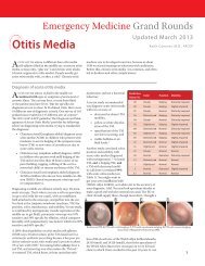
Appendix I: Oxygenation: the A-a gradient<br />
Evaluating the pO 2 on room air is easy: the lab usually prints out normal values for you, and even puts a little<br />
asterisk next to low values. But what if the patient's pO 2 is low normal, but the pCO 2 is below normal. Could this<br />
mean the patient is having to work hard to keep the pO 2 up, causing a low pCO 2 ? Could this mean the patient has a<br />
big pulmonary embolism or a bad pneumonia, despite a "normal" pO 2 ? You betcha. ‡‡<br />
Is there any scientific way to factor the pCO 2 into your figuring? Sure enough. This calculation has the<br />
intimidating name of the Aa Gradient. And, deriving it from first principles can be difficult. But using the formula is<br />
incredibly simple. Just keep it written down somewhere so you can find it when you need it.<br />
A-a Gradient (Alveolar-arterial Oxygen Gradient)<br />
Use: To quantify ABG results to estimate the average gradient across the alveolar membrane. An increased A-a<br />
gradient is seen in pulmonary embolism, pneumonias, ARDS, etc.<br />
Normal A-a gradient:<br />
Age<br />
Upper Limit of<br />
Normal<br />
20 17<br />
30 21<br />
40 24<br />
50 27<br />
60 31<br />
70 34<br />
80 38<br />
From: Morris A, Kamer R, Crapo R, et al. Clinical pulmonary function: a manual of uniform laboratory procedures,<br />
2E. Salt Lake City: InterMountain Thoracic Society, 1984:45. Quoted in Emergency Medicine Reports, 12/14/92.<br />
Normal values increase with age, and smoking or occupational pneumoconiosis will also increase the A-a gradient.<br />
Equation: A-a gradient = (713 (FIO 2 ) - 1.2 (pCO 2 )) - (pO 2 )<br />
For room air: A-a gradient is: 150 - 1.2(pCO 2 ) - pO 2<br />
Derivation:<br />
(pAlveolarO 2 ) = (Fractional Inspired O 2 ) * (Atmospheric Pressure - partial pressure of water in saturated air) -<br />
(pAlveolar CO 2 /R) -- (where R is the respiratory coefficient, the amount of CO 2 produced per molecule of O 2<br />
used) or:<br />
‡‡ One note: if trying to diagnose pulmonary embolism in a patient with chest pain, syncope, or unexplained tachycardia, don’t use the A-a gradient, or a pO2, or<br />
even a pulse oximeter reading to help you decide if it’s a PE. It just doesn’t help. Yes, by all means check the patient’s oxygenation to see how sick the patient is<br />
and whether the patient needs oxygen. But the majority of people with a PE will not have low oxygenation. Indeed, it’s even said (correctly) that, if you have two<br />
young healthy women with pleuritic chest pain, and one has a normal pulse ox and the other has a low pulse ox, the one with the normal pulse ox is more likely<br />
to have a PE. True. The one with the low pulse is is more likely to have a pneumonia.<br />
<strong>Page</strong> 8 of 9
<strong>Conover's</strong> ABG Handout<br />
pAO 2 = (FIO 2 (760-47)) - (pACO 2 /R) or,<br />
since rapid diffusion of CO 2 across the alveolar membrane means that pACO 2 and paCO 2 are the same:<br />
pAO 2 = 713(FIO 2 ) - 1.2(paCO 2 )<br />
therefore: A-a O 2 = (713(FIO 2 ) - 1.2(pCO 2 )) - pO 2<br />
Appendix II: Hypothermia and blood gases<br />
You may hear about "correcting blood gases for hypothermia." The blood gas blood is warmed to room<br />
temperature when the syringe is injected into the ABG machine. Therefore, the lab wants to know if the patient is<br />
hypothermic, so they can "correct" the numbers. Lab people want to "correct" the numbers so that they give you<br />
exactly correct numbers, reflecting exactly what the pH and pCO 2 are in the patient's blood. Despite these good<br />
intentions of the lab people, LIE TO THEM. If they ask if the patient is hypothermic, say no. If someone is taking the<br />
gas to the lab, and might tell the lab the patient is hypothermic, use duct tape over the person's mouth before<br />
sending him or her to the lab.<br />
Why? One very important reason.<br />
You've invested considerable time and energy in learning about room temperature blood gases. But, do you<br />
have any idea what a normal pH or pCO 2 is at 30°C? At 25°C? I don't, and hypothermia is one of my specialties.<br />
But it turns out that if you warm the blood gas to a normal temperature, and don't correct for hypothermia, then the<br />
numbers you get compare almost perfectly with a room-temperature gas. For example, the patient is hypothermic<br />
to 25°C. You get an uncorrected pH of 7.4. This means the patient is not acidotic (acidemic), and not alkalotic<br />
(alkalemic). If you got a "corrected" gas, you'd have to go look up in a table what the normal pH is for a core<br />
temperature of 25 °C. The normal pH at 25°C is not 7.4. Same thing for pCO 2 . The normal pCO 2 at 25°C is not 40.<br />
Got it?<br />
There is one exception, though. Correcting for temperature is appropriate when you're considering pO 2 on an<br />
arterial gas. Here's the trick I use: send the gas up without any mention of hypothermia, then use the pCO 2 and pH<br />
to figure out the patient's acid-base status. Then call the lab and say "oh, by the way, the patient's core temperature<br />
is 25°C, so you could you tell me the corrected pO 2 ?" And when they try to give you the corrected pH and pCO,<br />
just put your fingers in your ears.<br />
Well, now you know why you keep the lab people in the dark about your patient's temperature: to keep them<br />
from confusing you, and to let you use your room-temp acid-base knowledge to properly manage your patient.<br />
1. Touger M, Gallagher EJ, Tyrell J. Relationship between venous and arterial carboxyhemoglobin levels in<br />
patients with suspected carbon monoxide poisoning. Ann Emerg Med 1995;25(4):481-3.<br />
2. Gennis PR, Skovron ML, Aronson ST, Gallagher EJ. The usefulness of peripheral venous blood in estimating<br />
acid-base status in acutely ill patients. Ann Emerg Med 1985;14(9):845-9.<br />
3. Weil MH, Rackow EC, Trevino R, Grundler W, Falk JL, Griffel MI. Difference in acid-base state between<br />
venous and arterial blood during cardiopulmonary resuscitation. N Engl J Med 1986;315(3):153-6.<br />
4. Durkin R, Gergits MA, Reed JF, 3rd, Fitzgibbons J. The relationship between the arteriovenous carbon<br />
dioxide gradient and cardiac index. J Crit Care 1993;8(4):217-21.<br />
5. Adrogue HJ, Rashad MN, Gorin AB, Yacoub J, Madias NE. Assessing acid-base status in circulatory failure.<br />
Differences between arterial and central venous blood. N Engl J Med 1989;320(20):1312-6.<br />
<strong>Page</strong> 9 of 9
ABG Card<br />
Check pH on blood gas: acidemia (•pH)?<br />
alkalemia (•pH)? eupHemia (normal pH)?<br />
Check bicarb on chem panel and pCO 2<br />
on gas.<br />
•pH with •[HCO 3-<br />
], or •pH with •[HCO 3-<br />
], is<br />
primary metabolic acidosis or alkalosis.<br />
•pH with •pCO 2<br />
, or •pH with •pCO 2<br />
, is primary<br />
respiratory acidosis or alkalosis.<br />
Check for expected compensation:<br />
For metabolic acidosis, Winters’ equation:<br />
pCO 2<br />
= 1.5([HCO3 - ])+8±2 = last 2 digits of pH<br />
(lactic acidosis may overcompensate 2˚ CNS effects)<br />
For metabolic alkalosis or acidosis:<br />
pCO 2<br />
= last 2 digits of the pH = [HCO3-] + 15<br />
(for [HCO3 - ] from 8 to 35)<br />
For respiratory acidosis:<br />
•pCO 2<br />
10 = •pH .08 (acute)<br />
•pCO 2<br />
10 = •pH .03 (later: metabolic comp.)<br />
For respiratory alkalosis:<br />
•pCO 2<br />
10 = •[HCO3 - ] 2 (acute)<br />
•pCO 2<br />
10 = •[HCO3 - ] 5 (chronic/compensated)<br />
For metabolic acidosis, is it anion gap or non-anion<br />
gap acidosis? mixed disorder? AG = [Na + ] - ([Cl - ] +<br />
[HCO3 - ]). If all acidosis from anion-gap acids, then<br />
•AG should = •[HCO3 - ].<br />
If AG 20 (nl 12), [HCO3 - ] 19 (nl 27), then all acidosis<br />
(•[HCO3-] 8) is from •AG from excess organic acids.<br />
Normal Venous <strong>Blood</strong> Gas values:<br />
pH 7.32-7.42 0.03 less than arterial (7.35-7.45)<br />
pCO2 46 6 more than arterial: (40)<br />
2.1 7/09 <strong>Keith</strong> Conover, M.D., FACEP conovers.org
ABG Card<br />
High Anion Gap<br />
Metabolic<br />
Acidoses<br />
PaO<br />
SaO 2<br />
@pH<br />
2<br />
7.3<br />
PaO 2<br />
@pH<br />
7.4<br />
PaO 2<br />
@pH<br />
7.5<br />
Normal Anion Gap (Hyperchloremic)<br />
Metabolic Acidoses<br />
99 171 158 143<br />
98 122 111 101<br />
97 101 92 84<br />
96 89 82 74<br />
95 82 74 68<br />
94 76 69 63<br />
93 72 66 60<br />
92 68 62 57<br />
91 66 60 55<br />
90 63 58 53<br />
88 59 54 49<br />
86 56 51 47<br />
84 53 49 44<br />
82 51 47 42<br />
80 49 45 41<br />
78 47 43 39<br />
76 45 41 38<br />
74 44 40 35<br />
72 42 39 34<br />
K<br />
U<br />
S<br />
M<br />
A<br />
L<br />
ketoacidosis<br />
uremia<br />
salicylates<br />
methanol/ethylene glycol/paraldehyde<br />
alcohol (EtOH)<br />
lactic acidosis<br />
High K +<br />
hyperal, •amino acid catabolism p •pCO 2<br />
(takes<br />
while for HCO 3<br />
-<br />
to •)<br />
dilutional<br />
hypoaldo (•renin, adrenal dysfunction)<br />
NH4Cl, CaCl 2<br />
, lysine, arginine (≈adds HCl)<br />
Low K +<br />
GI – diarrhea, fistulas (K + loss, aldo)<br />
GU – surgical ureteral conduits<br />
RTA – Renal Tubular Acidosis. Complex.<br />
2.1 7/09 <strong>Keith</strong> Conover, M.D., FACEP conovers.org