pneumothorax BTS guidelines for the management of spontaneous
pneumothorax BTS guidelines for the management of spontaneous
pneumothorax BTS guidelines for the management of spontaneous

You also want an ePaper? Increase the reach of your titles
YUMPU automatically turns print PDFs into web optimized ePapers that Google loves.
Downloaded from thorax.bmj.com on 17 August 2008<br />
ii42<br />
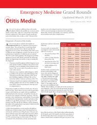
Henry, Arnold, Harvey<br />
• A lateral chest or lateral decubitus radiograph<br />
should be per<strong>for</strong>med if <strong>the</strong> clinical suspicion <strong>of</strong><br />
<strong>pneumothorax</strong> is high, but a PA radiograph is<br />
normal. [B]<br />
• CT scanning is recommended when differentiating a<br />
<strong>pneumothorax</strong> from complex bullous lung disease,<br />
when aberrant tube placement is suspected, and<br />
when <strong>the</strong> plain chest radiograph is obscured by<br />
surgical emphysema. [C]<br />
• The clinical history is not a reliable indicator <strong>of</strong><br />
<strong>pneumothorax</strong> size. [C]<br />
Clinical history and physical examination usually suggest <strong>the</strong><br />
presence <strong>of</strong> a <strong>pneumothorax</strong>, although clinical manifestations<br />
29 30<br />
are not reliable indicators <strong>of</strong> size. In general, <strong>the</strong> clinical<br />
symptoms associated with secondary pneumothoraces are<br />
more severe than those associated with primary pneumothoraces,<br />
and most patients with a secondary <strong>pneumothorax</strong><br />
complain <strong>of</strong> breathlessness which is out <strong>of</strong> proportion to <strong>the</strong><br />
31 32<br />
size <strong>of</strong> <strong>the</strong> <strong>pneumothorax</strong>. Many patients, particularly<br />
those with primary pneumothoraces, do not seek medical<br />
advice <strong>for</strong> several days, 46% waiting more than 2 days with<br />
symptoms. 9 This feature is important because <strong>the</strong> occurrence<br />
<strong>of</strong> re-expansion pulmonary oedema (RPO) after re-inflation<br />
may be related to <strong>the</strong> length <strong>of</strong> time <strong>the</strong> lung has been<br />
33 34<br />
collapsed (see section 4.4.1).<br />
Arterial blood gas measurements are frequently abnormal<br />
in patients with <strong>pneumothorax</strong> with <strong>the</strong> arterial oxygen tension<br />
(PaO 2<br />
) being less than 10.9 kPa (80 mm Hg) in 75% <strong>of</strong><br />
patients. 35 The presence <strong>of</strong> underlying lung disease along with<br />
<strong>the</strong> size <strong>of</strong> <strong>pneumothorax</strong> predicts <strong>the</strong> degree <strong>of</strong><br />
hypoxaemia. 35 Arterial PaO 2<br />
was below 7.5 kPa (55 mm Hg)<br />
and PaCO 2<br />
above 6.9 kPa (50 mm Hg) in 16% <strong>of</strong> cases <strong>of</strong><br />
secondary <strong>pneumothorax</strong> in <strong>the</strong> largest reported series. 36 Pulmonary<br />
function tests are weakly sensitive measures <strong>of</strong> <strong>the</strong><br />
presence or size <strong>of</strong> <strong>pneumothorax</strong> and are not<br />
recommended. 8<br />
In both primary and secondary <strong>spontaneous</strong> <strong>pneumothorax</strong><br />
<strong>the</strong> diagnosis is normally established by plain chest radiography.<br />
In general, expiratory radiographs add little and are<br />
not indicated as a routine investigation, even in <strong>the</strong> case <strong>of</strong> a<br />
37 38<br />
suspected small apical <strong>pneumothorax</strong>. When a <strong>pneumothorax</strong><br />
is suspected but not confirmed by standard posteroanterior<br />
(PA) chest radiographs, lateral radiographs provide<br />
added in<strong>for</strong>mation in up to 14% <strong>of</strong> cases. 39 The lateral decubitus<br />
radiograph is superior to <strong>the</strong> erect or supine chest<br />
radiograph and is felt to be as sensitive as CT scanning in<br />
<strong>pneumothorax</strong> detection. 40 The upright lateral or lateral decubitus<br />
radiograph is clinically helpful where findings on <strong>the</strong><br />
upright PA radiograph are unclear. While such small<br />
pneumothoraces may not have much clinical relevance in<br />
patients without underlying lung disease, in patients with<br />
suspected secondary pneumothoraces, even small pneumothoraces<br />
may have significant implications and here lateral or<br />
lateral decubitus radiographs are probably valuable. In<br />
patients with severe bullous lung disease CT scanning will differentiate<br />
emphysematous bullae from pneumothoraces and<br />
save <strong>the</strong> patient an unnecessary and potentially dangerous<br />
aspiration. 41<br />
3 SIZE OF PNEUMOTHORAX<br />
• The previous classification <strong>of</strong> <strong>the</strong> size <strong>of</strong> a <strong>pneumothorax</strong><br />
tends to underestimate its volume. In <strong>the</strong>se<br />
new <strong>guidelines</strong> <strong>the</strong> size <strong>of</strong> a <strong>pneumothorax</strong> is divided<br />
into “small” or “large” depending on <strong>the</strong> presence <strong>of</strong><br />
a visible rim <strong>of</strong> 2 cm between <strong>the</strong> lung<br />
margin and <strong>the</strong> chest wall.<br />
The plain PA radiograph is a poor method <strong>of</strong> quantifying <strong>the</strong><br />
size <strong>of</strong> a <strong>pneumothorax</strong> as it usually underestimates it. Exact<br />
methods <strong>of</strong> estimating size from PA chest radiographs are<br />
cumbersome and generally used only as a research tool. 42 The<br />
size <strong>of</strong> a <strong>pneumothorax</strong>, in terms <strong>of</strong> volume, is difficult to<br />
assess accurately from a chest radiograph which is a two<br />
dimensional image. In <strong>the</strong> 1993 <strong>guidelines</strong> 19 pneumothoraces<br />
were classified into three groups:<br />
• “small”: defined as a “small rim <strong>of</strong> air around <strong>the</strong> lung”;<br />
• “moderate”: defined as lung “collapsed halfway towards<br />
<strong>the</strong> heart border”; and<br />
• “complete”: defined as “airless lung, separate from <strong>the</strong> diaphragm”.<br />
This attempt to quantify a <strong>pneumothorax</strong> tends to underestimate<br />
<strong>the</strong> volume <strong>of</strong> anything greater than <strong>the</strong> smallest <strong>of</strong><br />
pneumothoraces. 1<br />
Since <strong>the</strong> volume <strong>of</strong> a <strong>pneumothorax</strong><br />
approximates to <strong>the</strong> ratio <strong>of</strong> <strong>the</strong> cube <strong>of</strong> <strong>the</strong> <strong>of</strong> <strong>the</strong> lung diameter<br />
to <strong>the</strong> hemithorax diameter, a <strong>pneumothorax</strong> <strong>of</strong> 1 cm on<br />
<strong>the</strong> PA chest radiograph occupies about 27% <strong>of</strong> <strong>the</strong> hemithorax<br />
volume if <strong>the</strong> lung is 9 cm in diameter and <strong>the</strong> hemithorax<br />
10 cm: (10 3 –9 3 )/10 3 = 27%. Similarly, a 2 cm radiographic<br />
<strong>pneumothorax</strong> occupies 49% <strong>of</strong> <strong>the</strong> hemithorax on <strong>the</strong> same<br />
basis (fig 3).<br />
In view <strong>of</strong> <strong>the</strong> proximity <strong>of</strong> <strong>the</strong> lung surface to <strong>the</strong> chest wall<br />
in a <strong>pneumothorax</strong> <strong>of</strong>