Technical Bulletin #455 Methods for Implantation of Corning ...
Technical Bulletin #455 Methods for Implantation of Corning ...
Technical Bulletin #455 Methods for Implantation of Corning ...

Create successful ePaper yourself
Turn your PDF publications into a flip-book with our unique Google optimized e-Paper software.
<strong>Technical</strong> <strong>Bulletin</strong> <strong>#455</strong><br />
<strong>Methods</strong> <strong>for</strong> <strong>Implantation</strong> <strong>of</strong> <strong>Corning</strong> ® Matrigel ® Matrix into Mice and Tissue<br />
Fixation<br />
1<br />
Kazuo Ohashi, M.D., Ph.D., 1 Takashi Yokoyama, M.D., 1 Yoshiyuki Nakajima, M.D., Ph.D., and 2 Marshall Kosovsky, Ph.D.<br />
1<br />
Nara Medical University, Nara City, Nara JAPAN; 2 <strong>Corning</strong> Incorporated, Tewksbury, MA, USA<br />
Introduction<br />
<strong>Corning</strong> Matrigel Matrix is a solubilized<br />
basement membrane preparation<br />
extracted from Engelbreth-Holm-Swarm<br />
(EHS) mouse sarcoma, a tumor rich in<br />
ECM proteins. Its major component<br />
is laminin, followed by collagen IV,<br />
heparan sulfate proteo-glycan, and<br />
entactin. <strong>Corning</strong> Matrigel Matrix<br />
is effective <strong>for</strong> the attachment and<br />
differentiation <strong>of</strong> both normal and<br />
trans<strong>for</strong>med anchorage-dependent<br />
epithelial and other cell types.<br />
<strong>Corning</strong> Matrigel Matrix is highly<br />
useful in various studies including 3D<br />
cell culture, cell invasion and migration<br />
assays, drug metabolism/toxicology, in<br />
vitro and in vivo angiogenesis assays.<br />
This report describes the use <strong>of</strong> <strong>Corning</strong><br />
Matrigel Matrix <strong>for</strong> in vivo applications<br />
such as angiogenesis and human tumor<br />
cell implantation in mice. 1-8<br />
1. <strong>Corning</strong> Matrigel Matrix (Cat. Nos.<br />
354234 and 356234) is suitable as a<br />
scaffold <strong>for</strong> supporting the implantation<br />
<strong>of</strong> various tumor cells. Growth<br />
Factor Reduced (GFR) <strong>Corning</strong><br />
Matrigel Matrix (Cat. Nos. 354230<br />
and 356230) is also available <strong>for</strong><br />
studies in which a reduced growth<br />
factor composition is required.<br />
2. <strong>Corning</strong> Matrigel Matrix phenol<br />
red-free (Cat. No. 356237) and<br />
GFR <strong>Corning</strong> Matrigel Matrix,<br />
phenol red-free (Cat. No. 356231)<br />
are typically used <strong>for</strong> the cyanmetrohemoglobin<br />
method that<br />
measures hemoglobin content<br />
(measurement <strong>of</strong> reddish-brown<br />
absorption) in angiogenesis studies.<br />
<strong>Corning</strong> Matrigel Matrix has been<br />
shown to enhance the process <strong>of</strong><br />
angiogenesis in vivo.<br />
3. <strong>Corning</strong> Matrigel Matrix High<br />
Concentration (HC) (Cat No. 354248)<br />
is suited <strong>for</strong> in vivo applications where<br />
a high protein concentration augments<br />
growth <strong>of</strong> tumors. The high protein<br />
concentration (18-22 mg/mL) also<br />
allows the <strong>Corning</strong> Matrigel Matrix<br />
plug to maintain its integrity after<br />
subcutaneous injection into mice. This<br />
keeps the injected tumor cells and/or<br />
angiogenic compounds localized <strong>for</strong><br />
in situ analysis and/or future excision.<br />
Procedures<br />
Subcutaneous injection <strong>of</strong> <strong>Corning</strong><br />
Matrigel Matrix into a mouse<br />
1. Since <strong>Corning</strong> Matrigel Matrix <strong>for</strong>ms<br />
a gel above 10°C, <strong>Corning</strong> Matrigel<br />
Matrix solution should be kept at<br />
low temperatures, and thus all equipment<br />
and reagents (syringes, needles,<br />
<strong>Corning</strong> Matrigel Matrix solution,<br />
etc.) should be chilled on ice prior to<br />
injection.<br />
2. After mixing <strong>Corning</strong> Matrigel Matrix<br />
with a cell suspension [Note 1], the<br />
<strong>Corning</strong> Matrigel mixture is injected<br />
into a mouse subcutaneously [Note<br />
2] (Figure 1). An appropriate needle<br />
size (21-25G) should be selected<br />
to prevent the destruction <strong>of</strong> cells.<br />
To increase the contact area <strong>of</strong> the<br />
injected <strong>Corning</strong> Matrigel mixture<br />
into subcutaneous tissues, a wide<br />
subcu-taneous pocket should be<br />
<strong>for</strong>med by swaying the needlepoint<br />
right and left after a routine subcutaneous<br />
insertion. The <strong>Corning</strong><br />
Matrigel mixture is then injected<br />
into the pocket. When the <strong>Corning</strong><br />
Matrigel mixture is injected into a<br />
particular area without swaying the<br />
needlepoint, the mixture will <strong>for</strong>m<br />
a large cell clump and a subsequent<br />
growth defect may result due to<br />
inefficient perfusion <strong>of</strong> nutrients to<br />
the cells within the core <strong>of</strong> the clump.<br />
Note 1: In this experiment, undiluted <strong>Corning</strong><br />
Matrigel Matrix alone was injected into<br />
the mouse. For tumor implantation applications,<br />
approximately 2x10 7 cells/mL <strong>of</strong> cell<br />
suspension should be mixed with <strong>Corning</strong><br />
Matrigel Matrix, resulting in a final cell<br />
concentration <strong>of</strong> ~10 6 cells/mL. To prevent<br />
incomplete gel <strong>for</strong>mation in mice, do not<br />
dilute <strong>Corning</strong> Matrigel Matrix to a final<br />
concentration below 4 mg/mL.<br />
Note 2: In this experiment, 0.7 mL <strong>of</strong> the<br />
<strong>Corning</strong> Matrigel was injected. The injection<br />
volume <strong>of</strong> <strong>Corning</strong> Matrigel takes into<br />
account the absorption <strong>of</strong> <strong>Corning</strong> Matrigel<br />
into the tissue and allows <strong>for</strong> easy removal<br />
<strong>of</strong> the resultant tissue ‘plug’. The optimal<br />
injection volume should be determined<br />
according to the requirements <strong>of</strong> your<br />
experiment.<br />
While the injection <strong>of</strong> ~0.1 mL <strong>of</strong> a <strong>Corning</strong><br />
Matrigel mixture into mice may be sufficient<br />
<strong>for</strong> the augmentation <strong>of</strong> tumor growth, the<br />
injection <strong>of</strong> at least 0.5 mL is recommended<br />
<strong>for</strong> in vivo angiogenesis studies.<br />
Figure 1. Subcutaneous injection site.<br />
Removal <strong>of</strong> the <strong>Corning</strong> Matrigel<br />
plug from the mouse<br />
3. After an appropriate incubation<br />
period [Note 3], the mouse is<br />
anaesthetized and a square segment<br />
<strong>of</strong> tissue is excised with scissors. To<br />
ensure complete excision <strong>of</strong> the plug,<br />
cut ~5 mm wider than the implantion<br />
site on all sides.To maintain the<br />
shape <strong>of</strong> the <strong>Corning</strong> Matrigel plug,<br />
excise the subcutaneous tissue,<br />
peritoneum, as well as skin. These<br />
tissues are then fixed with <strong>for</strong>malin.<br />
Figure 3 shows the implanted<br />
<strong>Corning</strong> Matrigel viewed from the<br />
peritoneal side following excision.<br />
The volume <strong>of</strong> the implanted <strong>Corning</strong><br />
Matrigel is reduced from the injected<br />
volume due to absorption and partial<br />
degradation <strong>of</strong> <strong>Corning</strong> Matrigel in<br />
vivo. The excised <strong>Corning</strong> Matrigel<br />
plug is usually clear yellowish in<br />
color. If blood vessels are <strong>for</strong>med<br />
within the <strong>Corning</strong> Matrigel plug, the<br />
color <strong>of</strong> the <strong>Corning</strong> Matrigel will<br />
appear red (Figure 4).<br />
Note 3: In this experiment, the <strong>Corning</strong><br />
Matrigel plug was removed after one week.<br />
When the quantity <strong>of</strong> hemoglobin is used<br />
to assess angiogenesis, <strong>Corning</strong> Matrigel<br />
containing VEGF and heparin should be<br />
injected to promote angiogenesis. After<br />
about three days, the <strong>Corning</strong> Matrigel plug<br />
containing newly <strong>for</strong>med blood vessels can<br />
be easily removed.
Figure 2. An arrow indicates the <strong>Corning</strong><br />
Matrigel injection site. A square indicates the<br />
excised region <strong>for</strong> the sample.<br />
Figure 3. The implanted <strong>Corning</strong> Matrigel plug<br />
viewed from the peritoneal side (needlepoint).<br />
Note 4: Fixation <strong>of</strong> <strong>Corning</strong> ® Matrigel ® under<br />
8°C may cause depolymerization <strong>of</strong> <strong>Corning</strong><br />
Matrigel. There<strong>for</strong>e, <strong>Corning</strong> Matrigel should<br />
be fixed at room temperature.<br />
5. The fixed <strong>Corning</strong> Matrigel plug can<br />
be embedded in paraffin to prepare<br />
sections <strong>for</strong> histochemical staining.<br />
Figure 7 shows a section <strong>of</strong> the<br />
<strong>Corning</strong> Matrigel plug stained with<br />
hematoxylin-eosin (HE). <strong>Corning</strong><br />
Matrigel appears pink to light<br />
reddish in color with HE staining.<br />
Figure 5. The removed tissue was<br />
fixed in a nylon bag. The skin side<br />
is facing upward.<br />
References<br />
1. Passaniti, A., et al., A simple, quantitative<br />
method <strong>for</strong> assessing angiogenesis and<br />
anti-angiogenic agents using reconstituted<br />
basement membrane, heparin, and fibroblast<br />
growth factor. Lab Invest. 67:519 (1992).<br />
2. Kragh, M., et al., In vivo chamber angiogenesis<br />
assay: An optimized Matrigel plug<br />
assay <strong>for</strong> fast assessment <strong>of</strong> anti-angiogenic<br />
activity. Int’l Journal <strong>of</strong> Oncology 22:305<br />
(2003).<br />
3 Bandyopadhyay, A., et al., Extracellular<br />
domain <strong>of</strong> TGFbeta type III receptor inhibits<br />
angiogenesis and tumor growth in human<br />
cancer cells. Oncogene 21:3541 (2002).<br />
4 Noel, A., et al., Heterotransplantation <strong>of</strong><br />
primary and established human tumor cells<br />
in nude mice. Anticancer Res. 15:1 (1995).<br />
5. Glondu, M., et al., A mutated cathepsin-D<br />
devoid <strong>of</strong> its catalytic activity stimulates the<br />
growth <strong>of</strong> cancer cells. Oncogene 20:6920<br />
(2001).<br />
6. Yue, W., et al., Tumor cells implantation.<br />
MCF-7 human breast carcinomas in nude<br />
mice as a model <strong>for</strong> evaluating aromatase<br />
inhibitors. J. Steroid Biochem. Molec. Biol.<br />
44(4-6):671 (1993).<br />
7. Isaji, M., et al., Angiogenesis evaluation by<br />
measuring hemoglobin content. Tranilast<br />
inhibits the proliferation, chemotaxis and<br />
tube <strong>for</strong>mation <strong>of</strong> human microvascular<br />
endothelial cells in vitro and angiogenesis<br />
in vivo. British Journal <strong>of</strong> Pharmacology<br />
122:1061 (1997).<br />
8. Ohashi, K., et al., Hepatocytes implantation.<br />
Sustained survival <strong>of</strong> human hepatocytes<br />
in mice: A model <strong>for</strong> in vivo infection with<br />
human hepatitis B and hepatitis delta<br />
viruses. Nature Medicine 6(3):327(2000).<br />
Data provided by Dr. Kazuo Ohashi and<br />
Dr. Takashi Yokoyama at Nara Medical<br />
University.<br />
Figure 4. The removed <strong>Corning</strong> Matrigel plug<br />
from the subcutaneous tissue.<br />
Fixation <strong>of</strong> tissues including<br />
<strong>Corning</strong> Matrigel Matrix<br />
4. The excised tissue should be stretched<br />
and put on a sheet <strong>of</strong> thick paper<br />
(e.g., poster board) to avoid the<br />
<strong>for</strong>mation <strong>of</strong> wrinkles. The tissue<br />
is then placed in a nylon bag <strong>for</strong><br />
protection. Fix the tissue in 10%<br />
neutralized <strong>for</strong>malin solution <strong>for</strong> at<br />
least one day at room temperature<br />
[Note 4]. This treatment will harden<br />
the tissue in preparation <strong>for</strong> slicing<br />
the sample (Figure 6). Care should<br />
be taken to ensure that the thickness<br />
<strong>of</strong> the slice is adequate to retain the<br />
implanted <strong>Corning</strong> Matrigel plug.<br />
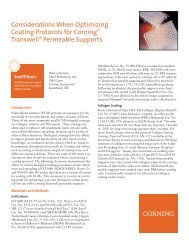
Figure 6. Arrows indicate the cut<br />
surface <strong>of</strong> the fixed <strong>Corning</strong> Matrigel<br />
plug. (Upper arrow = Skin; Middle<br />
arrow = <strong>Corning</strong> Matrigel; Lower<br />
arrow = muscle layer).<br />
Figure 7. HE stain image.<br />
<strong>Corning</strong> acquired the Discovery Labware Business including the Matrigel ® brand.<br />
For in<strong>for</strong>mation, visit www.corning.com/discoverylabware.<br />
For Research Use Only. Not <strong>for</strong> use in diagnostic or therapeutic procedures.<br />
For a listing <strong>of</strong> trademarks, visit us at www.corning.com/lifesciences/trademarks.<br />
All other trademarks are property <strong>of</strong> their respective owners.<br />
<strong>Corning</strong> Incorporated, One Riverfront Plaza, <strong>Corning</strong>, NY 14831-0001<br />
Skin<br />
<strong>Corning</strong> Matrigel Matrix<br />
Muscle Layer<br />
<strong>Corning</strong> Incorporated<br />
Life Sciences<br />
836 North St.<br />
Building 300, Suite 3401<br />
Tewksbury, MA 01876<br />
t 800.492.1110<br />
t 978.442.2200<br />
f 978.442.2476<br />
www.corning.com/lifesciences<br />
02/13 CLS-DL-CC-036<br />
© 2012, 2013 <strong>Corning</strong> Incorporated Printed in USA