Genetics characterization, nutritional and phytochemicals potential of gedi leaves (Abelmoschus manihot (L.) Medik) growing in the North Sulawesi of Indonesia as a candidate of poultry feed
Gedi, local name of Abelmoschus manihot (L.) Medik was used by local people in Northern Sulawesi-Indonesia as vegetable, because of its medicinal properties. The potency of gedi leaves in broiler diet has not been reported in literatures. The objective of this research was to investigate a genetic diversity of gedi commonly consumed as a gourmet cuisine in the North Sulawesi of Indonesia, and exploring the potential of this plant as a herb plant for a candidate of poultry feedstuff. Eight morphologically different gedi leaves (GH1, GH2, GH3, GH4, GH5, GH6, GM1 and GM2) that grow in Manado area, North Sulawesi of Indonesia were collected and identified. The leaves were extracted for DNA isolation followed by PCR and DNA sequencing analysis. During DNA isolation, 3 of 6 GH (GH4, GH5, GH6) were discontinued because of difficulty in separating the mucilage properties. Following PCR analysis, GH2 and GH3 did not produce bands and consequently were excluded from further analysis. In addition to that, chemical analysis was also performed to determine the phytochemical and nutritional contents .The results indicated that all gedi leaf samples showed similarity (99%) to species member of Abelmoschus manihot, and tribe of Malvaceae. In terms of proximate analysis, gedi leaves showed high crude protein (18.76 - 24.16%) and calcium (2.92-3.70%) content. Also, showed high crude fibre (13.06-17.53%). Together with the presence of alkaloid and steroidal saponin gedi leaves may offer beneficial effects as poultry feedstuff to a special production trait such as cholesterol-less meat.
Gedi, local name of Abelmoschus manihot (L.) Medik was used by local people in Northern Sulawesi-Indonesia as vegetable, because of its medicinal properties. The potency of gedi leaves in broiler diet has not been reported in literatures. The objective of this research was to investigate a genetic diversity of gedi commonly consumed as a gourmet cuisine in the North Sulawesi of Indonesia, and exploring the potential of this plant as a herb plant for a candidate of poultry feedstuff. Eight morphologically different gedi leaves (GH1, GH2, GH3, GH4, GH5, GH6, GM1 and GM2) that grow in Manado area, North Sulawesi of Indonesia were collected and identified. The leaves were extracted for DNA isolation followed by PCR and DNA sequencing analysis. During DNA isolation, 3 of 6 GH (GH4, GH5, GH6) were discontinued because of difficulty in separating the mucilage properties. Following PCR analysis, GH2 and GH3 did not produce bands and consequently were excluded from further analysis. In addition to that, chemical analysis was also performed to determine the phytochemical and nutritional contents .The results indicated that all gedi leaf samples showed similarity (99%) to species member of Abelmoschus manihot, and tribe of Malvaceae. In terms of proximate analysis, gedi leaves showed high crude protein (18.76 - 24.16%) and calcium (2.92-3.70%) content. Also, showed high crude fibre (13.06-17.53%). Together with the presence of alkaloid and steroidal saponin gedi leaves may offer beneficial effects as poultry feedstuff to a special production trait such as cholesterol-less meat.

You also want an ePaper? Increase the reach of your titles
YUMPU automatically turns print PDFs into web optimized ePapers that Google loves.
M<strong>and</strong>ey et al., 2014<br />
diethyl e<strong>the</strong>r. It is added with one drop <strong>of</strong> H 2 SO 4 <strong>and</strong> one<br />
drop <strong>of</strong> CH 3 COOH anhydrate. The presence <strong>of</strong> steroids<br />
w<strong>as</strong> <strong>in</strong>dicated by <strong>the</strong> alteration <strong>of</strong> violet to blue or green<br />
color. The formation <strong>of</strong> reddish violet color to <strong>the</strong><br />
<strong>in</strong>terface w<strong>as</strong> formed that <strong>in</strong>dicat<strong>in</strong>g positive sign for<br />
triterpenoids.<br />
Test for hydroqu<strong>in</strong>ons<br />
One gram sample w<strong>as</strong> boiled with methanol for<br />
few m<strong>in</strong>utes. The filtrate w<strong>as</strong> allowed to cool <strong>and</strong> <strong>the</strong>n<br />
added with 3 drops <strong>of</strong> NaOH 10%. The appearance <strong>of</strong><br />
red color <strong>in</strong>dicated <strong>the</strong> presence <strong>of</strong> hydroqu<strong>in</strong>one.<br />
Nutritional Analysis<br />
The proximate analysis were carried out <strong>in</strong><br />
duplicates <strong>and</strong> <strong>the</strong> results obta<strong>in</strong>ed were <strong>the</strong> average<br />
values. The proximate analysis (prote<strong>in</strong>, crude fiber,<br />
crude fat, carbohydrate <strong>and</strong> <strong>as</strong>h) <strong>of</strong> five types <strong>of</strong> <strong>gedi</strong> leaf<br />
were determ<strong>in</strong>ed by us<strong>in</strong>g <strong>the</strong> Association <strong>of</strong> Official <strong>of</strong><br />
Analytical Chemists (AOAC) methods (1980). Nutrient<br />
contents were valued <strong>in</strong> percentage. The energy value<br />
w<strong>as</strong> determ<strong>in</strong>ed by bomb calorie meter.<br />
RESULTS AND DISCUSSION<br />
Plant Identification<br />
Two typical colors <strong>of</strong> <strong>gedi</strong> <strong>leaves</strong> (green <strong>and</strong><br />
reddish green <strong>leaves</strong>) <strong>grow<strong>in</strong>g</strong> at eight locations <strong>in</strong><br />
Manado area were presented <strong>in</strong> Figure 1. All <strong>leaves</strong> <strong>of</strong><br />
this plant do not have <strong>the</strong> same size or even appearance.<br />
They vary <strong>in</strong> size, color, <strong>and</strong> even shape. The results <strong>of</strong><br />
plant identification <strong>of</strong> eight accessions <strong>of</strong> <strong>gedi</strong> leaf were<br />
summarized <strong>in</strong> Table 1. Those have been recognized that<br />
all <strong>of</strong> eight accessions <strong>of</strong> <strong>gedi</strong> leaf <strong>in</strong> this research were<br />
species <strong>of</strong> <strong>Abelmoschus</strong> <strong>manihot</strong> (L.) <strong>Medik</strong>, tribe<br />
Malvaceae. Breen (2012) reported that <strong>leaves</strong> are <strong>of</strong>ten<br />
<strong>the</strong> b<strong>as</strong>is for identify<strong>in</strong>g plants s<strong>in</strong>ce <strong>the</strong>y are so e<strong>as</strong>ily<br />
observed.<br />
The boundaries <strong>of</strong> <strong>the</strong> eight accessions <strong>of</strong> <strong>gedi</strong><br />
from <strong>the</strong> different locations <strong>of</strong> Manado area were b<strong>as</strong>ed<br />
on morphological features <strong>of</strong> <strong>the</strong> species. The<br />
phylogenetic hypo<strong>the</strong>ses were tested us<strong>in</strong>g chloropl<strong>as</strong>t<br />
DNA sequence <strong>of</strong> ndhF. Total genomic DNA were<br />
extracted from eight accessions <strong>of</strong> fresh leaf material,<br />
<strong>and</strong> <strong>the</strong> ndhF gene w<strong>as</strong> amplified <strong>in</strong> PCR us<strong>in</strong>g primer.<br />
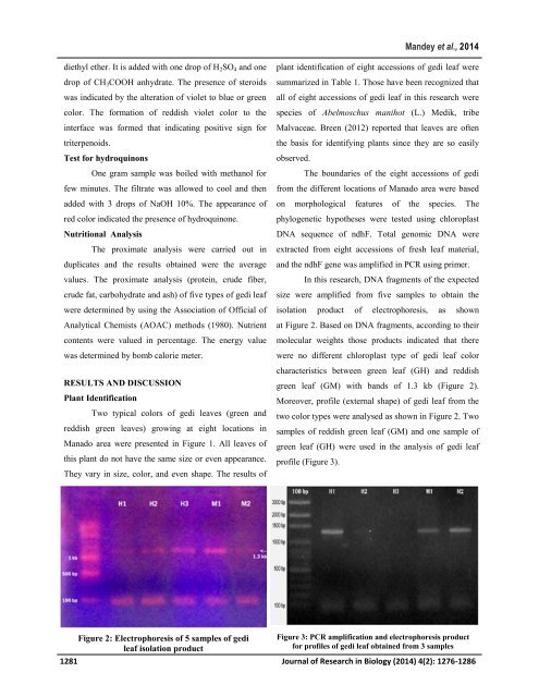
In this research, DNA fragments <strong>of</strong> <strong>the</strong> expected<br />
size were amplified from five samples to obta<strong>in</strong> <strong>the</strong><br />
isolation product <strong>of</strong> electrophoresis, <strong>as</strong> shown<br />
at Figure 2. B<strong>as</strong>ed on DNA fragments, accord<strong>in</strong>g to <strong>the</strong>ir<br />
molecular weights those products <strong>in</strong>dicated that <strong>the</strong>re<br />
were no different chloropl<strong>as</strong>t type <strong>of</strong> <strong>gedi</strong> leaf color<br />
characteristics between green leaf (GH) <strong>and</strong> reddish<br />
green leaf (GM) with b<strong>and</strong>s <strong>of</strong> 1.3 kb (Figure 2).<br />
Moreover, pr<strong>of</strong>ile (external shape) <strong>of</strong> <strong>gedi</strong> leaf from <strong>the</strong><br />
two color types were analysed <strong>as</strong> shown <strong>in</strong> Figure 2. Two<br />
samples <strong>of</strong> reddish green leaf (GM) <strong>and</strong> one sample <strong>of</strong><br />
green leaf (GH) were used <strong>in</strong> <strong>the</strong> analysis <strong>of</strong> <strong>gedi</strong> leaf<br />
pr<strong>of</strong>ile (Figure 3).<br />
Figure 2: Electrophoresis <strong>of</strong> 5 samples <strong>of</strong> <strong>gedi</strong><br />
leaf isolation product<br />
Figure 3: PCR amplification <strong>and</strong> electrophoresis product<br />
for pr<strong>of</strong>iles <strong>of</strong> <strong>gedi</strong> leaf obta<strong>in</strong>ed from 3 samples<br />
1281 Journal <strong>of</strong> Research <strong>in</strong> Biology (2014) 4(2): 1276-1286