neuropathy: gait changes in the diabetic foot - Wounds UK
neuropathy: gait changes in the diabetic foot - Wounds UK
neuropathy: gait changes in the diabetic foot - Wounds UK

You also want an ePaper? Increase the reach of your titles
YUMPU automatically turns print PDFs into web optimized ePapers that Google loves.
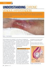
Review<br />
NEUROPATHY: GAIT CHANGES<br />
IN THE DIABETIC FOOT<br />
Motor <strong>neuropathy</strong> <strong>in</strong> patients with diabetes can lead to weakness <strong>in</strong> <strong>the</strong> muscles of <strong>the</strong> <strong>foot</strong> and lower<br />
leg, which <strong>in</strong> turn can lead to characteristic <strong>changes</strong> to <strong>the</strong> structure of <strong>the</strong> <strong>foot</strong>, such as an altered<br />
arch profile. Such structural <strong>changes</strong> often occur at sites of abnormally high pressure, which can<br />
result <strong>in</strong> tissue breakdown and ulceration particularly <strong>in</strong> <strong>in</strong>dividuals who also have sensory <strong>neuropathy</strong>.<br />
Veronica Newton and Carol<strong>in</strong>e McIntosh<br />
are Senior Lecturers <strong>in</strong> Podiatry, Centre<br />
for Health and Social Care Research,<br />
University of Huddersfield<br />
Peripheral <strong>neuropathy</strong> is <strong>the</strong><br />
dysfunction of <strong>the</strong> peripheral<br />
nerves (<strong>the</strong> nerves that are<br />
situated outside <strong>the</strong> bra<strong>in</strong> and<br />
sp<strong>in</strong>al cord), and it is a common<br />
problem for many people liv<strong>in</strong>g<br />
with diabetes. Published data<br />
has suggested that up to 50% of<br />
people with diabetes who present<br />
at <strong>diabetic</strong> <strong>foot</strong> cl<strong>in</strong>ics may have<br />
<strong>the</strong> condition (Edmonds and<br />
Foster, 1999).<br />
Peripheral <strong>neuropathy</strong> can<br />
affect many of <strong>the</strong> body’s nerve<br />
pathways, <strong>in</strong>clud<strong>in</strong>g <strong>the</strong> sensory<br />
nerves, autonomic nerves and<br />
motor nerves. Loss of sensation,<br />
known as sensory <strong>neuropathy</strong>,<br />
is commonly observed <strong>in</strong> <strong>the</strong><br />
<strong>diabetic</strong> <strong>foot</strong> (because raised<br />
levels of circulat<strong>in</strong>g glucose<br />
<strong>in</strong> <strong>the</strong> blood stream can over<br />
time permanently <strong>in</strong>terfere with<br />
normal nerve function. The<br />
typical cl<strong>in</strong>ical features <strong>in</strong>clude a<br />
loss of sensitivity to touch, pa<strong>in</strong>,<br />
temperature and vibration.<br />
Common cl<strong>in</strong>ical tests that<br />
specifically assess sensory<br />
loss <strong>in</strong>clude monofilaments, for<br />
light touch, and tun<strong>in</strong>g forks,<br />
for vibration perception. The<br />
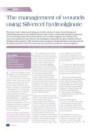
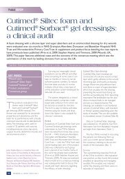
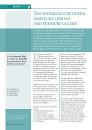
Clawed toes<br />
10g monofilament (National<br />
Institute for Health and Cl<strong>in</strong>ical<br />
Excellence [NICE], 2004) is a<br />
nylon fibre mounted on a hand<br />
held wand/pen structure. The<br />
fibre compresses at a given<br />
force when applied to nom<strong>in</strong>ated<br />
areas <strong>in</strong>form<strong>in</strong>g <strong>the</strong> practitioner if<br />
<strong>the</strong> person has <strong>the</strong> ability to feel<br />
pressure/pa<strong>in</strong>. The hand-held<br />
128Hz tun<strong>in</strong>g fork is p<strong>in</strong>ched<br />
toge<strong>the</strong>r at its far<strong>the</strong>st po<strong>in</strong>t and<br />
released to generate a vibration.<br />
The s<strong>in</strong>gle end of <strong>the</strong> fork is <strong>the</strong>n<br />
gently applied to <strong>the</strong> nom<strong>in</strong>ated<br />
area <strong>in</strong>form<strong>in</strong>g <strong>the</strong> practitioner if<br />
<strong>the</strong> person has <strong>the</strong> ability to feel<br />
vibration (Baker et al, 2005).<br />
Peripheral <strong>neuropathy</strong>, which is<br />
secondary to diabetes, does not<br />
just affect <strong>the</strong> sensory nerves — it<br />
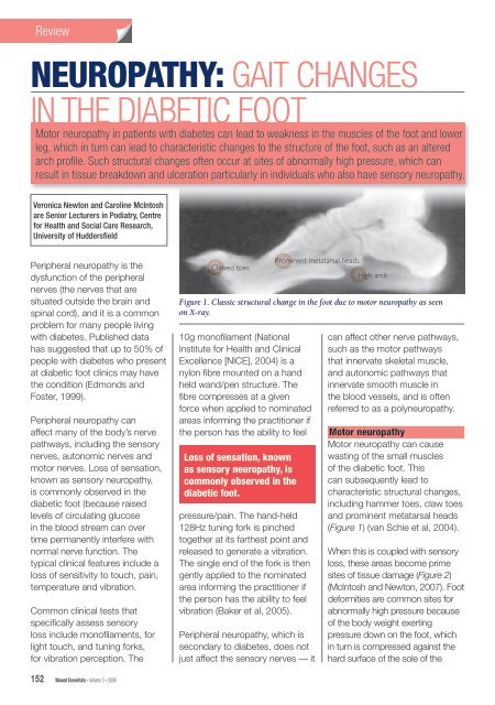
Prom<strong>in</strong>ent metatarsal heads<br />
High arch<br />
Figure 1. Classic structural change <strong>in</strong> <strong>the</strong> <strong>foot</strong> due to motor <strong>neuropathy</strong> as seen<br />
on X-ray.<br />
Loss of sensation, known<br />
as sensory <strong>neuropathy</strong>, is<br />
commonly observed <strong>in</strong> <strong>the</strong><br />
<strong>diabetic</strong> <strong>foot</strong>.<br />
can affect o<strong>the</strong>r nerve pathways,<br />
such as <strong>the</strong> motor pathways<br />
that <strong>in</strong>nervate skeletal muscle,<br />
and autonomic pathways that<br />
<strong>in</strong>nervate smooth muscle <strong>in</strong><br />
<strong>the</strong> blood vessels, and is often<br />
referred to as a poly<strong>neuropathy</strong>.<br />
Motor <strong>neuropathy</strong><br />
Motor <strong>neuropathy</strong> can cause<br />
wast<strong>in</strong>g of <strong>the</strong> small muscles<br />
of <strong>the</strong> <strong>diabetic</strong> <strong>foot</strong>. This<br />
can subsequently lead to<br />
characteristic structural <strong>changes</strong>,<br />
<strong>in</strong>clud<strong>in</strong>g hammer toes, claw toes<br />
and prom<strong>in</strong>ent metatarsal heads<br />
(Figure 1) (van Schie et al, 2004).<br />
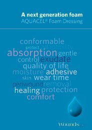
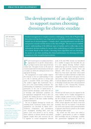
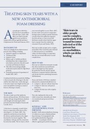
When this is coupled with sensory<br />
loss, <strong>the</strong>se areas become prime<br />
sites of tissue damage (Figure 2)<br />
(McIntosh and Newton, 2007). Foot<br />
deformities are common sites for<br />
abnormally high pressure because<br />
of <strong>the</strong> body weight exert<strong>in</strong>g<br />
pressure down on <strong>the</strong> <strong>foot</strong>, which<br />
<strong>in</strong> turn is compressed aga<strong>in</strong>st <strong>the</strong><br />
hard surface of <strong>the</strong> sole of <strong>the</strong><br />
152 Wound Essentials • Volume 3 • 2008
Review<br />
Figure 2. Structural <strong>changes</strong> with<br />
ulceration <strong>in</strong> a patient with diabetes.<br />
shoe and external floor surfaces,<br />
result<strong>in</strong>g <strong>in</strong> tissue damage.<br />
Repetitive high pressure at sites<br />
of deformity can be particularly<br />
detrimental for <strong>in</strong>dividuals with<br />
diabetes as tissue breakdown can<br />
occur. Callosities can also develop<br />
at sites of high pressure — <strong>the</strong>se<br />
can thicken, haemorrhage and<br />
eventually ulcerate (Figure 3) (van<br />
Schie et al, 2004).<br />
Motor <strong>neuropathy</strong> and <strong>gait</strong><br />
Many of <strong>the</strong> <strong>gait</strong> abnormalities<br />
recognised <strong>in</strong> patients<br />
with diabetes are a direct<br />
consequence of motor<br />
<strong>neuropathy</strong>. Basically, motor<br />
<strong>neuropathy</strong>, or dysfunction of <strong>the</strong><br />
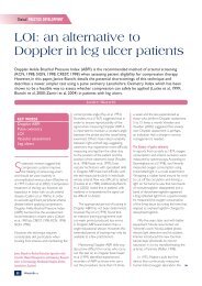
motor nerves, can lead to wast<strong>in</strong>g<br />
of <strong>the</strong> muscle belly (<strong>the</strong> fleshy part<br />
of <strong>the</strong> muscle), known as muscle<br />
atrophy (Figure 4).<br />
which muscles are affected, can<br />
give rise to an altered arch profile,<br />
such as a high arched <strong>foot</strong> (pes<br />
cavus) or low arched <strong>foot</strong> (pes<br />
planus) (Figure 5).<br />
Altered arch profiles can also give<br />
rise to areas of high pressure on<br />
<strong>the</strong> <strong>foot</strong> dur<strong>in</strong>g walk<strong>in</strong>g, which<br />
can contribute to abnormally high<br />
pressure on <strong>the</strong> plantar surface<br />
(sole of <strong>the</strong> <strong>foot</strong>).<br />
Gait <strong>changes</strong> due to<br />
motor <strong>neuropathy</strong><br />
Wastage <strong>in</strong> <strong>the</strong> muscles of <strong>the</strong><br />
leg can also have a detrimental<br />
effect on a patient’s <strong>gait</strong>. Figure 6<br />
provides a guide to some of <strong>the</strong><br />
key muscles of <strong>the</strong> lower limb and<br />
<strong>the</strong>ir role <strong>in</strong> <strong>the</strong> <strong>gait</strong> cycle.<br />
Proprioception and altered <strong>gait</strong><br />
Proprioception is def<strong>in</strong>ed as<br />
a patient’s perception of <strong>the</strong>ir<br />
body position<strong>in</strong>g. Impaired<br />
proprioception largely occurs due<br />
to sensory <strong>neuropathy</strong> and it can<br />
have a detrimental effect on <strong>gait</strong>.<br />
Patients with impaired<br />
proprioception may present with<br />
an ataxic (uncoord<strong>in</strong>ated) <strong>gait</strong><br />
as <strong>the</strong>y will be unaware of <strong>the</strong><br />
position of <strong>the</strong>ir lower limbs and<br />
feet when walk<strong>in</strong>g. This can also<br />
lead to postural <strong>in</strong>stability, feel<strong>in</strong>gs<br />
of <strong>in</strong>stability, balance deficits<br />
and an <strong>in</strong>creased risk of fallrelated<br />
<strong>in</strong>jury (Van Deursen and<br />
Simoneau, 1999).<br />
Assessment<br />
When undertak<strong>in</strong>g a neurological<br />
assessment on a patient with<br />
diabetes, it is important to<br />
assess <strong>the</strong> presence of motor<br />
<strong>neuropathy</strong>, particularly if<br />
sensory <strong>neuropathy</strong> is present.<br />
The comb<strong>in</strong>ation of sensory<br />
and motor <strong>neuropathy</strong> can be<br />
particularly detrimental and can<br />
render <strong>the</strong> <strong>foot</strong> vulnerable to<br />
deformities and an <strong>in</strong>creased risk<br />
of trauma and <strong>foot</strong> ulceration (van<br />
Schie et al, 2004).<br />
A basic assessment of motor<br />
<strong>neuropathy</strong> should <strong>in</strong>clude <strong>the</strong><br />
follow<strong>in</strong>g measures:<br />
8Observe <strong>the</strong> patient walk<strong>in</strong>g<br />
and note any coord<strong>in</strong>ation or<br />
balance problems. Listen for<br />
an audible <strong>foot</strong> slap, which<br />
may <strong>in</strong>dicate atrophy of <strong>the</strong><br />
tibialis anterior muscle<br />
(Figure 6)<br />
8Ask <strong>the</strong> patient whe<strong>the</strong>r <strong>the</strong>y<br />
feel unsteady, or have a history<br />
of falls/balance problems<br />
Atrophy of <strong>the</strong> muscles <strong>in</strong> <strong>the</strong> feet<br />
can have a profound <strong>in</strong>fluence on<br />
activities such as walk<strong>in</strong>g (<strong>gait</strong>).<br />
Muscle atrophy, dependent on<br />
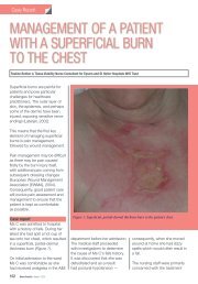
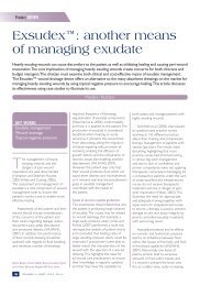
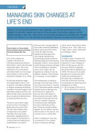
Figure 3. Structural <strong>changes</strong> to <strong>the</strong> <strong>foot</strong> without ulceration <strong>in</strong> a patient with diabetes.<br />
This is a classic image of a deformed high arch <strong>foot</strong> (pes cavus) affected by motor<br />
<strong>neuropathy</strong> where <strong>the</strong> muscle bulk <strong>in</strong> <strong>the</strong> arch region is much reduced.<br />
Wound Essentials • Volume 3 • 2008 153
Review<br />
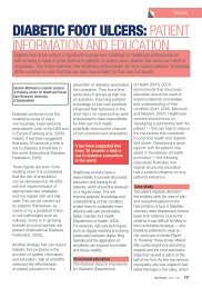
8Look for any obvious <strong>foot</strong><br />
deformities, such as hammer<br />
toes, claw toes or prom<strong>in</strong>ent<br />
metatarsal heads. Ask <strong>the</strong><br />
patient about <strong>the</strong> history of <strong>the</strong><br />
deformities — have <strong>the</strong>y recently<br />
developed or have <strong>the</strong>y always<br />
been present?<br />
8Compare both of <strong>the</strong> patient’s<br />
legs. Are <strong>the</strong>y similar? Is <strong>the</strong>re<br />
visible muscle wastage? A<br />
simple measurement of limb<br />
circumference can be used to<br />
compare <strong>the</strong> two legs<br />
8If <strong>the</strong> healthcare worker is<br />
sufficiently tra<strong>in</strong>ed <strong>in</strong> <strong>the</strong> relevant<br />
techniques, he or she should<br />
test for knee, ankle and plantar<br />
reflexes and conduct muscle<br />
power tests. If <strong>the</strong> healthcare<br />
worker is not tra<strong>in</strong>ed <strong>in</strong> <strong>the</strong>se<br />
techniques, <strong>the</strong> patient should<br />
be referred to a specialist nurse<br />
or podiatrist<br />
8Knee reflex — on this test<br />
<strong>the</strong> knee (patellar) tendon is<br />
tapped with a reflex hammer<br />
with <strong>the</strong> knee flexed and leg<br />
hang<strong>in</strong>g freely. Under normal<br />
circumstances once stimulated<br />
(tapped) <strong>the</strong> receptor with<strong>in</strong><br />
<strong>the</strong> tendon should <strong>in</strong>itiate an<br />
<strong>in</strong>voluntary muscle contraction<br />
and <strong>the</strong> practitioner would<br />
observe a brief rapid extension<br />
of <strong>the</strong> lower leg — known as <strong>the</strong><br />
knee reflex<br />
8Ankle reflex — this occurs<br />
when <strong>the</strong> Achilles tendon (at<br />
<strong>the</strong> back of <strong>the</strong> ankle) is tapped<br />
with a reflex hammer while <strong>the</strong><br />
<strong>foot</strong> is flexed towards <strong>the</strong> body<br />
(dorsiflexed). A positive result<br />
would be <strong>the</strong> jerk<strong>in</strong>g of <strong>the</strong> <strong>foot</strong><br />
towards its plantar surface (away<br />
from <strong>the</strong> body)<br />
8Planter reflex — <strong>the</strong> lateral side<br />
(outside) of <strong>the</strong> sole of <strong>the</strong> <strong>foot</strong> is<br />
rubbed with a blunt implement<br />
(so as not to cause pa<strong>in</strong>,<br />
discomfort or <strong>in</strong>jury) from <strong>the</strong> heel<br />
Cell body<br />
Muscle<br />
Nerve cells<br />
Axon<br />
Atrophied muscle<br />
Figure 4. Atrophy (wastage) of <strong>the</strong><br />
muscle due to motor <strong>neuropathy</strong>.<br />
along a curve to <strong>the</strong> ball of <strong>the</strong><br />
<strong>foot</strong>.A normal response should<br />
be observation of toes flex<strong>in</strong>g<br />
<strong>in</strong>wards<br />
8Muscle power test — muscle<br />
power can be def<strong>in</strong>ed as work<br />
done over a given period of<br />
time. The tissues be<strong>in</strong>g tested<br />
are skeletal tissues. Cl<strong>in</strong>ically<br />
this can be simply assessed<br />
by a person’s ability to resist<br />
applied force to a given<br />
anatomical area.<br />
Know<strong>in</strong>g when to refer<br />
It is important for healthcare<br />
workers to recognise that <strong>the</strong><br />
management of <strong>in</strong>dividuals with<br />
motor <strong>neuropathy</strong> requires a<br />
multidiscipl<strong>in</strong>ary approach.<br />
Patients who present with a<br />
high or low arch profile, or any<br />
o<strong>the</strong>r <strong>foot</strong> deformities, should<br />
be referred to a podiatrist for<br />
assessment. A podiatrist will be<br />
able to undertake a biomechanical<br />
assessment, <strong>in</strong>clud<strong>in</strong>g a <strong>gait</strong><br />
analysis, and prescribe treatment<br />
such as orthotic <strong>in</strong>soles to<br />
offload vulnerable pressure areas<br />
and m<strong>in</strong>imise <strong>the</strong> risk of new<br />
or recurrent episodes of <strong>foot</strong><br />
ulceration. Podiatrists may also<br />
work closely with orthotists to<br />
provide <strong>the</strong>rapeutic <strong>foot</strong>wear, often<br />
with extra depth to accommodate<br />
any <strong>foot</strong> deformities.<br />
Arch type<br />
Normal arch<br />
High arch<br />
Altered arch profiles can give<br />
rise to areas of high pressure on<br />
<strong>the</strong> <strong>foot</strong> dur<strong>in</strong>g <strong>gait</strong> which can<br />
contribute to abnormally high<br />
pressure on <strong>the</strong> plantar surface<br />
(sole of <strong>the</strong> <strong>foot</strong>).<br />
Flat<strong>foot</strong><br />
Figure 5. Commonly observed arch<br />
profiles of <strong>the</strong> <strong>foot</strong>.<br />
It is important to establish<br />
glycaemic control when<br />
attempt<strong>in</strong>g to prevent sensory<br />
and motor <strong>neuropathy</strong>. Ga<strong>in</strong><strong>in</strong>g<br />
glycaemic control means plasma<br />
glucose levels of 6–7 mmol/l or<br />
HbA 1c<br />
of 6–7% as recommended<br />
by National Service Framework<br />
for Diabetes (2001) and NICE<br />
guidel<strong>in</strong>es (National Institute for<br />
Health and Cl<strong>in</strong>ical Excellence,<br />
2004) to prevent <strong>the</strong> development<br />
or deterioration of peripheral<br />
<strong>neuropathy</strong>. Hence figures<br />
above or below this represent<br />
poor glycaemic control with<br />
<strong>in</strong>creased likelihood of develop<strong>in</strong>g<br />
or accelerat<strong>in</strong>g peripheral<br />
<strong>neuropathy</strong>.<br />
There are currently no<br />
pharmaceutical <strong>the</strong>rapies that<br />
prevent or slow <strong>the</strong> progression<br />
of motor <strong>neuropathy</strong> (van Schie<br />
et al, 2004).<br />
Conclusion<br />
Motor <strong>neuropathy</strong> <strong>in</strong> patients with<br />
diabetes can lead to weakness<br />
<strong>in</strong> <strong>the</strong> muscles of <strong>the</strong> <strong>foot</strong> and<br />
lower leg, which <strong>in</strong> turn can lead<br />
to characteristic <strong>changes</strong> to <strong>the</strong><br />
structure of <strong>the</strong> <strong>foot</strong>.<br />
Deformities such as hammer toes,<br />
clawed toes, prom<strong>in</strong>ent metatarsal<br />
heads and an altered arch profile<br />
are common.<br />
154 Wound Essentials • Volume 3 • 2008
Review<br />
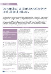
The <strong>gait</strong> cycle Tibialis anterior muscle Gastrocnemius<br />
Action:<br />
The <strong>gait</strong> cycle consists of <strong>in</strong>itial<br />
contact when <strong>the</strong> heel strikes <strong>the</strong><br />
ground; this is followed by <strong>the</strong><br />
fore<strong>foot</strong> contact<strong>in</strong>g <strong>the</strong> ground<br />
and <strong>the</strong>n toe off to propel <strong>the</strong><br />
<strong>in</strong>dividual onto <strong>the</strong>ir next <strong>foot</strong> step.<br />
The muscle groups <strong>in</strong> <strong>the</strong> leg<br />
and <strong>foot</strong> are not represented on<br />
this figure but are active dur<strong>in</strong>g<br />
different phases of <strong>the</strong> <strong>gait</strong> cycle<br />
The tibialis anterior muscle<br />
(coloured <strong>in</strong> orange) on <strong>the</strong> front<br />
of <strong>the</strong> leg contracts to ease <strong>the</strong><br />
<strong>foot</strong> down to <strong>the</strong> ground dur<strong>in</strong>g<br />
<strong>the</strong> <strong>in</strong>itial contact of <strong>the</strong> <strong>gait</strong> cycle<br />
The gastrocnemius muscle (<strong>in</strong><br />
blue) on <strong>the</strong> back of <strong>the</strong> leg is a<br />
very powerful muscle that has<br />
a role at <strong>the</strong> propulsive push off<br />
stage of <strong>the</strong> <strong>gait</strong> cycle. It assists<br />
with lift<strong>in</strong>g <strong>the</strong> heel from <strong>the</strong><br />
ground and controllably propell<strong>in</strong>g<br />
<strong>the</strong> body forwards.<br />
Effects of motor<br />
<strong>neuropathy</strong>:<br />
Muscle wastage (atrophy) due<br />
to motor <strong>neuropathy</strong> can result<br />
<strong>in</strong> an altered <strong>gait</strong> pattern, which<br />
can make <strong>the</strong> <strong>foot</strong> vulnerable to<br />
trauma and ulceration.<br />
If <strong>the</strong> tibialis anterior muscle is<br />
affected by motor <strong>neuropathy</strong><br />
consequences would be of a rapid<br />
uncontrolled <strong>foot</strong> slap/<strong>foot</strong> drop<br />
dur<strong>in</strong>g <strong>the</strong> <strong>in</strong>itial contact period of<br />
<strong>gait</strong> after <strong>the</strong> heel makes contact<br />
with <strong>the</strong> ground. This would<br />
reduce <strong>the</strong> capacity of <strong>the</strong> <strong>foot</strong> to<br />
absorb shock.<br />
The cl<strong>in</strong>ical consequences of<br />
this muscle wast<strong>in</strong>g are reduced<br />
muscle strength.<br />
This can result <strong>in</strong> pull<strong>in</strong>g <strong>the</strong> <strong>foot</strong><br />
<strong>in</strong>to a high arch position (pes<br />
cavus <strong>foot</strong> type), which would<br />
have a reduced ability to absorb<br />
shock.<br />
Figure 6. A guide to some of <strong>the</strong> key muscles of <strong>the</strong> lower limb and <strong>the</strong>ir role <strong>in</strong> <strong>the</strong> <strong>gait</strong> cycle.<br />
Such structural <strong>changes</strong> are<br />
often sites of abnormally high<br />
pressure, which can result <strong>in</strong><br />
tissue breakdown and ulceration,<br />
particularly <strong>in</strong> patients who also<br />
have sensory <strong>neuropathy</strong>.<br />
It is essential, <strong>the</strong>refore, that<br />
healthcare workers consider and<br />
assess patients for <strong>the</strong> presence<br />
of motor <strong>neuropathy</strong> when<br />
undertak<strong>in</strong>g rout<strong>in</strong>e neurological<br />
assessments. If evidence is found,<br />
patients should be referred to<br />
specialist multidiscipl<strong>in</strong>ary teams<br />
for fur<strong>the</strong>r assessment and<br />
management to m<strong>in</strong>imise <strong>the</strong> risk<br />
of new or recurrent episodes of<br />
<strong>foot</strong> ulceration. WE<br />
Baker N, Murali-Krishnan S,<br />
Rayman G (2005) A user’s<br />
guide to <strong>foot</strong> screen<strong>in</strong>g. Part 1:<br />
Peripheral <strong>neuropathy</strong>. Diabetic<br />
Foot 8(1): 28–37<br />
Edmonds M, Foster A (1999)<br />
Manag<strong>in</strong>g <strong>the</strong> Diabetic Foot.<br />
Blackwell Science, Oxford<br />
McIntosh C, Newton V (2007)<br />
Diabetic <strong>foot</strong> ulcers. In: Ousey<br />
K, McIntosh C (Eds). Lower<br />
Extremity <strong>Wounds</strong>: A problembased<br />
approach. John Wiley and<br />
Sons, Chichester: 191–230<br />
National Institute for Health<br />
and Cl<strong>in</strong>ical Excellence (2004)<br />
Cl<strong>in</strong>ical Guidel<strong>in</strong>es for Type<br />
2 Diabetes: Prevention and<br />
management of <strong>foot</strong> problems.<br />
NICE, London<br />
Van Deursen RW, Simoneau GG<br />
(1999) Foot and ankle sensory<br />
<strong>neuropathy</strong>, proprioception<br />
and postural stability. J Orthop<br />
Sports Phys Ther 29(12):<br />
718–26<br />
van Schie CHM, Vermigli C,<br />
Carr<strong>in</strong>gton AL et al (2004) Muscle<br />
weakness and <strong>foot</strong> deformities<br />
<strong>in</strong> diabetes. Diabetes Care 27(7):<br />
1668–73<br />
Wound Essentials • Volume 3 • 2008 155