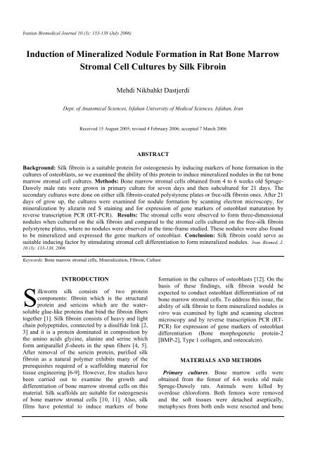
Induction of Mineralized Nodule Formation in Rat Bone Marrow ...
Induction of Mineralized Nodule Formation in Rat Bone Marrow ...
Induction of Mineralized Nodule Formation in Rat Bone Marrow ...

Create successful ePaper yourself
Turn your PDF publications into a flip-book with our unique Google optimized e-Paper software.
Iranian Biomedical Journal 10 (3): 133-138 (July 2006)<br />
<strong>Induction</strong> <strong>of</strong> <strong>M<strong>in</strong>eralized</strong> <strong>Nodule</strong> <strong>Formation</strong> <strong>in</strong> <strong>Rat</strong> <strong>Bone</strong> <strong>Marrow</strong><br />
Stromal Cell Cultures by Silk Fibro<strong>in</strong><br />
Mehdi Nikbahkt Dastjerdi<br />
Dept. <strong>of</strong> Anatomical Sciences, Isfahan University <strong>of</strong> Medical Sciences, Isfahan, Iran<br />
Received 15 August 2005; revised 4 February 2006; accepted 7 March 2006<br />
ABSTRACT<br />
Background: Silk fibro<strong>in</strong> is a suitable prote<strong>in</strong> for osteogenesis by <strong>in</strong>duc<strong>in</strong>g markers <strong>of</strong> bone formation <strong>in</strong> the<br />
cultures <strong>of</strong> osteoblasts, so we exam<strong>in</strong>ed the ability <strong>of</strong> this prote<strong>in</strong> to <strong>in</strong>duce m<strong>in</strong>eralized nodules <strong>in</strong> the rat bone<br />
marrow stromal cell cultures. Methods: <strong>Bone</strong> marrow stromal cells obta<strong>in</strong>ed from 4 to 6 weeks old Spruge-<br />
Dawely male rats were grown <strong>in</strong> primary culture for seven days and then subcultured for 21 days. The<br />
secondary cultures were done on either silk fibro<strong>in</strong>-coated polystyrene plates or free-silk fibro<strong>in</strong> ones. After 21<br />
days <strong>of</strong> grow up, the cultures were exam<strong>in</strong>ed for nodule formation by scann<strong>in</strong>g electron microscopy, for<br />
m<strong>in</strong>eralization by alizar<strong>in</strong> red S sta<strong>in</strong><strong>in</strong>g and for expression <strong>of</strong> gene markers <strong>of</strong> osteoblast maturation by<br />
reverse transcription PCR (RT-PCR). Results: The stromal cells were observed to form three-dimensional<br />
nodules when cultured on the silk fibro<strong>in</strong> and compared to the stromal cells cultured on the free-silk fibro<strong>in</strong><br />
polystyrene plates, where no nodules were observed <strong>in</strong> the time-frame studied. These nodules were also found<br />
to be m<strong>in</strong>eralized and expressed the gene markers <strong>of</strong> osteoblast. Conclusion: Silk fibro<strong>in</strong> could serve as<br />
suitable <strong>in</strong>duc<strong>in</strong>g factor by stimulat<strong>in</strong>g stromal cell differentiation to form m<strong>in</strong>eralized nodules. Iran. Biomed. J.<br />
10 (3): 133-138, 2006<br />
Keywords: <strong>Bone</strong> marrow stromal cells, M<strong>in</strong>eralization, Fibro<strong>in</strong>, Culture<br />
INTRODUCTION<br />
S<br />
ilkworm silk consists <strong>of</strong> two prote<strong>in</strong><br />
components: fibro<strong>in</strong> which is the structural<br />
prote<strong>in</strong> and seric<strong>in</strong>s which are the watersoluble<br />
glue-like prote<strong>in</strong>s that b<strong>in</strong>d the fibro<strong>in</strong> fibers<br />
together [1]. Silk fibro<strong>in</strong> consists <strong>of</strong> heavy and light<br />
cha<strong>in</strong> polypeptides, connected by a disulfide l<strong>in</strong>k [2,<br />
3] and it is a prote<strong>in</strong> dom<strong>in</strong>ated <strong>in</strong> composition by<br />
the am<strong>in</strong>o acids glyc<strong>in</strong>e, alan<strong>in</strong>e and ser<strong>in</strong>e which<br />
form antiparallel β-sheets <strong>in</strong> the spun fibers [4, 5].<br />
After removal <strong>of</strong> the seric<strong>in</strong> prote<strong>in</strong>, purified silk<br />
fibro<strong>in</strong> as a natural polymer exhibits many <strong>of</strong> the<br />
prerequisites required <strong>of</strong> a scaffold<strong>in</strong>g material for<br />
tissue eng<strong>in</strong>eer<strong>in</strong>g [6-9]. However, few studies have<br />
been carried out to exam<strong>in</strong>e the growth and<br />
differentiation <strong>of</strong> bone marrow stromal cells on this<br />
material. Silk scaffolds are suitable for osteogenesis<br />
<strong>of</strong> bone marrow stromal cells [10, 11]. Also, silk<br />
films have potential to <strong>in</strong>duce markers <strong>of</strong> bone<br />
formation <strong>in</strong> the cultures <strong>of</strong> osteoblasts [12]. On the<br />
basis <strong>of</strong> these f<strong>in</strong>d<strong>in</strong>gs, silk fibro<strong>in</strong> would be<br />
expected to conduct osteoblast differentiation <strong>of</strong> rat<br />
bone marrow stromal cells. To address this issue, the<br />
ability <strong>of</strong> silk fibro<strong>in</strong> to form m<strong>in</strong>eralized nodules <strong>in</strong><br />
vitro was exam<strong>in</strong>ed by light and scann<strong>in</strong>g electron<br />
microscopy and by reverse transcription PCR (RT-<br />
PCR) for expression <strong>of</strong> gene markers <strong>of</strong> osteoblast<br />
differentiation (<strong>Bone</strong> morphogenetic prote<strong>in</strong>-2<br />
[BMP-2], Type 1 collagen, and osteocalc<strong>in</strong>).<br />
MATERIALS AND METHODS<br />
Primary cultures. <strong>Bone</strong> marrow cells were<br />
obta<strong>in</strong>ed from the femur <strong>of</strong> 4-6 weeks old male<br />
Spruge-Dawely rats. Animals were killed by<br />
overdose chlor<strong>of</strong>orm. Both femora were removed<br />
and the s<strong>of</strong>t tissues were detached aseptically,<br />
metaphyses from both ends were resected and bone
134 Nikbakht Dastjerdi Iran. Biomed. J., July 2006<br />
marrow cells were collected by flush<strong>in</strong>g the<br />
diaphisis with a culture medium. A suspension <strong>of</strong><br />
bone marrow cells was obta<strong>in</strong>ed by repeated<br />
aspiration <strong>of</strong> the cell preparation through a 20-gauge<br />
needle, cells obta<strong>in</strong>ed from each femur, plated <strong>in</strong>to<br />
60 mm tissue culture dishes (Falcon, USA) , and<br />
<strong>in</strong>cubated at 37ºC for 1 h to promote adherence to<br />
the substrate. Growth medium (2 ml), [alpha<br />
m<strong>in</strong>imal essential medium (alpha-MEM, GIBCO-<br />
BRL, USA), pH 7.2 (conta<strong>in</strong><strong>in</strong>g 2.2 g/l sodium<br />
bicarbonate +15% FCS (Atlanta Biologicals, USA)<br />
+100 units/ml <strong>of</strong> penicill<strong>in</strong>/100 µg/ml <strong>of</strong><br />
streptomyc<strong>in</strong> (GIBCO-BRL, USA) was added to<br />
each dish and then an additional 2 ml was added<br />
after 24 h and cultured for seven days as primary<br />
cultures.<br />
Preparation <strong>of</strong> silk fibro<strong>in</strong>. Raw silkworm silk<br />
was degummed twice with 0.4 g/l NaHCO 3 solutions<br />
at 100°C for 1 h that removed the seric<strong>in</strong> and then<br />
washed with distilled water. Degummed silk was<br />
dissolved <strong>in</strong> 9.5 mol/l LiBr (Sigma, USA) solution at<br />
a ratio <strong>of</strong> 3 g/l. After dialysis aga<strong>in</strong>st distilled water<br />
for three days, the solution was filtered, and the silk<br />
fibro<strong>in</strong> solution was obta<strong>in</strong>ed [13]. A 5-ml volume<br />
<strong>of</strong> the silk fibro<strong>in</strong> solution (5 mg/ml) was added to<br />
each well <strong>of</strong> a six-well polystyrene plate (Falcon<br />
1018, USA) and <strong>in</strong>cubated at 25°C for 5 m<strong>in</strong>,<br />
followed by decantation <strong>of</strong> the solution. After<br />
keep<strong>in</strong>g the plate at 30°C for 2 h, 5 ml <strong>of</strong> the fibro<strong>in</strong><br />
solution was added aga<strong>in</strong> to the well, and the plate<br />
was <strong>in</strong>cubated at 25°C for 5 m<strong>in</strong>. The plate was<br />
dried at 50°C for 1 h. A 5-ml volume <strong>of</strong> 50%<br />
methanol was added to the well and <strong>in</strong>cubated at<br />
25°C for 10 m<strong>in</strong>. After dry<strong>in</strong>g the plate aga<strong>in</strong> at<br />
50°C for 1 h, the silk fibro<strong>in</strong>-coated plate was<br />
prepared.<br />
Secondary cultures. After seven days <strong>in</strong> primary<br />
cultures, the confluent monolayer was released with<br />
0.25% tryps<strong>in</strong>-EDTA (Sigma, USA), and then 4 ml<br />
<strong>of</strong> medium (alpha-MEM) conta<strong>in</strong><strong>in</strong>g 1-3 × 10 6 cells<br />
which supplemented with 10 mM sodium betaglycerophosphate<br />
(GIBCO-BRL, USA) and<br />
50 µg/ml ascorbic acid (Sigma, USA) was placed<br />
onto silk fibro<strong>in</strong>-coated plates. The plates with cells<br />
were <strong>in</strong>cubated <strong>in</strong> 5% CO 2 , at 37°C for 21 days.<br />
Growth medium was changed every second day. For<br />
control groups, 4 ml <strong>of</strong> the above mentioned<br />
medium conta<strong>in</strong><strong>in</strong>g 1-3 × 10 6 cells was directly<br />
placed onto a six-well polystyrene plate (Falcon<br />
1018, USA) and <strong>in</strong>cubated at 37°C, 5% CO 2 for 21<br />
days. Each experiment was repeated six times <strong>in</strong><br />
each group.<br />
Culture exam<strong>in</strong>ation. Cultures were exam<strong>in</strong>ed<br />
daily us<strong>in</strong>g <strong>in</strong>verted microscope (Olympus, Japan).<br />
Some cultures were fixed <strong>in</strong> 10% neutral buffered<br />
formal<strong>in</strong> and sta<strong>in</strong>ed with either H and E or toluid<strong>in</strong>e<br />
blue (Sigma, USA).<br />
Scann<strong>in</strong>g electron microscopy. After 21 days,<br />
cells <strong>in</strong> secondary cultures were fixed <strong>in</strong> 1.5%<br />
glutaraldehyde at 4°C for 30 m<strong>in</strong> and then, postfixed<br />
<strong>in</strong> 1% osmium tetroxide at 4°C for 1 h. Then<br />
were dehydrated through a series <strong>of</strong> <strong>in</strong>creas<strong>in</strong>g<br />
concentrations <strong>of</strong> ethanol and dried us<strong>in</strong>g<br />
hexamethyldisilazane. Samples were sputter coated<br />
with gold and viewed us<strong>in</strong>g a scann<strong>in</strong>g electron<br />
microscope (S-4500, Hitachi, Japan).<br />
M<strong>in</strong>eralization assay. M<strong>in</strong>eralization <strong>of</strong> nodules<br />
was determ<strong>in</strong>ed us<strong>in</strong>g alizar<strong>in</strong> red S sta<strong>in</strong><strong>in</strong>g which<br />
is a common histochemical technique used to detect<br />
calcium deposits <strong>in</strong> m<strong>in</strong>eralized tissues and cult [14,<br />
15]. Cells <strong>in</strong> secondary cultures after 21 days were<br />
fixed <strong>in</strong> 70% ethanol and sta<strong>in</strong>ed with 1% alizar<strong>in</strong><br />
red S (Sigma, USA), pH 6.4, for 5 m<strong>in</strong>. Cells were<br />
then washed with distilled water and viewed under<br />
the <strong>in</strong>verted microscope.<br />
RT-PCR. Total RNA was isolated from each silk<br />
fibro<strong>in</strong>-coated plate and after 21 days, controls<br />
subcultured us<strong>in</strong>g Trizol reagent (GIBCO-BRL,<br />
USA). The RNA pellet is washed with 70% ethanol.<br />
RNA Concentration was determ<strong>in</strong>ed at 260 nm and<br />
was analyzed for markers <strong>of</strong> osteoblast maturation:<br />
BMP-2, Type 1 collagen, and osteocalc<strong>in</strong>.<br />
Glyceraldehyde 3-phosphate dehydrogenase<br />
(GAPDH) was utilized as a housekeep<strong>in</strong>g gene.<br />
Equivalent amount <strong>of</strong> RNA samples (0.5 mg) was<br />
<strong>in</strong>itially reverse transcribed for first strand cDNA<br />
synthesis (GIBCO-BRL, USA). Synthesis <strong>of</strong> cDNA<br />
entailed us<strong>in</strong>g oligo (dT) as a RT primer, which<br />
b<strong>in</strong>ds to the poly A tail <strong>of</strong> the mRNA. After the RT<br />
reaction, rema<strong>in</strong><strong>in</strong>g RNA was removed by RNase<br />
treatment. Template DNA was then used <strong>in</strong> gene<br />
specific PCR for GAPDH, BMP-2, Type I collagen,<br />
and osteocalc<strong>in</strong> [16]. cDNA was amplified by PCR<br />
with oligonucleotide primers (Table1). All RT-PCR<br />
products were visualized on 1.5% agarose gel with<br />
0.5 mg/ml ethidium bromide. Photographs were<br />
taken under ultraviolet illum<strong>in</strong>ation (Bio-Rad, USA)<br />
and qualitative assessments were made <strong>of</strong> relative<br />
gene expression.<br />
http://IBJ.pasteur.ac.ir
Iran. Biomed. J., July 2006 <strong>Nodule</strong> <strong>Formation</strong> <strong>in</strong> Stromal Cell Cultures by Silk Fibro<strong>in</strong> 135<br />
Table 1. Primers sequences and cycle conditions used for the RT-PCR.<br />
Gene Sequence Cycl<strong>in</strong>g conditions Amplicon<br />
F: 5-CCTGGTAAAGATGGTGCC-3<br />
Type I collagen<br />
R: 5-CACCAGG TTCAC CTTTCGCACC-3 25 cycles; 94°C 30 s; 58°C 30 s; 72°C 1 m<strong>in</strong> 222 bp<br />
BMP-2<br />
F: 5-AGTTCTGTCCCCAGTGACGAGTTT-3<br />
R: 5-GTACAACATGGAGATTGCGCTGAG-3 36 cycles; 95°C one m<strong>in</strong>; 63°C 30 s; 72°C 1 m<strong>in</strong> 708bp<br />
Osteocalc<strong>in</strong><br />
F: 5-CCTCAGTCCCCAGCCCAGATCC-3<br />
R: 5-CAGGGCAGAGAGAGAGGACAGG-3 25 cycles; 94°C 30 s; 58°C 30 s; 72°C 1 m<strong>in</strong> 219 bp<br />
BMP-2, <strong>Bone</strong> morphogenetic prote<strong>in</strong>-2.<br />
RESULTS AND DISCUSSION<br />
Subcultures. In the fibro<strong>in</strong>-coated cultures, nearly<br />
all <strong>of</strong> the stromal cells attached to silk fibro<strong>in</strong> up to<br />
6-8 h <strong>of</strong> culture and formed cell colonies<br />
represent<strong>in</strong>g early nodule formation at day 14 (Fig.<br />
1) while <strong>in</strong> the control groups, the stromal cells<br />
attached to floor <strong>of</strong> culture dish up to 24 h and they<br />
did not form any colonies up to 21 days but they<br />
formed a monolayer <strong>of</strong> cells (Fig. 2).There were not<br />
any mean<strong>in</strong>gful differences between cultures <strong>in</strong> each<br />
group.<br />
It has shown that three-dimensional silk fibro<strong>in</strong><br />
nets support the attachment, spread and growth <strong>of</strong> a<br />
variety <strong>of</strong> human cell types <strong>of</strong> diverse tissue orig<strong>in</strong>s.<br />
These <strong>in</strong>cludes epithelial, fibroblast, glial,<br />
kerat<strong>in</strong>ocyte and osteoblast cells [17, 18]. Cell<br />
adhesion is important because it directly <strong>in</strong>fluences<br />
cell growth and differentiation. In the present study,<br />
the silk fibro<strong>in</strong> improved cell adhesion when<br />
compared to the control cultures. This improvement<br />
can be attributed to the changes <strong>in</strong> surface texture.<br />
Fig. 1. Cell colonies <strong>in</strong> secondary cultures on silk fibro<strong>in</strong>coated<br />
plates at day 14 (Tolid<strong>in</strong>e blue sta<strong>in</strong><strong>in</strong>g, × 100).<br />
Fig. 2. Stromal cells <strong>in</strong> secondary cultures <strong>of</strong> control groups at<br />
day 14.They are form<strong>in</strong>g a monolayer <strong>of</strong> cells and no cell<br />
colonies were seen (H and E sta<strong>in</strong><strong>in</strong>g, × 400).<br />
Also fibro<strong>in</strong> is considered to be hydrophilic prote<strong>in</strong><br />
[18], and this property might be favorable for the<br />
<strong>in</strong>teraction with the negatively-charged surface <strong>of</strong><br />
animal cells. S<strong>in</strong>ce the outermost surface <strong>of</strong><br />
biomaterials directly <strong>in</strong>terfaces with the cells,<br />
the biocompatibility <strong>of</strong> silk fibro<strong>in</strong> would<br />
be different from that <strong>of</strong> the polystyrene plate<br />
as the control. Therefore, the behavior <strong>of</strong> the<br />
stromal cells cultured on the free-silk fibro<strong>in</strong><br />
plates and the silk fibro<strong>in</strong>-coated plates would<br />
be different.<br />
<strong>Nodule</strong> formation and m<strong>in</strong>eralization. At day 21,<br />
three-dimensional nodule formation was observed <strong>in</strong><br />
the fibro<strong>in</strong>-coated cultures as shown <strong>in</strong> Figure 3 by<br />
scann<strong>in</strong>g electron microscopy. No nodule formation<br />
was observed <strong>in</strong> control groups at day 21. As shown<br />
<strong>in</strong> Figure 4 the nodules <strong>in</strong> the fibro<strong>in</strong>-coated cultures<br />
were sta<strong>in</strong>ed heavily with alizar<strong>in</strong> red S. It was<br />
found that control cultures showed no alizar<strong>in</strong> red<br />
S sta<strong>in</strong><strong>in</strong>g.<br />
http://IBJ.pasteur.ac.ir
136 Nikbakht Dastjerdi Iran. Biomed. J., July 2006<br />
(A)<br />
(B)<br />
Fig. 3. Scann<strong>in</strong>g electron micrographs <strong>of</strong> three-dimensional<br />
calcified nodules from stromal cells cultured for 21 days on silk<br />
fibro<strong>in</strong>-coated plates: (A), Low power <strong>of</strong> three nodules (× 200);<br />
(B), Higher power <strong>of</strong> one nodule (× 1200).<br />
m<strong>in</strong>eralized nodule-<strong>in</strong>duc<strong>in</strong>g factor. This is<br />
important with regard to develop<strong>in</strong>g materials for<br />
bone repair or bone tissue eng<strong>in</strong>eer<strong>in</strong>g/regeneration<br />
because many materials are biocompatible<br />
with regard to certa<strong>in</strong> cell types but <strong>of</strong>ten require<br />
addition <strong>of</strong> growth factors to improve the cell<br />
responses [24].<br />
An <strong>in</strong>terest<strong>in</strong>g f<strong>in</strong>d<strong>in</strong>g is the significant <strong>in</strong>crease<br />
<strong>in</strong> TGFß1; one <strong>of</strong> the primary cytok<strong>in</strong>es<br />
associated with the growth and regulation <strong>of</strong><br />
repair <strong>of</strong> bone [25]; observed <strong>in</strong> the silk fibro<strong>in</strong><br />
cultures compared to the synthetic polymeric<br />
material controls [26].<br />
Increased differentiation <strong>of</strong> osteoblast precursors<br />
by <strong>in</strong>duc<strong>in</strong>g mesenchymal cells to differentiate<br />
<strong>in</strong>to osteoblasts [26] and proliferation <strong>of</strong> osteoblasts<br />
[28], and <strong>in</strong>creased collagen synthesis [29] has<br />
been shown <strong>in</strong> the presence <strong>of</strong> TGFß1<br />
<strong>in</strong> culture. Thus, the effect <strong>of</strong> silk fibro<strong>in</strong><br />
on osteoblast differentiation and formation<br />
<strong>of</strong> m<strong>in</strong>eralized nodule <strong>in</strong> part may be due<br />
to stimulation <strong>of</strong> stromal cells to express TGFß1<br />
and it should be studied <strong>in</strong> the future.<br />
Weak expression <strong>of</strong> the genes <strong>in</strong> the control<br />
groups <strong>in</strong>dicate that the stromal cells have<br />
potential to differentiate to osteoblasts without<br />
any <strong>in</strong>ducer supplements but it is not enough to<br />
produce m<strong>in</strong>eralized nodules.<br />
Collectively, these results <strong>in</strong>dicate that silk fibro<strong>in</strong><br />
could serve as suitable <strong>in</strong>duc<strong>in</strong>g factor by<br />
stimulat<strong>in</strong>g stromal cell differentiation to form<br />
m<strong>in</strong>eralized nodules.<br />
RT-PCR. RT-PCR analysis was performed to<br />
assess progression <strong>of</strong> BMSC towards the osteoblast<br />
l<strong>in</strong>eage at the mRNA gene expression level.<br />
Expression <strong>of</strong> genes cod<strong>in</strong>g for BMP-2, Type I<br />
collagen, and osteocalc<strong>in</strong> <strong>in</strong> BMSC after 21 days <strong>of</strong><br />
culture was assessed. Remarkably, the<br />
osteo<strong>in</strong>ductive effects <strong>of</strong> silk fibro<strong>in</strong>-coated plates<br />
were obvious. In the control cultures, there was no<br />
expression or weak expression <strong>of</strong> these genes (Fig.<br />
5). Previous studies have shown that stromal cell<br />
cultures only form m<strong>in</strong>eralized nodules <strong>in</strong> the<br />
presence <strong>of</strong> additional factors like dexamethasone<br />
[20-23]. In the present study, m<strong>in</strong>eralized nodules<br />
were observed <strong>in</strong> the absence <strong>of</strong> dexamethasone<br />
demonstrat<strong>in</strong>g the remarkable ability <strong>of</strong> the silk<br />
fibro<strong>in</strong> to cause m<strong>in</strong>eralized nodule formation<br />
and <strong>in</strong>dicate that this prote<strong>in</strong> serve as suitable<br />
Fig. 4. Light micrograph <strong>of</strong> bone nodules from stromal cells<br />
cultured for 21 days on silk fibro<strong>in</strong>-coated plates. Positive dark<br />
red sta<strong>in</strong><strong>in</strong>g shows presence <strong>of</strong> calcium deposits i.e.<br />
m<strong>in</strong>eralization (alizar<strong>in</strong> red S sta<strong>in</strong><strong>in</strong>g, × 100).<br />
http://IBJ.pasteur.ac.ir
Iran. Biomed. J., July 2006 <strong>Nodule</strong> <strong>Formation</strong> <strong>in</strong> Stromal Cell Cultures by Silk Fibro<strong>in</strong> 137<br />
Fig. 5. Gene expression <strong>of</strong> BMP-2, Type I collagen, and<br />
osteocalc<strong>in</strong> us<strong>in</strong>g RT-PCR. GAPDH was used as a<br />
housekeep<strong>in</strong>g gene. Note to strong expression <strong>in</strong> the silk fibro<strong>in</strong>coated<br />
plates and weak expression <strong>in</strong> the control plates. SF, silk<br />
fibro<strong>in</strong>-coated plates; C, Control plates.<br />
REFERENCES<br />
1. Yamada, H., Nakao, H., Takasu, Y. and Tsubouchi,<br />
K. (2001) Preparation <strong>of</strong> undegraded native<br />
molecular fibro<strong>in</strong> solution from silkworm cocoons.<br />
Mater. Sci. Eng. C 14: 41-44.<br />
2. Tanaka, K., Kajiyama, N., Ishikura, K., Waga, S.,<br />
Kikuchi, A., Ohtomo, K., Takagi, T. and Mizuno, S.<br />
(1999) Determ<strong>in</strong>ation <strong>of</strong> the site <strong>of</strong> disulfide l<strong>in</strong>kage<br />
between heavy and light cha<strong>in</strong>s <strong>of</strong> silk fibro<strong>in</strong><br />
produced by Bombyx mori. Biochim. Biophis. Acta<br />
1432: 92-103.<br />
3. Zhou, C.Z., Confalonieri, F., Med<strong>in</strong>a, N., Zivanovic,<br />
Y., Esnault, C., Yang, T., Jacquet, M., Jan<strong>in</strong>, J.,<br />
Duguet, M., Perasso, R. and Li, Z.G. (2000) F<strong>in</strong>e<br />
organization <strong>of</strong> Bombyx mori fibro<strong>in</strong> heavy cha<strong>in</strong><br />
gene. Nucleic Acids Res. 28: 2413-2419.<br />
4. Fedic, R., Zurovec, M. and Sehnal, F. (2003)<br />
Correlation between fibro<strong>in</strong> am<strong>in</strong>o acid sequence and<br />
physical silk properties. J. Biol. Chem. 278 (37):<br />
35255- 35264.<br />
5. Asakura, T., Yao, J., Yamane, T., Umemura, K. and<br />
Ulrich, A.S. (2002) Heterogeneous structure <strong>of</strong> silk<br />
fibers from Bombyx mori resolved by 13 C solid-state<br />
NMR spectroscopy. J. Am. Chem. Soc. 124: 8794-<br />
8795.<br />
6. Altman, G.H, Diaz, F., Jakuba, C., Calabro, T.,<br />
Horan, R.L., Chen, J., Lu, H. and Richmond Jand<br />
Kaplan D.L. (2003) Silk-based biomaterials.<br />
Biomaterials 24: 401-416.<br />
7. Petr<strong>in</strong>i, P., Parolari, C. and Tanzi, M.C. (2001) Silk<br />
fibro<strong>in</strong>-polyurethane scaffolds for tissue eng<strong>in</strong>eer<strong>in</strong>g.<br />
J. Mater Sci. Mater Med. 12: 849-853.<br />
8. Altman, G.H., Horan, R.L, Lu, H.H., Moreau, J.,<br />
Mart<strong>in</strong>, I., Richmond, J.C. and Kaplan, D.L. (2002)<br />
Silk matrix for tissue eng<strong>in</strong>eered anterior cruciate<br />
ligaments. Biomaterials 23: 4131-4141.<br />
9. Kim, U.J., Park, J., Kim, H.J., Wada, M. and Kaplan,<br />
D.L. (2005) Three-dimensional aqueous-derived<br />
biomaterial scaffolds from silk fibro<strong>in</strong>. Biomaterials<br />
26: 2775-2785.<br />
10. Me<strong>in</strong>el, L., Karageorgiou, V., Fajardo, R., Snyder,<br />
B., Sh<strong>in</strong>de-Patil, V., Zichner, L., Kaplan, D., Langer,<br />
R. and Vunjak-Novakovic, G. (2004) <strong>Bone</strong> tissue<br />
eng<strong>in</strong>eer<strong>in</strong>g us<strong>in</strong>g human mesenchymal stem cells:<br />
effects <strong>of</strong> scaffold material and medium flow. Ann.<br />
Biomed. Eng. 32: 112-122.<br />
11. Me<strong>in</strong>el, L., Karageorgiou, V., H<strong>of</strong>mann, S., Fajardo,<br />
R., Snyder, B., Li, C., Zichner, L., Langer, R.,<br />
Vunjak-Novakovic, G. and Kaplan, D.L. (2004)<br />
Eng<strong>in</strong>eer<strong>in</strong>g bone-like tissue <strong>in</strong> vitro us<strong>in</strong>g human<br />
bone marrow stem cells and silk scaffolds. J.<br />
Biomed. Matern. Res. 71A: 25-34.<br />
12. S<strong>of</strong>ia, S., McCarthy, M.B., Gronowicz, G. and<br />
Kaplan, D.L. (2001) Functionalized silk-based<br />
biomaterials for bone formation. J. Biomed. Mater.<br />
Res. 54: 139-148.<br />
13. Chen, X., Li, W., Zhong, W., Lu, Y. and Yu, T.Y.<br />
(1997) pH sensitivity and ion sensitivity <strong>of</strong><br />
hydrogels based on complex-form<strong>in</strong>g chitosan/silk<br />
fibro<strong>in</strong> <strong>in</strong>terpenetrat<strong>in</strong>g polymer network. J. Appl.<br />
Polym. Sci. 65: 2257-2262.<br />
14. Coelho, M.J., Trigo, A. and Fernandes, M.H. (2000)<br />
Human bone cell cultures <strong>in</strong> biocompatibility test<strong>in</strong>g.<br />
Part I: osteoblastic differentiation <strong>of</strong> serially<br />
passaged human bone marrow cells cultured <strong>in</strong> α-<br />
MEM and <strong>in</strong> DMEM. Biomaterials 21: 1087-1094.<br />
15. Chang, Y., Stanford, C.M. and Keller, J.C. (2000)<br />
Calcium and phosphate supplementation promotes<br />
bone cell m<strong>in</strong>eralization: implications for<br />
hydroxyapatite (HA)-enhanced bone formation. J.<br />
Biomed. Mater. Res. 52: 270-278.<br />
16. Y<strong>in</strong>g, Z., Hong, G., Shang-Feng, L., Rong-Cong, W.<br />
and Zhao, W. (2005) Overexpression <strong>of</strong> QM <strong>in</strong>duces<br />
cell differentiation and m<strong>in</strong>eralization <strong>in</strong> MC3T3-E1.<br />
Biol. Pharm. Bull. 28 (8): 1371-1376.<br />
17. M<strong>in</strong>oura, N., Aiba, S., Gotoh, Y., Tsukada, M. and<br />
Imai, Y. (1995) Attachment and growth <strong>of</strong> cultured<br />
fibroblast cells on silk prote<strong>in</strong> matrices. J. Biomed.<br />
Mater. Res. 29: 1215-1221.<br />
18. Inouye, K., Kurokawa, M., Nishikawa, S. and<br />
Tsukada, M.(1998) Use <strong>of</strong> Bombyx mori silk fibro<strong>in</strong><br />
as a substratum for cultivation <strong>of</strong> animal cells. J.<br />
Biochem. Biophys. Methods. 37: 159-164.<br />
19. J<strong>in</strong>, H.J. and Kaplan, D.L. (2003) Mechanism <strong>of</strong> silk<br />
process<strong>in</strong>g <strong>in</strong> <strong>in</strong>sects and spiders. Nature 424<br />
(6952): 1057-1061.<br />
20. Bizios, R. (1994) Osteoblasts: an <strong>in</strong> vitro model <strong>of</strong><br />
bone-implant <strong>in</strong>teractions. Biotechnol. Bioeng. 43:<br />
582-585.<br />
21. Schiller, P.C., D’Ippolito, G., Balkan, W., Roos, B.A.<br />
and Howard, G.A. (2001) Gap-junctional<br />
communication mediates parathyroid hormone<br />
stimulation <strong>of</strong> m<strong>in</strong>eralization <strong>in</strong> osteoblastic cultures.<br />
<strong>Bone</strong> 28 (1): 38-44.<br />
22. Malaval, L., Modrowski, D., Gupta, A.K. and Aub<strong>in</strong>,<br />
J.E. (1994) Cellular expression <strong>of</strong> bone-related<br />
prote<strong>in</strong>s dur<strong>in</strong>g <strong>in</strong> vitro osteogenesis <strong>in</strong> rat bone<br />
marrow stromal cell cultures. J. Cell Physiol. 158<br />
http://IBJ.pasteur.ac.ir
138 Nikbakht Dastjerdi Iran. Biomed. J., July 2006<br />
(3): 555-572.<br />
23. Akbari, M., Nikbakht, M. and Sobhani, A. (2001)<br />
Expression <strong>of</strong> alkal<strong>in</strong>e phosphatase dur<strong>in</strong>g osteogenic<br />
differentiation <strong>of</strong> rat bone marrow stromal cells.<br />
Yakhteh (The Cell) 3 (11): 131-136.<br />
24. Mastrogiacomo, M., Muraglia, A., Komlev, V.,<br />
Peyr<strong>in</strong>, F., Rustichelli, F., Crovace, A. and Cancedda,<br />
R. (2005) Tissue eng<strong>in</strong>eer<strong>in</strong>g <strong>of</strong> bone: search for a<br />
better scaffold. Orthod. Crani<strong>of</strong>ac. Res. 8 (4):277-<br />
284.<br />
25. Andreasen, J.O. and Rud, J. (1972) Modes <strong>of</strong> heal<strong>in</strong>g<br />
histologically after endodontic surgery <strong>in</strong> 70 cases.<br />
Int. J. Oral. Surg. 1: 148-160.<br />
26. Barnes, G.L., Kostenuik, P.J., Gerstenfeld, L.C. and<br />
E<strong>in</strong>horn, T.A. (1999) Growth factor regulation <strong>of</strong><br />
fracture repair. J. <strong>Bone</strong> M<strong>in</strong>er. Res. 4 (11): 1805-<br />
1815.<br />
27. Centrella, M., Horowitz, M.C., Wozney, J.M. and<br />
McCarthy, T.L. (1994) Transform<strong>in</strong>g growth factor<br />
[beta] gene family members and bone. Endocr. Rev.<br />
15: 27-39.<br />
28. Centrella, M., McCarthy, T.L. and Canalis, E. (1987)<br />
Transform<strong>in</strong>g growth factor [beta] is a bifunctional<br />
regulator <strong>of</strong> replication and collagen synthesis <strong>in</strong><br />
osteoblast-enriched cell cultures from fetal rat bone.<br />
J. Biol. Chem. 262: 2869-2874.<br />
29. Centrella, C.M., Massague, J. and Canalis, E. (1986)<br />
Human platelet-derived transform<strong>in</strong>g growth factorbeta<br />
stimulates parameters <strong>of</strong> bone growth <strong>in</strong> fetal rat<br />
calvariae. Endocr<strong>in</strong>ology 119: 2306–2312.<br />
http://IBJ.pasteur.ac.ir