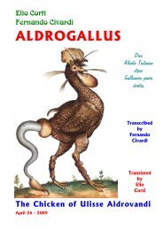
Hamburger and Hamilton - Summa gallicana
Hamburger and Hamilton - Summa gallicana
Hamburger and Hamilton - Summa gallicana

Create successful ePaper yourself
Turn your PDF publications into a flip-book with our unique Google optimized e-Paper software.
DEVELOPMENTAL DYNAMICS 195231-272 (1992)<br />
Reprinted from the JOURNAL OF MORPEOLOCY<br />
Vol. 88, No. 1, January 1951<br />
A SERIES OF NORMAL STAGES IN THE<br />
DEVELOPMENT OF THE<br />
CHICK EMBRYO<br />
VIKTOR HAMBURGER<br />
Department of Zoology, Washington University, St. Louis, Nissouri<br />
HOWARD L. HAMILTON<br />
Department of Zoolo8gy <strong>and</strong> Entom2070gy, Iowa State College, Ames<br />
FORTY-FIVE FIGURES<br />
The preparation of a series of normal stages of the chick<br />
embryo does not need justification at a time when chick ernbryos<br />
are not only widely used in descriptive <strong>and</strong> experimental<br />
embryology but are proving to be increasingly<br />
valuable in medical research, as in work on viruses <strong>and</strong> cancer.<br />
The present series was planned in connection with the<br />
preparation of a new edition of Lillie’s DeueZopmerzt of the<br />
Chick by the junior author. It is being published separately<br />
to make it accessible immediately to a large group of workers.<br />
Ever since Aristotle “discovered” the chick embryo as the<br />
ideal, object for embryological studies, the embryos have been<br />
described in terms of the length of time of incubation, <strong>and</strong><br />
this arbitrary method is still in general use, except for the<br />
first three days of incubation during which more detailed<br />
characteristics such as the numbers of somites are applied.<br />
The shortcomings of a classification based on chronological<br />
age are obvious to every worker in this field, for enormous<br />
variations may occur in embryos even though all eggs in a<br />
setting are plmaced in the incubator at the same time. Many<br />
factors are responsible for the lack of correlation between<br />
chronological <strong>and</strong> structural age. Among these are : genetic<br />
differences in the rate of development of different breccls<br />
(eg., the embryo of the White Leghorn breed develops more<br />
49<br />
Address renrint reauests to Dr. Joshua R. Sanes. DeDartment of<br />
Anatomy <strong>and</strong> Neurobiology, Washington University’ Medical School,<br />
St. Louis, MO 63110.<br />
Q 1993 WILEY-LISS, INC.
232 HAMBURGER AND HAMILTON<br />
50 V. HAMBURGER AND H. L. HAMILTON<br />
rapidly than that of the Barred Plymouth Rock <strong>and</strong> hatches<br />
approximately a day earlier) ; seasonal differences in the<br />
viability <strong>and</strong> vigor of embryos; differences in the stage of<br />
development when incubation is started ; differences in the<br />
“freshness” of eggs, Le., the lapse of time between laying<br />
<strong>and</strong> incubation; differences in the temperature of eggs when<br />
placed in the incubator, <strong>and</strong> in the size of individual eggs;<br />
differences in the temperature of incubation, <strong>and</strong> in type <strong>and</strong><br />
size of incubator.<br />
The wide variations in external form which occur at any<br />
given chronological age are clearly seen in tables 1 <strong>and</strong> 2<br />
which show the distribution of 296 embryos from the 4th day<br />
until hatching when classified according to our series of<br />
stages. For example, a “6-day” embryo may range anywhere<br />
from stage 27 + to stage 31 (table 1). It will also be noted that<br />
the data in table 1 are based on an incubation-temperature of<br />
103°F. (ca. 39.4”C.) whereas those in table 2 are based on<br />
a temperature of 375°C. This difference has resulted in the<br />
skipping of the “9-day” embryo altogether! It is not surprising,<br />
therefore, that the use of chronology with its lack of<br />
precision in the designation of embryos has actually led to<br />
misunderst<strong>and</strong>ings <strong>and</strong> controversies which could readily<br />
have been avoided by the use of an adequate series of morphological<br />
stages.<br />
Keibel <strong>and</strong> Abraham (1900) worked out a series of stages<br />
of the chick embryo based on morphological characters. This<br />
series never became popular <strong>and</strong> it has been rarely used <strong>and</strong><br />
quoted. Among its shortcomings are its inadequate illustrations<br />
which often make the identification of an embryo<br />
difficult, the incomplete coverage of older stages, <strong>and</strong> perhaps<br />
also the format <strong>and</strong> relative inaccessibility of the Normentafeln.<br />
M. Duval’s masterful Atlas d’EmbryoZo,gie (1889) with<br />
its artistically perfect drawings is unfortunately incomplete<br />
in that it does not go beyond the 8th day of incubation.<br />
Our own work covers the entire period of incubation. Its<br />
aim is to serve the practical purpose of identifying <strong>and</strong> designating<br />
embryos on the basis of external characters. The uii-
c<br />
d<br />
E<br />
rl<br />
?<br />
e<br />
L<br />
3<br />
v1<br />
a<br />
4<br />
m<br />
Lam<br />
di<br />
m<br />
t<br />
m<br />
c?<br />
-<br />
.-<br />
3<br />
t<br />
N<br />
c?<br />
N<br />
m<br />
t<br />
3<br />
.*<br />
ti<br />
m<br />
t<br />
0<br />
c2<br />
0<br />
m<br />
t<br />
3)<br />
N<br />
m<br />
N<br />
t<br />
zi<br />
N<br />
m<br />
N<br />
t<br />
c<br />
N<br />
,-<br />
N<br />
t<br />
c2N<br />
(0<br />
N<br />
t<br />
u3<br />
31<br />
3.3<br />
Y<br />
t<br />
d<br />
N<br />
d<br />
N<br />
-1<br />
N<br />
N<br />
NORMAL STAGES OF THE CHICK<br />
NORMAL STAGES OF THE CHICK 51<br />
ww<br />
W<br />
dd<br />
m<br />
M<br />
he<br />
‘a<br />
P<br />
tn d<br />
P<br />
I<br />
P<br />
t<br />
d<br />
n<br />
-<br />
P<br />
N<br />
d<br />
I<br />
N<br />
P<br />
t<br />
P<br />
ti<br />
rl<br />
P<br />
I<br />
P<br />
t<br />
0<br />
P<br />
a<br />
d<br />
3<br />
I<br />
0<br />
d<br />
t<br />
m<br />
n<br />
m<br />
n<br />
I<br />
n<br />
m<br />
t<br />
Ii<br />
r‘3<br />
m<br />
I<br />
n<br />
m<br />
c3<br />
t<br />
t-3<br />
t- 7<br />
I<br />
c<br />
P<br />
t<br />
a<br />
m<br />
(0<br />
m<br />
M<br />
Nr-<br />
rl<br />
c3<br />
.-<br />
r-<br />
ri<br />
ri<br />
olm<br />
m<br />
cfl i<br />
m<br />
n<br />
n<br />
233
234 HAMBURGER AND HAMILTON<br />
52 V. HAMBURGER AND 13. L. IlAMILTON<br />
exc.ellec1 series of stages of Ainblystoma by Harrison lias<br />
served as a model. Our series is independent of chronological<br />
age <strong>and</strong> of size of embryos, as is the ,4?r&Zystoma series. The<br />
photographs <strong>and</strong> drawings show most of the diagnostic<br />
criteria ; this, we hope, will facilitate a rapid identification.<br />
A brief text is added, in which the distinguishing criteria are<br />
listed for each stage.<br />
We are aware of the complications which derive from the<br />
iiidepeiideiit variations of different characters. For instance,<br />
the progress of differentiation in the visceral arches may lag<br />
behind that in the limb-buds, when compared with an average<br />
sequence. For this reason, the amnion <strong>and</strong> allantois, <strong>and</strong> the<br />
iiuinber of pairs of somites beyond 22 are of no diagnostic<br />
value. We have tried to establish average or “st<strong>and</strong>ard”<br />
types by comparing a considerable number of embryos in each<br />
stage, aiid me have selected for illustrations those embryos<br />
which appeared typical.<br />
Dixi-iiig the different phases of development, different characters<br />
become prominent, <strong>and</strong> theref ore particularly useful<br />
for the diagnosis. For the second day of incubation we have<br />
adopted the conventional designation of embryos according<br />
to numbers of pairs of somites. We have chosen intervals of<br />
three somites as “stages”; this makes it possible to designate<br />
embryos with intermediate numbers of somites by a +<br />
or - sign. Somites mere not counted unless fully formed <strong>and</strong><br />
coiiipletely separated by clefts from the adjacent mesoderm.<br />
The first somite was not included in the counts beyond stage<br />
10 when it begins to dwindle away.<br />
During the third day of incubation, or, more precisely,<br />
from the stage of 22 somites oiiwai-d (stage 14), the rapid<br />
progress in development of the limbs provides the most com<br />
venient diagnostic criteria. Preliminary \vork oii these stages<br />
has lieen done by <strong>Hamburger</strong> (’38, ’42) <strong>and</strong> by Saiinders<br />
(’48). Our stages 15 to 21 are identical with stages 1 to 7 of<br />
these authors. The original work was carefully rechecked<br />
<strong>and</strong> detailed descriptions of all characters were added.<br />
Stages 8 <strong>and</strong> 9 of Xawiders are combined in 0111’ stage 22;
NORMAL STUES OF THE CHICK<br />
235<br />
NORMAL STAGES OF THE CEICK 53<br />
stage 10 of Saunders is identical with our stage 23. The developmental<br />
phase between 4 <strong>and</strong> 9 days of incubation is<br />
characterized by rapid changes in the wings, legs, <strong>and</strong> visceral<br />
arches. From the 8th to the 12th days, feather-germs <strong>and</strong><br />
eyelids provide the most useful criteria. The designation of<br />
stages during the last phase of incubation is difficult because<br />
practically no new structures are formed <strong>and</strong> there is mainly<br />
just growth of what almready exists. Hence, we have had to<br />
make use of measurements of the lengths of the beak <strong>and</strong> of<br />
the toes.<br />
The senior author is responsible for stages 14 to 35 <strong>and</strong> the<br />
junior author for all the others.<br />
All illustrations <strong>and</strong> descriptions are based on material<br />
fixed in Bouin's solution or formalin. It is possible that<br />
minor distortions have occurred due to differential shrinkage,<br />
for instance in the amnion. The embryos used for stages<br />
14 to 35 came from a flock of White Leghorns at St. Louis.<br />
They were incubated in a small size Buckeye incubator (for<br />
350 eggs) without forced draft, at a temperature of 1Q3"F.<br />
(ca. 39.4"C.). The embryos used for the other stages were<br />
of several breeds (White Leghorn, Barred Plymouth Rock,<br />
<strong>and</strong> Rhode Isl<strong>and</strong> Red) from the Iowa State College Poultry<br />
Farm, <strong>and</strong> were incubated in a forced-draft incubator at a<br />
constant temperature of 375°C. During the course of this<br />
work several hundred embryos have been examined <strong>and</strong> classified<br />
from the second day of incubation until hatching.<br />
We wish to express our great appreciation of the expert<br />
advice <strong>and</strong> help which Dr. Mary E. Rawles, Johns Hopkins<br />
University, <strong>and</strong> Dr. Nelson T. Spratt, University of Minnesota,<br />
have given us in the difficult matter of selecting stages<br />
1 to 6. Dr. Rawles has generously supplied data on the range<br />
of time within which a given stage may usually be obtained,<br />
based on records of 7 0 embryos incubated at 38°C. Her<br />
data are included iii the text for stages 5-14 <strong>and</strong> 22. Dr.<br />
Spratt has supplied photographs <strong>and</strong> slides for illustrating
236 HAMBURGER AND HAMILTON<br />
54 V. HSMBUKGEB AND H. L. HAMILTON<br />
the prc-somitie stages <strong>and</strong> has given estimates of incubationtime<br />
for stages 2-4.<br />
The photographic work for stages 22 to 35 was done by Mr.<br />
L. Pinkers <strong>and</strong> Mr. D. Bucklin at Washington University, <strong>and</strong><br />
that for the remaining stages by Mi-. John Staby of the Iowa<br />
State College Experiment Station. All drawings were made<br />
by Mix. Elsie Herbold Froeschner of Ames, Iowa. Additional<br />
assistance was given by Miss Thelma Dunnebacke <strong>and</strong> lzliss<br />
Mary Lee Winkler, both of Washington University. We wish<br />
to thank all our helpers for their efficient <strong>and</strong> h inti ring cooperation.<br />
The work was supported, in part, by a Research<br />
Grant of the Rockefel’ler Foundation to the Department<br />
of Zoiilogy of Washington University, <strong>and</strong> by the Industrial<br />
Science Research Institute of Iowa State College.<br />
The description which follows should be used in conjunction<br />
with the ill’ustrations (plates 1-14) which are numbered<br />
according to stages.<br />
Xiage I. Pre-Streak: Prior to the appearance of the primitive<br />
streak. An “embryonic shield” may be visible, due to the accumulation<br />
of cells toward thc posterior half of the blastoderm.<br />
(See Spratt, ’42, pp. 71-72.)<br />
Rtage 2. Initial Streak: (‘( Short-Sroad beginning-streak” of Spratt,<br />
’42). A rather transitory stage in which the primitke streak<br />
first appears as a short, conical thickening, almost as broad as<br />
long (0.3-0.5 mm in length), at the posterior border of the<br />
pellucid area. Usually obtained after 6-7 hours of incubation.<br />
Sfage 3. Intermediate Xtreak: (12-13 hrs.). The primitive streak<br />
extends from the posterior margin to approximately the center<br />
of the pellucid area. The streak is relatively broad throughout<br />
its length, <strong>and</strong> is flared out where it touches the opaque area. No<br />
primitive groove.<br />
Stage 4. Definitive Xtreak: (18-19 hrs.). The primitive streak has<br />
reached its maximal length (average length = 1.88 mm, Spratt,<br />
’46). The primitive groove, primitive pit, <strong>and</strong> Hensen’s node are<br />
present. The area pellucida has become pear-shaped <strong>and</strong> the<br />
streak extends over two-thirds to three-fourths of its length.<br />
Stage 5. Head-Process: (19-22 hrs.). The notochord or headprocess<br />
is visible as a rod of condensed mesoderm extending
NORMAL STAGES OF THE CHICK<br />
237<br />
NORMAL STAGES OF TEE CHICK 55<br />
forward from the anterior edge of Hensen’s node. The head-fold<br />
has not yet appeared. Since the length of the notochord increases<br />
during this stage, it is suggested that the length of the notochord<br />
in millimeters be appended to the number of the stage for further<br />
precision (e.g., “Stage 5-0.2” would designate a notochordal<br />
blastoderm with notochord 0.2 mm in length).<br />
Btage 6. HeabPold: (23-25 hrs.). A definite fold of the blastoderm<br />
anterior to the notochord now marks the anterior end of the<br />
embryo proper. No somites have yet appeared in the mesoderin<br />
lateral to the notochord. This is a transitory stage, since the<br />
head-fold <strong>and</strong> the first pair of somites develop rather clos~ly in<br />
time.<br />
Stages 7 to 14 are based primarily on the numbers of pairs of<br />
somites which are clearly visible. The number of somites appears to<br />
be the simplest criterion for staging this phase of development, <strong>and</strong><br />
it is sufficiently accurate for practical purposes. A stage is assigned<br />
to every third pair of somites which is added; embryos with inbetween<br />
numbers of somites are designated by adding a + or -<br />
sign to the appropriate stage. Thus, stage 7 designates an embryo<br />
with one pair of somites; stage 7 + = two pairs; stage 8 - = three<br />
pairs; stage 8 = four pairs; etc. (See plates 2 <strong>and</strong> 3.)<br />
Stage 7. One somite: (23-26 hrs.). This is actually the second<br />
somite of the series; number one is not yet clearly defined.<br />
Neural folds are visible in the region of the head.<br />
Stage 8. Pour somaites: (26-29 hrs.). Neural folds meet at level<br />
of midbrain. Blood-isl<strong>and</strong>s are present in posterior half of<br />
blastoderm.<br />
Stage 9. Seven. somites: (29-33 hrs.). Primary optic vesicles are<br />
present,. Paired primordia of heart begin to fuse.<br />
Stage 10. Ten somites: (33-38 hrs.). The first somite is becoming<br />
dispersed ; it is not included in the counts far subsequent stages.’<br />
First indication of cranial flexure. Three primary brain-vesicles<br />
are clearly visible. Optic vesicles not constricted at bases. Heart<br />
bent slightly to right.<br />
Stage 11. Tlawteen somites: (4045 hrs.) Slight cranial flexarr<br />
Five nenronieres of hindbrain are distinct. Anterior neuropore<br />
is closing. Optic vesicles are constricted at bases. Heart bent<br />
to right.<br />
’ It is suggested that embryos which haye gained oiie soiiiite beyond Stage 10,<br />
but have lost s. 1 in the meantime, be designated as Stage 10 2; Stage 10 +<br />
would then Itale 11 s., not counting the rudimentaiy one; stage 11 - = 12 s., not<br />
couiitiiig the rudimentary one, etc.
238<br />
56 V. HAMBURGER AND H. L. HAMILTON<br />
Xtage 12. Sixteen somites: (45-49 hrs.). Head is turning onto left<br />
side. Anterior nenropore closeil. Telencephalon indicated. Primary<br />
optic vesicles <strong>and</strong> optic stalk well established. Auditory<br />
pit is deep, but wide open. Heart is slightly S-shaped. Headfold<br />
of amnion covers entire region of forebrain.<br />
Stage 13. Nineteen somites: (48-52 hrs.). Head is partly to fully<br />
turned to the left. Cranial <strong>and</strong> cervical flexures make broad<br />
cnrves. Distinct enlargement of telencephalon. Slight narrowing<br />
of opening to deep auditory pit. No indication of hppophysis.<br />
Atrio-ventricular canal indicated by constriction.<br />
Head-fold of amnion covers forebrain, midbrain, <strong>and</strong> anterior<br />
part of hindbrain.<br />
Siage 14. Twenty-two ~sowites: (50-53 hra.).<br />
Flexures aad rotatim. Cranial flexure: axes of forebrain <strong>and</strong><br />
hindbrain form abont a right angle. Cervical flexure a broad<br />
curve. Rotation of body back as far as somites 7-9. Behind<br />
this level, a slight flexure makes its appearance which will be<br />
referred to as “trunk-flexure.”<br />
T7isceral arches 1 <strong>and</strong> 2, <strong>and</strong> clefts 1 <strong>and</strong> 2 are distinct. Posterior<br />
arches not distinct.<br />
Primary optic vesicle begins to invaginate ; lens-placode is<br />
formed. Opening of nuatory pit constricted. Ratkke’s pouch<br />
can be recognized. Ventricular loop of heart now ventral to<br />
atrio-ventricular canal. Anamhz extends to somites 7-10.<br />
Beyond stage 14 the number of somites becomes increasingly<br />
difficult to determine with accuracy. This is due in port to the dispersal<br />
of the mesoderm of the anteriormost somites, <strong>and</strong>, in later<br />
stages, to the curvature of the tail. Total somite-counts given for<br />
the following stages are typical, but sufficiently variable m as not to<br />
be diagnostic. For these reasons, the limb-buds, visceral arches, <strong>and</strong><br />
other externally visible structures are used as identifying criteria<br />
from stage 15 onward.<br />
Stage 15. (<strong>Hamburger</strong>, ’38; Sannders, ’48, stage 1 ; ca. 50-55 hrs.) .<br />
1. Lateral body-folds extend to anterior end of wing-level<br />
(somites 15-17).<br />
2. Limb-prinaordia: prospective limb-areas flat, not yet demarcated.<br />
Inconspicuous condensation of mesoderm in wing-level.<br />
3. Smaites: 24-27.<br />
4. Amnion extends to somites 7-14.<br />
5. Flexures <strong>and</strong> rotatiom. Cranial flexure: axes of forebrain<br />
<strong>and</strong> hindbrain form an acute angle. The ventral contours of<br />
forebrain <strong>and</strong> hindbrain are nearly parallel. Cervical flexure
NORMAL STAGES OF THE CHICK<br />
239<br />
NORMAL STAGES OF THE CHICK 57<br />
a broad curve. The trunk is distinct. Rotation extends to<br />
somites 11 to 13.<br />
6. V%soeral arches: Visceral arch 3 <strong>and</strong> cleft 3 are distinct.<br />
The latter is shorter than cleft 2 <strong>and</strong> usually oval in shape.<br />
7. Eye: Optic cup is completely formed; double contour distinct<br />
in region of iris.<br />
Stage 16. (<strong>Hamburger</strong>-Sannders stage 2 ; ca. 51-56 hrs.).<br />
1.<br />
Lateral body-folds extend to somites 17-20, between levels<br />
of wings <strong>and</strong> legs.<br />
Limbs. Wing is lifted off blastoderm by infolding of lateral<br />
2.<br />
body-fold. It is represented by a thickened ridge. Primordinm<br />
of leg is. still flat; represented by a condensation of niesoderm.<br />
3. flomites: 26-28.<br />
4. Amnion extends to somites 10-18.<br />
5. Plexures <strong>and</strong> rotaiion: All flexures are more accentiiated<br />
than in stage 15. Rotation extends to somites 14-15.<br />
6. Tail-bwd a short, straight cone, delimited from blastoderm.<br />
7. Vislceral arches: Third cleft still oval in shape.<br />
8. Forebrain lengthened ; constrictions between brain-parts are<br />
deepened. Epiphysis indistinct or not yet formed.<br />
Xtage 17. (<strong>Hamburger</strong>-Saunders stage 3 ; ca. 52-64 hrs.).<br />
1. Lateral body-folds extend around the entire circumference<br />
of the body.<br />
2. Limb-buds: both wing- <strong>and</strong> leg-buds lifted off blastoderill<br />
by infolding of the body-folds. Both are distinct swellings of<br />
approximately equal size (see plate 5).<br />
3. Xomdtes: 29-32<br />
4. Amnion: Considerable variability, ranging from a condition<br />
in which posterior trunk <strong>and</strong> tail, from approximately somite<br />
26, are uncovered, to complete closure except for an oval hole<br />
over somites 28-36. Intermediate stages with an anterior fold<br />
covering as far back as somite 25 <strong>and</strong> a posterior fold<br />
covering part of the tail are common.<br />
5. Plextcres <strong>and</strong> rotation: Cranial flexure is unchanged. Cervical<br />
flexure is more sharply bent than in preceding stages,<br />
but its angle is still larger than 90". Trunk-flexure is distinct<br />
in brachial level. Rotation extends to somites 17-18.<br />
6. Tad-bud bent ventrad. Its mesodern1 unsegmented.<br />
7. Epiphysis: a distinct knob. Indication of nasal pzts.<br />
8. Allantois: not yet formed.
240 HAMBURGERANDHAMILTON<br />
58 V. HAMBURGEE AND H. L. HA1\!tILTON<br />
Stage 28. (Haiiiburger-Sa~iiider stage 4; ca. 65-69 hrs.).<br />
1. Linab-B~ls enlarged ; leg-buds slightly larger than wing-buds<br />
(see plates 4 <strong>and</strong> 5). L/W of wing = 6 or < 6 (L = length =<br />
anterior-posterior dimension as measured along the body-wall ;<br />
W = width = distance from body-wall to apex; see stage 20,<br />
plate 3).<br />
2. Xomites: 30-36; extend beyond level of leg-bud.<br />
3. Amnion: Usually closed; occasionally an oval hole in lumbar<br />
region.<br />
4. Flexures <strong>and</strong> rstation: At the cervical flexure, the axis of<br />
the medulla forms approximately a right angle to the axis of<br />
the posterior trunli. The trunk-flexure has shifteldl to the<br />
lumbar region. The rotation extends now to the posterior part<br />
of the body ; hence, the leg-buds are no longer in the horizontal<br />
plane.<br />
5. The tail-Dud is turned to the right, at about an angle of<br />
90” to the axis of the posterior trunk.<br />
6. Visceral arches: Maxillary process absent or inconspicuous.<br />
Fourth visceral cleft indistinct or absent.<br />
‘7. Allamtois: A short, thicli-walled pocket ; not yet vesicular.<br />
(<strong>Hamburger</strong>-Saunders stage 5 ; ca: 68- 72 hrs.).<br />
1. Linab-buds: Enlarged, symmetrical. Leg-buds slightly larger<br />
<strong>and</strong> bulkier than wing-buds (see plate 5). L/W of wing-<br />
Sfage 19.<br />
2.<br />
3.<br />
4<br />
5<br />
buds = 4-6.<br />
Sonaites: 3740; extend into tail; but the end of the tail<br />
which is directed forward is unsegmented.<br />
Flexup-es <strong>and</strong> rotation: In the cervical flexure the axis of the<br />
medulla forms an acute angle with the axis of the trunk. The<br />
trunk-flexure has nearly or entirely disappeared due to the<br />
rotation of the entire body. The contour of the posterior part<br />
of the trunk is straight to the base of the tail.<br />
Tad-b~d curved, its tip pointing forward.<br />
Tizsce~-al arches: The maxillary process is a distinct swelling<br />
of approximately the same length as the m<strong>and</strong>ibular process.<br />
The first visceral cleft is an open narrow slit at its dorsal<br />
part. It continues into a ‘shallow furrow. The second arch<br />
projects slightly over the surface. The 4th cleft is a fairly<br />
distinct slit at its dorsal part <strong>and</strong> continues ventrally as a<br />
shallow groove. It does not perforate into the pharynx as a<br />
true (open) cleft, but is, nevertheless, IiomoiogoLis to the other<br />
three clefts.<br />
6. Allantois: A small pocket of variable size; not yet vesicular.<br />
‘7. Eyes unpigmentecl.
NORMAL STAGES OF THE CHICK<br />
241<br />
NOlX1RIAL STAGES OF THE CHICK 59<br />
Stage 20. (<strong>Hamburger</strong>-Saunders stage 6 ; ca. 70 - 72 hrs.) .<br />
1. LimB-buds enlarged ; leg-buds are (distinctly larger from no~v<br />
on than wing-buds. The wing-buds are still approximately<br />
symmetrical ; the leg-buds are slightly asymmetrical (see plate<br />
5). L/W of wing = 3-3.9 ; L/W of leg = 3-2.3.<br />
2. Somites: 40-43 ; tip of tail still unsegmented.<br />
3. Flexures <strong>and</strong> rotation: Cervical flexure more accentuated<br />
than in stage 19. The bend in the tail-region begins to extend<br />
forward into the lunibo-sacral region. Contour of mid-trunk a<br />
straight line. Rotation completed.<br />
4. Visceral arches: Maxillary process distinct, equals or exceeds<br />
the m<strong>and</strong>ibular process in length. Second arch projects<br />
over surface. Fourth arch less prominent <strong>and</strong> smaller than<br />
third arch. Fourth cleft shorter than third cleft; a narrow<br />
slit at its dorsal part, continuing into a shallow groove.<br />
5. Allantois: Vesicular, variable in size; 011 the average of the<br />
size of the midbrain.<br />
6. Eye-pigment. A faint grayish hue.<br />
Stage 21.<br />
(Saunders stage 7 ; ca. 34 days).<br />
1. Linzbs: Enlarged; both wing- <strong>and</strong> leg-buds are slightly<br />
asymmetrical ; their proximo-distal axes are directed caudad,<br />
<strong>and</strong> the apex of the bud lies posterior to the midline bisecting<br />
the base of the bud. The posterior contours of wing- <strong>and</strong><br />
leg-buds are steeper than the anterior contours; they meet the<br />
baseline at an angle of approximately 90". L/W of wing =<br />
2.3-2.7; L/W of leg = 2.0-2.5.<br />
2. Somztes: 43-44; extreme tip of tail unsegmented.<br />
3. Flexures: The posterior c-urvatore includes the lumbo-sacral<br />
region. The dorsal contour of the trunk is straight or slightly<br />
bent.<br />
4. Visceral arches: Maxillary process is defiiiitely longer than<br />
5.<br />
m<strong>and</strong>ibular process, extending approximately to the middle<br />
of the eye. The second arch extends distinctly over the surface<br />
<strong>and</strong> overlaps the third arch ventrally. Fourth arch<br />
distinot; 4th cleft visible as a slit.<br />
Alla?ztois: Variable, usually larger than in stage 20; may<br />
extend to head.<br />
6. Eye-pigmentation: Faint.<br />
Stage 22. (Saunders stages 8 <strong>and</strong> 9 combined ; ca. 34 days).<br />
1. Linzbs: Elongated buds, pointing caudad. The anterior <strong>and</strong><br />
posterior contours are nearly parallel at their bases (see<br />
plate 7). L/W of wing = 1.5-2; L/W of leg = 1.3-1.8.
242 HAMBURGER AND HAMILTON<br />
60 V. TIAMBURGER AND H. L. HAMILTON<br />
2. Sondes: Extend to tip of tail.<br />
3. Plexures: Little change. The dorsal contour of the trunk is<br />
a straight line or curved.<br />
4. Visceral arches: Little change compared with stage 21.<br />
Maxillary process enlarged; 4th cleft distinct as a slit.<br />
5. Allantois: Variable in size; extends to head <strong>and</strong> may overlap<br />
the forebrain.<br />
6. Eye-pigmentcrtion: Distinct.<br />
Stage 23. (Saunders stage 10 ; ca. 3+ 4 days).<br />
1.<br />
Limbs: Longer than in stage 22; particularly the proximal<br />
parts in which anterior <strong>and</strong> posterior contours run parallel<br />
are lengthened ; otherwise, little change in shape. Both wing-<br />
<strong>and</strong> leg-buds approximately as long as they are wide.<br />
Visceral arches (see plates 7 <strong>and</strong> 8) : Maxillary process is<br />
2.<br />
lengthened further. The first visceral cleft is represented by a<br />
broken line. Its dorsal part is a distinct slit. A slight protuberance<br />
(“a’,) is noticeable anterior to the dorsal slit. The claudal<br />
part of the second arch is distinctly elevated over the surface.<br />
Arches 3 <strong>and</strong> 4 are still completely exposed. Visceral cleft 3 is<br />
a distinct groove, <strong>and</strong> cleft 4 is reduced to a narrow oval pit at<br />
its dorsal end.<br />
3. Flexures: The dorsal contour from hindbrain to tail is a<br />
curved line.<br />
Stage 24.<br />
(ca. 4 days).<br />
1. Limbs: Wing- <strong>and</strong> leg-buds distinctly longer than wide.<br />
Digital plate in wing not yet demarcated. Toe-plate in leg-bud<br />
2.<br />
Staye 25.<br />
distinct. Toes not yet demarcated.<br />
Visceral arches (see plates 7 <strong>and</strong> 8) : First visceral cleft a<br />
distinct curved line. Slight intdication of two protuberances<br />
(‘‘ a, ” “b’ ’) on m<strong>and</strong>ibular process <strong>and</strong> of three protuberances<br />
(l,,,,, C L e , ” C l f , , )<br />
on second arch. Part “c” of m<strong>and</strong>ibular<br />
process is receding. Second arch longer ventrally (at “f ’,) <strong>and</strong><br />
much wider than m<strong>and</strong>ibular process. Third arch reduced <strong>and</strong><br />
partly overgrown by second arch; 4th arch flattened. Both are<br />
sunk beneath the surface. Thirld visceral cleft is an elongated<br />
groove. Fourth visceral cleft reduced to a siiiall pit.<br />
(ca. 4t days).<br />
1. Liinibs: Elbow <strong>and</strong> knee-joints distinct (in dorsal or ventral<br />
view). Digital plate in wing distinct, but no demarcation of<br />
digits. Indication of faint grooves demarcating the third toe<br />
on leg.<br />
2. Visceral al-ches (see plates 7 <strong>and</strong> 8) : Maxillary process lengthened;<br />
it meets the wall of the nasal groove (notice the notch at
NORMAL STAGES OF THE CHICK 61<br />
point of fusion). Three protuberances on each side of first visceral<br />
cleft (“a” to “f”). In dorsal view, “a,” “b,” <strong>and</strong> “d”<br />
appear as round knobs, <strong>and</strong> c1c77 as a flat ridge. Part “f” is<br />
conspicuous <strong>and</strong> projects distinctly over the surface. It mill be<br />
referred to as the “collar.” Dorsal part of third arch still<br />
visible. Third <strong>and</strong> 4th visceral clefts reduced to small circular<br />
pits.<br />
Stage 26. (ca. 43-5 days).<br />
1. Limbs: Considerably lengthened. Contour of digital plate<br />
rounded. Indication of faint groove between second <strong>and</strong><br />
third digit. Demarcation of the first three toes distinct.<br />
2. Visceral nrrhps (see plates 8 <strong>and</strong> 9) : Contour of maxillary<br />
process a broken line. M<strong>and</strong>ibular process lengthened ventrally.<br />
Protuberances “a” <strong>and</strong> “b” project over the surface. The<br />
middle protuberance (11 b”) is subdivided by a shallow groove.<br />
A small knob is distinct at the dorsal edge of l1 c. ” On the second<br />
arch, protuberances l1 d” <strong>and</strong> “e” are only slightly elevated<br />
over the surfaoe. The (“f”) has broadened <strong>and</strong> overgrown<br />
visceral arches I11 <strong>and</strong> IV. A deep groove separates<br />
“f” from “c.” The two pits represent,ing the 3rd <strong>and</strong> 4th<br />
visceral clefts are no longer visible.<br />
Stage 27.<br />
(ca. 5 days).<br />
1. Limbs: Contour of digital plate angular in region o€ first<br />
digit. Grooves between first, second, <strong>and</strong> third digits indicated.<br />
Grooves between toes are distinct on outer <strong>and</strong> inner<br />
surfaces of toe-plate. First toe projects over the tibia1 part<br />
2.<br />
at an obtuse angle. Tip of third toe not yet pointed.<br />
Visceral at-ches (see plates 8 <strong>and</strong> 9) : Contour of maxillary<br />
process is a curved, broken line. M<strong>and</strong>ibular process has broadened<br />
ventrally (at “c”) <strong>and</strong> grown forward. Protuberances<br />
“a” <strong>and</strong> “b” project over the surface. Parts “d” <strong>and</strong> “e”<br />
are flat. Protuberances “b” <strong>and</strong> “e” are close to fusion, but a<br />
separating line is still distinct. The ‘‘ collar” (‘ ‘ f ’’) has broadened<br />
<strong>and</strong> continned its growth backward. It rises conspicuously<br />
above the surface. The groove between “c” <strong>and</strong> “f” has<br />
w i deiic d<br />
3. Beak: Barely recognizable.<br />
Sfage 28. (ca. 56 days).<br />
1. Limbs: Second digit <strong>and</strong> third toe longer than others, which<br />
gives the digital <strong>and</strong> toe-plates a pointed contour. Three<br />
digits <strong>and</strong> 4 toes distinct. No indication of 5th toe.<br />
2. Visceral arclzes (see plates 8 <strong>and</strong> 9) : Protnberance “a” still<br />
projects over the surface. M<strong>and</strong>ibular process has lengthened
244 HAMBURGER AND HAMILTON<br />
62 T7. HAMBURGER AND JT. L. HAMILTON<br />
<strong>and</strong> grown forward. Parts “b” <strong>and</strong> “err have fused; a fine<br />
suture line is occasionally still visible. Parts “b,” “d,” <strong>and</strong><br />
“e” no longer project above the surface. External auditory<br />
opening is now very distinct between “a,” “b,” <strong>and</strong> “d.”<br />
l1 Collar ’?<br />
(‘‘ f ”) projects distinctly over the surface. The neck<br />
between “collar” <strong>and</strong> m<strong>and</strong>ible has lrngthenril<br />
3. Beak: A distinct outgrowth is visible in profile.<br />
Stage 29. (aa. 6 days).<br />
1. Limbs: Wing bent in elbow. Second digit distinctly longer<br />
than the others. Shallow grooves between first, second, <strong>and</strong><br />
third digits. Second to 4th toes st<strong>and</strong> out as ridges separatrd<br />
by distinct grooves, <strong>and</strong> u. ith iiidiclations of webs between<br />
them. Distal contours of w3bs are straight lines, occasionally<br />
with indication of convexity. Rudiment of 5th toe visible.<br />
2. Visceral arches: M<strong>and</strong>ib~ilar process lengthened (compare<br />
with stage 28). M<strong>and</strong>ibular process <strong>and</strong> secoiid arch are<br />
broadly fused. Auditory ineatus distinct at dorsal end of<br />
fusion. All protuberances have flattened. Neck between<br />
“collar 77 <strong>and</strong> m<strong>and</strong>ibular process has lengthened. “Collar”<br />
st<strong>and</strong>s out conspicuously.<br />
3. Beak: More prominent than in stage 28. No egg-tooth<br />
visible as yet.<br />
Stage 30. (ca. 64 days).<br />
1. Limbs: The threc major segments of wing <strong>and</strong> leg are<br />
clearly demarcated. Wing bent in elbow-joint. Leg bent in<br />
knee-joint. Distinct grooves between first <strong>and</strong> second digits.<br />
Contours of webs between first two digits <strong>and</strong> between all<br />
toes are slightly curved concave lines.<br />
2. Visceral arches : The m<strong>and</strong>ibular process approaches the<br />
beak, but the gap between the two is still conspicuous.<br />
TJengthening of neck between l1 collar ’’ <strong>and</strong> m<strong>and</strong>ible is very<br />
3.<br />
conspicuous. “ Collar’ begins to flatten.<br />
Feather-germs: Two dorsal rows to either side of the spinal<br />
cord at the brachial level. Three rows at the level of the<br />
legs; they are rather indistinct at thoracic level. None on<br />
thigh.<br />
4. Scleral papillae: One on either side of choroid fissure; soinetimes<br />
indistinct but never more than two.<br />
5. Egg-tooth distinct, slightly protruding. Beak more pronoanced<br />
than iii previous stage.<br />
Stagr 31. (ca.‘i<br />
days).<br />
1. Limbs: Indication of a web between first <strong>and</strong> secoiid digits.<br />
Rudinient of 5th toe still distinct.
NORMAL STAGES OF THE CHICK<br />
2.<br />
3.<br />
NORMAL STAGES OF THE CHICK 63<br />
Visceral arches: The gap between m<strong>and</strong>ible <strong>and</strong> beak has<br />
narrowed to a small notch. “Collar” inconspicuous or absent.<br />
Peather-germs: On dorsal surface, continuous from brachial<br />
to lumbo-sacral level. Approximately 7 rows at limbo-sacral<br />
level. Distinct feather papillae on thigh. One indistinct row<br />
on each lateral edge of the tail.<br />
Xcleral papillae: Usually 6; 4 on the dorsal side near the<br />
4.<br />
choroid fissure, <strong>and</strong> two on the opposite side.<br />
Stage 32. (ca. ‘it days).<br />
1. Limbs: All digits <strong>and</strong> 4 toes have lengthened conspicuously.<br />
Rudiment of 5th toe has disappeared. Webs between digits <strong>and</strong><br />
toes are thin <strong>and</strong> their contours are concave. Differences in<br />
2.<br />
size of individual digits <strong>and</strong> toes become conspicuous.<br />
Visceral arches: Anterior tip of m<strong>and</strong>ible has reached the<br />
beak. “ Collar” has disappeared or is faintly recognizable.<br />
Peather-germs: Eleven rows or more on dorsal surface at<br />
3.<br />
level of the legs. One row on tail distinct, second row in-<br />
Idistinct. Scapular <strong>and</strong> flight feather-germs barely perceptible<br />
at optimal illumination or absent.<br />
4. Scleral papillae: Six to 8, in two groups; one group on<br />
dorsal <strong>and</strong> one on ventral side. Circle not yet closed.<br />
Stage 33. (ca. 74-8 days).<br />
1. Limbs: Web on radial margin of arm <strong>and</strong> first digit becomes<br />
discernible. All digits <strong>and</strong> toes lengthened.<br />
2. Visceral arches: M<strong>and</strong>ible <strong>and</strong> neck have lengthened con-<br />
3.<br />
spicuously. (Compare the ventral contour of boldy, from heartregion,<br />
along neck to tip of m<strong>and</strong>ible, in this <strong>and</strong> the preceding<br />
stages.)<br />
Peather-germs: Scapular <strong>and</strong> flight feather-germs not much<br />
advanced over stage 32. Tail: three rows distinct, the middle<br />
row considerably larger than the others.<br />
4. Scleral papillae: Thirteen, forming an almost complete<br />
circle, with gap for one missing papilla at a ventral point near<br />
the middle of the jaw.<br />
Stage 34.<br />
(ca. 8 (days).<br />
1. Limbs: Differential growth of second digit <strong>and</strong> third toe<br />
conspicuous. Contours of webs between digits <strong>and</strong> toes are<br />
2.<br />
3.<br />
concave <strong>and</strong> arched.<br />
Visceral arches: Lengthening of m<strong>and</strong>ible <strong>and</strong> of neck con-<br />
tinues (see previous stage).<br />
Peather-germs: On scapula, on ventral side of neck, on pro-<br />
coracoid, <strong>and</strong> posterior (flight) edge of wing, feather-germs<br />
are visible under good illumination. Feather-germs next to
HAMBURGER AND HAMILTON<br />
64 T. HA1\4BURGER AXD H. L. HAMILTON<br />
dorsal midline, particularly at limbo-sacral level, extend<br />
slightly over surface when viewed in profile. Feather-germs<br />
on thigh protrude conspicuously. One roK on inner side of<br />
each eye. None around umbiIim.1 cord.<br />
4 Sclera-1 papillae: Thirteen or 14.<br />
5 Nictitating waenbbrane extends halfway between outer rim<br />
of eye (eyelid) <strong>and</strong> scleral papillae.<br />
Stage 35. (ca. 8-9 days).<br />
1. Limbs: webs between digits <strong>and</strong> toes become inconspicuous.<br />
A transitory protuberance on the ulnar side of the second<br />
digit is probably a remnant of the web. Phalanges in toes are<br />
distinct.<br />
2 Visceral arch~s: Lengthening of beak continues. Compare<br />
the distance between the eye <strong>and</strong> the tip of the beak, in this<br />
<strong>and</strong> the preceding stages.<br />
3 Feather-germs: All are more conspicuous. Mid-dorsal line<br />
st<strong>and</strong>s out distinctly in profile view. At least 4 rows on inner<br />
side of each eye. New appearance of feather-germs near midventral<br />
line, close to sternum, <strong>and</strong> extending to both sides of<br />
umbilical cord.<br />
4 Nictitating membrane has grown conspicuously <strong>and</strong> approaches<br />
the outer scleral papillae. Eyelids (external to nictitating<br />
membrane) have extended towards the beak <strong>and</strong> have<br />
began to overgrow the eye-ball. The circuniference of the eyelids<br />
has become ellipsoidal.<br />
Stage 36.<br />
(cal. 10 days).<br />
1. Limhs: Distal segments of both wing <strong>and</strong> leg are proportionately<br />
much longer. Length of third toe, from its tip to the<br />
middle of its metatarsal joint = 5.4 i- 0 3 mm. Tapering primordia<br />
of claws are just visible on termini of the toes <strong>and</strong> on<br />
digit 1 of the wing. Protuberance on posterior side of digit<br />
2<br />
3<br />
2 of wing is missing.<br />
Visceral arches: Primordium of the comb appears as a prominent<br />
ridge with slightly serrated edge along the dorsal midline<br />
of the beak. A horizontal groove (the “labial groove”)<br />
is clearly visible at the tip of the upper jaw, hut is barely<br />
indicated on the tip of the m<strong>and</strong>ible. Nostril has narrowed to<br />
a slit. Length of beak from anterior aiigle of nostril to tip<br />
of bill = 2.5 nun.<br />
Feather-germs: Flight-feathers are conspicuous ; coverts are<br />
jubt visible in web of wing. Feather-germs now cover the tibiofihular<br />
portion of the leg. At least 9-10 rows of feather-germs<br />
between each upper eyelid <strong>and</strong> the dorsal midline. Sternal<br />
tracts prominent, with 3-4 rows on each side of ventral mid-
NORMAL STAGES OF THE CHICK<br />
247<br />
NORMAL STAGES OF THE CHICK 65<br />
line when counted in anterior part of sternum, merging into<br />
many rows around the umbilicus.<br />
4. EyeZids: Nictitating membrane covers anteriormost scleral<br />
papillae <strong>and</strong> approaches cornea. Lower lid has grown upward<br />
to level of cornea. Circumference of lids is a narrowing ellipse<br />
with its ventral edge flattened.<br />
Stage 37.<br />
(ca. 11 days).<br />
1. Lzmbs: Claws of toes are flattened laterally <strong>and</strong> curved ventrally<br />
; dorsal tips are opaque. indicating onset of cornification.<br />
Tip of claw on wing is also opaque. Pads on plantar surface<br />
of foot are conspicuous. Transverse ridges along the superior<br />
surfaoes of the metatarsus <strong>and</strong> phalanges are first indication<br />
2.<br />
3.<br />
of scales. Length of third toe = 7.4 & 0 3 mm.<br />
Visceral arches: Labial groove on m<strong>and</strong>ible is now clearly<br />
marked off. The comb is more prominent <strong>and</strong> clearly serrated.<br />
Length of beak from anterior angle of nostril to tip of bill =<br />
3.0 mm.<br />
Feather-gei-ms: Much more numerous, <strong>and</strong> in most-advanced<br />
tracts (e.g., along back <strong>and</strong> on tail) elongated into long,<br />
much-tapered cones. External auditory meatus is nearly surrounded<br />
by feather-germs. Circumference of eyelids is bordered<br />
by a single row of just-visible primordia; none on<br />
remainder of lids. Sternal tracts contain 5-6 prominent rows<br />
when counted at anterior end of sternum.<br />
4. Eyelids: Nictitating inembrane has reached anterior edge<br />
of cornea. Upper lid has reached dorsal edge of cornea. Lower<br />
lid has covered one-third to one-half of cornea. Circumference<br />
of lids now bounds a much-narrowed <strong>and</strong> ventrally-flattened<br />
biconvex area<br />
Xfage 38.<br />
(ca. 12 days).<br />
1. 1,zwzbs: Primordia of scales are marked off over entire surface<br />
of leg; ridges have not 5-et grown out to overlap surface.<br />
Tips of toes show a ventral center of cornification as well as<br />
the more extensive dorsal one. Main plantar pad is ridged<br />
2.<br />
3.<br />
when seen in profile. Length of third toe = 8.4 i 0.3 mm.<br />
Visceral arches: Labial groove marlred off by a deep furrow<br />
at the end of each jaw. Length of beak from anterior angle of<br />
nostril to tip of bill = 3.1 mm.<br />
Feather-gernzs: Coverts of web of wing are beconiing coni-<br />
cal. External auditory meatus is surrounded by feather-germs.<br />
Sternum is covered with feather-germs except along midline.<br />
IJpper eyelid is covered with newlyformed feather-germs ;<br />
lower lid is naked except for 2-3 rows at its edge.
248 HAMBURGER AND HAMILTON<br />
66 V. HAMBURGER AND H. L. IIAMILTON<br />
4. Eyelids: Lower lid covers two-thipds to three-fourths of<br />
cornea. Opening between lids is much reduced.<br />
Stage 39. (ca. 13 days).<br />
1. Limbs: Scales overlapping on superior surface of leg. Major<br />
pads of phalanges corered with papillae ; minor pads are<br />
smooth. Length of third toe = 9.8 f 0 3 mm.<br />
2. Visceral awhes: M<strong>and</strong>ible <strong>and</strong> maxilla cornified (opaque)<br />
back as far as level of proximal edge of “egg-tooth.” The<br />
channel of the auditory meatus can be seen only at the PO
NORMAL STAGES OF THE CHICK 249<br />
NORMAL STAGES OF THZ CHICK 67<br />
2. Thiid toe: Length =18 6 I.0.8 mm.<br />
Stage 44. (ca. 18 days),<br />
1. Beak: Length from anterior angle of nostril to tip of upper<br />
bill = 5.7 mm. The translucent peridermal covering of the<br />
beak is starting to peel off proximally.<br />
2. Third toe: Length = 20 4 t 0.8 mm.<br />
Btage 45. (ca. 19-20 days).<br />
1. Beak: Length is no longer diagnostic; in fact, the beak is<br />
usually shorter than in stage 44, due to a loss (by sloughing<br />
off) of its entire peridermal covering. As a consequence, the<br />
beak is now shiny all over <strong>and</strong> more blunt at its tip. Both<br />
labial grooves have disappeared with the periderm.<br />
2. Third toe: Average length is essentially unchanged from<br />
that of stage 44, except in those breeds with a longer period of<br />
incubation (21 days) <strong>and</strong> a heavier build of body. For these<br />
latter, length of third toe = ca. 21.4 i 0.8 mm.<br />
3. Extra-embryonic membranes: Yolk-sac is half-enclosed in<br />
body-cavity. Chorio-allantoic membrane contains less blood<br />
<strong>and</strong> is “sticky” in the living embryo.<br />
Stage 46. Newly-hatched chick (20-21 days).<br />
REFERENCES<br />
DUVAL, MATHIAS 1889 Atlas d’Embryologie. (116 pp., 40 plates). Paris.<br />
HAMBURGER, VIKTOR 1938 Morphogenetic <strong>and</strong> axial self-differentiation of<br />
transplanted limb primordia of 2-day chick embryos. J. Exp. Zool.,<br />
77: 379-399.<br />
1942 A Manual of Experiinental Embryology. 213 pp. University<br />
of Chicago Press.<br />
IIELBEL, F., AND K. ABRAHAM 1900 Normentaf el zur Entwicklungsgeschiclite<br />
des Huhnes (Gallus domesticus). 132 pp. Jena.<br />
SPRATT, NELSON T., JR. 1942 Location of organ-specific regions <strong>and</strong> their relationship<br />
to the development of the primitive streak in the early<br />
chick blastoderm. J. Exp. Zool., 89: 69-101.<br />
_~ 1946 Formcation of the primitive streak in the explanted chick<br />
blastoderm marked with carbon particles.. J. Exp. Zool., 103: 259-<br />
304.<br />
SAUNDERS, JOHN W., JR. 1948 The proximo-distal sequence of origin of the<br />
parts of the chick wing <strong>and</strong> the role of the ectoderm. J. Exp. Zool.,<br />
108: 363403.
EXPLANATION OF PLATES<br />
All numbers in the following plates refer to the corresponding stage numbers in<br />
the text. The description of each stage should be consulted for a more complete<br />
explanation of the figures.<br />
PLATE 1<br />
EXPLANATION OF FIGURES<br />
Stages 1-3+, illustrated by photographs provided by Dr. Nelson T. Spratt, Jr.<br />
(Stages 1 <strong>and</strong> 2 are published in J. Exp. Zool., 103: 265 <strong>and</strong> 274.) X 20.<br />
68
NORMAL STAGES OF THE CHICK<br />
251<br />
NOItMAL STAGES OF TJIE CHlCK<br />
V. HAMBURGER AND 1%. L. HAAIILTON<br />
PLATE 1
252 HAMBURGER AND HAMILTON<br />
PLATE 2<br />
IIXPLANATION OF FJCr1JRF.S<br />
Stages 4-9! x 20. Stage 10, x 1%. (Stages -L> 5, aiid 8- were pliotographed froin<br />
slides provided by Dr. Nelson 7'. Rpratt,, .Ti,. All otlievs nre froill the Iowa State<br />
College collec.tion.)
254 HAMBURGER AND HAMILTON<br />
I’l,i\TK 3<br />
ESPLAXATIOX OF FIGDRliS<br />
Stagcs 11-14, X I!.<br />
74
NORMAL STAGES OF THE CHICK 255<br />
PLATE 3<br />
75
256 HAMBURGER AND HAMILTON<br />
PLATE 4<br />
EXPLANAlION OF FIGURES<br />
Stages 15-18, x 12. Contours of limbs for stages 17 <strong>and</strong> 18 are shown in the<br />
drawings on plate 5.<br />
76
NORMAL STAGES OF THE CHICK 257<br />
PLATE 4
...I./.<br />
258<br />
HAMBURGER AND HAMILTON<br />
PLATE 5<br />
WINGS<br />
LEGS<br />
;) .:. 1<br />
..<br />
i:<br />
......<br />
j<br />
,<br />
. ...<br />
.i:<br />
.;;.<br />
....<br />
. ::.<br />
. ,,,.,<br />
.;. ... 2,.<br />
.:)<br />
STAGE 17<br />
...','.<br />
...... .<br />
.,. ..I,...<br />
STAGE 18<br />
......<br />
.: .:>'. ..:.,<br />
.....<br />
')<br />
. .<br />
...... ..:.<br />
:;::i:l:<br />
.....:I.<br />
:,.<br />
.....<br />
......<br />
.I.'<br />
.......<br />
......<br />
t:.:. : I:<br />
. ,.:*:<br />
........ ...:-.<br />
.._.<br />
....: >.<br />
.. .:i<br />
.: jj.;<br />
.;.::$p<br />
,..cx<br />
...!..?
NORMAL STAGES OF THE CHICK<br />
259<br />
NORRIAL STAGES OF TI-IE CIIICli<br />
\,. HAXULRGIR 4XD H. L. II\lII,TON<br />
Stages 19-21 (clenied embiyos), X 12. Stage 21 (olxtq~te)), X 8, nit11 contours of linibs<br />
slio\vn in the clruwiilys below, ca. X 12.
260 HAMBURGER AND HAMILTON<br />
I’1,AT‘d<br />
E M ’ T A N AT1 0 f; 0 L”<br />
i<br />
PIG I’ KE S<br />
x S. Tlic limbs for stage 22 are tlr:lwi1igs, cn. x 12; all others<br />
Stages 22-25,<br />
are pliotograplis, x 8. E’or details of visceunl arclics of stages 23-25, see plate 8.<br />
80
NORMAL STAGES OF THE CHICK 261<br />
PLATE 7<br />
81
262 HAMBURGER AND HAMILTON<br />
1'1-ATE 8<br />
Drawings of the region of the visceral arclies, iiiade from camera lueida tracings. Stages<br />
23-25, x 7. Stages 26-28, X 4.2. I-IV = visceral arches; mx., md. = maxillary <strong>and</strong> manrlibular<br />
processes of visceral arch I; 4 = 4th visceral cleft. see text for explallation of letters a-f.<br />
82
NORMAL STAGES OF THE CHICK<br />
263<br />
NORMAL STAGES OF THE CHICK<br />
V. HAMBURGER AND H. L. HAMILTON<br />
PLATE 9<br />
Stage 26, embryo <strong>and</strong> limbs, X 8. Stages 27-28, x 5.<br />
83
264 HAMBURGER AND HAMILTON<br />
PLATE 10<br />
EXPLANATION OF FIGURES<br />
Stages 29-30, X 5. Stages 31-32, x 4.<br />
84
NORMAL STAGES OF THE CHICK<br />
265<br />
NOIZMA4L STAGES OF. TIIE CHICK<br />
V. HIMBLRGER AXD H. L. HAHILTON<br />
PLATE 10<br />
85
266 HAMBURGER AND HAMILTON
NORMAL STAGES OF THE CHICK 267<br />
87
268 HAMBUFtGERAND HAMILTON<br />
I’TATI? 12
NORMAL STAGES OF THE CHICK<br />
269<br />
NORMAL STBGES OF THE CIIICK<br />
V. IXAMBURUBR .4XD H. L. IIAALILTON<br />
PLATE 12<br />
Rtnges 36-39, X 2.<br />
A9
270<br />
HAMBURGER AND HAMILTON<br />
NORMAI, STAGES OF THE ('IIICK<br />
V. HAMH~RGLR<br />
AND H I H~MIIITON<br />
PLATE 13<br />
00
NORMAL STAGES OF THE CHICK<br />
271<br />
NORMAL STAGES OF THE CHICK<br />
V. HAMBURGEX AND 71, L. HAMIMTON<br />
PLATE 13<br />
Stages 40-43, x I$. The white arrows on the leg of stage 42 indicate the points between<br />
which mensurenients are made to determine the leiigth of the third (longest) toe in stages 36-45.<br />
91
272 HAMBURGER AND HAMILTON<br />
92