Treacher Collins Syndrome - Erlanger Health System
Treacher Collins Syndrome - Erlanger Health System
Treacher Collins Syndrome - Erlanger Health System

Create successful ePaper yourself
Turn your PDF publications into a flip-book with our unique Google optimized e-Paper software.
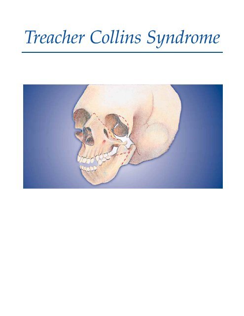
<strong>Treacher</strong> <strong>Collins</strong> <strong>Syndrome</strong>
TREACHER COLLINS<br />
SYNDROME<br />
<strong>Treacher</strong> <strong>Collins</strong> syndrome (also<br />
called mandibulofacial dysostosis<br />
and Franceschetti <strong>Syndrome</strong>) is a<br />
highly complex disease process. The<br />
basic etiology is unknown, but it is<br />
generally thought to be inherited as<br />
an autosomal dominant trait with<br />
variable penetrance. It is characterized<br />
by hypoplasia of the facial bones,<br />
especially the zygoma and the<br />
mandible. Facial clefting causes this<br />
hypoplastic appearance, with possible<br />
deformities or deficiencies of the<br />
ear, orbital, midface, and lower jaw<br />
regions. The clinical appearance is a<br />
result of the zygoma (malar bone)<br />
failing to fuse with the maxilla,<br />
frontal, and temporal bones. Highly<br />
variant degrees of involvement (complete,<br />
incomplete, and abortive<br />
forms) can be seen, but common<br />
facial features may include:<br />
(1) Hypoplastic cheeks, zygomatic<br />
arches, and mandible;<br />
(2) Microtia with possible hearing<br />
loss;<br />
(3) High arched or cleft palate;<br />
(4) Macrostomia (abnormally<br />
large mouth);<br />
(5) Anti-mongoloid slant to the eyes;<br />
(6) Colobomas;<br />
(7) Increased anterior facial height;<br />
(8) Malocclusion (anterior open bite);<br />
(9) Small oral cavity and airway with<br />
a normal-sized tongue;<br />
(10) Pointed nasal prominence.<br />
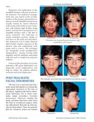
Preoperative<br />
<strong>Treacher</strong> <strong>Collins</strong> <strong>Syndrome</strong><br />
Severe mandibular hypoplasia in the patient with<br />
<strong>Treacher</strong> <strong>Collins</strong> syndrome.<br />
Preoperative<br />
Postoperative<br />
Postoperative<br />
Postoperative result after mandibular reconstruction and chin<br />
advancement to correct malocclusion and improve profile.<br />
The craniofacial team’s geneticist<br />
should evaluate all <strong>Treacher</strong> <strong>Collins</strong><br />
©1997 <strong>Erlanger</strong> <strong>Health</strong> <strong>System</strong> Tennessee Craniofacial Center 1(800) 418-3223
patients and their families to determine<br />
if the disease has been caused by inheritance<br />
of a family trait or as the result of a<br />
spontaneous gene mutation. If the disease<br />
has been inherited by one child in a<br />
family, there is a 50% chance that the parents<br />
will give birth to another involved<br />
child. If neither parents nor other family<br />
members are affected and a child is born<br />
with the condition, then a mutation has<br />
occurred. There is a 50% chance that this<br />
child will pass the trait on to future generations.<br />
Fortunately, genetic advances<br />
and careful prenatal screening have<br />
made <strong>Treacher</strong> <strong>Collins</strong> syndrome<br />
extremely rare.<br />
An extensive array of complications<br />
can affect treatment. Because of the small<br />
jaw and airway, combined with the normal<br />
size of the tongue, breathing problems<br />
can occur at birth and during sleep<br />
for a child with <strong>Treacher</strong> <strong>Collins</strong> syndrome<br />
when the base of the tongue<br />
obstructs the small hypopharynx. This<br />
situation can cause serious problems<br />
during the induction of general anesthesia.<br />
Consequently, a tracheostomy may<br />
be required to adequately control the airway.<br />
Learning and speech difficulties can<br />
also occur depending on the degree of<br />
conductive hearing loss common in the<br />
syndrome. Learning disabilities can<br />
potentially create a significant social stigma<br />
for the child. As with other disfiguring<br />
conditions, assessing and treating the<br />
psychological needs of the <strong>Treacher</strong><br />
<strong>Collins</strong> patient is a vital function of the<br />
true craniofacial center.<br />
Treatment of the hard and soft tissues<br />
of the face can require a number of surgical<br />
interventions, the first being the<br />
correction of eyelid coloboma in the first<br />
years of life (depending on the severity).<br />
The next stage is orbital reconstruction<br />
with calvarial bone grafts and correction<br />
of the lateral canthal displacement.<br />
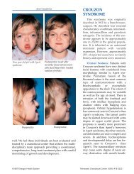
<strong>Treacher</strong> <strong>Collins</strong> <strong>Syndrome</strong><br />
Identical twins with <strong>Treacher</strong> <strong>Collins</strong> syndrome.<br />
Multi-stage ear reconstruction follows at about 5-7<br />
years of age. Correcting the lower face and jaws<br />
involves close coordination between the craniofacial<br />
surgeon and the pedodontist/orthodontist,<br />
with orthodontic intervention beginning with the<br />
eruption of the patient’s permanent teeth. After the<br />
teeth are aligned to their proper axis (or as closely<br />
as is possible), treatment of the lower face then<br />
involves orthognathic surgery to reposition the<br />
mandible and the maxilla, usually done during the<br />
patient’s teen years. This can be a one- or two-step<br />
procedure. The combined procedure involves rotating<br />
the midfacial segment around a transverse axis<br />
at the frontonasal angle (for severe maxillary<br />
hypoplasia) and lengethening the mandibular<br />
ramus. For less severe cases, a LeFort I type<br />
osteotomy technique is used to lower the maxillary<br />
tuberosities along with the ramus lengthening procedure<br />
for the mandible. As the child’s face continues<br />
to grow, additional procedures may be<br />
required to correct any developing deformities.<br />
Complimentary procedures such as rib cartilage<br />
grafts on the zygoma, closure of macrostomia, and<br />
secondary genioplasties are performed according<br />
to individual cases. A well-planned treatment regimen<br />
can produce excellent results with the ultimate<br />
goal being the complete restoration of form and<br />
function, thus enabling the patient to adapt to a<br />
“normal” way of life.<br />
©1997 <strong>Erlanger</strong> <strong>Health</strong> <strong>System</strong> Tennessee Craniofacial Center 1(800) 418-3223
CANTHAL SURGERY<br />
Many congenital and acquired deformities<br />
are associated with medial and lateral canthal<br />
displacement. Patients with blepharophimosis<br />
(telecanthus, epicanthal folds, ptosis), hypertelorism,<br />
Down’s <strong>Syndrome</strong>, craniofacial<br />
sysostosis, and acquired deformities may all<br />
have canthal deformities. Evaluation of the<br />
position and shape of the canthal area is a<br />
necessity in the planning of all orbital surgery.<br />
The contour and position of the canthi are<br />
important components in the aesthetic balance<br />
and symmetry of the face.<br />
Canthal surgery essentially consists of repositioning<br />
the involved canthal tendon to the<br />
desired position and securing it to the bone.<br />
This would seem to be a simple, straight-forward<br />
procedure. However, because of the complex<br />
anatomy of the medial canthal area and<br />
the difficulty in obtaining normal, symmetrical<br />
soft tissue contours, this procedure is neither as<br />
easy nor as predictable as it would appear to<br />
be. Attention to surgical technique, soft tissue<br />
tension and contour, bone mobilization and<br />
position, and healing of the tendon to bone —<br />
all are important in the ultimate results of canthal<br />
surgery.<br />
Canthal Surgery<br />
Preoperative<br />
Displacement of medial canthi and medial orbital walls<br />
due to facial trauma.<br />
Postoperative<br />
Medial orbital wall osteotomies and medial canthopexies<br />
performed to correct deformity.<br />
Preoperative<br />
Displacement of both medial canthi is present in this<br />
patient due to facial trauma.<br />
Postoperative<br />
Medial canthopexies are performed to return the medial<br />
canthi to their normal position.<br />
©1997 <strong>Erlanger</strong> <strong>Health</strong> <strong>System</strong> Tennessee Craniofacial Center 1(800) 418-3223