Mannitol Salt Agar (7143) - mibius
Mannitol Salt Agar (7143) - mibius
Mannitol Salt Agar (7143) - mibius
You also want an ePaper? Increase the reach of your titles
YUMPU automatically turns print PDFs into web optimized ePapers that Google loves.
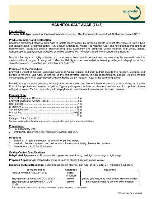
MANNITOL SALT AGAR (<strong>7143</strong>)<br />
Intended Use<br />
<strong>Mannitol</strong> <strong>Salt</strong> <strong>Agar</strong> is used for the isolation of staphylococci. This formula conforms to the US Pharmacopeia (USP). 1<br />
Product Summary and Explanation<br />
Chapman formulated <strong>Mannitol</strong> <strong>Salt</strong> <strong>Agar</strong> to isolate staphylococci by inhibiting growth of most other bacteria with a high<br />
salt concentration. 2 Chapman added 7.5% Sodium Chloride to Phenol Red <strong>Mannitol</strong> <strong>Agar</strong>, and noted pathogenic strains of<br />
staphylococci (coagulase-positive staphylococci) grew luxuriantly and produced yellow colonies with yellow zones.<br />
Nonpathogenic staphylococci produced small red colonies with no color change to the surrounding medium.<br />
<strong>Mannitol</strong> <strong>Salt</strong> <strong>Agar</strong> is highly selective, and specimens from heavily contaminated sources may be streaked onto this<br />
medium without danger of overgrowth. 3 <strong>Mannitol</strong> <strong>Salt</strong> <strong>Agar</strong> is recommended for isolating pathogenic staphylococci from<br />
clinical specimens, cosmetics, and microbial limit tests. 1,3-4<br />
Principles of the Procedure<br />
Enzymatic Digest of Casein, Enzymatic Digest of Animal Tissue, and Beef Extract provide the nitrogen, vitamins, and<br />
carbon in <strong>Mannitol</strong> <strong>Salt</strong> <strong>Agar</strong>. D-<strong>Mannitol</strong> is the carbohydrate source. In high concentrations, Sodium Chloride inhibits<br />
most bacteria other than staphylococci. Phenol Red is the pH indicator. <strong>Agar</strong> is the solidifying agent.<br />
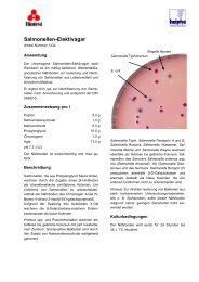
Bacteria that grow in the presence of a high salt concentration and ferment mannitol produce acid products, turning the<br />
Phenol Red pH indicator from red to yellow. Typical pathogenic staphylococci ferment mannitol and form yellow colonies<br />
with yellow zones. Typical non-pathogenic staphylococci do not ferment mannitol and form red colonies.<br />
Formula / Liter<br />
Enzymatic Digest of Casein ......................................................5 g<br />
Enzymatic Digest of Animal Tissue...........................................5 g<br />
Beef Extract...............................................................................1 g<br />
D-<strong>Mannitol</strong>...............................................................................10 g<br />
Sodium Chloride......................................................................75 g<br />
Phenol Red ........................................................................0.025 g<br />
<strong>Agar</strong>.........................................................................................15 g<br />
Final pH: 7.4 ± 0.2 at 25°C<br />
Formula may be adjusted and/or supplemented as required to meet performance specifications.<br />
Precautions<br />
1. For Laboratory Use.<br />
2. IRRITANT. Irritating to eyes, respiratory system, and skin.<br />
Directions<br />
1. Suspend 111 g of the medium in one liter of purified water.<br />
2. Heat with frequent agitation and boil for one minute to completely dissolve the medium.<br />
3. Autoclave at 121°C for 15 minutes.<br />
Quality Control Specifications<br />
Dehydrated Appearance: Powder is homogeneous, free flowing, and light red-orange to light beige.<br />
Prepared Appearance: Prepared medium is trace to slightly hazy and peach to pink.<br />
Expected Cultural Response: Cultural response on <strong>Mannitol</strong> <strong>Salt</strong> <strong>Agar</strong> at 35°C after 24 - 48 hours incubation.<br />
Microorganism Response Reactions<br />
Proteus mirabilis ATCC® 12453<br />
partial<br />
--<br />
inhibition<br />
Staphylococcus aureus ATCC® 25923 growth yellow colonies, may have yellow around colonies<br />
Staphylococcus epidermidis ATCC®<br />
12228<br />
growth pink colonies with no zone of color change<br />
The organisms listed are the minimum that should be used for quality control testing.<br />
PI <strong>7143</strong>, Rev 04, 03//10/05
Test Procedure<br />
Inoculate specimen on medium as a primary isolation or inoculate isolated colonies onto medium for differentiation.<br />
Results<br />
Staphylococci will grow on this medium, while the growth of most other bacteria will be inhibited. Coagulase-positive<br />
staphylococci will produce luxuriant growth of yellow colonies with yellow zones. Coagulase-negative staphylococci will<br />
produce small pink to red colonies with no color change to medium.<br />
Storage<br />
Store sealed bottle containing the dehydrated medium at 2 - 30°C. Once opened and recapped, place container in a low<br />
humidity environment at the same storage temperature. Protect from moisture and light by keeping container tightly<br />
closed.<br />
Expiration<br />
Refer to expiration date stamped on the container. The dehydrated medium should be discarded if not free flowing, or if<br />
appearance has changed from the original color. Expiry applies to medium in its intact container when stored as directed.<br />
Limitation of the Procedure<br />
Due to nutritional variation, some strains may be encountered that grow poorly or fail to grow on this medium.<br />
Packaging<br />
<strong>Mannitol</strong> <strong>Salt</strong> <strong>Agar</strong> Code No. <strong>7143</strong>A 500 g<br />
<strong>7143</strong>B 2 kg<br />
<strong>7143</strong>C 10 kg<br />
References<br />
1. United States Pharmacopeial Convention. 1995. The United States pharmacopeia, 23 rd ed. The United States Pharmacopeial Convention,<br />
Rockville, MD.<br />
2. Chapman, G. H. The significance of sodium chloride in studies of staphylococci. J. bacteriol. 50:201.<br />
3. Kloos, W. E., and T. L. Bannerman. 1995. Staphylococcus and Micrococcus. In P. R. Murray, E. J. Baron, M. A. Pfaller, F. C. Tenover, and R. H.<br />
Yolken (eds.). Manual of clinical microbiology, 6 th ed. American Society for Microbiology, Washington, D.C.<br />
4. Hitchins, A. D., T. T. Tran, and J. E. McCarron. 1995. Microbiology methods for cosmetics, p. 23.01-23.12. In Bacteriological analytical manual,<br />
8 th ed. AOAC International, Gaithersburg, MD.<br />
Technical Information<br />
Contact Acumedia Manufacturers, Inc. for Technical Service or questions involving dehydrated culture media preparation or performance at (517)372-<br />
9200 or fax us at (517)372-2006.<br />
PI <strong>7143</strong>, Rev 04, 03/10/05


