Spinal Cord Compression Superior Vena Cava Obstruction
Spinal Cord Compression Superior Vena Cava Obstruction
Spinal Cord Compression Superior Vena Cava Obstruction

You also want an ePaper? Increase the reach of your titles
YUMPU automatically turns print PDFs into web optimized ePapers that Google loves.
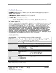
<strong>Spinal</strong> <strong>Cord</strong> <strong>Compression</strong><br />
and<br />
<strong>Superior</strong> <strong>Vena</strong> <strong>Cava</strong> <strong>Obstruction</strong><br />
Devin Schellenberg MD FRCPC<br />
Radiation Oncology<br />
Fraser Valley and Vancouver Centres<br />
British Columbia Cancer Agency
Aims of the Talk<br />
1. Med school level: Describe the pathologic process<br />
<br />
What is actually going on<br />
2. Resident Level: Learn how to recognize these<br />
emergencies<br />
<br />
<br />
Clinical symptoms<br />
Xray and CT findings<br />
3. Your Level: Understand how to treat these emergencies<br />
<br />
<br />
<br />
What should I give the patient<br />
What should I order<br />
Who should I call
Part 1: <strong>Spinal</strong> <strong>Cord</strong> <strong>Compression</strong>
<strong>Spinal</strong> <strong>Cord</strong> <strong>Compression</strong>: Incidence<br />
Loblaw D, et al Clinical Oncology 2003<br />
population study to review incidence, management and<br />
outcome of <strong>Spinal</strong> cord compression (SCC) in Ontario<br />
(1990-1995)<br />
cumulative probability of experiencing at least one episode<br />
of SCC in the 5 years preceding death from cancer was<br />
2.5% overall (ranged by cancer site)<br />
median survival following the first episode of SCC was 2.9<br />
months
Function of Anatomy<br />
Tumour grows in epidural space, obstruct epidural plexus /<br />
direct compression of cord, vasogenic edema and ensuing<br />
spinal cord infarction<br />
Intramedullary<br />
Cerebrospinal Fluid
Both cause similar clinical features<br />
1. Back pain 85-95%<br />
The most common first symptom<br />
Can be radicular<br />
2. Motor Findings<br />
Weakness 60-85%<br />
Hyper-reflexia (cauda equina depressed DTR)<br />
3. Sensory Changes<br />
4. Bowel and Bladder dysfunction<br />
Late finding, up to 50%
Xray<br />
<strong>Cord</strong> <strong>Compression</strong> Suspected<br />
What next?<br />
Urgent CT scan (specify level)<br />
Urgent MRI provides more accurate view of<br />
compression (but is harder to get)<br />
If Cancer diagnosis – call oncologist<br />
If no cancer diagnosis – start looking for one and<br />
call neurosurgery
CT Scan images<br />
10<br />
With and Without bone disease<br />
MRI imaging is more detailed (accurate)<br />
but harder to get urgently
11<br />
SCC – With Bony Disease
12<br />
SCC – With Bony Disease
13<br />
SCC – Without Bony Disease
14<br />
SCC – Without Bony Disease
15<br />
MRI – <strong>Cord</strong> <strong>Compression</strong>
Good prognostic factors<br />
‣ Responsive tumor to Radiotherapy or Chemotherapy<br />
‣ Lymphoma/Myeloma, Small Cell<br />
‣ Gradual onset with slow progression<br />
‣ Able to walk, normal bladder function<br />
‣ Good general condition
Aims of Management<br />
Return blood flow to spinal cord<br />
Reduce swelling<br />
•Steroids - dexamethasone<br />
Decrease pressure from mass<br />
1. Surgery and Radiation<br />
2. Radiation treatment alone
Management - Steroids<br />
Steroids beneficial in retaining motor function<br />
No evidence to support use of high dose steroids<br />
(>16mg/day)<br />
High dose steroids associated with more adverse<br />
events<br />
OK to start 10 mg IV<br />
Then 16 mg/day<br />
Select patients may not require steroids (e.g.<br />
asymptomatic with radiographic finding alone)
Management: Surgery<br />
<br />
<br />
<br />
Patchell et al: Radiation vs Surgery + Radiation<br />
Patient Characteristics<br />
• 3m life expectancy, Medically<br />
operable, Surgery within 24 hours<br />
Tumour<br />
• 1 site of compression<br />
• No cauda equina syndrome<br />
• Not Lymphoma<br />
Results<br />
Surgery + RT gave better: Return of ambulation, duration of<br />
ambulation, continence, functional ability & overall survival<br />
Issues<br />
Most patients do not meet inclusion criteria
Management: Radiotherapy<br />
Most common treatment for <strong>Cord</strong> <strong>Compression</strong><br />
Multiple studies examine role of radiotherapy alone<br />
in management<br />
Radiotherapy effective in treatment of SCC<br />
decrease pain<br />
increase functional outcomes<br />
increase sphincter function<br />
Final gait function dependent on gait at time of<br />
diagnosis
Radiotherapy: the sooner the better<br />
% of ambulatory pts who are ambulatory post 95%<br />
% of assisted ambulation who are ambulatory post 60%<br />
% of paraparetic who are ambulatory post 40%<br />
% of paraplegic who are ambulatory post 10%<br />
Loblaw et. al. 2004
The Surgery vs. Radiation Decision<br />
Surgery<br />
<br />
<br />
<br />
<br />
<strong>Spinal</strong> instability<br />
Bony compression<br />
Patients with no/remote cancer<br />
diagnosis (establish Path)<br />
Prior RT or neurological<br />
progression while on RT<br />
Radiation<br />
<br />
<br />
<br />
<br />
Medically inoperable<br />
Diffuse disease<br />
Radioresponsive tumors<br />
(lymphoma, myeloma, germ<br />
cell…)<br />
Have cancer diagnosis
SCC Conclusions<br />
<br />
<br />
Decisions regarding treatment should consider<br />
Medical status, ambulatory status, structural factors, anticipated<br />
outcome, treatment goals<br />
Treatment goals should be to improve or maintain highest<br />
quality of life possible<br />
Pain relief<br />
Restoration of function<br />
<br />
Refer for management early to obtain best results<br />
High index of suspicion (hx of cancer & back pain)<br />
Steroids<br />
CT or MRI spine
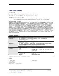
Part 2: <strong>Superior</strong> <strong>Vena</strong> <strong>Cava</strong><br />
<strong>Obstruction</strong>
<strong>Superior</strong> <strong>Vena</strong> <strong>Cava</strong> <strong>Obstruction</strong><br />
“One of your take away points should be that this is<br />
not an emergency most of the time.”<br />
Dr. Leong
SVC <strong>Obstruction</strong> - Anatomy
SVCO - Definition<br />
UpToDate: SVCO is often a prolonged process<br />
developing over a period of weeks or longer prior<br />
to clinical presentation. The duration of symptoms<br />
has no influence on treatment outcomes [7].<br />
Deferring therapy until a full diagnostic work-up<br />
has been completed does not pose a hazard for<br />
most patients, provided the evaluation is efficient<br />
and the patient is clinically stable…
When is SVCO an Emergency?<br />
Emergency if:<br />
Stridor, tachypnea<br />
Laryngeal edema (voice changes)<br />
Hemodynamically unstable<br />
Altered level of consciousness<br />
If only dilated veins/swelling<br />
Urgent, but not emergent<br />
If only an imaging diagnosis of SVCO<br />
Even less urgent<br />
SVC is still largely a Clinical diagnosis!
Not SVCO
Not SVCO -<br />
The report will say: tumor is against or compressing SVC
SVCO – On XRay<br />
Very little to see in terms of widened mediastinum
SVCO – Swelling and Dilated veins<br />
Lots to see clinically
SCVO – My Case
SVCO – Arm Swelling (into hand)
SVCO – “I’ve lost by elbow”
SVCO Symptoms and Signs<br />
Edema<br />
Altered consciousness<br />
http://www.scielo.br/pdf/jbpneu/v31n6/en_27958.pdf
SVCO: Clinical diagnosis<br />
Clinical symptoms<br />
Flushing<br />
Edema<br />
Venous engorgement<br />
Hoarseness (laryngeal edema)<br />
Impaired Mental Status (reduced perfusion)<br />
CT Scans<br />
Confirm diagnosis/plan treatment<br />
Venography and MR venography (rarely needed)
38<br />
SVCO Suspected – CT ordered
SVCO – CT findings
SVCO – CT findings
SVCO – CT findings
SVCO found: What should you do?<br />
42<br />
1. Make the diagnosis clinically and order an urgent<br />
CT<br />
2. If known cancer – call the oncologist (or to ER)<br />
3. If no pathology then we will need some…<br />
Respirology: Bronchoscopy +/- EBUS<br />
Thoracic Surgery : Bronch +/- EBUS<br />
CT guided biopsy<br />
Non-Thoracic primary: Can biopsy another site
Should I give steroids?<br />
SVCO<br />
Less helpful if lung cancer or non-lymphoma<br />
Can be very helpful in setting of KNOWN lymphoma<br />
BUT… If you suspect lymphoma and haven’t made a<br />
diagnosis then DO NOT give steroids<br />
Given if you think it can reduce symptoms from<br />
XRT/Chemo<br />
Given if you think there is laryngeal edema
Should I Give Diuretics<br />
SVCO<br />
Not well studied<br />
Likely of limited benefit (and of limited harm)<br />
We tend not to give them
Should I give something else?<br />
Try to control symptoms<br />
Pain Control<br />
Breathing symptoms<br />
• Puffers<br />
• Oxygen<br />
Dizziness (reduce BP meds)<br />
Edema (comfort compression)<br />
• Gloves
Definitive Treatment<br />
46<br />
Most commonly Radiation<br />
Chemotherapy for lymphomas/Small Cell Lung<br />
Interventional Radiology – SVC stent<br />
Good: Quick symptom relief<br />
Bad: Doesn’t treat underlying cause<br />
Problem: Often not an emergency so questionable if<br />
you need rapid relief
SVCO Conclusions<br />
47<br />
Recognize the symptoms and signs<br />
Understand if this is an EMERGENCY or just an urgency<br />
Order appropriate CT<br />
(and/or Ultrasound if clot suspected)<br />
Refer promptly
Thanks to:<br />
48<br />
Drs. Chad Lund / Paris-Ann Ingledew for slides<br />
Drs. Anand Karvat / Frances Wong for forwarding<br />
patients
Symptoms<br />
Dyspnea<br />
Distension<br />
edema of face with erythema (plethora)<br />
edema of arms<br />
Dilated chest wall veins<br />
Impaired mental status<br />
Decreased cerebral perfusion pressure<br />
• CPP = MAP – ICP or SVP (whichever is greater)<br />
Hoarseness<br />
Vocal cord edema
Management<br />
• Steroids (dexamethasone
SVCO – Clinical Picture (Internet)
SVCO - Stenting<br />
52<br />
http://www.ajronline.org/doi/abs/10.2214/AJR.08.1904
SVCO - Etiology<br />
Benign 20% Malignant 80%<br />
Aortic aneurysm<br />
Bronchogenic<br />
Sarcoidosis<br />
SCLC<br />
Mediastinitis<br />
NSCLC<br />
Thrombosis<br />
Lymphoma<br />
Retrosternal thyroid Germ cell<br />
Other
Cumulative Incidence
Radiotherapy<br />
Radiotherapy effective in treatment of MSCC<br />
decrease pain<br />
increase functional outcomes<br />
increase sphincter function<br />
Earlier institution of radiotherapy prior to decreased<br />
functional status preferred
SVCO – My patient
Poor prognostic factors<br />
Vertebral collapse<br />
Radioresistant /chemoresistant tumour<br />
• Melanoma<br />
• Renal cell<br />
Acute onset with rapid progression<br />
Inability to walk, loss of bladder function<br />
Poor general condition