SIMULATION CASEBOOK - MyCourses
SIMULATION CASEBOOK - MyCourses
SIMULATION CASEBOOK - MyCourses

You also want an ePaper? Increase the reach of your titles
YUMPU automatically turns print PDFs into web optimized ePapers that Google loves.
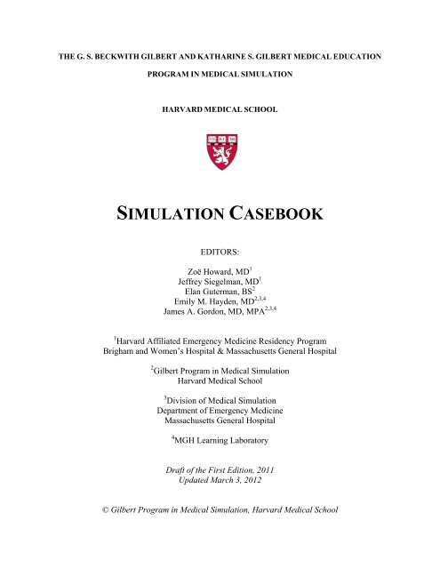
THE G. S. BECKWITH GILBERT AND KATHARINE S. GILBERT MEDICAL EDUCATION<br />
PROGRAM IN MEDICAL <strong>SIMULATION</strong><br />
HARVARD MEDICAL SCHOOL<br />
<strong>SIMULATION</strong> <strong>CASEBOOK</strong><br />
EDITORS:<br />
Zoë Howard, MD 1<br />
Jeffrey Siegelman, MD 1<br />
Elan Guterman, BS 2<br />
Emily M. Hayden, MD 2,3,4<br />
James A. Gordon, MD, MPA 2,3,4<br />
1 Harvard Affiliated Emergency Medicine Residency Program<br />
Brigham and Women’s Hospital & Massachusetts General Hospital<br />
2 Gilbert Program in Medical Simulation<br />
Harvard Medical School<br />
3 Division of Medical Simulation<br />
Department of Emergency Medicine<br />
Massachusetts General Hospital<br />
4 MGH Learning Laboratory<br />
Draft of the First Edition, 2011<br />
Updated March 3, 2012<br />
© Gilbert Program in Medical Simulation, Harvard Medical School
Gilbert Program in Medical Simulation<br />
Simulation Casebook<br />
Harvard Medical School Draft of the 1 st edition, (2011); updated 3/2/12<br />
Table of Contents<br />
CARDIOVASCULAR<br />
Anterior Myocardial Infarction……………….………………..……...…………... 6<br />
Aortic Stenosis with Atrial Fibrillation……………….……………...……………. 14<br />
Cardiogenic Shock………………………………………………………..……….. 25<br />
Inferior Myocardial Infarction……………………………………………...……... 33<br />
Pulmonary Embolism…….…………………………………………………..……. 42<br />
ENDOCRINE<br />
Alcoholic Ketoacidosis……………………………………………………………. 51<br />
Diabetic Ketoacidosis…………………………………………………………… 59<br />
Hyperemesis Gravidarum………………………………………………………….. 67<br />
Hypoglycemia………………………………….………………………………. 77<br />
Rhabdomyolysis…………………………………………………..……………... 81<br />
GASTROINTESTINAL<br />
Acute Cholecystitis…………………………………………………………….. 92<br />
Lower Gastrointestinal Bleed…………………………………….……………. 99<br />
Upper Gastrointestinal Bleed………………………………….……………… 108<br />
NEUROLOGICAL<br />
Migraine………………………………………………………………………….. 116<br />
OVERDOSE<br />
Cocaine Intoxication………………………………………………………………. 124
Gilbert Program in Medical Simulation<br />
Simulation Casebook<br />
Harvard Medical School Draft of the 1 st edition (2011), updated 3/2/12<br />
Ecstasy (MDMA) Intoxication…………………………………………………….. 132<br />
Narcotic Intoxication……………………………….……………………………… 138<br />
Organophosphate Poisoning…………………………………….…………………. 144<br />
RESPIRATORY<br />
Asthma Exacerbation……………………………………………………..……….. 150<br />
COPD Exacerbation………………………………….………….…………...……. 157<br />
Pneumonia: Community-Acquired…………………………….………………..… 164<br />
TRAUMA<br />
Head and Musculoskeletal Injury………………………………………………….. 173<br />
3
Gilbert Program in Medical Simulation<br />
Simulation Casebook<br />
Harvard Medical School Draft of the 1 st edition (2011), updated 3/2/12<br />
NOTE FROM THE EDITORS<br />
This casebook comprises a collection of selected simulation cases developed, refined, and<br />
deployed in the teaching laboratories of the Gilbert Program in Medical Simulation at Harvard<br />
Medical School. They have been drafted, edited, and adapted by several classes of teaching<br />
fellows and core faculty who comprise the Gilbert Simulation Team. Collectively, these cases<br />
have been used to teach thousands of students.<br />
We have edited and enhanced these cases to ensure medical accuracy and uniformity<br />
(N.B. the standardized case template used throughout was adapted from the Simulation Case<br />
Template developed by the Society for Academic Emergency Medicine Simulation Interest<br />
Group). As written, the cases are intended for a broad array of learners, ranging from preclinical<br />
medical students and doctoral graduate students to interns and advanced residents. There are<br />
optional case progressions, with case variations and suggestions to prompt the learner or guide<br />
the case. For advanced learners, a more challenging scenario can be adapted by utilizing the<br />
points in italics. Of note, basic variants of several of these cases have been used to teach<br />
sophisticated biomedical concepts to high school and college students as part of the Harvard<br />
Summer PreMedical Institute and Harvard Medical School Bioscience Program for High School<br />
Students.<br />
To enhance the simulation experience, multimedia links are provided to EKG,<br />
radiography, and other resources on the internet.<br />
We hope that a consolidated, central source of successfully tested and deployed<br />
simulation cases will be a valuable addition to many medical teaching programs, and will help<br />
stimulate further development of standardized approaches to simulation-based teaching and<br />
assessment.<br />
We are grateful for the support of G.S. Beckwith Gilbert and the Gilbert Family<br />
Foundation. Special thanks to the Simulation Fellows, Staff, and Faculty who made this work<br />
possible.<br />
-<br />
4
Gilbert Program in Medical Simulation<br />
Simulation Casebook<br />
Harvard Medical School Draft of the 1 st edition, (2011); updated 3/2/12<br />
CARDIOVASCULAR CASES<br />
Gilbert Program in Medical Simulation<br />
Harvard Medical School
Gilbert Program in Medical Simulation<br />
Simulation Casebook<br />
Harvard Medical School Draft of the 1 st edition (2011), updated 3/2/12<br />
Anterior Myocardial Infarction<br />
I. Target Audience: Medical Students, Residents<br />
II.<br />
Learning and Assessment Objectives<br />
Participants are expected to understand the scientific and humanistic issues underlying the disease<br />
assessment and treatment plan, and to critically consider and deploy the therapeutic options described<br />
Participants should provide a concise presentation of the patient to each physician consultant who<br />
participates in the exercise. Debriefing sessions by on-site clinical faculty is essential to discuss critical<br />
thinking and knowledge pathways, and to provide a forum for individual and team reflection on learning<br />
and practice goals. While the case descriptions are written with medical terminology, it is important that<br />
the provider and patient (i.e. the simulator) engage in authentic dialogue with lay terminology to reflect<br />
an actual patient encounter.<br />
Critical Actions Checklist:<br />
DONE CRITICAL ACTION<br />
Telemetry monitoring<br />
Patient history<br />
Physical examination<br />
Supplemental O 2<br />
IV Access<br />
Immediate ECG and portable CXR (within 10 minutes of start of case)<br />
Obtain appropriate laboratory studies: cardiac biomarkers<br />
Administer immediate aspirin<br />
Administer pressor agent<br />
Administer anticoagulation medication<br />
Consult cardiology for further treatment<br />
III.<br />
IV.<br />
Environment<br />
A. Simulation room set up: Emergency Department<br />
B. Manikin set up:<br />
1. High fidelity patient simulator<br />
2. No moulage needed<br />
3. Lines needed<br />
C. Props:<br />
1. Code blue cart<br />
2. Lab values (see Appendix A)<br />
3. Images (see Appendix B)<br />
D. Distracters: none<br />
Actors<br />
A. Nurse: facilitate scenario<br />
B. Consultants (optional for higher level residents who can provide interpretation on their<br />
own)<br />
1. Radiologist: reads chest x-ray<br />
2. Cardiologist: reads ECG, recommends treatment plan<br />
6
Gilbert Program in Medical Simulation<br />
Simulation Casebook<br />
Harvard Medical School Draft of the 1 st edition (2011), updated 3/2/12<br />
Case Narrative<br />
PATIENT: 60 year old<br />
CC: Chest pain, “There is an elephant sitting on my chest.”<br />
HPI: Use lay terminology as the voice of the patient<br />
Patient complains of crushing substernal chest pain radiating to his neck and jaw on the left side.<br />
Symptoms started one hour ago during a business meeting. Patient had to excuse himself from the<br />
meeting as he became obviously diaphoretic and pale.<br />
Patient reports nausea and lightheadedness after the onset of the “crushing” chest pain. Patient denies<br />
fevers, chills, vomiting, and palpitations. Patient reports mild shortness of breath and one previous<br />
episode of chest pain that lasted about 15 minutes one week ago that resolved spontaneously while he was<br />
in Japan.<br />
PMHx:<br />
Hypertension<br />
Lisinopril<br />
MEDICATIONS<br />
Codeine<br />
Shellfish<br />
ALLERGIES<br />
PSHx:<br />
Hernia repair, age 22<br />
SOCIAL Hx:<br />
EtOH:<br />
Tobacco:<br />
Illicits:<br />
Occupation:<br />
Additional:<br />
Occasional<br />
Denies<br />
Denies<br />
Ambassador to the U.S.<br />
Married<br />
FAMILY Hx:<br />
Father: Gastric cancer, expired age 80<br />
ROS:<br />
(+) Chest pain with radiation to neck/jaw, mild shortness of breath, diaphoresis, nausea,<br />
lightheadedness<br />
(-) Denies palpitations, vomiting, headache, blurred vision, numbness/motor weakness, abdominal<br />
pain, urinary symptoms, or fever/chills<br />
7
Gilbert Program in Medical Simulation<br />
Simulation Casebook<br />
Harvard Medical School Draft of the 1 st edition (2011), updated 3/2/12<br />
PHYSICAL EXAM: Those signs not demonstrable by the mannequin should be verbalized when<br />
students perform/verbalize the examination maneuver<br />
HR BP Temperature ( o C) O 2 Sats (RA) RR<br />
110 88/60 37.5 o 92% 24<br />
GENERAL:<br />
HEENT:<br />
NECK:<br />
CV:<br />
PULM:<br />
ABD:<br />
EXT:<br />
NEURO:<br />
A&OX3, moderate distress<br />
PERRL/EOMI<br />
Supple, no JVD<br />
2/6 systolic apical murmur, tachycardia<br />
Diffuse rales all lung fields<br />
Soft, NT/ND, + BS<br />
No C/C/E, palpable pulses all extremities<br />
WNL, MAE X 4, grossly intact<br />
LABS: See Appendix A<br />
Amylase/Lipase Level<br />
Comprehensive Metabolic Panel<br />
Arterial Blood Gas<br />
Hepatic Panel<br />
Basic Metabolic Panel X Lactate/Cortisol Level<br />
Cardiac Markers X Thyroid Panel<br />
Coagulation Profile X Toxicology Screen<br />
Complete Blood Count (CBC)<br />
Urinalysis<br />
CBC with differential X Urine HCG<br />
IMAGES: See Appendix B<br />
8<br />
Additional Labs: none<br />
Angiogram ECG X<br />
CT Scan, with contrast<br />
MRI<br />
CT Scan, without contrast X-Ray X<br />
Echocardiogram<br />
Ultrasound<br />
Additional Images: none<br />
CONSULTS:<br />
Cardiology – Dr. Jones: ECG will be read as STEMI in leads V1-V6 and leads I and AVL. Cardiology<br />
will recommend preparing the patient for cardiac catheterization: aspirin, Plavix, heparin, and “… if it’s<br />
safe in light of the patient’s vital signs,” B-blocker and nitroglycerin. Indicate that the catheterization<br />
team will need about 20 minutes to get in and that the patient must be stabilized prior to catheterization.<br />
Cardiologist asks the students to tell the patient that they will need to have a cardiac catheterization.<br />
If vitals have not been stabilized, tell participants to call back after blood pressure and other vitals<br />
improve. If participants ask about increasing the pressure safely, recommend pressors (dopamine).
Gilbert Program in Medical Simulation<br />
Simulation Casebook<br />
Harvard Medical School Draft of the 1 st edition (2011), updated 3/2/12<br />
Radiology – Dr. Smith: CXR shows diffuse pulmonary edema consistent with congestive heart failure.<br />
CLINCAL PROGRESSION:<br />
History and physical, IV/O 2 /monitor, and immediate aspirin. Pressors should be started after physical<br />
exam and stat portable CXR indicative of cardiogenic pulmonary edema with hypotension. ECG will<br />
indicate AMI either after participants’ own interpretation or after consultation.<br />
*** If over 500 CC’s IV fluids given or supplemental O 2 not initiated within the first 10-15 minutes of<br />
case, patient will continue providing history in short one word (monosyllabic) answers and indicate that<br />
shortness of breath is getting worse. O 2 saturation will drop:<br />
HR BP Temperature ( o C) O 2 Sats (RA) RR<br />
116 88/60 37.5 o 88% 26<br />
***If Morphine or Nitroglycerin given (sublingual or IV) blood pressure will drop, heart rate will<br />
increase but rhythm stays regular, patient will become less responsive:<br />
HR BP Temperature ( o C) O 2 Sats (RA) RR<br />
122 68/50 37.5 o 92% 24<br />
***If B-blockers given, heart rate and blood pressure will decrease, pt will become unresponsive:<br />
HR BP Temperature ( o C) O 2 Sats (RA) RR<br />
100 68/50 37.5 o 92% 24<br />
V. Instructor Notes<br />
A. Tips to keep scenario flowing<br />
1. If students are unsure of pathology, instructor can prompt the students to create<br />
differential diagnosis and lead them towards imaging and laboratory studies<br />
necessary to confirm diagnosis. Prompting can come in form of a primary care<br />
physician calling to check in on their patient.<br />
2. If supplemental O 2 is not provided, nurse can verbalize concern as patient becomes<br />
increasingly dyspneic<br />
B. Scenario programming<br />
1. Optimal management path:<br />
O 2 /IV/monitor<br />
History and physical examination<br />
Immediate aspirin<br />
Appropriate lab workup: CBC, BMP, cardiac markers, coagulation<br />
profile<br />
Appropriate imaging: stat portable CXR, ECG within 10 minutes<br />
Administer pressor agent<br />
Administer anticoagulation medication (e.g. Heparin, Plavix, +/- IIb/IIIa)<br />
Consider administering morphine, B-blocker, and nitrates<br />
Consult cardiology for further treatment<br />
2. Potential complications/errors path(s):<br />
Failure to administer O 2<br />
9
Gilbert Program in Medical Simulation<br />
Simulation Casebook<br />
Harvard Medical School Draft of the 1 st edition (2011), updated 3/2/12<br />
<br />
<br />
Administering over 500 CC’s IV fluid<br />
Administering large dose of Nitroglycerin, B-blocker, Morphine<br />
VI.<br />
Debriefing Plan<br />
A. Method of debriefing: Group with multimedia teaching materials<br />
B. Debriefing materials: See Appendix C<br />
C. Potential debriefing topics<br />
1. Team dynamics<br />
a. Leadership<br />
b. Collaboration<br />
c. Communication<br />
d. Professionalism<br />
2. Didactic material<br />
a. Presentation<br />
i. Appropriate differential diagnosis<br />
ii. Varying presentation of MI in different location<br />
1. Contrast fluid overload requiring + inotropy from anterior<br />
MI with preload dependence and need for IV fluids in<br />
inferior MI<br />
b. Pathophysiology<br />
i. Atherosclerotic vs. nonatherosclerotic causes<br />
ii. Laboratory results: troponin, CKMB levels<br />
c. Treatment<br />
i. Need for immediate diagnosis and reperfusion for the acute MI<br />
1. “Time is muscle”<br />
ii. Role of aspirin therapy: decrease mortality/reinfarction rates<br />
iii. Role of other antiplatelet therapy<br />
1. Clopidogrel (Plavix) at 600mg dose if emergent CABG not<br />
anticipated<br />
iv. Role of pressors vs. fluids in the anterior MI patient<br />
v. Role of anticoagulants<br />
1. Heparin: indicated in recurrent/persistent chest pain, AMI,<br />
positive biomarkers, dynamic EKG changes. Dose is<br />
60U/kg bolus followed by 12U/kg infusion, titrating to apt<br />
1.5-2.5 times control<br />
2. LMWH at 1mg/kg BID, adjusted for renal insufficiency<br />
3. GP IIb/IIIa inhibitor<br />
vi. Role of acute beta-blockade and nitrates<br />
1. B-blocker: heart rate control and resultant decrease of<br />
myocardial O 2 demand to reduce rates of reinfarction,<br />
recurrent ischemia and potentially mortality<br />
2. Nitrates: preload reduction and symptomatic relief<br />
3. Contraindications in the hypotensive MI patient<br />
a. Hold NTG for SBP < 90<br />
b. Hold BB if signs of cardiogenic shock<br />
vii. Treatment options: thrombolytic therapy vs. heart catheterization<br />
(PCI) vs. coronary bypass graft<br />
10
Gilbert Program in Medical Simulation<br />
Simulation Casebook<br />
Harvard Medical School Draft of the 1 st edition (2011), updated 3/2/12<br />
VII.<br />
Development and Deployment<br />
This case, along with it precursors (reference Gordon, below) and variants have been used over several<br />
years for a wide range of students, including high school, college, masters/PhD candidates, medical<br />
students (preclinical and clinical) and resident trainees. The presentation and progression is tailored to the<br />
level of the learner; often the Anterior MI case is paired with the Inferior MI case to allow students to<br />
compare and contrast diagnosis, anatomy, physiology, and management. It is commonly used as part of a<br />
“train the trainer” curriculum for faculty development in the use of simulation.<br />
VIII.<br />
Authors/Contributors<br />
Case drafted by Rami Ahmed, D.O, with contributions and updates by the Gilbert Simulation Team.<br />
Compiled and formatted by Elan Guterman.<br />
Reviewed and edited with references by Jeffrey Siegelman, M.D. and Zoë Howard, M.D.<br />
IX.<br />
References<br />
Antman EM, Anbe DT, Armstrong PW, Bates ER, Green LA, Hand M, Hochman JS, Krumholz HM,<br />
Kushner FG, Lamas GA, Mullany CJ, Ornato JP, Pearle DL, Sloan MA, Smith SC Jr. ACC/AHA<br />
guidelines for the management of patients with ST-elevation myocardial infarction. A report of the Am<br />
Coll of Cardiol/Am Heart Assoc Task Force on Practice Guidelines (Committee to revise the 1999<br />
guidelines). Bethesda (MD): American College of Cardiology, American Heart Association; 2004.<br />
Brady, WJ, Harrigan, RA, and Theodore Chan. “Acute Coronary Syndromes.” In: Rosen’s Emergency<br />
Medicine: Concepts and Clinical Practice, 6 th ed. Marx, et al, eds. pp 1154-99.<br />
Gordon, JA. Macy Cases for Realistic Patient Simulation in Critical Care and Emergency Medicine.<br />
Harvard Medical School, Boston: President and Fellows of Harvard College, 2002<br />
11
Gilbert Program in Medical Simulation<br />
Simulation Casebook<br />
Harvard Medical School Draft of the 1 st edition (2011), updated 3/2/12<br />
X. Appendix A: Lab Values<br />
BASIC METABOLIC PANEL<br />
Reference Range<br />
Sodium 137 135-147 mmol/L<br />
Potassium 3.9 3.5-5.2 mmol/L<br />
Chloride 105 95-107 mmol/L<br />
CO 2 27 22-30 mmol/L<br />
Urea Nitrogen (BUN) 15 7-20 mg/dL<br />
Creatinine 1.1 0.5-1.2 mg/dL<br />
Glucose 103 60-110 mg/dL<br />
CARDIAC MARKERS<br />
Reference Range<br />
Creatine Kinase-BB 0% 0%<br />
Creatine Kinase-MB (cardiac) 2% 0 - 3.9%<br />
Creatine Kinase-MM 98% 96 – 100%<br />
Creatine phosphokinase (CPK) 135 8 – 150 IU/L<br />
Troponin I 0.01 < 0.03 ng/mL<br />
COAGULATION PROFILE<br />
Reference Range<br />
Partial thromboplastin time (PTT) 35 30 – 45 sec<br />
Prothrombin time (PT) 12 10 – 12 sec<br />
International Normalized Ratio (INR) 1.0 1 – 2<br />
Fibrinogen 180 170 – 420 mg/dL<br />
12
Gilbert Program in Medical Simulation<br />
Simulation Casebook<br />
Harvard Medical School Draft of the 1 st edition (2011), updated 3/2/12<br />
COMPLETE BLOOD COUNT<br />
Reference Range<br />
WITH DIFFERENTIAL<br />
Male<br />
Female<br />
White Blood Cell (WBC) 9,700 4,500 - 10,000 K/uL<br />
Segmented neutrophils (%) 57 54 – 62%<br />
Band forms (%) 4 3 - 5 (>8% indicates L shift)<br />
Basophils (%) 0.75 0 – 1% 0 - 0.75%<br />
Eosinophils (%) 2 0 – 3% 1 – 3%<br />
Lymphocytes (%) 32 24 – 44% 25 – 33%<br />
Monocytes (%) 4 3 – 6% 3 – 7%<br />
Hemoglobin (HGB) 15.0 13.5 - 16.5 g/dL 12.0 - 15.0 g/dL<br />
Hematocrit (HCT) 39.0 41 – 50% 36 – 44%<br />
Red blood cell (RBC) 4.8 4.5 - 5.5 M/uL 4.0 - 4.9 M/uL<br />
RBC Distribution Width 10.0 < 14.5%<br />
MCV 92 80 – 100 fL<br />
MCH 32 26 – 34 pg<br />
MCHC 34 31 – 37 g/dL<br />
Platelet 420,000 100,000 - 450,000 K/uL<br />
XI. Appendix B: Diagnostic Studies<br />
Chest X-Ray: Diffuse cardiogenic pulmonary edema bilaterally<br />
http://www.meddean.luc.edu/lumen/meded/medicine/pulmonar/cxr/diffuse/Dscn017.jpg<br />
ECG:<br />
http://www.rcsed.ac.uk/fellows/bcpaterson/images/chest_12.jpg<br />
XII. Appendix C: Teaching Materials<br />
Angioplasty Video<br />
http://www.youtube.com/watch?v=gcrLzOkACgk&feature=channel<br />
13
Gilbert Program in Medical Simulation<br />
Simulation Casebook<br />
Harvard Medical School Draft of the 1 st edition (2011), updated 3/2/12<br />
Aortic Stenosis: Angina Followed by Atrial Fibrillation<br />
I. Target Audience: Medical Students, Residents<br />
II. Learning and Assessment Objectives<br />
Participants are expected to understand the scientific and humanistic issues underlying the disease<br />
assessment and treatment plan, and to critically consider and deploy the therapeutic options described<br />
Participants should provide a concise presentation of the patient to each physician consultant who<br />
participates in the exercise. Debriefing sessions by on-site clinical faculty is essential to discuss critical<br />
thinking and knowledge pathways, and to provide a forum for individual and team reflection on learning<br />
and practice goals. While the case descriptions are written with medical terminology, it is important that<br />
the provider and patient (i.e. the simulator) engage in authentic dialogue with lay terminology to reflect<br />
an actual patient encounter.<br />
Critical Actions Checklist:<br />
DONE CRITICAL ACTION<br />
PART I<br />
Telemetry monitoring<br />
Patient history<br />
Physical examination<br />
Supplemental O 2<br />
Immediate ECG and CXR<br />
Provide IV fluids<br />
Obtain appropriate laboratory studies: cardiac biomarkers<br />
Admit patient for stress test<br />
<br />
<br />
<br />
<br />
<br />
PART II<br />
Obtain additional history<br />
Repeat physical examination and immediate ECG<br />
Electrical or chemical cardioversion<br />
Initiate anticoagulant therapy<br />
Consider beginning longer acting rate/rhythm control agents after<br />
successful cardioversion<br />
III.<br />
Environment<br />
A. Simulation room set up: PART I in emergency department, PART II in either emergency<br />
department or on medical floor<br />
B. Manikin set up:<br />
1. High fidelity patient simulator<br />
2. No moulage needed<br />
3. Lines needed<br />
C. Props:<br />
1. Code blue cart<br />
2. Lab values (see Appendix A)<br />
3. Images (see Appendix B)<br />
D. Distracters: none<br />
14
Gilbert Program in Medical Simulation<br />
Simulation Casebook<br />
Harvard Medical School Draft of the 1 st edition (2011), updated 3/2/12<br />
IV.<br />
Actors<br />
A. Nurse: facilitate scenario<br />
B. Consultants (optional for higher level residents who can provide interpretation on their<br />
own)<br />
1. Radiologist: reads chest x-ray<br />
2. Cardiologist: reads ECG, recommends treatment plan<br />
V. Case Narrative: Part I<br />
PATIENT: 68 year old<br />
CC: Chest pain, shortness of breath<br />
HPI: Use lay terminology as the voice of the patient<br />
Patient presents with chest pain and shortness of breath that began while carrying pipes on a construction<br />
site. Patient feels like s/he is suffocating and describes chest pain as a dull pressure that has never been<br />
experienced before.<br />
Patient reports that many years ago, a primary care physician said s/he may need some “work” on her/his<br />
heart but patient “doesn’t know what that means.” Patient no longer sees the doctor.<br />
PMHx:<br />
Hypertension<br />
Rheumatic fever, as a child<br />
Prior asbestos exposure, shipyard<br />
Atenolol<br />
MEDICATIONS<br />
Aspirin<br />
ALLERGIES<br />
PSHx: Denies<br />
SOCIAL Hx:<br />
EtOH:<br />
Tobacco:<br />
Illicits:<br />
Occupation:<br />
Additional:<br />
Occasional<br />
Denies<br />
Denies<br />
Plumber<br />
Married, 2 children<br />
FAMILY Hx:<br />
Mother: Lung cancer, died age 74<br />
Father: Diabetes, still alive<br />
ROS:<br />
(+) Diaphoresis, shortness of breath, chest pain<br />
(-) Nausea, vomiting, fever/chills, constipation/diarrhea, headache, blurred vision, lightheadedness,<br />
palpitations, numbness/motor weakness, abdominal pain, or urinary symptoms<br />
15
Gilbert Program in Medical Simulation<br />
Simulation Casebook<br />
Harvard Medical School Draft of the 1 st edition (2011), updated 3/2/12<br />
PHYSICAL EXAM: Those signs not demonstrable by the mannequin should be verbalized when<br />
students perform/verbalize the examination maneuver<br />
HR BP Temperature ( o C) O 2 Sats (RA) RR<br />
92 170/100 37.0 o 94% 18<br />
GENERAL:<br />
HEENT:<br />
NECK:<br />
PULM:<br />
CV:<br />
ABD:<br />
EXT:<br />
NEURO:<br />
A&Ox3, anxious, diaphoretic<br />
Unremarkable<br />
Supple, no JVD<br />
Rales scattered (nonspecific)<br />
Systolic ejection murmur and s4 present<br />
Soft, NT/ND, + BS<br />
No C/C/E, palpable pulses all extremities<br />
MAE X 4, grossly intact<br />
LABS: See Appendix A<br />
Amylase/Lipase Level<br />
Comprehensive Metabolic Panel<br />
Arterial Blood Gas<br />
Hepatic Panel<br />
Basic Metabolic Panel X Lactate/Cortisol Level<br />
Cardiac Markers X Thyroid Panel<br />
Coagulation Profile X Toxicology Screen<br />
Complete Blood Count (CBC)<br />
Urinalysis<br />
CBC with differential X Urine HCG<br />
IMAGES: See Appendix B<br />
16<br />
Additional Labs: none<br />
Angiogram ECG X<br />
CT Scan, with contrast<br />
MRI<br />
CT Scan, without contrast X-Ray X<br />
Echocardiogram<br />
Ultrasound<br />
Additional Images: none<br />
CONSULTS:<br />
Cardiology – Dr. Jones: ECG is suggestive of left ventricular hypertrophy (LVH) but there are no other<br />
significant findings. Cardiology will recommend admitting the patient for a stress test, echocardiogram to<br />
evaluate aortic valve function and potentially cath.<br />
Cardiology consultant will encourage residents to establish diagnosis and management plan themselves,<br />
only providing minimal guidance as needed.<br />
CLINICAL PROGRESSION:<br />
History and physical, IV access, supplemental O 2 and monitor. Participants must rule out MI with<br />
appropriate laboratory studies and images, administer IV fluids, and determine whether or not to admit
Gilbert Program in Medical Simulation<br />
Simulation Casebook<br />
Harvard Medical School Draft of the 1 st edition (2011), updated 3/2/12<br />
patient. Cardiology consult will recommend admitting patient for further evaluation including stress test<br />
with imaging, echo, possible cath.<br />
***If nitroglycerin administered (with or without IV fluids), patient will complain of worsening<br />
symptoms and lightheadedness as blood pressure decreases:<br />
HR BP Temperature ( o C) O 2 Sats (RA) RR<br />
92 120/90 37.0 o 93% 20<br />
***If IV fluids provided, pain and shortness of breath will moderately resolve:<br />
HR BP Temperature ( o C) O 2 Sats (RA) RR<br />
90 150/100 37.0 o 96% 16<br />
**Debriefing of first case can occur now (guide for debriefing found after PART II of case)<br />
VI.<br />
Case Narrative: Part II<br />
***Patient name, past medical history, social and family history as above.<br />
CC: Chest pain, shortness of breath<br />
HPI: Use lay terminology as the voice of the patient<br />
Patient (above) was admitted to the inpatient unit and back to baseline vitals, more or less, but is having a<br />
second episode of chest pain and dyspnea, this time with lightheadedness. Atrial fibrillation with rapid<br />
ventricular response is displayed on the monitor. ROS: Initially as above<br />
PHYSICAL EXAM (Atrial Fibrillation): Those signs not demonstrable by the mannequin should be<br />
verbalized when students perform/verbalize the examination maneuver<br />
HR BP Temperature ( o C) O 2 Sats (RA) RR<br />
156 80/40 37.0 o 95% 20<br />
GENERAL:<br />
HEENT:<br />
NECK:<br />
PULM:<br />
CV:<br />
ABD:<br />
EXT:<br />
NEURO:<br />
A&Ox3, anxious, diaphoretic<br />
Unremarkable<br />
Slighty elevated JVD<br />
Rales scattered (nonspecific)<br />
Irregularly irregular tachycardia<br />
Soft, NT/ND, + BS<br />
No C/C/E, palpable pulses all extremities<br />
MAE X 4, grossly intact<br />
LABS: See Appendix A<br />
Amylase/Lipase Level<br />
Arterial Blood Gas<br />
Basic Metabolic Panel<br />
Comprehensive Metabolic Panel<br />
Hepatic Panel<br />
Lactate/Cortisol Level<br />
17
Gilbert Program in Medical Simulation<br />
Simulation Casebook<br />
Harvard Medical School Draft of the 1 st edition (2011), updated 3/2/12<br />
Cardiac Markers (optional) X Thyroid Panel<br />
Coagulation Profile<br />
Toxicology Screen<br />
Complete Blood Count (CBC)<br />
Urinalysis<br />
CBC with differential<br />
Urine HCG<br />
Additional Labs: none<br />
IMAGES: See Appendix B<br />
Angiogram ECG X<br />
CT Scan, with contrast<br />
MRI<br />
CT Scan, without contrast<br />
X-Ray<br />
Echocardiogram<br />
Ultrasound<br />
Additional Images: none<br />
CONSULTS:<br />
Cardiology – Dr. Jones: ECG will indicate atrial fibrillation with rapid ventricular response. Cardiology<br />
will recommend immediate cardioversion given the hemodynamic instability of the patient. Depending<br />
on the level of the learner, cardiology may give options to the novice learner (chemical cardioversion with<br />
beta-blocker, calcium-channel blocker, amiodarone, digoxin, or synchronized electrical cardioversion at<br />
50 joules) and without specific examples of interventions for more advanced learners. .<br />
CLINICAL PROGRESSION:<br />
IV access and monitoring already established. Obtain additional history, repeat physical exam, and<br />
provide supplemental O 2 and IV fluids as patient progresses to atrial fibrillation. ECG will indicate atrial<br />
fibrillation with RVR and participants may take one of the following courses of treatment to cardiovert<br />
patient. They should also consider administering anticoagulant (e.g. heparin).<br />
***If participants fail to order ECG after 3 minutes of atrial fibrillation, patient will become increasingly<br />
hypotensive and less responsive. Nurse will prompt participants to obtain ECG and consult cardiology.<br />
Option #1: Chemical cardioversion<br />
***If participants administer AV-nodal blocking agent along with IV fluids, patient’s heart rate and<br />
rhythm will revert to sinus tachycardia and vitals will read as below.<br />
Option #2: Electrical cardioversion<br />
***Chemical cardioversion will fail to convert rhythm. Only if participants electrically cardiovert patient,<br />
heart rate and rhythm will revert to sinus tachycardia and vitals will read as below.<br />
HR BP Temperature ( o C) O 2 Sats (RA) RR<br />
102 140/100 37.0 o 96% 16<br />
18
Gilbert Program in Medical Simulation<br />
Simulation Casebook<br />
Harvard Medical School Draft of the 1 st edition (2011), updated 3/2/12<br />
VII.<br />
Instructor Notes<br />
A. Tips to keep scenario flowing<br />
1. If need for further evaluation not recognized (Case: Part I), primary care provider<br />
will call and request cardiology consult. Cardiology consult will admit patient. If<br />
unsure of management strategy (Case: Part II), cardiology consult can call to check<br />
on their admitted patient and provide guidance with cardioversion.<br />
2. Nurse will prompt participants to obtain ECG if not ordered after patient becomes<br />
unresponsive due to atrial fibrillation<br />
3. Encourage participants to develop plans independently as much as possible.<br />
B. Scenario programming<br />
1. Optimal management path: Part I<br />
O 2 /IV/monitor<br />
History and physical examination<br />
Follow rule-out MI protocol<br />
o Labs: CBC, BMP, cardiac markers, coagulation profile<br />
o Images: ECG, CXR<br />
IV fluid resuscitation<br />
Admit patient<br />
Order stress test and echocardiogram<br />
2. Optimal management path: Part II<br />
O 2 /IV/monitor (assumed present)<br />
Additional history and physical examination<br />
IV fluids<br />
Continue rule-out MI (optional)<br />
Immediate cardioversion<br />
Administer anticoagulant<br />
3. Potential complications/errors path(s):<br />
Part I: Administration of nitrates<br />
Part II: Failure to obtain immediate ECG<br />
4. Program debugging: N/A<br />
VIII.<br />
Debriefing Plan<br />
A. Method of debriefing: Group with multimedia teaching materials<br />
B. Debriefing materials: See Appendix C<br />
C. Potential debriefing topics<br />
1. Team dynamics<br />
a. Leadership<br />
b. Collaboration<br />
c. Communication<br />
d. Professionalism<br />
2. Didactic material: Part I<br />
e. Etiology/Presentation<br />
i. Rheumatic fever<br />
ii. Systolic ejection murmur<br />
iii. EKG criteria for LVH, demonstration of LVH strain pattern<br />
19
Gilbert Program in Medical Simulation<br />
Simulation Casebook<br />
Harvard Medical School Draft of the 1 st edition (2011), updated 3/2/12<br />
f. Treatment<br />
i. Effect of nitroglycerin: increased chest pain when preload and<br />
afterload drops in setting of aortic stenosis<br />
ii. Role of IV fluids: increase back flow to coronary arteries<br />
iii. More definitive management: will need echocardiography for the<br />
diagnosis and assessment of AS severity.<br />
iv. Stress imaging will also be important given the need to ROMI,<br />
however, if significant AS is found on echo, together with symptoms<br />
of angina, coronary angiography should be considered, given that<br />
noninvasive tests for cardiac ischemia are less accurate in AS<br />
patients<br />
3. Didactic material: Part II<br />
a. Pathophysiology<br />
i. LVH as a response to increased afterload<br />
1. Resulting S4<br />
2. Hypotension due to loss of atrial kick in atrial fibrillation<br />
and impaired filling of stiff ventricle<br />
ii. Rales: interstitium "popping open" against water surface tension<br />
iii. Atrial fibrillation and implications of rapid ventricular response<br />
iv. Troponin elevation due to leak secondary to strain from rapid<br />
ventricular response<br />
b. Treatment<br />
i. Effect of B-blocker on contractility and pressure<br />
ii. Urgent cardioversion to control rate given hemodynamic instability<br />
iii. Rate control if patient is hemodynamically stable<br />
iv. More thorough discussion on chemical/electrical cardioversion and<br />
aftercare<br />
v. Comment on classic symptoms of AS (syncope, angina, heart<br />
failure/DOE) and dramatic increase in mortality once patients<br />
become symptomatic<br />
IX.<br />
Development and Deployment<br />
This two step/paired case was developed as an exercise within the medical school’s physiology class, and<br />
is a required experience for the entire first-year class. It has been used for several years.<br />
X. Authors/Contributors<br />
Case drafted by James Gordon, M.D., with contributions and updates by the Gilbert Simulation Team.<br />
Compiled and formatted by Elan Guterman.<br />
Reviewed and edited with references by Jeffrey Siegelman, M.D. and Zoë Howard, M.D.<br />
XI.<br />
References<br />
Bonow, RO, Carabello, BA, Chatterjee, K, et al. ACC/AHA 2006 guidelines for the management of<br />
patients with valvular heart disease. A report of the American College of Cardiology/American Heart<br />
20
Gilbert Program in Medical Simulation<br />
Simulation Casebook<br />
Harvard Medical School Draft of the 1 st edition (2011), updated 3/2/12<br />
Association Task Force on Practice Guidelines (Writing committee to revise the 1998 guidelines for the<br />
management of patients with valvular heart disease). J Am Coll Cardiol 2006; 48(3):e1-148.<br />
Valvular emergencies. In: Tintinalli JE, Kelen GD, Stapczynski JS, eds. Emergency Medicine: A<br />
Comprehensive Study Guide. 6 th ed. New York: McGraw-Hill; 2004:54.<br />
Dunmire, SM. “Infective Endocarditis and Valvular Heart Disease.” In: Rosen’s Emergency Medicine:<br />
Concepts and Clinical Practice, 6th ed. Marx, et al, eds. pp 1306-1307.<br />
21
Gilbert Program in Medical Simulation<br />
Simulation Casebook<br />
Harvard Medical School Draft of the 1 st edition (2011), updated 3/2/12<br />
XII.<br />
Appendix A: Lab Values<br />
BASIC METABOLIC PANEL<br />
Reference Range<br />
Sodium 142 135-147 mmol/L<br />
Potassium 4.5 3.5-5.2 mmol/L<br />
Chloride 97 95-107 mmol/L<br />
CO 2 27 22-30 mmol/L<br />
Urea Nitrogen (BUN) 13 7-20 mg/dL<br />
Creatinine 1.2 0.5-1.2 mg/dL<br />
Glucose 110 60-110 mg/dL<br />
CARDIAC MARKERS<br />
Reference Range<br />
Part I<br />
Creatine Kinase-BB 0 0%<br />
Creatine Kinase-MB (cardiac) 2.0 0 - 3.9%<br />
Creatine Kinase-MM 98 96 – 100%<br />
Creatine phosphokinase (CPK) 85 8 – 150 IU/L<br />
Troponin I 0.0 < 0.03 ng/mL<br />
Part II<br />
Creatine Kinase-BB 0 0%<br />
Creatine Kinase-MB (cardiac) 2.3 0 - 3.9%<br />
Creatine Kinase-MM 99 96 – 100%<br />
Creatine phosphokinase (CPK) 101 8 – 150 IU/L<br />
Troponin I 0.02 < 0.03 ng/mL<br />
22
Gilbert Program in Medical Simulation<br />
Simulation Casebook<br />
Harvard Medical School Draft of the 1 st edition (2011), updated 3/2/12<br />
COAGULATION PROFILE<br />
Reference Range<br />
Partial thromboplastin time (PTT) 35 30 – 45 sec<br />
Prothrombin time (PT) 12 10 – 12 sec<br />
International Normalized Ratio (INR) 1.0 1 - 2<br />
Fibrinogen 180 170 – 420 mg/dL<br />
COMPLETE BLOOD COUNT<br />
Reference Range<br />
WITH DIFFERENTIAL<br />
Male<br />
Female<br />
White Blood Cell (WBC) 7,500 4,500 - 10,000 K/uL<br />
Neutrophils (%) 60 54 – 62%<br />
Band forms (%) 4 3 - 5 (>8% indicates L shift)<br />
Basophils (%) 0.45 0 – 1% 0 - 0.75%<br />
Eosinophils (%) 1 0 – 3% 1 – 3%<br />
Lymphocytes (%) 30 24 – 44% 25 – 33%<br />
Monocytes (%) 4 3 – 6% 3 – 7%<br />
Hemoglobin (HGB) 14.0 13.5 - 16.5 g/dL 12.0 - 15.0 g/dL<br />
Hematocrit (HCT) 40.2 41 – 50% 36 – 44%<br />
Red blood cell (RBC) 4.8 4.5 - 5.5 M/uL 4.0 - 4.9 M/uL<br />
RBC Distribution Width 10.0 < 14.5%<br />
MCV 90 80 – 100 fL<br />
MCH 27 26 – 34 pg<br />
MCHC 33 31 – 37 g/dL<br />
Platelet 200,000 100,000 - 450,000 K/uL<br />
23
Gilbert Program in Medical Simulation<br />
Simulation Casebook<br />
Harvard Medical School Draft of the 1 st edition (2011), updated 3/2/12<br />
XIII. Appendix B: Diagnostic Studies<br />
Chest X-Ray: Normal or mild Congestive Heart Failure<br />
http://www.heartfailurematters.org/PublishingImages/chest_xray.jpg<br />
ECG (Part I): Left Ventricular Hypertrophy<br />
http://www.heartandmetabolism.org/images/HM9/hm9ccfig1.gif<br />
ECG (Part II): Rapid AF<br />
http://www.ecglibrary.com/af_fast.html<br />
XIV. Appendix C: Interesting articles to discuss/reference<br />
a) Zile, MR, Gaasch WH. Heart failure in aortic stenosis - improving diagnosis and treatment. N<br />
Engl J Med 2003; 348:1735.<br />
b) Julius, BK, Spillman, M, Vassalli, et al. Angina pectoris in patients with aortic stenosis and<br />
normal coronary arteries. Mechanisms and pathophysiologic concepts. Circulation 1997; 95:892.<br />
c) Vincentelli, A, Susen, S, Le Tourneau, T, et al. Acquired von Willebrand syndrome in aortic<br />
stenosis. N Engl J Med 2003; 349:343<br />
24
Gilbert Program in Medical Simulation<br />
Simulation Casebook<br />
Harvard Medical School Draft of the 1 st edition (2011), updated 3/2/12<br />
Cardiogenic Shock<br />
I. Target Audience: Medical Students, Residents<br />
II.<br />
Learning and Assessment Objectives<br />
Participants are expected to understand the scientific and humanistic issues underlying the disease<br />
assessment and treatment plan, and to critically consider and deploy the therapeutic options described<br />
Participants should provide a concise presentation of the patient to each physician consultant who<br />
participates in the exercise. Debriefing sessions by on-site clinical faculty is essential to discuss critical<br />
thinking and knowledge pathways, and to provide a forum for individual and team reflection on learning<br />
and practice goals. While the case descriptions are written with medical terminology, it is important that<br />
the provider and patient (i.e. the simulator) engage in authentic dialogue with lay terminology to reflect<br />
an actual patient encounter.<br />
Critical Actions Checklist:<br />
DONE CRITICAL ACTION<br />
Telemetry monitoring<br />
Patient history<br />
Physical examination<br />
Supplemental O 2<br />
IV Access<br />
Immediate ECG and portable CXR (within 10 minutes of start of case)<br />
Obtain appropriate laboratory studies: troponin, cbc, chemistry<br />
Administer immediate aspirin<br />
Increase cardiac output with pressor agent<br />
Administer anticoagulation medication<br />
Consult cardiology for further treatment<br />
III.<br />
IV.<br />
Environment<br />
A. Simulation room set up: Emergency Department<br />
B. Manikin set up:<br />
1. High fidelity patient simulator<br />
2. No moulage needed<br />
3. Lines needed<br />
C. Props:<br />
1. Code blue cart<br />
2. Lab values (see Appendix A)<br />
3. Images (see Appendix B)<br />
D. Distracters: none<br />
Actors<br />
A. Nurse: facilitate scenario<br />
B. Consultants: Cardiology for EKG interpretation and management, Primary care provider<br />
if the students need prompting in general (optional for higher level residents who can provide<br />
interpretation on their own)<br />
25
Gilbert Program in Medical Simulation<br />
Simulation Casebook<br />
Harvard Medical School Draft of the 1 st edition (2011), updated 3/2/12<br />
V. Case Narrative<br />
PATIENT: 64 year old<br />
CC: Chest pain, shortness of breath<br />
HPI: Use lay terminology as the voice of the patient<br />
Patient complains of worsening substernal chest pain over the past 3-4 hours that radiates to the left arm.<br />
Patient is short of breath, diaphoretic, and nauseated. Pain began while sitting and reading at home and is<br />
rated at a 10/10. Patient notes recent episodes of chest pain during exertion, resolving with rest.<br />
PMHx:<br />
Hypercholesterolemia<br />
Hypertension<br />
Lipitor<br />
Atenolol<br />
MEDICATIONS<br />
NKDA<br />
ALLERGIES<br />
PSHx: Denies<br />
SOCIAL Hx:<br />
EtOH:<br />
Tobacco:<br />
Illicits:<br />
Occupation:<br />
Additional:<br />
10 drinks/week<br />
½ pack/day<br />
Denies<br />
Waiter/waitress<br />
Lives with retired spouse<br />
FAMILY Hx:<br />
Father: Coronary Artery Disease, died of MI, age 84<br />
ROS:<br />
(+) Chest pain radiating to L arm, shortness of breath, nausea, diaphoresis, lightheadedness<br />
(-) Fever/chills, vomiting, diarrhea/constipation, blurred vision, numbness/motor weakness,<br />
abdominal pain, or urinary symptoms<br />
PHYSICAL EXAM: Those signs not demonstrable by the mannequin should be verbalized when<br />
students perform/verbalize the examination maneuver<br />
HR BP Temperature ( o C) O 2 Sats (RA) RR<br />
114 96/54 37.5 o 92% 18<br />
GENERAL: A&Ox3, anxious diaphoretic<br />
HEENT: PERRLA, EOMI, MMM<br />
NECK: Supple, no JVD<br />
PULM: Diffuse rales all fields<br />
CV: Tachycardic, regular rhythm, no murmur<br />
ABD: Soft, NT/ND, + BS, no mass/HSM<br />
26
Gilbert Program in Medical Simulation<br />
Simulation Casebook<br />
Harvard Medical School Draft of the 1 st edition (2011), updated 3/2/12<br />
EXT: No C/C/E, palpable pulses all extremities<br />
NEURO: No focal deficit<br />
LABS: See Appendix A<br />
***Pending throughout case (if participants appear to be struggling, can give them a positive troponin)<br />
Amylase/Lipase Level<br />
Comprehensive Metabolic Panel<br />
Arterial Blood Gas<br />
Hepatic Panel<br />
Basic Metabolic Panel X Lactate/Cortisol Level<br />
Cardiac Markers X Thyroid Panel<br />
Coagulation Profile X Toxicology Screen<br />
Complete Blood Count (CBC) X Urinalysis<br />
CBC with differential<br />
Urine HCG<br />
IMAGES: See Appendix B<br />
Additional Labs: none<br />
Angiogram ECG X<br />
CT Scan, with contrast<br />
MRI<br />
CT Scan, without contrast X-Ray X<br />
Echocardiogram<br />
Ultrasound<br />
Additional Images: none<br />
CONSULTS:<br />
Cardiologist – Dr. Kelly: Can help interpret EKG. Will prompt participants for aspirin, and heparin if<br />
not already given. Will also prompt for cardiac catheterization after EKG is read as ST elevation MI.<br />
Primary Care Provider--Dr. Myers: If participants do not obtain EKG, recognize MI or do not activate<br />
catheterization lab, will call unprompted to advise EKG, rule-out MI protocol, possible cardiology<br />
consult.<br />
CLINICAL PROGRESSION:<br />
History and physical, IV/O 2 /monitor with immediate aspirin, stat portable CXR and ECG indicative of<br />
anterior MI. Consider intubation if necessary to maintain O 2 saturation (however, be prepared for a large<br />
portion of the debriefing to focus on airway management for this—avoid clinical indications for<br />
intubation if you want to avoid this). After physical exam and ECG reading, participants should activate<br />
catheterization lab and administer alpha/beta agonists (pressor agent and ino/chronotropes), and<br />
anticoagulants.<br />
If O 2 not provided or IV fluids initiated, patient complains of worsening shortness of breath:<br />
HR BP Temperature ( o C) O 2 Sats (RA) RR<br />
116 96/54 37.5 o 88% 22<br />
27
Gilbert Program in Medical Simulation<br />
Simulation Casebook<br />
Harvard Medical School Draft of the 1 st edition (2011), updated 3/2/12<br />
If diuretics, nitroglycerin, morphine, or B-blocker given, patient complains of lightheadedness:<br />
HR BP Temperature ( o C) O 2 Sats (RA) RR<br />
120 80/50 37.5 o 90% 18<br />
If pressor agent given (except if phenylephrine by itself), blood pressure increases and patient improves:<br />
HR BP Temperature ( o C) O 2 Sats (RA) RR<br />
110 110/64 37.5 o 94% 16<br />
VI.<br />
Instructor Notes<br />
A. Tips to keep scenario flowing<br />
1. If students are unsure of pathology, instructor can prompt the students to<br />
create differential diagnosis and lead them towards imaging and laboratory<br />
studies necessary to confirm diagnosis.<br />
B. Scenario programming<br />
1. Optimal management path:<br />
O 2 /IV/monitor<br />
History and physical examination<br />
Immediate aspirin<br />
Appropriate lab workup: CBC, BMP, cardiac markers, coagulation<br />
profile<br />
Appropriate imaging: stat portable CXR, ECG within 10 minutes<br />
Recognize shock and administer pressor agent (e.g. Dopamine)<br />
Administer anticoagulation medication (e.g. Heparin)<br />
Consult cardiology and request to activate catheterization lab<br />
2. Potential complications/errors path(s):<br />
Failure to administer O 2<br />
Administering over 500 CC’s IV fluid<br />
Administering large dose of Nitroglycerin, B-blocker, Morphine<br />
3. Program debugging: N/A<br />
VII.<br />
Debriefing Plan<br />
A. Method of debriefing: Group with multimedia teaching materials<br />
B. Debriefing materials: See Appendix C<br />
C. Potential debriefing topics<br />
1. Team dynamics<br />
a. Leadership<br />
b. Collaboration<br />
c. Communication<br />
d. Professionalism<br />
2. Didactic material<br />
a. Shock pathophysiology<br />
i. Define shock: Perfusion insufficient to meet metabolic demand<br />
(often noted by low blood pressure < 90, MAP < 65, or other<br />
markers e.g. lactic acid)<br />
ii. Blood pressure is factor of heart rate, stroke volume, systemic<br />
vascular resistance.<br />
28
Gilbert Program in Medical Simulation<br />
Simulation Casebook<br />
Harvard Medical School Draft of the 1 st edition (2011), updated 3/2/12<br />
Drugs<br />
b. Different types of shock<br />
i. Volume problem: Hypovolemic (eg, - Hemorrhagic)<br />
ii. Pump problem: Cardiogenic<br />
iii. Other problem: Septic, Neurogenic, Anaphylactic<br />
c. Cardiogenic shock: presentation and etiology<br />
d. Treatment Can just focus on cardiogenic shock, but also may want to<br />
compare to treatment of other types of shock, such as septic shock written<br />
below<br />
i. Septic shock: infection/systemic inflammatory response leading to<br />
decreased vascular tone and capillary leakage leading to depleted<br />
intravascular volume<br />
1. Fill ‘tank’- add volume until MAP >65 or tank full [CVP 8-<br />
12]<br />
2. If tank adequately filled [CVP] but not enough to maintain<br />
blood pressure, increase tone with pressors<br />
3. Source control: identify causative agent and treat early<br />
ii. Cardiogenic shock: Pump problem. CO = SV x HR<br />
1. Increase CO [remember that HR and SV are related factors]<br />
2. Address underlying cause of pump failure (e.g. STEMI <br />
reperfusion/cath)<br />
3. Medications:<br />
Receptors<br />
Alpha (1) Beta 1 Beta 2<br />
Vasoconstriction<br />
“pressors”<br />
Contractility/HR, vaso/bronchodilation<br />
“ino-/chrono-tropes”<br />
Epinephrine ++++ ++++ ++<br />
Norepi/Levophed ++++ ++ 0<br />
Phenylephrine/Neo ++++ 0 0<br />
Dobutamine* 0 ++++ ++<br />
Dopamine<br />
ALL, in dose dependent fashion B2, B1, then Alpha as dose increases<br />
*Not technically a “pressor” as it lacks Alpha-1 properties<br />
***NOTE: Teaching points in addition to shock can include EKG reading and treatment for ACS<br />
VIII.<br />
Development and Deployment<br />
This case was developed for a widely subscribed fourth year medical school elective (emergency<br />
medicine/transition to internship), and has been used over several years as part of an instructional module<br />
on the physiology and management of shock.<br />
29
Gilbert Program in Medical Simulation<br />
Simulation Casebook<br />
Harvard Medical School Draft of the 1 st edition (2011), updated 3/2/12<br />
IX.<br />
References<br />
Jones, Alan E, and Jeffrey Kline. “Chapter 4 – Shock.” Rosen's emergency medicine : concepts and<br />
clinical practice.—6th ed. Marx, J, ed, Mosby Elsevier, 2006.<br />
Rivers E, Nguyen B, Havstad S, et al. Early goal-directed therapy in the treatment of severe sepsis and<br />
septic shock. N Engl J Med. 2001 Nov 8;345(19):1368-77.<br />
X. Authors/Contributors<br />
Case drafted by Tania Fatovich, M.D., with contributions and updates by the Gilbert Simulation Team<br />
Compiled and formatted by Elan Guterman<br />
Reviewed and edited with references by Jeffrey Siegelman, M.D. and Zoë Howard, M.D.<br />
30
Gilbert Program in Medical Simulation<br />
Simulation Casebook<br />
Harvard Medical School Draft of the 1 st edition (2011), updated 3/2/12<br />
XI.<br />
Appendix A: Lab Values<br />
BASIC METABOLIC PANEL<br />
Reference Range<br />
Sodium 137 135-147 mmol/L<br />
Potassium 3.9 3.5-5.2 mmol/L<br />
Chloride 105 95-107 mmol/L<br />
CO 2 27 22-30 mmol/L<br />
Urea Nitrogen (BUN) 15 7-20 mg/dL<br />
Creatinine 1.1 0.5-1.2 mg/dL<br />
Glucose 103 60-110 mg/dL<br />
CARDIAC MARKERS<br />
Reference Range<br />
Creatine Kinase-BB 0% 0%<br />
Creatine Kinase-MB (cardiac) 2.0% 0 - 3.9%<br />
Creatine Kinase-MM 98% 96 – 100%<br />
Creatine phosphokinase (CPK) 144 8 – 150 IU/L<br />
Troponin I 0.01 < 0.03 ng/mL<br />
COAGULATION PROFILE<br />
Reference Range<br />
Partial thromboplastin time (PTT) 35 30 – 45 sec<br />
Prothrombin time (PT) 12 10 – 12 sec<br />
International Normalized Ratio (INR) 1.0 1 – 2<br />
Fibrinogen 180 170 – 420 mg/dL<br />
COMPLETE BLOOD COUNT<br />
Reference Range<br />
WITH DIFFERENTIAL<br />
Male<br />
Female<br />
White Blood Cell (WBC) 9,700 4,500 - 10,000 K/uL<br />
Neutrophils (%) 57 54 – 62%<br />
Band forms (%) 8 3 - 5 (>8% indicates L shift)<br />
31
Gilbert Program in Medical Simulation<br />
Simulation Casebook<br />
Harvard Medical School Draft of the 1 st edition (2011), updated 3/2/12<br />
Basophils (%) 0.75 0 – 1% 0 - 0.75%<br />
Eosinophils (%) 2 0 – 3% 1 – 3%<br />
Lymphocytes (%) 32 24 – 44% 25 – 33%<br />
Monocytes (%) 4 3 – 6% 3 – 7%<br />
Hemoglobin (HGB) 15.0 13.5 - 16.5 g/dL 12.0 - 15.0 g/dL<br />
Hematocrit (HCT) 39.0 41 – 50% 36 – 44%<br />
Red blood cell (RBC) 4.8 4.5 - 5.5 M/uL 4.0 - 4.9 M/uL<br />
RBC Distribution Width 10.0 < 14.5%<br />
MCV 92 80 – 100 fL<br />
MCH 32 26 – 34 pg<br />
MCHC 34 31 – 37 g/dL<br />
Platelet 420,000 100,000 - 450,000 K/uL<br />
XII.<br />
Appendix B: Diagnostic Studies<br />
Chest X-Ray: Diffuse cardiogenic pulmonary edema bilaterally<br />
http://www.meddean.luc.edu/lumen/meded/medicine/pulmonar/cxr/diffuse/Dscn017.jpg<br />
ECG:<br />
http://upload.wikimedia.org/wikipedia/commons/1/11/12_lead_generated_anterior_MI.JPG<br />
XIII.<br />
Appendix C: Teaching Materials<br />
Angioplasty Video<br />
http://www.youtube.com/watch?v=gcrLzOkACgk&feature=channel<br />
32
Gilbert Program in Medical Simulation<br />
Simulation Casebook<br />
Harvard Medical School Draft of the 1 st edition (2011), updated 3/2/12<br />
Inferior Myocardial Infarction<br />
I. Target Audience: Medical Students, Residents<br />
II.<br />
Learning and Assessment Objectives<br />
Participants are expected to understand the scientific and humanistic issues underlying the disease<br />
assessment and treatment plan, and to critically consider and deploy the therapeutic options described<br />
Participants should provide a concise presentation of the patient to each physician consultant who<br />
participates in the exercise. Debriefing sessions by on-site clinical faculty is essential to discuss critical<br />
thinking and knowledge pathways, and to provide a forum for individual and team reflection on learning<br />
and practice goals. While the case descriptions are written with medical terminology, it is important that<br />
the provider and patient (i.e. the simulator) engage in authentic dialogue with lay terminology to reflect<br />
an actual patient encounter.<br />
Critical Actions Checklist:<br />
III.<br />
IV.<br />
DONE CRITICAL ACTION<br />
Telemetry monitoring<br />
Patient history<br />
Physical examination<br />
Supplemental O 2<br />
IV Access<br />
Obtain appropriate laboratory studies: cardiac biomarkers<br />
Immediate ECG (within 10 minutes of start of case)<br />
Stat portable CXR<br />
Administer aggressive IV fluids<br />
Administer immediate aspirin<br />
Administer pressor agent<br />
Administer anticoagulation medication<br />
Consult cardiology for further treatment<br />
Environment<br />
A. Simulation room set up: Emergency Department<br />
B. Manikin set up:<br />
1. High fidelity patient simulator<br />
2. No moulage needed<br />
3. Lines needed<br />
C. Props:<br />
1. Code blue cart<br />
2. Lab values<br />
3. Images<br />
D. Distracters: none<br />
Actors<br />
A. Nurse: facilitate scenario<br />
B. Consultants (optional for higher level residents who can provide interpretation on their<br />
own)<br />
1. Radiologist: reads chest x-ray<br />
2. Cardiologist: reads ECG, recommends treatment plan<br />
33
Gilbert Program in Medical Simulation<br />
Simulation Casebook<br />
Harvard Medical School Draft of the 1 st edition (2011), updated 3/2/12<br />
V. Case Narrative<br />
PATIENT: 62 year-old<br />
CC: Mid-epigastric discomfort<br />
HPI: Use lay terminology as the voice of the patient<br />
Patient presents with 2 hours of mid-epigastric pain that is not relieved by antacids. Pain began while<br />
patient was out walking. Patient has had similar “ulcer” symptoms while working in the yard or walking<br />
up stairs during the past few weeks. This time, the pain is not going away and is non-radiating.<br />
PMHx:<br />
GERD<br />
IDDM<br />
Insulin<br />
Ranitidine<br />
MEDICATIONS<br />
Penicillin<br />
ALLERGIES<br />
PSHx: Denies<br />
SOCIAL Hx:<br />
EtOH:<br />
Tobacco:<br />
Illicits:<br />
Occupation:<br />
Occasional<br />
1 pack/day, x30 years<br />
Denies<br />
Accountant<br />
FAMILY Hx:<br />
Brother: MI at age 55<br />
ROS:<br />
(+) Substernal chest pain, diaphoresis, nausea, dyspnea<br />
(-) No fever, chills, palpitations, vomiting, headache, blurred vision, numbness/motor weakness,<br />
melena, blood in stools or urinary symptoms<br />
PHYSICAL EXAM: Those signs not demonstrable by the mannequin should be verbalized when<br />
students perform/verbalize the examination maneuver<br />
HR BP Temperature ( o C) O 2 Sat (RA) RR<br />
40 90/50 37.0 o 97% 24<br />
GENERAL:<br />
HEENT:<br />
NECK:<br />
PULM:<br />
CV:<br />
ABD:<br />
A&OX3, uncomfortable<br />
WNL<br />
Mild JVD<br />
CTAB<br />
Bradycardic, regular rhythm, no murmur/rub/gallop<br />
Soft, NT/ND, +BS<br />
34
Gilbert Program in Medical Simulation<br />
Simulation Casebook<br />
Harvard Medical School Draft of the 1 st edition (2011), updated 3/2/12<br />
EXT:<br />
NEURO:<br />
No C/C/E, palpable pulses all extremities<br />
WNL, MAE X 4, grossly intact, anxious during exam<br />
LABS: See Appendix A<br />
Amylase/Lipase Level<br />
Arterial Blood Gas<br />
Basic Metabolic Panel X Lactate/Cortisol Level<br />
Cardiac Markers X Thyroid Panel<br />
Coagulation Profile X Toxicology Screen<br />
Complete Blood Count (CBC) X Urinalysis<br />
CBC with differential<br />
Urine HCG<br />
Comprehensive Metabolic Panel<br />
Hepatic Panel<br />
IMAGES: See Appendix B<br />
Additional Labs: none<br />
Angiogram ECG X<br />
CT Scan, with contrast<br />
MRI<br />
CT Scan, without contrast X-Ray X<br />
Echocardiogram<br />
Ultrasound<br />
Additional Images: none<br />
CONSULTS:<br />
Cardiology – Dr. Jones: ECG will be read as STEMI in inferior leads. Cardiology will recommend<br />
preparing the patient for cardiac catheterization: Aspirin, Plavix, heparin, and “if it’s safe in light of the<br />
patients’ vital signs,” nitroglycerin. Indicate that the patient needs to be stabilized prior to catheterization.<br />
Ask the students to notify the patient of the catheterization procedure.<br />
Cardiology can also suggest giving atropine and/or transcutaneous pacing for bradycardia.<br />
If vitals have not been stabilized, tell participants to call back after blood pressure and other vitals<br />
improve. If participants ask about increasing the pressure safely, recommend IV fluids.<br />
Cardiology consultant will encourage participants to establish diagnosis and management plan<br />
themselves, providing guidance with medication recommendations.<br />
Radiology – Dr. Smith: CXR will be read as normal x-ray. (advanced participants should interpret)<br />
CLINICAL PROGRESSION:<br />
History and physical, IV/O 2 /monitor with immediate aspirin, stat portable CXR and ECG indicative of<br />
inferior MI. Students should start aggressive reperfusion with 2 liters of Normal Saline after physical<br />
exam and ECG reading as well as administer pharmacologic therapy including pressor agent if<br />
hypotension persists, and anticoagulants. Students should consider administering atropine and must<br />
discuss further treatment with interventional cardiology and admit patient to catheterization lab.<br />
35
Gilbert Program in Medical Simulation<br />
Simulation Casebook<br />
Harvard Medical School Draft of the 1 st edition (2011), updated 3/2/12<br />
*** If IV fluids given, raise blood pressure to 110/70:<br />
HR BP Temperature ( o C) O 2 Sat (RA) RR<br />
40 110/70 37.0 o 97% 16<br />
***If Atropine given or pacing started, patient will begin to feel better and heart rate and blood pressure<br />
will increase:<br />
HR BP Temperature ( o C) O 2 Sat (RA) RR<br />
60 105/70 37.0 o 97% 18<br />
*** If Morphine and Nitroglycerin given (sublingual or IV) drop BP and patient becomes increasingly<br />
less responsive.<br />
HR BP Temperature ( o C) O 2 Sat (RA) RR<br />
40 70/40 37.0 o 97% 12<br />
***If B-blockers given, decrease the heart rate to 30 BPM, drop BP into the upper 60’s and pt becomes<br />
unresponsive.<br />
HR BP Temperature ( o C) O 2 Sat (RA) RR<br />
30 70/40 37.0 o 97% 16<br />
VI.<br />
Instructor Notes<br />
A. Tips to keep scenario flowing<br />
1. If students are unsure of pathology, instructor can prompt the students to create<br />
differential diagnosis and lead them towards imaging and laboratory studies<br />
necessary to confirm diagnosis<br />
2. If IV fluids not provided, nurse can verbalize need for supportive care as patient<br />
becomes increasingly hypotensive<br />
B. Scenario programming<br />
1. Optimal management path:<br />
O 2 /IV/monitor<br />
History and physical examination<br />
Aggressive IV fluid resuscitation with 2 large bore IV’s<br />
Immediate aspirin<br />
Appropriate lab workup: CBC, BMP, cardiac markers, coagulation<br />
profile<br />
Appropriate imaging: stat portable CXR, ECG within 10 minutes<br />
Administer pressor<br />
Administer anticoagulation medication (e.g. Heparin)<br />
Consider administering atropine<br />
Consult cardiology for further treatment<br />
2. Potential complications/errors path(s):<br />
Failure to administer IV fluids<br />
Administering large dose of Nitroglycerin, B-blocker, Morphine<br />
36
Gilbert Program in Medical Simulation<br />
Simulation Casebook<br />
Harvard Medical School Draft of the 1 st edition (2011), updated 3/2/12<br />
VII.<br />
Debriefing Plan<br />
A. Method of debriefing: Group with multimedia teaching materials<br />
B. Debriefing materials: See Appendix C<br />
C. Potential debriefing topics<br />
1. Team dynamics<br />
a. Leadership<br />
b. Collaboration<br />
c. Communication<br />
d. Professionalism<br />
2. Didactic material<br />
a. Presentation<br />
i. Varying presentation of MI in different locations of heart;<br />
importance of preload dependent inferior MIs<br />
ii. Similarity in presentation of inferior MI and peptic ulcer disease or<br />
“indigestion”<br />
b. Pathophysiology<br />
i. Atherosclerotic vs. nonatherosclerotic causes<br />
ii. Laboratory results: troponin, CK levels and why they would be<br />
normal in setting of acute infarct<br />
c. Treatment<br />
i. Need for immediate diagnosis and reperfusion for the acute MI<br />
ii. Role of IV fluids in the hypotensive patient with suspected<br />
inferior/right ventricle infarct<br />
iii. Role of aspirin therapy: decrease mortality and reinfarction rates<br />
iv. Role of atropine for symptomatic bradycardia in inferior MI (and<br />
pathophysiology behind bradycardia)<br />
v. Role of pressors in an inferior MI patient<br />
vi. Role of anticoagulation (literature regarding heparin vs low weight<br />
molecular heparin)<br />
vii. Role of acute beta-blockade, nitrates, and morphine<br />
1. B-blocker: heart rate control and resultant decrease of<br />
myocardial O 2 demand to reduce rates of reinfarction,<br />
recurrent ischemia and potentially mortality<br />
2. Nitrates: preload reduction and symptomatic relief but no<br />
apparent impact on mortality rate<br />
3. Contraindications in the hypotensive MI patient<br />
viii. Role of platelet glycoprotein (GP) IIb/IIIa-receptor antagonist<br />
ix. Discussion of Right sided EKG<br />
x. Treatment options: thrombolytic therapy vs. heart catheterization<br />
(PCI) vs. coronary bypass graft<br />
VIII.<br />
Development and Revisions<br />
This case, along with it precursors (reference Gordon, below) and variants have been used over several<br />
years for a wide range of students, including high school, college, masters/PhD candidates, medical<br />
students (preclinical and clinical) and resident trainees. The presentation and progression is tailored to the<br />
level of the learner; often the Anterior MI case is paired with the Inferior MI case to allow students to<br />
37
Gilbert Program in Medical Simulation<br />
Simulation Casebook<br />
Harvard Medical School Draft of the 1 st edition (2011), updated 3/2/12<br />
compare and contrast diagnosis, anatomy, physiology, and management. It is commonly used as part of a<br />
“train the trainer” curriculum for faculty development in the use of simulation.<br />
IX.<br />
Authors/Contributors<br />
Case drafted by the Gilbert Simulation Team, with group contributions and updates.<br />
Compiled and formatted by Elan Guterman.<br />
Reviewed and edited with references by Jeffrey Siegelman, M.D. and Zoë Howard, M.D.<br />
.<br />
38
Gilbert Program in Medical Simulation<br />
Simulation Casebook<br />
Harvard Medical School Draft of the 1 st edition (2011), updated 3/2/12<br />
X. Appendix A: Lab Values<br />
BASIC METABOLIC PANEL<br />
Reference Range<br />
Sodium 137 135-147 mmol/L<br />
Potassium 3.9 3.5-5.2 mmol/L<br />
Chloride 105 95-107 mmol/L<br />
CO 2 27 22-30 mmol/L<br />
Urea Nitrogen (BUN) 15 7-20 mg/dL<br />
Creatinine 1.1 0.5-1.2 mg/dL<br />
Glucose 103 60-110 mg/dL<br />
CARDIAC MARKERS<br />
Reference Range<br />
Creatine Kinase-BB 0% 0%<br />
Creatine Kinase-MB (cardiac) 2.0% 0 - 3.9%<br />
Creatine Kinase-MM 98% 96 – 100%<br />
Creatine phosphokinase (CPK) 100 8 – 150 IU/L<br />
Troponin I
Gilbert Program in Medical Simulation<br />
Simulation Casebook<br />
Harvard Medical School Draft of the 1 st edition (2011), updated 3/2/12<br />
Neutrophils (%) 57 54 – 62%<br />
Band forms (%) 4 3 - 5 (>8% indicates L shift)<br />
Basophils (%) 0.75 0 – 1% 0 - 0.75%<br />
Eosinophils (%) 2 0 – 3% 1 – 3%<br />
Lymphocytes (%) 32 24 – 44% 25 – 33%<br />
Monocytes (%) 4 3 – 6% 3 – 7%<br />
Hemoglobin (HGB) 15.0 13.5 - 16.5 g/dL 12.0 - 15.0 g/dL<br />
Hematocrit (HCT) 39.0 41 – 50% 36 – 44%<br />
Red blood cell (RBC) 4.8 4.5 - 5.5 M/uL 4.0 - 4.9 M/uL<br />
RBC Distribution Width 10.0 < 14.5%<br />
MCV 92 80 – 100 fL<br />
MCH 32 26 – 34 pg<br />
MCHC 34 31 – 37 g/dL<br />
Platelet 420,000 100,000 - 450,000 K/uL<br />
XI. Appendix B: Diagnostic Studies<br />
Chest X-Ray: Normal<br />
http://www.med.yale.edu/intmed/cardio/imaging/findings/normal_chest_pa2/index.html<br />
http://www.rctradiology.com/icons/normalchest.jpg<br />
ECG: Inferior STE, bradycardia<br />
http://1.bp.blogspot.com/_ES0GyVWZYyw/TKoOtvelRI/AAAAAAAAAaw/gxeujTTZ8FA/s1600/Inferior+STEMI+due+to+Circumflex+occlusion.jpg<br />
XII. Appendix C: Teaching Materials<br />
Angioplasty Video<br />
http://www.youtube.com/watch?v=gcrLzOkACgk&feature=channel<br />
XIII. Appendix C: Interesting articles to discuss/reference<br />
a) Canto J, et al. Prevalence, Clinical Characteristics, and Mortality Among Patients With<br />
Myocardial Infarction Presenting Without Chest Pain. JAMA Jun 28 2000; 283 (24): 3223 –<br />
3229.<br />
b) Henrigues J, et al. Primary Percutaneous Coronary Intervention Versus Thrombolytic Treatment:<br />
Long Term Follow Up According to Infarct Location. Heart Jan 2006; 92 (1): 75 – 79.<br />
40
Gilbert Program in Medical Simulation<br />
Simulation Casebook<br />
Harvard Medical School Draft of the 1 st edition (2011), updated 3/2/12<br />
c) Mahaffey K, et al. High-Risk Patients with Acute Coronary Syndromes Treated with Low-<br />
Molecular-Weight or Unfractionated Heparin: Outcomes at 6 months and 1 year in the<br />
SYNERGY trial. JAMA Nov 23 2005; 294 (20): 2594 – 2600.<br />
d) Antman EM et al. ACC/AHA Guidelines for the Management of Patients With ST-Elevation<br />
Myocardial Infarction: Executive Summary. J Am Coll Cardiol 2004;44:671-719.<br />
e) Gordon, JA. Macy Cases for Realistic Patient Simulation in Critical Care and Emergency<br />
Medicine. Harvard Medical School, Boston: President and Fellows of Harvard College, 2002<br />
41
Gilbert Program in Medical Simulation<br />
Simulation Casebook<br />
Harvard Medical School Draft of the 1 st edition (2011), updated 3/2/12<br />
Pulmonary Embolism<br />
I. Target Audience: Medical Students, Residents<br />
II.<br />
Learning and Assessment Objectives<br />
Participants are expected to understand the scientific and humanistic issues underlying the disease<br />
assessment and treatment plan, and to critically consider and deploy the therapeutic options described<br />
Participants should provide a concise presentation of the patient to each physician consultant who<br />
participates in the exercise. Debriefing sessions by on-site clinical faculty is essential to discuss critical<br />
thinking and knowledge pathways, and to provide a forum for individual and team reflection on learning<br />
and practice goals. While the case descriptions are written with medical terminology, it is important that<br />
the provider and patient (i.e. the simulator) engage in authentic dialogue with lay terminology to reflect<br />
an actual patient encounter<br />
Critical Actions Checklist:<br />
DONE CRITICAL ACTION<br />
Telemetry monitoring<br />
Patient history<br />
Physical examination<br />
Supplemental O 2<br />
IV Access<br />
Obtain laboratory studies: cbc, chemistries, troponin, coagulation studies<br />
Obtain imaging: chest xray, Chest CT angiogram<br />
Administer anticoagulant<br />
III.<br />
Environment<br />
A. Simulation room set up: Part I in emergency department, Part II on medical floor or<br />
emergency department<br />
B. Manikin set up:<br />
1. High fidelity patient simulator<br />
2. No moulage needed<br />
3. Lines needed<br />
C. Props:<br />
1. Code blue cart<br />
2. Lab values (see Appendix A)<br />
3. Images (see Appendix B)<br />
D. Distracters: none<br />
IV.<br />
Actors<br />
A. Nurse: facilitate scenario<br />
B. Consultants (optional for higher level residents who can provide interpretation on their<br />
own)<br />
1. Radiologist: reads chest x-ray<br />
2. Cardiologist: reads ECG, recommends treatment plan<br />
42
Gilbert Program in Medical Simulation<br />
Simulation Casebook<br />
Harvard Medical School Draft of the 1 st edition (2011), updated 3/2/12<br />
V. Case Narrative: Part I<br />
PATIENT: 68 year old<br />
CC: Chest pain, shortness of breath<br />
HPI: Use lay terminology as the voice of the patient<br />
Patient presents with mild left-sided chest pain and significant dyspnea that began while working on<br />
computer in the morning. Patient reports that he had recently been traveling on business across the<br />
country. Chest pain is worsened by taking in a deep breath, sharp in nature.<br />
PMHx:<br />
COPD<br />
Hypertension<br />
Albuterol<br />
Spiriva<br />
HCTZ<br />
Lisinopril<br />
MEDICATIONS<br />
NKDA<br />
ALLERGIES<br />
PSHx: Denies<br />
SOCIAL Hx:<br />
EtOH:<br />
Tobacco:<br />
Illicits:<br />
Occupation:<br />
Additional:<br />
Occasional<br />
1-2 pack/day, x 40 years<br />
Denies<br />
Patent lawyer<br />
Married, lives with spouse<br />
FAMILY Hx: Unremarkable<br />
ROS:<br />
(+) Chest pain, dyspnea, diaphoresis<br />
(-) Nausea, vomiting, fever/chills, diarrhea/constipation, headache, blurred vision, lightheadedness,<br />
palpitations, numbness/motor weakness, abdominal pain, or urinary symptoms<br />
PHYSICAL EXAM: Those signs not demonstrable by the mannequin should be verbalized when<br />
students perform/verbalize the examination maneuver<br />
HR BP Temperature ( o C) O 2 Sats (RA) RR<br />
115 120/70 37.5 o 84% 24<br />
GENERAL: A&Ox3, well-appearing, no acute distress<br />
HEENT: Unremarkable, MMM<br />
NECK: Supple, no JVD<br />
PULM: bilateral wheeze<br />
CV: Tachycardic, regular rhythm, no murmur<br />
43
Gilbert Program in Medical Simulation<br />
Simulation Casebook<br />
Harvard Medical School Draft of the 1 st edition (2011), updated 3/2/12<br />
ABD: Soft, NT/ND, + BS, no mass/HSM<br />
EXT: No C/C/E, palpable pulses all extremities<br />
NEURO: No focal deficit<br />
LABS: See Appendix A<br />
Amylase/Lipase Level<br />
Comprehensive Metabolic Panel<br />
Arterial Blood Gas X Hepatic Panel<br />
Basic Metabolic Panel X Lactate/Cortisol Level<br />
Cardiac Markers X Thyroid Panel<br />
Coagulation Profile X Toxicology Screen<br />
Complete Blood Count (CBC)<br />
Urinalysis<br />
CBC with differential X Urine HCG<br />
IMAGES: See Appendix B<br />
Additional Labs: D-Dimer<br />
Angiogram ECG X<br />
CT Scan, with contrast<br />
MRI<br />
CT Scan, without contrast X-Ray X<br />
Echocardiogram<br />
Ultrasound<br />
Additional Images: none<br />
CONSULTS:<br />
Cardiologist-Will suggest EKG, can help interpret EKG, will suggest admitting for rule-out MI<br />
Pulmonologist-Will suggest ABG.<br />
CLINICAL PROGRESSION:<br />
History and physical, IV access, supplemental O 2 and monitor. Participants must build an appropriate<br />
differential diagnosis, order any laboratory studies and images, and decide whether to admit patient.<br />
Cardiology consult will advise participants to admit for rule out MI (with ambiguity over CAD vs. COPD<br />
vs. CHF vs. PNA vs. PE).<br />
***If participants administer supplemental O 2 along with nebulized β-agonist (albuterol), antimuscarinic<br />
(Ipratropium) or epinephrine, wheezing improves but patient remains dyspneic and cannot maintain O 2<br />
saturation on room air:<br />
HR BP Temperature ( o C) O 2 Sats (RA) RR<br />
120 120/70 37.5 o 90% 22<br />
***If participants order chest CT, (with or without ordering D-Dimer first) pulmonary embolism will be<br />
confirmed.<br />
***If participants do not consider chest CT, cardiology consult will lead participants to admit patient for<br />
routine rule-out MI and COPD exacerbation.<br />
Debriefing of Part I can occur prior to Part II. Suggestions for debriefing topics listed after Part II.<br />
44
Gilbert Program in Medical Simulation<br />
Simulation Casebook<br />
Harvard Medical School Draft of the 1 st edition (2011), updated 3/2/12<br />
VI.<br />
Case Narrative: Part II<br />
***Patient name, past medical history, social and family history as above.<br />
CC: Worsening shortness of breath, lightheadedness<br />
HPI: Use lay terminology as the voice of the patient<br />
Patient is now on the inpatient ward with a diagnosis of chest pain rule-out MI and COPD (or perhaps a<br />
diagnosis of pulmonary embolism if the participants got that far). Patient is complaining of<br />
lightheadedness and has a declining level of consciousness.<br />
As patient is complaining (3-5 minutes), patient has a PEA arrest.<br />
ROS: As above, apart from increasing lightheadedness.<br />
PHYSICAL EXAM: Physical findings as above<br />
HR BP Temperature ( o C) O 2 Sats (RA) RR<br />
130 80/50 37.5 o 76% 28<br />
***Labs and images available are listed above.<br />
CONSULTS:<br />
Cardiology-Suggest to go through the “H’s and T’s” for cause of PEA.<br />
Advanced participants may consider cardiology consultation for stat echocardiogram or cardiothoracic<br />
surgery for embolectomy.<br />
CLINICAL PROGRESSION:<br />
Participants must re-examine patient, obtain any additional history, and re-initiate O 2 therapy. As patient<br />
falls into PEA arrest, participants must perform immediate CPR. If started, after one round of chest<br />
compressions, the patient will recover a blood pressure, previous vitals will return, and anti-coagulation<br />
and/or TPA should be ordered, and case will end.<br />
***If supplemental O 2 is provided, patient improves slightly but O 2 saturation stays below 90%<br />
EKG and CXR if ordered are unchanged. Cardiac enzymes are negative. Oxygen helps some but not<br />
enough to get the O2 sat into the 90s.<br />
VII.<br />
Instructor Notes<br />
A. Tips to keep scenario flowing<br />
1. Cardiology or pulmonary consult can be used to prompt participants to admit patient<br />
in Part I of case if they appear unsure about further management.<br />
B. Scenario programming<br />
1. Optimal management path:<br />
O 2 /IV/monitor<br />
History and physical examination<br />
45
Gilbert Program in Medical Simulation<br />
Simulation Casebook<br />
Harvard Medical School Draft of the 1 st edition (2011), updated 3/2/12<br />
Appropriate lab workup: CBC, BMP, cardiac enzymes, coagulation<br />
profile, D-Dimer<br />
Appropriate imaging: CXR, ECG, CT angiography (Chest)<br />
Administer anticoagulant (e.g. Heparin)<br />
2. Potential complications/errors path(s):<br />
Failure to initiate O 2 therapy<br />
3. Program debugging: N/A<br />
VIII.<br />
IX.<br />
Debriefing Plan<br />
A. Method of debriefing: Group with multimedia teaching materials<br />
B. Debriefing materials: See Appendix C<br />
C. Potential debriefing topics<br />
1. Team dynamics<br />
a. Leadership<br />
b. Collaboration<br />
c. Communication<br />
d. Professionalism<br />
2. Didactic material<br />
a. Part I:<br />
i. Differential diagnosis of chest pain<br />
ii. Differential diagnosis of shortness of breath<br />
b. Part II<br />
i. Diagnosis<br />
1. Decision Support (PERC score, Well’s criteria)<br />
2. D-Dimer, CT angiography, V/Q scan<br />
3. EKG findings (sinus tachycardia, uncommon RV strain<br />
pattern: S1Q3T3)<br />
ii. Treatment<br />
1. Anti-coagulation vs thrombolysis vs embolectomy<br />
Development and Deployment<br />
This case was developed for second-year medical students as part of a core respiratory<br />
physiology/pathophysiology class, and has been used as teaching module for third-year clerkship<br />
students.<br />
X. Authors/Contributors<br />
Case drafted by Emily Hayden, M.D., with contributions and updates by the Gilbert Simulation Team.<br />
Compiled and formatted by Elan Guterman.<br />
Reviewed and edited with references by Jeffrey Siegelman, M.D. and Zoë Howard, M.D.<br />
46
Gilbert Program in Medical Simulation<br />
Simulation Casebook<br />
Harvard Medical School Draft of the 1 st edition (2011), updated 3/2/12<br />
XI.<br />
References<br />
a) Kline, Jeffrey, and Michael Runyon. “Chapter 87 – Pulmonary Embolism and Deep<br />
Venous Thrombosis.” Rosen’s Emergency Medicine: Concepts and Clinical Practice<br />
: 6th ed. Marx, J, ed. Mosby, 2006.<br />
b) Kline JA, et al: Clinical criteria to prevent unnecessary diagnostic testing in<br />
emergency department patients with suspected pulmonary embolism. J Thromb<br />
Haemost 2004; 2:1247.<br />
c) Wells PS, et al: Excluding pulmonary embolism at the bedside without diagnostic<br />
imaging: Management of patients with suspected pulmonary embolism presenting to<br />
the emergency department by using a simple clinical model and D-dimer. Ann Intern<br />
Med 2001; 135:98.<br />
47
Gilbert Program in Medical Simulation<br />
Simulation Casebook<br />
Harvard Medical School Draft of the 1 st edition (2011), updated 3/2/12<br />
XII.<br />
Appendix A: Lab Values<br />
Part I (Room Air) ARTERIAL BLOOD GAS Reference Range<br />
pH 7.40 7.35-7.45<br />
pCO 2 39 35-45 mmHg<br />
pO 2 70 60-100 mmHg<br />
BASIC METABOLIC PANEL<br />
Reference Range<br />
Sodium 136 135-147 mmol/L<br />
Potassium 4.1 3.5-5.2 mmol/L<br />
Chloride 102 95-107 mmol/L<br />
CO 2 34 22-30 mmol/L<br />
Urea Nitrogen (BUN) 12 7-20 mg/dL<br />
Creatinine 0.8 0.5-1.2 mg/dL<br />
Glucose 110 60-110 mg/dL<br />
CARDIAC MARKERS<br />
Reference Range<br />
Creatine Kinase-MB (cardiac) 1.5 0 - 3.9%<br />
Creatine phosphokinase (CPK) 100 8 – 150 IU/L<br />
Troponin I < 0.03 < 0.03 ng/mL<br />
D-Dimer 1325
Gilbert Program in Medical Simulation<br />
Simulation Casebook<br />
Harvard Medical School Draft of the 1 st edition (2011), updated 3/2/12<br />
Hemoglobin (HGB) 15 13.5 - 16.5 g/dL 12.0 - 15.0 g/dL<br />
Hematocrit (HCT) 45 41 – 50% 36 – 44%<br />
Red blood cell (RBC) 5.0 4.5 - 5.5 M/uL 4.0 - 4.9 M/uL<br />
Platelet 225 100,000 - 450,000 K/uL<br />
XIII.<br />
Appendix B: Diagnostic Studies<br />
Chest X-Ray: Hyperinflation<br />
http://images.radiopaedia.org/images/266487/d35992af9e42aba7d71001c13843c3_gallery.jpg<br />
CT with Angiography<br />
Image courtesy of Dr Jeffrey Siegelman, Massachusetts General Hospital<br />
49
Gilbert Program in Medical Simulation<br />
Simulation Casebook<br />
Harvard Medical School Draft of the 1 st edition (2011), updated 3/2/12<br />
ENDOCRINE CASES<br />
Gilbert Program in Medical Simulation<br />
Harvard Medical School<br />
50
Gilbert Program in Medical Simulation<br />
Simulation Casebook<br />
Harvard Medical School Draft of the 1 st edition (2011), updated 3/2/12<br />
Alcoholic Ketoacidosis<br />
I. Target Audience: Medical Students, Residents<br />
II.<br />
Learning and Assessment Objectives<br />
Participants are expected to understand the scientific and humanistic issues underlying the disease<br />
assessment and treatment plan, and to critically consider and deploy the therapeutic options described<br />
Participants should provide a concise presentation of the patient to each physician consultant who<br />
participates in the exercise. Debriefing sessions by on-site clinical faculty is essential to discuss critical<br />
thinking and knowledge pathways, and to provide a forum for individual and team reflection on learning<br />
and practice goals. While the case descriptions are written with medical terminology, it is important that<br />
the provider and patient (i.e. the simulator) engage in authentic dialogue with lay terminology to reflect<br />
an actual patient encounter<br />
Critical Actions Checklist:<br />
DONE CRITICAL ACTION<br />
Telemetry monitoring<br />
Patient history<br />
Physical examination<br />
Supplemental O 2<br />
IV Access<br />
IV glucose<br />
IV fluids<br />
III.<br />
Environment<br />
A. Simulation room set up: Emergency Department<br />
B. Manikin set up:<br />
1. High fidelity patient simulator<br />
2. No moulage needed<br />
3. Lines needed<br />
C. Props:<br />
1. Code blue cart<br />
2. Lab values (see Appendix A)<br />
3. Images (see Appendix B)<br />
D. Distracters: none<br />
IV.<br />
Actors<br />
A. Nurse: facilitate scenario<br />
B. Consultants (optional for higher level residents who can provide interpretation on their<br />
own)<br />
1. Radiologist: reads chest x-ray<br />
2. Cardiologist: reads ECG<br />
51
Gilbert Program in Medical Simulation<br />
Simulation Casebook<br />
Harvard Medical School Draft of the 1 st edition (2011), updated 3/2/12<br />
V. Case Narrative<br />
PATIENT: 56 year old<br />
CC: Vomiting, chills, acute abdominal pain<br />
HPI: Use lay terminology as the voice of the patient<br />
Patient presents with uncontrollable nausea and vomiting with acute abdominal pain. Patient claims to<br />
not have been able to keep food down for two days. “Maybe something I ate?”<br />
The patient denies diarrhea.<br />
PMHx:<br />
Denies diabetes, blood pressure, heart issues<br />
MEDICATIONS<br />
Denies<br />
PSHx: Denies<br />
SOCIAL Hx:<br />
Penicillin<br />
ALLERGIES<br />
EtOH:<br />
Tobacco:<br />
Illicits:<br />
Occupation:<br />
Additional:<br />
Needs to be asked directly and pressed to admit alcohol use. Eventually admits history of<br />
alcohol abuse or nurse will relate the fact from past hospital admission records.<br />
“I haven’t had a drop in over two days.” Eventually admits a binge three days ago at a<br />
friend’s house. “He bought some of this fancy black rum thing.”<br />
Occasional<br />
Vigorously denies, latches on to that part of the question<br />
Temp secretary, registered with a local agency but is currently “between assignments.”<br />
Lives alone<br />
FAMILY Hx: Unremarkable<br />
ROS:<br />
(+) Nausea, vomiting, abdominal pain, dyspnea, dizziness<br />
(-) Muscle pain, diarrhea, syncope, seizure, or melena<br />
PHYSICAL EXAM: Those signs not demonstrable by the mannequin should be verbalized when<br />
students perform/verbalize the examination maneuver<br />
HR BP Temperature ( o C) O 2 Sats (RA) RR<br />
112 90/50 36.0 o 99% 24<br />
GENERAL:<br />
HEENT:<br />
NECK:<br />
PULM:<br />
In mild distress, but nontoxic appearing, smelling of ketones (nail polish remover on<br />
cotton balls, placed by the head of the mannequin)<br />
Dry mucous membranes<br />
Supple, no JVD<br />
Clear to auscultation bilaterally<br />
52
Gilbert Program in Medical Simulation<br />
Simulation Casebook<br />
Harvard Medical School Draft of the 1 st edition (2011), updated 3/2/12<br />
CV:<br />
ABD:<br />
EXT:<br />
NEURO:<br />
Tachycardic but regular<br />
Soft, ND, diffusely tender, + BS<br />
No C/C/E, palpable pulses all extremities<br />
MAE X 4, grossly intact<br />
LABS: See Appendix A<br />
Amylase/Lipase Level<br />
Comprehensive Metabolic Panel<br />
Arterial Blood Gas X Hepatic Panel<br />
Basic Metabolic Panel X Lactate/Cortisol Level<br />
Cardiac Markers<br />
Thyroid Panel<br />
Coagulation Profile Toxicology Screen X<br />
Complete Blood Count (CBC) X Urinalysis X<br />
CBC with differential<br />
Urine HCG<br />
IMAGES: See Appendix B<br />
Additional Labs: serum ketone levels<br />
Angiogram<br />
ECG<br />
CT Scan, with contrast<br />
MRI<br />
CT Scan, without contrast X-Ray X<br />
Echocardiogram<br />
Ultrasound<br />
Additional Images: none<br />
CLINCAL PROGRESSION:<br />
History and physical, IV access, supplemental O 2 and monitor. Participants must recognize likely<br />
acidosis, administer thiamine, check fingerstick glucose, administer glucose/dextrose, and order<br />
appropriate laboratory studies. Dehydration should be recognized and IV fluids should also be initiated<br />
promptly. Once completed, patient’s symptoms will resolve and participants must admit patient. Without<br />
glucose/dextrose or IV fluids, patient’s acidosis will worsen throughout case, patient will become more<br />
obtunded with worsening tachypnea and tachycardia.<br />
VI.<br />
Instructor Notes<br />
A. Tips to keep scenario flowing<br />
1. If students are unsure of pathology, instructor can prompt the students to create<br />
differential diagnosis and lead them towards imaging and laboratory studies<br />
necessary to confirm diagnosis.<br />
2. If IVF are not provided, nurse can verbalize patient’s appearance of dehydration or<br />
note increasing tachypnea.<br />
B. Scenario programming<br />
1. Optimal management path:<br />
O 2 /IV/monitor<br />
<br />
<br />
History and physical examination<br />
Appropriate lab work up: BMP, CBC, UA, ABG, toxicology screen,<br />
serum ketones<br />
53
Gilbert Program in Medical Simulation<br />
Simulation Casebook<br />
Harvard Medical School Draft of the 1 st edition (2011), updated 3/2/12<br />
Aggressive IV fluid resuscitation<br />
Thiamine administration<br />
Dextrose/glucose administration<br />
Correct electrolyte imbalances<br />
Imaging as indicated (CXR)<br />
2. Potential complications/errors path(s):<br />
Failure to administer dextrose/glucose<br />
Failure to administer IVF<br />
Failure to recognize acidosis<br />
3. Program debugging: N/A<br />
VII.<br />
Debriefing Plan<br />
A. Method of debriefing: Group with multimedia teaching materials<br />
B. Debriefing materials: See Appendix C<br />
C. Potential debriefing topics<br />
1. Team dynamics<br />
a. Leadership<br />
b. Collaboration<br />
c. Communication<br />
d. Professionalism<br />
2. Didactic material<br />
a. Presentation/Etiology<br />
i. Appropriate differential diagnosis<br />
ii. Abdominal pain, nausea/vomiting is common and nonspecific<br />
iii. Breath can have fruity odor, indicating ketonemia<br />
iv. Understanding respiratory compensation<br />
b. Pathophysiology<br />
i. Emphasize NADH/NAD ratio, normal glucose, depleted carbohydrate<br />
and protein stores, elevated fatty acids<br />
ii. Compare and contrast with DKA and starvation ketosis<br />
iii. Understand why glucose can be low/high/normal<br />
iv. Understand why ketonuria but little/no glucosuria<br />
v. Importance to consider and distinguish among ethanol, methanol and<br />
ethylene glycol toxicity<br />
vi. Anion gap: Na - (Cl + HCO 3 )<br />
c. Treatment<br />
i. Proper laboratory evaluation – considering all etiologies of metabolic<br />
acidosis, consider evaluating for osmolar gap<br />
ii. Need for vitamin supplementation (thiamine), careful repletion of<br />
electrolytes<br />
iii. Discussion of thiamine before glucose and potential to increase the risk<br />
of precipitating Wernicke’s encephalopathy/Korsakoff’s syndrome<br />
(evidence for/against)<br />
iv. Avoidance of insulin administration for the ketosis<br />
v. Avoidance of bicarbonate administration for the acidosis<br />
54
Gilbert Program in Medical Simulation<br />
Simulation Casebook<br />
Harvard Medical School Draft of the 1 st edition (2011), updated 3/2/12<br />
VIII.<br />
Development and Deployment<br />
This case was developed as part of a graduate-level science course (college, PhD students), and has been<br />
deployed as a recurrent course offering.<br />
IX.<br />
Authors/Contributors<br />
Case drafted by Julia Khodor, Ph.D. with Emily Hayden, M.D.; additional contributions and updates by<br />
the Gilbert Simulation Team. Compiled and formatted by Elan Guterman.<br />
Reviewed and edited with references by Jeffrey Siegelman, M.D. and Zoë Howard, M.D.<br />
55
Gilbert Program in Medical Simulation<br />
Simulation Casebook<br />
Harvard Medical School Draft of the 1 st edition (2011), updated 3/2/12<br />
X. Appendix A: Lab Values<br />
ARTERIAL BLOOD GAS<br />
Reference Range<br />
pH 7.13 7.35-7.45<br />
pCO 2 18 35-45 mmHg<br />
pO 2 84 60-100 mmHg<br />
HCO 3 16 24-28 mmol/L<br />
Sa O 2 99 97-100%<br />
FiO 2 .21 .21<br />
Anion gap: 26 (7-13)<br />
BASIC METABOLIC PANEL<br />
Sodium 140 135-147 mmol/L<br />
Potassium 3.3 3.5-5.2 mmol/L<br />
Chloride 104 95-107 mmol/L<br />
CO 2 10 22-30 mmol/L<br />
Urea Nitrogen (BUN) 45 7-20 mg/dL<br />
Creatinine 1.5 0.5-1.2 mg/dL<br />
Glucose 120 60-110 mg/dL<br />
Reference Range<br />
COMPLETE BLOOD COUNT<br />
Reference Range<br />
WITH DIFFERENTIAL Male Female<br />
White Blood Cell (WBC) 7,000 4,500 - 10,000 K/uL<br />
Neutrophils (%) 59 54 – 62%<br />
Band forms (%) 4 3 - 5 (>8% indicates L shift)<br />
Basophils (%) 0.35 0 – 1% 0 - 0.75%<br />
Eosinophils (%) 2 0 – 3% 1 – 3%<br />
Lymphocytes (%) 32 24 – 44% 25 – 33%<br />
Monocytes (%) 4 3 – 6% 3 – 7%<br />
Hemoglobin (HGB) 12.0 13.5 - 16.5 g/dL 12.0 - 15.0 g/dL<br />
Hematocrit (HCT) 36.0 41 – 50% 36 – 44%<br />
Red blood cell (RBC) 4.0 4.5 - 5.5 M/uL 4.0 - 4.9 M/uL<br />
RBC Distribution Width 16.0 < 14.5%<br />
MCV 105 80 – 100 fL<br />
MCH 32 26 – 34 pg<br />
MCHC 33 31 – 37 g/dL<br />
56
Gilbert Program in Medical Simulation<br />
Simulation Casebook<br />
Harvard Medical School Draft of the 1 st edition (2011), updated 3/2/12<br />
Platelet 150,000 100,000 - 450,000 K/uL<br />
SERUM KETONES<br />
Reference Range<br />
Beta Hydroxybutyrate Very high 0-0.3<br />
Acetoacetate Very high 0.3-2.0<br />
TOXICOLOGY SCREEN<br />
Reference Range<br />
Acetaminophen
Gilbert Program in Medical Simulation<br />
Simulation Casebook<br />
Harvard Medical School Draft of the 1 st edition (2011), updated 3/2/12<br />
Urobilinogen 0.3 0.2 – 1.0 Ehr U/L<br />
HCG 0 0 – 5 IU/L<br />
XI.<br />
Appendix B: Diagnostic Studies<br />
Chest X-Ray: Normal<br />
http://www.rctradiology.com/icons/normalchest.jpg<br />
XII. Appendix C: Interesting articles to discuss/reference<br />
a) Wrenn, KD, Slovis, CM, Minion, GE, Rutkowski, R. The syndrome of alcoholic ketoacidosis.<br />
Am J Med 1991; 91:119.<br />
b) Marinella MA. Alcoholic ketoacidosis presenting with extreme hypoglycemia. Am J Emerg<br />
Med. May 1997;15(3):280-1.<br />
c) Hojer, J. Severe metabolic acidosis in the alcoholic: differential diagnosis and management. Hum<br />
Exp Toxicol 1996; 15:482.<br />
d) Miller, PD, Heinig, RE, Waterhouse, C. Treatment of alcoholic acidosis. The role of dextrose and<br />
phosphorus. Arch Intern Med 1978; 138:67.<br />
58
Gilbert Program in Medical Simulation<br />
Simulation Casebook<br />
Harvard Medical School Draft of the 1 st edition (2011), updated 3/2/12<br />
Diabetic Ketoacidosis: New Onset Diabetes Mellitus Type I<br />
I. Target Audience: Medical Students, Residents<br />
II.<br />
Learning and Assessment Objectives<br />
Participants are expected to understand the scientific and humanistic issues underlying the disease<br />
assessment and treatment plan, and to critically consider and deploy the therapeutic options described<br />
Participants should provide a concise presentation of the patient to each physician consultant who<br />
participates in the exercise. Debriefing sessions by on-site clinical faculty is essential to discuss critical<br />
thinking and knowledge pathways, and to provide a forum for individual and team reflection on learning<br />
and practice goals. While the case descriptions are written with medical terminology, it is important that<br />
the provider and patient (i.e. the simulator) engage in authentic dialogue with lay terminology to reflect<br />
an actual patient encounter<br />
Critical Actions Checklist:<br />
DONE CRITICAL ACTION<br />
Telemetry monitoring<br />
Patient history<br />
Physical examination<br />
Supplemental O 2<br />
Obtain appropriate laboratory: cbc, chemistries, blood gas<br />
IV fluid resuscitation<br />
Insulin drip<br />
III.<br />
Environment<br />
A. Simulation room set up: Emergency Department<br />
B. Manikin set up:<br />
1. High fidelity patient simulator<br />
2. No moulage needed<br />
3. Lines needed<br />
C. Props:<br />
1. Code blue cart<br />
2. Lab values<br />
3. Images<br />
D. Distracters: none<br />
IV.<br />
Actors<br />
A. Nurse: facilitate scenario<br />
B. Consultants (optional for higher level residents who can provide interpretation on their<br />
own)<br />
1. Endocrinologist: will recommend laboratory studies and ICU consult<br />
2. Intensivist: will recommend management and admit patient to ICU<br />
59
Gilbert Program in Medical Simulation<br />
Simulation Casebook<br />
Harvard Medical School Draft of the 1 st edition (2011), updated 3/2/12<br />
V. Case Narrative<br />
PATIENT: 19 year old<br />
CC: Abdominal pain, vomiting<br />
HPI: Use lay terminology as the voice of the patient<br />
Patient presents complaining of nausea, vomiting, and abdominal pain which started last night. Patient<br />
reports feeling weak, confused, and thirsty for the last 3 days. Patient is actively throwing up during the<br />
H&P. When asked about abdominal pain, patient reports pain is diffuse, does not localize and does not<br />
migrate. Patient reports onset of subjective fever this morning after waking up (did not take temperature).<br />
Patient denies any recent illness, change in diet, foreign travel, chemical exposure or sick contacts.<br />
If asked, patient reports urinating more frequently in the last 2-3 days. Denies dysuria.<br />
PMHx:<br />
Denies<br />
Denies<br />
MEDICATIONS<br />
Penicillin<br />
ALLERGIES<br />
SOCIAL Hx:<br />
ETOH:<br />
Tobacco:<br />
Illicits:<br />
Occupation:<br />
Additional:<br />
Occasional<br />
Denies<br />
Denies<br />
Auto mechanic<br />
Lives alone<br />
FAMILY Hx:<br />
Adopted, does not know family history.<br />
ROS:<br />
(+) Fever, nausea, vomiting, abdominal pain, increased urinary frequency, shortness of breath,<br />
polydipsia over last three days until last night<br />
(-) No chest pain, headache, neck pain, cough, dysuria, diarrhea/constipation, blood in stool,<br />
hematemesis<br />
PHYSICAL EXAM: Those signs not demonstrable by the mannequin should be verbalized when<br />
students perform/verbalize the examination maneuver<br />
HR BP Temperature O 2 Sats (RA) RR<br />
140 80/60 37.5 96% 30<br />
GENERAL:<br />
HEENT:<br />
CV:<br />
PULM:<br />
ABD:<br />
Alert to person, place only (A&Ox2).<br />
Dry mucous membranes, neck supple, no meningismus<br />
Tachycardic, no murmurs<br />
Rapid deep breaths, clear to auscultation<br />
Diffuse tenderness, no localizing pain, soft, non-distended, + BS.<br />
60
Gilbert Program in Medical Simulation<br />
Simulation Casebook<br />
Harvard Medical School Draft of the 1 st edition (2011), updated 3/2/12<br />
EXT:<br />
NEURO:<br />
No edema, distal pulses palpable<br />
WNL<br />
LABS: See Appendix A<br />
Amylase/Lipase Level X Comprehensive Metabolic Panel<br />
Arterial Blood Gas X Hepatic Panel X<br />
Basic Metabolic Panel X Lactate/Cortisol Level<br />
Cardiac Markers<br />
Thyroid Panel<br />
Coagulation Profile Toxicology Screen X<br />
Complete Blood Count (CBC) Urinalysis X<br />
CBC with differential X Urine HCG<br />
IMAGES: See Appendix B<br />
Additional Labs: serum ketone levels, lactic acid level<br />
Angiogram ECG X<br />
CT Scan, with contrast<br />
MRI<br />
CT Scan, without contrast X X-Ray X<br />
Echocardiogram<br />
Ultrasound<br />
Additional Images: none<br />
CONSULTS:<br />
Endocrinologist or PCP: Dr. Myers: Recommend laboratory studies (if not ordered- CBC, BMP, serum<br />
ketones), 2 liters of fluid, and insulin drip (0.1 mg/kg/hr). Recommendation for further management after<br />
ICU consult and admission<br />
Intensivist: Dr. Adams: Recommend continued fluids, correction of electrolytes (IV potassium),<br />
additional laboratory studies/studies (lactate, toxicology screen, ECG, ABG), and admission to ICU<br />
CLINICAL PROGRESSION:<br />
Obtain history and physical, laboratory studies, ECG, initiate two liters of Normal Saline +/- insulin<br />
bolus, initiation of insulin drip, correction of electrolytes, discussion with PCP or endocrinologist,<br />
discussion with intensivist and admission to ICU.<br />
***If patient sent for CT scan for abdominal pain work-up but no treatment beforehand, blood pressure<br />
and heart rate will worsen<br />
HR BP Temperature O 2 Sats (RA) RR<br />
160 70/50 37.5 95% 30<br />
***Insulin (one-time dose) with fluids: If one time dose of insulin given with IV fluids, patient improves<br />
slightly but will continue to vomit:<br />
HR BP Temperature O 2 Sats (RA) RR<br />
132 94/60 37.5 96% 28<br />
61
Gilbert Program in Medical Simulation<br />
Simulation Casebook<br />
Harvard Medical School Draft of the 1 st edition (2011), updated 3/2/12<br />
***Insulin (one-time dose) with no fluids: If one time dose of insulin given without IV fluids, patient<br />
continues to worsen:<br />
HR BP Temperature O 2 Sats (RA) RR<br />
145 82/60 37.5 96% 30<br />
***Insulin drip: If insulin drip given with fluids, patient shows more significant improvement:<br />
HR BP Temperature O 2 Sats (RA) RR<br />
110 100/60 37.5 96% 20<br />
Once appropriate initial treatment provided, can provide one hour laboratory studies showing K 3.2,<br />
glucose 350. Students are expected to add potassium to IV fluids, and to request hourly lab draws, with<br />
plan to add glucose when level < 250.<br />
VI.<br />
VII.<br />
Instructor Notes<br />
A. Tips to keep scenario flowing<br />
1. If students are unsure of pathology, prompting can come in form of a primary care<br />
physician calling to check in on their patient.<br />
2. laboratory studiesIf supplemental O 2 is not provided, nurse can verbalize concern as<br />
patient becomes increasingly dyspneic<br />
B. Scenario programming<br />
1. Optimal management path:<br />
O 2 /IV/monitor<br />
History and physical examination<br />
Appropriate lab workup: CBC, BMP, blood gas, toxicology screen,<br />
urinalysis, hepatic panel, lactic acid level, lipase level, serum ketones<br />
Appropriate imaging: CXR, ECG<br />
Aggressive IV fluid resuscitation<br />
Initiate insulin drip<br />
Correct electrolyte imbalance<br />
Transfer patient to ICU<br />
2. Potential complications/errors path(s):<br />
Failure to administer IV fluids<br />
3. Program debugging: N/A<br />
Debriefing Plan<br />
A. Method of debriefing: Group with multimedia teaching materials<br />
B. Debriefing materials: See Appendix C<br />
C. Potential debriefing topics<br />
1. Team dynamics<br />
i. Leadership<br />
ii. Collaboration<br />
iii. Communication<br />
iv. Professionalism<br />
2. Didactic material<br />
i. Pathophysiology<br />
1. Type I vs. Type II Diabetes<br />
2. Pathophysiology of ketoacidosis: under low-insulin conditions<br />
(regardless of plasma glucose level) liver acts as if body is starving and<br />
metabolizes fats, producing ketone bodies/lowering blood pH<br />
62
Gilbert Program in Medical Simulation<br />
Simulation Casebook<br />
Harvard Medical School Draft of the 1 st edition (2011), updated 3/2/12<br />
3. Anion gap: Na - (Cl + HCO 3 )<br />
4. Corrected sodium: measured Na + 0.016 x (serum glucose - 100)<br />
5. Diagnosis of DKA is not enough, need to determine precipitant<br />
6. Comparison with HONC (Hyperosmolar Nonketotic Coma)<br />
ii. Presentation<br />
1. Abdominal pain, nausea, vomiting<br />
2. Polyuria and polydipsia: osmotic diuresis<br />
3. Kussmaul breathing with increasing acidosis: breath becomes slow and<br />
labored reducing blood CO 2<br />
4. High blood-sugar levels and hypokalemia<br />
iii. Etiology (infection, med non-compliance, newly diagnosed diabetes, less<br />
common MI, CVA, trauma)<br />
iv. Treatment<br />
1. Insulin drip<br />
2. IV fluids<br />
3. Electrolyte repletion<br />
v. Complications<br />
1. Hypoglycemia<br />
2. Missing underlying infection<br />
3. Electrolyte abnormalities<br />
VIII.<br />
Development and Deployment<br />
This case was developed for independent study sessions for first and second year medical students.<br />
Variants have been deployed as a required component of the second year endocrine<br />
physio/pathophysiology core module; for college and master’s/PhD science courses; and as part of<br />
“transition to the wards” integration curricula for rising third year medical students.<br />
IX.<br />
Authors/Contributors<br />
Case drafted Case drafted by Rami Ahmed, D.O, with contributions and updates by the Gilbert<br />
Simulation Team. Compiled and formatted by Elan Guterman.<br />
Reviewed by Jeffrey Siegelman, M.D. and Zoë Howard, M.D.<br />
X. References<br />
a) Brandenburg MA, Dire DJ. Comparison of arterial and venous blood gas values in the initial<br />
emergency department evaluation of patients with diabetic ketoacidosis. Ann Emerg<br />
Med. Apr 1998;31(4):459-65. (To discuss the utility of ABG over VBG)<br />
b) Wolfsdorf J, Craig ME, Daneman D, Dunger D, Edge J, Lee WR, et al. Diabetic ketoacidosis.<br />
ISPAD Clinical Practice Consensus Guidelines 2006-2007. Pediatr Diabetes. Feb 2007;8(1):28-43<br />
(special considerations in pediatrics, different fluid recommendations)<br />
63
Gilbert Program in Medical Simulation<br />
Simulation Casebook<br />
Harvard Medical School Draft of the 1 st edition (2011), updated 3/2/12<br />
XI.<br />
Appendix A: Lab Values<br />
ARTERIAL BLOOD GAS<br />
Reference Range<br />
pH 7.15 7.35-7.45<br />
pCO 2 25 35-45 mmHg<br />
pO 2 80 60-100 mmHg<br />
HCO 3 10 24-28 mmol/L<br />
Sa O 2 96 97-100%<br />
FiO 2 .21 .21<br />
BASIC METABOLIC PANEL<br />
Reference Range<br />
Sodium 128 135-147 mmol/L<br />
Potassium 5.6 3.5-5.2 mmol/L<br />
Chloride 95 95-107 mmol/L<br />
CO 2 10 22-30 mmol/L<br />
Urea Nitrogen (BUN) 48 7-20 mg/dL<br />
Creatinine 1.3 0.5-1.2 mg/dL<br />
Glucose 650 60-110 mg/dL<br />
CHEMISTRY TESTS<br />
Reference Range<br />
Lactic Acid 2.0 0 – 1.5 mEq/L<br />
Lipase 34 7 – 60 IU/L<br />
64
Gilbert Program in Medical Simulation<br />
Simulation Casebook<br />
Harvard Medical School Draft of the 1 st edition (2011), updated 3/2/12<br />
COMPLETE BLOOD COUNT<br />
Reference Range<br />
WITH DIFFERENTIAL<br />
Male<br />
Female<br />
White Blood Cell (WBC) 16,000 4,500 - 10,000 K/uL<br />
Neutrophils (%) 59 54 – 62%<br />
Band forms (%) 6 3 - 5 (>8% indicates L shift)<br />
Basophils (%) 0.35 0 – 1% 0 - 0.75%<br />
Eosinophils (%) 2 0 – 3% 1 – 3%<br />
Lymphocytes (%) 28 24 – 44% 25 – 33%<br />
Monocytes (%) 4 3 – 6% 3 – 7%<br />
Hemoglobin (HGB) 15.0 13.5 - 16.5 g/dL 12.0 - 15.0 g/dL<br />
Hematocrit (HCT) 45.0 41 – 50% 36 – 44%<br />
Red blood cell (RBC) 4.8 4.5 - 5.5 M/uL 4.0 - 4.9 M/uL<br />
RBC Distribution Width 10.0 < 14.5%<br />
MCV 90 80 – 100 fL<br />
MCH 32 26 – 34 pg<br />
MCHC 33 31 – 37 g/dL<br />
Platelet 150,000 100,000 - 450,000 K/uL<br />
HEPATIC PANEL<br />
Reference Range<br />
Age 25-60 Age >61<br />
Albumin 4.5 3.5 – 5.0 g/dL<br />
Alkaline Phosphatase 76 33 – 131 IU/L 51 – 153 IU/L<br />
Aspartate Aminotransferase (AST) 42 15-50 IU/L<br />
Alanine Aminotransferase (ALT) 15 6-50 IU/L<br />
T. Bilirubin 0.5 0.1 – 1.2 mg/dL<br />
D. Bilirubin 0.1 0 – 0.3 mg/dL<br />
Total Protein 6.9 6.3 – 8.5 g/dL<br />
SERUM KETONES<br />
Reference Range<br />
Beta Hydroxybutyrate Very high 0-0.3<br />
Acetoacetate Very high 0.3-2.0<br />
65
Gilbert Program in Medical Simulation<br />
Simulation Casebook<br />
Harvard Medical School Draft of the 1 st edition (2011), updated 3/2/12<br />
TOXICOLOGY SCREEN<br />
Reference Range<br />
Acetaminophen
Gilbert Program in Medical Simulation<br />
Simulation Casebook<br />
Harvard Medical School Draft of the 1 st edition (2011), updated 3/2/12<br />
XII.<br />
Appendix B: Diagnostic Studies<br />
Chest X-Ray: Normal<br />
http://www.rctradiology.com/icons/normalchest.jpg<br />
ECG: Sinus tachycardia, rate 140<br />
http://www.emedu.org/ecg/images/st_1a.jpg<br />
If ordered:<br />
CT Abdomen/Pelvis: Normal<br />
Image courtesy of Dr Jeffrey Siegelman, Massachusetts General Hospital<br />
67
Gilbert Program in Medical Simulation<br />
Simulation Casebook<br />
Harvard Medical School Draft of the 1 st edition (2011), updated 3/2/12<br />
Hyperemesis Gravidarum<br />
I. Target Audience: Medical Students, Residents<br />
II.<br />
Learning and Assessment Objectives<br />
Participants are expected to understand the scientific and humanistic issues underlying the disease<br />
assessment and treatment plan, and to critically consider and deploy the therapeutic options described<br />
Participants should provide a concise presentation of the patient to each physician consultant who<br />
participates in the exercise. Debriefing sessions by on-site clinical faculty is essential to discuss critical<br />
thinking and knowledge pathways, and to provide a forum for individual and team reflection on learning<br />
and practice goals. While the case descriptions are written with medical terminology, it is important that<br />
the provider and patient (i.e. the simulator) engage in authentic dialogue with lay terminology to reflect<br />
an actual patient encounter<br />
Critical Actions Checklist:<br />
DONE<br />
<br />
<br />
<br />
<br />
<br />
<br />
<br />
<br />
<br />
CRITICAL ACTION<br />
Vital signs<br />
Patient history<br />
Physical examination<br />
IV Access<br />
Antiemetics<br />
IV fluid resuscitation<br />
Administer Insulin/glucose<br />
Obtain appropriate laboratory studies: cbc, chemistries<br />
Clearance of ketonuria<br />
III.<br />
IV.<br />
Environment<br />
A. Simulation room set up: Emergency Department<br />
B. Manikin set up:<br />
1. High fidelity patient simulator<br />
2. No moulage needed<br />
3. Lines needed<br />
C. Props:<br />
1. Code blue cart<br />
2. Lab values (see Appendix A)<br />
3. Images (see Appendix B)<br />
D. Distracters: none<br />
Actors<br />
A. Nurse: facilitate scenario<br />
B. Consultants (optional for higher level residents who can provide interpretation on their<br />
own)<br />
1. Radiologist: reads chest x-ray<br />
2. Cardiologist: reads ECG<br />
3. Ob/Gyn: recommends treatment plan<br />
68
Gilbert Program in Medical Simulation<br />
Simulation Casebook<br />
Harvard Medical School Draft of the 1 st edition (2011), updated 3/2/12<br />
PATIENT: 25 year old, female<br />
CC: Vomiting, “I can’t stop throwing up”<br />
HPI: Use lay terminology as the voice of the patient<br />
Case Narrative<br />
Patient complains of uncontrollable nausea and vomiting. She is 13 weeks pregnant according to her last<br />
normal menstrual period. She has been pregnant 1 time before, but did not have this much nausea and<br />
vomiting, nor did it last as long. She has tried eating when she first wakes up in the morning before she<br />
arises from bed, and also been trying ginger tea as her aunt told her it worked for her. She has not gained<br />
any weight in the current pregnancy so far, and in fact may have lost 5-8lbs since conception. This<br />
current visit to the ER was brought on by inability to keep any food down over the previous 18 hours.<br />
Patient reports trying to drink water or Gatorade, but only being successful “sometimes.” If asked, she<br />
had been admitted to the hospital twice before this pregnancy, for IV fluids and “something to make me<br />
stop throwing up.”<br />
PMHx:<br />
Denies, except for 1 uncomplicated pregnancy 2 years ago.<br />
MEDICATIONS<br />
ALLERGIES<br />
Tylenol as needed<br />
NKDA<br />
***If asked: Did not yet have a glucose tolerance test in this pregnancy but denies developing gestational<br />
diabetes in her first pregnancy.<br />
PSHx: Denies<br />
SOCIAL Hx:<br />
EtOH:<br />
Tob:<br />
Illicits:<br />
Occupation:<br />
Additional:<br />
Occasional when not pregnant<br />
Denies<br />
Denies<br />
Pharmacy tech at local CVS<br />
Lives with boyfriend<br />
FAMILY Hx: Unremarkable<br />
ROS:<br />
(+) Nausea, vomiting, diaphoresis, slight lightheadedness<br />
(-) Denies fevers, chills, abdominal pain, diarrhea, vaginal discharge and vaginal bleeding<br />
PHYSICAL EXAM: Those signs not demonstrable by the mannequin should be verbalized when<br />
students perform/verbalize the examination maneuver<br />
HR BP Temperature O 2 Sats (RA) RR<br />
112 100/60 37.8 99% Normal<br />
GENERAL:<br />
HEENT:<br />
Ill but nontoxic appearing, with dry mucous membranes<br />
PERRLA, EOMI, dry MM<br />
69
Gilbert Program in Medical Simulation<br />
Simulation Casebook<br />
Harvard Medical School Draft of the 1 st edition (2011), updated 3/2/12<br />
NECK:<br />
PULM:<br />
CV:<br />
ABD:<br />
NEURO:<br />
Supple, No JVD<br />
CTAB<br />
Tachycardic but regular<br />
soft, NT/ND, uterus inferior to the umbilicus<br />
WNL, MAE X 4, grossly intact<br />
LABS: See Appendix A<br />
Amylase/Lipase Level X Comprehensive Metabolic Panel<br />
Arterial Blood Gas X Hepatic Panel X<br />
Basic Metabolic Panel X Lactate/Cortisol Level<br />
Cardiac Markers<br />
Thyroid Panel<br />
Coagulation Profile Toxicology Screen X<br />
Complete Blood Count (CBC) Urinalysis X<br />
CBC with differential X Urine HCG<br />
IMAGES: See Appendix B<br />
Additional Labs: serum ketone levels, lactic acid level<br />
Angiogram ECG X<br />
CT Scan, with contrast<br />
MRI<br />
CT Scan, without contrast<br />
X-Ray<br />
Echocardiogram Ultrasound X<br />
70<br />
Additional Images: none<br />
CONSULTS:<br />
OB – Dr. Kelly: Confirms 2 previous hospital admissions, for fluids and antiemetics, due to continual<br />
vomiting. Recommends similar course now, with an overnight admission and lab follow ups. For now,<br />
recommends laboratory studies (if not ordered- CBC, BMP), 2 liters of iv fluids, antiemetics, NO insulin.<br />
Requests follow-up if patient does not improve.<br />
CLINCAL PROGRESSION:<br />
History and physical, IV fluids and antiemetics. If students provide fluids and antiemetic, patient will feel<br />
slightly better but still not able to keep fluids down. After passage of some time (30 minutes to 2 hours),<br />
blood pressure increases to 150/90, heart rate increases to 130, temperature decreases to 36.6. OB/Gyn<br />
consultation will recommend 5% dextrose IV after which the patient will improve and be admitted for<br />
observation.<br />
V. Instructor Notes<br />
A. Tips to keep scenario flowing<br />
1. If students are unsure of pathology, prompting can come in form of a primary care<br />
physician or OB/Gyn calling to check in on their patient.<br />
2. If IV fluids are not provided, nurse can verbalize patient’s appearance of dehydration<br />
or note worsening clinical status.<br />
B. Scenario programming<br />
1. Optimal management path:
Gilbert Program in Medical Simulation<br />
Simulation Casebook<br />
Harvard Medical School Draft of the 1 st edition (2011), updated 3/2/12<br />
IV/monitor<br />
History and physical examination<br />
Appropriate lab workup: CBC, BMP, hepatic panel, amylase/lipase, UA,<br />
lactic acid, toxicology screen<br />
IV fluid resuscitation<br />
Dextrose administration<br />
Antiemetics administration<br />
Consult ObGyn<br />
Admit patient to the hospital<br />
2. Potential complications/errors path(s):<br />
Failure to administer glucose/dextrose containing fluids<br />
Failure to begin IV fluid resuscitation<br />
Failure to admit to hospital<br />
VI.<br />
Debriefing Plan<br />
A. Method of debriefing: Group with multimedia teaching materials<br />
B. Debriefing materials: See Appendix C<br />
C. Potential debriefing topics<br />
1. Team dynamics<br />
a. Leadership<br />
b. Collaboration<br />
c. Communication<br />
d. Professionalism<br />
2. Didactic material<br />
a. Presentation<br />
i. Appropriately broad differential diagnosis of N/V<br />
1. Medications, Tox, Infectious, CNS, GI<br />
ii. Metabolic derangements, such as hypokalemia, metabolic alkalosis,<br />
ketonemia and ketonuria<br />
b. Etiology/Pathophysiology<br />
i. Multiple theories: hormonal changes, abnormal gastric<br />
motility/H Pylori, psychological factors<br />
ii.<br />
Diagnosis largely clinical (persistent vomiting accompanied<br />
by weight loss exceeding 5 percent of pre-pregnancy body<br />
weight and ketonuria in the first trimester, unrelated to other<br />
causes)<br />
c. Treatment<br />
i. Importance of IV fluid resuscitation, Glucose/Dextrose<br />
containing fluids<br />
ii.<br />
iii.<br />
iv.<br />
Discuss need for vitamin supplementation<br />
Need for ultrasound: to confirm IUP and exclude gestational<br />
trophoblastic disease and multiple gestation (associated with<br />
hyperemesis)<br />
Additional management: Antihistamines (benadryl, meclizine),<br />
Vitamin B6, Antiemetics (Ondansetron, Promethazine,<br />
Prochlorperazine, Metoclopramide), Antacids<br />
v. Discuss steroid indications<br />
vi. Factors determining decision to admit<br />
71
Gilbert Program in Medical Simulation<br />
Simulation Casebook<br />
Harvard Medical School Draft of the 1 st edition (2011), updated 3/2/12<br />
d. Complications<br />
i. Rare, but do occur: Mallory-Weiss tears, severe<br />
dehydration/malnutrition<br />
VII.<br />
Development and Deployment<br />
This case was developed as part of a graduate-level science course (college, PhD students), and has been<br />
deployed as a recurrent course offering.<br />
VIII.<br />
Authors/Contributors<br />
Case drafted by Julia Khodor, Ph.D, with Emily Hayden, M.D.; additional contributions and updates by<br />
the Gilbert Simulation Team. Compiled and formatted by Elan Guterman.<br />
Reviewed and edited with references by Jeffrey Siegelman, M.D. and Zoë Howard, M.D.<br />
72
Gilbert Program in Medical Simulation<br />
Simulation Casebook<br />
Harvard Medical School Draft of the 1 st edition (2011), updated 3/2/12<br />
X. Appendix A: Lab Values<br />
Initial:<br />
ARTERIAL BLOOD GAS<br />
Reference Range<br />
pH 7.19 7.35-7.45<br />
pCO 2 25 35-45 mmHg<br />
pO 2 80 60-100 mmHg<br />
HCO 3 8 24-28 mmol/L<br />
Sa O 2 99 97-100%<br />
FiO 2 .21 .21<br />
*Anion gap 17.9 (7-13)<br />
At initial presentation<br />
BASIC METABOLIC PANEL<br />
Reference Range<br />
Sodium 141 135-147 mmol/L<br />
Potassium 2.5 3.5-5.2 mmol/L<br />
Chloride 98 95-107 mmol/L<br />
CO 2 8 22-30 mmol/L<br />
Urea Nitrogen (BUN) 36 7-20 mg/dL<br />
Creatinine 1.4 0.5-1.2 mg/dL<br />
Glucose 86 60-110 mg/dL<br />
***Glu, post-consult, if no sugar provided: 68 mg/dL<br />
CHEMISTRY TESTS<br />
Reference Range<br />
Lactic Acid 1.3 0 – 1.5 mEq/L<br />
Lipase 34 7 – 60 IU/L<br />
COMPLETE BLOOD COUNT<br />
Reference Range<br />
WITH DIFFERENTIAL<br />
Male<br />
Female<br />
White Blood Cell (WBC) 11,000 4,500 - 10,000 K/uL<br />
73
Gilbert Program in Medical Simulation<br />
Simulation Casebook<br />
Harvard Medical School Draft of the 1 st edition (2011), updated 3/2/12<br />
Neutrophils (%) 59 54 – 62%<br />
Band forms (%) 4 3 - 5 (>8% indicates L shift)<br />
Basophils (%) 0.35 0 – 1% 0 - 0.75%<br />
Eosinophils (%) 2 0 – 3% 1 – 3%<br />
Lymphocytes (%) 32 24 – 44% 25 – 33%<br />
Monocytes (%) 4 3 – 6% 3 – 7%<br />
Hemoglobin (HGB) 15.0 13.5 - 16.5 g/dL 12.0 - 15.0 g/dL<br />
Hematocrit (HCT) 45.0 41 – 50% 36 – 44%<br />
Red blood cell (RBC) 4.8 4.5 - 5.5 M/uL 4.0 - 4.9 M/uL<br />
RBC Distribution Width 10.0 < 14.5%<br />
MCV 90 80 – 100 fL<br />
MCH 32 26 – 34 pg<br />
MCHC 33 31 – 37 g/dL<br />
Platelet 150,000 100,000 - 450,000 K/uL<br />
HEPATIC PANEL<br />
Reference Range<br />
Age 25-60 Age >61<br />
Albumin 4.5 3.5 – 5.0 g/dL<br />
Alkaline Phosphatase 76 33 – 131 IU/L 51 – 153 IU/L<br />
Aspartate Aminotransferase (AST) 42 15-50 IU/L<br />
Alanine Aminotransferase (ALT) 15 6-50 IU/L<br />
T. Bilirubin 0.5 0.1 – 1.2 mg/dL<br />
D. Bilirubin 0.1 0 – 0.3 mg/dL<br />
Total Protein 6.9 6.3 – 8.5 g/dL<br />
SERUM KETONES<br />
Reference Range<br />
Beta Hydroxybutyrate Very high 0-0.3<br />
Acetoacetate Very high 0.3-2.0<br />
74
Gilbert Program in Medical Simulation<br />
Simulation Casebook<br />
Harvard Medical School Draft of the 1 st edition (2011), updated 3/2/12<br />
TOXICOLOGY SCREEN<br />
Reference Range<br />
Acetaminophen
Gilbert Program in Medical Simulation<br />
Simulation Casebook<br />
Harvard Medical School Draft of the 1 st edition (2011), updated 3/2/12<br />
XI.<br />
Appendix B: Diagnostic Studies<br />
ECG: sinus tachycardia<br />
http://www.emedu.org/ecg/images/st_1a.jpg<br />
Ultrasound: Intrauterine pregnancy consistent with 13 weeks<br />
http://www.ob-ultrasound.net/images/us13.jpg<br />
XII. Appendix C: Interesting articles to discuss/reference<br />
a) Holmgren, C, Aagaard-Tillery, KM, Silver, RM, et al. Hyperemesis in pregnancy: an evaluation of<br />
treatment strategies with maternal and neonatal outcomes. Am J Obstet Gynecol 2008; 198:56.<br />
b) Jewell, D, Young, G. Interventions for nausea and vomiting in early pregnancy. Cochrane Database Syst<br />
Rev 2003; :CD000145.<br />
c) Abell, T, Riely, C. Hyperemesis gravidarum. Gastroenterol Clin North Am 1992; 21:835.<br />
d) Fell, DB, Dodds, L, Joseph, KS, et al. Risk factors for hyperemesis gravidarum requiring hospital<br />
admission during pregnancy. Obstet Gynecol 2006; 107:277.<br />
76
Gilbert Program in Medical Simulation<br />
Simulation Casebook<br />
Harvard Medical School Draft of the 1 st edition (2011), updated 3/2/12<br />
Hypoglycemia<br />
I. Target Audience: Medical Students, Residents<br />
II.<br />
Learning and Assessment Objectives<br />
Participants are expected to understand the scientific and humanistic issues underlying the disease<br />
assessment and treatment plan, and to critically consider and deploy the therapeutic options described<br />
Participants should provide a concise presentation of the patient to each physician consultant who<br />
participates in the exercise. Debriefing sessions by on-site clinical faculty is essential to discuss critical<br />
thinking and knowledge pathways, and to provide a forum for individual and team reflection on learning<br />
and practice goals. While the case descriptions are written with medical terminology, it is important that<br />
the provider and patient (i.e. the simulator) engage in authentic dialogue with lay terminology to reflect<br />
an actual patient encounter<br />
Critical Actions Checklist:<br />
DONE CRITICAL ACTION<br />
Vital signs<br />
Patient history<br />
Physical examination<br />
Supplemental O 2<br />
IV Access<br />
Supplemental glucose<br />
III.<br />
Environment<br />
A. Simulation room set up: Emergency Department<br />
B. Manikin set up:<br />
1. High fidelity patient simulator<br />
2. No moulage needed<br />
3. Lines needed<br />
C. Props:<br />
1. Code blue cart<br />
2. Lab values (see Appendix A)<br />
3. Images (see Appendix B)<br />
D. Distracters: none<br />
IV.<br />
Actors<br />
A. Nurse: facilitate scenario<br />
B. Consultants (optional for higher level residents who can provide interpretation on their<br />
own)<br />
C.<br />
1. Radiologist: reads chest x-ray and head CT, if ordered<br />
2. Cardiologist: reads ECG<br />
77
Gilbert Program in Medical Simulation<br />
Simulation Casebook<br />
Harvard Medical School Draft of the 1 st edition (2011), updated 3/2/12<br />
V. Case Narrative<br />
PATIENT: 20 year old<br />
CC: Altered mental status<br />
HPI: Use lay terminology as the voice of the patient<br />
Patient brought in by ambulance after friends called 911 when patient was “acting funny.” Patient<br />
appears intoxicated. Patient is a limited historian due to altered mental status. Patient refuses to answer<br />
the questions pertaining to the event and his/her past medical history, continually responding with<br />
inappropriate or unrelated statements and a desire to “get outta here.”<br />
***After normalization with glucose, patient calms down and reports that s/he had been out with friends<br />
all day and taken daily dose of insulin but had forgotten to eat.<br />
PMHx:<br />
DM Type I<br />
Insulin<br />
MEDICATIONS<br />
NKDA<br />
ALLERGIES<br />
PSHx: Denies<br />
SOCIAL Hx:<br />
EtOH:<br />
Tobacco:<br />
Illicits:<br />
Occupation:<br />
Additional:<br />
Occasional<br />
Rarely<br />
Denies<br />
College student<br />
Lives in a dorm at school<br />
FAMILY Hx: Unremarkable<br />
ROS (initially):<br />
(+) Altered mental status. Otherwise, impossible to retrieve.<br />
PHYSICAL EXAM: Those signs not demonstrable by the mannequin should be verbalized when<br />
students perform/verbalize the examination maneuver<br />
HR BP Temperature ( o C) O 2 Sats (RA) RR<br />
114 128/70 37.0 o 98% 12<br />
GENERAL:<br />
HEENT:<br />
NECK:<br />
PULM:<br />
CV:<br />
ABD:<br />
EXT:<br />
Atraumatic, agitated, confused<br />
Cheeks flushed, TM clear bilaterally, PERRL, EOMI, nares clear, OP erythematous<br />
Supple, JVP not elevated<br />
CTAB<br />
Tachycardia, regular rhythm<br />
Soft, NT/ND, no masses, +BS<br />
No C/C/E, palpable pulses all extremities<br />
78
Gilbert Program in Medical Simulation<br />
Simulation Casebook<br />
Harvard Medical School Draft of the 1 st edition (2011), updated 3/2/12<br />
NEURO:<br />
Increasingly unresponsive, moving all extremities, not following commands but does<br />
withdraw to pain<br />
LABS: See Appendix A<br />
Amylase/Lipase Level Comprehensive Metabolic Panel X<br />
Arterial Blood Gas<br />
Hepatic Panel<br />
Basic Metabolic Panel<br />
Lactate/Cortisol Level<br />
Cardiac Markers<br />
Thyroid Panel<br />
Coagulation Profile Toxicology Screen X<br />
Complete Blood Count (CBC) Urinalysis X<br />
CBC with differential X Urine HCG<br />
IMAGES: See Appendix B<br />
Additional Labs: per facilitator’s discretion, fingerstick<br />
Angiogram ECG X<br />
CT Scan, with contrast<br />
MRI<br />
CT Scan, without contrast X X-Ray X<br />
Echocardiogram<br />
Ultrasound<br />
CONSULTS:<br />
Additional Images: none<br />
***Lab technician: can call for critical value of glucose to prompt treatment (either after or before to<br />
guide away from intubation depending on timing needs.)<br />
CLINICAL PROGRESSION:<br />
Patient history can only be elicited after normalization with glucose. Physical examination is difficult to<br />
complete, patient will not accept non-rebreather mask but can gain IV access and hook up to monitor.<br />
If participants give glucose as a bolus (not just glucose added to IV fluids), patient normalizes and can<br />
give history.<br />
If participants fail to give glucose in first 5-10 minutes of case—or they only give glucose as D5 in a liter<br />
of fluids, patient has worsening mental status to the point of unresponsiveness with decreased respiratory<br />
rate and an O 2 saturation possibly requiring intubation*:<br />
HR BP Temperature ( o C) O 2 Sats (RA) RR<br />
120 110/70 37.0 o 93%, decreasing 7<br />
*To avoid intubation, see consult above or notes below<br />
Possible addition to case: hypoglycemic seizure – if participants have already thought about glucose and<br />
can promptly treat patient.<br />
79
Gilbert Program in Medical Simulation<br />
Simulation Casebook<br />
Harvard Medical School Draft of the 1 st edition (2011), updated 3/2/12<br />
VI.<br />
VII.<br />
Instructor Notes<br />
A. Tips to keep scenario flowing<br />
1. Nurse can ask which laboratory studies to send, and prompt for a fingerstick<br />
B. Scenario programming<br />
1. Optimal management path:<br />
O 2 /IV/monitor<br />
History and physical examination<br />
Dextrose IV<br />
2. Program debugging: N/A<br />
Debriefing Plan<br />
A. Method of debriefing: Group with multimedia teaching materials<br />
B. Debriefing materials: See Appendix C<br />
C. Potential debriefing topics<br />
1. Team dynamics<br />
a. Leadership<br />
b. Collaboration<br />
c. Communication<br />
d. Professionalism<br />
2. Didactic material<br />
a. Presentation: history includes HA, confusion, sweating, anxiety, tremor,<br />
nausea, and somnolence.<br />
b. Etiology: DDx includes electrolyte abnormalities (Hyper and hyponatremia,<br />
hypercalcemia), hypoglycemia, uremia, hepatic encephalopathy, infection,<br />
trauma, toxic ingestion, hypoxia, hypercarbia, primary neurologic process<br />
c. Pathophysiology<br />
d. Treatment<br />
i. Type of glucose administration (ampule of D50 vs. D5 in fluids)<br />
e. Complications (obtundation with respiratory compromise, seizure)<br />
VIII.<br />
Development and Deployment<br />
This case was developed for a widely subscribed fourth year medical school elective (emergency<br />
medicine/transition to internship), and variants have been used over several years as part of an<br />
instructional module for the physiology and management of altered mental status.<br />
IX.<br />
Authors/Contributors<br />
Case drafted by Tania Fatovich, M.D., with contributions and updates by the Gilbert Simulation Team.<br />
Compiled and formatted by Elan Guterman.<br />
Reviewed and edited with references by Jeffrey Siegelman, M.D. and Zoë Howard, M.D.<br />
X. Appendix A: Lab Values<br />
Labs pending during case or if prefer- can give normal laboratory studies except for glucose. [Can discuss<br />
after the case what lab abnormalities could explain a change in MS.] FS can be available if requested.<br />
80
Gilbert Program in Medical Simulation<br />
Simulation Casebook<br />
Harvard Medical School Draft of the 1 st edition (2011), updated 3/2/12<br />
XI.<br />
Appendix B: Diagnostic Studies<br />
Chest X-Ray: Normal<br />
http://www.rctradiology.com/icons/normalchest.jpg<br />
ECG: Sinus tachycardia, rate 140<br />
http://www.emedu.org/ecg/images/st_1a.jpg<br />
81
Gilbert Program in Medical Simulation<br />
Simulation Casebook<br />
Harvard Medical School Draft of the 1 st edition (2011), updated 3/2/12<br />
Rhabdomyolysis<br />
I. Target Audience: Medical Students, Residents<br />
II.<br />
Learning and Assessment Objectives<br />
Participants are expected to understand the scientific and humanistic issues underlying the disease<br />
assessment and treatment plan, and to critically consider and deploy the therapeutic options described<br />
Participants should provide a concise presentation of the patient to each physician consultant who<br />
participates in the exercise. Debriefing sessions by on-site clinical faculty is essential to discuss critical<br />
thinking and knowledge pathways, and to provide a forum for individual and team reflection on learning<br />
and practice goals. While the case descriptions are written with medical terminology, it is important that<br />
the provider and patient (i.e. the simulator) engage in authentic dialogue with lay terminology to reflect<br />
an actual patient encounter<br />
Critical Actions Checklist:<br />
DONE CRITICAL ACTION<br />
Telemetry monitoring<br />
Patient history<br />
Physical examination<br />
Supplemental O 2<br />
IV Access<br />
Obtain appropriate laboratory studies: cbc, chemistries, total CK, UA<br />
IV fluid resuscitation<br />
Appropriate treatment of hyperkalemia<br />
Monitor urine output<br />
Cooling<br />
III.<br />
IV.<br />
Environment<br />
A. Simulation room set up: Emergency Department<br />
B. Manikin set up:<br />
1. High fidelity patient simulator<br />
2. No moulage needed<br />
3. Lines needed<br />
C. Props:<br />
1. Code blue cart<br />
2. Lab values (see Appendix A)<br />
3. Images (see Appendix B)<br />
D. Distracters: none<br />
Actors<br />
A. Nurse: facilitate scenario<br />
B. Consultants (optional for higher level residents who can provide interpretation on their<br />
own)<br />
1. Radiologist: reads chest x-ray<br />
2. Cardiologist: reads ECG<br />
3. Primary care provider, can provide background information<br />
4. Nephrologist, recommends treatment plan<br />
5. Intensivist, agrees with admission, discusses case<br />
82
Gilbert Program in Medical Simulation<br />
Simulation Casebook<br />
Harvard Medical School Draft of the 1 st edition (2011), updated 3/2/12<br />
V. Case Narrative<br />
PATIENT: 68 year old<br />
CC: “I Passed out”<br />
HPI: Use lay terminology as the voice of the patient<br />
*** Case will start with a presentation by EMS: 68 year old running a marathon had a sudden collapse<br />
during mile 16. Patient was seen staggering for a hundred yards before he suddenly fell down on the<br />
pavement. Patient started dry heaving while on the ground and then had a syncopal episode. Bystanders<br />
report patient was unconscious for 5 minutes until paramedics arrived. Patient is confused and agitated.<br />
EMS started a peripheral IV and gave 500 CC’s bolus of normal saline en route to the hospital:<br />
Per the patient’s son, paramedics learn that the patient has a medical history of hypertension and is<br />
allergic to penicillin and morphine. Patient has no history of tobacco or drugs.<br />
Patient reports, “I was running in the marathon and collapsed. I was feeling hotter than usual. Then I<br />
started vomiting.” Patient will then start to say unusual phrases, making incoherent statements but<br />
speaking clearly (avoid intubation). For example:<br />
- I was eating..mile 15…paramedic..then went to green…shoes.<br />
- Staying Africa…exam…policeman..the marathon…Rick..finish…line<br />
PMHx:<br />
Hypertension<br />
Appendectomy, age 30<br />
Lisinopril<br />
MEDICATIONS<br />
Morphine<br />
Penicillin<br />
ALLERGIES<br />
PSHx: Unknown<br />
SOCIAL Hx:<br />
EtOH:<br />
Tobacco:<br />
Illicits:<br />
Occupation:<br />
Additional:<br />
FAMILY Hx:<br />
Son:<br />
Denies<br />
Denies<br />
Denies<br />
Retired Lieutenant Colonel<br />
Widowed<br />
Cerebral Palsy<br />
ROS:<br />
(+) Diaphoresis, palpitations, muscle weakness, nausea/vomiting<br />
(-) Fever/chills, constipation/diarrhea, blood in stool, hematemesis<br />
83
Gilbert Program in Medical Simulation<br />
Simulation Casebook<br />
Harvard Medical School Draft of the 1 st edition (2011), updated 3/2/12<br />
PHYSICAL EXAM: Those signs not demonstrable by the mannequin should be verbalized when<br />
students perform/verbalize the examination maneuver<br />
HR BP Temperature ( o C) O 2 Sats (RA) RR<br />
122 80/60 42.0 o 98% 28<br />
GENERAL:<br />
HEENT:<br />
CV:<br />
PULM:<br />
ABD:<br />
EXT:<br />
NEURO:<br />
Alert and oriented to person only (unsure of time or place), diaphoretic<br />
PERRL/EOMI, CN II-XII intact, flat neck veins<br />
Sinus tachycardia<br />
CTAB<br />
Soft, NT/ND, +BS<br />
No C/C/E, diminished peripheral pulses, no signs of compartment syndrome<br />
Does not follow commands, no focal deficit, flexion withdrawal to painful stimuli<br />
LABS: See Appendix A<br />
Amylase/Lipase Level<br />
Comprehensive Metabolic Panel<br />
Arterial Blood Gas Hepatic Panel X<br />
Basic Metabolic Panel X Lactate/Cortisol Level X<br />
Cardiac Markers<br />
Thyroid Panel<br />
Coagulation Profile Toxicology Screen X<br />
Complete Blood Count (CBC) Urinalysis X<br />
CBC with differential X Urine HCG<br />
IMAGES: See Appendix B<br />
84<br />
Additional Labs: Creatine kinase level<br />
Angiogram ECG X<br />
CT Scan, with contrast<br />
MRI<br />
CT Scan, without contrast X X-Ray X<br />
Echocardiogram<br />
Ultrasound<br />
Additional Images: none<br />
CONSULTS:<br />
PCP- Dr. Myers: Participants may page the Primary care provider to get background information on the<br />
patient, PMH, drugs, social hx, etc. No other clinical assistance will be provided.<br />
Nephrologist- Dr. Jefferson: Participants may page nephrology due to the elevated BUN/creatinine.<br />
Recommendation will include aggressive fluid resuscitation, cooling of the patient, and obtaining a<br />
creatine kinase (CK) level. Consult will also recommend treating the hyperkalemia and referring patient<br />
to ICU. (If the participants ask how to treat hyperkalemia, recommend sodium bicarbonate, calcium<br />
chloride or calcium gluconate, and insulin/glucose.)<br />
Intensivist- Dr. Adams: Recommend aggressive IV fluid resuscitation with normal saline and consider<br />
sodium bicarbonate therapy. Place a Foley catheter to measure urine output. Follow creatine kinase<br />
levels. Spray the patient with water and place a large fan next to him for evaporative cooling.
Gilbert Program in Medical Simulation<br />
Simulation Casebook<br />
Harvard Medical School Draft of the 1 st edition (2011), updated 3/2/12<br />
CLINCAL PROGRESSION:<br />
Patient will be unable to provide an adequate history. Participants will get initial HPI and history from<br />
EMS that can be repeated at the onset per the confederate EMT/Paramedic or overhead voice.<br />
Participants should start IV/O 2 /monitor, begin assessing patient, provide IV fluids, and build a broad<br />
differential diagnosis. Students may request a consult from any one of the above consultants. Participants<br />
will be advised to continue fluids, cool the patient down, and admit to the ICU. If fluids and cooling<br />
measures are initiated the case will end with disposition to the ICU.<br />
If students fail to provide fluids within 10 minutes of start of case, patient vitals will worsen:<br />
HR BP Temperature ( o C) O 2 Sats (RA) RR<br />
126 70/50 39.0 o 98% 28<br />
VI.<br />
VII.<br />
Instructor Notes<br />
A. Tips to keep scenario flowing<br />
1. If students start treating hyperkalemia but have not considered rhabdomyolysis, nurse<br />
can provide abnormal CK level to prompt participants for further management<br />
2. Nurse can highlight abnormal BUN/creatinine to prompt participants to call<br />
nephrology<br />
B. Scenario programming<br />
1. Optimal management path:<br />
O 2 /IV/monitor<br />
History and physical examination<br />
Aggressive fluid resuscitation<br />
Appropriate lab work-up: BMP, CBC, urinalysis, toxicology screen,<br />
lactate level, creatine kinase level, hepatic panel<br />
Appropriate imaging: ECG, CXR, head CT<br />
Treat hyperkalemia with insulin/glucose and calcium<br />
Initiate cooling measures<br />
2. Potential complications/errors path(s):<br />
Failure to provide fluids<br />
3. Program debugging: N/A<br />
Debriefing Plan<br />
A. Method of debriefing: Group with multimedia teaching materials<br />
B. Debriefing materials: See Appendix C<br />
C. Potential debriefing topics<br />
1. Team dynamics<br />
i. Leadership<br />
ii. Collaboration<br />
iii. Communication<br />
iv. Professionalism<br />
2. Didactic material<br />
i. Presentation<br />
a. Diversity of presenting complaints from muscle aches to acute renal failure<br />
and coma<br />
b. Broad differential diagnosis (can use a systems approach)<br />
c. Range of CK levels and timing of peak serum CK concentration<br />
85
Gilbert Program in Medical Simulation<br />
Simulation Casebook<br />
Harvard Medical School Draft of the 1 st edition (2011), updated 3/2/12<br />
d. Exercise hyperthermia<br />
e. Important metabolic abnormalities in rhabdomyolysis (hyperkalemia,<br />
hypocalcemia, hyperphosphatemia, metabolic acidosis)<br />
b. Etiology: exercise, drugs, medications (ACE inhibitor, HCTZ, statins), direct muscle<br />
injury, seizure, DKA, infections, heat related syndromes, myopathies<br />
c. Pathophysiology<br />
i. necrosis of striated muscles<br />
ii. ARF: major theories (volume depletion resulting in renal<br />
ischemia, direct tubular injury)<br />
d. Treatment<br />
i. IVF resuscitation: volume repletion and to establish a diuresis<br />
ii. Management/diagnosis of underlying cause<br />
iii. Serial measurements important (serum potassium, calcium,<br />
phosphate, and creatinine, urine pH, urine output<br />
iv. Discuss indications for dialysis<br />
v. Discuss theory behind urine alkalinization (although no longer<br />
recommended over aggressive IV fluid resuscitation<br />
vi. Discuss EKG findings/management of hyperkalemia<br />
e. Complications<br />
i. Cardiac arrest, acute renal failure<br />
VIII.<br />
Development and Deployment<br />
This case was developed for independent study sessions for first and second year medical students.<br />
Variants have been deployed as part of “transition to the wards” integration curricula for rising third year<br />
medical students.<br />
IX.<br />
Authors/Contributors<br />
Case drafted by Rami Ahmed, D.O, with contributions and updates by the Gilbert Simulation Team.<br />
Compiled and formatted by Elan Guterman.<br />
Reviewed and edited with references by Jeffrey Siegelman, M.D. and Zoë Howard, M.D.<br />
86
Gilbert Program in Medical Simulation<br />
Simulation Casebook<br />
Harvard Medical School Draft of the 1 st edition (2011), updated 3/2/12<br />
X. Appendix A: Lab Values<br />
BASIC METABOLIC PANEL<br />
Reference Range<br />
Sodium 147 135-147 mmol/L<br />
Potassium 7.0 3.5-5.2 mmol/L<br />
Chloride 110 95-107 mmol/L<br />
CO 2 12 22-30 mmol/L<br />
Urea Nitrogen (BUN) 85 7-20 mg/dL<br />
Creatinine 4.8 0.5-1.2 mg/dL<br />
Glucose 100 60-110 mg/dL<br />
CHEMISTRY TESTS<br />
Reference Range<br />
Lactate 2.2 0 – 1.5 mEq/L<br />
Creatine Kinase 21,000 25 – 200 IU/L<br />
HCG 0 0 – 5 IU/L<br />
COMPLETE BLOOD COUNT<br />
Reference Range<br />
WITH DIFFERENTIAL<br />
Male<br />
Female<br />
White Blood Cell (WBC) 22,000 4,500 - 10,000 K/uL<br />
Neutrophils (%) 65 54 – 62%<br />
Band forms (%) 4 3 - 5 (>8% indicates L shift)<br />
Basophils (%) 0.35 0 – 1% 0 - 0.75%<br />
Eosinophils (%) 2 0 – 3% 1 – 3%<br />
Lymphocytes (%) 24 24 – 44% 25 – 33%<br />
Monocytes (%) 4 3 – 6% 3 – 7%<br />
Hemoglobin (HGB) 15.0 13.5 - 16.5 g/dL 12.0 - 15.0 g/dL<br />
Hematocrit (HCT) 45.0 41 – 50% 36 – 44%<br />
87
Gilbert Program in Medical Simulation<br />
Simulation Casebook<br />
Harvard Medical School Draft of the 1 st edition (2011), updated 3/2/12<br />
Red blood cell (RBC) 4.8 4.5 - 5.5 M/uL 4.0 - 4.9 M/uL<br />
RBC Distribution Width 10.0 < 14.5%<br />
MCV 90 80 – 100 fL<br />
MCH 32 26 – 34 pg<br />
MCHC 33 31 – 37 g/dL<br />
Platelet 100,000 100,000 - 450,000 K/uL<br />
HEPATIC PANEL<br />
Reference Range<br />
Age 25-60 Age >61<br />
Albumin 4.0 3.5 – 5.0 g/dL<br />
Alkaline Phosphatase 220 33 – 131 IU/L 51 – 153 IU/L<br />
Aspartate Aminotransferase (AST) 1000 15-50 IU/L<br />
Alanine Aminotransferase (ALT) 1200 6-50 IU/L<br />
T. Bilirubin 0.7 0.1 – 1.2 mg/dL<br />
D. Bilirubin 0.1 0 – 0.3 mg/dL<br />
Total Protein 7.4 6.3 – 8.5 g/dL<br />
SERUM KETONES<br />
Reference Range<br />
Beta Hydroxybutyrate 0.1 0-0.3<br />
Acetoacetate 0.8 0.3-2.0<br />
TOXICOLOGY SCREEN<br />
Reference Range<br />
Acetaminophen
Gilbert Program in Medical Simulation<br />
Simulation Casebook<br />
Harvard Medical School Draft of the 1 st edition (2011), updated 3/2/12<br />
Amphetamines Negative Negative<br />
URINALYSIS<br />
Reference Range<br />
Color Tea colored Pale to dark yellow, amber<br />
pH 7.3 5 – 8<br />
Specific gravity 1.002 1.002 – 1.035<br />
Protein Positive Negative/trace<br />
Glucose Negative Negative<br />
Ketones Negative Negative<br />
Nitrites Negative Negative<br />
Bilirubin Negative Negative<br />
Blood Positive Negative<br />
Leukocyte Negative Negative<br />
Urobilinogen 0.3 0.2 – 1.0 Ehr U/L<br />
MICROSCOPIC URINALYSIS<br />
Reference Range<br />
Red blood cell 0.0 0 – 2/HPF<br />
White blood cell 0.0 0 – 2/HPF<br />
XI.<br />
Appendix B: Diagnostic Studies<br />
Chest X-Ray: Normal<br />
http://www.rctradiology.com/icons/normalchest.jpg<br />
ECG: Sinus tachycardia with peaked T-waves<br />
http://www.frca.co.uk/images_main/resources/ECG/ECGresource56.jpg<br />
Head CT: Normal<br />
http://www.urmc.rochester.edu/smd/Rad/neuroimages/CThead.jpg<br />
89
Gilbert Program in Medical Simulation<br />
Simulation Casebook<br />
Harvard Medical School Draft of the 1 st edition (2011), updated 3/2/12<br />
XII.<br />
Appendix C: Teaching Materials<br />
Characteristic Myoglobinuria<br />
http://www.maricopaemergencymedicine.com/gallery/CaseImage/16/16_xlarge.jpg<br />
XIII. Appendix D: Interesting articles to discuss/reference<br />
a) Curry, SC, Chang, D, Connor, D. Drug- and toxin-induced rhabdomyolysis. Ann Emerg Med 1989;<br />
18:1068.<br />
b) Sinert R, et al: Exercised-induced rhabdomyolysis. Ann Emerg Med 1994; 23:1301.<br />
c) Grossman RA, et al: Nontraumatic rhabdomyolysis and acute renal failure. N Engl J Med 1974; 291:807.<br />
d) Singhal PC, et al: Rhabdomyolysis and acute renal failure associated with cocaine abuse. Clin<br />
Toxicol 1990; 28:321.<br />
90
Gilbert Program in Medical Simulation<br />
Simulation Casebook<br />
Harvard Medical School Draft of the 1 st edition (2011), updated 3/2/12<br />
GASTROINTESTINAL CASES<br />
Gilbert Program in Medical Simulation<br />
Harvard Medical School<br />
91
Gilbert Program in Medical Simulation<br />
Simulation Casebook<br />
Harvard Medical School Draft of the 1 st edition (2011), updated 3/2/12<br />
Acute Cholecystitis<br />
I. Target Audience: Medical Students, Residents<br />
II.<br />
Learning and Assessment Objectives<br />
Participants are expected to understand the scientific and humanistic issues underlying the disease<br />
assessment and treatment plan, and to critically consider and deploy the therapeutic options described<br />
Participants should provide a concise presentation of the patient to each physician consultant who<br />
participates in the exercise. Debriefing sessions by on-site clinical faculty is essential to discuss critical<br />
thinking and knowledge pathways, and to provide a forum for individual and team reflection on learning<br />
and practice goals. While the case descriptions are written with medical terminology, it is important that<br />
the provider and patient (i.e. the simulator) engage in authentic dialogue with lay terminology to reflect<br />
an actual patient encounter<br />
Critical Actions Checklist:<br />
DONE<br />
<br />
<br />
<br />
<br />
<br />
<br />
<br />
<br />
<br />
CRITICAL ACTION<br />
Vital signs<br />
Patient history<br />
Physical examination<br />
IV fluid resuscitation<br />
Pain management<br />
Appropriate imaging: abdominal ultrasound and/or CT scan<br />
Appropriate laboratory studies: cbc, chemistry, LFTs, lipase<br />
Initiate antibiotic therapy<br />
Surgical consult<br />
III.<br />
Environment<br />
A. Simulation room set up: Emergency Department<br />
B. Manikin set up:<br />
1. High fidelity patient simulator<br />
2. No moulage needed<br />
3. Lines needed<br />
C. Props:<br />
1. Code blue cart<br />
2. Lab values (see Appendix A)<br />
3. Images (see Appendix B)<br />
D. Distracters: none<br />
IV.<br />
Actors<br />
A. Nurse: facilitate scenario<br />
B. Consultants (optional for higher level residents who can provide interpretation on their<br />
own)<br />
C.<br />
1. Radiologist: reads chest x-ray, ultrasound<br />
2. Cardiologist: reads ECG<br />
3. Surgery: recommends treatment plan<br />
92
Gilbert Program in Medical Simulation<br />
Simulation Casebook<br />
Harvard Medical School Draft of the 1 st edition (2011), updated 3/2/12<br />
V. Case Narrative<br />
PATIENT: 42 year old<br />
CC: RUQ pain<br />
HPI: Use lay terminology as the voice of the patient<br />
Patient presents with persistent, sharp, severe RUQ pain for the past five hours. Patient states that s/he<br />
started experiencing “indigestion” about an hour after going out for breakfast with friends (ate fried<br />
potatoes, an omelet, and buttered toast); symptoms have not abated since that time. Patient has had<br />
similar symptoms in the past after eating fatty foods, but this is lasting a little longer than usual. Patient<br />
tried taking some antacids, but this didn’t help.<br />
PMHx:<br />
GERD<br />
Obesity<br />
MEDICATIONS<br />
If female, oral contraceptives<br />
Multivitamin<br />
Antacids OTC, as needed<br />
PSHx:<br />
None<br />
NKDA<br />
ALLERGIES<br />
Social Hx:<br />
EtOH: 5 drinks/week<br />
Tobacco: Denies<br />
Illicits: Denies<br />
Occupation: Works as a high school biology teacher<br />
Additional: Divorced and dating currently, 3 children (ages 8, 10, and 12)<br />
FHx:<br />
Father: MI at age 70<br />
ROS:<br />
(+) Slight headache<br />
(-) No fever/chills, nausea, vomiting, infectious exposure, melena or other changes in stools/bowel<br />
habits, recent dietary changes, or recent travel,<br />
PHYSICAL EXAM: Those signs not demonstrable by the mannequin should be verbalized when<br />
students perform/verbalize the examination maneuver<br />
HR BP Temperature O 2 Sats (RA) RR<br />
102 144/86 37.0 99% 18<br />
GENERAL:<br />
Uncomfortable-appearing, obese.<br />
93
Gilbert Program in Medical Simulation<br />
Simulation Casebook<br />
Harvard Medical School Draft of the 1 st edition (2011), updated 3/2/12<br />
HEENT:<br />
NECK:<br />
CV:<br />
PULM:<br />
ABD:<br />
SKIN:<br />
EXT:<br />
NEURO:<br />
No abnormalities<br />
Supple, JVP not elevated<br />
Tachycardia. Regular rhythm. No m/r/g.<br />
CTAB, with good bilateral air entry<br />
Non-distended, soft, tender to palpation in RUQ. positive Murphy’s sign, normal bowel<br />
sounds, no organomegaly or masses. Rectal: guaiac negative brown stool, non-tender.<br />
No rashes, warm to touch, cap refill
Gilbert Program in Medical Simulation<br />
Simulation Casebook<br />
Harvard Medical School Draft of the 1 st edition (2011), updated 3/2/12<br />
***If pain managed appropriately, heart rate and blood pressure improve to normal ranges:<br />
HR BP Temperature O 2 Sats (RA) RR<br />
92 130/80 37.0 99% 12<br />
VI.<br />
Instructor Notes<br />
A. Tips to keep scenario flowing<br />
1. Patient will continue asking for pain medication if not administered.<br />
2. If students are unsure what to do, nurse will prompt surgery consult who will ask for<br />
presentation of patient and will recommend abdominal ultrasound.<br />
B. Scenario programming<br />
1. Optimal management path:<br />
O 2 /IV/monitor<br />
History and physical examination<br />
Appropriate lab work-up: BMP, CBC, hepatic panel, amylase/lipase<br />
Appropriate imaging: CXR, abdominal U/S<br />
IV fluid resuscitation<br />
Manage pain (analgesic)<br />
Initiate antibiotic therapy<br />
Recommend for cholecystectomy<br />
2. Potential complications/errors path(s): failure to administer antibiotics or<br />
appropriate consultation<br />
3. Program debugging: N/A<br />
VII.<br />
Debriefing Plan<br />
A. Method of debriefing: Group with multimedia teaching materials<br />
B. Debriefing materials: See Appendix C<br />
C. Potential debriefing topics<br />
1. Team dynamics<br />
i. Leadership<br />
ii. Collaboration<br />
iii. Communication<br />
iv. Professionalism<br />
2. Didactic material<br />
i. Presentation<br />
1. Murphy sign<br />
2. Systemic symptoms (fever)<br />
ii. Etiology and Pathophysiology<br />
iii. Treatment<br />
1. Decision about antibiotic selection<br />
2. Decision to admit patient for surgery: biliary colic v. cholecystitis<br />
iv. Complications<br />
1. Perforation/rupture<br />
2. Ascending cholangitis<br />
3. Surgery complications: bile leak, wound infection, bleeding, etc.<br />
95
Gilbert Program in Medical Simulation<br />
Simulation Casebook<br />
Harvard Medical School Draft of the 1 st edition (2011), updated 3/2/12<br />
VIII.<br />
Development and Deployment<br />
This case was developed as part of an integrated clinical teaching module for second-year medical<br />
students, and has been used over several years..<br />
IX.<br />
Authors/Contributors<br />
Case drafted by Emily Hayden, M.D. with contributions and updates by the Gilbert Simulation Team.<br />
Compiled and formatted by Elan Guterman.<br />
Reviewed and edited with references by Jeffrey Siegelman, M.D. and Zoë Howard, M.D.<br />
X. References<br />
a) Strasberg SM. Clinical practice. Acute calculous cholecystitis. N Engl J Med. Jun 26<br />
2008;358(26):2804-11.<br />
96
Gilbert Program in Medical Simulation<br />
Simulation Casebook<br />
Harvard Medical School Draft of the 1 st edition (2011), updated 3/2/12<br />
XI.<br />
Appendix A: Lab Values<br />
BASIC METABOLIC PANEL<br />
Reference Range<br />
Sodium 136 135-147 mmol/L<br />
Potassium 3.5 3.5-5.2 mmol/L<br />
Chloride 100 95-107 mmol/L<br />
CO 2 24 22-30 mmol/L<br />
Urea Nitrogen (BUN) 20 7-20 mg/dL<br />
Creatinine 1.2 0.5-1.2 mg/dL<br />
Glucose 100 60-110 mg/dL<br />
CHEMISTRY TESTS<br />
Reference Range<br />
Lipase 34 7 – 60 IU/L<br />
Urine HCG Negative N/A<br />
COMPLETE BLOOD COUNT<br />
Reference Range<br />
WITH DIFFERENTIAL Male Female<br />
White Blood Cell (WBC) 16,000 4,500 - 10,000 K/uL<br />
Neutrophils (%) 65 54 – 62%<br />
Band forms (%) 8 3 - 5 (>8% indicates L shift)<br />
Basophils (%) 0.75 0 – 1% 0 - 0.75%<br />
Eosinophils (%) 2 0 – 3% 1 – 3%<br />
Lymphocytes (%) 10 24 – 44% 25 – 33%<br />
Monocytes (%) 4 3 – 6% 3 – 7%<br />
Hemoglobin (HGB) 13.0 13.5 - 16.5 g/dL 12.0 - 15.0 g/dL<br />
Hematocrit (HCT) 39.2 41 – 50% 36 – 44%<br />
Red blood cell (RBC) 4.8 4.5 - 5.5 M/uL 4.0 - 4.9 M/uL<br />
RBC Distribution Width 10.0 < 14.5%<br />
MCV 90 80 – 100 fL<br />
MCH 32 26 – 34 pg<br />
MCHC 33 31 – 37 g/dL<br />
Platelet 400,000 100,000 - 450,000 K/uL<br />
97
Gilbert Program in Medical Simulation<br />
Simulation Casebook<br />
Harvard Medical School Draft of the 1 st edition (2011), updated 3/2/12<br />
HEPATIC PANEL<br />
Reference Range<br />
Age 25-60 Age >61<br />
Alkaline Phosphatase 285 33 – 131 IU/L 51 – 153 IU/L<br />
Aspartate Aminotransferase (AST) 35 15-50 IU/L<br />
Alanine Aminotransferase (ALT) 25 6-50 IU/L<br />
T. Bilirubin 0.3 0.1 – 1.2 mg/dL<br />
D. Bilirubin 0.1 0 – 0.3 mg/dL<br />
Total Protein 7.3 6.3 – 8.5 g/dL<br />
XII.<br />
Appendix B: Diagnostic Studies<br />
Chest X-Ray: Normal<br />
http://www.med.yale.edu/intmed/cardio/imaging/findings/normal_chest_pa2/index.html<br />
Abdominal Ultrasound: Gallstones in neck causing acoustic shadowing, with gall bladder wall thickening<br />
>4mm: http://emedicine.medscape.com/article/774352-media<br />
Abdominal CT: http://www.aic.cuhk.edu.hk/web8/Hi res/Cholecystitis.jpg<br />
98
Gilbert Program in Medical Simulation<br />
Simulation Casebook<br />
Harvard Medical School Draft of the 1 st edition (2011), updated 3/2/12<br />
Lower Gastrointestinal Bleeding<br />
I. Target Audience: Medical students, Residents<br />
II.<br />
Learning and Assessment Objectives<br />
Participants are expected to understand the scientific and humanistic issues underlying the disease<br />
assessment and treatment plan, and to critically consider and deploy the therapeutic options described<br />
Participants should provide a concise presentation of the patient to each physician consultant who<br />
participates in the exercise. Debriefing sessions by on-site clinical faculty is essential to discuss critical<br />
thinking and knowledge pathways, and to provide a forum for individual and team reflection on learning<br />
and practice goals. While the case descriptions are written with medical terminology, it is important that<br />
the provider and patient (i.e. the simulator) engage in authentic dialogue with lay terminology to reflect<br />
an actual patient encounter<br />
Critical Actions Checklist:<br />
DONE<br />
<br />
<br />
<br />
<br />
<br />
<br />
<br />
<br />
CRITICAL ACTION<br />
Telemetry monitoring<br />
Patient history<br />
Physical examination<br />
Order laboratory studies: cbc, chemistries, coagulation studies, lactate<br />
Aggressive IV fluid resuscitation<br />
Stool guaiac<br />
Type and cross/Type and screen<br />
GI consultation<br />
III.<br />
Environment<br />
A. Simulation room set up: Emergency Department<br />
B. Manikin set up:<br />
1. High fidelity patient simulator<br />
2. No moulage needed<br />
3. Lines needed<br />
C. Props:<br />
4. IV line<br />
5. Lab values<br />
6. Images<br />
D. Distracters: none<br />
IV.<br />
Actors<br />
A. Nurse: facilitate scenario<br />
B. Consultants: Gastroenterologist - recommends treatment plan<br />
99
Gilbert Program in Medical Simulation<br />
Simulation Casebook<br />
Harvard Medical School Draft of the 1 st edition (2011), updated 3/2/12<br />
V. Case Narrative<br />
PATIENT: 45 year-old<br />
CC: Diarrhea and rectal bleeding<br />
HPI: Use lay terminology as the voice of the patient<br />
Ms./Mr. Jones is a 45-year-old African American (wo)man who presents with diarrhea and rectal<br />
bleeding. Patient was in her usual state of good health until one day ago when patient developed some<br />
crampy lower abdominal pain and loose stools. The pain is associated with bowel movements, and<br />
improves after passing stool. The pain does not radiate, and is 4-5/10 in intensity.<br />
Patient had 8-9 bowel movements yesterday, and then overnight had several more loose bowel<br />
movements that were mainly red blood with a small amount of liquid stool. Patient stated that the blood<br />
seemed to fill the toilet bowl, and that it caused a lot anxiety as patient has never had that much rectal<br />
bleeding before. Patient has hemorrhoids and has had intermittent bleeding in the past, but never as much<br />
as in the past 24 hours. Patient has not had any fevers, but did have some nausea without any vomiting.<br />
Patient also states that his/her bowel movements have decreased in frequency over the past several hours,<br />
with last bowel movement being five hours prior.<br />
PMHx:<br />
Hypertension<br />
Depression<br />
MEDICATIONS<br />
Multivitamin daily<br />
Aspirin 81 mg daily<br />
Hydrochlorothiazde 12.5 mg daily<br />
Penicillin<br />
ALLERGIES<br />
SOCIAL Hx:<br />
EtOH:<br />
Tobacco:<br />
Illicits:<br />
Occupation:<br />
Additional:<br />
3-4 drinks/week<br />
Denies<br />
Denies<br />
Lawyer<br />
Married, lives with spouse and two children, does not take any herbal medications or<br />
other over-the-counter medications.<br />
FAMILY Hx:<br />
Father: MI, age 67<br />
Mother: Colon cancer diagnosis, age 49<br />
Sister: Healthy<br />
Brother: Prostate cancer<br />
No family history of inflammatory bowel disease<br />
100
Gilbert Program in Medical Simulation<br />
Simulation Casebook<br />
Harvard Medical School Draft of the 1 st edition (2011), updated 3/2/12<br />
ROS:<br />
(+) Diarrhea, slight nausea, rectal bleeding, abdominal pain, orthostasis<br />
(-) No melena, vomiting, fever/chills, abdominal distention, headache, dysuria, hematuria, weight<br />
loss, rashes, joint pains, headaches, vision changes, shortness of breath, cough, or chest pain.<br />
PHYSICAL EXAM: Those signs not demonstrable by the mannequin should be verbalized when<br />
students perform/verbalize the examination maneuver<br />
HR BP Temperature ( o C) O 2 Sats (RA) RR<br />
108 122/82 37.4 o 100% 16<br />
GENERAL:<br />
HEENT:<br />
PULM:<br />
CV:<br />
ABD:<br />
EXT:<br />
RECTAL:<br />
SKIN:<br />
NEURO:<br />
Alert. Appears anxious, pale, and diaphoretic, but not in any acute distress<br />
Anicteric sclera. Pale conjunctiva. Supple neck. Dry mucosal membranes. No cervical or<br />
supraclavicular lymphadenopathy<br />
CTAB, no dullness to percussion<br />
Regular tachycardia. No murmurs<br />
Soft, non-distended, very mild tenderness on the left lower quadrants on deep palpation.<br />
No inguinal or femoral hernias. No masses. No HSM<br />
No peripheral edema<br />
No external hemorrhoids, no internal masses, non-tender, no stool in the rectal vault, but<br />
gross blood on glove<br />
No rashes and no jaundice.<br />
Grossly normal.<br />
LABS: See Appendix A<br />
Amylase/Lipase Level X Comprehensive Metabolic Panel<br />
Arterial Blood Gas Lactate/Cortisol Level X<br />
Basic Metabolic Panel X Hepatic Panel X<br />
Cardiac Markers<br />
Thyroid Panel<br />
Coagulation Profile X Toxicology Screen<br />
Complete Blood Count (CBC) Urinalysis X<br />
CBC with differential X Urine HCG<br />
IMAGES: See Appendix B<br />
Additional Labs: none<br />
Angiogram<br />
CT Scan, with contrast<br />
CT Scan, without contrast<br />
Echocardiogram<br />
ECG<br />
MRI<br />
X-Ray<br />
Ultrasound<br />
101<br />
Additional Images: Endoscopy<br />
CONSULTS:<br />
Gastroenterologist – Dr. Cole: Consult may advise participants to provide IV fluids if they fail to do so.
Gilbert Program in Medical Simulation<br />
Simulation Casebook<br />
Harvard Medical School Draft of the 1 st edition (2011), updated 3/2/12<br />
CLINICAL PROGRESSION:<br />
History and physical, IV/O2/monitor. Students are expected to recognize possible GI bleeding, consult<br />
GI, order 2 large bore IVs, type and cross for blood transfusion. Once students consult GI and discuss<br />
future management, case will end.<br />
***If the students give 1-2 liters of fluid or blood products within the first 10-15 minutes of the case:<br />
HR BP Temperature O 2 Sats (RA) RR<br />
100 122/82 37.4 100% 16<br />
***If fail to give fluids within 15 minutes of the start of the case, vitals will worsen with blood pressure<br />
gradually dropping and a slight increase in heart rate. If deemed necessary, GI or Primary care provider<br />
will call for guidance:<br />
HR BP Temperature O 2 Sats (RA) RR<br />
115 90/52 37.4 100% 16<br />
VI.<br />
VII.<br />
Instructors Notes<br />
A. Tips to keep scenario flowing<br />
1. If IV fluids are not provided, instructor can verbalize need to stabilize as patient<br />
becomes increasingly hypotensive in order to prompt supportive care.<br />
2. If student are unsure of imaging modality to confirm diagnosis, GI consult will<br />
recommend endoscopy<br />
B. Scenario programming<br />
1. Optimal management path:<br />
O 2 /IV/monitor<br />
History and physical examination<br />
Aggressive fluid resuscitation WITH 2 large bore IVs<br />
Appropriate lab workup<br />
o Include type and cross OR type and screen depending upon<br />
suspected blood loss<br />
o Hct<br />
o Coagulation panel<br />
Transfusion<br />
Plan for colonoscopy<br />
Consider need for antibiotic therapy<br />
Consult specialist for definitive care/determine need for surgical<br />
intervention<br />
Transfer to OR for laparotomy, if required<br />
2. Potential complications/errors path(s):<br />
Failure to administer fluids<br />
Failure to recognize need for specialist/surgical intervention<br />
3. Program debugging: N/A<br />
Debriefing Plan<br />
A. Method of debriefing: Group with multimedia teaching materials<br />
B. Debriefing materials: See Appendix C<br />
C. Potential debriefing topics<br />
1. Team dynamics<br />
i. Leadership<br />
102
Gilbert Program in Medical Simulation<br />
Simulation Casebook<br />
Harvard Medical School Draft of the 1 st edition (2011), updated 3/2/12<br />
ii. Collaboration<br />
iii. Communication<br />
iv. Professionalism<br />
2. Didactic material<br />
a. Appropriate differential diagnosis of lower GIB<br />
i. Broad differential diagnosis: anatomic (diverticulosis); vascular (angiodysplasia,<br />
ischemic, radiation-induced); inflammatory (infectious, idiopathic); neoplastic<br />
b. Pathophysiology of diverticular bleed<br />
c. Workup/Treatment options<br />
i. Discuss why endoscopy and colonoscopy are both warranted when searching for<br />
source of GIB<br />
ii. Discuss definitive way to determine origin of GIB<br />
iii. Other studies (radionuclide imaging, angiography)<br />
iv. Appropriate disposition – ICU vs floor, Surgical consultation (Hemodynamic<br />
instability, active bleeding..)<br />
d. Colon cancer<br />
i. Familial history<br />
ii. Surveillance<br />
VIII.<br />
Development and Deployment<br />
This case was developed as part of an elective GI pathophysiology module for second year medical<br />
students; the module combines cognitive and procedural (GI endoscopy) simulation, and has been<br />
deployed as a recurrent offering.<br />
IX.<br />
Authors/Contributors<br />
Case drafted by Steven Kappler, M.D. with Emily Hayden, M.D. and Helen Shields, M.D.; additional<br />
contributions and updates by the Gilbert Simulation Team.<br />
Compiled and formatted by Elan Guterman.<br />
Reviewed and edited with references by Jeffrey Siegelman, M.D. and Zoë Howard, M.D.<br />
103
Gilbert Program in Medical Simulation<br />
Simulation Casebook<br />
Harvard Medical School Draft of the 1 st edition (2011), updated 3/2/12<br />
X. Appendix A: Lab Values<br />
BASIC METABOLIC PANEL<br />
Reference Range<br />
Sodium 136 135-147 mmol/L<br />
Potassium 3.5 3.5-5.2 mmol/L<br />
Chloride 100 95-107 mmol/L<br />
CO 2 24 22-30 mmol/L<br />
Urea Nitrogen (BUN) 25 7-20 mg/dL<br />
Creatinine 1.0 0.5-1.2 mg/dL<br />
Glucose 100 60-110 mg/dL<br />
CHEMISTRY TESTS<br />
Reference Range<br />
Amylase 136 53 – 123 IU/L<br />
Lipase 3.5 7 – 60 IU/L<br />
Lactate 2.4 4.5 – 19.4 mg/dL<br />
COAGULATION PROFILE<br />
Reference Range<br />
Partial thromboplastin time (PTT) 40 30 – 45 sec<br />
Prothrombin time (PT) 12 10 – 12 sec<br />
International Normalized Ratio (INR) 1 1 - 2<br />
104
Gilbert Program in Medical Simulation<br />
Simulation Casebook<br />
Harvard Medical School Draft of the 1 st edition (2011), updated 3/2/12<br />
COMPLETE BLOOD COUNT<br />
Reference Range<br />
WITH DIFFERENTIAL<br />
Male<br />
Female<br />
White Blood Cell (WBC) 10,000 4,500 - 10,000 K/uL<br />
Neutrophils (%) 59 54 – 62%<br />
Band forms (%) 4 3 - 5 (>8% indicates L shift)<br />
Basophils (%) 0.35 0 – 1% 0 - 0.75%<br />
Eosinophils (%) 2 0 – 3% 1 – 3%<br />
Lymphocytes (%) 32 24 – 44% 25 – 33%<br />
Monocytes (%) 4 3 – 6% 3 – 7%<br />
Hemoglobin (HGB) 8.0 13.5 - 16.5 g/dL 12.0 - 15.0 g/dL<br />
Hematocrit (HCT) 24.0 41 – 50% 36 – 44%<br />
Red blood cell (RBC) 4.8 4.5 - 5.5 M/uL 4.0 - 4.9 M/uL<br />
RBC Distribution Width 10.0 < 14.5%<br />
MCV 90 80 – 100 fL<br />
MCH 32 26 – 34 pg<br />
MCHC 33 31 – 37 g/dL<br />
Platelet 250,000 100,000 - 450,000 K/uL<br />
HEPATIC PANEL<br />
Reference Range<br />
Age 25-60 Age >61<br />
Albumin 4.0 3.5 – 5.0 g/dL<br />
Alkaline Phosphatase 70 33 – 131 IU/L 51 – 153 IU/L<br />
Aspartate Aminotransferase (AST) 35 15-50 IU/L<br />
Alanine Aminotransferase (ALT) 23 6-50 IU/L<br />
T. Bilirubin 0.5 0.1 – 1.2 mg/dL<br />
D. Bilirubin 0.2 0 – 0.3 mg/dL<br />
Total Protein 6.8 6.3 – 8.5 g/dL<br />
105
Gilbert Program in Medical Simulation<br />
Simulation Casebook<br />
Harvard Medical School Draft of the 1 st edition (2011), updated 3/2/12<br />
URINALYSIS<br />
Reference Range<br />
Color Tea colored Pale to dark yellow, amber<br />
pH 7.3 5 – 8<br />
Specific gravity 1.002 1.002 – 1.035<br />
Protein Negative Negative/trace<br />
Glucose Negative Negative<br />
Ketones Negative Negative<br />
Nitrites Negative Negative<br />
Bilirubin Negative Negative<br />
Blood Negative Negative<br />
Leukocyte Negative Negative<br />
Urobilinogen 0.3 0.2 – 1.0 Ehr U/L<br />
MICROSCOPIC URINALYSIS<br />
Reference Range<br />
Red blood cell 7.0 0 – 2/HPF<br />
White blood cell 0.0 0 – 2/HPF<br />
106
Gilbert Program in Medical Simulation<br />
Simulation Casebook<br />
Harvard Medical School Draft of the 1 st edition (2011), updated 3/2/12<br />
XI.<br />
Appendix B: Teaching Materials<br />
http://www.nygastrodoctor.com/id1.html<br />
XII. Appendix C: Interesting articles to discuss/reference<br />
a) Zuccaro, G. Management of the adult patient with acute lower gastrointestinal bleeding. Am J<br />
Gastroenterol 1998; 93:1202.<br />
b) Lim J, Ahmed A. Endoscopic Approach to the Treatment of Gastrointestinal Bleeding.<br />
Techniques in Vascular and Interventional Radiology 2004; 7(3):123-129.<br />
c) Davila, RE, Rajan, E, Adler, DG, et al. ASGE Guideline: the role of endoscopy in the patient with<br />
lower-GI bleeding. Gastrointest Endosc 2005; 62:656.<br />
d) Witting M, et al. Usefulness and Validity of Diagnostic Nasogastric Aspiration in Patients<br />
without Hematemesis Annals of Emergency Medicine 2004; 43: 525-532.<br />
e) Olds, GD, Cooper, GS, Chak, A, et al. The yield of bleeding scans in acute lower gastrointestinal<br />
hemorrhage. J Clin Gastroenterol 2005; 39:273.<br />
107
Gilbert Program in Medical Simulation<br />
Simulation Casebook<br />
Harvard Medical School Draft of the 1 st edition (2011), updated 3/2/12<br />
Upper Gastrointestinal Bleed<br />
I. Target Audience: Medical Students, Residents<br />
II.<br />
Learning and Assessment Objectives<br />
Participants are expected to understand the scientific and humanistic issues underlying the disease<br />
assessment and treatment plan, and to critically consider and deploy the therapeutic options described<br />
Participants should provide a concise presentation of the patient to each physician consultant who<br />
participates in the exercise. Debriefing sessions by on-site clinical faculty is essential to discuss critical<br />
thinking and knowledge pathways, and to provide a forum for individual and team reflection on learning<br />
and practice goals. While the case descriptions are written with medical terminology, it is important that<br />
the provider and patient (i.e. the simulator) engage in authentic dialogue with lay terminology to reflect<br />
an actual patient encounter<br />
Critical Actions Checklist:<br />
DONE<br />
<br />
<br />
<br />
<br />
<br />
<br />
<br />
<br />
CRITICAL ACTION<br />
Telemetry monitoring<br />
Patient history<br />
Physical examination<br />
Make patient NPO<br />
Aggressive IV fluid resuscitation<br />
Obtain laboratory studies: cbc, chemistries, coagulation studies, lactate<br />
Consult GI for endoscopy<br />
Administer IV proton pump inhibitor<br />
III.<br />
IV.<br />
Environment<br />
A. Simulation room set up: Emergency Department<br />
B. Manikin set up:<br />
1. High fidelity patient simulator<br />
2. No moulage needed<br />
3. Lines needed<br />
C. Props:<br />
1. IV line<br />
2. Lab values<br />
3. Images<br />
4. PRBC<br />
D. Distracters: none<br />
Actors<br />
A. Nurse: facilitate scenario<br />
B. Consultants(optional for higher level residents who can provide interpretation on their<br />
own)<br />
1. Gastroenterologist, recommends treatment plan<br />
2. Radiologist, interprets images<br />
108
Gilbert Program in Medical Simulation<br />
Simulation Casebook<br />
Harvard Medical School Draft of the 1 st edition (2011), updated 3/2/12<br />
V. Case Narrative<br />
PATIENT: 54 year old<br />
CC: Vomiting blood<br />
HPI: Use lay terminology as the voice of the patient<br />
Patient reports multiple episodes of vomiting and mild epigastric pain beginning this morning. Emesis<br />
was initially coffee-ground, now becoming bright red with some clots. Recent burning epigastric pain,<br />
not relieved by Maalox.<br />
PMHx:<br />
Recent admission for r/o MI: Normal exercise treadmill stress EKG at that time<br />
Hypertension<br />
Hypercholesterolemia<br />
Arthritis<br />
MEDICATIONS<br />
Ibuprofen ~600 mg BID x 1 year (knee pain, bilaterally)<br />
HCTZ<br />
Simvastatin<br />
Metoprolol<br />
PSHx: Appendectomy,age 19<br />
NKDA<br />
ALLERGIES<br />
SOCIAL Hx:<br />
EtOH:<br />
Tobacco:<br />
Illicits:<br />
Occupation:<br />
Additional:<br />
History of heavy abuse<br />
Quit 2 years ago after heavy use<br />
Denies<br />
Line cook<br />
Divorced<br />
FAMILY Hx: No h/o cardiac diseases or ulcers that the patient is aware of<br />
ROS:<br />
(+) Abdominal pain, nausea, melena, orthostasis<br />
(-) No headache, fevers/chills, constipation/diarrhea, diaphoresis, shortness of breath<br />
PHYSICAL EXAM: Those signs not demonstrable by the mannequin should be verbalized when<br />
students perform/verbalize the examination maneuver<br />
HR BP Temperature ( o C) O 2 Sats (RA) RR<br />
140 95/60 37.0 99% 16<br />
GENERAL:<br />
HEENT:<br />
PULM:<br />
Pale, AAOX3<br />
Pale conjunctiva, non-icteric sclera<br />
CTAB<br />
109
Gilbert Program in Medical Simulation<br />
Simulation Casebook<br />
Harvard Medical School Draft of the 1 st edition (2011), updated 3/2/12<br />
CV:<br />
ABD:<br />
RECTAL:<br />
EXT:<br />
Tachycardic but regular, no murmurs/gallops<br />
Soft, mild mid-epigastric TTP, no masses/organomegaly<br />
Guaiac positive black stool<br />
No c/c/e<br />
LABS: See Appendix A<br />
Amylase/Lipase Level<br />
Comprehensive Metabolic Panel<br />
Arterial Blood Gas<br />
Hepatic Panel<br />
Basic Metabolic Panel X Lactate/Cortisol Level<br />
Cardiac Markers X Thyroid Panel<br />
Coagulation Profile X Toxicology Screen X<br />
Complete Blood Count (CBC) X Urinalysis X<br />
CBC with differential<br />
Urine HCG<br />
IMAGES: See Appendix B<br />
Angiogram ECG X<br />
CT Scan, with contrast<br />
MRI<br />
CT Scan, without contrast X-Ray X<br />
Echocardiogram<br />
Ultrasound<br />
CONSULTS:<br />
Gastroenterologist – Dr. Cole: recommends endoscopy, IV PPI, NG lavage<br />
Additional Labs: none<br />
Additional Images: Esophagogastroduodenoscopy<br />
CLINICAL PROGRESSION:<br />
History and physical, adequate IV access, IV fluids, and monitor. Students are expected to recognize GI<br />
bleeding, consider NG lavage (if they do lavage, coffee grounds are present, no gross blood), order<br />
appropriate laboratory studies, consult GI and determine need for blood products. Students may<br />
administer acid suppressors (proton pump inhibitors). Once students consult GI and discuss future<br />
management, case will end. Patients can require more aggressive fluid resuscitation, and scenario may<br />
include increasing emesis with intubation for airway protection if time allows.<br />
***If the students give 1-2 liters of fluid or blood products within the first 10 minutes of the case:<br />
HR BP Temperature ( o C) O 2 Sats (RA) RR<br />
110 145/90 37.0 o 99% 16<br />
***Failure to give fluids within 10 minutes of the start of the case OR failure to consider GI bleeding as<br />
part of differential diagnosis, vitals will worsen with blood pressure gradually dropping and a slight<br />
increase in heart rate. If deemed necessary, GI or Primary care provider will call for guidance.<br />
HR BP Temperature ( o C) O 2 Sats (RA) RR<br />
150 70/40, 37.0 o 99% 16<br />
110
Gilbert Program in Medical Simulation<br />
Simulation Casebook<br />
Harvard Medical School Draft of the 1 st edition (2011), updated 3/2/12<br />
VI.<br />
Instructors Notes<br />
A. Tips to keep scenario flowing<br />
B. If students fail to properly resuscitate patient, prompting can come in form of a primary<br />
care physician calling to check in on their patient.<br />
1. If IV fluids are not provided, patient becomes increasingly hypotensive as case<br />
progresses.<br />
2. If student are unsure of imaging modality, gastroenterology will recommend upper<br />
endoscopy<br />
C. Scenario programming<br />
1. Optimal management path:<br />
O 2 /IV/monitor<br />
History and physical examination<br />
Aggressive fluid resuscitation<br />
Appropriate lab workup<br />
o Include type and cross OR type and screen depending upon<br />
suspected blood loss<br />
o Include rule-out MI<br />
o Include HCT, coagulation panel<br />
Order endoscopy (esophagogastroduodenoscopy)<br />
Administer proton pump inhibitor<br />
Consult specialist for definitive care/determine need for surgical<br />
intervention<br />
Transfer to OR for laparotomy, if required<br />
2. Potential complications/errors path(s):<br />
Failure to administer fluids<br />
If pertinent, failure to recognize need for surgical intervention<br />
3. Program debugging: N/A<br />
VII.<br />
Debriefing Plan<br />
A. Method of debriefing: Group with multimedia teaching materials<br />
B. Potential Debriefing topics<br />
a. Appropriate differential diagnosis<br />
i. Discuss definitive way to determine origin of GIB<br />
ii. Discuss why endoscopy and colonoscopy are both warranted when searching for<br />
source of GIB<br />
b. Management of gastrointestinal bleed<br />
i. ABC’s<br />
ii. resuscitation with IVFs and blood products if necessary<br />
iii. appropriate laboratory studies<br />
iv. IV PPI<br />
v. octreotide<br />
c. Etiology<br />
i. NSAIDs, H Pylori, alcohol, and acid contributes to ulcers<br />
111
Gilbert Program in Medical Simulation<br />
Simulation Casebook<br />
Harvard Medical School Draft of the 1 st edition (2011), updated 3/2/12<br />
VIII.<br />
Development and Deployment<br />
This case was developed as part of an elective GI pathophysiology module for second-year medical<br />
students; the module combines cognitive and procedural (GI endoscopy) simulation, and has been<br />
deployed as a recurrent offering<br />
IX.<br />
Authors/Contributors<br />
Case adapted by Emily Hayden, M.D. with Steven Kappler, M.D. and Helen Shields, M.D. based on case<br />
material by Shaffer, Shields and Upton (reference below), with contributions and updates by the Gilbert<br />
Simulation Team. Compiled and formatted by Elan Guterman.<br />
Reviewed and edited with references by Jeffrey Siegelman, M.D. and Zoë Howard, M.D.<br />
X. References<br />
a) Adler DG, Leighton JA, Davila RE, Hirota WK, Jacobson BC, Quereshi WA, Rajan E, Zuckerman<br />
MJ, Fanelli RD, Hambrick RD, Baron T, Faigel DO. ASGE guideline: the role of endoscopy in acute<br />
non-variceal upper-GI hemorrhage. Gastrointest Endosc 2004 Oct;60(4):497-504.<br />
b) Lau, et al. Omeprazole before endoscopy in patients with gastrointestinal bleeding. N Engl J Med.<br />
2007 Apr 19;356(16):1631-40.<br />
c) Romagnuolo, J, et al. Simple clinical predictors may obviate urgent endoscopy in selected patients<br />
with nonvariceal upper gastrointestinal tract bleeding. Arch Intern Med. 2007; 167(3):265-70.<br />
d) Shaffer K, Shields H, Upton M. “Chest Pain and Anemia in a Middle-Aged Man: A Mystery in Five<br />
Acts,” A case for the Gastrointestinal Pathophysiology Website, Harvard Medical School.<br />
112
Gilbert Program in Medical Simulation<br />
Simulation Casebook<br />
Harvard Medical School Draft of the 1 st edition (2011), updated 3/2/12<br />
XI.<br />
Appendix A: Lab Values<br />
BASIC METABOLIC PANEL<br />
Reference Range<br />
Sodium 136 135-147 mmol/L<br />
Potassium 3.5 3.5-5.2 mmol/L<br />
Chloride 100 95-107 mmol/L<br />
CO 2 24 22-30 mmol/L<br />
Urea Nitrogen (BUN) 40 7-20 mg/dL<br />
Creatinine 1.2 0.5-1.2 mg/dL<br />
Glucose 100 60-110 mg/dL<br />
CARDIAC MARKERS<br />
Reference Range<br />
Creatine Kinase-BB 0% 0%<br />
Creatine Kinase-MB (cardiac) 0% 0 - 3.9%<br />
Creatine Kinase-MM 100% 96 – 100%<br />
Creatine phosphokinase (CPK) 85 8 – 150 IU/L<br />
Troponin I < 0.03 < 0.03 ng/mL<br />
COAGULATION PROFILE<br />
Reference Range<br />
Partial thromboplastin time (PTT) 41 30 – 45 sec<br />
Prothrombin time (PT) 10 10 – 12 sec<br />
International Normalized Ratio (INR) 1.0 1 - 2<br />
113
Gilbert Program in Medical Simulation<br />
Simulation Casebook<br />
Harvard Medical School Draft of the 1 st edition (2011), updated 3/2/12<br />
COMPLETE BLOOD COUNT<br />
Reference Range<br />
Male<br />
Female<br />
White Blood Cell (WBC) 10,000 4,500 - 10,000 K/uL<br />
Hemoglobin (HGB) 9 13.5 - 16.5 g/dL 12.0 - 15.0 g/dL<br />
Hematocrit (HCT) 27 41 – 50% 36 – 44%<br />
Platelet 150,000 100,000 - 450,000 K/uL<br />
TOXICOLOGY SCREEN<br />
Reference Range<br />
Acetaminophen 0
Gilbert Program in Medical Simulation<br />
Simulation Casebook<br />
Harvard Medical School Draft of the 1 st edition (2011), updated 3/2/12<br />
NEUROLOGICAL CASES<br />
Gilbert Program in Medical Simulation<br />
Harvard Medical School<br />
115
Gilbert Program in Medical Simulation<br />
Simulation Casebook<br />
Harvard Medical School Draft of the 1 st edition (2011), updated 3/2/12<br />
Migraine<br />
I. Target Audience: Medical Students, Residents<br />
II.<br />
Learning and Assessment Objectives<br />
Participants are expected to understand the scientific and humanistic issues underlying the disease<br />
assessment and treatment plan, and to critically consider and deploy the therapeutic options described<br />
Participants should provide a concise presentation of the patient to each physician consultant who<br />
participates in the exercise. Debriefing sessions by on-site clinical faculty is essential to discuss critical<br />
thinking and knowledge pathways, and to provide a forum for individual and team reflection on learning<br />
and practice goals. While the case descriptions are written with medical terminology, it is important that<br />
the provider and patient (i.e. the simulator) engage in authentic dialogue with lay terminology to reflect<br />
an actual patient encounter<br />
Critical Actions Checklist:<br />
DONE<br />
<br />
<br />
<br />
<br />
CRITICAL ACTION<br />
Vital signs<br />
Patient history<br />
Physical examination, including complete neurologic exam<br />
Analgesia<br />
III.<br />
Environment<br />
A. Simulation room set up: Emergency Department<br />
B. Manikin set up:<br />
1. High fidelity patient simulator<br />
2. No moulage needed<br />
3. Lines needed<br />
C. Props:<br />
1. Code blue cart<br />
2. Lab values (see Appendix A)<br />
3. Images (see Appendix B)<br />
D. Distracters: none<br />
IV.<br />
Actors<br />
A. Nurse: facilitate scenario<br />
B. Consultants (optional for higher level residents who can provide interpretation on their<br />
own)<br />
1. Radiologist: reads head CT<br />
116
Gilbert Program in Medical Simulation<br />
Simulation Casebook<br />
Harvard Medical School Draft of the 1 st edition (2011), updated 3/2/12<br />
V. Case Narrative<br />
PATIENT: 29 year old<br />
CC: Headache<br />
HPI: Use lay terminology as the voice of the patient<br />
Patient presents with a throbbing left-sided headache that began gradually in the morning. This is similar<br />
to past headaches the patient has had, though somewhat more severe. Pain is localized behind left eye and<br />
started “out of the blue” this morning as patient woke up about 5 hours prior to arrival at emergency<br />
department. Headache intensified when patient got up to use the bathroom this morning. Patient tried<br />
Tylenol with no relief and is now starting to feel as though his/her vision may be getting a little blurry on<br />
the left side where it really hurts.<br />
Patient is scared that this is a stroke and doesn’t want to move his head because any movement makes the<br />
pain worse. Patient reports some slight nausea and felt the need to vomit earlier but has not yet. Patient<br />
feels a little dizzy and some possible numbness around the lips, mostly on the left side.<br />
Patient has not eaten anything since last night where s/he reports partying a little more than usual, but<br />
nothing really out of the ordinary, “had a glass of red wine and a few beers, came home and went to bed.”<br />
Patent reports smoking a few cigarettes although typically not a smoker unless at a party and typically<br />
doesn’t drink wine.<br />
PMHx: Denies<br />
MEDICATIONS<br />
Ibuprofen, as needed<br />
PSHx:<br />
Appendectomy, age 16<br />
Tonsilectomy, age 8<br />
NKDA<br />
ALLERGIES<br />
SOCIAL Hx:<br />
EtOH:<br />
Tobacco:<br />
Illicits:<br />
Occupation:<br />
Additional:<br />
Socially<br />
Occasionally<br />
Denies<br />
Architect<br />
Dating, lives alone and has two healthy brothers<br />
FAMILY Hx:<br />
Father: “May have had a stroke when he was in his 60s”<br />
Mother: Hypertension, migraines<br />
ROS:<br />
(+) Headache, photophobia, phonophobia, blurry vision on left side, nausea,<br />
lightheadedness/dizziness<br />
117
Gilbert Program in Medical Simulation<br />
Simulation Casebook<br />
Harvard Medical School Draft of the 1 st edition (2011), updated 3/2/12<br />
(-) No focal weakness, fever/chills, weight loss, night sweats, diarrhea/constipation, chest pain,<br />
palpitations, abdominal pain, sick contacts or recent travel<br />
PHYSICAL EXAM: Those signs not demonstrable by the mannequin should be verbalized when<br />
students perform/verbalize the examination maneuver<br />
HR BP Temperature ( o C) O 2 Sats (RA) RR<br />
105 120/60 37.5 o 98% 14<br />
GENERAL:<br />
HEENT:<br />
NECK:<br />
PULM:<br />
CV:<br />
ABD:<br />
EXT:<br />
NEURO:<br />
Uncomfortable appearing, pale<br />
No post-nasal drip, no maxillary/sinus tenderness<br />
No retinal hemorrhage, disc margins sharp, PERRLA, EOMI, visual acuity grossly intact<br />
Supple, no meningismus<br />
CTAB<br />
Regular rate and rhythm<br />
Soft, NT/ND<br />
WNL<br />
CN II-XII intact, motor strength 5/5 bilateral upper and lower extremities, normal<br />
sensation to light touch, patient refuses to ambulate<br />
Negative Kernig’s (extend knee when hip flexed 90 o )<br />
Negative Burdzinski’s (flex hips with passive flexion of the neck)<br />
LABS: See Appendix A<br />
Amylase/Lipase Level<br />
Comprehensive Metabolic Panel<br />
Arterial Blood Gas<br />
Hepatic Panel<br />
Basic Metabolic Panel X Lactate/Cortisol Level<br />
Cardiac Markers<br />
Thyroid Panel<br />
Coagulation Profile X Toxicology Screen X<br />
Complete Blood Count (CBC)<br />
Urinalysis<br />
CBC with differential X Urine HCG<br />
IMAGES: See Appendix B<br />
Angiogram<br />
ECG<br />
CT Scan, with contrast<br />
MRI<br />
CT Scan, without contrast X X-Ray<br />
Echocardiogram<br />
Ultrasound<br />
CONSULTS:<br />
Radiology – Dr. Smith: Reads CT head as negative, if needed<br />
Additional Labs: +/- lumbar puncture<br />
Additional Images: none<br />
118
Gilbert Program in Medical Simulation<br />
Simulation Casebook<br />
Harvard Medical School Draft of the 1 st edition (2011), updated 3/2/12<br />
CLINICAL PROGRESSION:<br />
History and physical, O 2 /IV/monitor. Students must treat pain and determine future management of<br />
patient. Case will end when students release patient, educating patient about possible triggers, how to<br />
recognize signs of aura, and need for abortive medication.<br />
***If NSAID, anti-emetic (prochlorperazine, metoclopramide), or triptan given (oral, IM, IV), headache<br />
improves, pain gradually reaches rating of 5 (1-10 scale)<br />
VI.<br />
Instructor Notes<br />
A. Tips to keep scenario flowing<br />
1. Patients asks “Doc, aren’t you going to give me something for my head?”<br />
2. Nurse suggests analgesics, as necessary.<br />
B. Scenario programming<br />
1. Optimal management path:<br />
O 2 /IV/monitor<br />
History and physical examination<br />
Analgesia<br />
2. Potential complications/errors path(s): N/A<br />
3. Program debugging: N/A<br />
VII.<br />
Debriefing Plan<br />
A. Method of debriefing: Group with multimedia teaching materials<br />
B. Debriefing materials: See Appendix C<br />
C. Potential Debriefing topics<br />
1. Team dynamics<br />
a. Leadership<br />
b. Collaboration<br />
c. Communication<br />
d. Professionalism<br />
2. Didactic material<br />
i. Aura vs. no aura<br />
ii. Systemic manifestations (nausea, vomiting, scotoma, paresthesias,<br />
photophobia)<br />
iii. Historical factors suggesting a more serious underlying cause of<br />
headache<br />
iv. Indications for imaging: unexplained neurological finding, atypical<br />
features, additional risk factor (e.g. HIV), sudden severe headache<br />
v. Migraine types: basilar type, menstrual, familial hemiplegic, etc.<br />
vi. Differential diagnosis: migraine, ruptured aneurysm, meningitis,<br />
“hangover,” brain tumor, opportunistic infection, drug-induced<br />
cerebral vasospasm, stroke<br />
b. Etiology<br />
i. Triggers: list of foods, environmental, etc.<br />
c. Pathophysiology<br />
d. Treatment<br />
i. Mechanism of Toradol<br />
ii. Mechanism of Triptan<br />
119
Gilbert Program in Medical Simulation<br />
Simulation Casebook<br />
Harvard Medical School Draft of the 1 st edition (2011), updated 3/2/12<br />
iii. Mechanism of antiemetics<br />
iv. Abortive medications<br />
v. Teaching patient to recognize signs of aura<br />
VIII.<br />
Development and Deployment<br />
This case was developed for independent study sessions for first and second year medical students, and<br />
also used as a teaching module for third year clerkship students.<br />
IX.<br />
Authors/Contributions<br />
Case drafted by Kimberly Khoury, M.D., with contributions and updates by the Gilbert Simulation Team.<br />
Compiled and formatted by Elan Guterman.<br />
Reviewed and edited with references by Jeffrey Siegelman, M.D. and Zoë Howard, M.D.<br />
X. Reference<br />
a) Edlow, J, et al. Clinical Policy: Critical Issues in the Evaluation and Management of Adult<br />
Patients Presenting to the Emergency Department With Acute Headache. Ann Emerg Med.<br />
2008;52:407-436.<br />
b) Headache classification subcommittee of the International Headache Society. The International<br />
Classification of Headache Disorders: 2nd edition. Cephalalgia 2004; 24 Suppl 1:9.<br />
c) Griffith JD, Mycyk MB, & Kyriacou DN. Metoclopramide Versus Hydromorphone for the<br />
Emergency Department Treatment of Migraine Headache. Journal of Pain, 2008. Vol 9 (1): p. 88<br />
– 94.<br />
d) Coppola M, Yealy DM, & Leibold RA. Randomized, Placebo-Controlled Evaluation of<br />
Prochlorperazine Versus Metoclopramide for Emergency Department Treatment of Migraine<br />
Headache. Annals of Emegency Medicine, Nov 1995. 26 (5): p. 541 – 546.<br />
e) Colman, I, Friedman, BW, Brown, MD, et al. Parenteral dexamethasone for acute severe<br />
migraine headache: meta-analysis of randomised controlled trials for preventing recurrence.<br />
BMJ 2008; 336:1359.<br />
120
Gilbert Program in Medical Simulation<br />
Simulation Casebook<br />
Harvard Medical School Draft of the 1 st edition (2011), updated 3/2/12<br />
XI.<br />
Appendix A: Lab Values<br />
BASIC METABOLIC PANEL<br />
Reference Range<br />
Sodium 136 135-147 mmol/L<br />
Potassium 4.2 3.5-5.2 mmol/L<br />
Chloride 102 95-107 mmol/L<br />
CO 2 246 22-30 mmol/L<br />
Urea Nitrogen (BUN) 10 7-20 mg/dL<br />
Creatinine 1.0 0.5-1.2 mg/dL<br />
Glucose 100 60-110 mg/dL<br />
COAGULATION PROFILE<br />
Reference Range<br />
Partial thromboplastin time (PTT) 41 30 – 45 sec<br />
Prothrombin time (PT) 10 10 – 12 sec<br />
International Normalized Ratio (INR) 1.0 1 - 2<br />
Fibrinogen 200 170 – 420 mg/dL<br />
COMPLETE BLOOD COUNT<br />
Reference Range<br />
Male<br />
Female<br />
White Blood Cell (WBC) 10,000 4,500 - 10,000 K/uL<br />
Hemoglobin (HGB) 10 13.5 - 16.5 g/dL 12.0 - 15.0 g/dL<br />
Hematocrit (HCT) 40 41 – 50% 36 – 44%<br />
Platelet 150,000 100,000 - 450,000 K/uL<br />
121
Gilbert Program in Medical Simulation<br />
Simulation Casebook<br />
Harvard Medical School Draft of the 1 st edition (2011), updated 3/2/12<br />
TOXICOLOGY SCREEN<br />
Reference Range<br />
Acetaminophen 0
Gilbert Program in Medical Simulation<br />
Simulation Casebook<br />
Harvard Medical School Draft of the 1 st edition (2011), updated 3/2/12<br />
TOXICOLOGY CASES<br />
Gilbert Program in Medical Simulation<br />
Harvard Medical School<br />
123
Gilbert Program in Medical Simulation<br />
Simulation Casebook<br />
Harvard Medical School Draft of the 1 st edition (2011), updated 3/2/12<br />
Cocaine Intoxication<br />
I. Target Audience: Medical Students, Residents<br />
II.<br />
Learning and Assessment Objectives<br />
Participants are expected to understand the scientific and humanistic issues underlying the disease<br />
assessment and treatment plan, and to critically consider and deploy the therapeutic options described<br />
Participants should provide a concise presentation of the patient to each physician consultant who<br />
participates in the exercise. Debriefing sessions by on-site clinical faculty is essential to discuss critical<br />
thinking and knowledge pathways, and to provide a forum for individual and team reflection on learning<br />
and practice goals. While the case descriptions are written with medical terminology, it is important that<br />
the provider and patient (i.e. the simulator) engage in authentic dialogue with lay terminology to reflect<br />
an actual patient encounter<br />
Critical Actions Checklist:<br />
DONE CRITICAL ACTION<br />
Telemetry monitoring<br />
Patient history<br />
Physical examination<br />
Supplemental O 2<br />
IV Access<br />
Immediate ECG and portable CXR<br />
(within 10 mins of start of case)<br />
Obtain laboratory studies: cbc, chemistry, troponin, tox screen, coags<br />
Administer immediate aspirin<br />
Administer nitroglycerin and/or morphine<br />
Administer benzodiazepine<br />
Admit patient or transfer patient to observation unit<br />
III.<br />
Environment<br />
A. Simulation room set up: Emergency Department<br />
B. Manikin set up:<br />
1. High fidelity patient simulator<br />
2. No moulage needed<br />
3. Lines needed<br />
C. Props:<br />
1. Code blue cart<br />
2. Lab values (see Appendix A)<br />
3. Images (see Appendix B)<br />
D. Distracters: none<br />
IV.<br />
Actors<br />
A. Nurse: facilitate scenario<br />
B. Consultants (optional for higher level residents who can provide interpretation on their<br />
own)<br />
1. Radiologist: reads chest x-ray<br />
2. Cardiologist: reads ECG, recommends treatment plan<br />
124
Gilbert Program in Medical Simulation<br />
Simulation Casebook<br />
Harvard Medical School Draft of the 1 st edition (2011), updated 3/2/12<br />
V. Case Narrative<br />
PATIENT: 27 year old<br />
CC: Chest pain<br />
HPI: Use lay terminology as the voice of the patient<br />
Patient complains of substernal chest pain with associated marked diaphoresis, anxiety, and shortness of<br />
breath. Symptoms began that evening at a dance club where patient had consumed large amounts of<br />
alcohol. Patient reluctantly admits that the chest pain began within minutes of snorting cocaine. Patient<br />
denies fever, chills, and vomiting.<br />
PMHx:<br />
Seizures: Cocaine associated, hospitalized<br />
MVC: Alcohol related car accident, ED visit<br />
Denies<br />
MEDICATIONS<br />
NKDA<br />
ALLERGIES<br />
PSHx: Denies<br />
SOCIAL Hx:<br />
EtOH:<br />
Tobacco:<br />
Illicits:<br />
Occupation:<br />
Additional:<br />
FAMILY Hx:<br />
Father:<br />
Daily, ~3/day<br />
½ pack/day<br />
Previous IVDA (heroin), occasional cocaine use (snorted)<br />
Struggling actor<br />
Single, lives with 4 roommates<br />
Hypertension<br />
ROS:<br />
(+) Chest pain, diaphoresis, shortness of breath<br />
(-) Negative for palpitations, fever, chills, vomiting, headache, blurred vision, numbness/motor<br />
weakness, abdominal pain, diarrhea/constipation, and urinary symptoms<br />
PHYSICAL EXAM: Those signs not demonstrable by the mannequin should be verbalized when<br />
students perform/verbalize the examination maneuver<br />
HR BP Temperature O 2 Sats (RA) RR<br />
120 180/90 38.0 98% 22<br />
GENERAL:<br />
HEENT:<br />
NECK:<br />
PULM:<br />
CV:<br />
A&Ox3, anxious, pressured speech, diaphoretic, aggressive and/or suicidal<br />
Unremarkable, dilated pupils (with possible nasal septal necrosis)<br />
Supple, no JVD<br />
CTAB<br />
Tachycardia<br />
125
Gilbert Program in Medical Simulation<br />
Simulation Casebook<br />
Harvard Medical School Draft of the 1 st edition (2011), updated 3/2/12<br />
ABD:<br />
EXT:<br />
NEURO:<br />
Soft, NT/ND, + BS<br />
No C/C/E, palpable pulses all extremities<br />
MAE X 4, grossly intact<br />
LABS: See Appendix A<br />
Amylase/Lipase Level Comprehensive Metabolic Panel X<br />
Arterial Blood Gas<br />
Hepatic Panel<br />
Basic Metabolic Panel<br />
Lactate/Cortisol Level<br />
Cardiac Markers X Thyroid Panel<br />
Coagulation Profile X Toxicology Screen X<br />
Complete Blood Count (CBC) X Urinalysis<br />
CBC with differential<br />
Urine HCG<br />
IMAGES: See Appendix B<br />
Additional Labs: none<br />
Angiogram ECG X<br />
CT Scan, with contrast<br />
MRI<br />
CT Scan, without contrast X-Ray X<br />
Echocardiogram<br />
Ultrasound<br />
Additional Images: none<br />
CONSULTS:<br />
Cardiology – Dr. Jones: ECG will be read as nonspecific but upon hearing positive cocaine levels in<br />
patient, indicate that the patient needs to be stabilized, and that benzodiazepines should be used for the<br />
patient’s high blood pressure.<br />
Radiology – Dr. Smith: CXR will be read as normal x-ray.<br />
CLINICAL PROGRESSION:<br />
History and physical, IV/O 2 /monitor, and immediate aspirin should be administered. Participants should<br />
order stat portable CXR, ECG, and laboratory studies. Pharmacologic treatment should first line include<br />
benzodiazepine (e.g. Ativan), then consider nitroglycerin, morphine and/or Ca-channel blocker, to<br />
improve symptoms. Participants must admit patient or send to observation unit for serial enzymes and<br />
ECGs.<br />
***If B-blockers given, increase blood pressure and heart rate as patient complains of worsening<br />
symptoms:<br />
HR BP Temperature O 2 Sats (RA) RR<br />
140 198/100 38.0 98% 24<br />
***If benzo given, patient feels better as blood pressure and heart rate decrease:<br />
HR BP Temperature O 2 Sats (RA) RR<br />
100 144/80 37.8 98% 22<br />
126
Gilbert Program in Medical Simulation<br />
Simulation Casebook<br />
Harvard Medical School Draft of the 1 st edition (2011), updated 3/2/12<br />
- If Ca-channel blocker administered along with nitroglycerin, morphine and benzodiazepine,<br />
further decrease blood pressure and heart to normal range<br />
VI.<br />
VII.<br />
Instructor Notes<br />
A. Tips to keep scenario flowing<br />
1. If participants are unsure of medication names but request to lower heart rate with an<br />
“anti sympathetic” drug, follow the benzodiazepine response and leave discussions of<br />
complications due to B-block for debrief<br />
2. If participants are unsure of pathology, instructor can prompt a cardiology<br />
consultation to lead participants in direction of care<br />
B. Scenario programming<br />
1. Optimal management path:<br />
O 2 /IV/monitor<br />
History and physical examination<br />
Aggressive IV fluid resuscitation with 2 large bore IVs<br />
Immediate aspirin<br />
Appropriate lab workup: CBC, CMP, cardiac markers, coagulation<br />
profile, toxicology screen<br />
Appropriate imaging: stat portable CXR, ECG within 10 minutes<br />
Administer Ca-channel blocker, benzodiazepine, nitroglycerin, morphine<br />
Determine need to admit patient<br />
Consult cardiology or observation unit for further treatment<br />
2. Potential complications/errors path(s):<br />
Failure to administer medication to combat sympathomimetic reaction<br />
Administering B-blocker medication<br />
3. Program debugging: N/A<br />
Debriefing Plan<br />
A. Method of debriefing: Group with multimedia teaching materials<br />
B. Debriefing materials: See Appendix C<br />
C. Potential debriefing topics<br />
1. Team dynamics<br />
a. Leadership<br />
b. Collaboration<br />
c. Communication<br />
d. Professionalism<br />
2. Didactic material<br />
a. Pathophysiology<br />
i. Reinforce/review the function of the sympathetic system<br />
ii. Discuss action of agonism at alpha and beta 1 and 2 function<br />
iii. Discuss direct (Beta agonist) vs. indirect (causing release of<br />
endogenous catecholamines) effects of sympathomeimetic agents<br />
iv. Differential is broad in cocaine chest pain: cardiomyopathy,<br />
myocarditis, ACS, dissection, pneumomediastinum or pneumothorax<br />
v. Cocaine + ethanol= increased vasospasm due to cocaethylene<br />
b. Treatment<br />
i. Contraindication of Beta antagonism in sympathomimetic overdose<br />
1. Discuss increase in blood pressure due to unopposed alpha<br />
and b2 antagonism<br />
127
Gilbert Program in Medical Simulation<br />
Simulation Casebook<br />
Harvard Medical School Draft of the 1 st edition (2011), updated 3/2/12<br />
ii. Benzos decrease adrenergic state and provide anxiolysis<br />
iii. Nitrates equally effective, may help with vasospasm<br />
iv. CCB is a coronary artery vasodilator and decreases afterload<br />
c. Complications<br />
i. Myocardial infarction, accelerated atherosclerosis<br />
ii. Possibility of rhabdomyolysis with hyperkalemia<br />
VIII.<br />
Development and Deployment<br />
This case was developed for a widely subscribed fourth year medical school elective (emergency<br />
medicine/transition to internship), and has been used in this course over several years as part of an<br />
instructional toxicology module. It has also been adapted for use as part of a graduate-level science<br />
course (college, PhD students).<br />
IX.<br />
Authors/Contibutors<br />
Case drafted by Tania Fatovich, M.D., and revised with Emily Hayden, M.D., and Nancy Oriol, M.D.<br />
Additional contributions and updates by the Gilbert Simulation Team.<br />
Compiled and formatted by Elan Guterman.<br />
Reviewed and edited with references by Jeffrey Siegelman, M.D. and Zoë Howard, M.D.<br />
X. References<br />
a) Hollander JE. The management of cocaine-associated myocardial ischemia. N Engl J Med. Nov 1995;<br />
333(19):1267-72.<br />
b) Rose JS. Cocaethylene: a current understanding of the active metabolite of cocaine and ethanol. Am J<br />
Emerg Med. Jul 1994; 12(4):489-90.<br />
c) Weber JE, Shofer FS, Larkin GL, Kalaria AS, Hollander JE. Validation of a Brief Observation Period for<br />
Patients with Cocaine-Associated Chest Pain. N Engl J Med. 2003; 348(6): 510-517.<br />
128
Gilbert Program in Medical Simulation<br />
Simulation Casebook<br />
Harvard Medical School Draft of the 1 st edition (2011), updated 3/2/12<br />
XI.<br />
Appendix A: Lab Values<br />
COMPREHENSIVE METABOLIC PANEL<br />
Reference Range<br />
Sodium 136 135-147 mmol/L<br />
Potassium 3.5 3.5-5.2 mmol/L<br />
Chloride 100 95-107 mmol/L<br />
CO 2 24 22-30 mmol/L<br />
Urea Nitrogen (BUN) 20 7-20 mg/dL<br />
Creatinine 1.2 0.5-1.2 mg/dL<br />
Glucose 100 60-110 mg/dL<br />
Calcium 9.3 8.7 - 10.7 mg/d<br />
Total Protein 7.0 6.3 – 8.5 g/dL<br />
Albumin 3.4 3.2 – 5 g/dL<br />
Bilirubin, total 1.0 0.1 – 1.2 mg/dL<br />
Alkaline Phosphatase 70 30 - 125 units/L<br />
Aspartate Aminotransferase (AST) 28 15 – 50 IU/L<br />
Alanine Aminotransferase (ALT) 25 6 – 50 IU/L<br />
CARDIAC MARKERS<br />
Reference Range<br />
Creatine Kinase-BB 0% 0%<br />
Creatine Kinase-MB (cardiac) 2% 0 - 3.9%<br />
Creatine Kinase-MM 98% 96 – 100%<br />
Creatine phosphokinase (CPK) 145 8 – 150 IU/L<br />
Troponin I 0.02 < 0.03 ng/mL<br />
COAGULATION PROFILE<br />
Reference Range<br />
Partial thromboplastin time (PTT) 35 30 – 45 sec<br />
Prothrombin time (PT) 12 10 – 12 sec<br />
International Normalized Ratio (INR) 1.0 1 - 2<br />
Fibrinogen 180 170 – 420 mg/dL<br />
129
Gilbert Program in Medical Simulation<br />
Simulation Casebook<br />
Harvard Medical School Draft of the 1 st edition (2011), updated 3/2/12<br />
COMPLETE BLOOD COUNT<br />
Reference Range<br />
WITH DIFFERENTIAL<br />
Male<br />
Female<br />
White Blood Cell (WBC) 10,700 4,500 - 10,000 K/uL<br />
Neutrophils (%) 57 54 – 62%<br />
Band forms (%) 3 3 - 5 (>8% indicates L shift)<br />
Basophils (%) 0.75 0 – 1% 0 - 0.75%<br />
Eosinophils (%) 2 0 – 3% 1 – 3%<br />
Lymphocytes (%) 32 24 – 44% 25 – 33%<br />
Monocytes (%) 4 3 – 6% 3 – 7%<br />
Hemoglobin (HGB) 15.0 13.5 - 16.5 g/dL 12.0 - 15.0 g/dL<br />
Hematocrit (HCT) 39.0 41 – 50% 36 – 44%<br />
Red blood cell (RBC) 4.8 4.5 - 5.5 M/uL 4.0 - 4.9 M/uL<br />
RBC Distribution Width 10.0 < 14.5%<br />
MCV 92 80 – 100 fL<br />
MCH 32 26 – 34 pg<br />
MCHC 34 31 – 37 g/dL<br />
Platelet 320,000 100,000 - 450,000 K/uL<br />
TOXICOLOGY SCREEN<br />
Reference Range<br />
Acetaminophen
Gilbert Program in Medical Simulation<br />
Simulation Casebook<br />
Harvard Medical School Draft of the 1 st edition (2011), updated 3/2/12<br />
XII.<br />
Appendix B: Diagnostic Studies<br />
Chest X-Ray: Normal<br />
http://www.rctradiology.com/icons/normalchest.jpg<br />
EKG: Nonspecific STTW changes<br />
http://bestpractice.bmj.com/best-practice/images/bp/139-3_default.jpg<br />
131
Gilbert Program in Medical Simulation<br />
Simulation Casebook<br />
Harvard Medical School Draft of the 1 st edition (2011), updated 3/2/12<br />
Ecstasy Intoxication: MDMA-Induced Seizure<br />
I. Target Audience - Medical Students, Residents<br />
II.<br />
Learning and Assessment Objectives<br />
Participants are expected to understand the scientific and humanistic issues underlying the disease<br />
assessment and treatment plan, and to critically consider and deploy the therapeutic options described<br />
Participants should provide a concise presentation of the patient to each physician consultant who<br />
participates in the exercise. Debriefing sessions by on-site clinical faculty is essential to discuss critical<br />
thinking and knowledge pathways, and to provide a forum for individual and team reflection on learning<br />
and practice goals. While the case descriptions are written with medical terminology, it is important that<br />
the provider and patient (i.e. the simulator) engage in authentic dialogue with lay terminology to reflect<br />
an actual patient encounter<br />
Critical Actions Checklist:<br />
DONE CRITICAL ACTION<br />
Telemetry monitoring<br />
Patient history<br />
Physical examination<br />
Supplemental O 2<br />
Active cooling<br />
Labs: Fingerstick, CBC, CMP, UA, Toxicology Screen: Urine/Serum,<br />
Cardiac Markers, Coagulation Profile<br />
Imaging: CXR, ECG<br />
Treat agitation/hypertension with benzodiazepines<br />
Administer charcoal for GI decontamination, ONLY to be performed if <<br />
1 hour has elapsed, patient is NOT sedated and intubated<br />
III.<br />
IV.<br />
Environment<br />
A. Simulation room set up: Emergency Department<br />
B. Manikin set up:<br />
1. High fidelity patient simulator<br />
2. No moulage necessary<br />
3. Lines needed<br />
C. Props:<br />
1. IV line<br />
2. Supplemental O 2 equipment<br />
3. Lab values<br />
4. Images<br />
D. Distracters: Friend of patient may be used as source of background distraction during<br />
scenario. Level of distraction at the discretion of instructor (less distractions for novice<br />
learners).<br />
Actors<br />
A. Nurse: facilitate scenario<br />
B. Consultants: Poison control/ Toxicology recommends treatment plan<br />
C. Friend: distracts participants from patient care, able to provide initial history<br />
132
Gilbert Program in Medical Simulation<br />
Simulation Casebook<br />
Harvard Medical School Draft of the 1 st edition (2011), updated 3/2/12<br />
V. Case Narrative<br />
PATIENT: 19 year old<br />
CC: Found “passed out” at a rave<br />
HPI: Use lay terminology as the voice of the patient<br />
Patient found down by a friend at a rave. Per the patient’s friend, who spoke to EMS, they had been<br />
partying since about 5 pm, but friend wouldn’t give any info as to exactly what they did except “drink a<br />
few beers.” Called 911, and when EMS arrived, patient was “having jerking movements of his arms and<br />
legs and looked a little blue.”<br />
Vitals on scene:<br />
HR BP Temperature O 2 Sats (RA) RR<br />
120 150/95 37.5 88% 22<br />
***Per EMS: Patient was unresponsive. They started O 2 and gave him 1 mg of Ativan, came to ED.<br />
Jerking movements resolved en route. Fingerstick: 100<br />
PMHx:<br />
Denies<br />
MEDICATIONS<br />
NKDA<br />
ALLERGIES<br />
PSHx:<br />
Appendectomy, age 14<br />
Social Hx:<br />
EtOH:<br />
Tobacco:<br />
Illicits:<br />
Occupation:<br />
Additional:<br />
FAMILY Hx:<br />
Brother:<br />
Occasional<br />
Denies<br />
Denies<br />
Unknown<br />
per friend, “parties occasionally.” Will not admit to more than ETOH use.<br />
per friend, heroin addict, in and out of rehab<br />
ROS:<br />
Unable to elicit from patient<br />
PHYSICAL EXAM: Those signs not demonstrable by the mannequin should be verbalized when<br />
students perform/verbalize the examination maneuver<br />
HR BP Temperature O 2 Sats (RA) RR<br />
115 150/95 39.0 96% 20<br />
GENERAL:<br />
HEENT:<br />
NECK:<br />
NEURO:<br />
Confused and combative, anxious, speech is pressured.<br />
Pupils dilated but reactive. Fuduscopy: No retinal hemorrhage<br />
Supple, no LAD<br />
Non-focal, but unable to cooperate with neurologic exam.<br />
133
Gilbert Program in Medical Simulation<br />
Simulation Casebook<br />
Harvard Medical School Draft of the 1 st edition (2011), updated 3/2/12<br />
CV:<br />
PULM:<br />
ABD:<br />
Reflexes:<br />
Regular, tachycardic<br />
CTAB<br />
Soft, Non-tender, non-distended, no mass/HSM<br />
Strength normal other than as noted above.<br />
LABS: See Appendix A<br />
Amylase/Lipase Level Comprehensive Metabolic Panel X<br />
Arterial Blood Gas<br />
Lactate/Corisol Level<br />
Basic Metabolic Panel<br />
Liver Function Test<br />
Cardiac Markers X Thyroid Panel<br />
Coagulation Profile X Toxicology Screen X<br />
Complete Blood Count (CBC) X Urinalysis X<br />
CBC with differential<br />
Urine HCG<br />
134<br />
Additional Labs: serum toxicology screen<br />
**Note: MDMA screen avoided due to high false negative rate. Management directed at clinical status.<br />
IMAGES: See Appendix B<br />
Angiogram ECG X<br />
CT Scan, with contrast<br />
MRI<br />
CT Scan, without contrast X-Ray X<br />
Echocardiogram<br />
Ultrasound<br />
Additional Images: none<br />
CONSULTS:<br />
Toxicologist – Dr. Frank: Will recommend any tests and imaging forgotten in lab workup and will advise<br />
treating for MDMA intoxication with active cooling measures, supplemental O 2 and benzodiazepines.<br />
Will recommend monitoring sodium level and continuously evaluating effect of management plan.<br />
CLINICAL PROGRESSION:<br />
Obtain history from both patient and friend, perform physical exam, minimize stimuli, administer<br />
supplemental O 2 and cool patient.<br />
***If benzodiazepines are administered, blood pressure will gradually decrease and patient will become<br />
less agitated<br />
HR BP Temperature O 2 Sats (RA) RR<br />
105 130/90 38.5 96% 16<br />
***If haldol administered, interference with heat dissipation and lowered seizure threshold may result in<br />
increased temperature and small seizure<br />
HR BP Temperature O 2 Sats (RA) RR<br />
130 150/95 40.5 92% 24
Gilbert Program in Medical Simulation<br />
Simulation Casebook<br />
Harvard Medical School Draft of the 1 st edition (2011), updated 3/2/12<br />
VI.<br />
VII.<br />
VIII.<br />
Instructors Notes<br />
A. Tips to keep scenario flowing<br />
B. Scenario programming<br />
1. Optimal management path:<br />
O 2 /IV/monitor<br />
History and physical examination<br />
Aggressive fluid resuscitation, benzos, cooling<br />
Appropriate laboratory studies and images obtained<br />
2. Potential complications/errors path(s):<br />
Failure to initiate cooling, appropriate benzos/IVF<br />
Administering large dose of B-blocker, Haldol<br />
Debriefing Plan<br />
A. Method of debriefing: Group with multimedia teaching materials<br />
B. Debriefing materials: See Appendix C<br />
C. Potential debriefing topics<br />
1. Team dynamics<br />
a. Leadership<br />
b. Collaboration<br />
c. Communication<br />
d. Professionalism<br />
2. Didactic material<br />
a. Appropriate differential diagnosis, including various illicit intoxication:<br />
Cocaine, Amphetamine, Crystal meth, Anticholinergics (e. reduced sweating<br />
and garbled speech, “mouthful of marbles”), MDMA (diaphoresis and<br />
normal but pressured speech)<br />
b. Pathophysiology<br />
i. Differences in isolated amphetamine toxicity<br />
ii.<br />
Cardiovascular/CNS stimulation, hyperthermia, hyponatremia,<br />
serotonergic<br />
c. Symptoms and patterns of intoxication/toxidromes<br />
d. Management options<br />
i. Medical management of drug-related issues<br />
ii. Emergency management of seizure in this context<br />
e. History of MDMA (3,4 methylenedioxyamphetamine)<br />
i. 1914: Initially appetite suppressant<br />
ii. 1970s: Psychotherapeutic but found to be too addictive<br />
iii. Now: “circuit party” drug that induces euphoria, disinhibition,<br />
wakefulness, intimacy, sexual arousal<br />
Development and Deployment<br />
This case was developed and deployed for independent study sessions among first and second year<br />
medical students.<br />
IX.<br />
Authors/Contributors<br />
Case drafted by Kimberly Khoury, M.D., with contributions and updates by the Gilbert Simulation Team.<br />
Compiled and formatted by Elan Guterman.<br />
Reviewed and edited with references by Jeffrey Siegelman, M.D. and Zoë Howard, M.D.<br />
135
Gilbert Program in Medical Simulation<br />
Simulation Casebook<br />
Harvard Medical School Draft of the 1 st edition (2011), updated 3/2/12<br />
X. Appendix A: Lab Values<br />
COMPLETE BLOOD COUNT<br />
Reference Range<br />
WITH DIFFERENTIAL<br />
Male<br />
Female<br />
White Blood Cell (WBC) 13,000 4,500 - 10,000 K/uL<br />
Neutrophils (%) 90 54 – 62%<br />
Band forms (%) 2 3 - 5 (>8% indicates L shift)<br />
Basophils (%) 0.75 0 – 1% 0 - 0.75%<br />
Eosinophils (%) 0 0 – 3% 1 – 3%<br />
Lymphocytes (%) 6 66 24 – 44% 25 – 33%<br />
Monocytes (%) 3 3 – 6% 3 – 7%<br />
Hemoglobin (HGB) 14.0 13.5 - 16.5 g/dL 12.0 - 15.0 g/dL<br />
Hematocrit (HCT) 42.0 41 – 50% 36 – 44%<br />
Red blood cell (RBC) 4.8 4.5 - 5.5 M/uL 4.0 - 4.9 M/uL<br />
RBC Distribution Width 10.0 < 14.5%<br />
MCV 92 80 – 100 fL<br />
MCH 32 26 – 34 pg<br />
MCHC 34 31 – 37 g/dL<br />
Platelet 430,000 100,000 - 450,000 K/uL<br />
COMPREHENSIVE METABOLIC PANEL<br />
Reference Range<br />
Sodium 124 135-147 mmol/L<br />
Potassium 4.0 3.5-5.2 mmol/L<br />
Chloride 113 95-107 mmol/L<br />
CO 2 25 22-30 mmol/L<br />
Urea Nitrogen (BUN) 20 7-20 mg/dL<br />
Creatinine 1.7 0.5-1.2 mg/dL<br />
Glucose 95 60-110 mg/dL<br />
Alkaline Phosphatase 50 33 – 131 IU/L 51 – 153 IU/L<br />
136
Gilbert Program in Medical Simulation<br />
Simulation Casebook<br />
Harvard Medical School Draft of the 1 st edition (2011), updated 3/2/12<br />
Aspartate Aminotransferase (AST) 35 15-50 IU/L<br />
Alanine Aminotransferase (ALT) 23 6-50 IU/L<br />
T. Bilirubin 0.9 0.1 – 1.2 mg/dL<br />
Total Protein 6.3 6.3 – 8.5 g/dL<br />
Albumin 4.0 3.5 – 5.0 g/dL<br />
TOXICOLOGY SCREEN<br />
Acetaminophen 8
Gilbert Program in Medical Simulation<br />
Simulation Casebook<br />
Harvard Medical School Draft of the 1 st edition (2011), updated 3/2/12<br />
Narcotic Overdose<br />
I. Target Audience: Medical Students, Residents<br />
II.<br />
Learning and Assessment Objectives<br />
Participants are expected to understand the scientific and humanistic issues underlying the disease<br />
assessment and treatment plan, and to critically consider and deploy the therapeutic options described<br />
Participants should provide a concise presentation of the patient to each physician consultant who<br />
participates in the exercise. Debriefing sessions by on-site clinical faculty is essential to discuss critical<br />
thinking and knowledge pathways, and to provide a forum for individual and team reflection on learning<br />
and practice goals. While the case descriptions are written with medical terminology, it is important that<br />
the provider and patient (i.e. the simulator) engage in authentic dialogue with lay terminology to reflect<br />
an actual patient encounter<br />
Critical Actions Checklist:<br />
DONE CRITICAL ACTION<br />
Telemetry monitoring<br />
Physical examination<br />
Supplemental O 2<br />
IV fluids<br />
Empiric Narcan and glucose<br />
Obtain appropriate laboratory studies: CBC, BMP, fingerstick,<br />
toxicology screen<br />
Obtain appropriate images: CXR, ECG<br />
Airway management<br />
III.<br />
Environment<br />
A. Simulation room set up: Emergency Department<br />
B. Manikin set up:<br />
1. High fidelity patient simulator<br />
2. No moulage needed<br />
3. Lines needed<br />
C. Props:<br />
1. Code blue cart<br />
2. Lab values (see Appendix A)<br />
3. Images (see Appendix B)<br />
D. Distracters: none<br />
IV.<br />
Actors<br />
A. Nurse: facilitate scenario<br />
B. Consultants (optional for higher level residents who can provide interpretation on their<br />
own)<br />
3. Radiologist: reads chest x-ray<br />
4. Cardiologist: reads ECG<br />
138
Gilbert Program in Medical Simulation<br />
Simulation Casebook<br />
Harvard Medical School Draft of the 1 st edition (2011), updated 3/2/12<br />
V. Case Narrative<br />
PATIENT: 31 year old<br />
CC: Altered mental status<br />
HPI: Use lay terminology as the voice of the patient<br />
***Per EMS: Friend called EMS after he found patient on apartment couch and was not acting himself.<br />
Friend reports patient has no known drug allergies or significant past medical history. No further history<br />
possible secondary to decreased mental status. Patient not following commands, occasionally yells “Get<br />
me outta here” or “leave me alone.”<br />
Vitals on scene:<br />
HR BP Temperature ( o C) O 2 Sats (RA) RR<br />
70 120/84 37.0 o 98% 8<br />
PMHx:<br />
***Per friend: Non-contributory<br />
Unknown<br />
MEDICATIONS<br />
NKDA<br />
ALLERGIES<br />
PSHx/SOCIAL Hx/FAMILY Hx/ROS: Unknown<br />
PHYSICAL EXAM: Those signs not demonstrable by the mannequin should be verbalized when<br />
students perform/verbalize the examination maneuver<br />
HR BP Temperature ( o C) O 2 Sats (RA) RR<br />
78 110/84 37.0 o 98% 6<br />
GENERAL:<br />
HEENT:<br />
NECK:<br />
PULM:<br />
CV:<br />
ABD:<br />
EXT:<br />
NEURO:<br />
Confused<br />
Atraumatic, 1mm pupils<br />
Supple, no JVD<br />
CTAB, decreased RR<br />
RRR, no murmurs/rubs/gallops, 2+ pulses<br />
Soft, NT/ND, +BS<br />
No trauma, no c/c/e<br />
Pinpoint pupils, MAE x 4, grossly intact, exam limited due to poor cooperation<br />
LABS: See Appendix A<br />
***Pending until patient given Narcan empirically:<br />
Amylase/Lipase Level Comprehensive Metabolic Panel X<br />
Arterial Blood Gas<br />
Hepatic Panel<br />
Basic Metabolic Panel X Lactate/Cortisol Level<br />
Cardiac Markers<br />
Thyroid Panel<br />
Coagulation Profile Toxicology Screen (Urine, Serum) X<br />
139
Gilbert Program in Medical Simulation<br />
Simulation Casebook<br />
Harvard Medical School Draft of the 1 st edition (2011), updated 3/2/12<br />
Complete Blood Count (CBC) X Urinalysis<br />
CBC with differential<br />
Urine HCG<br />
IMAGES: See Appendix B<br />
Additional Labs: fingerstick<br />
Angiogram ECG X<br />
CT Scan, with contrast X MRI<br />
CT Scan, without contrast X-Ray X<br />
Echocardiogram<br />
Ultrasound<br />
Additional Images: none<br />
CLINICAL PROGRESSION:<br />
Patient is initially responsive, complaining and confused, but becomes increasingly somnolent. Attempt<br />
to obtain a patient history followed by rapid physical and IV/O 2 /monitor.<br />
Events: pt unable to provide much history, somnolent, decrease RR until Narcan given.<br />
As desired, can introduce more complex scenarios: polysubstance use, body packing, etc.<br />
***If no Narcan given within 10 minutes, patient becomes apneic and O 2 saturation drops, necessitating<br />
intubation:<br />
HR BP Temperature ( o C) O 2 Sats (RA) RR<br />
88 110/84 37.0 o Rapidly decreasing 0<br />
***If Narcan given, patient awakens and is combative, vomiting.<br />
HR BP Temperature ( o C) O 2 Sats (RA) RR<br />
80 110/84 37.0 o 98% 10<br />
VI.<br />
Instructor Notes<br />
A. Tips to keep scenario flowing<br />
1. If participants are unsure of treatment, nurse can suggest contacting poison control<br />
for assistance which should be tailored to participant knowledge level.<br />
B. Scenario programming<br />
1. Optimal management path:<br />
O 2 /IV/monitor<br />
Physical examination<br />
IV fluid resuscitation<br />
Empiric Narcan and glucose<br />
Appropriate lab workup: CBC, BMP, fingerstick, toxicology screen<br />
(urine, serum)<br />
Appropriate imaging: CXR, ECG<br />
2. Potential complications/errors path(s):<br />
Failure to administer Narcan<br />
140
Gilbert Program in Medical Simulation<br />
Simulation Casebook<br />
Harvard Medical School Draft of the 1 st edition (2011), updated 3/2/12<br />
3. Program debugging: N/A<br />
VII.<br />
VIII.<br />
Debriefing Plan<br />
A. Method of debriefing: Group with multimedia teaching materials<br />
B. Debriefing materials: See Appendix C<br />
C. Potential debriefing topics<br />
1. Team dynamics<br />
a. Leadership<br />
b. Collaboration<br />
c. Communication<br />
d. Professionalism<br />
2. Didactic material<br />
a. Appropriate differential diagnosis of altered mental status: Electrolytes<br />
(hyper/hypo: Na, glucose, Ca), uremia, hepatic encephalopathy, infection<br />
(central and otherwise), head trauma, illicit substances, hypoxia/ hypercarbia,<br />
primary neurologic process<br />
b. Pathophysiology<br />
c. Treatment<br />
i. Initial workup: laboratory studies, studies, diagnosis-guided<br />
treatment<br />
ii. Discuss empiric role of Narcan/glucose<br />
iii. Discuss why Flumazenil is not given empircally<br />
d. Complications of mixed overdose<br />
Development and Deployment<br />
This case was developed for a widely subscribed fourth year medical school elective (emergency<br />
medicine/transition to internship), and has been used over several years as part of an instructional module<br />
for altered mental status/toxicology.<br />
IX.<br />
Authors/Contributors<br />
Case drafted by Tania Fatovich, MD and Nancy Oriol, M.D., with contributions and updates by the<br />
Gilbert Simulation Team. Compiled and formatted by Elan Guterman.<br />
Reviewed and edited with references by Jeffrey Siegelman, M.D. and Zoë Howard, M.D.<br />
X. References<br />
a. Clarke, SFJ, et al. Naloxone in opioid poisoning: walking the tightrope. Emerg Med J<br />
2005;22:612-616 .<br />
141
Gilbert Program in Medical Simulation<br />
Simulation Casebook<br />
Harvard Medical School Draft of the 1 st edition (2011), updated 3/2/12<br />
XI.<br />
Appendix A: Lab Values<br />
BASIC METABOLIC PANEL<br />
Reference Range<br />
Sodium 137 135-147 mmol/L<br />
Potassium 4.0 3.5-5.2 mmol/L<br />
Chloride 113 95-107 mmol/L<br />
CO 2 25 22-30 mmol/L<br />
Urea Nitrogen (BUN) 20 7-20 mg/dL<br />
Creatinine 0.7 0.5-1.2 mg/dL<br />
Glucose 95 60-110 mg/dL<br />
COMPLETE BLOOD COUNT<br />
Reference Range<br />
WITH DIFFERENTIAL<br />
Male<br />
Female<br />
White Blood Cell (WBC) 9,000 4,500 - 10,000 K/uL<br />
Neutrophils (%) 55 54 – 62%<br />
Band forms (%) 24 3 - 5 (>8% indicates L shift)<br />
Basophils (%) 0.75 0 – 1% 0 - 0.75%<br />
Eosinophils (%) 2 0 – 3% 1 – 3%<br />
Lymphocytes (%) 32 24 – 44% 25 – 33%<br />
Monocytes (%) 4 3 – 6% 3 – 7%<br />
Hemoglobin (HGB) 14.0 13.5 - 16.5 g/dL 12.0 - 15.0 g/dL<br />
Hematocrit (HCT) 42.0 41 – 50% 36 – 44%<br />
Red blood cell (RBC) 4.8 4.5 - 5.5 M/uL 4.0 - 4.9 M/uL<br />
RBC Distribution Width 10.0 < 14.5%<br />
MCV 92 80 – 100 fL<br />
MCH 32 26 – 34 pg<br />
MCHC 34 31 – 37 g/dL<br />
Platelet 430,000 100,000 - 450,000 K/uL<br />
142
Gilbert Program in Medical Simulation<br />
Simulation Casebook<br />
Harvard Medical School Draft of the 1 st edition (2011), updated 3/2/12<br />
Glucose<br />
FINGERSTICK<br />
Normal<br />
Reference Range<br />
TOXICOLOGY SCREEN<br />
Acetaminophen 8
Gilbert Program in Medical Simulation<br />
Simulation Casebook<br />
Harvard Medical School Draft of the 1 st edition (2011), updated 3/2/12<br />
Organophosphate Poisoning<br />
I. Target Audience: Medical Students, Residents<br />
II.<br />
Learning and Assessment Objectives<br />
Participants are expected to understand the scientific and humanistic issues underlying the disease<br />
assessment and treatment plan, and to critically consider and deploy the therapeutic options described<br />
Participants should provide a concise presentation of the patient to each physician consultant who<br />
participates in the exercise. Debriefing sessions by on-site clinical faculty is essential to discuss critical<br />
thinking and knowledge pathways, and to provide a forum for individual and team reflection on learning<br />
and practice goals. While the case descriptions are written with medical terminology, it is important that<br />
the provider and patient (i.e. the simulator) engage in authentic dialogue with lay terminology to reflect<br />
an actual patient encounter<br />
Critical Actions Checklist:<br />
DONE CRITICAL ACTION<br />
Telemetry monitoring<br />
Patient history<br />
Physical examination<br />
Supplemental O 2<br />
IV Access<br />
Secure airway<br />
Administer antidote<br />
Decontamination<br />
III.<br />
Environment<br />
A. Simulation room set up: Emergency Department<br />
B. Manikin set up:<br />
1. High fidelity patient simulator<br />
2. No moulage needed<br />
3. Lines needed<br />
C. Props:<br />
1. Code blue cart<br />
2. Lab values (see Appendix A)<br />
3. Images (see Appendix B)<br />
D. Distracters: none<br />
IV.<br />
Actors<br />
A. Nurse: facilitate scenario<br />
B. Consultants (optional for higher level residents who can provide interpretation on their<br />
own)<br />
1. Radiologist: reads chest x-ray<br />
2. Cardiologist: reads ECG<br />
3. Poison control / Toxicologist: recommends treatment plan<br />
144
Gilbert Program in Medical Simulation<br />
Simulation Casebook<br />
Harvard Medical School Draft of the 1 st edition (2011), updated 3/2/12<br />
V. Case Narrative<br />
PATIENT: 26 year old<br />
CC: Nausea, vomiting, abdominal cramping<br />
HPI: Use lay terminology as the voice of the patient<br />
Patient was brought in by coworkers complaining of nausea, vomiting, and abdominal pain. Patient is<br />
confused and having trouble breathing as s/he relates that the symptoms began while at work on a<br />
landscaping job. The worker was alone in one area of the yard spraying for bugs when coworkers noticed<br />
patient to be vomiting and confused.<br />
PMHx:<br />
Denies<br />
Denies<br />
MEDICATIONS<br />
NKDA<br />
ALLERGIES<br />
PSHx: Denies<br />
SOCIAL Hx:<br />
EtOH:<br />
Tobacco:<br />
Illicits:<br />
Occupation:<br />
Additional:<br />
Social<br />
½ pack/day<br />
Marijuana use<br />
Landscaper<br />
Lives with wife, 2 kids<br />
FAMILY Hx: Non-contributory<br />
ROS:<br />
(+) Nausea, vomiting, diarrhea, shortness of breath, diaphoresis, abdominal cramping, confusion,<br />
anxiety<br />
(-) Fever/chills, chest pain, blood in stool<br />
PHYSICAL EXAM: Those signs not demonstrable by the mannequin should be verbalized when<br />
students perform/verbalize the examination maneuver<br />
HR BP Temperature ( o C) O 2 Sats (RA) RR<br />
46 100/64 37.0 o 90% 32<br />
GENERAL:<br />
HEENT:<br />
PULM:<br />
CV:<br />
ABD:<br />
EXT:<br />
Salivating, “crying,” vomiting, diaphoretic<br />
Pinpoint pupils<br />
Wheezing/rhonchorous<br />
Sinus bradycardia<br />
Diffuse tenderness to palpation without focality, rebound or guarding, hyperactive bowel<br />
sounds<br />
Occasional fasciculations<br />
145
Gilbert Program in Medical Simulation<br />
Simulation Casebook<br />
Harvard Medical School Draft of the 1 st edition (2011), updated 3/2/12<br />
NEURO:<br />
Largely nonfocal without rigidity but unable to fully comply with exam<br />
LABS: Pending throughout the case<br />
IMAGES: See Appendix B<br />
Angiogram ECG X<br />
CT Scan, with contrast<br />
MRI<br />
CT Scan, without contrast X-Ray X<br />
Echocardiogram<br />
Ultrasound<br />
Additional Images: none<br />
CONSULTS:<br />
Toxicologist – Dr. Frank: Will recommend any tests and imaging forgotten in lab workup and will advise<br />
treating for organophosphate intoxication with airway control and adequate oxygenation, aggressive<br />
atropine, consideration of pralidoxime (2-PAM) and benzodiazepines and appropriate HAZMAT<br />
decontamination. Will recommend continuous close monitoring and re-evaluation.<br />
CLINICAL PROGRESSION:<br />
History and physical, IV/O 2 /monitor. Students should recognize strong likelihood of organophosphate<br />
poisoning, take decontaminating actions, order laboratory studies and imaging studies. Patient will<br />
continue to complain about the symptoms: “Oh, my stomach! Please stop the vomiting and diarrhea” and<br />
ask for something to make her/him feel better. Labs will be pending throughout the case so students are<br />
expected to take action with suspected diagnosis of organophosphate overdose based on clinical<br />
presentation alone.<br />
***If students give atropine the heart rate and blood pressure will normalize, if they the antidote (2-<br />
PAM), the muscle fasiculations will stop.<br />
HR BP Temperature ( o C) O 2 Sats (RA) RR<br />
60 120/74 37.0 o 90% 32<br />
VI.<br />
Instructor Notes<br />
A. Tips to keep scenario flowing<br />
1. If students are unsure of pathology, instructor can prompt the students to create<br />
differential diagnosis by calling in as a consultant<br />
2. If airway management, oxygen therapy and atropine not initiated, nurse can verbalize<br />
need for supportive care<br />
B. Scenario programming<br />
1. Optimal management path:<br />
O 2 /IV/monitor<br />
History and physical examination<br />
Aggressive airway management/oxygenation<br />
Decontamination<br />
Consider administering atropine<br />
146
Gilbert Program in Medical Simulation<br />
Simulation Casebook<br />
Harvard Medical School Draft of the 1 st edition (2011), updated 3/2/12<br />
Consult toxicology/poison control<br />
2. Potential complications/errors path(s):<br />
Failure to protect airway, provide oxygen<br />
Failure to provide antidote<br />
3. Program debugging: N/A<br />
VII.<br />
VIII.<br />
Debriefing Plan<br />
A. Method of debriefing: Group with multimedia teaching materials<br />
B. Debriefing materials: See Appendix C<br />
C. Potential debriefing topics<br />
1. Team dynamics<br />
a. Leadership<br />
b. Collaboration<br />
c. Communication<br />
d. Professionalism<br />
2. Didactic material<br />
a. Pathophysiology<br />
i. Review/reinforce parasympathetic function<br />
ii. Rest and digest Cholinergic excess<br />
iii. ACh accumulation throughout the nervous system results in<br />
overstimulation of muscarinic and nicotinic receptors<br />
iv. Pesticide poisonings are the most common modes of poisoning<br />
fatalities<br />
b. Presentation<br />
i. SLUDGE: salivation, lacrimation, urination, defecation, gastric<br />
distress, emesis<br />
ii. BBB: bradycardia, bronchorrhea, bronchospasm<br />
iii. DUMBBELS: defecation-diaphoresis, urination, miosis,<br />
bronchorrhea, bradycardia, emesis, lacrimation, salivation<br />
c. Treatment<br />
i. Atropine, atropine, atropine (and more atropine until respiratory<br />
secretions decrease)<br />
ii. Pralidoxime (2-PAM)<br />
iii. Emphasis that organophosphate toxicity is a clinical diagnosis<br />
Development and Deployment<br />
This case was developed as part of a graduate-level science course (college, PhD students), and has been<br />
deployed as a standard course offering. Variants have also been used to teach medical students as part of<br />
an instructional pharmacology module.<br />
IX.<br />
Authors/Contributors<br />
Case drafted and revised by Nancy Oriol, M.D., Tania Fatovich, M.D., and Emily Hayden, M.D., with<br />
contributions and updates by the Gilbert Simulation Team. Compiled and formatted by Elan Guterman.<br />
Reviewed and edited with references by Jeffrey Siegelman, M.D. and Zoë Howard, M.D.<br />
147
Gilbert Program in Medical Simulation<br />
Simulation Casebook<br />
Harvard Medical School Draft of the 1 st edition (2011), updated 3/2/12<br />
X. Appendix A: Lab Values<br />
***If requested, pending throughout the case<br />
XI.<br />
Appendix B: Diagnostic Studies<br />
Chest X-Ray: Normal<br />
http://www.rctradiology.com/icons/normalchest.jpg<br />
ECG: Sinus Bradycardia<br />
www.emedu.org/ecg/crapsanyall.php<br />
XII. Appendix C: Teaching Materials<br />
http://depts.washington.edu/opchild/images/ach.jpg<br />
XIII. Appendix C: Interesting articles to discuss/reference<br />
a) Chuang FR, Jang SW, Lin JL, Chern MS, Chen JB, Hsu KT. QTc prolongation indicates a poor<br />
prognosis in patients with organophosphate poisoning. Am J Emerg Med. Sep 1996;14(5):451-3.<br />
b) Schier, JG, Hoffman, RS. Treatment of sarin exposure. JAMA 2004; 291:182.<br />
148
Gilbert Program in Medical Simulation<br />
Simulation Casebook<br />
Harvard Medical School Draft of the 1 st edition (2011), updated 3/2/12<br />
PULMONARY CASES<br />
Gilbert Program in Medical Simulation<br />
Harvard Medical School<br />
149
Gilbert Program in Medical Simulation<br />
Simulation Casebook<br />
Harvard Medical School Draft of the 1 st edition (2011), updated 3/2/12<br />
Asthma Exacerbation<br />
I. Target Audience: Medical Students, Residents<br />
II.<br />
Learning and Assessment Objectives<br />
Participants are expected to understand the scientific and humanistic issues underlying the disease<br />
assessment and treatment plan, and to critically consider and deploy the therapeutic options described<br />
Participants should provide a concise presentation of the patient to each physician consultant who<br />
participates in the exercise. Debriefing sessions by on-site clinical faculty is essential to discuss critical<br />
thinking and knowledge pathways, and to provide a forum for individual and team reflection on learning<br />
and practice goals. While the case descriptions are written with medical terminology, it is important that<br />
the provider and patient (i.e. the simulator) engage in authentic dialogue with lay terminology to reflect<br />
an actual patient encounter<br />
Critical Actions Checklist:<br />
DONE CRITICAL ACTION<br />
Telemetry monitoring<br />
Patient history<br />
Physical examination<br />
Supplemental O 2<br />
Administer β-agonist (albuterol)<br />
Administer antimuscarinic (Ipraropium)<br />
Administer corticosteroid (Prednisone)<br />
III.<br />
IV.<br />
Environment<br />
A. Simulation room set up: Emergency Department<br />
B. Manikin set up:<br />
1. High fidelity patient simulator<br />
2. No moulage needed<br />
3. Lines needed<br />
C. Props:<br />
1. Code blue cart<br />
2. Lab values<br />
3. Images<br />
D. Distracters: none<br />
Actors<br />
A. Nurse: facilitate scenario<br />
B. Consultants (optional for higher level residents who can provide interpretation on their<br />
own)<br />
1. Radiologist: reads chest x-ray<br />
2. Cardiologist: reads ECG<br />
150
Gilbert Program in Medical Simulation<br />
Simulation Casebook<br />
Harvard Medical School Draft of the 1 st edition (2011), updated 3/2/12<br />
V. Case Narrative<br />
PATIENT: 25 year old<br />
CC: Shortness of breath<br />
HPI: Use lay terminology as the voice of the patient<br />
Patient has been previously healthy. Patient went on his/her daily run but decided to take a new route this<br />
morning. As the patient was out running past a construction site, s/he started having difficulty breathing.<br />
Patient slowed down the pace but still became increasingly short of breath and anxious. After returning<br />
home, patient immediately decided to drive to the hospital.<br />
PMHx: Asthma<br />
MEDICATIONS<br />
’inhaler’ but ran out several months ago<br />
ALLERGIES<br />
Seasonal, “never this bad”<br />
PSHx: Unremarkable<br />
SOCIAL Hx:<br />
EtOH:<br />
Tobacco:<br />
Illicits:<br />
Occupation:<br />
Additional:<br />
FAMILY Hx:<br />
Father:<br />
Occasional<br />
Denies<br />
Occasional marijuana<br />
Student<br />
Lives with 2 roommates, takes many vitamins<br />
MI, age 55 (still alive)<br />
ROS:<br />
(+) Dyspnea, diaphoresis<br />
(-) No nausea, vomiting, fever/chills, diarrhea/constipation, headache, blurred vision,<br />
lightheadedness, palpitations, numbness/motor weakness, abdominal pain, nor urinary symptoms.<br />
No sick contacts nor recent hospitalizations.<br />
PHYSICAL EXAM: Those signs not demonstrable by the mannequin should be verbalized when<br />
students perform/verbalize the examination maneuver<br />
HR BP Temperature ( o C) O 2 Sats (RA) RR<br />
112 130/90 37.5 o 94%, decreasing 24<br />
GENERAL:<br />
HEENT:<br />
NECK:<br />
PULM:<br />
CV:<br />
ABD:<br />
Anxious, short of breath and speaking in 3 word sentences<br />
PERRL/EOMI, MMM<br />
Supple, No JVD<br />
Bilateral wheezes with prolonged expiration<br />
Tachycardic, no murmurs<br />
Soft, NT/ND, +BS<br />
151
Gilbert Program in Medical Simulation<br />
Simulation Casebook<br />
Harvard Medical School Draft of the 1 st edition (2011), updated 3/2/12<br />
EXT:<br />
NEURO:<br />
No edema or rashes<br />
WNL, MAE X 4, grossly intact<br />
LABS: See Appendix A<br />
Amylase/Lipase Level<br />
Comprehensive Metabolic Panel<br />
Arterial Blood Gas<br />
Hepatic Panel<br />
Basic Metabolic Panel X Lactate/Cortisol Level<br />
Cardiac Markers<br />
Thyroid Panel<br />
Coagulation Profile<br />
Toxicology Screen<br />
Complete Blood Count (CBC)<br />
Urinalysis<br />
CBC with differential X Urine HCG<br />
IMAGES: See Appendix B<br />
152<br />
Additional Labs: none<br />
Angiogram ECG X<br />
CT Scan, with contrast<br />
MRI<br />
CT Scan, without contrast X-Ray X<br />
Echocardiogram<br />
Ultrasound<br />
CONSULTS:<br />
Cardiology – Dr. Jones: ECG, if obtained, is read as normal sinus tachycardia.<br />
Radiology – Dr. Smith: CXR is normal.<br />
Additional Images: none<br />
CLINICAL PROGRESSION:<br />
History and physical, IV access, and monitor. Patient has decreasing O 2 saturation, increasing fatigue,<br />
and increasingly short of breath until nebulized albuterol administered and supplemental O 2 provided.<br />
Participants are expected to recognize and treat low O 2 saturation, build a thorough differential diagnosis,<br />
order appropriate laboratory studies/images, and determine need for admission. Once participants<br />
administer O 2 , bronchodilators, and steroidal medication, case will end. Alternatively, can progress to<br />
increased dyspnea with expectation that advanced interventions such as magnesium, heliox, and<br />
intubation will be employed.<br />
***If inhaler is used to provide medication, patient cannot take in a dose before needing to take a breath<br />
and does not improve.<br />
***Failure to give supplemental O 2 and nebulized medication within the first 10 minutes of the case will<br />
result in increasing tachycardia and decreased O 2 saturation, the vitals will read:<br />
HR BP Temperature ( o C) O 2 Sats (RA) RR<br />
130 130/75 37.0 o 88% 32<br />
***If single dose of nebulized β-agonist (albuterol) and/or antimuscarinic (ipratropium) administered<br />
alongside supplemental O 2 , shortness of breath and wheezing improve:
Gilbert Program in Medical Simulation<br />
Simulation Casebook<br />
Harvard Medical School Draft of the 1 st edition (2011), updated 3/2/12<br />
HR BP Temperature ( o C) O 2 Sats (NRB) RR<br />
120 130/90 37.5 o 98% 18<br />
***If continual nebulized albuterol, antimuscarinic medication (ipratropium), and steroids (prednisone)<br />
administered, blood pressure and respiratory rate normalize. Shortness of breath and wheezing are<br />
completely resolved.<br />
VI.<br />
VII.<br />
Instructor Notes<br />
A. Tips to keep scenario flowing<br />
1. If students are unsure of pathology, prompting can come in form of a primary care<br />
physician calling to check in on his patient.<br />
2. If supplemental O2 is not provided, nurse can verbalize need for supportive care as<br />
patient becomes increasingly dyspneic<br />
B. Scenario programming<br />
1. Optimal management path:<br />
O2/IV/monitor<br />
History and physical examination<br />
Appropriate lab workup (none needed): CBC, BMP<br />
Appropriate imaging (none needed): CXR, ECG<br />
Stacked nebulized β-agonist and antimuscarinic<br />
Administer corticosteroid (prednisone)<br />
2. Potential complications/errors path(s):<br />
Failure to administer O2<br />
Failure to administer medications via nebulizer<br />
3. Program debugging: N/A<br />
Debriefing Plan<br />
A. Method of debriefing: Group with multimedia teaching materials<br />
B. Debriefing materials<br />
C. Potential debriefing topics<br />
1. Team dynamics<br />
a. Leadership<br />
b. Collaboration<br />
c. Communication<br />
d. Professionalism<br />
2. Didactic material<br />
a. Presentation<br />
i. A decreased respiratory rate does not always mean the patient is<br />
improving<br />
b. Pathophysiology<br />
c. Etiology<br />
i. Exacerbation triggers<br />
ii. Differential diagnosis: pneumothorax, CHF exacerbation,<br />
pneumonia, pleural effusion, foreign body<br />
d. Treatment<br />
i. Treatment strategies: short-acting vs. long-acting medications<br />
ii. Factors determining whether to admit patient to hospital<br />
iii. Chest xray useful in first time wheeze, or apparent infectious<br />
symptoms<br />
153
Gilbert Program in Medical Simulation<br />
Simulation Casebook<br />
Harvard Medical School Draft of the 1 st edition (2011), updated 3/2/12<br />
iv. RSI medication selection in asthma (consider use of ketamine)<br />
v. Ventilator settings in the intubated asthma patient<br />
1. high FiO2<br />
2. “Permissive hypercapnia” to minimize airway pressures<br />
(decreased tidal volumes and/or respiratory rate) and avoid<br />
pneumothorax<br />
VIII.<br />
Development and Deployment<br />
This case, along with it precursors (reference Gordon, below) and variants have been used over several<br />
years for a wide range of students, including high school, college, masters/PhD candidates, medical<br />
students (preclinical and clinical) and resident trainees. The presentation and progression is tailored to the<br />
level of the learner; often the Asthma case is paired with the COPD case to allow students to compare and<br />
contrast diagnosis, anatomy, physiology, and management. It is commonly used as part of a “train the<br />
trainer” curriculum for faculty development in the use of simulation.<br />
IX.<br />
Authors/Contributors<br />
Case drafted by the Gilbert Simulation Team with group updates and contributions.<br />
Compiled and formatted by Elan Guterman.<br />
Reviewed and edited with references by Jeffrey Siegelman, M.D. and Zoë Howard, M.D.<br />
X. References<br />
a) US Department of Health and Human Services, National Institute of Health, National Heart,<br />
Lung, and Blood Institute, Expert Panel Report 3: guidelines for the diagnosis and management<br />
of asthma. Accessed February 25, 2010.<br />
http://www.nhlbi.nih.gov.ezp-prod1.hul.harvard.edu/guidelines/asthma/asthgdln.pdf<br />
b) Gordon, JA. Macy Cases for Realistic Patient Simulation in Critical Care and Emergency<br />
Medicine. Harvard Medical School, Boston: President and Fellows of Harvard College, 2002<br />
154
Gilbert Program in Medical Simulation<br />
Simulation Casebook<br />
Harvard Medical School Draft of the 1 st edition (2011), updated 3/2/12<br />
XI.<br />
Appendix A: Lab Values<br />
BASIC METABOLIC PANEL<br />
Reference Range<br />
Sodium 139 135-147 mmol/L<br />
Potassium 3.7 3.5-5.2 mmol/L<br />
Chloride 98 95-107 mmol/L<br />
CO 2 26 22-30 mmol/L<br />
Urea Nitrogen (BUN) 51 7-20 mg/dL<br />
Creatinine 1.1 0.5-1.2 mg/dL<br />
Glucose 100 60-110 mg/dL<br />
COMPLETE BLOOD COUNT<br />
Reference Range<br />
WITH DIFFERENTIAL<br />
Male<br />
Female<br />
White Blood Cell (WBC) 7,500 4,500 - 10,000 K/uL<br />
Neutrophils (%) 57 54 – 62%<br />
Band forms (%) 8 3 - 5 (>8% indicates L shift)<br />
Basophils (%) 0.75 0 – 1% 0 - 0.75%<br />
Eosinophils (%) 3 0 – 3% 1 – 3%<br />
Lymphocytes (%) 16 24 – 44% 25 – 33%<br />
Monocytes (%) 4 3 – 6% 3 – 7%<br />
Hemoglobin (HGB) 13.0 13.5 - 16.5 g/dL 12.0 - 15.0 g/dL<br />
Hematocrit (HCT) 39.2 41 – 50% 36 – 44%<br />
Red blood cell (RBC) 4.8 4.5 - 5.5 M/uL 4.0 - 4.9 M/uL<br />
RBC Distribution Width 10.0 < 14.5%<br />
MCV 90 80 – 100 fL<br />
MCH 37 26 – 34 pg<br />
MCHC 33 31 – 37 g/dL<br />
Platelet 200,000 100,000 - 450,000 K/uL<br />
155
Gilbert Program in Medical Simulation<br />
Simulation Casebook<br />
Harvard Medical School Draft of the 1 st edition (2011), updated 3/2/12<br />
XII.<br />
Appendix B: Diagnostic Studies<br />
Chest X-Ray: Normal<br />
http://www.rctradiology.com/icons/normalchest.jpg<br />
ECG: Sinus tachycardia<br />
http://www.emedu.org/ecg/images/st_1a.jpg<br />
156
Gilbert Program in Medical Simulation<br />
Simulation Casebook<br />
Harvard Medical School Draft of the 1 st edition (2011), updated 3/2/12<br />
COPD Exacerbation<br />
I. Target Audience: Medical Students, Residents<br />
II.<br />
Learning and Assessment Objectives<br />
Participants are expected to understand the scientific and humanistic issues underlying the disease<br />
assessment and treatment plan, and to critically consider and deploy the therapeutic options described<br />
Participants should provide a concise presentation of the patient to each physician consultant who<br />
participates in the exercise. Debriefing sessions by on-site clinical faculty is essential to discuss critical<br />
thinking and knowledge pathways, and to provide a forum for individual and team reflection on learning<br />
and practice goals. While the case descriptions are written with medical terminology, it is important that<br />
the provider and patient (i.e. the simulator) engage in authentic dialogue with lay terminology to reflect<br />
an actual patient encounter<br />
Critical Actions Checklist:<br />
DONE CRITICAL ACTION<br />
Vital signs<br />
Patient history<br />
Physical examination<br />
Supplemental O 2<br />
IV Access<br />
Obtain appropriate laboratory studies: cbc, BMP, troponin<br />
Chest xray<br />
Administer β-agonist (albuterol)<br />
Administer antimuscarinic (Ipraropium)<br />
Consider corticosteroid (Prednisone)<br />
Consider antibiotic<br />
III.<br />
Environment<br />
A. Simulation room set up: Emergency Department<br />
B. Manikin set up:<br />
1. High fidelity patient simulator<br />
2. No moulage needed<br />
3. Lines needed<br />
C. Props:<br />
1. Code blue cart<br />
2. Lab values<br />
3. Images<br />
D. Distracters: none<br />
IV.<br />
Actors<br />
A. Nurse: facilitate scenario<br />
B. Consultants (optional for higher level residents who can provide interpretation on their<br />
own)<br />
1. Radiologist: reads chest x-ray<br />
157
Gilbert Program in Medical Simulation<br />
Simulation Casebook<br />
Harvard Medical School Draft of the 1 st edition (2011), updated 3/2/12<br />
2. Cardiologist: reads ECG<br />
V. Case Narrative<br />
PATIENT: 63 year old<br />
CC: Shortness of breath, cough<br />
HPI: Use lay terminology as the voice of the patient<br />
Patient presents with productive cough with increasing sputum and shortness of breath for 2-3 days,<br />
cough x 4 months and shortness of breath x 2 days, which became markedly worse this morning. Cough<br />
was initially non-productive, then gradually began bringing up non-bloody, yellowish phlegm. Patient<br />
was hospitalized for ‘breathing issues” last year but has no history of intubations.<br />
***If asked, patient has history of heavy tobacco use and “mild asthma.”<br />
PMHx:<br />
COPD: last steroids was 7 months ago<br />
Arthritis<br />
MEDICATIONS<br />
Albuterol and Ipratropium inhalers<br />
Advair<br />
Naprosyn<br />
PSHx: Denies<br />
NKDA<br />
ALLERGIES<br />
SOCIAL Hx:<br />
EtOH: Social<br />
Tobacco: Quit<br />
***If pressed further, quit 3 days ago after 2 pack/day x 34 years<br />
Illicits: Denies<br />
Occupation: Owner, public relations company<br />
Marital status: Lives with wife, has 2 healthy kids (age 25, age 27)<br />
FAMILY Hx: Unremarkable<br />
ROS:<br />
(+) Dyspnea, productive cough<br />
(-) No nausea, vomiting, fever/chills, chest pain, headache, blurred vision, lightheadedness,<br />
palpitations, numbness/motor weakness, abdominal pain, or urinary symptoms<br />
158
Gilbert Program in Medical Simulation<br />
Simulation Casebook<br />
Harvard Medical School Draft of the 1 st edition (2011), updated 3/2/12<br />
PHYSICAL EXAM: Those signs not demonstrable by the mannequin should be verbalized when<br />
students perform/verbalize the examination maneuver<br />
HR BP Temperature ( o C) O 2 Sats (RA) RR<br />
92 120/80 37.0 o 88% 24<br />
GENERAL:<br />
HEENT:<br />
NECK:<br />
PULM:<br />
CV:<br />
ABD:<br />
SKIN:<br />
EXT:<br />
Appears tired, able to speak in short sentences<br />
Ruddy faced, TM clear bilaterally, PERRL, EOMI, nares clear, OP erythematous<br />
Supple, JVP not elevated<br />
Somewhat barrel chested; diffuse wheezing bilaterally with poor air movement<br />
S1, S2, tachycardic, regular, no M/R/G<br />
Soft, benign, no masses<br />
No rashes, warm to touch, cap refill
Gilbert Program in Medical Simulation<br />
Simulation Casebook<br />
Harvard Medical School Draft of the 1 st edition (2011), updated 3/2/12<br />
***If inhaler is used to provide medication, patient cannot take in a dose before needing to take a breath<br />
and does not improve.<br />
***Failure to give supplemental O 2 and nebulized medication within the first 10 minutes of the case will<br />
result in increasing tachycardia and decreased O 2 saturation, the vitals will read:<br />
HR BP Temperature ( o C) O 2 Sats (RA) RR<br />
102 120/80 37.0 o 85% 27<br />
***If single dose of nebulized β-agonist (albuterol), antimuscarinic (ipratropium) administered alongside<br />
supplemental O 2 , shortness of breath and wheezing improve but patient cannot maintain O 2 saturation on<br />
room air:<br />
HR BP Temperature ( o C) O 2 Sats (RA) RR<br />
106 120/80 37.0 o 91% (O 2 ) 20<br />
***If continual nebulized albuterol and antimuscarinic medication (ipratropium) administered along with<br />
O 2 therapy, blood pressure and respiratory rate normalize. Shortness of breath and wheezing are<br />
completely resolved.<br />
For advanced participants, consider continuing scenario to include worsening respiratory<br />
decompensation after nebs, requiring PPV vs. intubation. Thus, after nebulizers, vitals read:<br />
HR BP Temperature ( o C) O 2 Sats (2L NC) RR<br />
125 140/92 37.0 o 79% (O 2 ) 36<br />
VI.<br />
Instructor Notes<br />
A. Tips to keep scenario flowing<br />
B. If students are unsure of pathology, prompting can come in form of a primary<br />
care physician calling to check in on their patient.<br />
1. laboratory studiesIf supplemental O 2 is not provided, nurse can verbalize need for<br />
supportive care as patient becomes increasingly dyspneic<br />
C. Scenario programming<br />
1. Optimal management path:<br />
O 2 /IV/monitor<br />
History and physical examination<br />
Appropriate lab workup: CBC, BMP, ABG<br />
Appropriate imaging: CXR, ECG<br />
Continual nebulized β-agonist and antimuscarinic<br />
Consider corticosteroid (Prednisone)<br />
Consider antibiotics<br />
2. Potential complications/errors path(s):<br />
Failure to administer O 2<br />
Failure to administer medications via nebulizer<br />
3. Program debugging: N/A<br />
160
Gilbert Program in Medical Simulation<br />
Simulation Casebook<br />
Harvard Medical School Draft of the 1 st edition (2011), updated 3/2/12<br />
VII.<br />
VIII.<br />
Debriefing Plan<br />
A. Method of debriefing: Group with multimedia teaching materials<br />
B. Debriefing materials: See Appendix C<br />
C. Potential debriefing topics<br />
1. Team dynamics<br />
i. Leadership<br />
ii. Collaboration<br />
iii. Communication<br />
iv. Professionalism<br />
2. Didactic material<br />
i. Presentation<br />
ii. Pathophysiology<br />
1. COPD vs. Asthma<br />
2. Chronic bronchitis vs. emphysema<br />
3. CO2 retention (ABG with metabolic compensation)<br />
iii. Etiology<br />
1. Disease onset<br />
2. Exacerbation triggers<br />
iv. Treatment<br />
1. Reversibility of condition<br />
2. Need to provide antibiotics<br />
3. Factors determining whether to admit patient to hospital<br />
4. COPD management<br />
5. Adjunctive treatments: Noninvasive ventilation<br />
Development and Deployment<br />
This case, along with it precursors (reference Gordon, below) and variants have been used over several<br />
years for a wide range of students, including high school, college, masters/PhD candidates, medical<br />
students (preclinical and clinical) and resident trainees. The presentation and progression is tailored to the<br />
level of the learner; often the COPD case is paired with the Asthma case to allow students to compare and<br />
contrast diagnosis, anatomy, physiology, and management. It is commonly used as part of a “train the<br />
trainer” curriculum for faculty development in the use of simulation.<br />
IX.<br />
Authors and their affiliations<br />
Case drafted by the Gilbert Simulation Team with group updates and contributions.<br />
Compiled and formatted by Elan Guterman.<br />
Reviewed and edited with references by Jeffrey Siegelman, M.D. and Zoë Howard, M.D.<br />
161
Gilbert Program in Medical Simulation<br />
Simulation Casebook<br />
Harvard Medical School Draft of the 1 st edition (2011), updated 3/2/12<br />
X. Appendix A: Lab Values<br />
ARTERIAL BLOOD GAS<br />
Reference Range<br />
pH 7.32 7.35-7.45<br />
pCO 2 50 35-45 mmHg<br />
pO 2 80 60-100 mmHg<br />
HCO 3 26 24-28 mmol/L<br />
Sa O 2 91 97-100%<br />
FiO 2 .21 .21<br />
BASIC METABOLIC PANEL<br />
Reference Range<br />
Sodium 136 135-147 mmol/L<br />
Potassium 3.5 3.5-5.2 mmol/L<br />
Chloride 100 95-107 mmol/L<br />
CO 2 28 22-30 mmol/L<br />
Urea Nitrogen (BUN) 20 7-20 mg/dL<br />
Creatinine 1.2 0.5-1.2 mg/dL<br />
Glucose 100 60-110 mg/dL<br />
COMPLETE BLOOD COUNT<br />
Reference Range<br />
WITH DIFFERENTIAL<br />
Male<br />
Female<br />
White Blood Cell (WBC) 8,500 4,500 - 10,000 K/uL<br />
Neutrophils (%) 57 54 – 62%<br />
Band forms (%) 8 3 - 5 (>8% indicates L shift)<br />
Basophils (%) 0.75 0 – 1% 0 - 0.75%<br />
Eosinophils (%) 2 0 – 3% 1 – 3%<br />
Lymphocytes (%) 32 24 – 44% 25 – 33%<br />
Monocytes (%) 4 3 – 6% 3 – 7%<br />
162
Gilbert Program in Medical Simulation<br />
Simulation Casebook<br />
Harvard Medical School Draft of the 1 st edition (2011), updated 3/2/12<br />
Hemoglobin (HGB) 13.0 13.5 - 16.5 g/dL 12.0 - 15.0 g/dL<br />
Hematocrit (HCT) 39.2 41 – 50% 36 – 44%<br />
Red blood cell (RBC) 4.8 4.5 - 5.5 M/uL 4.0 - 4.9 M/uL<br />
RBC Distribution Width 10.0 < 14.5%<br />
MCV 90 80 – 100 fL<br />
MCH 32 26 – 34 pg<br />
MCHC 33 31 – 37 g/dL<br />
Platelet 200,000 100,000 - 450,000 K/uL<br />
XI.<br />
Appendix B: Diagnostic Studies<br />
Chest X-Ray: Hyperinflated, no opacities<br />
http://radiopaedia.org/images/266487<br />
ECG: Sinus tachycardia<br />
http://www.emedu.org/ecg/images/st_1a.jpg<br />
XII. Appendix C: Teaching Materials<br />
Diseased vs Healthy lungs: http://www.simplescience.org/Lessons/Medicine/Breath/Lesson0.htm<br />
Anatomy of diseased alveoli: http://myhealth.ucsd.edu/library/healthguide/enus/support/topic.asp?hwid=zm2394<br />
XIII.<br />
Appendix D: References<br />
a) Brochard L, et al. Noninvasive Ventilation for Acute Exacerbations of Chronic Obstructive<br />
Pulmonary Disease. NEJM 1995; 333(13): 817-22.<br />
b) Skorodin MS, Tenholder MF, Yetter B, Owen KA, Waller RF, Khandelwahl S, et al. Magnesium<br />
sulfate in exacerbations of chronic obstructive pulmonary disease. Arch Intern Med. Mar<br />
13 1995;155(5):496-500.<br />
c) Saint S, et al. Antibiotics in Chronic Obstructive Pulmonary Disease Exacerbations. A Meta-<br />
Analysis. JAMA Mar 22 – 29 1995; 273 (12): 957 – 960.<br />
d) Aaron SD, et al. Outpatient Oral Prednisone after Emergency Treatment of Chronic Obstructive<br />
Pulmonary Disease. NEJM 2003; 348 (26):2618-25.<br />
e) Gordon, JA. Macy Cases for Realistic Patient Simulation in Critical Care and Emergency<br />
Medicine. Harvard Medical School, Boston: President and Fellows of Harvard College, 2002<br />
163
Gilbert Program in Medical Simulation<br />
Simulation Casebook<br />
Harvard Medical School Draft of the 1 st edition (2011), updated 3/2/12<br />
Community Acquired Pneumonia<br />
I. Target Audience: Medical Students, Residents<br />
II.<br />
Learning and Assessment Objectives<br />
Participants are expected to understand the scientific and humanistic issues underlying the disease<br />
assessment and treatment plan, and to critically consider and deploy the therapeutic options described<br />
Participants should provide a concise presentation of the patient to each physician consultant who<br />
participates in the exercise. Debriefing sessions by on-site clinical faculty is essential to discuss critical<br />
thinking and knowledge pathways, and to provide a forum for individual and team reflection on learning<br />
and practice goals. While the case descriptions are written with medical terminology, it is important that<br />
the provider and patient (i.e. the simulator) engage in authentic dialogue with lay terminology to reflect<br />
an actual patient encounter<br />
Critical Actions Checklist:<br />
DONE CRITICAL ACTION<br />
Vital signs<br />
Patient history<br />
Physical examination<br />
Supplemental O 2<br />
IV fluid resuscitation<br />
Obtain appropriate laboratory studies: cbc, BMP<br />
Obtain chest xray<br />
Administer antipyretics<br />
Administer antibiotics<br />
III.<br />
IV.<br />
Environment<br />
A. Simulation room set up: Emergency Department<br />
B. Manikin set up:<br />
1. High fidelity patient simulator<br />
2. No moulage needed<br />
3. Lines needed<br />
C. Props:<br />
1. Code blue cart<br />
2. Lab values<br />
3. Images<br />
D. Distracters: none<br />
Actors<br />
A. Nurse: facilitate scenario<br />
B. Consultants (optional for higher level residents who can provide interpretation on their<br />
own)<br />
1. Radiologist: reads chest x-ray<br />
2. Cardiologist: reads ECG<br />
164
Gilbert Program in Medical Simulation<br />
Simulation Casebook<br />
Harvard Medical School Draft of the 1 st edition (2011), updated 3/2/12<br />
Case Narrative<br />
PATIENT: 57 year old<br />
CC: Shortness of breath, cough<br />
HPI: Use lay terminology as the voice of the patient<br />
Patient presents with fever and dyspnea. Patient reports 3 day history of SOB, sore throat, malaise, and<br />
low-grade fever. Patient recently developed productive cough. This morning, her/his fever rose to 103 o F at<br />
which point s/he decided to drive to the hospital. Patient presents with yellow sputum, myalgias, right<br />
sided chest pain that is worse with deep breathing.<br />
PMHx: Denies<br />
MEDICATIONS<br />
Ibuprofen, as needed<br />
Tylenol, as needed<br />
PSHx:<br />
Appendectomy, age 18<br />
NKDA<br />
ALLERGIES<br />
SOCIAL Hx:<br />
EtOH:<br />
Tobacco:<br />
Illicits:<br />
Occupation:<br />
Additional:<br />
2 beers/night, “sometimes more on weekends”<br />
Denies<br />
Denies<br />
Furniture dealer<br />
Divorced and dating currently<br />
FAMILY Hx:<br />
Mother: Died of an MI at age 75<br />
Father: Alive, age 86 but has colon cancer<br />
ROS:<br />
(+) Dyspnea, diaphoresis, subjective fever, myalgia<br />
(-) No nausea, vomiting, diarrhea, chills, headache, blurred vision, lightheadedness, palpitations,<br />
numbness/motor weakness, abdominal pain, urinary symptoms. No sick contacts, recent<br />
hospitalizations, nor history of similar symptoms in the past.<br />
PHYSICAL EXAM: Those signs not demonstrable by the mannequin should be verbalized when<br />
students perform/verbalize the examination maneuver<br />
HR BP Temperature ( o C) O 2 Sat (RA) RR<br />
112 130/75 39.0 o 92% 24<br />
GENERAL:<br />
HEENT:<br />
Nontoxic appearing, increased work of breathing, splinting with deep inspiration<br />
Pale conjunctiva, non-icteric sclera, MMM<br />
165
Gilbert Program in Medical Simulation<br />
Simulation Casebook<br />
Harvard Medical School Draft of the 1 st edition (2011), updated 3/2/12<br />
PULM:<br />
CV:<br />
ABD:<br />
SKIN:<br />
EXT:<br />
Right (or left if L-sided pneumonia) chest with crackles, decreased breath sounds<br />
S1, S2, tachycardic, regular, no M/R/G<br />
Soft, NT/ND, +BS<br />
No rash, warm to touch, cap refill
Gilbert Program in Medical Simulation<br />
Simulation Casebook<br />
Harvard Medical School Draft of the 1 st edition (2011), updated 3/2/12<br />
***Failure to give supplemental O 2 and antipyretic within the first 10 minutes of the case will result in<br />
increasing tachycardia and decreased O 2 saturation, the vitals will read:<br />
HR BP Temperature ( o C) O 2 Sats (RA) RR<br />
128 130/75 39.0 o 90% 28<br />
V. Instructor Notes<br />
A. Tips to keep scenario flowing<br />
B. If students are unsure of pathology, prompting can come in form of a primary<br />
care physician calling to check in on their patient.<br />
1. laboratory studiesIf supplemental O 2 is not provided, nurse can verbalize need for<br />
supportive care as patient becomes increasingly dyspneic<br />
C. Scenario programming<br />
1. Optimal management path:<br />
O 2 /IV/monitor<br />
History and physical examination<br />
Appropriate lab workup: CBC, CMP, and consider ABG, blood culture,<br />
sputum culture<br />
Appropriate imaging: CXR, ECG<br />
Administer antipyretics<br />
Administer antibiotics<br />
Determine need for hospital admission<br />
2. Potential complications/errors path(s):<br />
Failure to administer O 2<br />
Failure to recognize need for antibiotics and possible hospital admission<br />
3. Program debugging: N/A<br />
VI.<br />
Debriefing Plan<br />
A. Method of debriefing: Group with multimedia teaching materials<br />
B. Debriefing materials: See Appendix C<br />
C. Potential debriefing topics<br />
1. Team dynamics<br />
a. Leadership<br />
b. Collaboration<br />
c. Communication<br />
d. Professionalism<br />
2. Didactic material<br />
a. Presentation<br />
i. Appropriate differential diagnosis<br />
ii. Based on the chest x-ray, discuss what would be expected on physical<br />
examination of the chest<br />
b. Pathophysiology<br />
i. Likely cause of acute febrile illness<br />
ii. Host defense mechanisms that are overcome for organism to cause<br />
pneumonia<br />
c. Etiology<br />
i. Understand differences between community acquired, nosocomial,<br />
and healthcare associated pneumonia<br />
167
Gilbert Program in Medical Simulation<br />
Simulation Casebook<br />
Harvard Medical School Draft of the 1 st edition (2011), updated 3/2/12<br />
d. Treatment<br />
i. Factors determining whether to admit patient to hospital<br />
ii. PORT/PSI/CURB-65 score calculators and decision support tools<br />
available online<br />
iii. Choice of antibiotics<br />
1. Empiric<br />
2. Organism specific<br />
iv. Ventilatory support<br />
VII.<br />
Development and Deployment<br />
This case was developed as part of an integrated clinical teaching module for second-year medical<br />
students, and has been used over several years.<br />
VIII.<br />
Authors and their affiliations<br />
Case drafted by Emily Hayden, MD, with contributions and updates by the Gilbert Simulation Team.<br />
Compiled and formatted by Elan Guterman.<br />
Reviewed and edited with references by Jeffrey Siegelman, M.D. and Zoë Howard, M.D.<br />
IX.<br />
References<br />
a) Mandell, LA, et al. IDSA/ATS Guidelines for CAP in Adults. Clin Infect Dis 2007:44 (Suppl 2)<br />
S27-72.<br />
b) http://www.clevelandclinicmeded.com/medicalpubs/diseasemanagement/infectiousdisease/community-acquired-pneumonia/#cesec6<br />
168
Gilbert Program in Medical Simulation<br />
Simulation Casebook<br />
Harvard Medical School Draft of the 1 st edition (2011), updated 3/2/12<br />
X. Appendix A: Lab Values<br />
ARTERIAL BLOOD GAS<br />
Reference Range<br />
pH 7.37 7.35-7.45<br />
pCO 2 35 35-45 mmHg<br />
pO 2 80 60-100 mmHg<br />
HCO 3 26 24-28 mmol/L<br />
Sa O 2 91 97-100%<br />
FiO 2 .21 .21<br />
BLOOD AND SPUTUM CULTURES<br />
Reference Range<br />
Blood Culture Pending N/A<br />
Sputum Culture Pending N/A<br />
COMPLETE BLOOD COUNT<br />
Reference Range<br />
WITH DIFFERENTIAL<br />
Male<br />
Female<br />
White Blood Cell (WBC) 10,500 4,500 - 10,000 K/uL<br />
Neutrophils (%) 85 54 – 62%<br />
Band forms (%) 8 3 - 5 (>8% indicates L shift)<br />
Basophils (%) 0.75 0 – 1% 0 - 0.75%<br />
Eosinophils (%) 0 0 – 3% 1 – 3%<br />
Lymphocytes (%) 4 24 – 44% 25 – 33%<br />
Monocytes (%) 3 3 – 6% 3 – 7%<br />
Hemoglobin (HGB) 13.0 13.5 - 16.5 g/dL 12.0 - 15.0 g/dL<br />
Hematocrit (HCT) 39.2 41 – 50% 36 – 44%<br />
Red blood cell (RBC) 4.8 4.5 - 5.5 M/uL 4.0 - 4.9 M/uL<br />
RBC Distribution Width 10.0 < 14.5%<br />
MCV 90 80 – 100 fL<br />
MCH 37 26 – 34 pg<br />
MCHC 33 31 – 37 g/dL<br />
Platelet 200,000 100,000 - 450,000 K/uL<br />
169
Gilbert Program in Medical Simulation<br />
Simulation Casebook<br />
Harvard Medical School Draft of the 1 st edition (2011), updated 3/2/12<br />
COMPREHENSIVE METABOLIC PANEL<br />
Reference Range<br />
Sodium 136 135-147 mmol/L<br />
Potassium 3.5 3.5-5.2 mmol/L<br />
Chloride 100 95-107 mmol/L<br />
CO 2 24 22-30 mmol/L<br />
Urea Nitrogen (BUN) 20 7-20 mg/dL<br />
Creatinine 1.2 0.5-1.2 mg/dL<br />
Glucose 100 60-110 mg/dL<br />
Calcium 9.3 8.7 - 10.7 mg/d<br />
Total Protein 7.0 6.3 – 8.5 g/dL<br />
Albumin 3.4 3.2 – 5 g/dL<br />
Bilirubin, total (T. Bili) 1.0 0.1 – 1.2 mg/dL<br />
Alkaline Phosphatase 70 30 - 125 units/L<br />
Aspartate Aminotransferase (AST) 28 15 – 50 IU/L<br />
Alanine Aminotransferase (ALT) 25 6 – 50 IU/L<br />
170
Gilbert Program in Medical Simulation<br />
Simulation Casebook<br />
Harvard Medical School Draft of the 1 st edition (2011), updated 3/2/12<br />
XI.<br />
Appendix B: Diagnostic Studies<br />
Chest X-Ray #1: RUL Pneumonia (Male)<br />
http://www.med-ed.virginia.edu/courses/rad/cxr/pathology3chest.html<br />
Chest X-Ray #2: LLL Pneumonia (Male)<br />
http://medinfo.ufl.edu/~bms5191/pulmon/p3.html<br />
Chest X-Ray #3: RML Pneumonia (Female) – select RML<br />
http://www.meddean.luc.edu/lumen/MedEd/medicine/pulmonar/cxr/atlas/cxratlas_f.htm<br />
ECG: Sinus tachycardia (fever, dehydration)<br />
http://www.emedu.org/ecg/images/st_1a.jpg<br />
XII.<br />
Appendix C: Teaching Materials<br />
Blood Culture: S. Pneumoniae<br />
http://pathmicro.med.sc.edu/Infectious%20Disease/s%20pneumoniae.jpg<br />
Sputum Culture: S. Pneumoniae<br />
http://www.buddycom.com/bacteria/gpc/spneumo4.jpg<br />
PORT Score Calculator: http://www.mdcalc.com/psi-port-score-pneumonia-severity-index-adult-cap<br />
171
Gilbert Program in Medical Simulation<br />
Simulation Casebook<br />
Harvard Medical School Draft of the 1 st edition (2011), updated 3/2/12<br />
TRAUMA CASES<br />
Gilbert Program in Medical Simulation<br />
Harvard Medical School<br />
172
Gilbert Program in Medical Simulation<br />
Simulation Casebook<br />
Harvard Medical School Draft of the 1 st edition (2011), updated 3/2/12<br />
Head and Musculoskeletal Trauma: SDH and pelvic fracture and open tib/fib fracture<br />
I. Target Audience: Medical Students, Residents<br />
II.<br />
Learning and Assessment Objectives<br />
Participants are expected to understand the scientific and humanistic issues underlying the disease<br />
assessment and treatment plan, and to critically consider and deploy the therapeutic options described<br />
Participants should provide a concise presentation of the patient to each physician consultant who<br />
participates in the exercise. Debriefing sessions by on-site clinical faculty is essential to discuss critical<br />
thinking and knowledge pathways, and to provide a forum for individual and team reflection on learning<br />
and practice goals. While the case descriptions are written with medical terminology, it is important that<br />
the provider and patient (i.e. the simulator) engage in authentic dialogue with lay terminology to reflect<br />
an actual patient encounter<br />
Critical Actions Checklist:<br />
DONE CRITICAL ACTION<br />
Telemetry monitoring<br />
Patient history<br />
Physical examination<br />
Supplemental O 2<br />
IV Access<br />
Fluid resuscitation<br />
Appropriate laboratory studies: cbc, BMP, LFT, T&S, coags, (+/- HCG)<br />
Appropriate imaging<br />
Pelvis stabilization<br />
Airway management<br />
Transfer to Level 1 trauma center<br />
III.<br />
Environment<br />
A. Simulation room set up: Emergency Department<br />
B. Manikin set up:<br />
1. High fidelity patient simulator<br />
2. Full-spinal immobilization<br />
3. Right proximal lower extremity injury (blood, deformity if available)<br />
4. Lines needed<br />
C. Props:<br />
1. Code blue cart<br />
2. Lab values (see Appendix A)<br />
3. Images (see Appendix B)<br />
D. Distracters: none, hysterical family member may be used as appropriate<br />
IV.<br />
Actors<br />
A. Nurse: facilitate scenario<br />
B. Consultants (optional for higher level residents who can provide interpretation on their<br />
own)<br />
1. Radiologist<br />
2. Trauma Surgeon<br />
173
Gilbert Program in Medical Simulation<br />
Simulation Casebook<br />
Harvard Medical School Draft of the 1 st edition (2011), updated 3/2/12<br />
V. Case Narrative<br />
PATIENT: 19 year old<br />
CC: Trauma, altered mental status, lower extremity injury<br />
HPI: Use lay terminology as the voice of the patient<br />
Patient is a young motorcyclist not wearing a helmet who spun out of control on the interstate and was<br />
brought by ambulance to a Level 3 trauma center. The medics found patient laying on the ground,<br />
combative and with an obvious deformity of his/her right proximal lower extremity which was splinted.<br />
At the scene, patient was not answering questions appropriately.<br />
PMHx/ PSHx/ SOCIAL Hx: Unknown<br />
Unknown<br />
MEDICATIONS<br />
Unknown<br />
ALLERGIES<br />
FAMILY Hx: Non-contributory<br />
ROS: Cannot elicit from patient<br />
PHYSICAL EXAM: Those signs not demonstrable by the mannequin should be verbalized when<br />
students perform/verbalize the examination maneuver<br />
HR BP Temperature ( o C) O 2 Sats (RA) RR<br />
124 90/50 37.0 o 98% 18<br />
GENERAL: Moderately combative, not answering questions appropriately, GCS 13<br />
HEENT: Right parietal hematoma, PERRL 4 to 2mm, no facial tenderness, no hemotympanum, no<br />
septal hematoma, midface stable, dentition intact<br />
NECK: C-collar in place, no midline tenderness or deformity; trachea midline<br />
PULM: Clear to auscultation bilaterally, minor abrasions<br />
CV: Tachycardic, reg rhythm<br />
ABD: Soft, non-tender, road rash on right flank; FAST exam negative. Pelvis unstable.<br />
BACK: No midline tenderness or deformity<br />
EXT: Right open tib-fib fracture with loss of distal pulses to right lower extremity<br />
NEURO: Moves all extremities<br />
GU: Blood at urethral meatus, no gross blood on rectal exam with normal tone<br />
LABS: See Appendix A<br />
Amylase/Lipase Level<br />
Arterial Blood Gas<br />
Basic Metabolic Panel<br />
Cardiac Markers<br />
Coagulation Profile<br />
Comprehensive Metabolic Panel<br />
Hepatic Panel<br />
Lactate/Cortisol Level<br />
Thyroid Panel<br />
Toxicology Screen<br />
174
Gilbert Program in Medical Simulation<br />
Simulation Casebook<br />
Harvard Medical School Draft of the 1 st edition (2011), updated 3/2/12<br />
Complete Blood Count (CBC)<br />
CBC with differential<br />
None available throughout case<br />
IMAGES: See Appendix B<br />
Urinalysis<br />
Urine HCG<br />
Additional Labs: none<br />
Angiogram<br />
ECG<br />
CT Scan, with contrast<br />
MRI<br />
CT Scan, without contrast x X-Ray X<br />
Echocardiogram Ultrasound X<br />
Additional Images: none<br />
CONSULTS:<br />
Radiology – Dr. Smith: If necessary, reads CT head as left subdural hemorrhage with midline shift, chest<br />
xray as normal, pelvic xray as open book pelvic fracture.<br />
Surgeon – Dr. Lester: Recommends stabilization then transfer to Level I trauma center.<br />
CLINICAL PROGRESSION:<br />
History and physical, large bore IV access, and monitor. Patient has progressive hypotension.<br />
Participants are expected to aggressively resuscitate with fluids and blood products. Upon recognition of<br />
the various injuries, participants should consult trauma surgery, neurosurgery, and orthopedics, but will<br />
find that none are available and will need to stabilize patient for transfer to Level I trauma center.<br />
***Failure to give IV fluids or blood within the first 5 minutes of the case will result in increasing<br />
tachycardia and decreased blood pressure. Similarly, after 10 minutes if the pelvis has not been wrapped,<br />
the vitals will progressively worsen, progressing to PEA arrest at the discretion of instructor:<br />
HR BP Temperature ( o C) O 2 Sats (RA) RR<br />
135 80/50 37.0 o 96% 20<br />
***After 2L IVF or PRBC transfusion, tachycardia improves.<br />
HR BP Temperature ( o C) O 2 Sats (RA) RR<br />
106 100/70 37.0 o 98% 18<br />
***For advanced participants, mental status can progressively worsen due to head trauma requiring<br />
difficult intubation.<br />
VI.<br />
Instructor Notes<br />
A. Tips to keep scenario flowing<br />
1. If students are unsure of pathology, instructor can prompt the students to create<br />
differential diagnosis and lead them towards imaging and laboratory studies<br />
necessary to confirm diagnosis<br />
2. If fluid resuscitation is not provided, nurse can verbalize need for supportive care as<br />
patient becomes increasingly hypotensive.<br />
B. Scenario programming<br />
1. Optimal management path:<br />
O 2 /IV/monitor<br />
175
Gilbert Program in Medical Simulation<br />
Simulation Casebook<br />
Harvard Medical School Draft of the 1 st edition (2011), updated 3/2/12<br />
History and physical examination<br />
Appropriate lab workup: CBC, Chemistry, Tox, Coags, Type and Cross<br />
Appropriate imaging: Portable chest and pelvis xray, FAST, leg xray,<br />
Head CT<br />
Pelvis stabilization<br />
Airway management<br />
Transfer to Level 1 trauma center<br />
2. Potential complications/errors path(s):<br />
Failure to administer fluids<br />
Failure to stabilize pelvis<br />
Failure to transfer<br />
3. Program debugging: N/A<br />
VII.<br />
VIII.<br />
Debriefing Plan<br />
A. Method of debriefing: Group with multimedia teaching materials<br />
B. Debriefing materials<br />
C. Potential debriefing topics<br />
1. Team dynamics<br />
a. Leadership<br />
b. Collaboration<br />
c. Communication<br />
d. Professionalism<br />
2. Didactic material<br />
a. Presentation: General approach to trauma (primary/secondary survey)<br />
b. Pathophysiology: Pelvis fracture classification, Intracranial injuries<br />
c. Treatment: Indications for transfer, RSI medication selection in head trauma<br />
patients<br />
Development and Deployment<br />
This case has broadly been developed for medical students and residents.<br />
IX.<br />
Authors and their affiliations<br />
Case drafted by Jeffrey Siegelman, M.D., and Zoë Howard, M.D. with the Gilbert Simulation Team.<br />
Compiled and formatted by Elan Guterman.<br />
Reviewed and edited with references by Jeffrey Siegelman, M.D. and Zoë Howard<br />
X. References<br />
a) Burgess AR, Eastridge BJ, Young JW, et al. Pelvic ring disruptions: effective<br />
classification system and treatment protocols. J Trauma. Jul 1990;30(7):848-56.<br />
b) DeAngelis NA, Wixted JJ, Drew J, Eskander MS, Eskander JP, French BG. Use of the<br />
trauma pelvic orthotic device (T-POD) for provisional stabilisation of anterior-posterior<br />
compression type pelvic fractures: a cadaveric study. Injury. Aug 2008;39(8):903-6.<br />
176
Gilbert Program in Medical Simulation<br />
Simulation Casebook<br />
Harvard Medical School Draft of the 1 st edition (2011), updated 3/2/12<br />
c) Smits M, et al. External validation of the Canadian CT Head Rule and the New Orleans<br />
Criteria for CT scanning in patients with minor head injury. JAMA Sept 28 2005; 294<br />
(12): 1519 – 1525.<br />
XI.<br />
Appendix A: Lab Values: none available during case<br />
XII.<br />
Appendix B: Diagnostic Studies<br />
Chest X-Ray: Normal<br />
http://www.rctradiology.com/icons/normalchest.jpg<br />
AP pelvis:<br />
http://www.itim.nsw.gov.au/images/Compound_Open_book_pelvic_fracture_2.jpg<br />
FAST exam: negative<br />
Right LE xray<br />
http://orthopedics.about.com/library/xrayimages/fxaptibfib.jpg<br />
Head CT: Subdural hematoma and midline shift<br />
Image courtesy of Dr Jeffrey Siegelman, Massachusetts General Hospital<br />
177