Research Stereo Microscope - Optoteam.at
Research Stereo Microscope - Optoteam.at
Research Stereo Microscope - Optoteam.at

You also want an ePaper? Increase the reach of your titles
YUMPU automatically turns print PDFs into web optimized ePapers that Google loves.
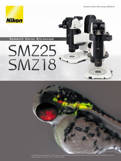
<strong>Research</strong> <strong>Stereo</strong> <strong>Microscope</strong> SMZ25/18<br />
R e s e arch S tereo M icroscope<br />
Live zebrafish expressing GFP- and RFP-neurons, imaged with Nikon’s l<strong>at</strong>est research stereo microscope, the SMZ25 (fluorescence and OCC).<br />
Image courtesy of Joe Fetcho, Ph.D., Cornell University.
Traditional boundaries between scientific fields such as molecular biology and developmental biology are<br />
rapidly disappearing as researchers seek to connect findings <strong>at</strong> the molecular level to those derived from<br />
cellular, tissue, and organismal studies. Fields including molecular biology, cell biology, neurobiology,<br />
embryology, developmental biology and systems biology have increasing needs for imaging systems th<strong>at</strong><br />
span sp<strong>at</strong>ial scales from single cells to whole organisms.<br />
With these demands in mind, Nikon has developed an all new stereo microscope th<strong>at</strong> fe<strong>at</strong>ures a large zoom<br />
r<strong>at</strong>io of 25:1, high resolution and exceptional fluorescence transmission capability.<br />
This l<strong>at</strong>est edition to the SMZ series represents a landmark in stereo microscope evolution – one th<strong>at</strong><br />
guarantees your research will be elev<strong>at</strong>ed to the next level.<br />
World’s largest zoom range and highest resolution in the SMZ series<br />
First stereo microscope to offer a 25:1 zoom range (SMZ25)<br />
Both eye p<strong>at</strong>hs boast numerical apertures (NA) of up to 0.156, using the SHR (Super High<br />
Resolution) Plan Apo 1x objective and SMZ25 zooming body<br />
Bright and high contrast fluorescent images<br />
2 days old Transgenic Zebrafish embryo, Tg (isl1-GFP)<br />
(using SHR Plan Apo 1x <strong>at</strong> zoom magnific<strong>at</strong>ion of 6x with SMZ25)<br />
Image courtesy of Hisaya Kakinuma, Ph.D.<br />
Labor<strong>at</strong>ory for Developmental Gene Regul<strong>at</strong>ion,<br />
Developmental Brain Science Group,<br />
RIKEN Brain Science Institute<br />
Fly eye lens ensures uniform brightness over the entire field of view even <strong>at</strong> the lowest<br />
magnific<strong>at</strong>ions<br />
Breakthroughs in optical design result in significantly improved signal to noise r<strong>at</strong>io and<br />
crystal clear fluorescent images<br />
Autom<strong>at</strong>ion and digital imaging<br />
Motorized focus and zoom oper<strong>at</strong>ion (SMZ25)<br />
Imaging Software NIS-Elements enables the use of multiple imaging, processing and<br />
analysis modalities including z-stack capture, time-lapse imaging, and the gener<strong>at</strong>ion of<br />
EDF images<br />
Easy to use<br />
User-friendly remote control (SMZ25)<br />
Easy-to-oper<strong>at</strong>e slim LED DIA base with OCC illumin<strong>at</strong>ion<br />
Wide range of illumin<strong>at</strong>ors and accessories accommod<strong>at</strong>e a variety of observ<strong>at</strong>ion methods<br />
Zooming observ<strong>at</strong>ion<br />
Zoom r<strong>at</strong>io<br />
Magnific<strong>at</strong>ion range<br />
Maximum<br />
magnific<strong>at</strong>ion<br />
Maximum FOV<br />
Maximum NA of<br />
objective<br />
Motorized zoom model<br />
with the highest zoom<br />
r<strong>at</strong>io and resolution<br />
in the SMZ series<br />
Manual zoom model<br />
providing advanced optical<br />
performance and incredibly<br />
bright fluorescence <strong>at</strong> an<br />
economical cost<br />
Motorized zoom<br />
Manual zoom<br />
BF/DF/FL/Simple polarizing<br />
BF/DF/FL/Simple polarizing<br />
25:1<br />
18:1<br />
0.63x ~ 15.75x<br />
0.75x ~ 13.5x<br />
315x* 1<br />
270x* 1<br />
ø70mm* 2<br />
ø59mm* 2<br />
0.312* 3 0.3* 3<br />
*1: Using SHR Plan Apo 2x/C-W 10x *2: Using SHR Plan Apo 0.5x/C-W 10x *3: Using SHR Plan Apo 2x<br />
2 3
World’s largest zoom range<br />
and incredible resolution<br />
Dynamic zoom r<strong>at</strong>io of 25:1<br />
An innov<strong>at</strong>ive optical system known as Perfect Zoom System provides the world’s first zoom r<strong>at</strong>io of 25:1 (zoom range:<br />
0.63x - 15.75x). Even with a 1x objective lens, the SMZ25 captures the entire 35mm dish and simultaneously delivers<br />
microscopic details.<br />
0.63x<br />
2.5x<br />
Higher NA in both eye p<strong>at</strong>hs coupled with a superior zoom r<strong>at</strong>io provides seamless viewing on<br />
the macro and micro levels.<br />
Superior resolution never before seen on a stereomicroscope<br />
Nikon’s SMZ25 series offers superior NA of 0.156 on the SHR Plan Apo 1x and 0.312 on the SHR Plan Apo 2x.<br />
Traditionally, researchers have had to switch to a higher magnific<strong>at</strong>ion microscope to view microscopic details after using a<br />
stereo microscope to view or manipul<strong>at</strong>e macroscopic structures. Nikon’s SMZ25/18 elimin<strong>at</strong>es this need by providing both<br />
macroscopic and microscopic imaging capabilities. For example, the SHR Plan Apo 2x objective allows for visualiz<strong>at</strong>ion of<br />
structures as small as a few microns in size, which was once considered to be impossible on a stereo microscope.<br />
Apochrom<strong>at</strong>ic correction is maintained in both the objective lens and the optical zoom system, virtually elimin<strong>at</strong>ing color<br />
aberr<strong>at</strong>ions.<br />
5x<br />
15.75x<br />
Individual olfactory nerve cells in a drosophila expressing a<br />
GFP-membrane marker are clearly resolved as black bodies<br />
encircled by fluorescent membranes (see circled area). This<br />
image demonstr<strong>at</strong>es the SMZ25’s incredible high resolution<br />
as the olfactory cells are typically only ø5μm in diameter<br />
Drosphila brain, GFP-G<br />
(using SHR Plan Apo 2x <strong>at</strong> zoom magnific<strong>at</strong>ion of 15.75x with SMZ25)<br />
Image courtesy of Hokuto Kazama, Ph.D.<br />
Labor<strong>at</strong>ory for Circuit Mechanisms of Sensory Perception RIKEN<br />
Auto Link Zoom (ALZ) supports<br />
seamless viewing <strong>at</strong> different scales<br />
ALZ autom<strong>at</strong>ically adjusts the zoom factor to maintain the same field of view<br />
when switching objective lenses. This function enables seamless switching<br />
between whole organism imaging <strong>at</strong> low magnific<strong>at</strong>ions and detailed imaging <strong>at</strong><br />
high magnific<strong>at</strong>ions.<br />
Arthromacra sp.<br />
(Using SHR Plan Apo 1x with SMZ25)<br />
Image courtesy of Japan Insect Associ<strong>at</strong>ion<br />
SHR Plan Apo 0.5x<br />
zoom 6x<br />
Maintains FOV <strong>at</strong> total magnific<strong>at</strong>ion of 3x<br />
SHR Plan Apo 2x<br />
zoom 1.5x<br />
Adult Drosophila, pebbled-Gal4 drive membrane-bound GFP expression in partial cells (with SMZ25)<br />
Image courtesy of Hokuto Kazama, Ph.D. Labor<strong>at</strong>ory for Circuit Mechanisms of Sensory Perception RIKEN<br />
The optical axis distance decreases <strong>at</strong><br />
low magnific<strong>at</strong>ion and increases <strong>at</strong><br />
high magnific<strong>at</strong>ion<br />
A single motor neuron expressing clusters<br />
of GFP-glycine receptors (resolved as<br />
individual puncta along the cell body and<br />
processes) imaged in a live zebrafish<br />
Zebrafish (GFP and OCC)<br />
(using SHR Plan Apo 2x <strong>at</strong> zoom magnific<strong>at</strong>ion of 15.75x<br />
with SMZ25)<br />
Image courtesy of Joe Fetcho , Ph.D., Cornell University<br />
Offers the highest zoom r<strong>at</strong>io thanks to<br />
Nikon’s Perfect Zoom System<br />
Newly developed high-performance<br />
objective lens<br />
Comparison of resolution by resolution chart<br />
Conventional model<br />
A breakthrough in stereoscope design, Perfect Zoom System dynamically change the<br />
distance between the two optical axes as the zoom factor is changed. This change in<br />
optical axis distance enables maximiz<strong>at</strong>ion of light entry into the optical system <strong>at</strong> every<br />
magnific<strong>at</strong>ion. The result is an uncompromised, large zoom range, high resolution in<br />
both eye p<strong>at</strong>hs, and minimal aberr<strong>at</strong>ions over the entire zoom-range. Furthermore, this<br />
breakthrough in optical design enables all of these desirable fe<strong>at</strong>ures to be housed in a<br />
compact zoom body, resulting in an ergonomic instrument design.<br />
Low zoom<br />
magnific<strong>at</strong>ion range<br />
High zoom<br />
magnific<strong>at</strong>ion range<br />
Nikon’s newly developed objective lens series, the<br />
SHR Plan Apo series, offers a high resolution of<br />
1100LP/mm (Observed value, using SHR Plan Apo 2x<br />
<strong>at</strong> maximum zoom). The new SHR Plan Apo series of<br />
lenses deliver brilliant images with true-to-life colors.<br />
4 5
Bright and high contrast<br />
fluorescent images<br />
Enhanced brightness and uniform illumin<strong>at</strong>ion in low magnific<strong>at</strong>ion range<br />
Even <strong>at</strong> low magnific<strong>at</strong>ion, the SMZ25 series captures the entire 35mm dish with equal brightness over the whole field<br />
of view*, making these new stereo microscopes ideal for live screening of developmental models such as C. elegans,<br />
drosophila, zebrafish, and mice to identify and select mutants. The SMZ25 series also allows brilliant images to be<br />
captured even with low excit<strong>at</strong>ion light levels, minimizing photo-bleaching and photo-toxicity which is harmful to live cells<br />
and organisms. *Using SMZ25/SHR Plan Apo 1x<br />
Newly developed epi-fluorescence <strong>at</strong>tachment captures clear fluorescence images.<br />
Improved S/N r<strong>at</strong>io and crystal clear fluorescent images<br />
thanks to an improved optical system<br />
Nikon’s newly developed optical system offers a drastic improvement in S/N r<strong>at</strong>io even <strong>at</strong> high magnific<strong>at</strong>ions. This<br />
improved S/N r<strong>at</strong>io makes it possible to capture cell division, which is difficult using conventional stereo microscopes, and<br />
samples with low excit<strong>at</strong>ion light.<br />
mRFP<br />
t=0 min t=20 min t=60 min<br />
The spindle appearing<br />
in cell division can be<br />
observed<br />
mRFP + GFP<br />
Time-lapse imaging of developing C. elegans embryos expressing RFP-histones and GFP-membrane markers<br />
allows researchers to screen for cytokinesis mutants prior to selection for downstream applic<strong>at</strong>ions<br />
Fertilized mouse egg, Green: Spindle<br />
(EGFP- α tubulin), Red: Nucleus<br />
(Histone H2B-mRFP1)<br />
(using SHR Plan Apo 1x <strong>at</strong> zoom<br />
magnific<strong>at</strong>ion of 13.5x with SMZ25)<br />
Image courtesy of Kazuo Yamag<strong>at</strong>a, Ph.D.<br />
Center for Genetic Analysis of Biological<br />
Responses, <strong>Research</strong> Institute for Microbial<br />
Diseases, Osaka University<br />
GFP<br />
C. elegans embryos (GFP and RFP; each ovoid is ø30μm in diameter)<br />
(using SHR Plan Apo 2x <strong>at</strong> zoom magnific<strong>at</strong>ion of 8x with SMZ25)<br />
Image courtesy of Julie C. Canman, Ph.D., Columbia University.<br />
Single fluorescent neurons can be visualized<br />
in live C. elegans<br />
12.5 days old mouse embryo, Red: Nucleus<br />
(Using SHR Plan Apo 0.5x <strong>at</strong> zoom magnific<strong>at</strong>ion 1.30x<br />
with SMZ18)<br />
Image courtesy of Kazuo Yamag<strong>at</strong>a, Ph.D.<br />
Center for Genetic Analysis of Biological Responses, <strong>Research</strong><br />
Institute for Microbial Diseases, Osaka University<br />
Fluorescence and OCC images of a live C. elegans expressing<br />
GFP- and RFP-neurons<br />
(using SHR Plan Apo 2x <strong>at</strong> zoom magnific<strong>at</strong>ion of 3x with SMZ25)<br />
Image courtesy of Julie C. Canman, Ph.D., Columbia University<br />
Fly eye lens ensures uniform brightness<br />
over the entire field of view<br />
Conventional epi-fluorescence<br />
<strong>at</strong>tachment<br />
New epi-fluorescence<br />
<strong>at</strong>tachment<br />
Zoom body with significant improvements in<br />
optical performance<br />
The SMZ25 series is the first stereo microscope in<br />
the world to use a fly eye lens on an epi-fluorescence<br />
<strong>at</strong>tachment. This innov<strong>at</strong>ive design ensures bright<br />
and uniform illumin<strong>at</strong>ion even <strong>at</strong> low magnific<strong>at</strong>ions,<br />
resulting in uncompromised uniformity in brightness<br />
across a large field of view.<br />
Poor illumin<strong>at</strong>ion<br />
coverage<br />
Fly eye lens uniformly<br />
illumin<strong>at</strong>es the entire<br />
field of view<br />
Nikon has succeeded in improving the signal and reducing noise in<br />
fluorescent images by using a short wavelength, high transmission<br />
Fluor lens. Combined with an innov<strong>at</strong>ive epi-fluorescence <strong>at</strong>tachment,<br />
the SMZ18/25 is better able to detect excit<strong>at</strong>ion light than conventional<br />
fluorescent stereo microscopes.<br />
6 7
A wide range of digital imaging capabilities with the Digital Sight series and NIS-Elements<br />
imaging software.<br />
User-friendly remote controller<br />
The all new remote controller provides easy access to zoom and focus controls and is designed for both right and left<br />
hand use. The remote controller contains an LCD monitor with an adjustable backlight which provides inform<strong>at</strong>ion<br />
regarding the zoom factor, objective lens, filter cube, and LED DIA brightness <strong>at</strong> a glance. The backlight on the LCD<br />
monitor can also be turned off to elimin<strong>at</strong>e interference with low-light imaging applic<strong>at</strong>ions. In addition to the remote<br />
controller, the microscope can also be oper<strong>at</strong>ed through a PC.<br />
The brightness of the LCD monitor<br />
backlight and LED indic<strong>at</strong>ors is<br />
adjustable<br />
Multichannel (multicolor)<br />
Multiple fluorescent channels can be captured in<br />
conjunction with other imaging methods such as OCC<br />
or brightfield.<br />
Individual cells resolved in a live drosophila embryo<br />
expressing GFP and mCherry<br />
(using SHR Plan Apo 2x <strong>at</strong> zoom magnific<strong>at</strong>ion of 8x with SMZ25)<br />
Image courtesy of Max V. Staller, Ph.D., Clarissa Scholes,<br />
and Angela DePace, Ph.D., Harvard Medical School<br />
Time lapse<br />
Easily setup a time-lapse imaging experiment with NIS-Elements.<br />
0 sec<br />
38.38 sec<br />
One software for all systems: NIS-Elements which is Nikon’s flagship, cross-pl<strong>at</strong>form imaging<br />
software can now be used with Nikon’s l<strong>at</strong>est stereomicroscope systems SMZ25 and SMZ18.<br />
NIS-Elements enables a wide range of advanced digital imaging capabilities, easily from a PC.<br />
Calcium-imaging: Time-lapse<br />
imaging of GCaMP expressing<br />
neurons inside a live zebrafish<br />
shows individual neurons firing <strong>at</strong><br />
different times (arrowheads). The<br />
last time-frame shows a whole<br />
cluster of neurons firing (asterisk).<br />
Images taken<br />
in individual<br />
colors<br />
Overlapping image with all colors<br />
Extended depth of focus (EDF)<br />
Capture multiple high resolution images <strong>at</strong> different focal depths to<br />
cre<strong>at</strong>e a single extended depth of focus image or quasi-3D image.<br />
57.14 sec<br />
71.43 sec<br />
Select the in-focus area (white square)<br />
and produces one all-in-focus image<br />
(using SHR Plan Apo 2x <strong>at</strong> zoom<br />
magnific<strong>at</strong>ion of 9x with SMZ25<br />
and camera head DS-Qi1)<br />
Image courtesy of Joe Fetcho, Ph.D.,<br />
Cornell University<br />
Zebrafish embryo<br />
(using SHR Plan Apo 2x <strong>at</strong> zoom<br />
magnific<strong>at</strong>ion of 3.4x with SMZ25)<br />
Image courtesy of Hisaya Kakinuma,<br />
Ph.D., Labor<strong>at</strong>ory for Developmental Gene<br />
Regul<strong>at</strong>ion, Developmental Brain Science<br />
Group, RIKEN Brain Science Institute<br />
Access the inform<strong>at</strong>ion you want quickly and easily<br />
Easily obtain the inform<strong>at</strong>ion you need, such as Z drive position, zoom factor, objective lens, filter cube, and LED DIA<br />
brightness by using the Digital Sight series and NIS-Elements or Digital Sight series DS-L3 together with the microscope.<br />
Scale tracking<br />
The scale bar autom<strong>at</strong>ically adjusts to accommod<strong>at</strong>e<br />
changes in magnific<strong>at</strong>ion.<br />
DS-L3 is an easy-to-use high-definition, large touch-panel monitor th<strong>at</strong> can be used<br />
to quickly capture images without a PC or monitor.<br />
Scene mode<br />
Optimal imaging parameters for each sample type and<br />
observ<strong>at</strong>ion method can easily be set using the icons.<br />
On-axis imaging for digital images<br />
Easily switch between stereo position<br />
<strong>Stereo</strong> position (stereoscopic view)<br />
(stereoscopic view) and mono position<br />
(on-axis view) when using the P2-RNI2<br />
Intelligent Nosepiece by simply sliding the<br />
objective lens.<br />
Digital images with uncompromised clarity<br />
can be captured using the mono position.<br />
Mono position (on-axis view)<br />
Select the perfect camera for your applic<strong>at</strong>ion.<br />
Ultra high-definition<br />
cooled color camera head<br />
High-resolution 12.7 megapixels<br />
Superior color reproduction<br />
-10°C ambient temper<strong>at</strong>ure cooling<br />
High-definition color camera head<br />
High-resolution 5.0 megapixels<br />
High-speed frame r<strong>at</strong>e<br />
Suitable for a wide range of<br />
applic<strong>at</strong>ions<br />
High-sensitivity cooled<br />
monochrome camera head<br />
High sensitivity and low noise<br />
High quantit<strong>at</strong>ive capability<br />
Cooled 1.5-megapixel CCD<br />
Switch from a stereo position to mono position by sliding the<br />
objective lens to the right<br />
*For more details, see the Digital Sight series c<strong>at</strong>alogues.<br />
8 9
Wide range of available accessories<br />
Base unit<br />
Nikon has improved ease of use by moving the controls to the front of the base, including the brightness adjustment dial and on/off switch.<br />
Fiber DIA base<br />
Fiber DIA base fe<strong>at</strong>ures condenser lenses<br />
th<strong>at</strong> can be switched between low and high<br />
magnific<strong>at</strong>ions. Furthermore, the Oblique<br />
Coherent Contrast (OCC) illumin<strong>at</strong>ion system<br />
allows high contrast illumin<strong>at</strong>ion.<br />
Slim bases<br />
The slimmer LED DIA Base and Plain Base help<br />
increase efficiency of sample manipul<strong>at</strong>ion by bringing<br />
the level of the sample closer to the table.<br />
Conventional base<br />
SHR Plan Apo series of Objective<br />
The SHR Plan Apo series fe<strong>at</strong>ures higher NA, wider field of view, and superior fl<strong>at</strong>ness and color aberr<strong>at</strong>ion correction.<br />
These objective lenses can be seamlessly switched because all magnific<strong>at</strong>ions have the same parfocal distance. The new bayonet mount design<br />
allows lenses to be safely and easily removed.<br />
1 P2-SHR Plan Apo 0.5x<br />
3 P2-SHR Plan Apo 1.6x<br />
2 P2-SHR Plan Apo 1x<br />
4 P2-SHR Plan Apo 2x<br />
Maximum<br />
NA<br />
Working distance<br />
Correction ring<br />
Wavelength<br />
SHR Plan<br />
Apo 0.5x<br />
0.078<br />
0.075<br />
71mm<br />
––<br />
SHR Plan<br />
Apo 1x<br />
0.156<br />
0.15<br />
60mm<br />
––<br />
380-700nm<br />
SHR Plan<br />
Apo 1.6x<br />
0.25<br />
0.24<br />
30mm<br />
––<br />
SHR Plan<br />
Apo 2x<br />
0.321<br />
0.3<br />
20mm<br />
3mm<br />
w<strong>at</strong>er depth<br />
1 P2-DBF Fiber Diascopic Illumin<strong>at</strong>ion Base<br />
Example applic<strong>at</strong>ions<br />
OCC illumin<strong>at</strong>or<br />
The new LED DIA Base with a built-in OCC illumin<strong>at</strong>or gener<strong>at</strong>es minimal<br />
he<strong>at</strong>, consumes little power and is long-life. This illumin<strong>at</strong>or can enhance the<br />
contrast of uneven surfaces, such as th<strong>at</strong> of an embryo.<br />
Conventional diascopic illumin<strong>at</strong>ion<br />
OCC illumin<strong>at</strong>or<br />
Zebrafish embryo (using SHR Plan Apo 1x <strong>at</strong> zoom magnific<strong>at</strong>ion of 5x with SMZ18)<br />
Image courtesy of Junichi Nakai, Ph.D. Saitama University Brain science Institute<br />
2 P2-DBL LED Diascopic 3 P2-PB Plain Base<br />
Illumin<strong>at</strong>ion Base<br />
Wh<strong>at</strong> is OCC illumin<strong>at</strong>ion?<br />
The acronym OCC stands for oblique<br />
coherent contrast (OCC), which is a form of<br />
oblique lighting method developed by Nikon.<br />
Compared to conventional diascopic<br />
illumin<strong>at</strong>ion th<strong>at</strong> illumin<strong>at</strong>es directly<br />
from below, OCC illumin<strong>at</strong>ion applies<br />
coherent light to samples in a diagonal<br />
direction, giving contrast to colorless and<br />
transparent sample structures.<br />
Slim base<br />
<strong>Microscope</strong>-Stage Autom<strong>at</strong>ic<br />
Thermo Control System<br />
Thermo pl<strong>at</strong>e MATS<br />
(Manufacturer: Tokai Hit Co., Ltd.)<br />
The fl<strong>at</strong> pl<strong>at</strong>e surface ensures<br />
easy oper<strong>at</strong>ion of the<br />
manipul<strong>at</strong>or and easy handling of<br />
specimens.<br />
Tubes<br />
Choose from two types of tilting<br />
trinocular tube and one type of low<br />
eyelevel trinocular tube. All tubes have<br />
a camera port for seamless integr<strong>at</strong>ion<br />
with the Digital Sight series.<br />
Nosepiece / Focus mount adapter<br />
There is the option of the nosepiece either single or double<br />
for purposes of expanding research for changing the<br />
magnific<strong>at</strong>ion range.<br />
Stage<br />
1 P2-TERG100 Trinocular Tilting Tube (eyepiece: port 100:0 / 0:100)<br />
2 P2-TERG50 Trinocular Tilting Tube (eyepiece: port 100:0/50:50)<br />
3<br />
P2-TL100 Trinocular Tube L (eyepiece: port 100:0 / 0:100)<br />
The stage fe<strong>at</strong>ures an XY stroke of 6x4* inches (150mm x<br />
100mm) and can be <strong>at</strong>tached to any of the bases, making it<br />
effective for capturing large images when used in combin<strong>at</strong>ion<br />
with the imaging software NIS-Elements. A sliding stage and<br />
tilting stage are also available.<br />
*Limited Y travel<br />
with 32mm<br />
column bases<br />
Focus unit<br />
The focus unit is combined with the base unit. Choose from either a manual<br />
or motorized focus unit.<br />
Stand / Focus mount<br />
Combine the stand with a focus mount for viewing<br />
and capturing images with reflected illumin<strong>at</strong>ion.<br />
1<br />
2<br />
P2-RNI2 Intelligent Nosepiece<br />
P2-FM Focus Mount Adapter<br />
Controller<br />
P-SXY64 XY Stage<br />
1 P2-FMDN<br />
Focus Mount<br />
2 P-PS32<br />
Plain Stand<br />
Nikon offers a remote control unit th<strong>at</strong> can be used to<br />
oper<strong>at</strong>e the microscope and capture images by hand. A<br />
footswitch is also offered, allowing the user to oper<strong>at</strong>e the<br />
microscope and capture images by foot, freeing the hands<br />
for sample manipul<strong>at</strong>ion.<br />
1 P2-MFU Motorized Focus Unit<br />
2 P2-FU Focus Unit<br />
P2-RC Remote Controller<br />
1<br />
2<br />
AZ-PCR Photo Release<br />
AZ-FSW Foot Switch<br />
10 11
Wide range of available accessories<br />
Specific<strong>at</strong>ions<br />
Epi-fluorescence light set<br />
Motorized epi-fluorescence light set<br />
The fluorescent turret can be oper<strong>at</strong>ed<br />
using the remote control or imaging<br />
software NIS-Elements.<br />
Manual epi-fluorescence light set<br />
An easy-to-use manual model for<br />
Nikon’s newly developed highperformance<br />
epi-fluorescence<br />
<strong>at</strong>tachment.<br />
Zooming Body<br />
Optical system<br />
Parallel-optics type (zooming type), apochrom<strong>at</strong>ic optical system<br />
Zoom Motorized Manual<br />
Zoom r<strong>at</strong>io 25:1 18:1<br />
Zoom range 0.63-15.75x 0.75-13.5x<br />
Aperture diaphragm Zooming body built-in Zooming body built-in<br />
Objectives NA, WD (mm)<br />
•<br />
P2-SHR Plan Apo 2x 0.312, 20 (with a correction ring for w<strong>at</strong>er 0 to 3mm in depth) 0.3, 20 (with a correction ring for w<strong>at</strong>er 0 to 3mm in depth)<br />
•<br />
P2-SHR Plan Apo 1.6× 0.25, 30 0.24, 30<br />
1 P2-EFLM Motorized Epi Fluorescence<br />
Attachment<br />
2 Light shading Pl<strong>at</strong>e (comes with Fluorescence Attachment)<br />
3 P2-EFL Filter Cube (GFP-B/GFP-L/RFP)<br />
Combin<strong>at</strong>ions<br />
with SMZ25<br />
4 P2-EFLBF Filter Cube (Bright Field, with /4 pl<strong>at</strong>e)<br />
5 P2-CTLA Control Box 6 P2-RC Remote Controller<br />
Fiber illumin<strong>at</strong>or set<br />
1 P2-EFLI Epi Fluorescence Attachment<br />
2 Light shading Pl<strong>at</strong>e (comes with Fluorescence Attachment)<br />
3 P2-EFL Filter Cube (GFP-B/GFP-L/RFP)<br />
4 P2-EFLBF Filter Cube (Bright Field, with /4 pl<strong>at</strong>e)<br />
5 P2-CTLB Control Box<br />
Combin<strong>at</strong>ions<br />
with SMZ18<br />
•<br />
P2-SHR Plan Apo 1× 0.156, 60 0.15, 60<br />
•<br />
P2-SHR Plan Apo 0.5× 0.078, 71 0.075, 71<br />
Total Magnific<strong>at</strong>ion 3.15-315x 3.75-270x<br />
(Using 10x eyepieces) (Depending on objective used) (Depending on objective used)<br />
Eyepieces (F.O.V. mm)<br />
•<br />
C-W 10x (22)<br />
•<br />
C-W 15x (16)<br />
•<br />
C-W 20x (12.5) • C-W 30x (7)<br />
Tubes (Eyepiece/Port)<br />
•<br />
P2-TERG 100 Trinocular Tilting tube (100/0 : 0/100)<br />
•<br />
P2-TERG 50 Trinocular Tilting tube (100/0 : 50/50)<br />
Inclin<strong>at</strong>ion angle : 0-30 degree<br />
Flexible double arm fiber illumin<strong>at</strong>ion set<br />
The direction and angle of<br />
illumin<strong>at</strong>ion can be changed to suit<br />
the sample by making adjustments<br />
with these double arms. The fiber<br />
holder position can also be changed<br />
to obtain the optimal position for<br />
illumin<strong>at</strong>ing samples.<br />
1<br />
2<br />
3<br />
C-FDF Flexible Double Arm<br />
Fiber Illumin<strong>at</strong>ion Unit<br />
C-FIDH Fiber Holder<br />
C-FLED2 LED Light Source for Fiber Illumin<strong>at</strong>or<br />
Combin<strong>at</strong>ions<br />
with SMZ18<br />
Ring fiber illumin<strong>at</strong>ion set<br />
This ring fiber illumin<strong>at</strong>ion<br />
set fe<strong>at</strong>ures an episcopic<br />
illumin<strong>at</strong>ion unit th<strong>at</strong> effectively<br />
captures images (can be used<br />
with 1x and 0.5x objective<br />
lenses).<br />
1<br />
2<br />
P2-FIR<br />
Ring Fiber Illumin<strong>at</strong>ion Unit<br />
C-FLED2 LED Light Source for<br />
Fiber Illumin<strong>at</strong>or<br />
Combin<strong>at</strong>ions<br />
with SMZ18<br />
•<br />
P2-TL100 Trinocular Tube L (100/0 : 0/100) Inclin<strong>at</strong>ion angle : 0-15 degree<br />
Focus Unit (Stroke from<br />
•<br />
P2-MFU Motorized Focus Unit (Up 96mm/Down 4mm)<br />
Objective's parfocal point)<br />
•<br />
P2-FU Focus Unit (Up 97mm/Down 5mm)<br />
Focus mount Adapter/Nosepiece<br />
•<br />
P2-FM Focus Mount Adapter<br />
•<br />
P2-FM Focus Mount Adapter<br />
•<br />
P2-RNI2 Intelligent Nosepiece<br />
•<br />
P2-RNI2 Intelligent Nosepiece<br />
(2 objectives can be <strong>at</strong>tached) (2 objectives can be <strong>at</strong>tached)<br />
•<br />
P2-FMDN Focus Mount (for P-PS32 Plan Stand)<br />
Bases/Stand<br />
•<br />
P2-PB Plain Base • P2-DBL LED Diascopic Illumin<strong>at</strong>ion Base (OCC illumin<strong>at</strong>or built-in)<br />
•<br />
P2-DBF Fiber Diascopic Illumin<strong>at</strong>ion Base • P-PS32 Plain Stand (Only for SMZ18)<br />
Stages<br />
•<br />
P-SXY64 Stage • C-SSL Dia-sliding Stage • C-TRS Tilting Stage<br />
Epi-Fluorescence Attachments 4 filter cubes mountable, Fly eye lens built-in<br />
•<br />
P2-EFLM Motorized Epi Fluorescence Attachment<br />
•<br />
P2-EFLI Epi Fluorescence Attachment<br />
Epi-Fluorescence light sources<br />
•<br />
HG Precentered Fiber illumin<strong>at</strong>or Intensilight C-HGFIE HG/C-HGFI HG (130W)<br />
Coaxial illumin<strong>at</strong>or<br />
The coaxial light illumin<strong>at</strong>or<br />
makes it possible to view light<br />
reflected from the surface of a<br />
sample, which is ideal for shooting<br />
shadow-less images of thick<br />
samples.<br />
1<br />
2<br />
P2-CI Coaxial Epi Illumin<strong>at</strong>or<br />
C-FLED2 LED Light Source for<br />
Fiber Illumin<strong>at</strong>or<br />
Darkfield observ<strong>at</strong>ion accessory<br />
Darkfield viewing is possible<br />
simply by <strong>at</strong>taching the dark<br />
field unit to the base.<br />
Combin<strong>at</strong>ions<br />
with SMZ18<br />
Ring LED illumin<strong>at</strong>or<br />
Ring LED illumin<strong>at</strong>or is equipped with<br />
high-intensity and long-life LEDs. The<br />
illumin<strong>at</strong>or’s dial adjusts the intensity of<br />
the white LED.<br />
1 P2-FIRL LED Ring Illumin<strong>at</strong>ion Unit<br />
Polarizing observ<strong>at</strong>ion accessory<br />
The analyzer is <strong>at</strong>tached to the<br />
objective and the polarizer to the<br />
base or stand to enable polarized<br />
viewing.<br />
Combin<strong>at</strong>ions<br />
with SMZ18<br />
Episcopic Illumin<strong>at</strong>ors<br />
•<br />
P2-FIRL LED Ring Illumin<strong>at</strong>ion Unit<br />
Use with Fiber light source<br />
•<br />
P2-CI Coaxial Epi Illumin<strong>at</strong>or • P2-FIR Ring Fiber Illumin<strong>at</strong>ion Unit<br />
•<br />
C-FDF Flexible Double Arm Fiber Illumin<strong>at</strong>ion Unit<br />
Episcopic light sources<br />
•<br />
C-FLED2 LED Light source for fiber illumin<strong>at</strong>or<br />
Observ<strong>at</strong>ion methods<br />
Bright Field, Epi Fluorescence, Simple Polarizing (with P2-POL Simple Polarizing Attachment),<br />
Dark Field (with P-DF LED Dark Field Unit), Oblique lighting<br />
Weight (approx.) 32kg 30kg<br />
(Motorized Epi Fluorescence Attachment (Epi Fluorescence Attachment configur<strong>at</strong>ion<br />
configur<strong>at</strong>ion with Trinocular Tilting Tube, with Trinocular Tilting Tube, Focus Unit,<br />
Motorized Focus Unit, Intelligent Nosepiece, Intelligent Nosepiece, LED DIA base and<br />
LED DIA base and Objectives 1x and 0.5x) Objectives 1x and 0.5x)<br />
Power consumption (approx.) 30W 10W<br />
(Motorized Epi Fluorescence Attachment (Epi Fluorescence Attachment configur<strong>at</strong>ion<br />
configur<strong>at</strong>ion with Trinocular Tilting Tube, with Trinocular Tilting Tube, Focus Unit,<br />
Motorized Focus Unit, Intelligent Nosepiece Intelligent Nosepiece and LED DIA base)<br />
and LED DIA base)<br />
1<br />
2<br />
P-DF LED Dark Field Unit<br />
Shading cover<br />
1<br />
P2-POL Simple Polarizing Attachment<br />
12 13
Dimensions<br />
System diagram<br />
(configured with motorized epi-fluorescence <strong>at</strong>tachment<br />
and LED DIA base)<br />
(configured with fiber DIA base)<br />
C-W10×<br />
Eyepiece<br />
C-W15×<br />
Eyepiece<br />
C-W20×<br />
Eyepiece<br />
C-W30×<br />
Eyepiece<br />
1/1.8” Camera 2/3” Camera<br />
C-mount Adapter<br />
0.55×<br />
C-mount Adapter<br />
0.7×<br />
C-mount CCD Camera<br />
C-mount<br />
TV Adapter A<br />
A<br />
LV-TV<br />
TV Adapter<br />
298<br />
298<br />
A A A<br />
AZ-PCR<br />
Photo Release<br />
P2-TERG100<br />
Trinocular Tilting Tube<br />
P2-TERG50<br />
Trinocular Tilting Tube<br />
P2-TL100<br />
Trinocular Tube L<br />
P2-RC<br />
Remote<br />
Controller<br />
AZ-FSW<br />
Foot Switch<br />
514 25<br />
514 25<br />
X<br />
P2-EFLM<br />
Motorized Epi<br />
Fluorescence Attachment<br />
(with Light Shading Pl<strong>at</strong>e)<br />
C-HGFI/C-HGFIE<br />
HG Precentered Fiber Illumin<strong>at</strong>or<br />
P2-EFL<br />
Filter Cube (GFP-B/GFP-L /RFP)<br />
471<br />
569<br />
459<br />
568<br />
E<br />
X<br />
P2-EFLI<br />
Epi Fluorescence<br />
Attachment<br />
(with Light Shading Pl<strong>at</strong>e)<br />
D<br />
P2-CI<br />
Coaxial Epi<br />
Illumin<strong>at</strong>or<br />
(with /4 pl<strong>at</strong>e)<br />
P2-EFLC<br />
Filter Cube<br />
P2-EFLBF<br />
Filter Cube<br />
(Bright Field)<br />
X<br />
P2-MFU<br />
Motorized Focus Unit<br />
Y<br />
P2-CTLA<br />
Control Box<br />
P2-FM<br />
Focus Mount Adapter<br />
B<br />
SU-AC AC Adapter<br />
444<br />
444<br />
X<br />
C<br />
SMZ18<br />
Zooming body<br />
X<br />
SMZ25<br />
Zooming body<br />
X<br />
P2-RNI2<br />
Intelligent Nosepiece<br />
X<br />
P2-RLY<br />
Relay Box<br />
Y<br />
P2-FU<br />
Focus Unit<br />
P2-CTLB<br />
Control Box<br />
(configured with epi-fluorescence <strong>at</strong>tachment<br />
and LED DIA base)<br />
(configured with plain stand and focus mount)<br />
P2-SHR Plan<br />
Apo 0.5×<br />
Objective<br />
P2-SHR Plan<br />
Apo 1×<br />
Objective<br />
P2-SHR Plan<br />
Apo 1.6×<br />
Objective<br />
P2-SHR Plan<br />
Apo 2×<br />
Objective<br />
B<br />
D<br />
C<br />
P2-RLYC<br />
Relay Cable<br />
298<br />
300<br />
E<br />
P2-FIRL<br />
LED Ring<br />
Illumin<strong>at</strong>ion<br />
Unit<br />
Z<br />
J<br />
P2-POL<br />
Simple Polarizing<br />
Attachment<br />
P2-FIR<br />
Ring Fiber Illumin<strong>at</strong>ion Unit<br />
Z<br />
AZLED-PCU Power Code<br />
Power cable<br />
514 25<br />
302<br />
337 (44)<br />
244<br />
P-DF<br />
LED Dark Field Unit<br />
Stage Adapters<br />
for AZ<br />
Z<br />
Shading<br />
cover<br />
C-FDF<br />
Flexible Double Arm<br />
Fiber Illumin<strong>at</strong>ion Unit<br />
AC Adapter 2<br />
C-FLED2<br />
LED Light Source for Fiber Illumin<strong>at</strong>or<br />
H<br />
P-SXY64<br />
XY Stage<br />
C-SSL<br />
DIA Sliding Stage<br />
I<br />
B<br />
C-TRS<br />
Tilting Stage<br />
G<br />
C-FIDH<br />
Fiber Holder<br />
C<br />
P2-FMDN<br />
Focus Mount<br />
471<br />
569<br />
429<br />
492<br />
H<br />
I<br />
J<br />
H<br />
I<br />
G<br />
H<br />
I<br />
G<br />
G<br />
G<br />
444<br />
P2-PB<br />
Plain Base<br />
Y<br />
AC Adapter 2<br />
P2-DBL LED Diascopic<br />
Illumin<strong>at</strong>ion Base<br />
P2-DBF<br />
Fiber Diascopic<br />
Illumin<strong>at</strong>ion Base<br />
P-PS32<br />
Plain Stand<br />
14 15
Specific<strong>at</strong>ions and equipment are subject to change without any notice or oblig<strong>at</strong>ion on the part of the manufacturer. May 2013 ©2013 NIKON CORPORATION<br />
N.B. Export of the products* in this c<strong>at</strong>alog is controlled under the Japanese Foreign Exchange and Foreign Trade Law. Appropri<strong>at</strong>e export procedures shall be required in case of export from Japan.<br />
*Products: Hardware and its technical inform<strong>at</strong>ion (including software)<br />
NIKON CORPORATION<br />
Shin-Yurakucho Bldg., 12-1, Yurakucho 1-chome<br />
Chiyoda-ku, Tokyo 100-8331 Japan<br />
Bioscience) phone:+81-3-3216-2375 fax:+81-3-3216-2385<br />
Industrial Instruments) phone:+81-3-3216-2384 fax:+81-3-3216-2388<br />
http://www.nikon.com/instruments/<br />
NIKON INSTRUMENTS INC.<br />
1300 Walt Whitman Road, Melville, N.Y. 11747-3064, U.S.A.<br />
phone: +1-631-547-8500; +1-800-52-NIKON (within the U.S.A. only)<br />
fax: +1-631-547-0306<br />
http://www.nikoninstruments.com/<br />
NIKON METROLOGY, INC.<br />
12701 Grand River Avenue, Brighton, MI 48116 U.S.A.<br />
phone: +1-810-220-4360 fax: +1-810-220-4300<br />
E-mail: sales_us@nikonmetrology.com<br />
http://www.nikonmetrology.com/<br />
NIKON INSTRUMENTS EUROPE B.V.<br />
Tripolis 100, Burgerweeshuispad 101, 1076 ER Amsterdam, The Netherlands<br />
phone: +31-20-7099-000 fax: +31-20-7099-298<br />
http://www.nikoninstruments.eu/<br />
NIKON METROLOGY EUROPE NV<br />
Geldenaaksebaan 329, 3001 Leuven, Belgium<br />
phone: +32-16-74-01-00 fax: +32-16-74-01-03<br />
Email: sales_europe@nikonmetrology.com<br />
http://www.nikonmetrology.com/<br />
NIKON INSTRUMENTS (SHANGHAI) CO., LTD.<br />
CHINA phone: +86-21-6841-2050 fax: +86-21-6841-2060<br />
(Beijing branch) phone: +86-10-5831-2028 fax: +86-10-5831-2026<br />
(Guangzhou branch) phone: +86-20-3882-0550 fax: +86-20-3882-0580<br />
Printed in Japan (1305-06) Am/M<br />
NIKON SINGAPORE PTE LTD.<br />
SINGAPORE phone: +65-6559-3618 fax: +65-6559-3668<br />
NIKON MALAYSIA SDN. BHD.<br />
MALAYSIA phone: +60-3-7809-3688 fax: +60-3-7809-3633<br />
NIKON INSTRUMENTS KOREA CO., LTD.<br />
KOREA phone: +82-2-2186-8400 fax: +82-2-555-4415<br />
NIKON INDIA PRIVATE LIMITED<br />
INDIA phone: +91-124-4688500 fax: +91-124-4688527<br />
NIKON CANADA INC.<br />
CANADA phone: +1-905-602-9676 fax: +1-905-602-9953<br />
NIKON INSTRUMENTS S.p.A.<br />
ITALY phone: +39-055-300-96-01 fax: +39-055-30-09-93<br />
NIKON AG<br />
SWITZERLAND phone: +41-43-277-28-67 fax: +41-43-277-28-61<br />
NIKON GMBH AUSTRIA<br />
AUSTRIA phone: +43-1-972-6111-00 fax: +43-1-972-6111-40<br />
NIKON BELUX<br />
BELGIUM phone: +32-2-705-56-65 fax: +32-2-726-66-45<br />
NIKON UK LTD.<br />
UNITED KINGDOM phone: +44-208-247-1717 fax: +44-208-541-4584<br />
NIKON METROLOGY UK LTD.<br />
UNITED KINGDOM phone: +44-1332-811-349 fax: +44-1332-639-881<br />
E-mail: sales_uk@nikonmetrology.com<br />
2CE-MPGH-1<br />
NIKON FRANCE S.A.S.<br />
FRANCE phone: +33-1-4516-45-16 fax: +33-1-4516-45-55<br />
NIKON METROLOGY SARL<br />
FRANCE phone: +33-1-60-86-09-76 fax: +33-1-60-86-57-35<br />
E-mail: sales_france@nikonmetrology.com<br />
NIKON GMBH<br />
GERMANY phone: +49-211-941-42-20 fax:+49-211-941-43-22<br />
NIKON METROLOGY GMBH<br />
GERMANY phone: +49-6023-91733-0 fax: +49-6023-91733-229<br />
E-mail: sales_germany@nikonmetrology.com