Cemento-osseous Dysplasia in African-American Men - Tennessee ...
Cemento-osseous Dysplasia in African-American Men - Tennessee ...
Cemento-osseous Dysplasia in African-American Men - Tennessee ...

Create successful ePaper yourself
Turn your PDF publications into a flip-book with our unique Google optimized e-Paper software.
<strong>Cemento</strong>-<strong>osseous</strong> <strong>Dysplasia</strong> <strong>in</strong> <strong>African</strong>-<strong>American</strong> <strong>Men</strong>:<br />
A Report of Two Cl<strong>in</strong>ical Cases<br />
Peter M. DiFiore, DDS, MS; Sarah Elizabeth Bowen, BS<br />
T<br />
D<br />
A<br />
EXAM #25<br />
Introduction<br />
<strong>Cemento</strong>-<strong>osseous</strong> dysplasia has been<br />
described and divided <strong>in</strong>to three types<br />
accord<strong>in</strong>g to its cl<strong>in</strong>ical presentation. 1-5<br />
1. Periapical cemento-<strong>osseous</strong><br />
dysplasia: s<strong>in</strong>gle or multiple<br />
lesions at the root apices of<br />
mandibular anterior teeth and<br />
occasionally maxillary anterior<br />
teeth.<br />
2. Focal cemento-<strong>osseous</strong>: focal<br />
or multifocal lesions <strong>in</strong> close<br />
proximity to the root apices of<br />
mandibular posterior teeth or <strong>in</strong><br />
mandibular posterior edentulous<br />
areas.<br />
3. Florid cemento-<strong>osseous</strong><br />
dysplasia: the most extensive<br />
form of this dysplasia, present<strong>in</strong>g<br />
as bilateral lesions <strong>in</strong> the toothbear<strong>in</strong>g<br />
areas of the mandible or<br />
of both the mandible and maxilla.<br />
<strong>Cemento</strong>-<strong>osseous</strong> dysplasia is an<br />
asymptomatic condition that is detected<br />
on rout<strong>in</strong>e radiographic exam<strong>in</strong>ation as<br />
radiolucent, mixed radiolucent/radiopaque<br />
or radiopaque periapical areas, depend<strong>in</strong>g<br />
on its stage of maturation from fibrous<br />
tissue proliferation to cemento-<strong>osseous</strong><br />
condensation. 1,3,4,6-11<br />
The lesions of cemento-<strong>osseous</strong><br />
dysplasia may <strong>in</strong>volve s<strong>in</strong>gle or multiple<br />
teeth and are most often associated with<br />
mandibular anterior teeth, often with<br />
mandibular posterior teeth and least<br />
often with maxillary teeth. 6,12-14 They are<br />
fairly well circumscribed round lesions<br />
that range <strong>in</strong> size from 1 to 10 mm 1,3,6 but<br />
may coalesce to form larger lesions 4,7,15<br />
or degenerate to form bone cysts. 16,17<br />
These non-neoplastic periapical lesions<br />
generally do not cause cortical bone<br />
expansion or perforation, 4,6,8,10,15 but rather<br />
slowly progress through dysplastic stages<br />
of maturation <strong>in</strong> the periapical areas of<br />
alveolar bone without attachment to the<br />
root apices. 1,4,7,9<br />
The pathogenesis of cemento<strong>osseous</strong><br />
dysplasia has not been clearly<br />
established, however, the most common<br />
ABSTRACT<br />
Two cl<strong>in</strong>ical cases of the unusual occurrence of cemento-<strong>osseous</strong> dysplasia <strong>in</strong><br />
men and the cl<strong>in</strong>ical, radiographic and demographic f<strong>in</strong>d<strong>in</strong>gs that formed the basis<br />
for their diagnosis and management are presented.<br />
Peter M. DiFiore, DDS, MS, is a Diplomate of the <strong>American</strong> Board of Endodontics<br />
and Associate Professor and Chair, Department of Endodontics at the University of<br />
<strong>Tennessee</strong> Health Science Center, College of Dentistry <strong>in</strong> Memphis, TN.<br />
Sarah Elizabeth Bowen, BS, is a Senior Dental Student at the University of <strong>Tennessee</strong><br />
Health Science Center, College of Dentistry <strong>in</strong> Memphis, TN.<br />
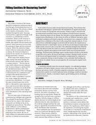
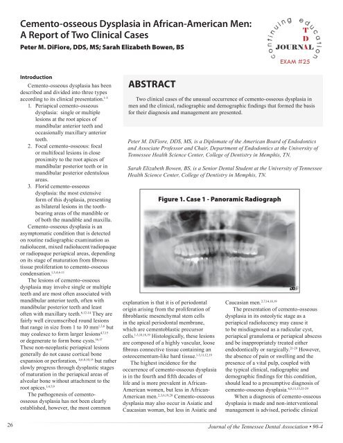
Figure 1. Case 1 - Panoramic Radiograph<br />
explanation is that it is of periodontal<br />
orig<strong>in</strong> aris<strong>in</strong>g from the proliferation of<br />
fibroblastic mesenchymal stem cells<br />
<strong>in</strong> the apical periodontal membrane,<br />
which are cementoblastic precursor<br />
cells. 1-3,10,18,19 Histologically, these lesions<br />
are composed of a highly vascular, loose<br />
fibrous connective tissue conta<strong>in</strong><strong>in</strong>g an<br />
osteocementum-like hard tissue. 1-3,11,12,19<br />
The highest <strong>in</strong>cidence for the<br />
occurrence of cemento-<strong>osseous</strong> dysplasia<br />
is <strong>in</strong> the fourth and fifth decades of<br />
life and is more prevalent <strong>in</strong> <strong>African</strong>-<br />
<strong>American</strong> women, but less <strong>in</strong> <strong>African</strong>-<br />
<strong>American</strong> men. 2,3,6,19,20 <strong>Cemento</strong>-<strong>osseous</strong><br />
dysplasia may also occur <strong>in</strong> Asiatic and<br />
Caucasian woman, but less <strong>in</strong> Asiatic and<br />
Caucasian men. 2,7,14,18,19<br />
The presentation of cemento-<strong>osseous</strong><br />
dysplasia <strong>in</strong> its osteolytic stage as a<br />
periapical radiolucency may cause it<br />
to be misdiagnosed as a radicular cyst,<br />
periapical granuloma or periapical abscess<br />
and be <strong>in</strong>appropriately treated either<br />
endodontically or surgically. 21-29 However,<br />
the absence of pa<strong>in</strong> or swell<strong>in</strong>g and the<br />
presence of a vital pulp, coupled with<br />
the typical cl<strong>in</strong>ical, radiographic and<br />
demographic f<strong>in</strong>d<strong>in</strong>gs for this condition,<br />
should lead to a presumptive diagnosis of<br />
cemento-<strong>osseous</strong> dysplasia. 6,8,11,13,21-29<br />
When a diagnosis of cemento-<strong>osseous</strong><br />
dysplasia is made and non-<strong>in</strong>terventional<br />
management is advised, periodic cl<strong>in</strong>ical<br />
26<br />
Journal of the <strong>Tennessee</strong> Dental Association • 90-4
and radiographic follow-up exam<strong>in</strong>ations<br />
are recommended for a m<strong>in</strong>imum of<br />
two years. If these lesions demonstrate<br />
unusual changes or become symptomatic,<br />
surgical <strong>in</strong>tervention would then be<br />
<strong>in</strong>dicated. 2,4,8,10,13,15,27,30<br />
Case Reports<br />
Case 1<br />
A 31-year-old <strong>African</strong>-<strong>American</strong><br />
male presented to the University of<br />
<strong>Tennessee</strong> College of Dentistry for<br />
an exam<strong>in</strong>ation of his wisdom teeth.<br />
His medical history revealed no<br />
significant f<strong>in</strong>d<strong>in</strong>gs and no history<br />
of jaw or face trauma. Panoramic<br />
and periapical radiographs were<br />
taken (Figures 1 and 2). Intra-oral<br />
exam<strong>in</strong>ation revealed a normal oral<br />
mucosa, the absence of soft and hard<br />
tissue swell<strong>in</strong>g and teeth of normal<br />
color. Periodontal exam<strong>in</strong>ation<br />
revealed the presence<br />
of severe generalized<br />
periodontal disease.<br />
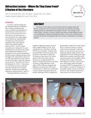
A discrete periapical<br />
radiolucency associated<br />
with tooth number 25 was<br />
noted. However, teeth<br />
numbers 7 through 10, 19<br />
through 24 and 26 through<br />
30 all had vary<strong>in</strong>g degrees<br />
of mixed-radiopaque/<br />
radiolucent periapical<br />
areas, of which tooth<br />
number 19 was the largest,<br />
<strong>in</strong>volv<strong>in</strong>g both the mesial<br />
and distal root apices<br />
which showed the most profound<br />
degree of opacity. All of these teeth<br />
were asymptomatic, with no pa<strong>in</strong><br />
or tenderness on percussion or<br />
palpation. The pulps of these teeth<br />
tested vital with<strong>in</strong> normal limits to<br />
both cold and electric stimulation,<br />
except for tooth 30, which had<br />
prior endodontic treatment. On the<br />
basis of the patient’s cl<strong>in</strong>ical and<br />
radiographic f<strong>in</strong>d<strong>in</strong>gs, age and ethnic<br />
background, a presumptive diagnosis<br />
of multifocal periapical cemento<strong>osseous</strong><br />
dysplasia, with the possibility<br />
of early stage florid cement-<strong>osseous</strong><br />
dysplasia, was made. The patient was<br />
treatment-planned for extraction of<br />
teeth numbers 17 and 32, periodontal<br />
treatment and further evaluation of<br />
this condition at scheduled follow-up<br />
exam<strong>in</strong>ations.<br />
Figure 2. Case 1 - Periapical<br />
Radiographs<br />
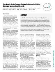
Figure 3. Case 2 - Panoramic Radiograph<br />
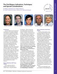
Figure 4. Case 2 - Periapical<br />
Radiographs<br />
Case 2<br />
A 63-year-old <strong>African</strong>-<br />
<strong>American</strong> male presented to the<br />
University of <strong>Tennessee</strong> College<br />
of Dentistry for a periodic oral<br />
exam<strong>in</strong>ation. His medical history<br />
revealed hypertension, gout,<br />
hypercholesterolemia, anxiety and<br />
depression. The medications he was<br />
prescribed for these conditions were<br />
felodip<strong>in</strong>, furosemide, potassium<br />
chloride, allopur<strong>in</strong>ol, atorvastat<strong>in</strong><br />
and citalopram. He had a history of<br />
regular dental care and was devoid of<br />
craniofacial <strong>in</strong>jury but had undergone<br />
sleep-apnea surgery. His <strong>in</strong>tra-oral<br />
soft and hard tissue exam<strong>in</strong>ation<br />
was unremarkable. His periodontal<br />
exam<strong>in</strong>ation revealed a moderate,<br />
generalized, chronic periodontitis.<br />
Panoramic and periapical<br />
radiographs (Figures 3<br />
and 4) revealed that tooth<br />
number 25 demonstrated<br />
a def<strong>in</strong>ite periapical<br />
radiolucency; however, all<br />
of the mandibular anterior<br />
teeth had vary<strong>in</strong>g degrees<br />
of mixed radiolucent/<br />
radiopaque periapical<br />
areas. These teeth were<br />
asymptomatic, <strong>in</strong>tact<br />
and uniformly normal <strong>in</strong><br />
coloration, not sensitive<br />
to palpation by percussion<br />
and vital <strong>in</strong> their response<br />
to cold and electric<br />
stimulation (Figure 5).<br />
Summariz<strong>in</strong>g these cl<strong>in</strong>ical and<br />
radiographic exam<strong>in</strong>ation f<strong>in</strong>d<strong>in</strong>gs<br />
and <strong>in</strong> consideration of the patient’s<br />
ethnic background, presumptive<br />
diagnosis was periapical cemento<strong>osseous</strong><br />
dysplasia. His treatment<br />
plan <strong>in</strong>cluded periodontal treatment<br />
and regular periodic cl<strong>in</strong>ical and<br />
radiographic follow-up exam<strong>in</strong>ation.<br />
Discussion<br />
These two cases of cemento-<strong>osseous</strong><br />
dysplasia are unique for two reasons:<br />
first, both uncommonly occurred <strong>in</strong> men,<br />
and second, one occurred at the lower<br />
end and the other at the upper end of<br />
the usual age range for this condition.<br />
Both patients had a def<strong>in</strong>itive periapical<br />
radiolucency associated with tooth number<br />
25. However, both patients had a number<br />
of other teeth with vary<strong>in</strong>g degree of<br />
mixed radiopaque/radiolucent periapical<br />
90-4 • <strong>Cemento</strong>-<strong>osseous</strong> <strong>Dysplasia</strong> <strong>in</strong> <strong>African</strong>-<strong>American</strong> <strong>Men</strong>: A Report of Two Cl<strong>in</strong>ical Cases<br />
27
lesions. In the first case, the<br />
patient had a more extensive<br />
cl<strong>in</strong>ical presentation of cement<strong>osseous</strong><br />
dysplasia <strong>in</strong>volv<strong>in</strong>g<br />
the mandibular anterior and<br />
posterior teeth and the maxillary<br />
<strong>in</strong>cisor teeth. In the second case,<br />
the patient had a less extensive<br />
cl<strong>in</strong>ical presentation of cement<strong>osseous</strong><br />
dysplasia <strong>in</strong>volv<strong>in</strong>g only<br />
the mandibular anterior teeth.<br />
Although these two cl<strong>in</strong>ical cases<br />
of cemento-<strong>osseous</strong> dysplasia<br />
were somewhat atypical for<br />
gender and age, both had the<br />
typical cl<strong>in</strong>ical and radiographic<br />
characteristics of cemento<strong>osseous</strong><br />
dysplasia but presented<br />
with different manifestations of<br />
lesions at multifocal sites. These two cases<br />
serve to emphasize that the recognition<br />
and proper diagnosis of cemento-<strong>osseous</strong><br />
dysplasia as a common benign condition<br />
among people of <strong>African</strong> descent will<br />
prevent unnecessary endodontic and/<br />
or surgical treatment and can establish<br />
a regimen for periodic observational<br />
management.<br />
References<br />
1. Waldron CA. Fibro-<strong>osseous</strong> Lesions of the Jaws. J Oral<br />
Maxillofac Surg, 1985;43:249-62.<br />
2. Summerl<strong>in</strong> DJ, Tomich CE. Focal <strong>Cemento</strong>-<strong>osseous</strong> <strong>Dysplasia</strong>: A<br />
Cl<strong>in</strong>ico-pathologic Study of 221 Cases. Oral Surg Oral Med Oral<br />
Pathol, 1994;78(5):611-20.<br />
3. Melrose RJ. The Cl<strong>in</strong>ico-pathologic Spectrum of <strong>Cemento</strong><strong>osseous</strong><br />
<strong>Dysplasia</strong>. Oral Maxillofac Cl<strong>in</strong> North Am,<br />
1997;9(4):643-53.<br />
Figure 5. Case 2 - Cl<strong>in</strong>ical Photograph<br />
Anterior Teeth<br />
4. Alawi F. Benign Fibro-<strong>osseous</strong> Diseases of the Maxillofacial<br />
Bones. Am J Cl<strong>in</strong> Pathol, 2002;118(S1):S50-70.<br />
5. MacDonald-Jankowski, DS. Fibro-<strong>osseous</strong> Lesions of the Face<br />
and Jaws. Cl<strong>in</strong>ical Radiology, 2004;59:11-25.<br />
6. Zegarelli EV, Kutscher AH, Napoli N, Iurono F, Hoffman P. The<br />
Ccementoma: A Study of 230 Patients with 435 <strong>Cemento</strong>mas.<br />
Oral Surg, 1964;17(2):219-24.<br />
7. Kawai T, Hiranuma H, Kish<strong>in</strong>o M, Jikko A, Saduka M. <strong>Cemento</strong><strong>osseous</strong><br />
Ddysplasia of the Jaws <strong>in</strong> 54 Japanese Patients. A<br />
Radiographic Study. Oral Surg Oral Med Oral Pathol Oral Radiol<br />
Endod, 1999;87(1):107-14.<br />
8. Scholl RJ, Kellet HM, Neumann DP, Lurie AG. Cysts and<br />
Cystic Lesions of the Mandible: Cl<strong>in</strong>ical and Radiologicalhistopathologic<br />
Review. RadioGraphics, 1999;19(5):1107-24.<br />
9. Dunfee Bl, Sakai O, Pisty R, Gohel A. Radiological and<br />
Pathological Characteristics of Benign and Malignant Lesions of<br />
the Mandible. RadioGraphics, 2006;26(6):1751-68.<br />
10. Dagistan S, Goregen M, Cakur B, Miloglu O. <strong>Cemento</strong>-<strong>osseous</strong><br />
<strong>Dysplasia</strong>s. Rev Cl<strong>in</strong> Pesq Odontol, 2007;3(1):43-9.<br />
11. Slootweg PJ. Bone Diseases of the Jaws. International Journal of<br />
Dentistry, 2010;1(1):1-7.<br />
12. Vegh T, Multiple <strong>Cemento</strong>mas (Periapical Cemental <strong>Dysplasia</strong>):<br />
Report of a Case. Oral Surg, 1976;42(3):402-6.<br />
13. Falace DA, Cunn<strong>in</strong>gham CJ. Periapical Cemental <strong>Dysplasia</strong>:<br />
Simultaneous Occurrence <strong>in</strong> Multiple Maxillary and Mandibular<br />
Teeth. J Endod, 1984;10(9):455-6.<br />
14. Tanaka H, Yoshimoto A, Toyama Y, Iwase T, Hayasaka N, Moro<br />
I. Periapical Cemental <strong>Dysplasia</strong> with Multiple Lesions. Int J Oral<br />
Maxillofac Surg, 1987;16(6):757-63.<br />
15. Manganaro AM, Millet GV. Periapical<br />
Cemental Dysplaia. General Dentistry,<br />
1996;44(4);336-9.<br />
16. Higuchi Y, Nakamura N, Tashiro H.<br />
Cl<strong>in</strong>icopathological Study of <strong>Cemento</strong><strong>osseous</strong><br />
<strong>Dysplasia</strong> Produc<strong>in</strong>g Cysts of the<br />
Mandible. Oral Surg Oral Med Oral Pathol,<br />
1988;65(3):339-42.<br />
17. Mahomed F, Alt<strong>in</strong>i M, Meer S, Coleman H.<br />
<strong>Cemento</strong>-<strong>osseous</strong> <strong>Dysplasia</strong> with Associated<br />
Simple Bone Cysts. J Oral Maxillofac Surg,<br />
2005;63:1549-54.<br />
18. Hamner JE, Scofield HH, Cornyn J. Benign<br />
Fibro-<strong>osseous</strong> Jaw Lesions of Periodontal<br />
Orig<strong>in</strong>: An Analysis of 249 Cases. Cancer,<br />
1968;22(4):861-78.<br />
19. Waldron CA, Giansanti JS. Benign<br />
Fibro-<strong>osseous</strong> Lesions of the Jaws: A<br />
Cl<strong>in</strong>ical-Radiographic-histologic Review of<br />
Sixty-five Cases. Part II: Benign Fibro-<strong>osseous</strong><br />
Lesions of Periodontal Orig<strong>in</strong>. Oral Surg,<br />
1973;35(3):340-50.<br />
20. Neville BW, Albenesius RJ. The Prevalence of<br />
Benign Fibro-<strong>osseous</strong> Lesions of Periodontal<br />
Orig<strong>in</strong> <strong>in</strong> Black Women: A Radiographic<br />
Survey. Oral Surg Oral Med Oral Pathol,<br />
1986;62(3):340-4.<br />
21. Forman GH. Periapical Cemental <strong>Dysplasia</strong><br />
Resembl<strong>in</strong>g Apical Granulomata and<br />
Radicular Cysts. Br Dent J, 1975;138(1):22-4.<br />
22. Wilcox LR, Walton R. A Case of Mistaken Identity Periapical<br />
Cemental <strong>Dysplasia</strong> <strong>in</strong> an Endodontically Treated Tooth. Endod<br />
Dent Traumatol, 1989;5(6):298-301.<br />
23. Ward MR. Periapical Cemental <strong>Dysplasia</strong>: A Case Report. New<br />
Zealand Dental Journal, 1993;89(4):53-4.<br />
24. Ghandour K. <strong>Cemento</strong>mas. Dental News, 1997;4(1):33-5.<br />
25. Smith S, Patel K, Hosk<strong>in</strong>son AE. Periapical Cemental <strong>Dysplasia</strong>:<br />
A Case ofMisdiagnosis. Br Dent J, 1998;185(?):122-3.<br />
26. Chandler NC, Love RM, Sundqvist G. Laser Doppler Flowmetry:<br />
An Aid <strong>in</strong> the Differential Diagnosis of Apical Radiolucencies.<br />
Oral Surg Oral Med Oral Pathol Oral Radiol Endod,<br />
1999;87(5):613-6.<br />
27. Drazic R, M<strong>in</strong>ic AJ. Focal <strong>Cemento</strong>-<strong>osseous</strong> <strong>Dysplasia</strong> <strong>in</strong> the<br />
Maxilla Mimick<strong>in</strong>g Periapical Granuloma. Oral Surg Oral Med<br />
Oral Pathol Oral Radiol Endod, 1999;88(1):87-9.<br />
28. Galgano C, Samson J, Kuffer R, Lombardi T. Focal <strong>Cemento</strong><strong>osseous</strong><br />
<strong>Dysplasia</strong> Involv<strong>in</strong>g a Mandibular Lateral Incisor.<br />
International Endodontic Journal, 2003;36:907-11.<br />
29. Resnick CM, Novell<strong>in</strong>e RA. <strong>Cemento</strong>-<strong>osseous</strong> <strong>Dysplasia</strong>, A<br />
Radiological Mimic of Periapical Abscess. Emerg Radiol,<br />
2008;15:367-74.<br />
30. Fantasia JE, Damm DD. Oral Diagnosis: Multifocal Mixed<br />
Radiolucencies and Radiopacities. General Dentistry,<br />
2002;50(3):286-8.<br />
Questions for Cont<strong>in</strong>u<strong>in</strong>g Education Article - CE Exam #25<br />
1. The types of cement-<strong>osseous</strong> dysplasia are classified<br />
as:<br />
a. periapical cement-<strong>osseous</strong> dysplasia<br />
b. focal cement-<strong>osseous</strong> dysplasia<br />
c. florid cement-<strong>osseous</strong> dysplasia<br />
d. all the above<br />
2. The lesions of cement-<strong>osseous</strong> dysplasia:<br />
a. always <strong>in</strong>volve maxillary teeth<br />
b. may <strong>in</strong>volve s<strong>in</strong>gle or multiple teeth<br />
c. never exceed 1mm <strong>in</strong> size<br />
d. always <strong>in</strong>volved with mandibular teeth<br />
3. <strong>Cemento</strong>-<strong>osseous</strong> dysplasia lesions:<br />
a. are non-neoplastic<br />
b. do not cause cortical bone expansion<br />
c. do not cause perforation of the cortical plate<br />
d. all the above<br />
4. The highest <strong>in</strong>cidence of cement-<strong>osseous</strong> dysplasia is:<br />
a. <strong>in</strong> the teenage years<br />
b. occurs more commonly <strong>in</strong> men across all<br />
ethnicities<br />
c. occurs more commonly <strong>in</strong> native americans<br />
d. occurs with the highest <strong>in</strong>cidence <strong>in</strong> the fourth and<br />
fifth decades of life<br />
5. When the cement-<strong>osseous</strong> dysplasia presents <strong>in</strong> its<br />
osteolytic stage as a periapical radiolucency, it must be<br />
dist<strong>in</strong>guished from:<br />
a. radicular cyst<br />
b. periapical granuloma<br />
c. periapical abcess<br />
d. all the above<br />
Publication date: Fall 2010. Expiration date: Fall 2013.<br />
28<br />
Journal of the <strong>Tennessee</strong> Dental Association • 90-4
Answer Form for TDA CE Credit Exam #25:<br />
<strong>Cemento</strong>-<strong>osseous</strong> <strong>Dysplasia</strong> <strong>in</strong> <strong>African</strong>-<strong>American</strong> <strong>Men</strong>: A Report of Two Cl<strong>in</strong>ical Cases<br />
Publication date: Fall 2010. Expiration date: Fall 2013.<br />
Circle the correct letter answer for each CE Exam question:<br />
1. a b c d<br />
T<br />
D<br />
A<br />
EXAM #25<br />
2. a b c d<br />
3. a b c d<br />
4. a b c d<br />
5. a b c d<br />
Cost per exam per person is $15.00 for one (1) cont<strong>in</strong>u<strong>in</strong>g education credit.<br />
This page may be duplicated for multiple use. Please pr<strong>in</strong>t or type.<br />
ADA ID Number (Dentist Only):<br />
License Number of RDH:<br />
Registration Number if RDA:<br />
Name (Last/First/Middle):<br />
Office Address:<br />
City/State/Zip:<br />
Daytime Phone Number : ( )<br />
Component Society (TDA Member Only):<br />
Dr.<br />
(Auxiliary Staff: Please provide name of Employer Dentist)<br />
Signature:<br />
All checks should be made payable to the <strong>Tennessee</strong> Dental Association. Return the Exam Form and<br />
your check or credit card <strong>in</strong>formation to:<br />
<strong>Tennessee</strong> Dental Association at 660 Bakers Bridge Ave., Suite 300 <strong>in</strong> Frankl<strong>in</strong>, TN 37067<br />
The form may be faxed to 615-628-0214 if us<strong>in</strong>g a credit card.<br />
Credit Card Information (Use your TDA/Bank of America card, MasterCard or Visa ONLY):<br />
Card #:<br />
Exp. Date:<br />
Three-digit CVV2 Code (on back of the card follow<strong>in</strong>g the card number):<br />
Name as it appears on the card:<br />
Do not write <strong>in</strong> this space - for TDA Adm<strong>in</strong>istration purposes only<br />
Check #: CC Paid w/doctor’s CC<br />
90-4 • <strong>Cemento</strong>-<strong>osseous</strong> <strong>Dysplasia</strong> <strong>in</strong> <strong>African</strong>-<strong>American</strong> <strong>Men</strong>: A Report of Two Cl<strong>in</strong>ical Cases Answer Form<br />
29