MMed Internal Medicine Dissertation 2012 Primus.pdf - muhas
MMed Internal Medicine Dissertation 2012 Primus.pdf - muhas
MMed Internal Medicine Dissertation 2012 Primus.pdf - muhas

Create successful ePaper yourself
Turn your PDF publications into a flip-book with our unique Google optimized e-Paper software.
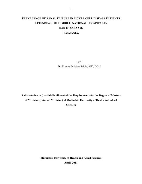
i<br />
PREVALENCE OF RENAL FAILURE IN SICKLE CELL DISEASE PATIENTS<br />
ATTENDING MUHIMBILI NATIONAL HOSPITAL IN<br />
DAR ES SALAAM,<br />
TANZANIA.<br />
By<br />
Dr. <strong>Primus</strong> Felician Saidia, MD, DGH<br />
A dissertation in (partial) Fulfilment of the Requirements for the Degree of Masters<br />
of <strong>Medicine</strong> (<strong>Internal</strong> <strong>Medicine</strong>) of Muhimbili University of Health and Allied<br />
Sciences<br />
Muhimbili University of Health and Allied Sciences<br />
April, 2011
ii<br />
CERTIFICATION<br />
The undersigned certify that he has read and hereby recommend for examination of a<br />
dissertation entitled ‘Prevalence of renal failure among sickle cell disease patients<br />
attending Muhimbili National Hospital, Dar es Salaam’ in the fulfilment of the<br />
requirements for the degree of Master of <strong>Medicine</strong> (<strong>Internal</strong> <strong>Medicine</strong>) of the Muhimbili<br />
University of Health and Allied sciences (MUHAS)<br />
This <strong>Dissertation</strong> Submitted in Partial Fulfilment of The Requirements for the degree of<br />
Master of <strong>Medicine</strong> (<strong>Internal</strong> <strong>Medicine</strong>) of the Muhimbili University of Health and Allied<br />
sciences (MUHAS)<br />
Prof. Eden Maro<br />
Supervisor<br />
Date…………………………………………….<br />
Dr. Onesmo Kisanga<br />
Co-supervisor<br />
Date………………………………………………..
iii<br />
DECLARATION AND COPYRIGHT<br />
I, Dr. <strong>Primus</strong> Felician Saidia, declare that this dissertation work is my own original work<br />
and that it has not been presented and will not be presented to any other university for<br />
similar or any other dgree award<br />
Signature________________________________ Date__________________<br />
This dissertation is copyright material protected under the Berne Convention, the<br />
Copyright Act 1999 and other international and national enactments, in that behalf, on<br />
interlectual property. It may not be reproduced by any means in fully or in part except for<br />
short extracts and fair dealings, for research or private study, critical scholary review or<br />
disclocourse with an acknowledgment, without permission of the Directorate of<br />
Postgraduate studies on behalf of both the auther and the Muhimbili University of Health<br />
and Allied Sciences
iv<br />
AKNOWLEDGEMENT<br />
I wish and remain thankful to God, the Almighty for the gift of life.<br />
I wish to thank my supervisor Professor Eden Maro and the co-supervisor Dr. Onesmo<br />
Kisanga for their overal guidance and review of this dissertation work.<br />
I wish to thank Professor Jain Sharad and Dr. Paschal Lugajo for their extensive review<br />
and contribution in the analysis and production of this dissertation work.<br />
I wish to thank The Department of <strong>Internal</strong> medicine, MUHAS for accepting my<br />
dissertation title and all the departmental support through out the dissertation proposal<br />
and report production.<br />
I wish to thank all the sickle cell team at Muhimbili National Hospital for all the support<br />
they have given to me during proposal writing and data collection at the clinic and the<br />
sickle cell laboratory.<br />
I wish to thank Mrs Vick and Mr. Kamogella, the MNH laboratory technicians for all<br />
their support during laboratory works.<br />
Many thanks to all the patients who voluntarily participated in the study<br />
I wish to thank Dr. Cyprian Makwaya for his tireless support in data handling and<br />
eventually, analysis.<br />
Last but not least, I wish to express my utmost gratitude to my dear wife Lucia and my<br />
sons Mwolisa, Joseph and Ngashase for their constant encouragement and inspiration<br />
throughout the disertation hardships.
v<br />
DEDICATION<br />
To my Mother for her educational belief and support<br />
To my wife Lucia and our sons, Mwolisa, Jose and Ngashase
vi<br />
ABSTRACT<br />
Background: Sickle-cell disease, is an inheritable hemoglobinopathy where red cells<br />
assume an abnormal, rigid, sickle shape leading to several complications, one of them,<br />
chronic renal failure. The disease is common among blacks. Renal failure in SCD<br />
patients ranges from 5 to 18 percent and contributes to 18% of the overall mortality in<br />
SCD patients. Despite its remarkable contribution in mortality, no data from Africa and<br />
especially Tanzania is available to address the extent of renal failure in SCD patients.<br />
Objective: This study aimed to determine the prevalence of renal failure and associated<br />
factors among sickle cell disease patients attending Muhimbili National Hospital in Dar<br />
es Salaam, Tanzania.<br />
Methods: A descriptive cross sectional study was done for a period of seven months,<br />
September 2010 through March 2011. During this period, patients attending the<br />
outpatient SCD clinic were enrolled and screened for features suggestive of renal failure.<br />
The data included demographic data, anthropometric measurements and clinical<br />
information. Blood for CBC, serum creatinine, urea and electrolytes was obtained,<br />
urinalysis inclusive. Renal ultrasound was performed for those with established renal<br />
failure. The end point of this study was renal failure defined by eGFR less than<br />
60ml/min/1.73m 2 . The collected data was recorded cleaned, validated and finally<br />
analysed using SPSS version 15.0. Various associations between the outcome and risk<br />
factors were assessed and p-value less than 0.05 was taken as statistically significant<br />
Results: A total of 313 sickle cell disease patients were enrolled into the study. Of these<br />
14.7% had established renal failure (i.e. with eGFR
vii<br />
failure (P>0.5). Conclusively, early diagnosis and treatment of renal failure in sickle cell<br />
disease patients may retard progression to end stage renal disease.
viii<br />
DEFINITION OF TERMS<br />
Anaemia severity : For the purpose of the study severity of anaemia was defined and<br />
classified as follows{
ix<br />
ABBREVIATIONS<br />
ARF<br />
BP<br />
CBC<br />
CKD<br />
CRF<br />
CRD<br />
DBP<br />
eCcr<br />
EDTA<br />
ESRD<br />
eGFR<br />
GFR<br />
Hb<br />
HbSS<br />
MD<br />
M.MED<br />
MPGN<br />
MSc<br />
MUHAS<br />
MNH<br />
NOS<br />
SBP<br />
SCD<br />
SCN<br />
= Acute Renal Failure<br />
= Blood Pressure<br />
= Complete Blood Count<br />
= Chronic Kidney Disease<br />
= Chronic Renal Failure<br />
= Chronic Renal Disease<br />
= Diastolic Blood Pressure<br />
= Estimated Creatinine Clearance<br />
= Ethylenedeamine-acetic acid<br />
= End Stage Renal Disease<br />
= estimated Glomerular Filtration Rate<br />
= Glomerular Filtration Rate<br />
= Haemoglobin<br />
= Homozygous hemoglobin S<br />
= Medical Doctor<br />
= Masters of <strong>Medicine</strong><br />
= Membranoproliferative Glomerulonephritis<br />
= Masters of Science<br />
= Muhimbili University of Health and Allied Sciences<br />
= Muhimbili National Hospital<br />
= Nitric Oxide Synthetase<br />
= Systolic Blood Pressure<br />
= Sickle Cell Disease<br />
= Sickle Cell Nephropathy
x<br />
TABLE OF CONTENTS<br />
Title………………………………………………………………………………………..i<br />
Ceritification………………………………………………………………………………ii<br />
Declaration and copyright protection…………………………………………………….iii<br />
Aknowledgement…………………………………………………………………………iv<br />
Dedication…………………………………………………………………………………v<br />
Abstract…………………………………………………………………………………...vi<br />
Definition of terms………………………………………………………………………viii<br />
List of acronyms…...……………………………………………………………………..ix<br />
Table of contents…………………………………………………………………………..x<br />
CHAPTER 1 : Introduction and Literature……………………………………………1<br />
1.1.1 Overview…………………………………………………………………………..1<br />
1.1.2 Cockcroft-Gault equation/formula………………………………………………..2<br />
1.1.3 Stages of chronic kidney disease …………………………………………………3<br />
1.1.4 Prevalence and mortality ………………………………………………………….3<br />
1.1.5 Pathophysiology ………………………………………………………………..…5<br />
1.1.6 Clinical features ……………………………………………………………...…...6<br />
1.1.7 Diagnosis ……………………………………………………………………..…..9<br />
1.1.8 Pathophysiology…………………………………………………………………..9<br />
1.1.9 Management of SCD renal failure……………………………………………….11<br />
1.2 Problem Statement………………………………………………………………..14<br />
1.3 Rationale…………………………………………………………………………..15<br />
1.4 Objectives……………………………………………………………………….…16<br />
CHAPTER 2 :<br />
Methodology………………………………………………………………………….. 17<br />
2.1 Study area………………………………………………………………………….. 17<br />
2.2 Study design…………………………………………………………………………17<br />
2.3 Study Population…………………………………………………………………….17<br />
2.4 Inclusion criteria……………………………………………………………………..17<br />
2.5 Sample size…………………………………………………………………………..17
xi<br />
2.6 Sampling procedure………………………………………………………………….18<br />
2.7 Data collection……………………………………………………………………….18<br />
2.8 Patient management………………………………………………………………….18<br />
2.9 Specimens and investigations……………………………………………………… .19<br />
2.10 Statistical analysis…………………………………………………………………..19<br />
2.11 Ethical issues ……………………………………………………………………….20<br />
2.12 Ethical clearance……………………………………………………………………20<br />
CHAPTER 3 : Results………………………………………………………………….21<br />
3.1 Demographic characteristics and prevalence of renal failure………………………..21<br />
3.2 Proteinuria and renal failure…………………………………………………………23<br />
3.3 Renal function in relation to past clinical events/crisis, anaemia severity, hypertension<br />
and blood urea levels…..…………………….………………………………………24<br />
3.4 Ultrasonographic findings in patients with renal failure……………………………..30<br />
CHAPTER 4 : Discussion………………………………………………………………27<br />
4.1 Major findings ……..………………………………………………………………...27<br />
4.2 Prevalence of renal failure by age and sex…………………………………………...27<br />
4.3 Proteinuria and renal function………….……………………………………………28<br />
4.4 Hematuria in SCD patients …………………………………………………………29<br />
4.6 Hypertension in SCD patients and renal failure .………………………………...…29.<br />
4.7 Clinical illnesses in SCD patients and renal failure…………………………………30<br />
4.8 Uraemia and renal failure……………………………………………………………30<br />
4.4 Anaemia and renal failure ……………………..……………………………………31<br />
4.9 Renal ultrasonography in SCD nephropathy...………………………………………31<br />
CHAPTER 5 : Study limitations, conclussion and Recomendations…………….….32<br />
5.1 Conclusion …………………………………………………………………………..32<br />
5.2 Recommendations …………………………………………………………………...32<br />
5.3 Study limitations………………………………………..……………………………32<br />
References ………………………………………………………………………………33
xii<br />
Annexes …………………………………………………………………………………38<br />
Appendices………………………………………………………………………………41
1<br />
CHAPTER ONE<br />
1.1 INTRODUCTION AND LITERATURE REVIEW<br />
1.1.1 Overview<br />
Sickle-cell disease (or drepanocytosis) is a life-long blood disorder characterized by red<br />
blood cells that assume an abnormal, rigid, sickle shape. Sickling decreases the cells'<br />
flexibility and results in a risk of various complications. The sickling occurs because of a<br />
mutation in the hemoglobin gene. (1)<br />
Mutations occur in the ß chain of Hb whereby the hydrophobic amino acid valine takes<br />
the place of hydrophilic glutamic acid at the sixth amino acid position of the polypeptide<br />
chain. This substitution creates a hydrophobic spot on the outside of the protein structure<br />
that sticks to the hydrophobic region of an adjacent haemoglobin molecule's beta chain.<br />
This clumping together (polymerization) of Hb S molecules into rigid fibers causes the<br />
"sickling" of red blood cells. (2)<br />
SCD has several complications, including chronic renal failure, manifesting with<br />
hypertension (high blood pressure) proteinuria (protein loss in the urine), hematuria (red<br />
blood cells in urine) and worsened anemia. Progression to end-stage renal failure confers<br />
poor prognosis (3).<br />
Over the past 20 years, early medical treatment has significantly increased the life<br />
expectancy of children with sickle cell disease. More than 90% of patients now reach the<br />
age of 20, and the median life expectancy of sickle cell patients is at least 50 years in<br />
countries with advanced healthcare systems. Gender wise, life expectancy averages to 42<br />
and 48 years for males and females, respectively. (1, 4)<br />
Renal failure manifests as acute or chronic in which acute renal failure (ARF) is a<br />
syndrome characterized by the rapid decline in glomerular filtration rate (hours to days),<br />
retention of nitrogenous waste products, and perturbation of extracellular fluid volume
2<br />
and electrolyte and acid-base homeostasis. Chronic renal disease (CRD) is a more<br />
gradual pathophysiologic process with multiple etiologies, resulting in the inexorable<br />
attrition of nephron number and function and frequently leading to end-stage renal<br />
disease (ESRD) evident in a period of three months or more ((5).<br />
1.1.2 Cockcroft – Gault equation/formula<br />
This formula is a commonly used surrogate marker for actual creatinine clearance, which<br />
may be used to calculate an Estimated Creatinine Clearance, which in turn estimates<br />
GFR.<br />
It is named after it’s founders and it uses creatinine measurements and a patient's weight<br />
to predict the creatinine clearance. (6)<br />
The formula, as originally published for creatinine in µmol/L, is:<br />
Where Constant is 1.23 for men and 1.04 for women.
3<br />
1.1.3 Stages of Chronic Kidney Disease (7)<br />
Glomerular<br />
Stage<br />
Description<br />
Filtration<br />
Rate<br />
(GFR)<br />
At increased<br />
risk<br />
Risk factors for kidney disease (e.g., diabetes, high<br />
blood pressure, family history, older age, ethnic<br />
group)<br />
More than 90<br />
1<br />
Kidney damage (protein in the urine) and normal<br />
GFR<br />
More than 90<br />
2 Kidney damage and mild decrease in GFR 60 to 89<br />
3 Moderate decrease in GFR 30 to 59<br />
4 Severe decrease in GFR 15 to 29<br />
5<br />
Kidney failure (dialysis or kidney transplant<br />
needed)<br />
Less than 15<br />
1.1.4 Prevalence and mortality<br />
In the US, sickle cell anemia usually occurs in black people, but sometimes occurs in<br />
Hispanic people. In the United States, about one in five hundred black births, and about<br />
one in 36,000 Hispanic births, have sickle cell anemia. (8)<br />
The prevalence of sickle cell disease is extremely high in Africa, where 150,000 to<br />
300,000 homozygous individuals are born every year. Africans were well aware of the<br />
disease before its first description in America (9). In a newborn screening study in<br />
Nigeria, the prevalence of HbSS was 2.8%. (10).
4<br />
In Tanzania, a study done to evaluate prevalence of sickle cell haemoglobin, HbSS in<br />
highlands and lowland revealed that lowlands had higher prevalence of high HbSS than<br />
highlands (HbS = 16.04% (98/611) versus 6.32% (36/570), P = 0.0001)(11). It has been<br />
estimated that birth incidence of SCD is 6-7 per 1000 children (12).<br />
Also, it is roughly estimated that the prevalence of the trait in Tanzania ranges between<br />
15 - 20%, which is among the highest in Africa (12) In Kenya prevalence of HbSS was<br />
found to be 0.8% in one of the coastal regions and constituted to 1.6% of all paediatric<br />
admissions. (13)<br />
Despite the fact that more than 70% of sufferers live in Africa, expenditure on the related<br />
care and research in the continent is negligible and most advances in the understanding<br />
and management of this condition have been based on research conducted in the North.<br />
(14)<br />
The prevalence of renal failure in sickle cell disease ranges from 5 to 18% of the total<br />
population of SCD patients (15). In a prospective, case-control study of patients with<br />
SCD compared with sickle cell hemoglobin C patients, 31 (4.2%) patients were affected<br />
by renal failure. The median age at the time of renal failure was 23.1 years. Survival time<br />
was 4 years with a median age of death of 27 years after the diagnosis of end-stage renal<br />
disease (ESRD) in spite of dialysis treatment. Proteinuria, hypertension, severe anaemia,<br />
and hematuria were reliable predictors of chronic renal failure (16). In this series, the<br />
presence of the inherited Central African Republic ß s -gene cluster haplotype in a patient<br />
with SCD increased significantly the risk of chronic renal failure (16). In a prospective<br />
survival analysis of 964 patients with sickle cell anaemia in adults, it was found that an<br />
18% overall mortality in adult SCD patients with 40% of these (7.6% of the total)<br />
manifesting overt renal failure. None received a kidney transplant. By multivariate<br />
regression analysis, renal failure was identified as the major risk factor for early mortality<br />
in adult patients with SCD (17, 18, 19)
5<br />
In a population study of 368 patients, documented chronic renal insufficiency was found<br />
in 4.6% of SCD patients and was significantly associated with proteinuria and increased<br />
age (20, 4). In patients with SCD, priapism episodes, especially those with postpubertal<br />
presentation and tricorporal disease (corpora cavernosa and corpus spongiosa<br />
involvement), were associated with increased risk of multiorgan failure, including kidney<br />
failure, which has shown to be one of the causes of increased hospital admissions in SCD<br />
patients (21, 22).<br />
There is no gender predilection for renal failure in most series. The U.S Renal Data<br />
System database shows a marked male predominance of sickle cell nephropathy (SCN),<br />
in which few patients were offered transplantation (23).<br />
1.1.5 Pathophysiology<br />
Chronic sickling underlies several mechanisms for kidney injury. The arterial side of the<br />
renal microvasculature has a low O 2 tension. The hypertonicity and low pH of the renal<br />
medulla promote the formation of hemoglobin polymers in the red cells with deformation<br />
of the sickled cells, resulting in an increase in the blood viscosity, functional venous<br />
engorgement, and interstitial edema, predisposing the renal microcirculation to ischemia<br />
and infarction. Obliteration of the medullary vasculature produces segmental scarring and<br />
interstitial fibrosis (structural papillectomy), with formation of dilated renal pelvic<br />
capillaries and veins. Hematuria results from rupture of vessels from the early venous<br />
engorgement or from the dilated vessels that result from scarring. The development of<br />
collateral vessels and their abnormal orientation in the medulla interferes with the<br />
countercurrent exchange mechanism, culminating through the years in irreversible loss of<br />
medullary tonicity. It has been postulated that renal cortical blood flow and eGFR are<br />
increased by the secretion of medullary vasodilator prostaglandins. (24)<br />
Hyperfiltration coupled with glomerular hypertrophy can lead to glomerulosclerosis<br />
(15,24,25). Once progression of the glomerular damage is evident, eGFR begins to<br />
decrease, worsened by the ingestion of analgesics that can independently induce<br />
interstitial nephropathy (15). There is a documented pattern of increased dextran
6<br />
permeability in the glomerular basement membrane of SCD patients, with an incremental<br />
increase in the pore radius. These changes could cause a nonselective proteinuria rather<br />
than the microalbuminuria associated with hyperfiltration (26).<br />
Another study using a transgenic sickle cell mouse model showed that there is an<br />
induction of nitric oxide synthase II (NOS II) in the glomeruli and distal nephron. This<br />
enzyme may increase the synthesis of nitric oxide leading to vasodilation and contribute<br />
subsequently to hyperfiltration (27).<br />
1.1.6 Clinical features<br />
The clinical manifestations of these pathophysiologic processes in SCD are well defined<br />
Hyposthenuria is the first clinical evidence of defective medullary tonicity. In SCD<br />
patients older than 10 years, the maximal urinary concentration is often reduced to 400<br />
mosmol/kg H 2 O. This urine concentrating capacity can be restored with multiple<br />
transfusions of normal erythrocytes, showing the reversible nature of the defect.<br />
However, in patients older than 15 years, the process is often irreversible. Both<br />
vasopressin generation and urinary diluting capacity in SCD renal failure patients remain<br />
unchanged (15). Hyposthenuria can produce a higher than usual obligatory urine output,<br />
thereby increasing the risk of dehydration.<br />
Hematuria<br />
Asymptomatic hematuria is one of the most prevalent features of the disease (15, 24) and<br />
occurs either in heterozygous or homozygous patients at any age. Gross hematuria may<br />
also be observed in patients with sickle cell trait, either alone or in combination with von<br />
Willebrand disease, even in the absence of extrarenal bleeding (15,24). The hematuria is<br />
usually unilateral with the left kidney four times more frequently involved than the right.<br />
This may be explained by increased venous pressure due to the greater length of the left<br />
renal vein (24). Most of the episodes are self-limited, although dramatic and prolonged<br />
periods of gross hematuria may be seen (15).
7<br />
Renal Tubular Acidosis<br />
In sickle cell trait, renal acidification is normal (15 ,24). The distal tubule of homozygous<br />
hemoglobin S (Hb-SS) patients requires a greater acidic stimulus to reach a maximum<br />
urine-to-blood hydrogen ion gradient. The acidification defect is consistent with less than<br />
maximal generation of titratable acid (24). Usually this defect does not cause systemic<br />
metabolic acidosis (15). There is no proximal loss of bicarbonate.<br />
Proteinuria<br />
Proteinuria is a frequent finding in SCD and is present in 30% of patients during longterm<br />
follow-up (24). Both proteinuria and renal insufficiency increase with age in a<br />
parallel pattern (15, 24, 28). Nephrotic syndrome is found in approximately 40% of the<br />
patients with sickle cell nephropathy (SCN) (15,42, 29, 30). In a summarized series it was<br />
suggested that the nephrotic syndrome in SCD is a predictor of progression to chronic<br />
renal failure.<br />
It is believed that glomerular capillary hypertension, thought to be present in sickle cell<br />
nephropathy, mediates proteinuria. This concept is supported by the decrease in protein<br />
excretion that is observed with the administration of angiotensin-converting enzyme<br />
inhibitors (16, 31, 32 and 33).<br />
Renal failure<br />
Renal failure occurs with either an acute or chronic presentation. Acute nonoliguric renal<br />
failure is present in 10% of patients hospitalized with SCD (34). Frequently, a<br />
concomitant infection or rhabdomyolysis is detected with the renal failure. Less often,<br />
renal vein thrombosis and intravascular hemolysis have been reported as causes of acute<br />
renal insufficiency in SCD patients (34). Despite the sparse literature, the prognosis<br />
appears to be favorable (34). In a retrospective case-control study of 12 SCD patients with<br />
acute renal failure, it was reported that 83% of patients survived with recovery of renal<br />
function in all patients who survived, without progression to ESRD (34).
8<br />
Usually, in sickle cell nephropathy the development of ESRD occurs between the third<br />
and fifth decades of life (15, 28). However, the renal abnormalities begin at earlier ages<br />
(35).<br />
Hyperfiltration is common in young patients with SCD (35) and is closely related to<br />
glomerular hypertrophy. Proteinuria in sickle cell nephropathy is associated with<br />
glomerulosclerosis on renal biopsy, which often progresses to renal failure. The presence<br />
of the nephrotic syndrome in patients with SCN is a clinical marker for ESRD, evolving<br />
from the progression of glomerulosclerosis. It is reasonable to argue that chronic renal<br />
failure is almost always the consequence of the progression of this mechanism of renal<br />
injury in SCN, clinically manifested by proteinuria and pathologically represented by<br />
glomerular hypertrophy and focal segmental glomerulosclerosis.<br />
Hypertension<br />
The incidence of hypertension in patients with Hb-SS ranges between 2 and 6% (24)<br />
compared with the published incidence for the black population in the United States of<br />
28%. A renal salt-losing state has been suggested to explain the rather low incidence of<br />
hypertension in patients with SCD (24), although this would suggest chronic volume<br />
depletion. A defect in vascular tone has also been suggested (36). Recent data from the<br />
Cooperative Study of Sickle Cell Disease demonstrated that individuals with SCD have<br />
blood pressure levels that are significantly lower than in the general population.<br />
Predictive variables of blood pressure by multiple regression analysis showed that in<br />
males under 18 years, there was a positive correlation between diastolic blood pressure<br />
and blood urea nitrogen and a negative correlation with the estimated creatinine<br />
clearance. Systolic blood pressure correlated with blood urea nitrogen in females over 17<br />
years of age. Alarmingly, values that could be considered normal or that represent mild<br />
hypertension in healthy individuals should be considered a risk factor for important<br />
cardiovascular complications in patients with SCD. Also, there is a positive association<br />
between blood pressure, stroke, and increased mortality in SCD. (37)
9<br />
1.1.7 Diagnosis<br />
Proteinuria<br />
Once proteinuria is detected by dipstick, it should be quantified, and renal function should<br />
be assessed (15). Diseases other than sickle cell nephropathy should be considered.<br />
Sudden oedema or massive proteinuria (>3 g/24 h) may initially suggest idiopathic<br />
nephrotic syndrome, although renal vein thrombosis should always be considered, due to<br />
the predisposition of patients with Hb-SS to experience venous thrombosis (15, 24).<br />
Hematuria<br />
Exclusion of other causes of hematuria is important before considering the diagnosis of<br />
sickle cell nephropathy. Renal and bladder ultrasound can identify bleeding from a stone<br />
or tumor. Increased echogenicity of the renal pyramids or calyceal clubbing by urography,<br />
in the absence of hypercalciuria or nephrocalcinosis, may suggest sickle cell disease (15).<br />
In gross hematuria, the use of cystoscopy may identify the source of bleeding.<br />
Coagulation tests are useful to rule out the concomitant appearance of von Willebrand<br />
disease in sickle trait patients.<br />
Hyperfiltration<br />
The upper limit for a normal eGFR is not certain even with inulin clearance (15).<br />
Simplified methods for nonisotopic iothalamate and para-aminohippurate measurement<br />
are now available, although for most clinical purposes an accurate measure of eGFR is<br />
not necessary (38). A decrease in eGFR over time in a patient with sickle cell disease,<br />
especially when accompanied by proteinuria, requires careful follow-up (15).<br />
1.1.8 Histopathology<br />
Glomerular enlargement was described as part of SCD in 1960 (38). In children, the<br />
finding has been reported more frequently beyond 2 years of age (39). This pattern is<br />
more obvious in the juxtamedullary glomeruli. A difference in size has been shown when<br />
glomeruli from SCD children are compared with healthy children (39). In adults with Hb-<br />
SS, the average diameter of the glomeruli (186 ± 14.5 µm) has been found to be greater<br />
than that in glomeruli from control biopsies (137.9 ± 19.3 µm) (32). In older patients with
10<br />
renal involvement, progressive ischemia and fibrosis lead to obliteration of the glomeruli<br />
(15).<br />
One histological study, observed perihilar focal and segmental glomerulosclerosis in eight<br />
of 10 biopsy specimens (32). The sclerotic segments were adherent to Bowman's capsule<br />
with areas of hyalinosis, lipid vacuolation, and foam cells (32). Adjacent to the glomeruli,<br />
focal interstitial fibrosis and tubular atrophy were noted. Immunofluorescence microscopy<br />
revealed only irregular staining for IgM, C3, and C1q in sclerotic areas. No electrondense<br />
immune complex-type deposits were seen. Some areas of subendothelial<br />
rarefaction had the appearance of basement membrane duplication (32).<br />
In an analysis of kidney biopsies from six nephrotic patients with Hb-SS, both collapse of<br />
the capillaries and mesangial atrophy and expansion of the obliterated tuft segment by<br />
mesangial matrix in maximally hypertrophied glomeruli in focal segmental<br />
glomerulosclerosis were described. The mean glomerular diameter was enlarged in<br />
nephrotic Hb-SS (233.6 ± 25.3 µm) and non-SCD nephrotic patients (243.0 ± 12.5 µm)<br />
compared with control subjects (158.0 ± 12.7 µm). Some peripheral capillary loops had<br />
the appearance of duplication, others a wrinkling appearance (40).<br />
In analysing 240 patients with SCD, twelve had nephrotic syndrome. In nine, the<br />
glomerular lesion consisted of mesangial expansion and basement membrane duplication<br />
(nonimmune membranoproliferative glomerulonephritis [MPGN]-like lesion) and global<br />
or focal segmental glomerulosclerosis (32).<br />
Immunofluorescence studies in one series of four young patients with Hb-SS and<br />
glomerular disease revealed MPGN like lesions associated with Ig and complement<br />
deposition (four patients), and renal tubular epithelial antigen deposition (two patients) in<br />
a granular pattern along the glomerular basement membrane (38). In the circulation of<br />
some patients, tubular epithelial antigens and cryoprecipitable renal tubular antigenantibody<br />
complexes have been detected (38). It is uncertain whether these were<br />
attributable to SCD.
11<br />
The composite picture of sickle cell glomerulopathy is one of glomerular hypertrophy and<br />
focal glomerulosclerosis (32), with either an expansive or collapsing pattern (40). The<br />
basement membrane lucencies and areas of apparent duplication present a variable<br />
nonimmune MPGN-like picture (17), without the lobular appearance of immune MPGN.<br />
In a few cases, an immune complex nephropathy has been reported (38, 41), although it is<br />
uncertain whether this is part of sickle cell nephropathy or an unusual appearance of an<br />
immune complex nephropathy modified by the presence of sickle cell disease.<br />
Medullary lesions consist of oedema, focal scarring, and interstitial fibrosis with<br />
consequent atrophy and mononuclear infiltration. Renal papillary necrosis appears<br />
focally, with a few collecting ducts surrounded by an extensive area of fibrosis (15, 24).<br />
Tubular hemosiderin deposits observed in biopsy specimens may play a role in the<br />
progression of the nephropathy. Magnetic resonance imaging in SCD patients shows a<br />
decrease in the renal cortical spin echo signal, suggesting defective renal cortical iron<br />
metabolism (42). In experimental models using rabbits, saccharated-iron complexes can<br />
produce the nephrotic syndrome (43).<br />
1.1.9 Management of SCD Renal Failure<br />
Prevention<br />
Treatment aims at preventing vaso-occlusive crises and control of infections which can<br />
worsen renal function, in addition to proper identification and management of renal<br />
complications. Early neonatal screening for SCD improves survival. (44)<br />
Altitudes above 2500 m without the use of supplementary oxygen should be avoided by<br />
SCD patients (24). Heavy exercise may be dangerous to these patients because of the risk<br />
of lactic acidosis. Sudden death has rarely been reported, but occurs even in sickle trait<br />
patients. Before elective surgery, it is recommended that transfusions with filtered normal<br />
red blood cells be performed.<br />
Fluids<br />
Close monitoring of fluid intake and output should be performed. Fluid deprivation,<br />
excessive fluid loss, and inability to ingest fluids can induce dehydration in Hb-SS more
12<br />
rapidly than in a normal population, thus exposing the patient to the additional risk of a<br />
potential sickle-cell crisis (24, 28). Urine output usually should be maintained above 2000<br />
ml/day in adults and in children in proportion to their size (24) to ensure adequate<br />
hydration. In states of circulatory overload in critically ill patients due to multiple<br />
transfusions, the use of furosemide is recommended (15).<br />
Hematuria<br />
Most episodes of gross hematuria in patients with SCD subside spontaneously. However,<br />
in a few patients hematuria may be massive. Bed rest is recommended to avoid dislodging<br />
clots. The use of hypotonic solutions (4 L/1.73 m 2 per day) in conjunction with diuretics<br />
(furosemide or thiazide) can efficiently eliminate clots from the bladder and<br />
concomitantly alleviate sickling and possibly prevent renal papillary necrosis (15).<br />
Transfusions may be necessary for excessive blood loss. The use of epsilon-aminocaproic<br />
acid for fibrinolysis may be necessary. To control hematuria, low doses may be adequate,<br />
starting from 1 g per 1.73 m 2 body surface area orally 3 times daily with a subsequent<br />
incremental increase until bleeding subsides (15).<br />
Arteriographic localization and local embolization of the affected renal segment are<br />
indicated in patients with uncontrolled bleeding (15). Rarely, nephrectomy is required.<br />
Proteinuria<br />
The avoidance of a high protein intake (greater than the recommended dietary allowance)<br />
may prevent further deterioration of the nephropathy. However, protein restriction is not<br />
recommended due to pre-existent growth failure and the low energy state of many Hb-SS<br />
patients (45).<br />
The use of angiotensin-converting enzyme inhibitors potentially can diminish the degree<br />
proteinuria in SCD patients with nephropathy. A 2 week trial with enalapril therapy<br />
involving 10 patients with mild nephropathy showed that proteinuria decreased by 57%,<br />
but returned to high levels after treatment withdrawal. Blood pressure, GFR, and effective<br />
renal plasma flow did not change significantly (32)
13<br />
Renal Transplant<br />
Renal transplantation in SCD patients is as well useful in SCD patients with ESRD.<br />
However, little is offered by physicians by assuming there is a poor chance for successful<br />
therapy. A study by Ojo and colleagues reported similar short-term survival of renal<br />
allografts in recipients with end-stage SCN with non sicklers with ESRD. However, long<br />
term benefits were comparatively low in SCD patients with ESRD. (46)<br />
Furthermore, renal transplant conferred better patient survival compared to dialysis in<br />
patients with ESRD. Similar results hold true for adolescent patients (47).<br />
Preparation prior to transplantation includes transfusion of lymphocyte-depleted packed<br />
cells (often required due to inherent erythropoietin resistance). A rise in the hematocrit<br />
and plasma viscosity may precipitate a plasma cell crisis, which is especially common in<br />
the first year after transplantation. The concurrent use of hydroxyurea to increase<br />
hemoglobin F production while decreasing the hematocrit may reduce the frequency of<br />
crises, thereby improving graft survival (48, 49).<br />
In summary, sickle cell nephropathy is an important cause of mortality in SCD patients,<br />
with specific genetic and clinical markers that can indicate further progression to ESRD.<br />
Chronic sickling promotes different mechanisms of kidney injury: structural<br />
papillectomy, urine concentration defects, hyperfiltration, and glomerular enlargement<br />
and sclerosis. Clinical detection of the manifestations of these processes, as well as<br />
detection of risk factors for medullary carcinoma, can permit the clinician to offer a<br />
rational treatment to the SCD patient.<br />
Chronic dialysis and transplantation represent reasonable options for those patients who<br />
develop ESRD.
14<br />
1.2 PROBLEM STATEMENT<br />
Sickle cell disease is common in Africa with 150,000 to 300,000 homozygous individuals<br />
born every year (8). ). It has been estimated that birth incidence of SCD is 6-7 per 1000<br />
children.<br />
In addition, it is roughly estimated that the prevalence of the trait in Tanzania ranges<br />
between 15 - 20%, which is among the highest in Africa (12)<br />
The World Health Organization has recently recognized that Sickle cell disease (SCD) as<br />
a problem of major public health significance. Despite the fact that more >70% of<br />
sufferers live in Africa, expenditure on the related care and research in the continent is<br />
negligible, and most advances in the understanding and management of this condition<br />
have been based on research conducted in the North (14).<br />
From previous published reports, the presence of renal failure in sickle cell disease (SCD)<br />
ranges between 5 - 18% of the total population of SCD patients (1)<br />
Chronic sickling underlies several mechanisms for kidney injury. The arterial side of the<br />
renal microvasculature has a low O 2 tension. The hypertonicity and low pH of the renal<br />
medulla promote the formation of hemoglobin polymers in the red cells with deformation<br />
of the sickled cells, resulting in an increase in the blood viscosity, functional venous<br />
engorgement, and interstitial edema, predisposing the renal microcirculation to ischemia<br />
and infarction. This results into glomerular damage and therefore real failure (15).<br />
Despite such a high prevalence of renal failure among sickle cell patients, no other study<br />
has been done in Tanzania to evaluate the extent of this condition for betterment of SCD<br />
patient management and improved outcomes.
15<br />
1.3 RATIONALE<br />
Tanzania is currently establishing specialized renal unit at Muhimbili National Hospital<br />
and other centers in order to improve health care for people with renal problems.<br />
However, little is known on the extent of renal failure in high-risk groups such as sickle<br />
cell disease patients.<br />
This study determined the prevalence of renal failure among sickle cell disease patients<br />
attending sickle cell clinic at Muhimbili National Hospital.<br />
This study identified sickle cell disease patients with renal failure at different stages and<br />
the indicators for the disease.<br />
The information obtained will help in the management of SCD patients and therefore<br />
improve quality of life and reduce mortality.
16<br />
1.4 OBJECTIVES<br />
Broad objective<br />
To determine the prevalence of renal failure and associated factors among SCD patients<br />
attending sickle cell disease clinic at Muhimbili National Hospital.<br />
Specific Objectives<br />
1. To determine the prevalence of renal failure in SCD patients according to age and sex.<br />
2. To describe the severity of renal failure in SCD patients by age and sex.<br />
3. To determine the relationship between renal failure and past clinical illness within six<br />
months<br />
4. To determine the relationship between renal failure in SCD patients and laboratory<br />
findings.
17<br />
CHAPTER TWO<br />
2.0 METHODOLOGY<br />
2.1 Study Area<br />
Muhimbili National Hospital, Dare es Salaam, Tanzania.<br />
2.2 Study Design<br />
A descriptive cross sectional study was done to determine the prevalence of renal failure<br />
among SCD patients attending sickle cell disease clinics.<br />
2.3 Study Population<br />
The study participants were sampled from the sickle cell disease adult and paediatric<br />
clinics at Muhimbili National Hospital<br />
2.4 Inclusion Criteria<br />
All sickle cell disease patients aged 5 years and above were considered for enrolment.<br />
Only those consenting and assenting for the study were studied.<br />
2.5 Sample size:<br />
The sample size will be calculated from the following formula<br />
N= Z 2 P (100- P)/ E 2<br />
Where: Z= critical value 1.96<br />
N = Estimated sample size<br />
E = Margin of error<br />
P = Prevalence of renal failure in SCD<br />
N = (1.96)²×18(100-18)/(4.2)²<br />
=308.
18<br />
2.6 Sampling procedure<br />
Convenience sampling procedure was used whereby all SCD patients with the above<br />
criteria were consecutively enrolled into the study after patients/parents/guardians<br />
provided written consent. For children above ten years, verbal assent to participate was<br />
obtained and the written consent was signed by the accompanying parent or guardian<br />
until the sample size was reached.<br />
Patients were enrolled into the study during clinic visits whereby the first 20 to 30<br />
patients were enrolled per each clinic visit.<br />
2.7 Data collection<br />
Data were collected using structured questionnaires designed for the purpose of the study.<br />
Data collected included demographic data, age, sex and past clinical illness/crisis as well<br />
as laboratory investigations. To minimize recall bias, the information about the past<br />
clinical events was taken within six months period.<br />
2.8 Patients’ management<br />
Routine clinical procedures in patient management were followed and, in addition,<br />
patient’s body weight was measured using a bathroom weighing scale. Blood pressure<br />
was measured using an electronic blood pressure machine type Omron(M6), where<br />
appropriate paediatric cuff was used for children. Blood pressure reading was taken three<br />
times and the average was recorded as the patient’s blood pressure.<br />
Systolic blood pressure of 140mmHg and above and diastolic blood pressure of 90mmHg<br />
above were recorded as hypertension for adults aged 18 years according to the European<br />
Society of hypertension classification 2007. Blood pressure above or equal to the 95 th<br />
percentiles in children below 18 years, by gender, height and age, were recorded as<br />
hypertension according to the American National High Blood Pressure Education<br />
Program Working Group on High Blood Pressure in Children and Adolescents tables<br />
2004. Specific investigations were performed including complete blood count, urinalysis,
19<br />
renal function tests (creatinine and urea) and serum electrolytes. Results were<br />
communicated to the attending doctor and the patient to aid in the general care for the<br />
patient.<br />
2.9 Specimens and Investigations<br />
Blood samples for RFT and electrolytes were collected aseptically in 5 ml red top<br />
vacutainers.(BD, NJ, USA). Samples for CBC were collected in vacutainers containing<br />
EDTA (Ethylenediaminetetra-acetic acid). Approximately 3 ml was collected.<br />
About 10 ml of mid-stream urine was collected in universal sterile clear bottles for<br />
urinalysis. Young children were assisted by their accompanying parents/guardians on<br />
collecting the midstream urine, where they were instructed to wait a few seconds as the<br />
child starts voiding, then collect the urine .<br />
RFT and electrolytes were performed in the laboratory using Architect Chemistry<br />
Analyser (Make: Abbot). Serum creatinine and urea were recorded for analysis.<br />
Complete blood count (CBC) was done using Cell-dyn 3500R analyser. Urinalysis was<br />
done macroscopically using urine dipstick and microscopy using microscope.<br />
Estimated Glomerular Filtration Rate ( eGFR) was calculated using the Cockroft- Gault<br />
equation/formula. eGFR of less than 60mL/min/1.73m 2 was described as established<br />
renal failure. Proteinuria in patients with eGFR >90 defined stage one renal failure.<br />
Renal ultrasonography was done to patients with eGFR less than 60mL/min/1.73m 2<br />
described as established renal failure to ascertain features of SCD involvement and to<br />
exclude other causes of renal failure including renal or bladder stones.<br />
2.10 Data management and Statistical analysis<br />
Data was entered, cleaned, validated and analyzed using SPSS version 15.0.<br />
The<br />
prevalence of renal failure was expressed in percentages for the entire study group and by<br />
age and sex.
20<br />
χ 2 test was used to examine the association between renal failure and<br />
categorical<br />
variables and Fisher’s exact test was used when the expected count was less than five.<br />
Student t-test was used to determine the association between two continuous variables. P<br />
values < 0.05 were taken as statistically significant.<br />
2.11 Ethical issues<br />
A written consent was obtained from the adult patients/parent/ guardian prior to<br />
enrolment and in addition, a verbal assent was obtained from older children (10-17<br />
years). The following information was given during patient/parent/guardian education to<br />
ensure that they have the information needed to make an informed choice: a complete<br />
description of the aims of the study, investigations that were to be performed, potential<br />
benefits and risks, specimen collection procedures, and assurance of confidentiality of<br />
any information given as well as test results. Any other requested additional information<br />
was provided to patients/parents/guardian by study personnel. Patients found with renal<br />
failure were referred to the hospital renal unit. Patient’s information and results were<br />
dealt with confidentially<br />
2.13 Ethical clearance<br />
Ethical clearance was obtained from the MUHAS high degree ethical committee of<br />
research and publication.
21<br />
CHAPTER THREE<br />
3.0 RESULTS<br />
3.1Demographic characteristics and prevalence of renal failure.<br />
During the study, a total number of 313 known sickle cell disease patients were enrolled,<br />
out of which 173 (55.3%) were females. The majority of the patients 160(51.1%) were<br />
between 10-19 years of age.<br />
All the patients were anaemic with haemoglobin range between 3.1-12g/dl (7.7±1.4,<br />
median , 7.6g/dl) (Table 1.)<br />
The study revealed that 14.7% of the study participants had established renal failure<br />
(eGFR
22<br />
Table1: Demographic,and laboratory characteristics in the study population<br />
(N = 313)<br />
Category Min value Max value Median Frequency Mean±std<br />
Sex<br />
Male<br />
Female<br />
140(44.7%)<br />
173(55.3%)<br />
Age (yrs) 5 44 16 16±7<br />
Pulse rate (bpm) 59 147 88 90±14<br />
SBP (mmHg)<br />
DBP (mmHg)<br />
90<br />
41<br />
153<br />
108<br />
111<br />
72<br />
113±15<br />
72±12<br />
Serum Creatinine (umol/L)<br />
26.4<br />
176.7<br />
50.2<br />
53.7±16.4<br />
BUN (mmol/L)<br />
1.0<br />
10.0<br />
2.2<br />
2.4±1.2<br />
GFR (mL/min/1.73m 2 )<br />
33.0<br />
183.0<br />
93<br />
92.9±29.3<br />
Calcium level (mmol/L)<br />
2.0<br />
2.7<br />
2.3<br />
2.3±0.2<br />
Phosphate Level (mmol/L)<br />
0.7<br />
2.1<br />
1.4<br />
1.4±0.2<br />
Potassium Level (mmol/L)<br />
3.1<br />
10.0<br />
4.2<br />
4.4±0.9<br />
Sodium Level(mmol/L)<br />
123.0<br />
157.0<br />
137<br />
138.8±5.4<br />
Chlorine level (mmol/L)<br />
96.0<br />
124.0<br />
107<br />
107.9±4.6<br />
Urine Protein<br />
0<br />
+2<br />
RBC Casts (present)<br />
9(2.9%)<br />
SG<br />
1.00<br />
1.03<br />
1.02±0.0<br />
RBC (M/uL)<br />
1.22<br />
5.75<br />
2.8<br />
2.94±0.75<br />
Hemoglobin level (g/dl)<br />
3.1<br />
12.0<br />
7.6<br />
7.7±1.4<br />
MCV(fL)<br />
54<br />
128<br />
88<br />
86.9±10.3<br />
MCHC(g/dl)<br />
23.1<br />
33.2<br />
30.8<br />
30.5±1.2<br />
MCH(pg)<br />
14.9<br />
41.0<br />
27.2<br />
26.7±3.7<br />
WBC (K/uL)<br />
3.6<br />
33.8<br />
13.4<br />
14.12±5.0
23<br />
Table 2: The Stages of Kidney function according to age and sex (N=313)<br />
Normal<br />
Stage II<br />
Stage III<br />
Total<br />
P-value<br />
GFR(ml/min/1.73m²)<br />
(≥90)<br />
(60-89)<br />
(30-59)<br />
Characteristic<br />
Age( Years)
24<br />
Table 3: Proteinuria and the level of kidney function (eGFR in mL/min/1.73m 2 )<br />
N=313<br />
Levels of GFR<br />
Total<br />
90+ 60-89.9 30-59.9<br />
Protein<br />
in urine<br />
absent 133<br />
80.1%<br />
90<br />
89.1%<br />
37<br />
80.4%<br />
260<br />
83.1%<br />
present<br />
33<br />
19.9%<br />
11<br />
10.9%<br />
9<br />
19.6%<br />
53<br />
16.9%<br />
Total<br />
166<br />
100.0%<br />
101<br />
100.0%<br />
46<br />
100.0%<br />
313<br />
100.0%<br />
P = 0.144<br />
3.3 Renal function in relation to past clinical events/crisis, anaemia severity,<br />
hypertension and blood urea levels<br />
The presence or absence of clinical events which included painful crisis, severe anaemia<br />
necessitating blood transfusion, acute chest syndrome, malaria or other infections within<br />
a six months period had no significant association with renal failure, p = 0.84 (table 4)<br />
Furthermore, the study has shown that anaemia was significantly more severe in patients<br />
with low eGFR, P
25<br />
Table 4. Renal function in relation to past clinical events/crisis, anaemia severity,<br />
hypertension and blood urea and nitrogen levels<br />
Level of eGFR<br />
26<br />
3.4 Renal Ultrasonography findings in patients with established renal failure<br />
From Figure 1 below, 59% of patient with established renal failure by GFR (less than 60<br />
mL/min/1.73m 2 ) appeared for renal ultrasound. Based on echogenicity and<br />
corticomedullary differentiation, 44.4% had established renal parenchyma disease.(fig 1)<br />
Fig 1.Renal Ultrasound Findings<br />
N= 46<br />
27(58%) 27(59%) 19(41%)<br />
No USS<br />
RPD12 (44.4%)<br />
Normal 15(55.6%)<br />
RPD= renal parenchyma disease
27<br />
CHAPTER FOUR<br />
4.0 DISCUSSION<br />
4.1 Major findings<br />
The major findings in this study are, firstly the prevalence of renal failure among sickle<br />
cell disease patients attending SCD clinics at MNH is relatively high. Secondly, the<br />
younger patients are more affected by renal failure with no gender predilection. Thirdly,<br />
severity of anaemia was statistically significant with the level of kidney function by<br />
eGFR, indicating that those with low eGFR have worsening anaemia. Fourthly,<br />
proteinuria and hypertension were relatively high with no association to renal failure.<br />
Lastly, there was evidence of renal parenchymal disease in patients with established renal<br />
failure by renal ultrasonography.<br />
4.2 Prevalence of renal failure by age and sex<br />
The prevalence of renal failure in this study was found to be 14.7%, (with eGFR less than<br />
60.) This is within ranges found by other studies where renal failure was estimated to be<br />
between 5 to 18 %.(15).<br />
In this study, it has been shown that renal insufficiency (i.e., those with eGFR of 60-89)<br />
is approximately 32.3%. This is higher than results from previous studies where chronic<br />
renal insufficient has been documented to be 4.6% in sickle cell disease patients (4, 20).<br />
This can be explained by the fact the Cockroft-Gault equation used to calculate the eGFR<br />
in this study, overestimates the eGFR by 5-23% when compared with other classical<br />
modalities such as inulin clearance (6) Therefore some of the patients classified as CKD<br />
stage two, may be in higher stages of renal failure.<br />
The study has also shown age predilection whereby renal failure affects younger patients<br />
between 5 to 9 years ( P =0.01). The mean age at established renal failure was 10.6 years<br />
This is contrary to other study findings where increased age was a risk factor for renal<br />
failure. Studies have shown age predilection, revealing that manifestation of CKD start
28<br />
around 10 years of age with hyposthenuria as the first clinical indicator. The median age<br />
at ESRD is 23.1 years (15,16).<br />
This can be explained by the fact that most of other studies to determine renal failure in<br />
sickle cell disease patients were done in developed countries where quality of life and<br />
health care is optimal, hence slow progression to organ damage in sickle cell disease<br />
patients. Early diagnosis, regular clinic follow-up and management of sickle cell patients<br />
retards chronic kidney damage (44). These services are rarely available in<br />
underdeveloped countries like Tanzania where the study has been conducted. Studies<br />
done in West Africa have demonstrated vey high prevalence of chronic renal failure,<br />
others recording as high as 31.8% (53)<br />
It has as well been documented that presence of the inherited Central African Republic<br />
bs-gene cluster haplotype in a patient with SCD increased significantly the risk of<br />
chronic renal failure even at young age (3). This study being done in Africa, there may be<br />
some inherited gene cluster haplotypes which may worsen and fasten the establishment of<br />
renal failure in young patients with sickle cell disease.<br />
The study has shown no gender predilection (p =0.2), thereby concurring with other<br />
studies. However, the US database system shows a marked male predominance (23).<br />
4.3 Proteinuria and renal function<br />
In this study, prevalence of proteinuria was 16.9%. Moreover, there was no statistically<br />
significant association between proteinuria and renal failure(P = 0.14). Other studies have<br />
documented that with many other predictors such as hypertension, severe anaemia and<br />
hematuria, proteinuria predicted renal failure (16). This can be explained by the fact that<br />
in this study only dipstick urine protein was performed which cannot detect very low<br />
levels of protein in urine.
29<br />
The study has also shown proteinuria to increase significantly with age (P=0.04). This<br />
has been documented by other studies which have shown proteinuria and renal failure<br />
increasing parallel with age(15,24,28)<br />
Proteinuria is a useful in staging renal failure or chronic kidney disease (CKD) in those<br />
patients with GFR more than 90, as the presence of protein in urine distinguishes between<br />
stage 1 and normal kidney function (7). In this study patients with protein in urine but<br />
with normal GFR were 33 out of 313 patients (10.6%) hence stage I renal failure, while<br />
42.5% had normal renal function.<br />
4.4 Hematuria in SCD patients<br />
The prevalence of hematuria was 2.9%. Its association with renal failure was not assessed<br />
because of its very low prevalence. Other studies have shown that asymptomatic<br />
hematuria is one of the most prevalent features of the sickle cell disease independent of<br />
age (15, 24), this can either be gross or microscopic. However, the hematuria so observed<br />
in this study was purely microscopic. Gross hematuria may also be observed in patients<br />
with sickle cell disease either alone or in combination with von Willebrand disease, even<br />
in the absence of extrarenal bleeding (15,24).<br />
It has been proved by studies that hematuria is usually unilateral frequently from the left<br />
kidney four times than the right. This may be explained by increased venous pressure due<br />
to the greater length of the left renal vein. Most of the episodes are self-limited, although<br />
dramatic and prolonged periods of gross hematuria may be seen (15,24). The study did<br />
not asses the laterality of the hematuria so observe. Therefore, exclusion of other causes<br />
of hematuria such as renal or bladder stones by ultra sound or other investigations are<br />
important before considering diagnosis of SCD nephropathy.<br />
4.5 Hypertension in SCD patients with renal failure<br />
The overall prevalence of hypertension among SCD patients was found to be 14.4%.<br />
When analysed separately 4.2% and 4.5% had isolated systolic hypertension and diastolic<br />
hypertension respectively. Five point eight percent had both SBP and DBP elevated.
30<br />
Further analysis revealed no association between renal failure and hypertension (p =<br />
0.16). Other studies have documented the prevalence of hypertension in SCD patients to<br />
range between 2 to 6% (24). Therefore, the study findings show slightly high prevalence<br />
as compared to other studies.<br />
However, this is still low concurring with the fact that the prevalence of hypertension in<br />
sickle cell disease is notably low as compared to the incidence in black population in the<br />
United States which is 28%. Renal salt loosing state, defect in vascular tone have been<br />
suggested to be the cause of low incidence of hypertension in sickle cell disease.(24,36)<br />
Studies have shown that blood pressure values that could be considered normal or mild<br />
hypertension in normal individuals are important risks for major cardiovascular events<br />
such as stroke and increased mortality in sickle cell disease patients (37)<br />
4.6 Clinical illness in SCD patients with renal failure<br />
In this study reported sickle cell crisis or clinical event/illness in the past six months<br />
which included painful crisis, acute chest syndrome, severe anaemia with blood<br />
transfusion, malaria and other infections did not predict increased risk of renal failure.(p<br />
= 0.84). In other studies, recurrent clinical events or sickle cell crisis have been shown to<br />
be associated with increased risk of multiple organ failure, including renal failure<br />
(21,22).However, other studies have documented occurrence of organ damage<br />
irrespective of the rate of clinical illness such as painful crisis concurring with the above<br />
findings(50). This study based on self reported clinical events or crisis in the past six<br />
months. Recall bias may have accounted for the above.<br />
4.7 Uraemia and renal function.<br />
Asymptomatic uraemia was present in 5% of all patients. Concurrently all patients with<br />
uraemia had established renal failure with eGFR less than 60 (P = 00001). This correlates<br />
with uraemia as defining finding in renal failure. In CKD, the level of serum urea<br />
increases with decreasing eGFR. However, symptomatic uraemia usually develops only<br />
after the creatinine clearance falls to less than 10 mL/min, although some patients may be
31<br />
symptomatic at higher clearance levels, especially if renal failure acutely develops. The<br />
syndrome may be heralded by the clinical onset of nausea, vomiting, fatigue, anorexia,<br />
weight loss, muscle cramps, pruritus, and change in mental status (51). However, in this<br />
study the lowest eGFR was 33mL/min/1.73m². Moreover, dehydration may have been a<br />
confounder.<br />
4.8 Anaemia and renal failure<br />
There was a very strong significant association between renal failure and severity of<br />
anaemia (P=0.000). Therefore increasing severity of anaemia predicted advanced stage of<br />
renal failure. This comply with other studies which have show that worsening anaemia<br />
predicts renal failure (15,24,28). Anaemia commonly worsens in patients with established<br />
renal failure due to erythropoietin deficiency , increased erythrocyte destruction due to<br />
uraemia, and bone marrow suppression. Moreover, studies have shown that anemia<br />
begins early in the course of chronic renal insufficiency therefore highly associated with<br />
established renal failure (52). Hence in the presence of sickle cell anaemia, the condition<br />
is worsened by presence of renal failure<br />
4.9 Renal ultrasonography in SCD nephropathy<br />
The study has revealed established renal parenchymal disease in 12 patients (44.4%) out<br />
of 27 patients with established renal failure who underwent renal-pelvic ultrasound.<br />
Established renal parenchyma disease was based on loss of corticomedullary<br />
differentiation and increased echogenicity. Other studies have shown that, with<br />
ultrasonography, there is increased echogenicity of the inner medulla, and in more<br />
advanced cases, a filling defect in the area of the medullary tip in patients with sickle cell<br />
disease nephropathy.(15) Therefore, the ultrasonographic findings have show presence of<br />
renal parenchymal disease in those with renal failure. However early parenchymal<br />
changes may have not been evident in the renal ultrasonography.
32<br />
CHAPTER FIVE<br />
5.0, CONCLUSION AND RECOMMENDATIONS, STUDY LIMITATIONS<br />
5.1 Conclusion<br />
- This study has estimated the presence renal failure (14.7%)among SCD patients<br />
attending the hospital clinic<br />
- Severe anaemia (Hb
33<br />
REFERENCES<br />
1. Platt OS, Brambilla DJ, Rosse WF, et al."Mortality in sickle cell disease. Life<br />
expectancy and risk factors for early death". N. Engl. J. Med. 1994<br />
330 (23): 1639-44<br />
2. A. Ashley-Koch, Q. Yang, and R. S. Olney.."Hemoglobin S Allele and Sickle<br />
Cell Disease." American Journal of Epidemiology 1998; 151 (9): 839-45<br />
3. Powars DR, Elliott-Mills DD, Chan L, et al. "Chronic renal failure in sickle cell<br />
disease: risk factors, clinical course, and mortality". Ann. Intern. Med1991; 115<br />
(8): 614–20.<br />
4. Girot R, Stankovic K, Lionnet F. “New issues in adult sickle cell disease”, Bull<br />
Acad Natl Med. 2008 Oct; 192(7):1395-1411.<br />
5. Harrison textbook of clinical medicine 16 th edition. Chapter 261(Soft copy)<br />
6. Gault MH, Longerich LL, Harnett JD, Wesolowski C. "Predicting glomerular<br />
function from adjusted serum creatinine". Nephron 1992;62 (3): 249–56<br />
7. http://www.kidney.org/kidneydisease/ckd/knowGFR.cfm web access 22/3/2010<br />
8. http://www.nhlbi.nih.gov/health/dci/Diseases/Sca/SCA_WhoIsAtRisk.html<br />
9. Diallo DA. “Sickle cell disease in Africa: current situation and strategies for<br />
improving the quality and duration of survival” Bull Acad Natl Med. 2008<br />
Oct;192(7):1361-73<br />
10. M. Odunvbun et al: “ Newborn screening for sickle cell disease in a Nigerian<br />
hospital” Public Health,2008; 122(10): 1111-1116<br />
11. Segeja MD et al, “Prevalence of glucose-6-phosphate dehydrogenase deficiency<br />
and haemoglobin S in high and moderate malaria transmission areas of Muheza,<br />
north-eastern Tanzania.” Tanzania J Health Res. 2008 Jan;10(1):9-13<br />
12. http://sicklecelltz.org/aboutus.html web access 20/01/2010<br />
13. Komba AN, Makani J, Sadarangani M, Ajala-Agbo T, Berkley JA, Newton CR,<br />
Marsh K, Williams TN “Malaria as a cause of morbidity and mortality in children<br />
with homozygous sickle cell disease on the coast of Kenya” Clin Infect Dis.<br />
2009;49(2):223-4
34<br />
14. Makani, J.; Williams, T.N. Marsh, K.“Sickle cell disease in Africa: burden and<br />
research priorities: Annals of Tropical <strong>Medicine</strong> and Parasitology 2007; 101(1):<br />
3-14<br />
15. Scheinman JI: Sickle cell nephropathy. In: Pediatric Nephrology, edited by<br />
Holliday M, Barratt TM, Avner ED, Baltimore, Williams & Wilkins, 1994, pp908<br />
-919<br />
16. Powars DR, Elliot-Mills DD, Chan L, Hiti AL, Opas LM, Johnson C: Chronic<br />
renal failure in sickle cell disease: Risk factors, clinical course, and mortality. Ann<br />
Intern Med 1991; 115 : 614-620,<br />
17. Platt RS, Brambilla DJ, Rosse WF, Milner PF, Castro O, Steinberg MH, Klug PP:<br />
Mortality in sickle cell disease. N Engl J Med 1994; 330:1639 -1644.<br />
18. Powars DR, Chan LS, Hiti A, Ramicone E, Johnson C “Outcome of sickle cell<br />
anemia: a 4-decade observational study of 1056 patients.” <strong>Medicine</strong> (Baltimore).<br />
2005;84(6):363-76<br />
19. Monique Morgado Loureiro, Suely Rozenfeld, Marilia Sá Carvalho, and Rodrigo<br />
Doyle Portugal “Factors associated with hospital readmission in sickle cell<br />
disease” BMC Blood Disorders 2009, 9;2:10.1186/1471-2326-9-2<br />
20. Donald E. Wesson, The Initiation and progression of Sickle cell Nephropathy.<br />
Kidney International 2002; 61: 2277-2286<br />
21. Sharpsteen JR, Powars D, Johnson C, Rogers ZR, Williams WD, Posch RJ:<br />
Multisystem damage associated with tricorporal priapism in sickle cell disease.<br />
Am J Med 1993; 941:289 -295,<br />
22. Abbott KC, Hypolite IO, Agodoa LY. Sickle cell nephropathy at end-stage renal<br />
disease in the United States: patient characteristics and survival. Clin Nephrol<br />
2002;58: 9-15.<br />
23. Nissenson AR, Port FK: Outcome of end stage renal disease in patients with rare<br />
causes of renal failure. Q J Med 1989; 73 : 1055-1062,<br />
24. Van Eps LWS, de Jong PE: Sickle cell disease. In: Diseases of the Kidney, edited<br />
by Schrier RW, Gottschalk CW, Boston, Little, Brown & Co., 1997, pp561 -590<br />
25. Donald E. Wesson, The Initiation and progression of Sickle cell Nephropathy.<br />
Kidney International2002; 61: 2277-2286
35<br />
26. Guasch A, Cua M, You W, Mitch WE: Sickle cell anemia causes a distinct pattern<br />
of glomerular dysfunction. Kidney Int. 1997;51 : 826-833.<br />
27. Bank N, Aynedran HS, Qiu JH, Osei SY, Ahima RS, Fabry ME, Nagel RL: Renal<br />
nitric oxide synthases in transgenic sickle cell mice. Kidney Int 1996; 50: 184-<br />
189.<br />
28. Van Beers: Sickle Cell Disese-related organ damage occurs irrespective of pain<br />
rate: implication for clinical practice”. Hematologica 2008 May; 98(5):757-760<br />
29. Marsenic O, Couloures KG, Wiley JM. “Proteinuria in children with sickle cell<br />
disease”. Nephrol Dial Transplant. 2008 Feb;23(2):715-20.<br />
30. Aleem A. “Renal abnormalities in patients with sickle cell disease: a single center<br />
report from Saudi Arabia.” Saudi J Kidney Dis Transpl. 2008 Mar;19(2):194-9.<br />
31. Falk RJ, Jenette JC: Sickle cell nephropathy. Adv Nephrol Necker Hosp 1994;<br />
23:133 -147.<br />
32. Falk RJ, Scheinman J, Phillips G, Orringer E, Johnson A, Jennette C: Prevalence<br />
and pathologic features of sickle cell nephropathy and response to inhibition of<br />
angiotensin-converting enzyme. N Engl J Med 1992;326: 910-915.<br />
33. Bakir AA, Hathiwala SC, Ainis H, Hryhorczuk, Rhee HL, Levy PS, Dunea G:<br />
Prognosis of nephrotic syndrome in sickle glomerulopathy. Am J Nephrol 1987;<br />
7:110 -115.<br />
34. Sklar AH, Perez JC, Harp RJ, Caruana RJ: Acute renal failure in sickle cell<br />
anemia. Int J Artif Organs1990; 13 : 347-351.<br />
35. Tejani A, Phadke K, Adamson O, Nicastri A, Chen CK, Sen D: Renal lesions in<br />
sickle cell nephropathy in children. Nephron 1985; 39:352 -355.<br />
36. Hatch FE: Altered vascular reactivity in sickle hemoglobinopathy: A possible<br />
protective factor from hypertension. Am J Hypertens 1989; 2: 2-8.<br />
37. Pegelow CH, Colangelo L, Steinberg M, Wright EC, Smith J, Phillips G,<br />
Vichinsky E: Natural history of blood pressure in sickle cell disease: Risks for<br />
stroke and death associated with relative hypertension in sickle cell anemia. Am J<br />
Med 1997; 102:171 -177
36<br />
38. Pardo V, Strauss J, Kramer H, Ozawa T, McIntosh RM: Nephropathy associated<br />
with sickle cell anemia: An autologous immune complex nephritis. Am J Med<br />
1975; 59:650 -659.<br />
39. Bernstein J, Whitten CF: A histologic appraisal of the kidney in sickle cell<br />
anemia. Arch Pathol 1960; 70 : 407-417.<br />
40. Bhathena DB, Sondheimer JH: The glomerulopathy of homozygous sickle<br />
hemoglobin (SS) disease: Morphology and pathogenesis. J Am Soc Nephrol 1991;<br />
1:1241 -1252.<br />
41. Iskandar SS, Morgann RG, Browning MC, Lorentz WB: Membranoproliferative<br />
glomerulonephritis associated with sickle cell disease in two siblings. Clin<br />
Nephrol 19 ; 35 : 47-51.<br />
42. Lande IM, Glazer GM, Sarnaik S, Aisen A, Rucknagel D, Martel W: Sickle cell<br />
nephropathy: MR Imaging. Radiology 1986; 158 : 379-383.<br />
43. Bucalew VM, Someren A: Renal manifestations of sickle cell disease. Arch Intern<br />
Med 1974133:660 -669.<br />
44. Powars D: Diagnosis at birth improves survival of children with sickle cell<br />
anemia. Pediatrics 1989; 83 : 830-833.<br />
45. Singhal A, Davies P, Wierenga KJJ, Thomas P, Serjeant G: Is there an energy<br />
deficiency in homozygous sickle cell disease? Am J Clin Nutr 1997; 66: 386-390.<br />
46. Ojo AO, Govaerts TC, Schmouder RL, Leichtman AB. Renal transplantation in<br />
end-stage sickle cell nephropathy. Transplantation:1999; 67:291-295.<br />
47. Chatterjee SN: National study on natural history of renal allografts in sickle cell<br />
disease or trait. Nephron 198; 025 : 199-201.<br />
48. Warady BA, Sullivan EK. Renal transplantation in children with sickle cell<br />
disease: a report of the North American Pediatric Renal Transplant Cooperative<br />
Study (NAPRTCS). Pediatr Transplant. 1998;2:130-133<br />
49. Allen A, Scoble J, Snowden S, Hambley H, Bellingham A. Hydroxyurea, sickle<br />
cell disease and renal transplantation. Nephron. 1997;75:106-107.<br />
50. Van Beers EJ, van Tuijn CFJ, Mac Gillavry MR, van der Giessen A, Schnog J-JB,<br />
and Biemond BJ “ Sickle cell disease-related organ damage occurs irrespective of
37<br />
pain rate: implications for clinical practice”. Haematologica 2008 May;93(5):757-<br />
760.<br />
51. Carrero JJ, Witasp A, Stenvinkel P, et al. Visfatin is increased in chronic kidney<br />
disease patients with poor appetite and correlates negatively with fasting serum<br />
amino acids and triglyceride levels. Nephrol Dial Transplant. 2010;25(3):901-6<br />
52. Kazmi WH, Kausz AT, Khan S, et al. Anemia: an early complication of chronic<br />
renal insufficiency. Am J Kidney Dis 2001; 38:803<br />
53. Niang A, Diouf B, Ndiaye/Sene FS, Fall S, Moreira/Diop T. Sickle Cell Disease<br />
and the Kidney. Saudi J Kidney Dis Transpl 2004;15:180-4
38<br />
Annex 1<br />
Classification of Hypertension according to the European Society of Hypertension<br />
Classification 2007
39<br />
Annex II: American National High Blood Pressure Education Program Working Group<br />
on High Blood Pressure in Children and Adolescents 2004. Tables for estimating Blood<br />
pressure in children and adolescents
41<br />
APPENDICES<br />
Appendix I : Questionnaire<br />
QIN [SN]<br />
PREVALENCE OF RENAL FAILURE AMONG SICKLE CELL DISEASE<br />
PATIENTS ATTENDING MNH<br />
1.0 PATIENT’S BACKGROUND INFORMATION.<br />
File No_________________________________[FN]<br />
SCD No________________________________[SCDN]<br />
Name __________________________________ [NAME]<br />
Phone number____________________________<br />
Date of enrolment____________________________ [DOE]<br />
Sex: 1. Male 2. Female ------------------------------------------------- [SEX]<br />
Date of birth_____________________[DOB]<br />
Age (years)______________________[AGE]<br />
2.0 Anthropometry:<br />
Temperature (oC)………………………………………………………………… [TEMP]<br />
Pulse rate (bpm)………………………………………………………………… [PULSE]<br />
Systolic Blood Pressure (mmHg)………………………………………………… [SBP]<br />
Diastolic Blood Pressure (mmHg)………………………………………………… [DBP]<br />
Oxygen saturation (%)……………………………………………………… [OXYGEN]<br />
Height (cm)…………………………………………………………………… [HEIGHT]<br />
Weight (kg)…………………………………………………………………… [WEIGHT]<br />
3.0 Clinical events in the past 6 months<br />
1. Have you ever had a sickle cell crisis in the past 6 months? [CRISIS]<br />
1) Yes<br />
2) No<br />
If Yes, How many times ……………………………………………….. [CRISFREQ]
42<br />
2. If yes what type of crisis/illness:………………………………….[CRISTYPE]<br />
1. Pain/ painful crisis<br />
2. Acute chest syndrome<br />
3. Severe anaemia necessitating blood transfusion<br />
4. Malaria/infections<br />
5. Others ……………………………………………..<br />
6. N/A<br />
3. How were you treated?........................................................................... [TREAT]<br />
1. Outpatient<br />
2. Admitted<br />
3. N/A<br />
4. Have you been on medication for treating pain for a long time?..[TREATPAIN]<br />
1. Yes<br />
2. No<br />
5. If yes, what type of medication?......................................................[MEDTYPE]<br />
1. NSAIDS<br />
2. Others<br />
3. N/A<br />
4.0 Laboratory Results<br />
1. Renal Function tests:a) Creatinine(umol/L.)___________________________CREAT]<br />
b) BUN(mmol/L)_________________________________________________[UREA]<br />
c) GFR_______________________________________________________[ GFR]<br />
3. Electrolytes:<br />
Ca (mmol/L)…………………………………………………………………………..[Ca].<br />
Phosp(mmol/L)…………………………………………………………………[PHOSPH]<br />
K(mmol/L)………………………………………………………………………[ K]<br />
Na (mmol/L)..........................................................................................................[ Na]<br />
Cl(mmol/L)………………………………………………………………………[ CL]
43<br />
4. Urinalysis: i) Urine Protein_____________________________________ [URIPOT]<br />
ii) RBC casts_______________________________________ [RBCcast]<br />
iii) Others: S.G_____________________ [SG]<br />
Pus Cells ________________ [PUScells]<br />
Sugar ___________________ [SUGAR]<br />
5. Hemogram:<br />
a) RBC_____________________________________________ [RBC]<br />
b) HB______________________________________________ [HB]<br />
c) HCT____________________________________________ [HCT]<br />
d) MCV___________________________________________ [MCV]<br />
e)MCHC_________________________________________ [MCHC]<br />
f)MCH ___________________________________________[MCH]<br />
g)RDW___________________________________________[RDW]<br />
h) WBC___________________________________________ [WBC]<br />
5.0 Ultrasound<br />
results...............................................................................................................................<br />
..........................................................................................................................................<br />
..........................................................................................................................................<br />
..........................................................................................................................................<br />
..........................................................................................................................................<br />
..............................................................................................................................
44<br />
Appendix II: CONSENT FORM<br />
Consent to participate in the study of Prevalence of renal failure in sickle cell<br />
patients attending MNH.<br />
Dear Sir/Madam,<br />
Greetings!<br />
My Name is Dr. <strong>Primus</strong> Saidia, a resident doctor in the Department of <strong>Internal</strong> <strong>Medicine</strong><br />
at MUHAS. I am conducting a study regarding the Prevalence of Renal Failure among<br />
Sickle Cell Disease Patients attending MNH. I am requesting your participation.<br />
PURPOSE OF THE STUDY:<br />
The aim of this study is to determine the prevalence of renal failure among sickle cell<br />
patients.<br />
HOW TO PARTICIPATE:<br />
Patients who will be ready to participate will sign a consent form to approve his/her<br />
willingness.<br />
Short interview will be done and sample for investigation such as blood and urine will be<br />
taken. For those with serious problems, ultrasound may be required.<br />
CONFIDENTIALITY:<br />
Information obtained from you will be confidential and will be of help in this study and<br />
better care for patients with sickle cell disease in the future.<br />
COSTS:<br />
You will not be required to pay anything for your participation.<br />
VOLUNTARY PARTICIPATION & RIGHTS TO WITHDRAW:<br />
Your participation is voluntary and you have the right to withdraw from participating in<br />
our study at any time. Whatever your decision may be, it will not affect in any way your<br />
rights to care and treatment.<br />
RISKS<br />
There’s no risk by drawing blood although you will feel some pain when the needle<br />
pierces your skin for drawing this blood.
45<br />
BENEFITS:<br />
Your participation in this study will help you know about the status of your kidney<br />
functions.<br />
You will as well get the benefit of getting appropriate treatment as per need.<br />
We hope that the information from this research will be useful in contributing to improve<br />
the quality of care in Sickle Cell Disease patients.<br />
CONTACT PERSONS:<br />
If you have any inquiries about this study, please do not hesitate to contact:<br />
Dr. <strong>Primus</strong> Saidia<br />
Principal Investigator<br />
Muhimbili University of Health and Allied Sciences (MUHAS)<br />
Department of <strong>Internal</strong> <strong>Medicine</strong><br />
P.O. Box 65001 Dar es Salaam.<br />
Tel. 0754 809742<br />
OR in case of any information about your rights as a participant in this study please<br />
contact:<br />
Professor E.F. Lyamuya.<br />
The Director<br />
Research and Publication Committee Research and Publication Committee<br />
Muhimbili University of Health and Allied Sciences (MUHAS)<br />
P.O. Box 65001 Dar es Salaam<br />
Tel. 2151489<br />
I will be grateful if you willingly agree to participate in this study
46<br />
I ____________________________<br />
Have understood the above information and my questions have been answered by the<br />
investigator to my satisfaction. I willingly agree to take part in this research.<br />
Name of the participant: ______________________________________<br />
Signature of the participant: __________________Date _____________<br />
Signature of Investigator ______________________ Date: __________
47<br />
Appendix III: CONSENT FORM - SWAHILI VERSION<br />
FOMU YA MAKUBALIANO YA KUSHIRIKI KATIKA UTAFITI<br />
Habari! Mimi ni Dk <strong>Primus</strong> Saidia na ni Daktari katika shahada ya Uzamili katika Chuo<br />
Kikuu Cha Sayansi Za Tiba cha Muhimbili. Nafanya utafiti kuhusu kiwango cha<br />
kuharibika kwa figo kwa wagonjwa wa Sickle Cell hapa Muhimbili hospitali.<br />
Ninaomba ushirikiano wako.<br />
Nia ya Utafiti;<br />
Dhumuni ni kujua ni wagonjwa wangapi wana uharibifu wa figo miongoni mwa<br />
wagonjwa wa sickle cell wanaotibiwa hapa Muhimbili.<br />
Jinsi ya Kushiriki:<br />
Mgonjwa ambaye yuko tayari kushiriki ataweka sahihi yake , ili kuonyesha utayari.<br />
Yatafuata maswali machache ya Utangulizi, kisha vipimo vya damu, na mkojo<br />
vitachukuliwa. Kwa wale wenye shida kubwa ya figo, watatakiwa kufanya kipimo cha<br />
ultra sound ya tumbo.<br />
Usiri:<br />
Taarifa ya magonjwa yako hazitatangazwa kwa yoyote zaidi ya mtafiti. Matokeo ya<br />
utafiti kwa ujumla yatasaidia kuboresha huduma ya tiba kwa kwa wagonjwa wa sickle<br />
cell.<br />
Gharama:<br />
Hutatakiwa kulipa gharama yoyote kwa kushiriki kwako.<br />
Utayari wakushiriki au kujitoa:<br />
Kushiriki kwako ni hiyari na waweza kujitoa. Lakini haitakunyima haki ya kupata tiba<br />
zingine.<br />
Faida:<br />
Kushiriki kwako katika utafiti huu, kutakusaidia kujua hali ya figo zako.<br />
Pia utafaidika kupata matibabu halisia kama itakavyokuwa inahitajika.<br />
Ni tumaini letu kuwa utafiti huu utasidia kuboresha huduma kwa wagonjwa wa sickle<br />
cell hapa kwetu na penginepo.<br />
Nitakushukuru kwa kushiriki kwako utafiti huu. Aksante.
48<br />
Iwapo utakuwa na swali lolote kuhusu utafiti huu wasiliana na Dr. <strong>Primus</strong> Saidia, Chuo<br />
kikuu Cha Afya Na Sayansi za Tiba Muhimbili; Idara ya Tiba; S.L.P 65001 Dar Es<br />
Salaam. Simu 0754 809742.<br />
AU endapo utakuwa na swali lolote kuhusu haki zako kama mshiriki katika utafiti huu<br />
wasiliana na:<br />
Prof E.F Lyamuya; Mkurugenzi wa kamati ya tafiti na matoleo chuoni. Chuo Kikuu Cha<br />
Afya na Sayansi za Tiba Muhimbili; S.L.P 65001 Dar Es Salaam; S.L.P 65001 Dar Es<br />
Salaam . Simu 2151489.<br />
Mimi………………………………nimeelezwa/ nimesoma yaliyomo katika fomu hii na<br />
nimeelewa maana yake. Nakubali kushiriki katika utafiti huu.<br />
Sahihi……………………………..(Mshiriki) Tarehe………………………..<br />
Sahihi…………………………….. ( Mtafiti) Tarehe………………………..