
You also want an ePaper? Increase the reach of your titles
YUMPU automatically turns print PDFs into web optimized ePapers that Google loves.
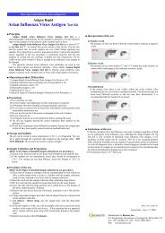
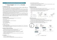
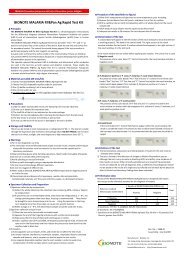
One step avian influenza virus type A and subtype<strong>H5</strong> antigen test<br />
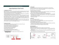
<strong>Anigen</strong> <strong>Rapid</strong> <strong>AIV</strong> <strong>Ag</strong>/<strong>H5</strong> <strong>AIV</strong> <strong>Ag</strong> <strong>Test</strong> <strong>Kit</strong><br />
■ Principles<br />
<strong>Anigen</strong> <strong>Rapid</strong> <strong>AIV</strong> <strong>Ag</strong>/<strong>H5</strong> <strong>AIV</strong> <strong>Ag</strong> <strong>Test</strong> <strong>Kit</strong> is a chromatographic<br />
immunoassay for the qualitative detection of avian influenza type A virus<br />
antigen and subtype <strong>H5</strong> in avian cloaca, trachea, kidney or feces.<br />
The <strong>Anigen</strong> <strong>Rapid</strong> <strong>AIV</strong> <strong>Ag</strong>/<strong>H5</strong> <strong>AIV</strong> <strong>Ag</strong> <strong>Test</strong> <strong>Kit</strong> has a letter of “T” as<br />
test line and “C” as control line on the surface of the device. The test line and<br />
control line in result window are not visible before applying any samples. The<br />
control line is used for procedural control. Control line should be always<br />
appeared if the test procedure is performed properly and the test reagents of<br />
control line are working. A purple test line respectively will be visible in the<br />
result window if there is enough avian influenza virus antigen in the sample.<br />
A monoclonal anti- avian influenza virus common nucleod protein (type<br />
A) and a monoclonal anti- avian influenza virus subtype <strong>H5</strong> used in test line as<br />
capture and detector materials respectively. These enable the <strong>Anigen</strong> <strong>Rapid</strong><br />
<strong>AIV</strong> <strong>Ag</strong>/<strong>H5</strong> <strong>AIV</strong> <strong>Ag</strong> <strong>Test</strong> <strong>Kit</strong> to identify avian influenza virus type A antigen<br />
and subtype <strong>H5</strong> in avian cloaca, trachea, kidney or feces with a high degree of<br />
accuracy<br />
■ Materials provided (10<strong>Test</strong>s/<strong>Kit</strong>)<br />
1) <strong>Anigen</strong> <strong>Rapid</strong> <strong>AIV</strong> <strong>Ag</strong>/<strong>H5</strong> <strong>AIV</strong> <strong>Ag</strong> <strong>Test</strong> Device x 10EA<br />
2) Sample Collection tubes containing 1ml of assay diluent x 10EA<br />
3) Sample collection swab x 10EA<br />
4) Disposable droppers x 10EA<br />
5) Instruction for use<br />
■ Precautions<br />
1) For veterinary use only.<br />
2) For best results, strict adherence to there instructions is required.<br />
3) All samples should be handled as being potentially infectious.<br />
4) Do not open or remove test kit from their individually sealed pouches until<br />
immediately before their use.<br />
5) Do not use the test kit if the pouch is damaged or the seal is broken.<br />
6) Do not reuse test kit.<br />
7) All reagents must be at room temperature before running the assay.<br />
8) Do not use reagents beyond the stated expiration date marked on the<br />
package label.<br />
9) Do not mix components from different lot numbers because the<br />
components in this kit have been quality control tested as standard batch<br />
unit.<br />
■ Storage and Stability<br />
The kit can be stored at room temperature (2~30 ℃ ) or refrigerated. The test<br />
kit is stable through the expiration date marked on the package label. DO<br />
NOT FREEZE. Do not store the test kit in direct sunlight.<br />
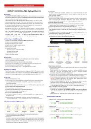
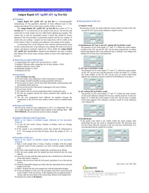
■ Interpretation of the test<br />
1) Negative result<br />
The presence of only one band within the result window on both of the <strong>AIV</strong><br />
<strong>Ag</strong> and <strong>H5</strong> <strong>AIV</strong> <strong>Ag</strong> test areas indicates a negative result.<br />
2) Simultaneous <strong>AIV</strong> type A and <strong>AIV</strong> subtype <strong>H5</strong> <strong>Ag</strong> Positive result<br />
The presence of two color bands (“T” and “C”) within the result window<br />
on both of the <strong>AIV</strong> <strong>Ag</strong> and <strong>H5</strong> <strong>AIV</strong> <strong>Ag</strong> test areas respectively, no matter<br />
which band appears first avian influenza type A subtype virus antigen and<br />
subtype <strong>H5</strong> in avian feces simultaneously.<br />
3) <strong>AIV</strong> type A <strong>Ag</strong> Positive result<br />
The presence of two color bands (“T” and “C”) within the result window<br />
on the <strong>AIV</strong> <strong>Ag</strong> test area, and the presence of only one band (“C”) within<br />
the result window on the <strong>H5</strong> <strong>AIV</strong> <strong>Ag</strong> test area, no matter which band<br />
appears first, indicates a positive result of Avian influenza virus type A.<br />
4) <strong>AIV</strong> subtype <strong>H5</strong> <strong>Ag</strong> Positive result<br />
The presence of two color bands (“T” and “C”) within the result window<br />
on the avian influenza subtype <strong>H5</strong> test area, and the presence of only one<br />
band (“C”) within the result window on the <strong>AIV</strong> <strong>Ag</strong> test area, no matter<br />
which band appears first, indicates a positive result of Avian influenza<br />
virus subtype <strong>H5</strong>.<br />
■ Sample Collection and Preparation<br />
[Refer to the Figure of detailed sample collection & test procedure<br />
enclosed]<br />
1) To get the best result, Cloaca, Trachea or Kidney swab are strongly<br />
recommended.<br />
2) If the sample is not immediately tested, they should be refrigerated at<br />
2~8 ℃ . For storage not less than 48 hours, freeze the sample at -20℃<br />
or<br />
below.<br />
■ Procedure of the test<br />
[Refer to the Figure of detailed sample collection & test procedure<br />
enclosed]<br />
1) Take a swab sample from a Cloaca, Trachea or Kidney with the sample<br />
collection swab. Or take scattered a wet feces with the sample collection<br />
swab.<br />
2) Insert the swab sample into the sample tube containing assay diluent.<br />
3) Mix the swab sample until the sample has been dissolved into the assay<br />
diluent.<br />
4) Leave the test tube until the large particles have settled down to the bottom<br />
of the tube (about 1 minute).<br />
5) Remove the test device from the foil pouch, and places it on a flat and dry<br />
surface.<br />
6) Using the disposable dropper provided, take the supernatant from extracted<br />
sample in the tube.<br />
7) Add 4 ~ 5 drops into the each sample hole with the disposable dropper.<br />
8) As the test begins to work, you will see purple color move across the each<br />
result window of the test device. If the migration has not appeared after 1<br />
minute, add one more drop of the mixed assay diluent to the sample hole.<br />
9) Interpret test results at 20 minutes.<br />
5) Invalid Result<br />
If the purple color band is not visible within the result window after<br />
performing the test, the result is considered invalid. The directions may<br />
not have been followed correctly or the test may have deteriorated. It is<br />
recommended that the sample be re-tested.<br />
Manufactured by BioNote, Inc.<br />
2-9 Seogu-dong, Hwaseong-si, Gyeonggi-do, Korea 445-170<br />
Tel.: +82 31 211 0516, Fax.: +82 31 8003 0618<br />
http://www.bionote.co.kr<br />
Date issued : June. 29th, 2008
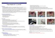
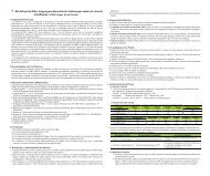
<strong>Anigen</strong> <strong>Rapid</strong> <strong>AIV</strong> <strong>Ag</strong>/<strong>H5</strong> <strong>AIV</strong> <strong>Ag</strong> <strong>Test</strong> <strong>Kit</strong><br />
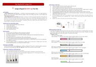
[Figure of Detailed Sample Collection & <strong>Test</strong> Procedure]<br />
A. Sample Collection Methods<br />
1. Cloaca feces swab method<br />
1) Hold the inside of both wings with one hand.<br />
2) Pull out the sample collection swab from the kidney.<br />
2) Hold the tail part to the upper direction with the other hand.<br />
3) Insert the sample collection swab to the cloaca of rectum.<br />
4) Smear the inside of the cloaca several times with sample<br />
collection swab.<br />
3) The outside of the sample collection swab may be seen a red.<br />
4. Scattered wet feces swab method<br />
1) Insert the sample collection swab to the feces.<br />
2) <strong>Ag</strong>itate the sample collection swab at the inside of feces.<br />
3) Pull out the sample collection swab.<br />
5) Pull out the sample collection swab.<br />
6) The outside of sample collection swab may be seen a gray mucoid<br />
or dark brown color.<br />
2. Trachea swab method<br />
1) Incise the trachea by the scissors.<br />
2) Insert the sample collection swab into the incised trachea and<br />
smear the inside of the trachea several times.<br />
[Cautions]<br />
1. Do not swab the feces too much. It may hinder the migration of<br />
buffer on the kit.<br />
2. Do not swab the feces too small. It may show negative reaction<br />
even though <strong>AIV</strong> infected feces.<br />
B. <strong>Test</strong> Procedures<br />
1) Insert the swab into the sample tube<br />
containing assay diluent and mix the<br />
swab until the sample has been dissolved<br />
into the assay diluent.<br />
3) Pull out the sample collection swab from the trachea.<br />
2) Using the disposable dropper provided,<br />
take the supernatant from extracted<br />
sample in the tube.<br />
3) Add 4(four) ~ 5(five) drops into the<br />
sample hole with disposable dropper.<br />
4) The outside of the sample collection swab may be seen a gray<br />
mucoid or dark brown color.<br />
4) Interpret test results at 20 minutes.<br />
3. Kidney swab method<br />
1) Find the kidney in poultry and insert the sample collection swab<br />
into the kidney and smear the inside of the kidney several times.