Case Study: A 44-Year-Old Woman With Type 1 ... - Clinical Diabetes
Case Study: A 44-Year-Old Woman With Type 1 ... - Clinical Diabetes
Case Study: A 44-Year-Old Woman With Type 1 ... - Clinical Diabetes

You also want an ePaper? Increase the reach of your titles
YUMPU automatically turns print PDFs into web optimized ePapers that Google loves.
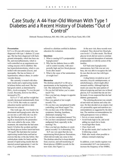
C A S E S T U D I E S<br />
<strong>Case</strong> <strong>Study</strong>: A <strong>44</strong>-<strong>Year</strong>-<strong>Old</strong> <strong>Woman</strong> <strong>With</strong> <strong>Type</strong> 1<br />
<strong>Diabetes</strong> and a Recent History of <strong>Diabetes</strong> “Out of<br />
Control”<br />
Deborah Thomas-Dobersen, RD, MS, CDE, and Terri Ryan-Turek, RD, CDE<br />
Presentation<br />
K.P. is a <strong>44</strong>-year-old woman who was<br />
diagnosed with type 1 diabetes 22 years<br />
ago. She has been diagnosed with background<br />
retinopathy, which has been stable,<br />
and microalbuminuria, which is<br />
well-controlled on an angiotensin-converting<br />
enzyme (ACE) inhibitor. She<br />
has hypercholesterolemia, which is controlled<br />
with simvastatin (Zocor), and<br />
neuropathy. She has no history of<br />
hypertension, tobacco abuse, or cardiovascular<br />
disease.<br />
She currently is treated with ultralente<br />
insulin and a rapid-acting insulin<br />
in boluses before each meal. She has<br />
had good control, as determined by<br />
HbA 1c levels averaging 7% over the past<br />
2 years. Recently, however her HbA 1c<br />
increased to 9%.<br />
K.P. is 5' 3" tall, and her weight has<br />
recently decreased unintentionally from<br />
115 to 110 lb. She works as a special<br />
education teacher and drives daily<br />
between two schools.<br />
One week ago, she was found to be<br />
disoriented and hypoglycemic after her<br />
car ran into a curb while she was driving.<br />
Her blood glucose level was 26<br />
mg/dl as measured by the paramedics<br />
who were called to the scene. After<br />
treatment, she stated that she had eaten<br />
lunch 2 h before the accident and that<br />
her blood glucose before lunch had been<br />
130 mg/dl. She had taken her normal<br />
rapid-acting insulin bolus and had no<br />
exercise that day or the day before. Her<br />
carbohydrate intake for lunch and the<br />
amount of rapid-acting insulin that she<br />
injected seemed appropriate (1 unit for<br />
every 15 g carbohydrate). She was<br />
referred to a dietitian certified in diabetes<br />
education for evaluation.<br />
Questions<br />
1. What is causing K.P.’s postprandial<br />
hypoglycemia?<br />
2. Why has her diabetes been so difficult<br />
to control recently, with unexpectedly<br />
high and low blood glucose<br />
levels after meals?<br />
3. What is the cause of her unintentional<br />
weight loss?<br />
Discussion<br />
The dietitian asked K.P. to fill out a<br />
questionnaire at the beginning of her<br />
visit. She indicated the following:<br />
• Do you feel full before you’ve eaten<br />
much? Yes.<br />
• Have you had any changes in appetite<br />
recently? Yes.<br />
• Have you gained or lost weight<br />
recently? Yes.<br />
• Do you have any unexplained trouble<br />
controlling your blood sugars? Yes.<br />
• Do you have any of the following<br />
symptoms: bloating, heartburn,<br />
abdominal cramping? Yes.<br />
• Do you have unexplained nausea? Yes.<br />
• Do you have unexplained vomiting of<br />
undigested food, especially in the<br />
morning? No.<br />
• Have you had any diarrhea or constipation<br />
recently? Is it alternating? No.<br />
K.P. was very upset that she was<br />
experiencing blood glucose fluctuations,<br />
which did not make any sense to her.<br />
The dietitian asked her to keep detailed<br />
food, exercise, and self-monitoring of<br />
blood glucose (SMBG) records for the<br />
next week. (See Figure 1.)<br />
At the next visit, these records were<br />
evaluated. They showed low blood glucose<br />
levels 1–2 h after meals. The blood<br />
glucose levels did not seem to correlate<br />
with the expected absorption of nutrients<br />
postprandially or with the action of the<br />
insulins.<br />
K.P. had some hypoglycemia<br />
unawareness, but if she was not very<br />
busy, she was able to recognize the subtle<br />
cues that she now has with hypoglycemia.<br />
A drug history revealed no use of<br />
narcotics, tricyclic antidepressants, or<br />
anticholinergics, which might affect<br />
stomach motility. Although high-fat<br />
meals can cause the same pattern of<br />
delayed emptying and later rise in blood<br />
glucose, K.P. recorded that she ate lowfat<br />
meals. It was suspected that she had<br />
diabetic gastroparesis.<br />
The referring physician was consulted<br />
and ruled out anemia and celiac disease.<br />
He then decided on an empiric trial<br />
of a promotility agent and nutrition recommendations<br />
as suggested by the dietitian.<br />
The nutrition recommendations for<br />
mild gastroparesis were as follows:<br />
• Eat six or more meals/day.<br />
• Avoid foods high in fat, and avoid<br />
adding too much fat to foods.<br />
• Avoid high-fiber foods.<br />
• Chew food well.<br />
• Sit up after meals; don’t recline for 1<br />
h following a meal.<br />
• Walk after meals when possible to<br />
enhance stomach emptying. 1<br />
In addition, a new insulin regimen was<br />
recommended. K.P. was instructed to<br />
increase the frequency of her SMBG,<br />
178<br />
Volume 19, Number 4, 2001 • CLINICAL DIABETES<br />
Downloaded from http://clinical.diabetesjournals.org/ by guest on January 25, 2014
C A S E S T U D I E S<br />
decrease her ultralente dose by 10%<br />
before bed, and give a divided rapid-acting<br />
insulin bolus—half immediately<br />
after the meal and half 2 h postprandially.<br />
The rapid-acting insulin bolus was<br />
calculated for the grams of carbohydrate<br />
in each meal, with any correction factors<br />
for high blood glucose added. Figure 2<br />
shows K.P.’s post-treatment SMBG<br />
records.<br />
Many patients with diabetic gastroparesis<br />
are asymptomatic or have vague<br />
symptoms and therefore go undiagnosed<br />
or undertreated. Gastroparesis develops<br />
in 40–50% of patients with longstanding<br />
type 1 diabetes and in 30–40% of those<br />
with longstanding type 2 diabetes. 2 In<br />
addition to the duration of diabetes, the<br />
degree of chronic or acute hyperglycemia<br />
seems to be associated with<br />
more severe gastrointestinal problems. 3<br />
Patients with a history of retinopathy,<br />
nephropathy, or neuropathy should be<br />
presumed to have gastrointestinal abnormalities<br />
until proven otherwise. 2<br />
Diabetic gastroparesis results in<br />
delayed stomach emptying, leading to<br />
retention of stomach contents. Other<br />
abnormalities include gastric dysrhythmia,<br />
abnormality of fundic relaxation, and<br />
antral hypomotility. Symptoms include<br />
bloating, early satiety, abdominal pain,<br />
nausea, or vomiting. Delayed stomach<br />
emptying may lead to gastroesophageal<br />
reflux, with symptoms of heartburn and<br />
vomiting of undigested food.<br />
Because gastroparesis makes stomach<br />
emptying unpredictable, blood glucose<br />
levels may be erratic and difficult to<br />
control (Figure 3). Because this condi-<br />
tion changes the timing of nutrient<br />
absorption, the typical diabetes therapy<br />
of matching carbohydrate absorption to<br />
insulin action is thrown off, with resultant<br />
wide swings in blood glucose levels.<br />
Diabetic gastroparesis also puts patients<br />
at risk for compromised nutritional status<br />
because of dietary inadequacy. 4<br />
Although there is a lack of evidencebased<br />
nutrition interventions for gastroparesis<br />
in diabetic patients, the following<br />
nutritional guidelines may be effective: 5<br />
1. Early satiety is one of the hallmarks<br />
of gastroparesis. Because larger volume<br />
of foods slow gastric emptying,<br />
smaller more frequent meals may<br />
help.<br />
2. Liquids usually empty from the<br />
stomach more easily and rapidly than<br />
solids. Solids require normal func-<br />
Figure 1. K.P.’s pre-treatment SMBG records. This pattern shows a typical elevated fasting blood glucose level. Two h postprandially,<br />
when nutrient absorption is at its peak (assuming a low-fat meal), the blood glucose drops considerably more than<br />
the 30–50 mg/dl expected, and yet rises to high levels before the next meal with no additional food intake.<br />
Figure 2. K.P.’s post-treatment SMBG records. This pattern shows a more normalized blood glucose excursion achieved by 1)<br />
decreasing ultralente and thus decreasing rebound (one cause of high fasting blood glucose) resulting from low blood glucose<br />
levels throughout night, and 2) giving split rapid-acting insulin boluses, one after the meal and another 2 h postprandially.<br />
CLINICAL DIABETES • Volume 19, Number<br />
Downloaded<br />
4, 2001<br />
from http://clinical.diabetesjournals.org/ by guest on January 25, 2014<br />
179
C A S E S T U D I E S<br />
gastroparesis in some patients.<br />
Optimal glycemic control is imperative<br />
for maximum utilization of nutritional<br />
intervention.<br />
• Medical nutrition therapy, patient education,<br />
and a change in insulin therapy<br />
may give relief.<br />
• Because of the nature of the subject<br />
matter discussed (nutrient absorption<br />
patterns), dietitians are often the member<br />
of the diabetes care team to first<br />
suspect a diagnosis of gastroparesis.<br />
Dietitians often have the opportunity to<br />
discover the presence of gastroparesis<br />
in their patients and can be proactive<br />
regarding treatment recommendations.<br />
Figure 3. K.P.’s continuous glucose monitoring system (CGMS) tracing. K.P. wore<br />
a CGMS monitor for the pre-treatment day shown in Figure 1. The monitor<br />
records 288 readings per day of interstitial glucose, which closely corresponds to<br />
blood glucose. K.P.’s delayed gastric emptying from gastroparesis wreaked havoc<br />
on her blood glucose levels. The symbols at the bottom of the figure (triangle and<br />
delta) represent meal and insulin bolus.<br />
tioning of the antrum and fundus for<br />
churning, mixing, and exiting.<br />
Because patients often report<br />
increased fullness as the day proceeds,<br />
consuming mostly liquids<br />
toward the end of the day may be<br />
considered.<br />
3. Fiber, especially pectin, is known to<br />
slow stomach emptying. If bezoar<br />
formation is a concern, patients<br />
should avoid oranges, persimmons,<br />
coconuts, berries, green beans, figs,<br />
apples, sauerkraut, brussel sprouts,<br />
potato peels, and legumes.<br />
4. Fatty foods or foods with a significant<br />
amount of fat added to them exit<br />
the stomach more slowly and may be<br />
poorly tolerated. However, many<br />
patients tolerate fat in liquid form,<br />
such as milkshakes, whole milk, and<br />
nutritional supplements.<br />
Other methods of insulin treatment may<br />
be used to help with gastroparesis. Some<br />
patients benefit from taking a bolus of<br />
half rapid-acting and half regular insulin<br />
after meals. For patients on an insulin<br />
pump, a dual wave may be used to more<br />
closely approximate the rise in blood<br />
glucose seen with delayed absorption.<br />
For patients who use insulin injections,<br />
the rapid-acting insulin or regular insulin<br />
given with a meal may be divided, with<br />
half given as the patient eats and half<br />
given 1–2 h postprandially.<br />
<strong>Clinical</strong> Pearls<br />
• Gastroparesis develops in 40–50% of<br />
patients with longstanding type 1 diabetes<br />
and 30–40% of those with longstanding<br />
type 2 diabetes.<br />
• Symptoms are often vague and may<br />
not be expressed at the clinic visit.<br />
Early satiety is one of the hallmarks<br />
of gastroparesis. Symptoms such as<br />
appetite changes and fluctuations in<br />
blood glucose are common.<br />
• Hyperglycemia can cause transient<br />
REFERENCES<br />
1 Lipp RW, Schnedl WJ, Hammer HF: Does<br />
postprandial walking influence delayed gastric<br />
emptying in patients with longstanding IDDM?<br />
<strong>Diabetes</strong> Care 19:1306–1310, 1996<br />
2 Merio R, Festa A, Bergman H, Eder, T, Eibl<br />
N, Stacher-Janotta G, Weber U, Budka C, Heckenberg<br />
A, Bauer P, Francesconi M, Schernthaner<br />
G, and Stacher G: Slow gastric emptying in type<br />
1 diabetes: relation to autonomic and peripheral<br />
neuropathy, blood glucose, and glycemic control.<br />
<strong>Diabetes</strong> Care 20:419–423, 1997<br />
3 Wolosin JD, Edelman SV: <strong>Diabetes</strong> and the<br />
gastrointestinal tract. <strong>Clinical</strong> <strong>Diabetes</strong><br />
18:148–151, 2000<br />
4 Goldberg KB, Lasichak A, Rock CL: Dietary<br />
adequacy in patients with diabetic gastroparesis. J<br />
Am Diet Assoc 97:420–422, 1997<br />
5 Parrish CR: Gastrointestinal issues in persons<br />
with diabetes. In Handbook of <strong>Diabetes</strong><br />
Medical Nutrition Therapy. 2nd ed. Powers M,<br />
ed. Gaithersburg, Md., Aspen Publishers, 1996, p.<br />
618–637<br />
Deborah Thomas-Dobersen, RD, MS,<br />
CDE, is a research coordinator and Terri<br />
Ryan-Turek, RD,CDE, is a clinical<br />
faculty member in the Endocrinology<br />
Department of University of Colorado<br />
Health Sciences Center in Denver.<br />
Note of disclosure: Ms. Thomas-<br />
Dobersen has received research funding<br />
from MiniMed, Inc., which manufactures<br />
and markets a continuous glucose monitoring<br />
system.<br />
180<br />
Volume 19, Number 4, 2001 •<br />
Downloaded from http://clinical.diabetesjournals.org/ by guest on January 25, 2014<br />
CLINICAL DIABETES
C A S E S T U D I E S<br />
<strong>Case</strong> <strong>Study</strong>: A 43-<strong>Year</strong>-<strong>Old</strong> Man <strong>With</strong> Perineal Pain<br />
and Swelling<br />
David J. Meier, MD<br />
Presentation<br />
J.T. is a 43-year-old man who presented<br />
to his primary care doctor after 4 days<br />
of progressive pain and swelling in his<br />
genital region, along with a low-grade<br />
fever. There was no history of similar<br />
symptoms. He denied recent trauma. He<br />
had no nausea, vomiting, diarrhea, constipation,<br />
abdominal pain, melena, or<br />
hematochezia. He also denied dysuria,<br />
urgency, and frequency. He was in a<br />
monogamous relationship and had no<br />
history of sexually transmitted diseases.<br />
Notably, he reported a 110-lb weight<br />
loss over 6 years.<br />
His medical history was positive<br />
only for a urethral stricture treated with a<br />
urethral dilation on cystoscopy in 1965.<br />
He had no allergies and was taking no<br />
medications. He did not smoke or drink<br />
alcohol. His family history was significant<br />
for his grandmother with type 2 diabetes.<br />
Physical examination showed a<br />
mildly ill-appearing man in moderate<br />
distress. He was awake and alert. His<br />
vital signs included a blood pressure of<br />
115/72 mmHg, heart rate of 110, respiratory<br />
rate of 13, and temperature of 99.7<br />
degrees. He was 5'9" and weighed 140<br />
lb. Head, eyes, ears, nose, and throat<br />
were normal. Lungs were clear. Heart<br />
was tachycardic with a normal s1 and s2<br />
with no murmurs or gallops and no rub.<br />
Abdomen was nontender and nondistended<br />
with normal bowel sounds. His<br />
extremities had no edema, and pulses<br />
were normal bilaterally.<br />
J.T.’s genital exam revealed diffuse<br />
erythema and edema of his scrotum and<br />
perineal area, along with severe tenderness.<br />
There were multiple areas of hem-<br />
orrhagic necrosis involving a large part<br />
of the scrotum but sparing the penis. His<br />
testicles were normal in size and contour<br />
and were not tender. His perirectal area<br />
was erythematous, but there was no evidence<br />
of fissures, ulcerations, or crepitus.<br />
Laboratory data included a normal<br />
hemoglobin and hematocrit but a<br />
markedly elevated white blood count to<br />
35,000 k/mm 3 . His chem. 7 panel was<br />
also normal except for a mildly<br />
decreased sodium of 132 mEq/l and an<br />
elevated glucose of 284 mg/dl. His<br />
HbA 1c concentration was 12.8% with a<br />
glycosylated hemoglobin of 17.7%.<br />
There were no other laboratory abnormalities.<br />
No other laboratory tests or<br />
imaging procedures were ordered.<br />
Questions<br />
1. What is the diagnosis?<br />
2. What are the potential sources?<br />
3. How is this patient predisposed to<br />
developing this condition?<br />
4. How should this patient be managed?<br />
Commentary<br />
This is a classic presentation of<br />
Fournier’s gangrene, which is defined as<br />
necrotizing cellulitis and fascitis of the<br />
perineum. Originally called “spontaneous<br />
fulminant gangrene of the scrotum,”<br />
Fournier’s gangrene was first<br />
described by Baurienne 1 in 1764 but<br />
named after Jean Alfred Fournier 2 in<br />
1883, after he characterized the disease<br />
as the abrupt onset of fulminating genital<br />
gangrene of idiopathic origin in previously<br />
healthy young patients.<br />
Since then, our understanding of this<br />
disease has changed considerably.<br />
Fournier’s gangrene is now considered to<br />
have a somewhat more indolent course,<br />
to have an identifiable source in<br />
75–100% of cases, to be associated with<br />
systemic disease, and to occur throughout<br />
a broader age range. 3,4 The incidence<br />
is higher in men than in women, with a<br />
ratio of 10:1. 5<br />
Fournier’s gangrene most often<br />
affects patients with systemic disease,<br />
most commonly diabetes or alcoholism.<br />
Studies have shown the prevalence of<br />
diabetes in patients with Fournier’s gangrene<br />
to be 32–60% and the prevalence<br />
of alcoholism to be between 25 and<br />
66%. 3,4 Chronic debilitation and<br />
immunosuppression secondary to transplantation,<br />
chemotherapy, or HIV infection<br />
are also significant risk factors. 3,4<br />
Patients often present with genital<br />
swelling, erythema, and tenderness, frequently<br />
accompanied by fever and systemic<br />
toxicity. Swelling quickly increases,<br />
and crepitus and gangrene develop.<br />
A rectal, dermal, or urinary source is<br />
often found by careful investigation. 3,4<br />
Rectal sources are suggested by pain,<br />
bleeding, and a history of anal fissures or<br />
dilatation, hemorrhoid banding, or perforation.<br />
Dermal sources are suggested by<br />
acute or chronic infections of the scrotum,<br />
recurrent hidradenitis suppurativa,<br />
balanitis, or recent epesiotomy. Urinary<br />
sources are suggested by dysuria, urethral<br />
discharge, recent urinary catheterization,<br />
or obstructed voiding, especially<br />
as a result of urethral strictures from sexually<br />
transmitted diseases. Of all the predisposing<br />
conditions, urethral strictures<br />
and perirectal abscesses are the most<br />
common. 6<br />
In contrast to historical descriptions,<br />
the microbiological etiology can often be<br />
CLINICAL DIABETES • Volume 19, Number 4, 2001<br />
Downloaded from http://clinical.diabetesjournals.org/ by guest on January 25, 2014<br />
181
C A S E S T U D I E S<br />
determined. The lesions are usually foul<br />
smelling, suggesting a polymicrobial<br />
infection with anaerobic-aerobic synergy.<br />
7 Organisms typically isolated include<br />
clostridia, klebsiella, streptococci, coliforms,<br />
staphylococci, bacteroides, and<br />
corynebacteria. 5 Local infection usually<br />
occurs next to the portal of entry. The<br />
inflammatory reaction then spreads to<br />
the deep fascial planes with resultant<br />
obliterative endarteritis. This results in a<br />
cascade of cutaneous and subcutaneous<br />
vascular necrosis, local ischemia and tissue<br />
necrosis, and hypoxia-driven proliferation<br />
of anaerobic organisms. 3<br />
Despite our knowledge of the etiology<br />
of Fournier’s gangrene, the diagnosis<br />
must remain clinical because it represents<br />
a surgical emergency. Often the illness<br />
is too fulminant to await the results<br />
of diagnostic tests. Skill is required to<br />
differentiate Fournier’s gangrene from<br />
scrotal cellulitis, abscesses, strangulated<br />
herniae, pyoderma ganrenosum, and vasculitis.<br />
3 If time permits, an abdominal X-<br />
ray can be used to evaluate for subcutaneous<br />
air in the abdominal wall.<br />
Similarly, a scrotal ultrasound can help<br />
to evaluate for subcutaneous air before<br />
the clinical detection of crepitus and to<br />
rule out other scrotal pathology. 3 Proctoscopy<br />
can be useful to evaluate for the<br />
source of infection and to document rectal<br />
and anal involvement. 4 Likewise, a<br />
retrograde urethrogram can demonstrate<br />
urinary extravasation, thus documenting<br />
the need for suprapubic diversion. 4<br />
Fournier’s gangrene is a surgical<br />
emergency. Treatment consists of<br />
aggressive, prompt surgical debridement,<br />
and intravenous antibiotics and fluids.<br />
Most experts recommend treating with a<br />
penicillin to cover clostridia, an aminoglycoside<br />
or third-generation<br />
cephalosporin for gram negative rods,<br />
and clindamycin (Cleocin) or metronidazole<br />
(Flagyl) for anaerobes. 5<br />
A wide debridement should be per-<br />
formed until normal fascia is found and<br />
repeated debridement is often required. 6<br />
Although occasionally compromised, the<br />
testes are frequently spared because of<br />
their independent blood supply. 3 A<br />
colostomy is performed if there is<br />
colonic or rectal perforation. Recently,<br />
hyperbaric oxygen has been shown to<br />
decrease length of stay, increase wound<br />
healing, and decrease the spread of<br />
infection. 8<br />
Mortality is ~16% 5 and has not<br />
improved much since the original<br />
description of this disease, likely<br />
because of the more complicated patient<br />
population and the increased average age<br />
of patients at diagnosis. 7 Interestingly,<br />
despite being the highest-risk group for<br />
developing Fournier’s gangrene, diabetic<br />
patients are not at increased risk of death<br />
compared to other patients with the disease.<br />
However, mortality is increased<br />
with advanced age, extensive disease,<br />
shock or sepsis, positive blood cultures,<br />
anorectal sources, and renal or hepatic<br />
dysfunction. 4,5,9<br />
J.T. was diagnosed as having type 2<br />
diabetes and Fournier’s gangrene and<br />
was taken to the operating room for<br />
debridement within several hours of<br />
being transferred to the emergency room<br />
from his primary care physician’s office.<br />
His testicles were preserved, and he did<br />
not require a colostomy. He was hospitalized<br />
for 10 days and underwent three<br />
surgical debridements and a flap closure<br />
of his wound. He remained on ampicillin<br />
(Unasyn) until discharge and was then<br />
placed on amoxicillin (Augmentin).<br />
He received diabetes education and<br />
performed self-monitoring of blood glucose<br />
four times a day. He was treated<br />
with insulin. His diabetes was then managed<br />
as an outpatient with no subsequent<br />
readmissions.<br />
<strong>Clinical</strong> Pearls<br />
• Fournier’s gangrene is a rare condition<br />
that requires emergency surgical treatment.<br />
• The clinical presentation can be variable<br />
but may consist of genital<br />
swelling, erythema, and tenderness,<br />
frequently accompanied by fever.<br />
Presentation often occurs before the<br />
development of crepitus. The disease<br />
may be indolent with progression over<br />
several days.<br />
• Suspect a systemic disease such as<br />
diabetes, alcoholism, or HIV infection<br />
in a patient diagnosed with Fournier’s<br />
gangrene.<br />
• Search for the portal of entry, which<br />
typically includes the dermis, rectum,<br />
or urethra.<br />
• Mortality is still almost 20% despite<br />
advances in modern medicine.<br />
REFERENCES<br />
1 Baurienne H: Sur une plaie contuse qui s’est<br />
terminee par le sphacele de la scrotum. J Med<br />
Chir Pharm 20:251–256, 1764<br />
2 Fournier JA: Gangrene foudroyante de la<br />
verge. Sem Med 3:345, 1883<br />
3 Smith GL, Bunker CB, Dinneen MD:<br />
Fournier’s gangrene. Br J Urol 81:347–355, 1998<br />
4 Vick R, Carson CC III: Infections in urology:<br />
Fournier’s disease. Urol Clin North Am<br />
26:841–849, 1999<br />
5 Eke N: Fournier’s gangrene: a review of<br />
1726 cases. Br J Surgery 87:718–728, 2000<br />
6 Corman JM, Moody JA, Aronson WJ:<br />
Fournier’s gangrene in a modern surgical setting:<br />
improved survival with aggressive management.<br />
BJU Interna 84:85–88, 1999<br />
7 Yaghan RJ, Al-Jaberi TM, Bani-Hani I:<br />
Fournier’s gangrene: changing face of the disease.<br />
Dis Colon Rectum 43:1300–1308, 2000<br />
8 Hollabaugh RS, Dmochowski RR, Hickerson<br />
WL: Fournier’s gangrene: therapeutic impact of<br />
hyperbaric oxygen. Plast Reconstr Surg<br />
101:94–100, 1998<br />
9 Olsofka JN, Carrillo EH, Spain DA, Polk<br />
HC: The continuing challenge of Fournier’s gangrene<br />
in the 1990s. Am Surgeon 65:1156–1159,<br />
1999<br />
David J. Meier, MD, is a house officer in<br />
the Department of Internal Medicine at<br />
the University of Michigan in Ann Arbor.<br />
182<br />
Volume 19, Number 4, 2001 •<br />
Downloaded from http://clinical.diabetesjournals.org/ by guest on January 25, 2014<br />
CLINICAL DIABETES
C A S E S T U D I E S<br />
<strong>Case</strong> <strong>Study</strong>: Hypertriglyceridemia in a <strong>Woman</strong> <strong>With</strong><br />
Insulin Resistance<br />
Liza L. Ilag, MD<br />
Presentation<br />
B.L. is a 24-year-old woman with a history<br />
of dermatomyositis diagnosed at<br />
age 3. She received treatment with<br />
cyclophosphamide (Cytoxan), methotrexate,<br />
and prednisone until she was 11<br />
years old with successful remission.<br />
When she was 16 years old, she presented<br />
with lipoatrophic facial features,<br />
hirsutism, amenorrhea, and acanthosis<br />
nigricans in the axillary and groin areas.<br />
She was lean and muscular but not virilized.<br />
An oral glucose tolerance test<br />
showed fasting glucose of 159 mg/dl and<br />
insulin of 538 u/ml (normal
C A S E S T U D I E S<br />
expression in adipose tissue. Troglitazone<br />
(Rezulin) and pioglitazone (Actos)<br />
have been shown to decrease triglyceride<br />
levels, and all of the drugs in this class<br />
appear to increase HDL levels. An<br />
increase in LDL levels has been reported,<br />
but changes in the size of the LDL<br />
cholesterol particles may make the cholesterol<br />
less susceptible to oxidation.<br />
B.L. was initially treated with troglitazone<br />
for her insulin resistance syndrome.<br />
Before treatment, her HbA 1c was<br />
5.8%, total cholesterol was 249 mg/dl,<br />
triglycerides were 1,563 mg/dl, and<br />
HDL was 17 mg/dl. <strong>With</strong> troglitazone,<br />
B.L.’s insulin levels fell, but her triglyceride<br />
levels remained high. When troglitazone<br />
was withdrawn from the market,<br />
she was switched to pioglitazone.<br />
For triglycerides that remain ≥400<br />
mg/dl (and LDL 2,000 mg/dl can precipitate<br />
pancreatitis.<br />
• Diet and improved glycemic control<br />
can often ameliorate moderate hypertriglyceridemia.<br />
• Fibrates and nicotinic acid are potent<br />
triglyceride-lowering medications that<br />
may be needed to effectively manage<br />
severe hypertriglyceridemia. Used with<br />
caution, they can help in the management<br />
of hypertriglyceridemia in diabetic<br />
patients.<br />
• Fish oil is an option to lower triglyceride<br />
levels in resistant hypertriglyceridemia<br />
inadequately controlled by<br />
diet and medications.<br />
SUGGESTED READINGS<br />
American <strong>Diabetes</strong> Association: Management<br />
of dyslipidemia in adults with diabetes (Position<br />
Statement). <strong>Diabetes</strong> Care 24 (Suppl.<br />
1):S58–S61, 2001<br />
Ballantyne CM, Grundy SM, Oberman A,<br />
Kreisberg RA, Havel RJ, Frost PH, Haffner SA:<br />
Hyperlipidemia: diagnostic and therapeutic perspectives.<br />
J Clin Endocrinol Metab<br />
85:2089–2112, 2000<br />
Connor WE, DeFrancesco CA, Connor SL:<br />
-3 fatty acids from fish oil: effects on plasma<br />
lipoproteins and hypertriglyceridemic patients.<br />
Ann NY Acad Sci 683:16–34, 1993<br />
Elam MB, Hunninghake DB, Davis KB, Garg<br />
R, Johnson C, Egan D, Kostis JB, Sheps DS,<br />
Brinton EA. Effect of niacin on lipid and lipoprotein<br />
levels and glycemic control in patients with<br />
diabetes and peripheral arterial disease: the<br />
ADMIT study: a randomized trial. Arterial Disease<br />
Multiple Intervention Trial. JAMA<br />
284:1263–1270, 2000<br />
Harris WS: Nonpharmacologic treatment of<br />
hypertriglyceridemia: focus on fish oils. Clin<br />
Cardiol 22 (Suppl. II):II-40–II-43, 1999<br />
Miller M: Current perspectives on the management<br />
of hypertriglyceridemia. Am Heart J<br />
140:232–240, 2000<br />
Liza L. Ilag, MD, is a lecturer and<br />
research fellow in the Division of<br />
Endocrinology and Metabolism at the<br />
University of Michigan in Ann Arbor.<br />
184<br />
Volume 19, Number 4, 2001 •<br />
Downloaded from http://clinical.diabetesjournals.org/ by guest on January 25, 2014<br />
CLINICAL DIABETES