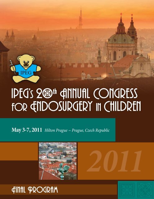
May 3-7, 2011 Hilton Prague ~ Prague, Czech Republic
May 3-7, 2011 Hilton Prague ~ Prague, Czech Republic
May 3-7, 2011 Hilton Prague ~ Prague, Czech Republic

Create successful ePaper yourself
Turn your PDF publications into a flip-book with our unique Google optimized e-Paper software.
<strong>May</strong> 3-7, <strong>2011</strong> <strong>Hilton</strong> <strong>Prague</strong> ~ <strong>Prague</strong>, <strong>Czech</strong> <strong>Republic</strong><br />
<strong>2011</strong>
Where?<br />
Who?<br />
<strong>Hilton</strong> <strong>Prague</strong> Hotel<br />
Pobrezni 1, <strong>Prague</strong> ~ <strong>Czech</strong> <strong>Republic</strong> 186 00<br />
Phone: +420.224.841.111 Fax: +420.224.842.378<br />
International Pediatric Endosurgery Group (IPEG)<br />
11300 W. Olympic Blvd., Suite 600 ~ Los Angeles, CA 90064<br />
Phone: +1.310.437.0553 Fax: +1.310.437.0585 Email: registration@ipeg.org<br />
COPYRIGHT<br />
The program and talks presented at the <strong>2011</strong> IPEG meeting are copyrighted products of the International Pediatric Endoscopic Group. Any reproduction<br />
or rebroadcast with out express written consent of IPEG is prohibited.<br />
CME & EVALUATION FORMS<br />
IPEG Registrants, please complete the IPEG CME & Evaluation form included in your registration bag and return to the IPEG Registration desk to have<br />
your CME certificate mailed to you after the meeting.<br />
Please allow 4-6 weeks for processing of all CME requests.<br />
IPEG’s 20th Annual Congress for Endosurgery in Children ~ <strong>May</strong> 3-7, <strong>2011</strong> ~ www.ipeg.org 1
Table of Contents<br />
General Information 2-3<br />
Program Chairs 4<br />
Meeting Leaders & Faculty 5<br />
Program Committee<br />
Executive Committee<br />
Past Presidents<br />
Faculty<br />
Schedule-at-a-Glance 6<br />
Complete Schedule 7-17<br />
Wednesday, <strong>May</strong> 4 7<br />
Thursday, <strong>May</strong> 5 8<br />
Friday, <strong>May</strong> 6 13<br />
Saturday, <strong>May</strong> 7 16<br />
CME Worksheet 18<br />
Faculty & Presenters Disclosures 19<br />
Long Term Research Fund Donors 20-21<br />
<strong>Prague</strong> Tours & Activities 22<br />
Abstracts 23-96<br />
Oral Abstracts 23<br />
Video Abstracts 47<br />
Poster List 51<br />
Poster Abstracts 57<br />
Commercial Bias Reporting Form 97<br />
<strong>2011</strong> Corporate Supporters<br />
Diamond Level<br />
Ethicon Endo-Surgery<br />
Karl Storz Endoscopy<br />
Stryker Endoscopy<br />
Exhibit Hall Hours (Chez Louis Meeting Room)<br />
Wednesday, <strong>May</strong> 4, <strong>2011</strong><br />
Welcome Reception in Exhibit Hall<br />
Thursday, <strong>May</strong> 5, <strong>2011</strong><br />
Hall Open<br />
Friday, <strong>May</strong> 6, <strong>2011</strong><br />
Hall Open<br />
6:00 pm – 7:30 pm<br />
9:30 am – 3:30 pm<br />
9:30 am – 3:30 pm<br />
Poster Hours (Palmovka – Tyrolka Meeting Room)<br />
Thursday, <strong>May</strong> 5, <strong>2011</strong> <br />
9:30 am – 3:30 pm<br />
Poster Tours <br />
12:30 pm – 1:30 pm<br />
Friday, <strong>May</strong> 6, <strong>2011</strong><br />
9:30 am – 3:30 pm<br />
Poster Tours <br />
12:30 pm – 1:30 pm<br />
Registration Hours (Grand Ballroom Foyer)<br />
Tuesday, <strong>May</strong> 3, <strong>2011</strong><br />
10:00 am – 6:00 pm<br />
Wednesday, <strong>May</strong> 4, <strong>2011</strong><br />
6:00 am – 6:00 pm<br />
Thursday, <strong>May</strong> 5, <strong>2011</strong><br />
6:00 am – 6:00 pm<br />
Friday, <strong>May</strong> 6, <strong>2011</strong><br />
6:00 am – 6:00 pm<br />
Saturday, <strong>May</strong> 7, <strong>2011</strong><br />
7:00 am – 12:00 pm<br />
SPEAKER READY ROOM (Troja Meeting Room)<br />
Tuesday, <strong>May</strong> 3, <strong>2011</strong><br />
10:00 am – 6:00 pm<br />
Wednesday, <strong>May</strong> 4, <strong>2011</strong><br />
6:00 am – 6:00 pm<br />
Thursday, <strong>May</strong> 5, <strong>2011</strong><br />
6:00 am – 6:00 pm<br />
Friday, <strong>May</strong> 6, <strong>2011</strong><br />
6:00 am – 6:00 pm<br />
Saturday, <strong>May</strong> 7, <strong>2011</strong><br />
7:00 am – 12:00 pm<br />
2<br />
IPEG’s 20th Annual Congress for Endosurgery in Children ~ <strong>May</strong> 3-7, <strong>2011</strong> ~ www.ipeg.org
Why IPEG?<br />
Now is an excellent time to become an IPEG member! Join IPEG now<br />
and receive a substantial discount on the meeting registration by being<br />
an IPEG member! Your dues also include a subscription to the Journal of<br />
Laparoendoscopic & Advanced Surgical Techniques and Part B: Videoscopy<br />
(a $1200 value is yours for Free with your paid IPEG membership).<br />
Who Should Attend?<br />
The 20th Annual Congress of the International Pediatric Endosurgery<br />
Group (IPEG) has elements that have been specifically designed to meet<br />
the needs of practicing pediatric surgeons, urologists, and other related<br />
specialties, physicians-in-training, GI assistants, and nurses who are<br />
interested in minimally invasive surgery in children and adolescents. The<br />
IPEG Program Committee recommends that participants design their own<br />
attendance schedule based on their own personal educational objectives.<br />
<strong>2011</strong> Meeting Objectives<br />
The objectives of the activity are to educate, expose and allow pediatric<br />
surgeons and urologist the opportunity to discuss the developing<br />
techniques and management principles regarding minimally invasive<br />
surgical techniques and scientific developments that will affect their<br />
patient population.<br />
Specific Objectives include:<br />
• Presentation of new and developing minimally invasive surgical<br />
techniques in a scientific environment.<br />
• Opportunity to interact with experts in the fields of minimally<br />
invasive pediatric surgery and urology via panel interactions and<br />
audience response systems.<br />
• Discussion of current and future controversial issues regarding<br />
minimally invasive surgery in infants and children.<br />
• Advance the use of minimally invasive surgical procedures in<br />
infants and children.<br />
• Encourage international interactions in the management and<br />
minimally invasive surgical interventions for infants and children.<br />
At the conclusion of this event the participant will be able to<br />
implement the information and techniques that were obtained<br />
during the event and by doing this the care of that population will be<br />
improved and will continue to advance.<br />
Best Science Award<br />
The Best Science Award will be a cash prize of US $1,000 to be<br />
presented on Saturday during the Awards Presentation Session.<br />
The Program Committee will select the Award recipient. The IPEG<br />
Executive Committee is committed to education and feels that this is a<br />
very concrete way to express that commitment.<br />
IRCAD Award<br />
As a result of a generous grant provided by Karl Storz Endoscopy,<br />
the best resident abstract presenters will be selected by the IPEG<br />
Publications Committee to receive the <strong>2011</strong> IRCAD Award. The Award<br />
recipients will travel to Strasbourg, France to participate in a course<br />
in pediatric minimally invasive surgery at the world famous European<br />
Institute of Telesurgery. This center at the University of Strasbourg is<br />
a state-of-the-art institute for instruction in all aspects of endoscopic<br />
surgery that is now providing a series of courses in pediatric surgery.<br />
Accreditation<br />
This activity has been planned and implemented in accordance<br />
with the Essentials and Standards of the Accreditation Council for<br />
Continuing Medical Education through the joint sponsorship of<br />
the Society of American Gastrointestinal and Endoscopic Surgeons<br />
(SAGES) and IPEG. SAGES is accredited by the ACCME to provide<br />
continuing medical education for physicians.<br />
The Society of American Gastrointestinal and Endoscopic Surgeons<br />
(SAGES) designates this live activity for a maximum of 27 AMA<br />
PRA Category 1 Credit(s). Physicians should claim only the credit<br />
commensurate with the extent of their participation in the activity.<br />
Wednesday, <strong>May</strong> 4, <strong>2011</strong><br />
Advance Endoscopic Course Lecture:<br />
“I Take You Through Challenging Cases” 3.5<br />
Postgraduate Course Lecture: MIS in Pediatric Oncology 3.5<br />
Advance Endoscopic Simulator Hands On Course 3.0<br />
Thursday, <strong>May</strong> 5, <strong>2011</strong><br />
Thursday Sessions 7.5<br />
Friday, <strong>May</strong> 6, <strong>2011</strong><br />
Friday Sessions 6.5<br />
Saturday, <strong>May</strong> 7, <strong>2011</strong><br />
Saturday Sessions 3.0<br />
CME & Evaluation Forms<br />
IPEG Registrants, please complete the IPEG CME & Evaluation form<br />
included in your registration bag and return to the IPEG Registration<br />
desk to have your CME certificate mailed to you after the meeting.<br />
Please allow 4-6 weeks for processing of all CME requests.<br />
IPEG Member Benefits<br />
IPEG exists to support excellence in Pediatric Minimal Access Surgery<br />
and Endoscopy through education and research; to provide a forum<br />
for the exchange of ideas in Pediatric Minimal Access Surgery and<br />
Endoscopy; and to encourage and support development of standards<br />
of training and practice in Pediatric Minimal Access Surgery and<br />
Endoscopy. Benefits of membership include:<br />
• Online subscription to the Journal of Laparoendoscopic & Advanced<br />
Surgical Techniques & Videoscopy and Part B: Videoscopy (A $1200<br />
value is yours for Free with your paid IPEG membership.)<br />
• Significant discounts on registration fees for the Annual Congress<br />
for Endosurgery in Children. (Note: registering for the IPEG<br />
Scientific Session, as a member, will save you the equivalent of one<br />
year’s dues.)<br />
• Access to our Video Library with Archived Presentations from<br />
previous meetings.<br />
• Affordable dues for surgeons and surgeons-in-training in any<br />
country.<br />
• Opportunities to meet and discuss pediatric minimally invasive<br />
surgery with leaders and innovators in the field.<br />
For more information and applications, please go to:<br />
www.ipeg.org/whyjoin.php<br />
IPEG’s 20th Annual Congress for Endosurgery in Children ~ <strong>May</strong> 3-7, <strong>2011</strong> ~ www.ipeg.org 3
Holger Till, MD, PhD<br />
Program Chair<br />
Leipzig, GERMANY<br />
Professor Holger Till is currently Chair Professor of Pediatric Surgery and Director of the Division of Pediatric<br />
Surgery at the University of Leipzig, Germany. He attended Medical School at the University of Goettingen and<br />
the University of California in San Diego (UCSD). He also participated in a student exchange program with the<br />
Harvard Medical School and got fascinated by pediatric surgery while working with Professor Patricia Donahoe<br />
at the Massachusetts General Hospital in Boston. After graduation in 1989 he completed his residency in General<br />
Surgery and his fellowship in Pediatric Surgery at the Ludwig-Maximilians University of Munich. His career as<br />
a Pediatric Surgeon started at the Dr. von Hauner Children’s Hospital of the University of Munich. In 2004 he<br />
became an Assistant Professor of Pediatric Surgery at the Chinese University of Hong Kong with Professor Yeung.<br />
In 2006 he returned to Germany and accepted the Professorship for Pediatric Surgery in Leipzig.<br />
Professor Till has a special interest in pediatric minimal invasive surgery and is the present director of the<br />
Single-Portal Laparoscopic Surgery (SPLS) training course at the IRDC (International Reference and Development<br />
Center for Surgical Technology) in Leipzig. He also chairs the training academy of the German Society of Pediatric<br />
Surgery. His present research introduces modern techniques like metabolomics and proteomics to malformations<br />
of the newborn as well as morbid obesity. He has published more than 130 scientific articles in national and<br />
international indexed journals and presented over 100 abstracts. Professor Till is a member of several professional<br />
societies and serves on the Editorial Board of many prestigious journals.<br />
Maria Marcela Bailez, MD<br />
Program Co-Chair<br />
Buenos Aires, ARGENTINA<br />
Dr. Maria Marcela Bailez is currently the Head of the Surgical Center at the Garrahan´s Children´s Hospital<br />
(University of Buenos Aires Argentina). She also serves as an Assistant Professor in the division of Pediatric<br />
Surgery at the same hospital starting in 1988.<br />
Dr. Bailez received her medical degree at the University of Buenos Aires. She completed her fellowship in<br />
Pediatric Surgery at The Gutierrez Children’s Hospital of Buenos Aires. She spent a year as a Visiting Assistant<br />
Professor at the Department of Pediatrics of The Johns Hopkins School of Medicine. She has a special interest<br />
in colorectal and female reconstructive surgery in pediatrics and was one of the pioneers in introducing MIS in<br />
children starting in 1992.<br />
She has a strong focus in education in MIS. She is the current Director of the Laparoscopic Pediatric Surgery<br />
Training Courses organized by the School of Medicine of the Northeast National University in Argentina and<br />
has been the Director of 10 Advanced Laparoscopic and Toracoscopic Hands on Courses in her institution .<br />
Dr. Bailez is the current secretary of IPEG and served as the Program chair in 2010 and CoChair of the<br />
Educational Comitee . She is a member of the Executive Board of the International Society of Intersex<br />
(ISHID). She serves on the editorial board of 3 major surgical journals and is the author of 193 abstracts /<br />
publications and 10 book chapters. She has made more than 170 presentations, conferences and living surgery<br />
demonstrations around the world on pediatric surgical topics and was the winner of the IRCAD Award in 2007.<br />
Dr. Sean Marven is Consultant Paediatric Surgeon at Sheffield Children’s Hospital in Yorkshire, England.<br />
Where he is the lead surgeon for minimally invasive surgery. He has trained at the Rocky Mountain Paediatric<br />
surgery Unit Denver, Colorado in 2001 and Red Cross Childrens Hospital, Cape Town, South Africa in 1997.<br />
He was granted MB ChB 1987 by Leicester University Medical School, and was awarded FRCS (Paediatric<br />
Surgery) at Edinburgh in 1999. He commenced his Paediatric surgical training at Sheffield Children’s Hospital<br />
in 1992 and has been a Consultant Paediatric Surgeon there since 2001. He has taught on the Leeds minimally<br />
invasive surgery workshop, the Royal College of Surgeons of England operative skills course and the Oxford<br />
Neonatal surgery course. He is has founded the British Association of Paediatric Surgeons Congenital Anomaly<br />
Surveillance System (BAPSCASS). He is a former secretary of the British Association of Paediatric Endoscopic<br />
Surgeons (BAPES).<br />
Sean Marven, FRCS<br />
Program Co-Chair<br />
Yorkshire, ENGLAND<br />
4<br />
IPEG’s 20th Annual Congress for Endosurgery in Children ~ <strong>May</strong> 3-7, <strong>2011</strong> ~ www.ipeg.org
Program Committee<br />
Aayed Al-Qahtani, MD<br />
Maria Marcela Bailez, MD - Co-Chair<br />
Sanjeev Dutta, MD<br />
Keith E. Gerogeson, MD<br />
Munther Haddad, FRCS<br />
Carroll “Mac” Harmon, MD, PhD<br />
Saleem Islam, MD<br />
Pablo Laje, MD<br />
Marc A. Levitt, MD<br />
Thom E. Lobe, MD<br />
Gordon A. MacKinlay, FRCS<br />
Marcelo H. Martinez Ferro, MD<br />
Giorlomo Mattioli, MD<br />
Sean Marven, FRCS - Co-Chair<br />
Philippe Montupet, MD<br />
Azad Najmaldin, FRCS<br />
Todd A. Ponsky, MD<br />
Olivier Reinberg, MD<br />
Steven S. Rothenberg, MD<br />
Shawn St. Peter, MD<br />
Phillip O. Szavay, MD<br />
Holger Till, MD, PhD - Co-Chair<br />
Jean-Stéphane Valla, MD<br />
John H.T. Waldhausen, MD<br />
Executive Committee<br />
President: Gordon A. MacKinlay, FRCS<br />
President-Elect: Thom E. Lobe, MD<br />
1st Vice President: Tadashi Iwanaka, MD, PhD<br />
2nd Vice President: Benno M. Ure, MD, PhD<br />
Secretary: Maria Marcela Bailez, MD<br />
Treasurer: Marc A. Levitt, MD<br />
Editor: Daniel J. Ostlie, MD<br />
America’s Representative: Carroll “Mac” Harmon, MD, PhD<br />
European Representative: Juergen Schleef, MD, PhD<br />
World-at-Large Representative: Miguel Guelfand, MD<br />
World-at-Large Representative: Aayed R. Al-Qahtani, MD<br />
Past-President: Marcelo H. Martinez Ferro, MD<br />
Past Presidents<br />
Marcelo H. Martinez Ferro, MD (2010)<br />
George W. Holcomb III, MD (2009)<br />
Jean-Stéphane Valla, MD (2008)<br />
Atsuyuki Yamataka, MD (2007)<br />
Keith Georgeson, MD (2006)<br />
Klaas (N) M.A. Bax, MD (2005)<br />
C. K. Yeung, MD (2004)<br />
Craig Albanese, MD (2003)<br />
Vincenzo Jasonni, MD (2002)<br />
Peter Borzi, MD (2001)<br />
Steven S. Rothenberg, MD (2000)<br />
Juergen Waldschmidt, MD (1999)<br />
Hock L. Tan, MD (1998)<br />
Takeshi Miyano, MD (1997)<br />
Steven Rubin, MD (1996)<br />
Gunter-Heinrich Willital, MD (1995)<br />
Faculty<br />
Hossein Allal, MD – Montpellier, FRANCE<br />
Aayed Al-Qahtani, MD – Riyadh, SAUDI ARABIA<br />
Georges Azzie, MD – Toronto, Ontario, CANADA<br />
Marcela Bailez, MD – Buenos Aires, ARGENTINA<br />
Katherine Barsness, MD – Chicago, Illinois, USA<br />
Francois Becmeur, MD – Strasbourg, FRANCE<br />
Simon A. Clarke, FRCS – London, UNITED KINGDOM<br />
Karen Diefenbach, MD – Stanford, California, USA<br />
Ciro Esposito, MD – Naples, ITALY<br />
Edward Esteves, MD – Goiania, BRAZIL<br />
Keith E. Georgeson, MD – Birmingham, Alabama, USA<br />
Stefan Gfoerer, MD – Frankfurt, GERMANY<br />
Miguel A. Guelfand, MD – Santiago, CHILE<br />
Munther J. Haddad, FRCS – London, UNITED KINGDOM<br />
Carroll “Mac” Harmon, MD, PhD – Birmingham, Alabama, USA<br />
George W. Holcomb III, MD – Kansas City, Missouri, USA<br />
Celeste Hollands, MD – Mobile, Alabama, USA<br />
Thomas H. Inge, MD, PhD – Cincinnati, Ohio, USA<br />
Tadashi Iwanaka, MD – Tokyo, JAPAN<br />
Vincenzo Jasonni, MD – Genoa, ITALY<br />
Timothy Kane, MD – Pittsburgh, Pennsylvania, USA<br />
Sanjeev Khurana, MD – Adelaide, AUSTRALIA<br />
Yuri Kozlov, MD – Irkutsk, RUSSIA<br />
Pablo Laje, MD – Philadelphia, Pennsylvania, USA<br />
Jimmy Lam, FRCS – Edinburgh, Scotland, UNITED KINGDOM<br />
Marc A. Levitt, MD – Cincinnati, Ohio, USA<br />
Long Li, MD – Beijing, CHINA<br />
Thom E. Lobe, MD – Des Moines, Iowa, USA<br />
Pedro-Jose Lopez, MD – Santiago, CHILE<br />
Gordon A. MacKinlay, FRCS – Edinburgh, Scotland, UNITED KINGDOM<br />
Marcelo H. Martinez Ferro, MD – Ciudad Autonoma, Buenos Aires, ARGENTINA<br />
Sean Marven, FRCS – Yorkshire, England, UNITED KINGDOM<br />
Girolamo Mattioli, MD – Genova, ITALY<br />
Amanda McCabe, FRCS – Edinburgh, Scotland, UNITED KINGDOM<br />
Merrill McHoney, FRCS – London, England, UNITED KINGDOM<br />
Milissa McKee, MD – New Haven, Connecticut, USA<br />
John J. Meehan, MD – Seattle, Washington, USA<br />
Oliver Muensterer, MD – Birmingham, Alabama, USA<br />
Fraser Munro, FRCS – Edinburgh, Scotland, UNITED KINGDOM<br />
Simona Nappo, MD – Rome, ITALY<br />
Thanh Liem Nguyen, MD – Hanoi, VIETNAM<br />
Daniel J. Ostlie, MD – Kansas City, Missouri, USA<br />
Dariusz Patkowski, MD – Wroclaw, POLAND<br />
Todd A. Ponsky, MD – Cleveland Heights, Ohio, USA<br />
Giovanna Riccipetitoni, MD – Milan, ITALY<br />
Steven S. Rothenberg, MD – Denver, Colorado, USA<br />
Juergen Schleef, MD, PhD – Trieste, ITALY<br />
Krishnasamy Selvarajan, MD – INDIA<br />
Shawn D. St. Peter, MD – Kansas City, Missouri, USA<br />
Henri Steyaert, MD – Nice, FRANCE<br />
Philipp O. Szavay, MD – Tuebingen, GERMANY<br />
Holger Till, MD, PhD – Leipzig, GERMANY<br />
Benno M. Ure, MD, PhD – Hannover, GERMANY<br />
Jean-Stéphane Valla, MD – Nice, FRANCE<br />
David van der Zee, MD – Utrecht, THE NETHERLANDS<br />
François Varlet, MD – St. Etienne, FRANCE<br />
Robin Wachowiak, MD – Sarchen, GERMANY<br />
Mark Wulkan, MD – Atlanta, Georgia, USA<br />
Atsuyuki Yamataka, MD – Tokyo, JAPAN<br />
C.K. Yeung, MD – Hong Kong, CHINA<br />
IPEG’s 20th Annual Congress for Endosurgery in Children ~ <strong>May</strong> 3-7, <strong>2011</strong> ~ www.ipeg.org 5
General Sessions in Grand Ballroom<br />
PRE-MEETING COURSES<br />
Wednesday, <strong>May</strong> 4, <strong>2011</strong><br />
8:00 am – 12:00 pm Advanced Endoscopic Course Lecture:<br />
“I Take You Through A Challenging Case”<br />
Chair: David van der Zee, MD<br />
12:00 pm – 1:00 pm Lunch (Participants on their own)<br />
1:00 pm – 4:00 pm Advanced Endoscopic Simulator<br />
hands On Course<br />
Chair: Todd A. Ponsky, MD<br />
Co-Chair: Maria Marcela Bailez, MD<br />
2:00 pm – 6:00 pm Postgraduate Course Lecture:<br />
miS in Pediatric Oncology<br />
Chair: Girolamo Mattioli, MD<br />
Co-Chair: Henri Steyaert, MD<br />
IPEG 20 th ANNUAL CONGRESS<br />
Wednesday, <strong>May</strong> 4, <strong>2011</strong><br />
6:00 pm – 7:30 pm Welcome Reception & Exhibit Hall Opening<br />
Thursday, <strong>May</strong> 5, <strong>2011</strong><br />
7:45 am – 8:45 am Morning Scientific Video Session: Coolest<br />
Tricks & Extraordinary Procedures<br />
Moderators: Simon A. Clarke, FRCS<br />
& Carroll “Mac” Harmon, MD, PhD<br />
8:45 am – 9:00 am Welcome Address<br />
Gordon A. MacKinlay, FRCS, <strong>2011</strong> IPEG President<br />
9:00 am – 10:00 am Scientific Session: Clinical & Basic Science<br />
Moderators: Shawn St. Peter, MD<br />
& Atsuyuki Yamataka,MD, PhD<br />
10:00 am – 10:30 am Break (in the Exhibit Hall)<br />
10:30 am – 11:30 am Scientific Session: Gastrointestinal<br />
& Hepatobiliary –Part I<br />
Moderators: Marc A. Levitt, MD<br />
& Marcelo H. Martinez Ferro, MD<br />
11:30 am – 12:00 pm Presidential Address & Lecture<br />
Gordon A. MacKinlay, FRCS, <strong>2011</strong> IPEG President<br />
12:00 pm – 1:30 pm Lunch (in the Exhibit Hall) – Chez Louis Salon<br />
12:00 pm – 1:30 pm Poster Tours<br />
Chair: Daniel J. Ostlie, MD<br />
Co-Chair: Ciro Esposito, MD<br />
1:30 pm – 2:30 pm Scientific Session: Thorax<br />
Moderators: Steven S. Rothenberg, MD<br />
& Giovanna Riccipetitoni, MD<br />
2:30 pm – 2:45 pm IPEG Lifetime Achievement Award<br />
Recipient: Mrs. Sybill Storz<br />
2:45 pm – 3:15 pm Karl Storz Lecture: Interventional Radiology<br />
Speaker: Derek Roebuck, FRCR<br />
3:15 pm – 3:30 pm Break (in the Exhibit Hall)<br />
3:30 pm – 5:00 pm Panel: IPEG Beyond Feasibility: Long-Term<br />
Results & Prospective Trials<br />
Chair: Maria Marcela Bailez, MD<br />
Co-Chair: George W. Holcomb, III, MD<br />
5:00 pm – 6:00 pm Panel: Emerging Technology<br />
Chair: Mark A. Wulkan, MD<br />
6:15 pm – 7:45 pm Covidien Energy Symposium<br />
Friday, <strong>May</strong> 6, <strong>2011</strong><br />
8:00 am – 9:00 am Morning Scientific Video Session: Unexpected<br />
Findings, Troubles & Complications<br />
Moderators: Thom E. Lobe, MD<br />
& Edward Esteves, MD<br />
9:00 am – 10:00 am Scientific Session: Urogenital MIS<br />
Moderators: CK Yeung, MD<br />
& Miguel Guelfand, MD<br />
10:00 am – 10:30 am Break (in the Exhibit Hall)<br />
10:30 am – 11:30 am Scientific Session: NOTES®, Single Incision,<br />
Robotics, & Other Emerging Technologies<br />
Moderators: Munther Haddad, FRCS<br />
& John Meehan, MD<br />
11:30 am – 12:00 pm Keynote Lecture: “A Pyramid of Care”<br />
Speaker: Kypros Nicolaides, FRCOG<br />
12:00 pm – 1:30 pm Lunch (in the Exhibit Hall) – Chez Louis Salon<br />
12:00 pm – 1:30 pm Poster Tours<br />
Chair: Daniel J. Ostlie, MD<br />
Co-Chair: Ciro Esposito, MD<br />
1:30 pm – 3:00 pm Panel: Controversies in Urological Surgery<br />
Chair: Jean-Stéphane Valla, MD<br />
Co-Chair: Pedro-José Lopez, MD<br />
3:00 pm – 3:30 pm Break (in the Exhibit Hall)<br />
3:30 pm – 5:00 pm Scientific Session: Gastrointestinal<br />
& Hepatobiliary –Part II<br />
Moderators: Long Li, MD & Holger Till, MD, PhD<br />
7:00 pm – 11:00 pm Main Event! <strong>Hilton</strong> <strong>Prague</strong> – Cloud 9 (Rooftop)<br />
Saturday, <strong>May</strong> 7, <strong>2011</strong><br />
8:00 am – 9:00 am Scientific Session: Miscellaneous<br />
Moderators: Tadashi Iwanaka, MD<br />
& Keith Georgeson, MD<br />
9:00 am – 10:00 am General Assembly<br />
10:00 am – 10:30 am Awards<br />
• Coolest Tricks<br />
• Basic Science<br />
• IRCAD<br />
10:30 am – 11:00 am Break (in the Grand Ballroom Foyer)<br />
11:00 am – 12:00 pm Scientific Session: Colorectal<br />
Moderators: Juergen Schleef, MD, PhD<br />
& Aayed Al-Qahtani, MD<br />
12:00 pm – 1:00 pm Panel: Quo vadis IPEG?<br />
Chair: Benno M. Ure, MD, PhD<br />
Co-Chair: Sean Marven, FRCS<br />
1:00 pm Closing Remarks & Presentation of the<br />
iPEG 2012 Meeting<br />
President: Gordon A. MacKinlay, FRCS<br />
Separate registration fee<br />
6<br />
IPEG’s 20th Annual Congress for Endosurgery in Children ~ <strong>May</strong> 3-7, <strong>2011</strong> ~ www.ipeg.org
Wednesday, <strong>May</strong> 4<br />
General Sessions in Grand Ballroom<br />
PRE-MEETING COURSES<br />
Wednesday, <strong>May</strong> 4, <strong>2011</strong><br />
8:00 am – 12:00 pm Advanced Endoscopic Course Lecture: “I Take You Through a Challenging Case”<br />
Chair: David van der Zee, MD<br />
Location: Karlin I – II Meeting Room<br />
Description: Experts in the field take you through challenging cases step-by-step, demonstrating the right steps, showing<br />
the pitfalls, and indicate the follow-up.<br />
Objectives: At the conclusion of this session, participants will be able to:<br />
• List three advantages and disadvantages of MIS TEF repair in premature infants.<br />
• List three advantages and disadvantages of MIS CCAM treatment.<br />
• List three advantages and disadvantages of MIS duodenal atresia repair.<br />
• List three advantages and disadvantages of MIS choledochal cyst management.<br />
8:00 am Introduction – David van der Zee, MD<br />
8:05 am TEF in a Preterm Infant – Fraser Munro, FRCS<br />
8:35 am Q & A<br />
8:50 am CCAM – Juergen Schleef, MD, PhD<br />
9:20 am Q & A<br />
9:35 am Break<br />
10:00 am Duodenal Atresia Repair – Steven S. Rothenberg, MD<br />
10:30 am Q & A<br />
10:45 am Choledochal Cyst Management – Nguyen T. Liem, MD<br />
11:15 am Q & A<br />
11:30 am Group Discussion<br />
12:00 pm – 1:00 pm Lunch (Participants on their own)<br />
1:00 pm – 4:00 pm Advanced Endoscopic Simulator Hands On Course<br />
Chair: Todd A. Ponsky, MD<br />
Co-Chair: Maria Marcela Bailez, MD<br />
Location: Palmovka – Tyrolka Meeting Room<br />
Description: Participants will get the opportunity to get acquainted with and practice several endoscopic techniques in a<br />
safe environment.<br />
Objectives: At the conclusion of this session, participants will be able to:<br />
• List three advantages and disadvantages of MIS intracorporeal suturing.<br />
• List three advantages and disadvantages of MIS pyloromyotomy.<br />
• List three advantages and disadvantages of MIS esophageal atresia repair.<br />
• List three advantages and disadvantages of MIS single incision laparoscopic surgery.<br />
• List three advantages and disadvantages of MIS obesity surgery.<br />
• List three advantages and disadvantages of MIS flexible endoscopy.<br />
Basic Suturing – Krishnasamy Selvarajan, MD & Robin Wachowiak, MD<br />
Single Incision – Miguel Guelfand, MD & Oliver Muensterer, MD<br />
Gastric Banding – Aayed Al-Quatani, MD & Thomas H. Inge, MD, PhD<br />
Fundamentals – Georges Azzie, MD, Katherine Barsness, MD & Phillip O. Szavay, MD<br />
Neonatal Trainer Model – Karen Diefenbach, MD & Milissa McKee, MD<br />
Pylorus Model – Timothy Kane, MD & Fraser Munro, MD<br />
TEF Trainer – Hossein Allal, MD & Darius Patkowski, MD<br />
IPEG acknowledges educational grants in support of this course from Ethicon Endo-Surgery and Karl Storz Endoscopy.<br />
IPEG acknowledges contributions in-kind in support of this course from Ethicon Endo-Surgery, Karl Storz Endoscopy, Montpellier, and University of Toronto.<br />
Separate registration fee<br />
IPEG’s 20th Annual Congress for Endosurgery in Children ~ <strong>May</strong> 3-7, <strong>2011</strong> ~ www.ipeg.org 7
Wed. <strong>May</strong> 4 & Thurs., <strong>May</strong> 5<br />
General Sessions in Grand Ballroom<br />
2:00 pm – 6:00 pm Postgraduate Course Lecture: MIS in Pediatric Oncology<br />
Chair: Girolamo Mattioli, MD<br />
Co-Chair: Henri Steyaert, MD<br />
Location: Karlin I – II Meeting Room<br />
Description: The aim of this session is to address two of the most controversial MIS procedures. We have chosen mediastinal<br />
and adrenal masses to exemplify bioptic and excision procedures respectively. The content of this session will primarily<br />
focus on these two patient groups.<br />
Objectives:<br />
1. Identification of surgical risk factors on preoperative imaging.<br />
2. Apply surgical risk factors to relevant cases (i.e. mediastinal and adrenal masses) and propose MIS approach when feasible.<br />
3. Avoiding or early detecting intraoperative complications.<br />
4. Defining postoperative advantages of MIS approach.<br />
At the conclusion of this session, participants will be able to:<br />
• List three advantages and disadvantages of MIS cancer surgery.<br />
• List three advantages and disadvantages of MIS mediastinal mass biopsy.<br />
• List three advantages and disadvantages of MIS adrenal mass excision.<br />
IPEG 20 th Annual Congress<br />
Wednesday, <strong>May</strong> 4, <strong>2011</strong><br />
2:00 pm Introduction – Girolamo Mattioli, MD & Henri Steyaert, MD<br />
2:05 pm Neurogenic Tumors (MIS vs. Open) – Gordon A. MacKinlay, FCRS & Benno Ure, MD, PhD<br />
2:35 pm Q & A<br />
2:50 pm Nephroblastoma (MIS vs. Open) – François Becmeur, MD & François Varlet, MD<br />
3:20 pm Q & A<br />
3:35 pm Break<br />
4:05 pm Ovarian Tumors (MIS vs. Single Incision) – Henri Steyaert, MD & Holger Till, MD, PhD<br />
4:35 pm Q & A<br />
4:50 pm Technical Points – Gordon A. MacKinlay, FRCS & Girolamo Mattioli, MD<br />
5:20 pm Q & A<br />
5:35 pm Discussion – Vincenzo Jasonni, MD<br />
6:00 pm – 7:30 pm Welcome Reception and Exhibit Hall Opening<br />
Location: Chez Louis Salon<br />
Thursday, <strong>May</strong> 5, <strong>2011</strong><br />
7:45 am – 8:45 am Morning Scientific Video Session: Coolest Tricks & Extraordinary Procedures<br />
Moderators: Simon A. Clarke, FCRS & Caroll “Mac” Harmon, MD, PhD<br />
7:45 am V001: LEFT TRANSAXILLARY ESOPHAGEAL LENGTHENING FOR SHORT ESOPHAGUS Earl C. Downey, MD, University of Utah<br />
7:53 am V002: RIGHT THORACOSCOPIC APPROACH TO EA/TEF WITH ABERRANT RIGHT SUBCLAVIAN ARTERY Thomas Inge, MD<br />
PhD, Timothy Lee, MD, Cincinnati Children’s Hospital Medical Center<br />
8:01 am V003: THORACOSCOPIC SEGMENTECTOMY FOR A CONGENITAL ARTERIO-VENOUS MALFORMATION IN THE SUPERIOR<br />
SEGMENT OF THE RIGHT LOWER LOBE Steven S. Rothenberg, MD, Suzanne Yoder, MD, The Rocky Mountain Hospital for Children<br />
8:09 am V004: THORACOSCOPIC PATCH REPAIR OF A LEFT SIDED CONGENITAL DIAPHRAGMATIC HERNIA IN A NEONATE Dave<br />
Lal, MD, John Miura, MD, Amy Wagner, MD, Children’s Hospital of Wisconsin, Medical College of Wisconsin<br />
8:15 am V005: SINGLE PORT LAPAROSCOPIC ADRENALECTOMY Nicole M. Chandler, MD, Paul D. Danielson, MD, All Children’s Hospital<br />
8:23 am V006: SINGLE SITE LAPAROSCOPIC INTUSSUSCEPTION Samir Pandya, MD, Mark L Wulkan, MD, Emory University and<br />
Children’s Healthcare of Atlanta<br />
8:29 am V007: LAPAROSCOPIC SINGLE STAGE RADICAL COLPOHYSTERECTOMY AND VAGINAL REPLACEMENT IN A PERSISTENT<br />
RHABDOMYOSARCOMA M. M. Bailez, MD, S. Cabral, MD, P. Flores, MD, Garrahan Children`s Hospital, Buenos Aires , Argentina<br />
8:27 am V008: LAPAROSCOPIC MITROFANOFF CONTINENT STOMA Santiago Weller, MD, Juan Ignacio I. Bortagaray, MD, Enrique Paz,<br />
MD, Juan P. Corbetta, MD, Victor Duran, MD, Carol Burek, MD, Cristian Sager, MD, Juan C. Lopez, MD, Hospital Nacional de<br />
Pediatria Juan P. Garrahan<br />
8<br />
IPEG’s 20th Annual Congress for Endosurgery in Children ~ <strong>May</strong> 3-7, <strong>2011</strong> ~ www.ipeg.org
Thursday, <strong>May</strong> 5<br />
General Sessions in Grand Ballroom<br />
8:45 am – 9:00 am Welcome Address<br />
Gordon A. MacKinlay, FRCS, <strong>2011</strong> IPEG President<br />
9:00 am – 10:00 am Scientific Session: Clinical & Basic Science<br />
Moderators: Shawn St. Peter, MD & Atsuyuki Yamataka, MD, PhD<br />
9:00 am S001: THE PROTECTIVE EFFECTS OF ISCHEMIC PRECONDITIONING AND POSTCONDITIONING ON LIVER ISCHEMIA/<br />
REPERFUSION INJURY IN A RAT MODEL OF CO2 PNEUMOPERITONEUM Zhu Jie, Department of Pediatric Surgery, Children’s<br />
Hospital Affiliated to Soochow University<br />
9:08 am S002: THE IMPACT OF INTEGRATED SERUM PROTEOMIC AND METABOLOMIC PROFILING FOLLOWING SLEEVE<br />
GASTRECTOMY IN CHILDREN AND ADOLESCENTS Andreas Oberbach, MD, PhD, MPH, Martin von Bergen, PhD, Holger Till,<br />
MD, PhD, University of Leipzig<br />
9:16 am S003: CHANGES IN CR3 AND FCγR III/II EXPRESSION ON PERITONEAL MACROPHAGE AFTER LAPAROSCOPIC AND OPEN<br />
ABDOMINAL SURGERY IN A MOUSE MODEL Jian Wang, Affiliated Children’s Hospital of Soochow University<br />
9:24 am S004: THE EXPERIMENTAL STUDIES AND CLINICAL APPLICATIONS OF LAPAROSCOPIC DUODENAL SINGLE-LAYER<br />
SUTURED ANASTOMOSIS IN INFANTS Suolin LI, MD, Zengwen YU, MD, Yang YANG, MD, Department of Pediatric Surgery, 2nd<br />
Hospital of Hebei Medical University<br />
9:30 am S005: LUNG RESECTION USING RADIO FREQUENCY ABLATION IN A PORCINE MODEL BY THORACOSCOPY AND<br />
THORACOTOMY: A COMPARATIVE STUDY Anna Poupalou, Tomos Periklis, Evaggelos Felekouras, Kontos Micheal, Dimitrios<br />
Papachristos, Apostolos Papalois, Dionisis Pavlopoulos, Fatima Yagoubi, Nikolaos Kavatzas, Alkiviadis Kostakis, Department<br />
of Pediatric Surgery, “P.& A. Kyriakou Children’s Hospital”; Department of Surgery and Department of Pathology, University of<br />
Athens, Laiko Hospital; Experimental Surgery, Elpen, Athens, Greece; and Hopital pour enfants, Lenval, Nice<br />
9:36 am S006: QUANTIFYING HEMODYNAMIC EFFECTS OF PECTUS EXCAVATUM AND NUSS REPAIR ON RIGHT AND LEFT<br />
VENTRICULAR FUNCTION BY HIGH SPEED “CARDIOVASCULAR MAGNETIC RESONANCE CINE-SSFP-IMAGING”, EARLY<br />
RESULTS OF THE PROSPECTIVE BERLIN-BUCH NUSS-CMR STUDY Klaus Schaarschmidt, Prof., MD, S. Polleichtner, MD, A.<br />
Zagrosek, MD, J. Schulz-Menger, Prof., MD, M. Lempe, MD, F. Schlesinger, MD, U. Jaeschke, MD, Helios Center for Pediatric &<br />
Adolescent Surgery and Cardiac MRI Team, Berlin-Buch, Germany<br />
9:42 am S007: INTRAABDOMINAL LAPAROSCOPY-ASSISTED “OPEN” VESSEL LIGATION. SUCCESSFUL OUTCOME ON TESTICULAR<br />
VESSELS HAS POTENTIAL APPLICATION FOR TREATING VARICOCELE. Go Miyano, MD, Katsumi Miyahara, PhD, Abudebieke<br />
Halibieke, MD, Geoffrey J. Lane, MD, Yoshifumi Kato, MD, Tadaharu Okazaki, MD, Atsuyuki Yamataka, MD, Department of<br />
Pediatric General and Urogenital Surgery. Juntendo University Hospital.<br />
9:48 am S008: STITCH VERSUS SCAR, EVALUATION OF LAPAROSCOPIC PEDIATRIC INGUINAL HERNIA REPAIR IN A RABBIT<br />
MODEL Jeffrey A. Blatnik, MD, Karem C. Harth, MD MHS, David M. Krpata, MD, Katherine B. Kelly, MD, Steve J. Schmosich, PhD,<br />
Todd A. Ponsky, MD, University Hospitals Case Medical Center<br />
9:54 am S009: TACKLING THE LEARNING CURVE: SCORE CARD TRAINING FOR MIS BEGINNERS Markus Duersch 1), MD, P. Romero<br />
2), MD, B. Reingruber 1), MD PhD, 1) University Department of Paediatric Surgery, Regensburg, Germany; 2) University Department<br />
of Paediatric Surgery, Heidelberg, Germany<br />
10:00 am – 10:30 am Break (in the Exhibit Hall)<br />
Location: Chez Louis Salon<br />
10:30 am – 11:30 am Scientific Session: Gastrointestinal & Hepatobiliary – Part I<br />
Moderators: Marc A. Levitt, MD & Marcelo H. Martinez Ferro, MD<br />
10:30 am S010: EARLY AND INTERMEDIATE OUTCOMES OF LAPAROSCOPIC CYSTECTOMY AND HEPATICODUODENOSTOMY<br />
VERSUS ROUX-EN-Y HEPATICOJEJUNOSTOMY FOR CHOLEDOCHAL CYST IN CHILDREN: A RANDOMIZED CLINICAL TRIAL<br />
Nguyen Thanh Liem, MD PhD, Pham Duy Hien, MD, Le Anh Dung, MD, Tran Ngoc Son, MD PhD, Vu Manh Hoan, MD, National<br />
Hospital of Pediatrics<br />
10:38 am S011: MIDDLE-TERM FOLLOW-UP RESULTS ON LAPAROSCOPIC VERSUS OPEN ROUX-Y HEPATOJEJUNOSTOMY FOR<br />
CHILDREN WITH CHOLEDOCHAL CYSTS Long Li, Professor, Mei Diao, MD, Shuli Liu, MD, Department of Surgery, Capital<br />
Institute of Pediatrics, Beijing, P.R. China<br />
10:46 am S012: IS THE LAPAROSCOPIC OPERATION AS SAFE AS OPEN OPERATION FOR CHOLEDOCHAL CYST IN CHILDREN? Nguyen<br />
Thanh Liem, MD PhD, Pham Duy Hien, MD, Vu Manh Hoan, MD, National Hospital of Pediatrics<br />
10:52 am S013: LAPAROSCOPIC VERSUS CONVENTIONAL KASAI PORTOENTEROSTOMY IN INFANTS WITH BILIARY ATRESIA: A<br />
PROSPECTIVE TRIAL Joachim F. Kuebler, MD, Nagoud Schukfeh, MD, Carsten Engelmann, MD, Jens Dingemann, MD, Claus<br />
Petersen, MD, Benno M. Ure, MD, Department of Pediatric Surgery, Medical University Hannover<br />
10:58 am S014: CHOLEDOCHOLITHIASIS. MANAGEMENT AND TREATMENT. EXPERIENCE IN A PEDIATRIC HOSPITAL S. Cabral, MD,<br />
M. Bailez, MD, C. Fraire, MD, H. Questa, MD, Garrahan Childrens Hospital Buenos Aires Argentina<br />
11:04 am S015: LAPAROSCOPIC LIVER RESECTION FOR TREATMENT OF LIVER TUMORS IN CHILDREN. Andrey Volobuev, Department<br />
of Pediatric Oncology, Andrey Rybov, nt of Pediatric Oncology, Polad Kerimov, nt of Pediatric Oncology, Diana Rybakova, nt of<br />
Pediatric Oncology, Scientific Research Institute of Children’s Oncology and Hematology of N. N. Blokhin RAMS<br />
11:12 am S016: LAPAROSCOPIC VERSUS OPEN REPAIR OF CONGENITAL DUODENAL OBSTRUCTION IN INFANTS Sarah J. Hill, MD, Curt S.<br />
Koontz, MD, Simone Langness, Mark L. Wulkan, MD, Emory Univeristy School of Medicine and Children’s Healthcare of Altanta<br />
IPEG’s 20th Annual Congress for Endosurgery in Children ~ <strong>May</strong> 3-7, <strong>2011</strong> ~ www.ipeg.org 9
Thursday, <strong>May</strong> 5<br />
General Sessions in Grand Ballroom<br />
11:30 am – 12:00 pm Presidential Address & Lecture: “The Twentieth Annual Congress of IPEG,<br />
A Milestone in the Development of Pediatric Endosurgery”<br />
Speaker: Gordon A. MacKinlay, FRCS<br />
Introduction by: Holger Till, MD, PhD<br />
Location: Grand Ballroom<br />
Gordon MacKinlay is the Senior Surgeon at The Royal Hospital for Sick Children, Edinburgh, Scotland. He is<br />
a general Paediatric Surgeon with interests in Minimal Access Surgery, Paediatric Surgical Oncology, Urology<br />
and Neonatal Surgery. Although born in Dunfermline, the ancient capital of Scotland, his schooling was in<br />
England and his medical training at Charing Cross Hospital Medical School, University of London. His post<br />
graduate training was in London, Cape Town and Edinburgh. He was appointed as Senior Lecturer in Clinical<br />
Surgery, University of Edinburgh and Consultant Paediatric Surgeon, The Royal Hospital for Sick Children<br />
Edinburgh in 1979 and has remained there since apart from a year’s secondment in 1982 as Senior Paediatric<br />
Surgeon, Tawam Hospital, Al Ain, UAE to set up the Paediatric Surgical Service there. Over the past 17 years<br />
he has had a passion for minimally invasive surgery and has attained global recognition as a pioneer in the use of laparoscopy<br />
and thoracoscopy in neonates, older children and those with childhood tumours. As well as President of IPEG, he is currently<br />
President of The British Association of Paediatric Surgeons (BAPS). He lives in Edinburgh with his wife Genevieve. He has three<br />
children and two grandchildren of whom he is most proud.<br />
12:00 pm – 1:30 pm Lunch (in the Exhibit Hall)<br />
Location: Chez Louis Salon<br />
12:00 pm – 1:30 pm Poster Tours<br />
Chair: Daniel J. Ostlie, MD<br />
Co-Chair: Ciro Esposito, MD<br />
Moderators: Simon A. Clarke, FRCS, Karen Diefanbach, MD, Stefan Gfoerer, MD, Celleste Hollands, MD, Sanjeev<br />
Khurana, MD, Pablo Laje, MD, Jimmy Lam, FRCS, Merrill McHoney, MD, Amanda McCabe, FRCS, Yuri Koslov, MD,<br />
oliver Muensterer, MD, & Philipp O. Szavay, MD<br />
Location: Palmovka – Tyrolka Meeting Room<br />
1:30 pm – 2:30 pm Scientific Session: Thorax<br />
Moderators: Steven S. Rothenberg, MD & Giovanna Riccipetitoni, MD<br />
1:30 pm S017: THORACOSCOPIC RESECTION FOR PEDIATRIC MEDIASTINAL NEUROGENIC TUMORS – A MULTICENTER STUDY<br />
Jose Carlos Fraga, PhD, MD, Steven Rothenberg, MD, Edward Kiely, MD, Agostino Pierro, MD, Pediatric Thoracic Surgery Unit/<br />
Pediatric Surgery Service, Hospital de Clinicas de Porto Alegre, Brazil / Pediatric Surgery Service, The Rocky Mountain Hospital For<br />
Children, Denver, USA / Surgical Unit of the Institute of Child Health and Great Ormo<br />
1:38 pm S018: THE USE OF HIGH FREQUENCY OSCILLATING VENTILATION TO FACILITATE STABILITY DURING NEONATAL<br />
THORACOSCOPIC OPERATIONS Vincent E. Mortellaro, MD, Obina O. Adibe, MD, Pablo Aguayo, MD, David Juang, MD, Dan J.<br />
Ostlie, MD, George W. Holcomb, MD, Shawn D. St. Peter, MD, Children’s Mercy Hospital and Clinics, Kansas City, Missouri<br />
1:44 pm S019: FIRST DECADES EXPERIENCE WITH THORACOSCOPIC TRACHEO-ESOPHAGEAL FISTULA REPAIR Steven S.<br />
Rothenberg, MD, The Rocky Mountain Hospital for Children<br />
1:50 pm S020: THORACOSCOPIC APPROACH FOR ESOPHAGEAL ATRESIA- OWN EXPERIENCE Katarzyna Mascianica, MD, Dariusz<br />
Patkowski, MD, PhD, Assos. Prof., Department of Pediatric Surgery and Urology, Medical University of Wroclaw, Poland<br />
1:56 pm S021: LONG-TERM OUTCOME FOLLOWING THORACOSCOPIC REPAIR OF LONG-GAP OESOPHAGEAL ATRESIA T. J.<br />
Bradnock, MD, G. A. Mackinlay, MD, F. D. Munro, MD, E. Broadis, MD, G. Jawaheer, MD, A. Dick, MD, S. R. Potts, MD, The Royal<br />
Hospital for Sick Children, Edinburgh, Scotland; The Royal Belfast Hospital for Sick Children; Birmingham Children’s Hospital<br />
2:02 pm S022: THORACOSCOPIC REPAIR OF CONGENITAL DIAPHRAGMATIC HERNIA IN NEWBORNS Alexander Razumovsky, Victor<br />
Rachkov, Svetlana Smirnova, Abdumanap Alhasov, Nadezhda Kulikova, Zoricto Mitupov, Filatov Children’s Hospital, Moscow,<br />
Russia; Russian State Medical University<br />
2:02 pm S023: THE CONGENITAL DIAPHRAGMATIC HERNIA: EVALUATION OF SELECTION CRITERIA FOR THORACOSCOPIC<br />
REPAIR IN THE NEONATAL PERIOD Cindy Gomes Ferreira, BS, Paul Philippe, MD, Manuel Lopez, MD, Guillaume Podevin, PhD,<br />
Pascal De Lagausie, PhD, François Becmeur, PhD, Departments of Pediatric Surgery, University Hospitals of: Strasbourg (France),<br />
Luxembourg (Luxembourg), Angers (France), St. Etienne (France), Marseille (France).<br />
2:16 pm S024: THE ROLE OF THORACOSCOPIC THYMECTOMY IN JUVENILE MYASTHENIA GRAVIS: ADJUVANT OR DEFINITIVE<br />
THERAPY? Emily R Christison-Lagay, MD, Bela Dharia, NP, Jiri Vajsar, MD, Peter C Kim, MD PhD, The Hospital for Sick Children<br />
10<br />
IPEG’s 20th Annual Congress for Endosurgery in Children ~ <strong>May</strong> 3-7, <strong>2011</strong> ~ www.ipeg.org
Thursday, <strong>May</strong> 5<br />
General Sessions in Grand Ballroom<br />
2:30 pm – 2:45 pm IPEG Lifetime Achievement Award<br />
Recipient: Mrs. Sybill Storz<br />
Presentation by: Gordon A. MacKinlay, FRCS<br />
Location: Grand Ballroom<br />
After completing her education with concentration on foreign languages in a boarding school in French<br />
Switzerland Sybill Storz began in the late fifties working alongside with her father, the company founder, in<br />
their family business learning every department of the medical instrument company. From the beginning<br />
Sybill Storz dedicated her work and her involvement likewise to the market-oriented as well as to productionand<br />
technical-oriented areas. Therefore she led soon after her start in the company on the one side the<br />
department for Distribution & Logistics and on the other side the department for controlling the system<br />
compatibility of the comprehensive product assortment. Sybill Storz gained a complex understanding of the<br />
diverse company processes, the activities and developments on the relevant markets and of course the responsibility and the<br />
challenge of entrepreneurial decision-making. With the continuous growth of the company Sybill Storz concentrated her work<br />
especially on the area Sales & Marketing but also accompanied the numerous product developments of the company. Already<br />
in the 1970s and 1980s she pursued the construction of a world-wide acting network of production sites, sales and marketing<br />
organisation in Europe and North America.<br />
After the death of Dr. med. h. c. Karl Storz in 1996 Sybill Storz took over the management as chief executive of the KARL<br />
STORZ group. Since then Sybill Storz proceeds the leadership in Research & Development and continues a successfully<br />
marketing strategy throughout the world. Since 1996 over 100 new patents could be registered and global turnover increased<br />
approximately by 15 – 20% each year.<br />
In creating a consistent company strategy Sybill Storz put the main emphasis on the following aspects. First, she strictly<br />
orientates by the requirements of the physicians and therefore maintains a direct and intensive communication with leading<br />
physicians, clinics and scientific groups for exchanging ideas for improving medical technique. In this respect Sybill Storz<br />
also supports the setting up of training centres around the world. Furthermore Sybill Storz advanced the expansion of KARL<br />
STORZ to a global organisation through using the specialised knowledge of local entities. Solely in the last fifteen years<br />
(1994-2009) twenty-seven subsidiaries (production sites and sales- and marketing organisations) in Europe, North and South<br />
America, Asia, Africa and Australia have been established.<br />
Sybill Storz proceeds the tradition of the company’s readiness to adopt new technologies and adapt these technical innovations<br />
to suit the requirements of endoscopy, thus, expanding the technical capacities of this medical branch. The performances of<br />
KARL STORZ in their over 60 years history helped to establish the Minimally Invasive Surgery as a standard application in<br />
medicine. The aim of Sybill Storz is not simply to maintain but to develop and of course to improve endoscopic techniques.<br />
This challenge Sybill Storz accomplish with a high amount of personal involvement, responsibility and creditability.<br />
2:45 pm – 3:15 pm Karl Storz Lecture: Interventional Radiology<br />
In 2005, Karl Storz, Inc. made a commitment to provide IPEG with a yearly donation in support of the IPEG Annual<br />
Meeting. In return, IPEG offered to honor this commitment by naming our annual keynote lecture after the respected<br />
inventor and businessman, Dr. Karl Storz. The IPEG Program Committee has the sole responsibility for selecting those who<br />
present this prestigious lecture. Past speakers include: Jacques Marescaux, MD, Carlos Pellegrini, MD, Michael Harisson,<br />
MD, and Donald Nuss, MB, Ch.B, FRCS, FACS, FAAP.<br />
Speaker: Derek Roebuck, FRCR<br />
Consultant Paediatric Radiologist at Great Ormond Street Hospital for Children<br />
Introduction by: Gordon A. MacKinlay, FRCS<br />
Location: Grand Ballroom<br />
Derek Roebuck, pediatric interventional radiologist, has special interests in airway intervention, oncology and<br />
arterial disease in childhood. He worked in Australia, the United States of America and China before being<br />
appointed to Great Ormond Street Hospital, London, in 1999.<br />
3:15 pm – 3:30 pm Break (in the Exhibit Hall)<br />
Location: Chez Louis Salon<br />
IPEG’s 20th Annual Congress for Endosurgery in Children ~ <strong>May</strong> 3-7, <strong>2011</strong> ~ www.ipeg.org 11
Thursday, <strong>May</strong> 5<br />
General Sessions in Grand Ballroom<br />
3:30 pm – 5:00 pm Panel: IPEG Beyond Feasibility: Long-Term Results & Prospective Trials<br />
Chair: Maria Marcela Bailez, MD<br />
Co-Chair: George W. Holcomb, III, MD<br />
Location: Grand Ballroom<br />
Description: The nuances of these controversial areas of MIS are not general knowledge. By facilitating discussion among<br />
experts on this topic the hope is to close this knowledge gap. By demonstrating various techniques, the panelists will<br />
help to close the performance gap.<br />
Objectives:<br />
• Define how to power an evidence based medicine trial in MIS in pediatrics.<br />
• List at least three MIS trials in MIS in pediatrics.<br />
• List two MIS trials in morbid obesity in pediatrics.<br />
• List three parameters of comparative long-term results between open and laparoscopic pullthrough for anorectal<br />
anomalies.<br />
3:30 pm Introduction – Maria Marcela Bailez, MD<br />
3:35 pm Evidence Based Medicine; Levels of Evidence, How to Power a Study, Early Studies – George W. Holcomb, III, MD<br />
3:50 pm Q & A<br />
3:55 pm Lessons Learned from our MIS Perspective Trials (Single Incisions, Fowler Stephens, etc.) – Shawn St. Peter, MD<br />
4:10 pm Q & A<br />
4:15 pm Bariatric Surgery Trials – Thomas H. Inge, MD, PhD<br />
4:30 pm Q & A<br />
4:35 pm Comparative Long-Term Results Between Open and Laparoscopic Assisted Pullthrough for Anorectal<br />
Abnormalities – Maria Marcela Bailez, MD<br />
4:50 pm Q & A<br />
5:00 pm – 6:00 pm Panel: Emerging Technology<br />
Chairs: Mark A. Wulkan, MD<br />
Co-Chair: Todd A. Ponsky, MD<br />
Location: Grand Ballroom<br />
Description: This session will consist of presentations by experts on the Ethics of Innovation, rules and regulations<br />
concerning Innovation, and examples of how Innovation happens. Audience participation in an extended question and<br />
answer session is encouraged.<br />
Objectives: At the conclusion of this session, participants will be able to:<br />
• Identify at least one ethical dilemma related to innovation.<br />
• Identify the regulatory agencies involved in innovation in at least two regions of the world.<br />
• Understand the difference between variation in care and Innovation.<br />
• Have a reasoned approach to introducing a new technology or technique to their institution.<br />
5:00 pm Ethics of Innovation – Todd A. Ponsky, MD<br />
5:15 pm Regulations and Risk – Keith Georgeson, MD<br />
5:30 pm Necessity is the Mother of Innovation – Mark Wulkan, MD<br />
5:45 pm Group Discussion<br />
6:15pm -7:45pm<br />
Covidien Energy Symposium:<br />
“Advanced Energy Applications in Pediatric Surgery: Clinical Benefits & Economic Value”<br />
Moderator: Steven S. Rothenberg, MD<br />
Speakers: Maria Marcela Bailez, MD & Pascal de Lagausie, MD, PhD<br />
Location: Grand Ballroom<br />
This symposium is hosted by Covidien, and is not planned nor accredited for CME by IPEG. Registration is free for all<br />
IPEG meeting attendees.<br />
12<br />
IPEG’s 20th Annual Congress for Endosurgery in Children ~ <strong>May</strong> 3-7, <strong>2011</strong> ~ www.ipeg.org
Friday, <strong>May</strong> 6<br />
General Sessions in Grand Ballroom<br />
Friday, <strong>May</strong> 6, <strong>2011</strong><br />
8:00 am – 9:00 am Morning Scientific Video Session: Unexpected Findings, Troubles & Complications<br />
Moderators: Thom E. Lobe, MD & Edward Esteves, MD<br />
8:00 am V009: ROBOTIC RESECTION WITH ROUX-EN-Y RECONSTRUCTION OF A CHOLEDOCHAL CYST WITH A LONG COMMON<br />
BILE DUCT/PANCREATIC CUCT COMMON CHANNEL John J. Meehan, MD, Jacob Stephenson, MD, Seattle Children’s Hospital<br />
8:06 am V010: LAPAROSCOPIC RESECTION OF A RIGHT ADRENAL PHEOCHROMOCYTOMA Kristin Shipman, Steven Rothenberg, The<br />
Rocky Mountain Hospital for Children<br />
8:12 am V011: LAPAROSOPIC RESECTION OF GASTRIC GIANT CAVERNOUS HEMANGIOMA IN A CHILD Carlos Garcia-Hernandez,<br />
MD, Sergio Landa Juarez, MD, Lourdes Carvajal Figueroa, MD, Roberto Suarez Gutierrez, MD, Judith Cueva-Carrillo, MD, Hospital<br />
Star Medica Infantil Privado. Mexico D.F.<br />
8:18 am V012: LAPAROSCOPIC RESECTION OF A PRIMARY RETROPERITONEAL TERATOMA William R. Leeper, MD BSc, Neil H.<br />
Merritt, FRCSC MD BSc, London Health Sciences Centre, Children’s Hospital of Western Ontario<br />
8:24 am V013: ROBOTIC THYMECTOMY Jacob Stephenson, MD, Daniel Ledbetter, MD, John J. Meehan, MD FACS, Seattle Childrens<br />
Hospital<br />
8:30 am V014: SINGLE INCISION LAPAROENDOSCOPIC OVARIAN CYSTECTOMY FOR A 22 CM SEROUS CYSTADENOMA IN AN<br />
ADOLESCENT FEMALE. Marcelo W. Hinojosa, MD, Nam Nguyen, MD, Jennifer A. Jolley, MD, Dean M. Anselmo, MD, Childrens<br />
Hospital Los Angeles<br />
8:36 am V015: SINGLE INCISION LAPAROSCOPIC REDUCTION AND SPLENOPEXY AS A TREATMENT FOR SPLENIC TORSION IN<br />
A PEDIATRIC PATIENT: A CASE REPORT Deiadra M. Garrett, MD, Dean M. Anselmo, MD, Manuel B. Torres, MD, Nam Nguyen,<br />
MD, 1) Miller Children’s Hospital 2) Childrens Hospital Los Angeles<br />
8:42 am V016: PLACEMENT OF A PERMANENT GASTRIC ELECTRICAL STIMULATOR Ami Shah, MD, Saleem Islam, MD MPH,<br />
University of Florida<br />
9:00 am – 10:00 am Scientific Session: Urogenital MIS<br />
Moderators: C.K. Yeung, MD & Miguel Guelfand, MD<br />
9:00 am S025: LAPAROSCOPIC PYELOPLASTY IN CHILDREN: EVALUATION OF SAFETY, EFFICACY AND GASTROINTESTINAL<br />
MORBIDITY. Haluk Emir, Emil Mammadov, Rahsan Ozcan, Mehmet Elicevik, Cenk Buyukunal, Yunus Soylet, Division of Pediatric<br />
Urology, Department of Pediatric Surgery, Cerrahpasa Medical Faculty, Istanbul University, Istanbul, Turkey.<br />
9:06 am S026: LAPAROSCOPIC VERSUS OPEN PYELOPLASTY FOR URETEROPELVIC JUNCTION OBSTRUCTION IN CHILDREN: A<br />
SYSTEMATIC REVIEW AND META-ANALYSIS Hong Mei, Dr., Jiarui Pu, Ms., Chunlei Yang, Mr., Huanyu Zhang, Mr., Qiangsong<br />
Tong, Prof., Department of Pediatric Surgery, Union Hospital of Tongji Medical College, Huazhong University of Science and<br />
Technology<br />
9:12 am S027: RETROPERITONEOSCOPIC PYELOPLASTY IN INFANTS Ravindra H. Ramadwar, Consultant Paediatric Surgeon MS MCh<br />
DNB, Kishore Adayanthaya, Consultant Paediatric Surgeon MS MCh, Bombay Hospital, Hinduja Hospital<br />
9:18 am S028: USING CONVENTIONAL 3- AND 5-MM STRAIGHT INSTRUMENTS IN LESS FOR CONSECUTIVE PEDIATRIC CASES OF<br />
PYELOPLASTY Yunli Bi, MD, Shuangsui Ruan, MD, Liangsheng Lu, MD, Children’s Hospital of Fudan University<br />
9:24 am S029: OUTCOMES OF ROBOTIC URETERAL REIMPLANTATION AFTER FAILED DEFLUX SURGERY David Chalmers, MD,<br />
Katherine Herbst, Christina Kim, MD FAAP, Connecticut Children’s Medical Center<br />
9:30 am S030: TRANSVESICOSCOPIC CROSS-TRIGONAL URETERONEOCYSTOSTOMY IN CHILDREN: A SINGLE CENTER<br />
EXPERIENCE Haluk Emir, Emil Mammadov, Mehmet Elicevik, Cenk Buyukunal, Yunus Soylet, Division of Pediatric Urology,<br />
Department of Pediatric Surgery, Cerrahpasa Medical Faculty, Istanbul University, Istanbul, Turkey.<br />
9:36 am S031: RETROPERITONEAL LAPAROSCOPIC ADRENALECTOMY FOR ADRENAL TUMOR IN CHILDREN Rongde Wu, MD, Rui<br />
Ma, MD, Qihai Yu, MD, Shandong Provincial Hospital<br />
9:42 am S032: TREATMENT OF PEDIATRIC VARICOCELES: A COMPARATIVE STUDY OF LAPAROSCOPIC VARICOCELE LIGATION<br />
WITH PALOMO AND IVANISSEVICH TECHNIQUE Liangsheng Lu, Yunli Bi, Yiqun Lu, Xiang Wang, Shuangsui Ruan, Department<br />
of Pediatric Surgery, Children’s Hospital of Fudan University,Shanghai, China<br />
9:48 am S033: META-ANALYSIS OF LYMPHATIC SPARING VERSUS LYMPHATIC NON-SPARING VARICOCELECTOMY IN CHILDREN<br />
AND ADOLESCENTS Qiangsong Tong, Prof., Huanyu Zhang, Mr., Chunlei Yang, Mr., Jiarui Pu, Ms., Hong Mei, Dr., Department of<br />
Pediatric Surgery, Union Hospital of Tongji Medical College, Huazhong University of Science and Technology<br />
10:00 am – 10:30 am Break (in the Exhibit Hall)<br />
Location: Chez Louis Salon<br />
IPEG’s 20th Annual Congress for Endosurgery in Children ~ <strong>May</strong> 3-7, <strong>2011</strong> ~ www.ipeg.org 13
Friday, <strong>May</strong> 6<br />
General Sessions in Grand Ballroom<br />
10:30 am – 11:30 am Scientific Session: NOTES®, Single Incision, Robotics, & Other Emerging Technologies<br />
Moderators: Munther Haddad, FRCS & John J. Meehan, MD<br />
10:30 am S034: EXPERIENCE WITH ENDOSCOPIC AND ENDOSCOPIC-ASSISTED MANAGEMENT OF PEDIATRIC SURGICAL PROBLEMS:<br />
RESULTS AND LESSONS Katherine P. Davenport, MD, Kevin P. Mollen, MD, Timothy D. Kane, MD, Steven S. Rothenberg, MD, Sheikh<br />
Zayed Institute for Pediatric Surgical Innovation, Children’s Hospital of Pittsburgh of UPMC, Rocky Mountain Hospital for Children<br />
10:36 am S035: EMBRYONIC NATURAL ORIFICE TRANSUMBILICAL-ANAL ENDOSCOPIC SURGERY (E-NOTES) FOR HIRSCHSPRUNG’S<br />
DISEASE Suolin LI, MD, Zengwen YU, MD, Chi SUN, MD, Department of Pediatric Surgery, 2nd Hospital of Hebei Medical University<br />
10:42 am S036: TRANSESOPHAGEAL RIGHT UPPER PULMONARY LOBECTOMY - IN VIVO PORCINE EXPERIMENTAL STUDY João<br />
Moreira-Pinto, MD, Aníbal Ferreira, MD, Alice Miranda, DVM, Carla Rolanda, MD, PhD, Jorge Correia-Pinto, MD, PhD, Life and<br />
Health Sciences Research Institute (ICVS), University of Minho, Braga; Pediatric Surgery, Centro Hospitalar do Porto;<br />
Gastroenterology, Hospital de Braga; Pediatric Surgery, Hospital São João, Porto, Portugal<br />
10:48 am S037: AUTONOMOUS INTERVENTIONS – PROGRESS ON IMAGE GUIDED ROBOTICS Daniel von Allmen, MD, Sonia Suda, BA,<br />
Kaicheng Liang, PhD, Edward D. Light, PhD, Stephen W. Smith, PhD, Cincinnati Children’s Hospital, Duke University<br />
10:54 am S038: SINGLE-INCISION LAPAROSCOPIC COLON AND RECTAL SURGERY FOR PEDIATRIC INFLAMMATORY BOWEL DISEASE:<br />
OUR FIRST 11 CASES D. D. Potter, MD, Jeanne Tung, MD, William Faubion, MD, Christopher R. Moir, MD, <strong>May</strong>o Clinic Rochester<br />
11:00 am S039: COMPARISON OF SINGLE PORT LAPAROSCOPIC SURGERY IN SMALL VS. LARGE CHILDREN Paul D. Danielson, MD,<br />
Nicole M. Chandler, MD, All Children’s Hospital<br />
11:06 am S040: IS SINGLE INCISION LAPAROSCOPIC CHOLECYSTECTOMY AS GOOD AS ADVERTISED?: A COMPARATIVE ANALYSIS<br />
TO THE STANDARD LAPAROSCOPIC CHOLECYSTECTOMY IN CHILDREN Claudia Emami, MD, Deiadra Garrett, MD, Dean<br />
Anselmo, MD, Shannon L. Castle, MD, Manuel Torres, MD, Nam Nguyen, MD, Children’s Hospital Los Angeles, Miller Children’s<br />
Hospital, Long Beach<br />
11:12 am S041: SINGLE-INCISION PEDIATRIC ENDOSURGICAL (SIPES) NISSEN FUNDOPLICATION: A TECHNIQUE IN EVOLUTION<br />
Oliver J Muensterer, MD, PhD, Lena Perger, MD, Erik N. Hansen, MD, MPH, Richard Keijzer, MD, PhD, Carroll M Harmon, MD,<br />
PhD, Children’s Hospital of Alabama, University of Alabama at Birmingham<br />
11:18 am S042: SINGLE-INCISION LAPAROSCOPIC SURGERY FOR PYLORIC STENOSIS Yury Kozlov, MD, Vladimir Novogilov, MD,<br />
Alexey Podkamenev, MD, Andrey Rasputin, Nicolay Sirkin, Pavel Krasnov, Marina Kononenko, Alexey Solovjev, MD, Irina Weber,<br />
MD, Department of Newborn Surgery, Municipal Pediatric Hospital, Irkutsk, Russia<br />
11:24 am S043: SINGLE INCISION THORACOSCOPIC SURGERY (SITS) IN CHILDREN: EQUIVALENT RESULTS WITH FEWER SCARS AS<br />
COMPARED TO TRADITIONAL MULTIPLE INCISION THORACOSCOPY Michael S. Katz, MD, Marshall Z. Schwartz, MD,<br />
Matthew L. Moront, MD, L. Grier Arthur, MD, Shaheen J Timmapuri, MD, Brittany K. Nagy, BS, Rajeev Prasad, MD, Department of<br />
Pediatric General Surgery, St. Christopher’s Hospital for Children/Drexel University College of Medicine, Philadelphia, PA<br />
11:30 am – 12:00 pm Keynote Lecture: “A Pyramid of Care”<br />
Speaker: Kypros Nicolaides, FRCOG, Director and Consultant in Fetal Medicine, King’s College Hospital<br />
Introduction by: Gordon A. MacKinlay, FRCS<br />
Location: Grand Ballroom<br />
Professor Nicolaides is Director of the Research Centre for Fetal Medicine, King’s College Hospital. This<br />
centre, which was opened in 1984 by Princess Diana, was the first Fetal Medicine Centre in Britain and is the<br />
biggest one in the world. More than 20,000 patients are examined each year and many of these patients are<br />
referred from other hospitals in Britain and other countries because of serious complications of pregnancy.<br />
In addition more than 200 doctors from all over the world visit the centre to observe and receive training.<br />
Also, Professor Nicolaides is the Founder and Chairman of the Fetal Medicine Foundation. This charity was set<br />
up in 1995. The main source of income is a private clinic which donates all its profits to the charity. The aims are<br />
to promote research and training in Fetal Medicine throughout the World. More than £8,000,000 have been donated to finance<br />
the training of many doctors from different countries and to carry out major multicentre studies on screening and prevention of<br />
preterm delivery and pre-eclampsia, which are the leading causes of perinatal mortality. The Foundation has also introduced the<br />
yearly World Congress in Fetal Medicine, implemented a series of educational courses internationally and has developed the Light<br />
for Life Campaign to train doctors and nurses in underdeveloped countries with high maternal and perinatal mortality.<br />
12:00 pm – 1:30 pm Lunch (in the Exhibit Hall)<br />
Location: Chez Louis Salon<br />
Poster Tours<br />
Chair: Daniel J. Ostlie, MD, Co-Chair: Ciro Esposito, MD<br />
Moderators: Simon A. Clarke, FRCS, Karen Diefanbach, MD, Stefan Gfoerer, MD, Celleste Hollands, MD, Sanjeev<br />
Khurana, MD, Yuri Kozlov, MD, Pablo Laje, MD, Jimmy Lam, FRCS, Merrill McHoney, FRCS, Amanda McCabe, FRCS,<br />
oliver Muensterer, MD, & Philipp O. Szavay, MD<br />
Location: Palmovka – Tyrolka Meeting Room<br />
14<br />
IPEG’s 20th Annual Congress for Endosurgery in Children ~ <strong>May</strong> 3-7, <strong>2011</strong> ~ www.ipeg.org
Friday, <strong>May</strong> 6<br />
General Sessions in Grand Ballroom<br />
1:30 pm – 3:00 pm Panel: Controversies in Urological Surgery<br />
Chair: Jean-Stéphane Valla, MD<br />
Co-Chair: Pedro-José Lopez, MD<br />
Location: Grand Ballroom<br />
Description: Potential barriers that may be faced by the physicians include the ability to procure the instrumentation<br />
and other surgical resources to accomplish the performance of the available techniques and knowledge that will be<br />
gained through the exercise. Additionally, real barriers that currently exist include the actual patient populations for<br />
individual providers, which vary significantly from country to country and institution to institution. This activity<br />
will open avenues of communication that will allow for the discussion and possible management scenarios between<br />
providers and institutions. Our mission is to support excellence in pediatric minimally invasive surgery and endoscopy<br />
through education and research; to provide a forum for the exchange of ideas in pediatric minimally invasive surgery<br />
and endoscopy; and to encourage and support development of standards of training and practice in pediatric minimal<br />
access surgery and endoscopy.<br />
Objectives: At the conclusion of this session, participants will be able to:<br />
• List 3 advantages and disadvantages of MIS pyeloplasty.<br />
• List 3 advantages and disadvantages of MIS ureteral reimplantation.<br />
• List 3 advantages and disadvantages of MIS nephrectomy.<br />
1:30 pm Introduction – Jean Stephane Valla, MD & Pedro-José Lopez, MD<br />
1:35 pm Pyeloplasty: Trans- or Retroperitoneal or Lap-assisted? – Jean-Stéphane Valla, MD, Pedro-José Lopez, MD<br />
& Simona Nappo, MD<br />
1:50 pm Q & A<br />
2:00 pm Transvesical or Extravesical Ureteric Reimplantation – C.K. Yeung, MD & François Varlet, MD<br />
2:15 pm Q & A<br />
2:25 pm Nephrectomy -Single or Several Accesses – Pedro-José Lopez, MD<br />
2:40 pm Q & A<br />
3:00 pm – 3:20 pm Break (in the Exhibit Hall)<br />
Location: Chez Louis Salon<br />
3:30 pm – 5:00 pm Scientific Session: Gastrointestinal & Hepatobiliary –Part II<br />
Moderators: Holger Till, MD, PhD & Long Li, MD<br />
3:30 pm S044: UTILITY OF LAPAROSCOPIC NISSEN FUNDOPLICATION IN THE CONTROL OF PULMONARY HYPERTENSION<br />
SECONDARY TO GASTROESOPHAGEAL REFLUX. Carlos García-Hernández, MD, Judith Cueva-Carrillo, MD, Agustin Diaz-<br />
Cristerna, MD, Lourdes Carvajal-Figueroa, MD, Roberto Suarez-Gutierrez, MD, Hopital Star Medica Infantil Privado. México D.F.<br />
3:38 pm S045: LONG-TERM OUTCOME OF LAPAROSCOPIC NISSEN PROCEDURE IN PEDIATRIC PATIENTS WITH GERD MESURED<br />
USING THE MODIFIED QPSG ROMA III ESPGHAN’S QUESTIONNAIRE Esposito Ciro, Ph D, Alicchio Francesca, MD, Farina<br />
Alessandra, MD, Giurin Ida, MD, Perricone Flavio, MD, Iaquinto Marianna, MD, Ascione Giuseppe, MD, Miele Erasmo, MD, De<br />
luca Concetta, MD, Staiano Annamaria, MD, Savanelli Antonio, MD, Settimi Ale, Department of Pediatrics, “Federico II”<br />
University” Naples<br />
3:46 pm S046: INITIAL EXPERIENCE WITH MAGNETICALLY ASSISTED SINGLE-TROCAR APPENDECTOMY (MASTA) Carolina<br />
Millan, MD, Guillermo Dominguez, MD, Horacio Bignon, MD, Enrique Buela, MD, Gaston Bellia, MD, Eugenia Elias, MD, Carlos<br />
Leite de Barros Carvalho, MD, Marcelo Martinez-Ferro, MD, Fundación Hospitalaria, Private Children´s Hospital, Buenos Aires,<br />
Argentina<br />
3:52 pm S047: COMPLICATIONS OF LAPAROSCOPIC SPLENIC PROCEDURES IN CHILDREN Igor V Poddoubnyi, MD, Kirill N Tolstov,<br />
MD, Elena V Fedorova, MD, Moscow State University of Medicine and Dentistry, Department of Pediatric Surgery; Izmailovo<br />
Children’s Hospital<br />
4:04 pm S048: SINGLE INCISION LAPAROSCOPIC (SIL) SPLENECTOMY IN PEDIATRIC PATIENTS Claudia N Emami, MD MPH, Deiadra<br />
Garrett, MD PhD, Nam X Nguyen, MD, Dean Anselmo, MD, Childrens Hospital Los Angeles<br />
4:10 pm S049: PERFORATED APPENDICITIS: IS SINGLE INCISION LAPAROSCOPIC APPENDECTOMY (SILS) A GOOD IDEA? Ismael<br />
Salas de Armas, MD, Ashwin Pimpalwar, MD FRCS Ped Surgery, Michael E. DeBakey Department of Surgery, Division of Surgery,<br />
Baylor College of Medicine. Texas Children’s Hospital, Houston, Texas.<br />
4:16 pm S050: ROLE OF MININVASIVE INTRAOPERATIVE ENTEROSCOPY IN NEONATES Giovanna Riccipetitoni, MD, Claudio Vella,<br />
MD, Luciano Maestri, MD, Milena Meroni, MD, Enrica Caponcelli, MD, Department of Pediatric Surgery Children’s Hospital “V.<br />
Buzzi “ - Milan<br />
IPEG’s 20th Annual Congress for Endosurgery in Children ~ <strong>May</strong> 3-7, <strong>2011</strong> ~ www.ipeg.org 15
Fri., <strong>May</strong> 6 & Sat. <strong>May</strong> 7<br />
General Sessions in Grand Ballroom<br />
4:22 pm S051: CARDIAPLICATION: A NOVEL SURGICAL TECHNIQUE FOR REFRACTORY GASTROESOPHAGEAL REFLUX IN THE<br />
PEDIATRIC POPULATION Sarah J. Hill, MD, Samir Pandya, MD, Matthew Clifton, MD, Amina Bhatia, MD, Mark L. Wulkan, MD,<br />
Emory University School of Medicine, Childrens Healthcare of Atlanta<br />
4:28 pm S052: LAPAROSCOPIC REPAIR OF CONGENITAL DUODENAL STENOSIS IN A PEDIATRIC REFERENCE CENTER IN PERU Lily<br />
J. Saldaña, MD, Luis E Cifuentes, MD, Instituto de Salud del Niño. Lima-Perú.<br />
4:34 pm S053: THE LEARNING CURVE OF SINGLE-INCISION PEDIATRIC ENDOSURGERY Nathalie Kremer, MD, Martin Lacher, MD,<br />
Carroll M Harmon, MD PhD, Keith E Georgeson, MD, Oliver J Muensterer, MD PhD, Children’s Hospital of Alabama, University of<br />
Alabama at Birmingham<br />
4:40 pm S054: SINGLE INCISION LAPAROSCOPIC APPROACH IN MANAGEMENT OF SPLENIC PATHOLOGIES IN CHILDREN Deiadra<br />
Garrett, MD, Claudia Emami, MD, Dean M. Anselmo, MD, Manuel B. Torres, MD, Nam Nguyen, MD, Children’s Hospital Los<br />
Angeles and Miller Children’s Hospital, Long Beach<br />
4:46 pm S055: INITIAL EXPERIENCE WITH TROCAR-LESS SINGLE INCISION LAPAROSCOPIC SURGERY IN INFANTS AND CHILDREN<br />
Makoto Yagi, MD PhD, Yuko Udatsu, MD PhD, Hideki Yoshida, MD, Yuji Morishita, MD PhD, Takashi Nogami, MD PhD, Division of<br />
Pediatric Surgery, Department of Surgery, Kinki University School of Medicine<br />
4:52 pm S056: CHILDREN EVALUATE THEIR POSTOPERATIVE PAIN AFTER CONVENTIONAL LAPAROSCOPIC VERSUS SILS<br />
APPENDECTOMY IN A PROSPECTIVE STUDY Steffi <strong>May</strong>er, MD, Antje Werner, MD, Robin Wachowiak, MD, Ulf Bühligen, MD,<br />
Roland Böhm, MD, Christian Geyer, MD, Holger Till, MD PhD, Department of Pediatric Surgery, University Hospital Leipzig,<br />
Leipzig, Germany<br />
7:00 pm – 11:00 pm Main Event!<br />
Location: <strong>Hilton</strong> <strong>Prague</strong> – Cloud 9 Skybar & Lounge (Rooftop)<br />
Saturday, <strong>May</strong> 7, <strong>2011</strong><br />
8:00 am – 9:00 am Scientific Session: Miscellaneous<br />
Moderators: Tadashi Iwanaka, MD & Keith Georgeson, MD<br />
8:00 am S057: LASER ABLATION OF PLACENTAL VESSELS IN TWIN-TO-TWIN TRANSFUSION SYNDROME – A PARADIGM FOR<br />
ENDOSCOPIC FETAL SURGERY Michele L Lombardo, MD, Debra Watson-Smith, RN, Christopher S. Muratore, MD, Stephen R.<br />
Carr, MD, Barbara M. O’Brien, MD, Francois I. Luks, MD, Hasbro Children’s Hospital and Warren Alpert Medical School of Brown<br />
University, Providence RI<br />
8:06 am S058: LAPAROSCOPIC ADRENALECTOMY IN CHILDREN: A MULTICENTER EXPERIENCE Shawn D. St. Peter, MD, Patricia<br />
A. Valusek, MD, Sarah Hill, MD, Mark L. Wulkan, MD, Marcelo Martinez Ferro, MD, Horacio Bignon, MD, Pablo Laje, MD, Peter<br />
Mattei, MD, Kathleen Graziano, MD, Oliver Muensterer, MD, Casey M Calkins, MD, Charles M Leys, M, 1. Children’s Mercy Hospital<br />
And Clinics, Kansas City, Mo 2. Children’s Healthcare of Atlanta At Egleston, Atlanta, Ga 3. Fundacion Hospitalaria Children´S<br />
Hospital Of Buenos Aires, Buenos Aires, Argentina 4. Children’s Hospital Of Philadelphia, Philadel<br />
8:12 am S059: LAPAROSCOPIC VERSUS OPEN INGUINAL HERNIOTOMY IN INFANTS AND CHILDREN: A META-ANALYSIS<br />
Abdulrahman Alzahem, King Saud University<br />
8:18 am S060: DIFFERENTIATED APPROACH TO LAPAROSCOPIC VAGINAL CONSTRUCTION IN ADOLESCENTS – 7 YEARS<br />
EXPERIENCE WITH 48 PATIENTS Igor V. Poddoubnyi, MD, Tatiana M. Glybina, MD, Alexei B. Okulov, MD, Mikhail Y. Kozlov,<br />
Moscow State University of Medicine and Dentistry, Department of Pediatric Surgery; Izmailovo Children’s Hospital<br />
8:24 am S061: BRONCHOSCOPIC REMOVAL OF ASPIRATED FOREIGN BODIES: AN ESSENTIAL SKILL FOR A PAEDIATRIC SURGEON<br />
WORKING IN THE DEVELOPING WORLD Milind Chitnis, MD, Colin Lazarus, MD, Itayi Simango, MD, Mie Elsen, MD, Dirk von<br />
Delft, MD, Carla van Rensburg, MD, Verena Linder, MD, Department of Paediatric Surgery, East London Hospital Complex, Walter<br />
Sisulu University, East London, South Africa<br />
8:30 am S062: ENDOSCOPIC SURGERY IN CHILDREN’S ONCOLOGY Andrey Volobuev, PhD, Andrey Rybov, PhD, Polad Kerimov, PhD,<br />
Anatoliy Kazantchev, Diana Rybakova, PhD, Mihail Rybansky, PhD, Scientific Research Institute of Children’s Oncology and<br />
Hematology of N. N. Blokhin RAMS<br />
8:36 am S063: IMPROVED INSULIN SENSITIVITY AND DECREASED INSULIN SECRETION IN ADOLESCENTS UNDERGOING<br />
GASTRIC BYPASS Thomas Inge, MD, PhD, Todd Jenkins, PhD, Lawrence Dolan, MD, David D’Alessio, MD, Cincinnati Children’s<br />
Hospital Medical Center<br />
8:42 am S064: A RETROSPECTIVE REVIEW OF GASTROSCOPY-ASSISTED CLOSURE OF PYRIFORM SINUS FISTULA IN 57 CHILDREN”<br />
Zhi-bao Lv, MD, Xian-min Xiao, MD, Shan Zheng, MD, Yi-ming Zhou, Department of Surgery,Shanghai Children’s Hospital,and<br />
Department of Surgery , Children’s Hospital , Fudan University Shanghai,China<br />
8:48 am S065: LAPAROSCOPIC MANAGEMENT OF URETERAL COMPLICATIONS AFTER UROLOGICAL LAPAROSCOPIC<br />
PROCEDURES IN CHILDREN Manuel Lopez, MD, Francois Varlet, University Hospital of Saint Etienne,France<br />
9:00 am – 10:00 am General Assembly<br />
16<br />
IPEG’s 20th Annual Congress for Endosurgery in Children ~ <strong>May</strong> 3-7, <strong>2011</strong> ~ www.ipeg.org
Saturday, <strong>May</strong> 7<br />
General Sessions in Grand Ballroom<br />
10:00 am – 10:15 am Awards: • Coolest Tricks – Presented by Carroll “Mac” Harmon, MD, PhD<br />
• Basic Science – Presented by Shawn St. Peter, MD<br />
• IRCAD – Presented by Daniel Ostlie, MD<br />
10:15 am – 10:30 am 2010 IPEG Research Grant Presentation: “Experimental Studies for Assessing Esophageal Atresia Repair by N.O.T.E.S.”<br />
Tiago Henriques-Coelho, MD<br />
10:30 am – 11:00 am Break<br />
Location: Grand Ballroom Foyer<br />
11:00 am – 12:00 pm Scientific Session: Colorectal<br />
Moderatos: Aayed Al-Qahtani, MD & Juergen Schleef, MD, PhD<br />
11:00 am S066: PRIMARY LAPAROSCOPIC-ASSISTED ENDORECTAL PULL-THROUGH FOR HIRSCHSPRUNG’S DISEASE: 5-YEAR<br />
OUTCOME DATA FOR A NATIONAL COHORT OF 28 CASES T. J. Bradnock, MD, G. Duthie, MD, R. Kronfli, MD, F. D. Munro,<br />
MD, G A. MacKinlay, MD, D. Wilson-Storey, MD, A. J. Sabharwal, MD, C. Davis, MD, G. M. Walker, MD, The Royal Hospitals for Sick<br />
Children, Glasgow and Edinburgh<br />
11:08 am S067: LAPAROSCOPIC-ASSISTED ENDORECTAL SOAVE PULL-THROUGH PROCEDURE FOR HIRSCHSPRUNG’S DISEASE:<br />
A 10-YEAR EXPERIENCE FROM A SINGLE INSTITUTION IN CHINA Shao-tao Tang, MD, Ying Yang, MD, Guo-bin Wang, MD,<br />
Department of Pediatric Surgery, Union Hospital of Huazhong University of Science and Technology, Wuhan 430022, China<br />
11:16 am S068: PERIOERATIVE OUTCOMES OF LAPAROSCOPICALLY-ASSISTED ANORECTAL PULL THROUGH: RECTAL MUCOSA<br />
PROLAPSE, IS IT A RARE COMPLICATION? Soo-Min Jung, MD, Hyun-Baek Shin, MD, Suk-Bae Moon, MD, Suk-Koo Lee, PhD,<br />
Jeong-Meen Seo, PhD, Department of Surgery, Samsung Medical Center, Sungkyunkwan University<br />
11:22 am S069: LONG TERM OUTCOMES AFTER PRIMARY LAPAROSCOPIC-ASSISTED ENDORECTAL PULL-THROUGH FOR<br />
HIRSCHSPRUNG DISEASE IN INFANTS Takeo Yonekura, MD PhD, Takuya Kosumi, MD PhD, Katuji Yamauchi, MD PhD, Takuya<br />
Kimura, MD PhD, Toshio Sawai, MD PhD, Department of Pediatric Surgery, Nara Hospital, Kinki University School of Medicine<br />
11:28 am S070: OUTCOME COMPARISON AMONG LAPAROSCOPIC-, LAPAROTOMIC-DUHAMEL AND TRANSANAL ENDORECTAL<br />
PULL-THROUGH: A SINGLE CENTER 18-YEAR EXPERIENCE Stefano Giuliani, MD PhD, Pietro Betalli, MD, Alessandra Narciso, MD,<br />
Paola Midrio, MD, PG Gamba, MD, Division of Pediatric Surgery, Department of Pediatrics – Salus Pueri, University of Padova, Italy<br />
11:36 am S071: LAPAROSCOPIC-ASSISTED RESTORATIVE PROCTOCOLECTOMY IN CHILDREN WITH ULCERATIVE COLITIS: IS<br />
DIVERTING ILEOSTOMY NECESSARY? Brian W. Gray, MD, Ronald Hirschl, MD, James Geiger, MD, University of Michigan, Ann<br />
Arbor, MI, USA<br />
11:42 am S072: LAPAROSCOPIC BOWEL RESECTION IN PEDIATRIC COLOPROCTOLOGY Igor V. Poddoubnyi, MD, Mikhail Y Kozlov,<br />
Elmira I. Alieva, MD, Kirill N Tolstov, MD, Moscow State University of Medicine and Dentistry, Department of Pediatric Surgery;<br />
Izmailovo Children’s Hospital<br />
11:48 am S073: LAPAROSCOPIC ASSISTED RECTAL PULL-THROUGH FOR PERSISTENT CLOACA Nguyen Thanh Liem, MD PhD, Tran<br />
Anh Quynh, MD, National Hospital of Pediatrics<br />
11:54 am S074: LAPAROSCOPIC TREATMENT OF CLOACAS Edward Esteves, PhD, Humberto B. Sousa-Filho, MD, Marcelo Calcagno-Silva,<br />
MD, Kelly C. Paiva, MD, Roneyara R. Valamiel, MD, Celio C. Chagas, MD, Agner A. Moreira, MD, University of Goias, Goiania City,<br />
Brazil. Hosp Joao Penido, Juiz Fora, Brazil<br />
12:00 pm – 1:00 pm Panel: Quo vadis IPEG?<br />
Chair: Benno M. Ure, MD, PhD<br />
Co-Chair: Sean Marven, FRCS<br />
Location: Grand Ballroom<br />
Description: Where is IPEG going next? What are the techniques and technologies for the future? What are the<br />
challenges for developing nations? How will we deliver the educational needs of surgeons?<br />
Objectives: At the conclusion of this session, participants will be able to:<br />
• Identify the technology on the horizon that may enter routine practice.<br />
• Employ the techniques appropriate for developing nations.<br />
• Introduce the concept of a simulation hierarchy and curriculum.<br />
• Understand how an educational program can enhance training.<br />
12:00 pm Introduction – Benno M. Ure, MD, PhD<br />
12:05 pm In Terms of Developing Techniques – Thomas H Inge, MD, PhD<br />
12:25 pm In Terms of Networks – Steven S. Rothenberg, MD<br />
12:45 pm In Terms of Education – Todd A. Ponsky, MD<br />
1:00 pm Closing Remarks & Presentation of the IPEG 2012 Meeting<br />
Speaker: Gordon A. MacKinlay, FRCS<br />
IPEG’s 20th Annual Congress for Endosurgery in Children ~ <strong>May</strong> 3-7, <strong>2011</strong> ~ www.ipeg.org 17
TIME ACTIVITY CREDITS AVAILABLE HOURS ATTENDED<br />
WEDNESDAY, MAY 4, <strong>2011</strong><br />
8:00 am – 12:00 pm Advanced Endoscopic Course Lecture:<br />
“I Take You Through A Challenging Case” 3.5 <br />
1:00 pm – 4:00 pm Advanced Endoscopic Simulator Hands On Course 3.0 <br />
2:00 pm –6:00 pm Postgraduate Course Lecture: Mis in Pediatric Oncology 3.5 <br />
THURSDAY, MAY 5, <strong>2011</strong><br />
7:45 am – 8:45 am Morning Scientific Video Session:<br />
Coolest Tricks & Extraordinary Procedures 1.0 <br />
8:45 am – 9:00 am Welcome Address 0 <br />
9:00 am – 10:00 am Scientific Session: Clinical & Basic Science 1.0 <br />
10:30 am – 11:30 am Scientific Session: Gastrointestinal & Hepatobiliary – Part I 1.0 <br />
11:30 am – 12:00 pm Presidential Address & Lecture: “The Twentieth Annual Congress of IPEG,<br />
a Milestone in the Development of Pediatric Surgery” 0.5 <br />
12:00 pm – 1:30 pm Poster Tours 0 <br />
1:30 pm – 2:30 pm Scientific Session: Thorax 1.5 <br />
2:45 pm – 3:15 pm Karl Storz Lecture: Interventional Radiology 0.5 <br />
3:30 pm – 5:00 pm Panel: IPEG Beyond Feasibility: Long-Term Results & Prospective Trials 1.5 <br />
5:00 pm – 6:00 pm Panel: Emerging Technology 1.0 <br />
TOTAL CREDITS AVAILABLE FOR THURSDAY 7.5 <br />
FRIDAY, MAY 6, <strong>2011</strong><br />
8:00 am – 9:00 am Morning Scientific Video Session:<br />
Unexpected Findings, Troubles & Complications 1.0 <br />
9:00 am – 10:00 am Scientific Session: Urogenital MIS 1.0 <br />
10:30 am – 11:30 pm Scientific Session: NOTES®, Single Incision, Robotics,<br />
& Other Emerging Technologies 1.0 <br />
11:30 am – 12:00 pm Keynote Lecture: “A Pyramid of Care” 0.5 <br />
12:00 pm – 1:30 pm Poster Tours 0 <br />
1:30 pm – 3:00 pm Panel: Controversies in Urological Surgery 1.5 <br />
3:30 pm – 5:00 pm Scientific Session: Gastrointestinal & Hepatobiliary – Part II 1.5 <br />
TOTAL CREDITS AVAILABLE FOR FRIDAY 6.5 <br />
SATURDAY, MAY 7, <strong>2011</strong><br />
8:00 am – 9:00 am Scientific Session: Miscellaneous 1.0 <br />
9:00 am – 10:00 am General Assembly 0 <br />
10:00 am – 10:30 am Awards 0 <br />
11:00 am – 12:00 pm Scientific Session: Colorectal 1.0 <br />
12:00 pm – 1:00 pm Panel: Quo vadis IPEG? 1.0 <br />
1:00 pm Closing Remarks, Presentation of the IPEG 2012 Meeting 0 <br />
TOTAL CREDITS AVAILABLE FOR SATURDAY 3.0 <br />
TOTAL POSSIBLE CREDITS 27.00 <br />
This is NOT your CME credit form. Please use the worksheet to track the number of CME hours you attend for each activity. All attendees wishing<br />
to receive a CME certificate for activities attended at the IPEG’s 20th Annual Congress for Endosurgery in Children, please complete a CME<br />
Request Form and turn it in at the Registration Desk prior to the end of the meeting. Or mail the form to IPEG office no later than June 10, <strong>2011</strong>.<br />
ipeg cme Department • 11300 W. Olympic Blvd. Suite 600 • Los Angeles, CA 90064, USA<br />
* An additional charge of $25 will be assessed for requests received after June 10, <strong>2011</strong><br />
18<br />
IPEG’s 20th Annual Congress for Endosurgery in Children ~ <strong>May</strong> 3-7, <strong>2011</strong> ~ www.ipeg.org
Name Commercial Interest What was received For what role<br />
Maria Marcela Bailez, MD* Covidien Consulting Fee latin American Advisory Board<br />
Keith E. Georgeson, MD*<br />
Pending<br />
Thomas H. Inge, MD, PhD Ethicon Grant Funding PI<br />
Thom E. Lobe, MD, PhD* BodyViz ownership<br />
neatStitch<br />
investor/Stockholder<br />
Gordon A. MacKinlay, FRCS<br />
Pending<br />
John J. Meehan, MD intuitive Surgical honorarium Speaking and/or Teaching<br />
intuitive Surgical honoraria Proctor Educational<br />
João Moreira-Pinto, MD Karl Storz other Financial Benefit Consulting<br />
Todd A. Ponsky, MD* Stryker honoraria Teaching<br />
Storz honoraria Teaching<br />
Benno M. Ure, MD, PhD Aesculap Consulting Fee Consulting<br />
IPEG <strong>2011</strong> Faculty & Presenters WITH NO Disclosures<br />
The following Presenters, Faculty, Executive and Program Committee Members do not have any relevant financial relationships or significant commercial<br />
interests associated with their participation at the IPEG’s 20th Annual Congress for Endosurgery in Children.<br />
Hossein Allal, MD<br />
Aayed Al-Qahtani, MD*<br />
Abdulrahman Alzahem, MD<br />
Dean M. Anselmo, MD<br />
Georges Azzie, MD<br />
Katherine Barsness, MD<br />
Francois Becmeur, MD<br />
Yunli Bi, MD<br />
Juan I. Bortagaray, MD<br />
T.J. Bradnock, MD<br />
Nicole M. Chandler, MD<br />
Milind Chitnis, MD<br />
Emily R. Christison-Lagay, MD<br />
Simon A Clarke, FRCS<br />
Paul D. Danielson, MD<br />
Katherine P. Davenport, MD<br />
Karen Diefenbach, MD<br />
Earl C. Downey, MD<br />
Markus Duersch, MD<br />
Sanjeev Dutta, MD*<br />
Claudia N. Emami, MD<br />
Haluk Emir, MD<br />
Ciro Esposito, MD<br />
Edward Esteves, MD<br />
Jose Carlos Fraga, MD<br />
Alicchio Francesca, MD<br />
Carlos Garcia-Hernandez, MD<br />
Stefan Gfroerer, MD<br />
Stefano Giuliani, MD<br />
Miguel A. Guelfand, MD<br />
Cindy Gomes Ferreira, MD<br />
Brian W. Gray, MD<br />
Miguel A. Guelfand, MD*<br />
Munther J. Haddad, FRCS*<br />
Carroll M. Harmon ,MD, PhD*<br />
Sarah J. Hill, MD<br />
Marcelo W. Hinojosa, MD<br />
George W. Holcomb III, MD*<br />
Celeste Hollands, MD<br />
Saleem Islam, MD*<br />
Tadashi Iwanaka, MD*<br />
Vincenzo Jasonni, MD<br />
Zhu Jie, MD<br />
Soo-Min Jung, MD<br />
Timothy Kane, MD*<br />
Michael S Katz, MD<br />
Sanjeev Khurana, MD<br />
Christina Kim, MD<br />
Yuri Kozlov, MD<br />
Nathalie Kremer, MD<br />
Joachim F Kuebler, MD<br />
Pablo Laje, MD*<br />
Dave R. Lal, MD<br />
Jimmy Lam, FRCS<br />
William R Leeper, MD<br />
Marc A. Levitt, MD*<br />
Long Li, MD<br />
Suolin Li, MD<br />
Michele L. Lombardo, MD<br />
Manuel Lopez, MD<br />
* Executive Committee Members and /or Program Committee Members<br />
Pedro-José Lopez, MD<br />
Liangsheng Lu, MD<br />
Zhi-bao Lv, MD<br />
Marcelo H. Martinez Ferro, MD*<br />
Sean Marven, MD*<br />
Girolamo Mattioli, MD*<br />
Steffi <strong>May</strong>er, MD<br />
Amanda McCabe, FRCS<br />
Merrill McHoney, FRCS<br />
Milissa McKee, MD<br />
Go Miyano, MD<br />
*Philipp Montupet, MD<br />
Vincent E. Mortellaro, MD<br />
Oliver J. Muensterer, MD<br />
Fraser Munro, MD<br />
Simona Nappo, MD<br />
Thanh Liem Nguyen, MD<br />
Kyros Nicolaides, FRCOG<br />
Andreas Oberbach, MD<br />
Daniel J. Ostlie, MD*<br />
Dariusz Patkowski, MD<br />
Igor V. Poddoubnyi, MD<br />
D.D. Potter, MD<br />
Anna Poupalou, MD<br />
Ravindra H. Ramadwar, MD<br />
Alexander Razumovsky, MD<br />
Olivier Reinberg, MD*<br />
Giovanna Riccipetitoni, MD<br />
Derek Roebuck, FRCR<br />
Steven S. Rothenberg, MD*<br />
Ismael Salas de Armas, MD<br />
Lily J. Saldana, MD<br />
Klaus Schaarschmidt, MD<br />
Juergen Schleef, MD, PhD<br />
Krishnasamy Selvarajan, MD<br />
Kristin Shipman, MD<br />
Shawn D. St. Peter, MD*<br />
Jacob Stephenson, MD<br />
Henri Steyaert, MD<br />
Philipp O. Szavay, MD*<br />
Shao-tao Tang, MD<br />
Holger Till, MD, PhD*<br />
Qiangsong Tong<br />
Jean Stephane Valla, MD*<br />
Patricia A. Valusek, MD<br />
David van der Zee, MD<br />
François Varlet, MD<br />
Andrey Volobuev, MD<br />
Daniel von Allmen, MD<br />
Robin Wachowiak, MD<br />
Jian Wang, MD<br />
Rongde Wu, MD<br />
Mark L. Wulkan, MD<br />
Makoto Yagi, MD<br />
Atsuyuki Yamataka, MD<br />
C.K. Yeung, MD<br />
Takeo Yonekura, MD<br />
IPEG’s 20th Annual Congress for Endosurgery in Children ~ <strong>May</strong> 3-7, <strong>2011</strong> ~ www.ipeg.org 19
In an effort to further IPEG’s mission of education, research and improved patient care, the IPEG Executive Committee formed the IPEG Long<br />
Term Research Fund (LTRF). The primary goal of the LTRF is to award an annual research grant to IPEG members. This grant is meant to<br />
stimulate and support high quality original research from IPEG members in basic science. The IPEG Research Grant is made possible by the<br />
donations of numerous IPEG members. Without your promotion and financial support of this grant, this award would not be possible.<br />
Contributions in total as of March 4, <strong>2011</strong>. THANK YOU TO ALL THOSE WHO HAVE DONATED!<br />
$1,000 and Above<br />
George W. Holcomb, III<br />
Thom E. Lobe, MD<br />
Todd A. Ponsky, MD<br />
Steven S. Rothenberg, MD<br />
$500 to $999<br />
Julio Justo Baez, MD<br />
Andrew R. Hong, MD<br />
Celeste Hollands, MD<br />
Tadashi Iwanaka, MD, PhD<br />
marcelo Martinez Ferro, MD<br />
Daniel J. Ostlie, MD<br />
$300 to $499<br />
Craig Albanese, MD<br />
Jean-Francois Colombani, MD<br />
James M. DeCou, MD<br />
Karen A. Diefenbach, MD<br />
James (Trey) W. Eubanks III, MD<br />
Munther J. Haddad, MD<br />
Jacob C. Langer, MD<br />
David P. Meagher Jr, MD<br />
Juergen Schleef, MD, PhD<br />
Benno Ure, MD, PhD<br />
Victor Valda, MD<br />
$100 to $299<br />
Mari Arai, MD Ciro Esposito, MD, PhD Gordon A. MacKinlay, MD, FRCS Heriberto L. Solano, MD<br />
Mariana Bachmann De Santos, MD Edward Esteves, MD Thomas McGill, MD José Sousa, MD<br />
Maria Marcela Bailez, MD Takao Fujimoto, MD, PhD Elizabeth J. McLeod, MD Amy B. Stanfill, MD<br />
Linda A. Baker, MD Carlos García-Hernández, MD Clemens-Magnus Meier, MD Henri Steyaert, MD<br />
Katherine A. Barsness, MD Andrew J.A. Holland, PhD Mario Mendoza-Sagaon, MD Makoto Suzuki, MD<br />
Bonnie L. Beaver, MD Thomas H. Inge, MD, PhD Sadasivam Muthurajan, MD Norio Suzuki, MD<br />
Marcos Bettolli, MD Joseph A. Iocono, MD Azad Najmaldin, MD, FRCS Burak Tander, MD<br />
Peter Borzi, MD Tetsuya Ishimaru, MD Dariusz Patkowski, MD Holger Till, MD, PhD<br />
Ioana Bratu, MD Toan Khuc, MD Mark Powis, MD Jean-Stephane Valla, Prof<br />
Michael G. Caty, MD Masayuki Kubota, MD Rajeev Prasad, MD David C. van der Zee, MD, PhD<br />
Kelvin L. Choo, MD, FRACS Masashi Kurobe, MD Gretchen Purcell Jackson, MD, PhD Ravindra Vegunta, MD<br />
Simon A. Clarke, FRCS Jean-Martin Laberge, MD Bhaskar N. Rao, MD Richard G. Weiss, MD<br />
Giovanni Cobellis, MD Vinh T. Lam, MD Masahito Sato, MD Atsuyuki Yamataka, MD<br />
Ralph C. Cohen, Prof Colin Lazarus, MD Robert Schlechter, MD C.K. Yeung, MD<br />
Catherine M. Cosentino, MD Marc A. Levitt, MD Osamu Segawa, MD Suzanne M. Yoder, MD<br />
Benjamin Del Rio Hernandez, MD Mary H. Li, MD Jeong-Meen Seo, MD Jeffrey Zitsman, MD<br />
Sanjeev Dutta, MD, FRCS Francois I. Luks, MD Hideki Soh, MD<br />
20<br />
IPEG’s 20th Annual Congress for Endosurgery in Children ~ <strong>May</strong> 3-7, <strong>2011</strong> ~ www.ipeg.org
$50 to $99<br />
Aayed R. Al-Qahtani, MD Christopher A. Gitzelmann, MD Maurício Macedo, PhD Horacio A. Questa, MD<br />
Joanne E. Baerg, MD Wojciech Gorecki, MD Claudia Marhuenda Irastorza, MD Troy M. Reyna, MD<br />
Thomas Baesl, MD Jonathan I. Greenfeld, MD Sean Marven, MD Daniel J. Robertson, MD<br />
Tahmina Banu, MD Miguel Guelfand, MD Antonio Messineo, Prof Bradley Rodgers, MD<br />
Klaas MA Bax, MD, PhD Anna Gunnarsdóttir, MD Kris G. Milbrandt, MD David H. Rothstein, MD<br />
Francisco Jose Berchi Garcia, MD James K.M. Hamill, MD Alastair J.W. Millar, MD Jan Rutqvist, MD<br />
Stephen W. Bickler, MD B.J. Hancock, MD Jesus D. Miranavarro, MD Medhat Safar Al-Sofyani, MD<br />
Christopher J. Bourke, MD William Hardin, MD Jorge Mogilner, MD Hanan Mohamed Ahmed Sais, MD<br />
Allen F. Browne, MD Carroll M. Harmon, MD, PhD Rodrigo Mon, MD Edgar Salamanca, MD<br />
Marcelo Calcagno Silva, MD Andre Hebra, MD Philippe Montupet, MD Mary C. Santos, MD<br />
Juan I. Camps, MD Holly L. Hedrick, MD James T. Moore, MD Klaus Schaarschmidt, Prof.<br />
Karen C. Cartwright, MD Ambrosio Hernandez, MD Kevin P. Moriarty, MD Marc Schlatter, MD<br />
A. Alfred Chahine, MD Jeffrey Horwitz, MD Eliane M. Moriya, MD Edward Shlasko, MD<br />
Steve C. Chen, MD Olajire Idowu, MD Philip N. Morreau, MD Albert Shun, MD<br />
Anthony Chung-ning Chin, MD Tamaki Iwade, MD Volker Mueller, MD SV Soundappan,<br />
Lars J. Cisek, MD Sidney M. Johnson, MD Fraser D. Munro, MD Troy L. Spilde, MD<br />
Zahavi Cohen, MD Michael Josephs, MD Bommayya Narayanaswamy, MD Shawn D. St Peter, MD<br />
C. Eric Coln, MD Carlos Eduardo R. Junqueira, MD Holly L. Neville, MD Henrik Steinbrecher, MD<br />
John Connor, MD Michel Juricic, MD Michael Glenn Nightingale, MB, ChB Gustavo Stringel, MD<br />
Mauricio Copete-Ortíz, MD Saundra Kay, MD Gustavo Nunez, MD Gerhard Stuhldreier, MD<br />
Jose C. Cortez, MD Alfred P. Kennedy, MD Ann O’Connor, MD Wendy T. Su, MD<br />
Richard A. Courtney, MD Brian D. Kenney, MD Carlos A.H. Peterson, MD Philipp O. Szavay, MD<br />
Robert Cywes, MD, PhD Edward M. Kiely, MD Paul Philippe, MD Barbara Tadini, MD<br />
Alistair Dick, MD Takuya Kimura, MD J. Duncan Phillips, MD Hiroo Takehara, DO<br />
Anthony Dilley, MD Yoshihiro Kitano, MD Agostino Pierro, Prof Yuk Him Tam, MD<br />
John E. Dinsmore, MD Hiroaki Komuro, MD Jorge Correia-Pinto, MD Takemaru Tanimizu, MD<br />
Steven Docimo, MD Keith A. Kuenzler, MD Guillaume Podevin, MD David Tashjian, MD<br />
Abderrahman Sadok El Kadhi, MD Seong-Cheol Lee, MD Elizabeth C. Pohlson, MD Juan Pablo Valdivieso, MD<br />
Sherif G. Emil, MD Yi-Horng Lee, MD William P. Potsic, MD Martin Van Niekerk, MD<br />
Edward G. Ford, MD Thomas S. Lendvay, MD D. Dean Potter, MD Andrew L. Wong, MD<br />
Piergiorgio Gamba, MD Pierre Lingier, MD David M. Powell, MD Kenneth K. Wong, MD<br />
Justin Gerstle, MD Deborah Loeff, MD Prem Puri, Prof Makoto Yagi, MD<br />
IPEG’s 20th Annual Congress for Endosurgery in Children ~ <strong>May</strong> 3-7, <strong>2011</strong> ~ www.ipeg.org 21
La-Boheme is the official DMC/Tour Company for the IPEG’s 20th<br />
Annual Congress. For more information on individual or group tours and<br />
activities, or to make a reservation, please contact La-Boheme directly:<br />
La-Boheme<br />
PHONE: +420.225.116.125<br />
FAX: +420.225.116.222<br />
Email: sales@la-boheme.cz<br />
Please note: All the tours in the city are walking, as <strong>Prague</strong> is an ideal city to<br />
walk and buses cannot go through the nice places.<br />
Panoramic Tour<br />
Walking tour, duration 3.5 hours<br />
The tour shows the historic center of the capital. From the Powder Tower<br />
the guests will walk along the Royal Route to the Old Town Square. The<br />
Old Town Square (Staromestske) is one of two main squares in the city<br />
center (the other is Wenceslas Square, a 5-minute walk away). With<br />
its ancient buildings and magnificent churches, this is one of the most<br />
beautiful historical sights in Europe. Afterwards, participants will visit<br />
the Jewish Quarter, continue along Karlova Street to the Charles Bridge<br />
from there they can see a breathtaking view of <strong>Prague</strong> Castle.<br />
Artistic Tour<br />
Walking tour, duration 3.5 hours<br />
The tour includes a visit to <strong>Prague</strong> Castle, which is one of the biggest<br />
castles in the world! The castle buildings represent virtually every<br />
architectural style of the last millennium. The <strong>Prague</strong> Castle includes<br />
Gothic St Vitus Cathedral, the Romanesque Basilica of St. George, a<br />
monastery, several palaces, gardens and defense towers. Most of the<br />
castle areas are open to tourists. Guests will also see Loreta Church, walk<br />
through Nerudova Street and will end the tour at the Charles Bridge.<br />
Jewish Quarter<br />
Walking tour, duration 2.5 hours<br />
The Jewish Quarter in <strong>Prague</strong>, known as Josefov, is located between the<br />
Old Town Square and the Vltava River. Its torrid history dates back to<br />
the 13th century, when the Jewish community in <strong>Prague</strong> was ordered<br />
to vacate their homes and settle in one area. During this walking tour<br />
the guests will see the only central European Jewish Town-Quarter that<br />
survived the holocaust. They will stroll through the Old Jewish Cemetery<br />
and visit various synagogues.<br />
Konopiste/Karlstejn<br />
All day tour<br />
It is possible to visit Konopiste and Karlstejn separately<br />
This castle was once the home of the successor to the Austrian throne,<br />
Ferdinand d’Este, who some say his assassination in Sarajevo in 1914<br />
served as a pretext to the start of World War I! Guest will visit the<br />
Konopiste Chateau, built in the 13th century and later rebuilt. Guests will<br />
also have the chance to see representative areas of the Chateau’s interior,<br />
which documents life there, as well valuable works of art. After lunch,<br />
guests will be taken to the Karlstejn Castle, which was built in the 14th<br />
century by the <strong>Czech</strong> King and Roman Emperor Charles 4th. At that<br />
time, the castle held the treasure of the Holy Roman Empire, including<br />
the imperial coronation jewels.<br />
South of Bohemia Cesky Krumlov<br />
All day tour<br />
On this all-day excursion the guests will have a chance to admire the<br />
beautiful South Bohemian countryside, with its picturesque villages and<br />
many ponds. Cesky Krumlov is a city on the UNESCO heritage list. The<br />
unique urban complex, comprising of three hundred historic houses and<br />
the National Castle and Chateau, the second largest after <strong>Prague</strong> Castle,<br />
draws many visitors every year and is roughly called the Renaissance<br />
Pearl.<br />
Kalovy Vary<br />
All day tour<br />
Karlovy Vary is the world-famous spa known for its 12 healing mineral<br />
springs. Charles IV founded the city in 1358 and is situated in West<br />
Bohemia. The guests will visit the city center and walk along the main<br />
street, which is surrounded, by the Tepla River. The main street will<br />
bring them to the famous colonnades where mineral springs rise. They<br />
can taste them there. The best known is the “Vádló” (“Geyser”), springing<br />
its healing water to a height of 12 meters. After lunch the guests will<br />
have the opportunity to buy some crystal or delicious local waffles. We<br />
recommend tasting the traditional “Becherovka” liqueur made of herbs<br />
(Becherovka is also called like 13th spring). Then the guests will visit the<br />
famous Moser glass factory and enjoy the tour of the facility.<br />
Welcome Reception & Exhibit Hall Opening<br />
Wednesday, <strong>May</strong> 4, <strong>2011</strong>, 6:00 pm – 7:30 pm<br />
Location: Exhibit Hall (Chez Louis Salon)<br />
Attire: Business Casual<br />
Main Event<br />
Friday, <strong>May</strong> 6, <strong>2011</strong>, 7:00 pm – 11:00 pm<br />
Location: <strong>Hilton</strong> <strong>Prague</strong> – Cloud 9 Skybar & Lounge<br />
Attire: Evening Cocktail/Casual, Tickets for additional Guest: $150<br />
Last year, the team from the Juntendo University Hospital, out of<br />
Tokyo, Japan, lead by Atsuyuki Yamataka, MD and Go Miyano, MD,<br />
earned their bragging rights with their Hula performance!<br />
And Kudos to Miguel Guelfand, MD and John Meehan, MD for their<br />
team efforts!<br />
Join us at the Cloud 9 Skybar & Lounge for a night of networking, great music, “Scottish Ceilidh” and a bit of competition!<br />
Please bring your dancing shoes this will be a theme mash up Scotland meets Bohemian! Wear your kilts or something tartan!<br />
22<br />
IPEG’s 20th Annual Congress for Endosurgery in Children ~ <strong>May</strong> 3-7, <strong>2011</strong> ~ www.ipeg.org
S001: THE PROTECTIVE EFFECTS OF ISCHEMIC PRECONDITIONING<br />
AND POSTCONDITIONING ON LIVER ISCHEMIA/REPERFUSION<br />
INJURY IN A RAT MODEL OF CO2 PNEUMOPERITONEUM Zhu Jie,<br />
Sun Qinglin Department of Pediatric Surgery, Children’s Hospital<br />
Affiliated to Soochow University<br />
Objective: To investigate the protective effects of ischemic preconditioning<br />
and postconditioning on liver ischemia/reperfusion injury in a rat model of<br />
CO2 pneumoperitoneum(P). Method: 24 male SD rats were randomly divided<br />
into 4 groups, pneumoperitoneum (P group), ischemic preconditioning<br />
(IP group), ischemic postconditioning (IPo group) and control (C group).<br />
C group was subjected to sham operation. Other groups were subjected to<br />
the CO2 pneumoperitoneum, 15mmHg intra-abdominal pressure (IAP). P<br />
group was subjected to 60 min of P, followed by 30 min of deflation(D). IP<br />
group was subjected to preconditioning prior to P/D, which consisted of 10<br />
min of P, followed by 10 min of D. IPo group was subjected to 60 min of P,<br />
followed by three cycles of 1 min of D and 1 min of P and 30 min of D. Plasma<br />
alanine aminotransferase(ALT) and aspartate aminotransferase(AST),as<br />
well as homogenized tissue malondialdehyde(MDA) and nitric oxide(NO)<br />
levels, glutathione(GSH) and superoxide dismutase(SOD) activities were<br />
measured respectively. The hepatic pathological changes were also observed<br />
by light microscopy. The expression of iNOS in liver tissue were examined<br />
by immunohistochemical technique in each group. Results: Plasma ALT<br />
and AST as well as liver MDA levels were significantly increased, whereas<br />
liver SOD values were decreased in groups P,IP and IPo, as compared to<br />
group C(P
Continued<br />
group, the continuous suture time took shorter than the interrupted<br />
suture time(t=2.984, p=0.017, p0.05). However, there was a rabbit that<br />
the pressure could only meintain 48cmH2O due to interrupted anastomotic<br />
mucosal eversion. Clinical application: The etiology of 12 infants was<br />
laparoscopically identified and all procedures were successfully performed.<br />
Six cases with duodenal diaphragmatic stenosis were encountered a partial<br />
excision of the diaphragm after vertical incision of the anterior part in the<br />
duodenum followed by a transverse suture. A diamond-shaped full-thickness<br />
duodenoduodenal anastomosis was completed in 4 cases with duodenal<br />
atresia and 2 annular pancreas. The operative time was 60-150min. There<br />
was no intraoperative complications and blood transfusion. Except 1 neonate<br />
with transient anastomotic leak was cured by draining for 3 days (from day<br />
2 to 4 postoperatively) without another intervention. All children were cured<br />
and the postoperative period of hospital stay was 7 to 12 days. Conclusion:<br />
The hand-sewn single-layer anastomosis is carried out that it avoids<br />
mucosal eversion. If the mucosal eversion occured, the anastomotic tolerant<br />
pressure would decrease significantly and prone to anastomotic leakage. The<br />
single-layer interrupted and continuous suture anastomosis could achieve<br />
satisfactory effect, but the continuous suture is more convenient and timesaving<br />
comparing with the interrupted suture under the laparoscope.<br />
S005: LUNG RESECTION USING RADIO FREQUENCY ABLATION IN<br />
A PORCINE MODEL BY THORACOSCOPY AND THORACOTOMY: A<br />
COMPARATIVE STUDY Anna Poupalou, Tomos Periklis, Evaggelos<br />
Felekouras, Kontos Micheal, Dimitrios Papachristos, Apostolos Papalois,<br />
Dionisis Pavlopoulos, Fatima Yagoubi, Nikolaos Kavatzas, Alkiviadis<br />
Kostakis Department of Pediatric Surgery, “P.& A. Kyriakou Children’s<br />
Hospital”, Department of Surgery and Department of Pathology,<br />
University of Athens, Laiko Hospital; Experimental Surgery, Elpen,<br />
Athens, Greece, and Hopital Pour Enfants, Lenval, Nice<br />
Introduction: Video-assisted minimally invasive techniques are gradually<br />
gaining popularity in thoracic surgery. Thoracoscopic lobectomy is<br />
a technically demanding procedure requiring both thoracic surgical<br />
experience and the dexterity to handle minimally invasive instruments.<br />
Despite the recent advances in methods of aerostasis and haemostasis,<br />
perioperative bleeding and air leak remain the most common and difficultto-control<br />
complications. At the same time radiofrequency ablation (RFA)<br />
is emerging as a promising minimally invasive tool for the treatment of<br />
lung malignancies and was proved to provide haemostasis in applied or<br />
experimental liver, spleen or renal surgery or trauma, by endoscopic or<br />
open technique. Aim: The aim of this study was to assess the feasibility,<br />
efficacy and safety of lingula resection using radiofrequency ablation (RFA)<br />
with internally cooled electrodes in a porcine model by thoracoscopy and<br />
thoracotomy, confronting the two methods. Methods: Twelve domestic<br />
pigs underwent RFA-assisted lingulectomy, six by video assisted thoracic<br />
surgery (VATS, group A), and six by thoracotomy (group B). RFA was<br />
the only aerostatic and hemostatic method applied during parenchymal<br />
dissection throughout the operation. After the end of the operation the<br />
animals returned to their cages and remained under close observation. After<br />
30 days the animals were sacrificed for the study of the macroscopic and<br />
microscopic effects of the technique. Results: RFA – assisted lingulectomy<br />
was successful in all 12 animals. No major bleeding, air leak or other<br />
complication was observed and no conversion to open thoracotomy was<br />
required in the group A. There was no mortality either during or after<br />
surgery. No signs of pneumothorax, blood loss or infection were observed<br />
up to 1 month postoperatively in the minimal invasive group. In the open<br />
group, significantly more adhesions and one infection were observed. There<br />
was a significant difference in the operating time (shorter in the minimal<br />
invasive group A), and a significant amount of adhesions in the group B.<br />
The histopathology of the lung revealed three clearly demarcated zone<br />
lesions. Conclusion: Our study demonstrates that thoracoscopic and open<br />
lingulectomy using RFA energy is technically feasible. RFA energy is an<br />
effective and safe aerostatic and hemostatic method in the porcine model.<br />
Between the benefits of the endoscopic technique were the shorter operative<br />
time and absence of adhesions in the operating field.<br />
S006: QUANTIFYING HEMODYNAMIC EFFECTS OF PECTUS<br />
EXCAVATUM AND NUSS REPAIR ON RIGHT AND LEFT<br />
VENTRICULAR FUNCTION BY HIGH SPEED “CARDIOVASCULAR<br />
MAGNETIC RESONANCE CINE-SSFP-IMAGING”, EARLY RESULTS<br />
OF THE PROSPECTIVE BERLIN-BUCH NUSS-CMR STUDY Klaus<br />
Schaarschmidt, Prof, MD, S Polleichtner, MD, A Zagrosek, MD, J Schulz-<br />
Menger, Prof MD, M Lempe, MD, F Schlesinger, MD, U Jaeschke, MD<br />
Helios Center for Pediatric & Adolescent Surgery and Cardiac MRI Team,<br />
Berlin-Buch, Germany<br />
Objective: Exercise intolerance in pectus excavatum is known, but true<br />
physiological impairment is difficult to prove. Controversial is, whether<br />
Nuss improves cardio-pulmonary performance although cardiac relief was<br />
reported 2006 by Coln echo-cardiographically. Cardiac nuclear magnetic<br />
resonance (CMR) has far less inter-observer variance but postoperative CMR<br />
shows severe interferences by ferro-magnetic Nuss bars and investigations<br />
are limited by scarce CMR capacities. Methods: 7/2009 - 7/2010 51 PE patients<br />
of 12.8-42.9y (21.1±8.6, 40 males) with a Haller of 8.3±3.6 (4.3-18.1) and BMI<br />
of 20.8±3.6 entered the study and 31 series free of artifacts allowed complete<br />
evaluation. This ongoing prospective study quantifies right +left ventricular<br />
function by CMR before, 2 weeks, 3months and 1 year after Nuss. The use<br />
of titanium bars (13´-17´) in all patients avoided bar interferences. Cardiac<br />
function was assessed by Cine-SSFP-imaging in axial orientation for left<br />
ventricular (LV) and right ventricular (RV) function. Phase contrast velocity<br />
encoding CMR above aortic and pulmonary valves measured the flows and<br />
compared RV- and LV stroke volumes (SV).<br />
Change of right ventricular shape after Nuss (pleural effusion in 2 wk CMR)<br />
Results: The right ventricle lies anteriorly and to the right and is<br />
predominantly compromised by pectus excavatum (picture). Thus right<br />
(and left) ventricular ejection fraction and right ventricular stroke volume<br />
(SV) are significantly increased 2 weeks (p
Continued<br />
and left ventricular morphological and functional parameters after several<br />
postoperative periods following Nuss repair. The study is planned for 100<br />
evaluable patients with the last measurement 2 years after bar removal.<br />
S007: INTRAABDOMINAL LAPAROSCOPY-ASSISTED “OPEN” VESSEL<br />
LIGATION. SUCCESSFUL OUTCOME ON TESTICULAR VESSELS HAS<br />
POTENTIAL APPLICATION FOR TREATING VARICOCELE Go Miyano,<br />
MD, Katsumi Miyahara, PhD, Abudebieke Halibieke, MD, Geoffrey<br />
J Lane, MD, Yoshifumi Kato, MD, Tadaharu Okazaki, MD, Atsuyuki<br />
Yamataka, MD, Department of Pediatric General and Urogenital<br />
Surgery, Juntendo University Hospital<br />
Aim: We developed a technique for ligating vessels intraabdominally<br />
in a similar fashion to conventional open ligation without the need for<br />
complicated maneuvering or clips. We applied our laparoscopy-assisted<br />
“open” ligation (LOL) technique to the testicular vessels to assess its efficacy<br />
for the surgical treatment of varicocele. Methods: Twenty male 8-week-old<br />
Wister rats were the subjects for this study. In the LOL group (n=10), a 5mm<br />
trocar was inserted in the epigastrium using the open Hasson technique,<br />
and carbon dioxide was insufflated to 3mmHg. A 0-degree laparoscope was<br />
introduced through the epigastric trocar. A 3mm grasper was inserted in<br />
the right lower quadrant without a trocar to expose the testicular artery<br />
and vein (TAV) by retracting fat tissue near the left inguinal ring toward the<br />
contralateral side. A lapa-her-closure (LHC) needle loaded with 3-0 SurgiPro<br />
was inserted directly into the left lower quadrant at the point where the TAV<br />
should be, advanced under the vessels, and the suture material released<br />
leaving the other end outside the insertion site. The LHC was then withdrawn<br />
a little and advanced again, but this time, over the vessels to grasp the end of<br />
the suture material just released and bring it outside the insertion site. The<br />
2 ends were then tied as in any conventional open procedure and the knot<br />
passed through the insertion site and tightened around the vessels. This<br />
whole procedure was repeated again proximally. The scope was then used<br />
to confirm that each tie was adequate before the vessels were ligated under<br />
laparoscopic control. In the open group (n=10), laparotomy was performed<br />
through a lower midline abdominal incision. The left TAV were ligated with<br />
two 3-0 SurgiPro ties, and then ligated between the ties. In both groups, right<br />
TAV were left intact. All rats were sacrificed 2 weeks postoperatively, and<br />
both testes were examined with hematoxylin and eosin (H&E) to observe if<br />
there was necrosis of germinal cells and seminiferous tubules. At sacrifice,<br />
the abdominal cavity was also examined macroscopically. Results: Treatment<br />
time ranged from 5 to 7 minutes for the LOL group, and 7 to 8 minutes<br />
for the open group. Postoperative recovery was uneventful. Macroscopic<br />
examination of the abdomen failed to identify any adhesions between the<br />
ligated TAV and the small bowel in any rat. Histopathology of the treated left<br />
testis showed coagulative necrosis of the germinal cells and seminiferous<br />
tubules in all rats from both groups. No histopathologic changes were<br />
observed in the untreated right testes from both groups. Conclusion: Our<br />
technique obviates the need for laparoscopic maneuvers and clips and allows<br />
the operating surgeon the comfort of hands-on control. It would appear our<br />
technique is as effective as open ligation and recommend it be considered for<br />
laparoscopic vessel ligation.<br />
S008: STITCH VERSUS SCAR, EVALUATION OF LAPAROSCOPIC<br />
PEDIATRIC INGUINAL HERNIA REPAIR IN A RABBIT MODEL<br />
Jeffrey A Blatnik, MD, Karem C Harth, MD, MHS, David M Krpata, MD,<br />
Katherine B Kelly, MD, Steve J Schmosich, PhD, Todd A Ponsky, MD,<br />
University Hospitals Case Medical Center<br />
Introduction: Laparoscopic repair of pediatric inguinal hernias has been<br />
described in great detail in the literature, and numerous techniques have<br />
been described. The majority of minimally invasive options rely on the<br />
placement of a simple ligating suture. However, when compared with the<br />
gold standard open repair, some studies have shown an increased hernia<br />
recurrence rate. The purpose of the current study was to evaluate the<br />
effectiveness of the ligating stitch in successful hernia repair. Methods: The<br />
male New Zealand white rabbit has a congenital inguinal hernia similar to<br />
a patent processus vaginalis seen in infants. Twenty two rabbits weighing<br />
2.7-3.0kg underwent laparoscopic repair of their inguinal hernia defect.<br />
Prior to hernia repair the gubernaculum was ligated with electrocautery to<br />
internalize the testicles. The defect was closed utilizing the Subcutaneous<br />
Endoscopically Assisted Ligation (SEAL) technique which involves placing<br />
a single ligating suture (2-0 polypropylene suture) around the hernia defect<br />
under laparoscopic guidance. Group 1 (6 rabbits) was used to determine the<br />
time to healing with simple suture placement. These animals underwent<br />
suture repair on both sides and had survival periods of 1, 2, 4, 6, 8, and<br />
12 weeks. Group 2 (16 rabbits) was done to evaluate the role of peritoneal<br />
trauma on repair. These animals underwent suture repair on the left, while<br />
on the right underwent trauma to the peritoneum with laparoscopic scissors<br />
prior to suture closure. Group 2 was allowed to survive for 2 or 4 weeks. At<br />
the time of necropsy the defects would initially be explored laparoscopically<br />
at 4mmhg pressure. The pneumoperitoneum was released and the suture<br />
used for repair was cut and removed. The abdomen was then reinsufflated<br />
to 4mmhg, and under direct visualization, the pressure was increased<br />
to 36mmhg looking for opening of the defect. Results: Group 1 — At all<br />
evaluated time points defects were closed upon initial examination, however<br />
after stitch removal all defects opened at an average pressure of 5mmHg. As<br />
the pneumoperitoneum was increased the defects enlarged nearly returning<br />
to original size. Group 2 — At 2 week evaluation, only 25% (2/8) of defects<br />
repaired with suture alone remained closed after stitch removal whereas<br />
87.5% (7/8) of defects which had peritoneal trauma remained closed when<br />
insufflated to maximum pressure. At 4 weeks the numbers remained similar<br />
with 17% (1/6) of defects repaired with suture alone remaining closed and<br />
100% (8/8) of defects repaired with peritoneal trauma remaining closed.<br />
Discussion: Using a rabbit model we have shown that a simple ligating suture<br />
alone may not be sufficient for closure of a patent processus vaginalis out<br />
to 12 weeks after repair. The addition of sharp trauma to the peritoneum in<br />
combination with suture placement may allow for a more permanent repair<br />
of the defect.<br />
S009: TACKLING THE LEARNING CURVE: SCORE CARD TRAINING<br />
FOR MIS BEGINNERS Markus Duersch, MD, P. Romero, MD, B.<br />
Reingruber, MD, PhD, 1) University Department of Paediatric Surgery,<br />
Regensburg, Germany, 2) University Department of Paediatric Surgery,<br />
Heidelberg, Germany<br />
Aims: Off-patient laparoscopic training is essential in the expanding variety<br />
of instruments, procedures and techniques available to an increasing<br />
number of minimally invasive paediatric surgeons and theatre staff alike. We<br />
presented our Endo-Paed-Trainer at the IPEG-meeting in Phoenix Arizona<br />
2009. In the last few years we designed a programmed systematic training for<br />
students and residents to assess their basic skills and monitor their further<br />
development in tasks like camera handling or bimanual interaction with a<br />
variety of instruments. In addition, sequential short sessions were compared<br />
with one long session in our aim to analyse the most effective way of<br />
training. Methods: 34 students and residents with no prior experience in MIS<br />
were instructed to perform clearly defined exercises. Every trainee repeated<br />
the same exercise 3 to 5 times per session. The first exercise was to use the<br />
30° scope and visualise several defined points of orientation in the abdomen<br />
from two sides. In the second exercise the trainees practised the bimanual<br />
use of instruments, in this case the dissector and the scissors, to excise and<br />
remove objects from the abdomen. Results: The sequential training group<br />
consisted of 11 students and residents, who underwent the 5 day workshop<br />
in groups of 3 or 4 trainees doing each exercise three times a day. The long<br />
session group included 23 trainees, who took part in a 5 hour workshop in<br />
groups of 4-5 and performed both exercises 5 times. The sequential training<br />
IPEG’s 20th Annual Congress for Endosurgery in Children ~ <strong>May</strong> 3-7, <strong>2011</strong> ~ www.ipeg.org 25
Continued<br />
group completed the first exercise in an average time of 155 seconds the<br />
first round and 36 seconds on day five. The long session group had an initial<br />
average time of 153 seconds, which was reduced to 55 seconds in the last<br />
round. The second exercise was completed by the sequential training group<br />
in 350 seconds in the first attempt and could be finally reduced to 124<br />
seconds. The long session group needed 342 seconds the first and 181 seconds<br />
in the 5th attempt. The intragroup improvements were 73 and accordingly<br />
65 per cent in the sequential, 64 and accordingly 47 per cent in the long<br />
session groups. In summary, the sequential training group achieved better<br />
improvements in both exercises. Discussion: The programmed systematic<br />
score card training had a very high acceptance by all trainees. It is possible<br />
to measure the individual progress in a realistic setting and to compare the<br />
own results with the rest of the group, which seemed to be a very strong<br />
motivator. We could prove that the sequential training is superior to a single<br />
long term session, altough it may be more difficult to implement it as the<br />
regular training. All trainees improved significantly throughout the training.<br />
The score card training is also suitable for the testing and implementation of<br />
new instruments and methods by advanced MI surgeons.<br />
S010: EARLY AND INTERMEDIATE OUTCOMES OF LAPAROSCOPIC<br />
CYSTECTOMY AND HEPATICODUODENOSTOMY VERSUS ROUX-<br />
EN-Y HEPATICOJEJUNOSTOMY FOR CHOLEDOCHAL CYST IN<br />
CHILDREN: A RANDOMIZED CLINICAL TRIAL Nguyen Thanh Liem,<br />
MD, PhD, Pham Duy Hien, MD, Le Anh Dung, MD, Tran Ngoc Son, MD,<br />
PhD, Vu Manh Hoan, MD, National Hospital of Pediatrics<br />
Objective: To compare the early and intermediate outcomes of laparoscopic<br />
cystectomy and hepaticoduodenostomy versus Roux-en-Y hepaticojejunostomy<br />
for choledochal cyst in children. Methods: An open randomized clinical<br />
trial was developed and a research protocol approved by the hospital ethics<br />
committee. Written consent was obtained from patient’s parents. Patients<br />
with choledochal cysts admitted to National Hospital of Pediatrics of<br />
Hanoi, Vietnam from December 2007 to August 2010 were enrolled in the<br />
study. The patients were randomized to undergo laparoscopic cystectomy<br />
and hepaticoduodenostomy or Roux-en-Y hepaticojejunostomy. The<br />
main outcome measures included operative time, rate of postoperative<br />
biliary leakage, bleeding, hospital stay, cholangitis, duodenal ulcer and<br />
anastomotic stenosis. Results: There were 242 patients. 121 patients<br />
underwent hepaticoduodenostomy and 121 patients underwent Rouxen-Y<br />
hepaticojejunostomy. There was no significant difference between<br />
the two groups in regards to the patient’s age, gender, and the diameter of<br />
the choledochal cyst. The mean operative time in hepaticoduodenostomy<br />
group was 151±3 minutes whereas it was 213 minutes ± 5 minutes in<br />
Roux-en-Y hepaticojejunostomy group. The difference was significant<br />
(p
Continued<br />
patients and have design limitations. Therefore, the long-term outcome<br />
after laparoscopic Kasai procedure remains to be established. Methods:<br />
Laparoscopic Kasai procedure was performed in a consecutive series of<br />
children from 2006 to 2007. Conventionally operated control patients<br />
consisted of a consecutive series of infants with biliary atresia operated<br />
from August 2003 to 2006. All data were ascertained prospectively using<br />
the European Biliary Atresia Registry / EBAR registration forms. Primary<br />
outcome measure was survival with own liver 6 months after Kasai without<br />
being listed for liver transplantation. An interim analysis was planned<br />
after data became available for the first 12 patients, who underwent the<br />
laparoscopic Kasai procedure. In case of a significantly different interim<br />
outcome, the follow-up period should be extended to 24 months until a final<br />
decision can be made. Results: Laparoscopic Kasai procedure was performed<br />
in 12 children without conversion or revision and there was no revision in<br />
the control group of 28 conventionally operated patients. Six months after<br />
operation, 5 out of 12 laparoscopically operated patients (42%) survived with<br />
own liver, compared to 23 out of 28 (82%) controls (p
Continued<br />
(p=0.335 and 0.378). The CDO was due to atresia (n=32), web (n=16) and<br />
annular pancreas (n=10). Median operative time for LAP was 116 minutes<br />
with a range of 73-164 while median time for OPEN was 103 minutes with<br />
a range of 71-220 (p=0.013). There was no difference in time to full feedings<br />
(p=0.69) or postoperative length of stay (p=0.682). Number of days of IV<br />
narcotic usage approached statistical significance (p=0.079). Ventilation time<br />
was 2 days with a range of 0-149 for LAP and ventilation time was 4 days with<br />
a range of 0-9 for OPEN (p=0.02). Complication rates between the groups were<br />
similar Conclusion: In the hands of a skilled surgeon, laparoscopy appears<br />
to be a safe and effective technique in managing CDO in neonates. In this<br />
retrospective study, laparoscopic management of CDO appeared to allow<br />
a lower narcotic pain medication requirement and shorter post-operative<br />
ventilator requirement with similar length of stay and time to full feedings.<br />
Operative time was slightly longer in the LAP group. Formal prospective trials<br />
are recommended to validate these findings.<br />
S017: THORACOSCOPIC RESECTION FOR PEDIATRIC MEDIASTINAL<br />
NEUROGENIC TUMORS - A MULTICENTER STUDY Jose Carlos Fraga,<br />
PhD MD, Steven Rothenberg, MD, Edward Kiely, MD, Agostino Pierro,<br />
MD Pediatric Thoracic Surgery Unit/Pediatric Surgery Service, Hospital<br />
de Clinicas de Porto Alegre, Brazil / Pediatric Surgery Service, The Rocky<br />
Mountain Hospital For Children, Denver, USA / Surgical Unit of the<br />
Institute of Child Health and Great Ormo<br />
Purpose: Thoracoscopy is the preferred method of surgical approach for<br />
adults with mediastinal neurogenic tumors. In children there is still<br />
controversy regarding the use of thoracoscopy for these kinds of tumors.<br />
The aim of this study is to evaluate the efficacy and safety of thoracoscopy<br />
resection in children with mediastinal neurogenic tumors. Methods: With<br />
ethics approval we reviewed the case notes of 15 children underwent<br />
thoracoscopic resection of mediastinal neurogenic tumors in three different<br />
tertiary hospitals, from July 1995 to August 2010. Results: There were 11<br />
(73%) male and 4 (27%) female, with median age of 16 months (range from<br />
7 days to 12 years old), and median weight of 11,9 kg (range from 3,2 to 55,1<br />
kg). Tumors were neuroblastoma in 9 (60%), ganglioneuroma in 4 (27%)<br />
and ganglioneuroblastoma in 2 (13%). The mean length of the operation<br />
was 118 ± 67,4 minutes, and all of tumors had complete thoracoscopic<br />
resection without need of open conversion. A chest tube drain was left for a<br />
mean of 1,2 ± 0,9 days in 10 children (67%); in 5 (33%) it was not necessary<br />
any drain. Two children (13%), one with ganglioneuroma and other<br />
with ganglioneuroblastoma, presented postoperative Horner syndrome.<br />
Hospital stay after surgery was 2,9 ± 1,2 days. There was not death and no<br />
recurrence has been noted at median follow-up of 3,6 years. Conclusions:<br />
Thoracoscopy resection of mediastinal neurogenic tumors in children is safe<br />
and effective. There have been short hospital stays and no recurrence with<br />
minimal postoperative complications. Major advantages of this approach<br />
are the avoidance of a thoracotomy and the enhanced surgical accuracy due<br />
improved visualization.<br />
S018: THE USE OF HIGH FREQUENCY OSCILLATING VENTILATION<br />
TO FACILITATE STABILITY DURING NEONATAL THORACOSCOPIC<br />
OPERATIONS Vincent E Mortellaro, MD, Obina O Adibe, MD, Pablo<br />
Aguayo, MD, David Juang, MD, Dan J Ostlie, MD, George W Holcomb,<br />
MD, Shawn D St. Peter, MD, Children’s Mercy Hospital and Clinics,<br />
Kansas City, Missouri<br />
Background: Thoracoscopy has become a surgical option for the repair of<br />
esophageal atresia and congenital diaphragmatic hernia. Insufflation of<br />
carbon dioxide combined with one lung ventilation creates an anesthetic<br />
challenge to control acidosis and maintain oxygenation while allowing<br />
optimal operative exposure. We have overcome these issues by utilizing the<br />
high frequency oscillating ventilator (HFOV) and herein report our early<br />
experience. Methods: We conducted a retrospective review from 2007 to<br />
2010 on neonates who underwent thoracoscopic operation utilizing HFOV.<br />
Patient demographics and intraoperative course were reviewed. Results: In<br />
the study timeframe, 17 neonates were identified, 12 with esophageal atresia<br />
and 5 with posterolateral congenital diaphragmatic hernia. The mean age<br />
at operation was 19 days (range 1 – 166 days), with a mean weight of 3.1 ±<br />
1.0 kg. Mean gestational age was 37 ± 3 weeks. Prior to surgery, 6 patients<br />
(35%) were on conventional mechanical ventilation, and no patient was<br />
on high frequency oscillating ventilation. The mean American Society of<br />
Anesthesiologist’s classification score was 3 (range 3 – 4). There were no<br />
intraoperative complications and mean operative time was 197 ± 72min. The<br />
mean intraoperative ventilator manipulation needed was 3 changes per case.<br />
The mean intraoperative pulse oximeter saturation was 97% ± 2%. Intraoperative<br />
arterial blood gas data were available in 16 patients, the other had<br />
venous sampling. In patients with an arterial blood gas, the mean pH was<br />
7.36 (range 7.18 -7.47), mean pCO2 was 41mmHg (range 25 – 63mmHg), and<br />
the mean pO2 was 156mmhg (range 41 – 426mmHg). Conclusion: The use of<br />
high frequency oscillator ventilation allows for good intraoperative exposure<br />
while allowing excellent oxygenation and elimination of carbon dioxide to<br />
prevent acidosis.<br />
S019: FIRST DECADES EXPERIENCE WITH THORACOSCOPIC<br />
TRACHEO-ESOPHAGEAL FISTULA REPAIR Steven S Rothenberg, MD,<br />
The Rocky Mountain Hospital for Children<br />
Purpose: The first thoracoscopic tracheo-esophageal fistula (TEF) repair<br />
was performed in March of 2009. This report evaluates the results after the<br />
first decade of thoracoscopic TEF repair. Methods: From March 2000 to<br />
September 2010 44 consecutive patients with Type 3 TEF and an additional<br />
6 pts with pure esophageal atresia (EA) were repaired by or under the direct<br />
supervision of a single surgeon. Patient weights ranged from 1.2 to 3.8 kg<br />
(Mean 2.6). 18 patients had significant associated congenital anomalies. The<br />
repairs were performed using a three ports. The fistula was ligated using<br />
a single endoscopic clip and the anastomosis was performed using single<br />
interrupted sutures. Transanastomotic tubes and chest drains were left in all<br />
cases. Results: Forty -nine of the 50 procedures were completed successfully<br />
thoracoscopically. Operative times ranged from 50 to 120 minutes (avg. 85<br />
minutes) stenosis. There were 2 leaks, one in a TEF and one in a EA, both<br />
resolved with conservative therapy. Oral feeds were started on day 4 or 5<br />
in all other patients. ten of 50 patients required dilations (1 to 9), and 17<br />
required a Nissen fundoplication for severe reflux. One patient required a<br />
thoracoscopic aortopexy for severe tracheomalacia. All patients are currently<br />
on full oral feeds. No patient has any evidence of chest wall asymmetry,<br />
winged scapula, or clinically significant scoliosis. There have been no<br />
recurrent fistulas. Conclusion: Thoracoscopic TEF repair has proven to be an<br />
effective and safe technique. Initial experience resulted in a higher stricture<br />
rate but this improved with experience and changes in technique. The results<br />
are superior to that of documented open series and avoid the morbidity of an<br />
open thoracotomy.<br />
S020: THORACOSCOPIC APPROACH FOR ESOPHAGEAL ATRESIA-<br />
OWN EXPERIENCE Katarzyna Mascianica, MD, Dariusz Patkowski, MD<br />
PhD, Assos Prof, Department of Pediatric Surgery and Urology, Medical<br />
University of Wroclaw, Poland<br />
Purpose: To evaluate the safety and efficacy of the thoracoscopic repair<br />
of esophageal atresia (EA) with or without tracheoesophageal fistula<br />
(TEF). Material & Methods: From August 2005 to July 2010, 53 patients<br />
(32 boys, 21girls) weighing 900g to 3900g underwent thoracoscopic repair<br />
of EA with or without TEF. Eight cases were operated in other hospitals<br />
in Poland. Two 5-mm and one 2.5-3,5mm trocars were placed. The 5-mm<br />
25- to 30-degree telescope was preferred. A pneumothorax was maintained<br />
with 4 to 6 mm Hg. The azygos vein was never divided. The TEF was closed<br />
with 5-mm titanic clips or by ligation. The esophageal anastomosis was<br />
28<br />
IPEG’s 20th Annual Congress for Endosurgery in Children ~ <strong>May</strong> 3-7, <strong>2011</strong> ~ www.ipeg.org
Continued<br />
made over 6-8French nasogastric tube by simple stitches of 5-0 Vicryl tied<br />
intracorporally. The chest tube was left without suction. The enteral feeding<br />
was usually started on postoperative day 3 or 4. Barium swallow was<br />
performed on postoperative day 5 or 6, then the chest tube was removed.<br />
Results: There were fifty cases with EA and distal TEF, and three cases with<br />
EA, long gap and proximal TEF (one was missed during the first operation).<br />
All procedures were completed without conversion. Primary esophageal<br />
anastomosis was performed in 49cases and delayed in 2 cases. In one case<br />
because of unstable condition only TEF was closed. In three cases with long<br />
gap Foker technique was used and in two cases esophageal anastomosis<br />
was completed in the second stage also by thoracoscopic approach. One<br />
case with severe brain damage was withdrawn from further treatment.<br />
There were 10 cases weighted under 1500g. The average operative time for<br />
EA and TEF was 113 minutes (range, 55–245 minutes) with significant<br />
improvement after gaining experience. There were two cases of accidental<br />
tracheal opening. The anastomotic leak rate was 11,7% (six cases), and all<br />
were healed on conservative treatment. Six cases required one to three<br />
courses of anastomotic stricture dilatations. There were 8 deaths (15%)<br />
of causes not related with performed operation, among them were three<br />
cases with Edwards syndrome. Conclusions: The thoracoscopic repair of<br />
EA with or without TEF is effective method, with potential benefits of<br />
avoiding thoracotomy. Based on our experience, it is the procedure of<br />
choice if performed by an experienced endoscopic pediatric surgeon. The<br />
intraoperative complications observed if properly managed have a good<br />
prognosis.<br />
S021: LONG-TERM OUTCOME FOLLOWING THORACOSCOPIC<br />
REPAIR OF LONG-GAP OESOPHAGEAL ATRESIA T J Bradnock,<br />
MD, G A Mackinlay, MD, F D Munro, MD, E Broadis, MD, G Jawaheer,<br />
MD, A Dick, MD, S R Potts, MD, The Royal Hospital for Sick Children,<br />
Edinburgh, Scotland; The Royal Belfast Hospital for Sick Children;<br />
Birmingham Children’s Hospital<br />
Introduction: Few, if any reports describing thoracoscopic repair of<br />
long-gap oesophageal atresia (OA) exist. The two senior authors (GAM<br />
and FDM) have repaired 5 long-gap OAs using this technique and present<br />
their experience. Methods: A retrospective case-note review of all infants<br />
undergoing thoracoscopic repair of long-gap OA was performed to describe<br />
outcomes and complications associated with this approach. Technique:<br />
All patients were managed with a Replogle tube and Stamm gastrostomy<br />
prior to definitve surgery. A ‘stressed’ contrast study was performed to<br />
determine the oesophageal gap. Following tracheal intubation infants<br />
were placed semi-prone with the right-side elevated. One 5mm and two<br />
3.5m ports were used for access. CO2 insufflation was used to collapse the<br />
lung. Plasmakinetic forceps were used to dissect and coagulate the azygous<br />
vein. After opening the mediastinal pleura, a stay suture was placed on the<br />
upper pouch, which was then mobilised. The lower pouch was mobilised<br />
down to the level of the diaphragm if necessary, preserving vascular<br />
branches to the oesophagus and vagal trunks. The tips of the pouches<br />
were excised and interrupted 5/0 vicryl sutures placed under graded<br />
traction to bring the ends together and effect a primary intracorporeal<br />
anastomosis over an 8Fr transanastomotic tube. A chest tube was left in<br />
situ. Results: 5 patients at three centres underwent thoracosopic repair<br />
of long-gap OA between 2-4 months of age. 4 patients had pure OA and 1<br />
patient had an upper pouch fistula. Pre-operative ‘stressed’ contrast studies<br />
revealed gaps of 2.5- 3.5 vertebral bodies. There were no conversions and<br />
no intra-operative complications. There were two anastomotic leaks,<br />
both managed conservatively with the chest tube. 4 patients developed<br />
an anastomotic stricture, which was managed successfully with a course<br />
of dilatations. Detailed follow-up data was available for 3 patients with<br />
3.5- 6 years follow-up. All of these patients were tolerating a normal diet<br />
with no restrictions. 2 patients had their gastrostomy closed and the third<br />
is awaiting closure. 2 patients underwent laparoscopic fundoplication for<br />
refractory gastro-oesophageal reflux and the third is currently awaiting<br />
fundoplication. One patient required pyloroplasty for gastric outlet<br />
obstruction. No patients required aortopexy. One patient with multiple<br />
congenital anomalies in whom the procedure was performed for palliation,<br />
subsequently died from their co-morbidity. Conclusions: Successful repair<br />
of long-gap OA using an intracorporeal anastomosis under tension can<br />
be safely performed thoracoscopically. All surviving patients in this<br />
series had normal tolerance of oral feeds and achieved closure of their<br />
gastrostomy providing evidence of good long-term oesophageal function.<br />
This technique allows the surgeon to provide linear, horizontal traction<br />
between the ends when constructing the anastomosis under tension. An<br />
unwanted, unavoidable by-product of the open approach is the inadvertent<br />
application of oblique traction as the ends are drawn up into the wound,<br />
thereby increasing tension on the anastomosis. The thoracoscopic approach<br />
combines the benefits of reduced musculoskeletal morbidity with an<br />
avoidance of the need for multiple operations. The excellent visualisation<br />
of the distal oesophagus allows precise and extensive mobilisation of the<br />
lower pouch reducing risk of nerve and vascular injury.<br />
S022: THORACOSCOPIC REPAIR OF CONGENITAL DIAPHRAGMATIC<br />
HERNIA IN NEWBORNS. Alexander Razumovsky, Victor Rachkov,<br />
Svetlana Smirnova, Abdumanap Alhasov, Nadezhda Kulikova, Zoricto<br />
Mitupov Filatov Children’s Hospital, Moscow, Russia, Russian State<br />
Medical University<br />
Congenital diaphragmatic hernia (CDH) occurs in 1 of every 2000 to 3000<br />
live births and accounts for 8% of all major congenital anomalies. CDH<br />
has traditionally been repaired via a thoracotomy. The use of minimally<br />
invasive surgery in the neonatal population is increasing. In this report, we<br />
describe our initial experience with thoracoscopic repair of CDH. Material:<br />
Since 2008 35 newborns with CDH were treated through thoracoscopic<br />
approach. All patients were stabilized preoperatively and underwent<br />
repair in the first days after birth. 11 patients were prenatally diagnosed.<br />
All were intubated shortly after birth. 3 neonatal had associated cardiac<br />
anomalies. The operation was carried out through three 3mm throacars<br />
approach. Pleural insufflation with carbon dioxide was maintained<br />
at a pressure of 2 to 4 mm Hg. The hernia defect was repaired using<br />
nonabsorbable interrupted sutures with extracorporeal knots. In case of<br />
the large diaphragmatic defect we used the synthetic patch. Chest tube<br />
was left in all cases. Results: The mean age at the moment of operation –<br />
1,3+/-0,3 days. Weight of newborns at operation ranged from 2300 gr to<br />
3600 gr. Average weight – 3150+/-45 gr. Left side CDH were found in 80%.<br />
12 neonates with large defects required closure with a synthetic patch,<br />
which was placed thoracoscopically. The average operating room time was<br />
60,2+/-15,5 minutes (in patients with synthetic patch (SP) – 81,0+/-16,4<br />
min). There were no instances of intraoperative respiratory or cardiac<br />
instability. No conversions, even in the need for patch closure. There<br />
were no intraoperative complications and mortality. Patients spent an<br />
average of 6.9+/-1.0 days on the ventilator following repair (SP – 14,1+/-<br />
4,2 days). The average time until full-enteral feeding was 5,3+/- 2.25<br />
days(SP – 7,1+/-3,2). There were 3 early deaths of patients with cardiac<br />
anomalies. In postoperative period 3 chylothorax (SP), 2 – hydrothorax<br />
and 1- pneumothorax occurred. All complications were successfully<br />
treated. Chest tube was removed on 8,0+/-2,5 day (on 17,3+/-4,2 day in SP).<br />
Average stay in intensive care unit – 7,8+/-2,1 days (SP – 16,2+/-4,1 days).<br />
The mean postoperative hospital stay was 8,7+/-2,7 days (SP -25,2+/-4,6<br />
days). There was no evidence of recurrence with a mean follow-up of 379<br />
days. Conclusion: Thoracoscopic repair of CDH is a safe, effective strategy.<br />
It is technically possible to perform thoracoscopic repair with a patch. The<br />
rehabilitation of CDH newborns with SP is longer, and more complicated.<br />
IPEG’s 20th Annual Congress for Endosurgery in Children ~ <strong>May</strong> 3-7, <strong>2011</strong> ~ www.ipeg.org 29
Continued<br />
S023: THE CONGENITAL DIAPHRAGMATIC HERNIA: EVALUATION<br />
OF SELECTION CRITERIA FOR THORACOSCOPIC REPAIR IN THE<br />
NEONATAL PERIOD Cindy Gomes Ferreira, BS, Paul Philippe, MD,<br />
Manuel Lopez, MD, Guillaume Podevin, PhD, Pascal DE Lagausie, PhD,<br />
François Becmeur, PhD, Departments of Pediatric Surgery, University<br />
Hospitals of Strasbourg (France), Luxembourg (Luxembourg), Angers<br />
(France), St. Etienne (France), Marseille (France)<br />
Objective: Minimal invasive surgery (MIS) gains more and more popularity<br />
among pediatric surgeons for the treatment of congenital diaphragmatic<br />
hernia (CDH) in the neonatal period. Publications multiply aiming to prove<br />
the feasibility and safety of the thoracoscopic approach: some teams use<br />
selection criteria, but none have proven statistical evidence. The aim of<br />
this retrospective study is to investigate selection criteria for thoracoscopic<br />
primary closure of CDH. Patients & Methods: Since 2006, thoracoscopy<br />
with intention to treat has been performed in 29 neonates in 5 centers. The<br />
complete prenatal, perinatal, postnatal, operative and postoperative data<br />
of 25 patients could be evaluated. Most of the selection criteria mentioned<br />
in the literature have been analyzed for each patient. Two subgroups of<br />
patients could be identified: 19 patients have benefited from primary<br />
closure, 6 patients required conversion for patch closure or presented major<br />
complications after thoracoscopic repair. Statistical tests have been used<br />
to compare these two subgroups. Results: In the first group, thoracoscopic<br />
primary repair was performed after postnatal stabilization period in 19<br />
neonates (12 boys, 7 girls). The 6 patients (4 boys, 2 girls) of the second group<br />
required conversion for patch closure in 5 cases or died in the immediate<br />
postoperative period in 1 case. No statistical difference was found for<br />
prenatal diagnosis, gestational age at birth, birth weight and age at operation<br />
between these populations. Significant statistical difference was found in<br />
the second group for immediate postnatal PaCO2 > 65mmHg, need of NOi<br />
in the postnatal stabilization period, intrathoracic position of the stomach<br />
on the first radiographic imaging, presence of pulmonary hypertension<br />
signs at the cardiac ultrasound done on the first day of life, and preoperative<br />
oxygenation index > 3.5. Conclusion: CDH can be safely closed in the<br />
neonatal period by thoracoscopy. The identification of selection criteria may<br />
optimize this approach. Even if thoracoscopic patch closure seems feasible<br />
and safe, it involves longer operative times that could threaten the stability<br />
of these newborns. With the help of selection criteria, some surgical teams<br />
trained in pediatric mini-invasive surgery can make their first attempts in<br />
the thoracoscopic repair of neonates with a diaphragmatic defect adequate<br />
for primary repair. Prospective studies are nonetheless necessary to validate<br />
these selection criteria.<br />
S024: THE ROLE OF THORACOSCOPIC THYMECTOMY IN JUVENILE<br />
MYASTHENIA GRAVIS: ADJUVANT OR DEFINITIVE THERAPY?<br />
Emily R Christison-Lagay, MD, Bela Dharia, NP, Jiri Vajsar, MD, Peter C<br />
Kim, MD, PhD The Hospital for Sick Children<br />
Background: Thymectomy is a well established treatment for generalized<br />
myasthenia gravis in adults, but predictors of long term efficacy and<br />
the optimum timing for intervention in juvenile myasthenia remain<br />
controversial. Purpose: To review the preoperative presentation, surgical<br />
experience, and long-term neuromuscular follow-up in patients undergoing<br />
thoracoscopic thymectomy in a single institution. Methods: A retrospective<br />
chart review of all patients undergoing thoracoscopic thymectomy for<br />
myasthenia gravis at a tertiary referral center between 2000-2010. Age at<br />
diagnosis, presurgical medications and hospitalizations, pre-operative chest<br />
imaging, presence of acetylcholinesterase antibodies, Osserman Stage,<br />
time to operative intervention, length of follow-up, and DeFilippi remission<br />
classification, as well as peri-operative and post-operative data (length<br />
of surgery, blood loss, need for chest tube, length of intubation, length of<br />
hospital stay, pathology, and complications) were recorded. Results: Fifteen<br />
patients were identified with a mean age of 11.2 years at time of diagnosis and<br />
an average treatment duration of 12.3 months prior to operative intervention.<br />
Of these patients, most presented with Osserman Stage IIB (8) or III (5)<br />
disease. Two patients presented with Osserman Stage IIa disease. All were<br />
on preoperative Mestinon, 11 (73%) were on steroids, 7 (47%) were receiving<br />
IVIG at the time of surgery. Average operative time was 145 minutes with an<br />
average estimated blood loss of 21 ml. There were no reported complications,<br />
no conversions to an open approach and an average length of stay of 2.6 days.<br />
Average length of follow-up was 37.5 months, available on 13 of 15 patients.<br />
Nine of thirteen (69%) were improved (DeFilippi Class 2 or 3) at one month<br />
follow-up, however the pattern of remission waxed and waned, with only<br />
50% reporting improvement at 1 year, 86% at 2 years and 75% at 3 years. One<br />
patient achieved complete remisson off all medication. Although one-third<br />
of patients presented with myasthenic respiratory crisis at diagnosis, no<br />
patients required ventilation or ICU admission for respiratory crisis after<br />
thymectomy. Conclusions: Thoracoscopic thymectomy offers a safe approach<br />
in children with JMG with little morbidity and a short hospital stay, but the<br />
role of surgery appears to be adjuvant to long-term medical management,<br />
making the disease more amenable to pharmacological treatment and<br />
reducing the likelihood of respiratory crisis.<br />
S025: LAPAROSCOPIC PYELOPLASTY IN CHILDREN: EVALUATION<br />
OF SAFETY, EFFICACY AND GASTROINTESTINAL MORBIDITY<br />
Haluk Emir, Emil Mammadov, Rahsan Ozcan, Mehmet Elicevik, Cenk<br />
Buyukunal, Yunus Soylet Division of Pediatric Urology, Department<br />
of Pediatric Surgery, Cerrahpasa Medical Faculty, Istanbul University,<br />
Istanbul, Turkey<br />
Aim: To evaluate the safety and efficacy of laparoscopic pyeloplasty through<br />
retrospective analysis of cases performed by a single institution. Patients<br />
& Method: Fourty three laparoscopic pyeloplasties were performed on 23<br />
female and 18 male (total: 41) patients between 2003-2010. Mean age was 6,5<br />
years (2 months -17 years). The affected side was left in 24, right in 14 and<br />
bilateral in 3 patients. Transperitoneal approach was used in all cases, and<br />
transmesocolic way was used on left side affected cases if exposition was<br />
enough. Ultrasonography was repeated during postoperative period and<br />
MAG-3 dynamic renal scintigraphy were repeated 1 year after operation.<br />
Gastrointestinal system was evaluated for possible adhesive complication.<br />
Results: All operations were completed without conversion to open surgery.<br />
Transmesocolic approach was successfull in 14 of 26 affected left kidney.<br />
Dismembered pyeloplasty with limited pelvic reduction was performed<br />
in 40 kidneys. Ureteropyelostomy was done in 2 with aberrant vessel and<br />
Fenger type pyeloplasty was done in 1 with intrarenal pelvis. DJ catheter<br />
insertion succeeded in all cases except 5 in which tube pyelostomy had to be<br />
performed. Perirenal urine collection in 1 and renal pelvicalyceal bleeding in<br />
another 1 were managed conservatively. Transient hypertensive attack was<br />
seen in 1 patient. One patient whose DJ catheter had to be removed on 8th<br />
postoperative day because of bladder irritation, developed perirenal urinoma<br />
which was treated with percutaneous drainage and DJ catheter re-insertion.<br />
The mean postoperative hospitalization time was 3 days (2-8 days). The<br />
mean postoperative follow-up was 1,7 years (3 months-7 years). Renal pelvis<br />
AP diameter decreased in all patients with a mean of 58% (range: 6-90%).<br />
Parenchymal thickness either increased or stayed steady in all patients with<br />
mean values of 32,7% (range: 10-75%) and split renal functions increased<br />
or stayed steady in all with a mean of 10,8% (range: 1,8-27,5), except one.<br />
Although decreased renal pelvis PA diameter, persistent hydronephrosis with<br />
signs of obstruction was seen in 2 patients. One of these patients underwent<br />
balloon dilatation. There were no gastrointestinal complications related to<br />
procedure during followup period. Conclusion: Laparoscopic pyeloplasty<br />
can be safely and efficiently performed in childhood period. Transmesocolic<br />
approach can be used in appropriate left side cases. Transperitoneal approach<br />
does not cause additional gastrointestinal morbidity.<br />
30<br />
IPEG’s 20th Annual Congress for Endosurgery in Children ~ <strong>May</strong> 3-7, <strong>2011</strong> ~ www.ipeg.org
Continued<br />
S026: LAPAROSCOPIC VERSUS OPEN PYELOPLASTY FOR<br />
URETEROPELVIC JUNCTION OBSTRUCTION IN CHILDREN: A<br />
SYSTEMATIC REVIEW AND META-ANALYSIS Hong Mei, Dr, Jiarui<br />
Pu, Ms, Chunlei Yang, Mr, Huanyu Zhang, Mr, Qiangsong Tong, Prof<br />
Department of Pediatric Surgery, Union Hospital of Tongji Medical<br />
College, Huazhong University of Science and Technology<br />
Background: Laparoscopic pyeloplasty (LP) is widespread performed for the<br />
surgical management of ureteropelvic junction (UPJ) obstruction. However,<br />
the true effectiveness and potential advantages of LP in children remains<br />
controversial. Purpose: To comprehensively review the available evidences in<br />
the literature on the use of LP versus open pyeloplasty (OP) for UPJ obstruction<br />
in children. Methods: Published studies until the end of July 2010 were<br />
searched from Medline, Embase, Web of Science, and Ovid databases, using<br />
“pyeloplasty”, “laparoscopy”, and “ureteropelvic junction obstruction” as the<br />
search terms, with English language as a limit. A systemic review and metaanalysis<br />
was performed using the odds ratios (ORs) for dichotomous variables,<br />
and weighted mean differences (WMDs) for continuous variables. Results:<br />
Out of 1381 studies, 8 studies for UPJ obstruction in children were selected<br />
for comprehensive review. Two randomized controlled trials (RCTs) and 6<br />
non-RCTs, comprising 674 cases of LP and 7315 cases of OP, met the inclusion<br />
criteria. The OP has significantly reduced operative time compared to LP (WMD<br />
= 59.00; 95% confidence interval [CI] = 41.15 to 76.85; P < 0.00001), whereas<br />
the duration of hospital stay was significantly shorter in the LP group (WMD =<br />
−0.40; 95% CI= −0.77 to -0.03; P = 0.03). No difference was observed between<br />
LP and OP regarding to overall postoperative complications (OR = 0.97; 95% CI<br />
= 0.55 to 1.70; P = 0.90) or the rate of success (OR = 1.66; 95% CI = 0.65 to 4.27;<br />
P = 0.29). Conclusions: LP is a minimally invasive, safe and effective therapy<br />
method for UPJ obstruction in children, with shorter hospital stay and excellent<br />
outcomes, and without additional risk of postoperative complications. Due to<br />
the publishing bias, a series of RCTs are necessary to explore the efficiencies of<br />
LP in the management of UPJ obstruction in children.<br />
S027: RETROPERITONEOSCOPIC PYELOPLASTY IN INFANTS<br />
Ravindra H Ramadwar, Consultant Paediatric Surgeon MS, MCh, DNB,<br />
Kishore Adayanthaya, Consultant Paediatric Surgeon MS MCh Bombay<br />
Hospital, Hinduja Hospital<br />
Aim: To evaluate i) the ability to perform retroperitoneoscopic pyeloplasty<br />
in infancy ii) to compare it with retroperitoneoscopic pyeloplasty in older<br />
children. Methods: All patients who underwent retroperitoneoscopic<br />
Anderson Hynes Pyeloplasty since January 2005 were studied prospectively<br />
the data were collected. The data were subdivided in two groups Group A - all<br />
children under one year of age and Group B - all children above one year of<br />
age. The groups were compared and results analysed. Results: Since January<br />
2005, 81 children (age 4 weeks to 14 years) (weight 2.5 kg to 42 kg) underwent<br />
retroperitoneoscopic Anderson Hynes pyeloplasty. Of these 32 patients (6<br />
right side, 26 left side) belonged to group A and 49 patients (11 right side,<br />
38 left side)belonged to group B. Mean operative time in group A was 128<br />
minutes and in group B was 114 minutes (p>0.05). Feeding was commenced<br />
12-18 hours after surgery in all patients. There was one conversion to open<br />
in group A and none in group B. The mean hospital stay was group A - 4.2<br />
days and Group B - 3.5 days (p>0.05). Conclusion: Retroperitoneoscopic<br />
pyeloplasty could be performed equally well in infants as compared to older<br />
children. The operative times were comparable. The size of the infant or left<br />
side pyeloplasties did not pose any significant technical challenges.<br />
S028: USING CONVENTIONAL 3- AND 5-MM STRAIGHT<br />
INSTRUMENTS IN LESS FOR CONSECUTIVE PEDIATRIC CASES OF<br />
PYELOPLASTY Yunli Bi, MD, Shuangsui Ruan, MD, Liangsheng Lu, MD,<br />
Children’s Hospital of Fudan University<br />
Background: Laparo-Endoscopic Single-site Surgery is still mainly limited<br />
to a few relatively simple procedures in pediatric surgery. We performed<br />
LESS pyeloplasty in consecutive hydronephrosis in children with<br />
conventional straight instruments and ports. The peri-operative data were<br />
reviewed in these cases to evaluate the technical feasibility. Methods: From<br />
Dec. 2009 to Aug. 2010, there are 22 consecutive cases of hydronephrosis,<br />
with mean age of 56.9 months (2-134); 18 of them were boys, the other 4<br />
were girls. Twelve of them were presented with recurrent flank pain, the<br />
others were detected by ultrasound. All were investigated with isotope<br />
renograph and ultrasound. For surgical technique, a semicircle incision<br />
was made on the circumference of the umbilical ring. Three conventional<br />
ports were inserted inside this incision, one 5mm for camera and two 3mm<br />
for instruments. Conventional laparoscopic instruments and 30 degree<br />
camera were used for Anderson-Hynes pyeloplasty. A F4.7 or F3 pigtail<br />
stent was introduced through one of the instrument port. A drainage is<br />
placed beside the anastomosis, and comes out from the umbilical ring<br />
incision. The stent was removed 6 weeks postoperatively. Results: All cases<br />
were successfully completed with no need for extra-umbilical incision.<br />
The mean operative time is 198 min (150-270). All could tolerate oral food<br />
intake on postoperative day 1. Of the 20 cases who have had the pigtail<br />
stent removed 6 weeks postoperatively, 1 (5%) developed symptoms<br />
of anastomosis obstruction and a renal stoma was placed through the<br />
flank incision. No other significant complications occurred in all cases.<br />
Conclusions: LESS pyeloplasty is technically feasible in children. Using<br />
3- and 5-mm ports and instruments is a viable option over the expensive<br />
multi-channel single port and bent or articulating instruments.<br />
S029: OUTCOMES OF ROBOTIC URETERAL REIMPLANTATION<br />
AFTER FAILED DEFLUX SURGERY David Chalmers, MD, Katherine<br />
Herbst, Christina Kim, MD, FAAP, Connecticut Children’s Medical<br />
Center<br />
Abstract Surgical options for correcting vesicoureteral reflux continue to<br />
grow. Robotically-assisted laparoscopic (RAL) ureteral reimplantation<br />
is a newer approach and its utilization continues to grow. A critical eye<br />
should be used to assess the success of new surgical techniques. One<br />
important focus is success rates when prior surgery has failed. Objective:<br />
After receiving institutional IRB approval, we performed a retrospective<br />
chart review of extravesical RAL ureteral reimplantations performed at our<br />
hospital between October 2007 and <strong>May</strong> 2010. We compared the operative<br />
times and success rates in patients with no prior surgery compared to<br />
the patients who had prior Deflux surgery. Results: A total of 17 cases (23<br />
ureters) were performed between October 2007 and <strong>May</strong> 2010. Of these<br />
cases, 4 patients (5 ureters) had prior, but failed Deflux injection. T-tests<br />
and chi-squared tests were run to compare age at surgery, length of stay,<br />
operative times and resolution on follow up x-rays. Mean age for patients<br />
with no prior surgery (group A) was 6.5 years versus 5.2 years in the<br />
patients with failed Deflux population (group B). Length of stay was similar<br />
in both groups at 1.31 days versus 1.25 days (Group A versus B). Length<br />
of follow up was 14.8 months versus 13 months (Group A versus B). The<br />
amount of time for docking the robot was similar at 2 hours and 4 minutes<br />
versus 2 hours and 12 minutes (Group A versus B). The resolution rate<br />
on follow up voiding cystourethrogram (VCUG) was 89% versus 100% in<br />
groups A and B respectively. Conclusion: Although our series is very small,<br />
it is encouraging. As with any new technique, it is important to understand<br />
success rates in multiple scenarios. Intuitively, we expected prior Deflux<br />
surgery to make the tissue dissection more challenging. We expected prior<br />
surgery to cause longer operative times and possibly lower success rates. It<br />
is exciting that the 4 patients (5 ureters) with prior Deflux surgery had high<br />
success rates and comparable operative times. RAL ureteral reimplantation<br />
provides another minimally invasive surgical technique for correction of<br />
reflux. We understand the strength of studies with a prospective design.<br />
Therefore, we are conducting a prospective study to analyze outcomes of<br />
Deflux, open ureteral reimplantation, and RAL ureteral reimplantation.<br />
IPEG’s 20th Annual Congress for Endosurgery in Children ~ <strong>May</strong> 3-7, <strong>2011</strong> ~ www.ipeg.org 31
Continued<br />
S030: TRANSVESICOSCOPIC CROSS-TRIGONAL<br />
URETERONEOCYSTOSTOMY IN CHILDREN: A SINGLE CENTER<br />
EXPERIENCE Haluk Emir, Emil Mammadov, Mehmet Elicevik, Cenk<br />
Buyukunal, Yunus Soylet Division of Pediatric Urology, Department<br />
of Pediatric Surgery, Cerrahpasa Medical Faculty, Istanbul University,<br />
Istanbul, Turkey<br />
Purpose: To evaluate the safety and efficacy of transvesicoscopic ureteric<br />
reimplantation technique in children. Patients & Methods: Seventeen<br />
ureteric units in eleven patients underwent transvesicoscopic “Cohen”<br />
ureteroneocystostomy between years 2003-2007 and the results were<br />
retrospectively analyzed. There were 4 boys and 7 girls. All patients had<br />
vesicoureteric reflux, except one with paraostial diverticula. Six patients<br />
underwent bilateral, 5 patients underwent unilateral transvesicoscopic<br />
reimplantation (a total of 17 units). Results: Transvesicoscopic procedure<br />
was successfully completed in all patients. Mean operation time was 3.6<br />
hours in unilateral and 5.1 hours in bilateral cases without perioperative<br />
complications except pneumoperitoneum development in two cases.<br />
In early postoperative period, two patients developed macroscopic<br />
hematuria. Mean hospital stay was 3.8 days (3-5 days) except for one<br />
patient who suffered from urinary tract infection and needed longer<br />
hospitalization. Mean follow-up period was 4.5 years (3-7 years). Only one<br />
patient with bilateral vesicoureteral reflux had passive unilateral grade<br />
I vesicoureteral reflux on postoperative cystogram with a success rate of<br />
92% (94.2% of ureters). That patient did not have clinical complaints and<br />
was followed conservatively. One patient has also recurrent urinary tract<br />
infections without reflux. Discussion: Transvesicoscopic cross-trigonal<br />
ureteroneocystostomy can be safely performed with high success rate in<br />
children.<br />
S031: RETROPERITONEAL LAPAROSCOPIC ADRENALECTOMY FOR<br />
ADRENAL TUMOR IN CHILDREN Rongde Wu, MD, Rui Ma, MD, Qihai<br />
Yu, MD Shandong Provincial Hospital<br />
Purpose: To confer the indication and procedures of retroperitoneal<br />
laparoscopic adrenalectomy for adrenal tumor in children. Methods:<br />
A retrospectively study of 12 children (aged 6 months-14 years) who<br />
underwent retroperitoneal laparoscopic adrenalectomy for adrenal<br />
tumors in a single unit from October 1998 to April 2008 was undertaken.<br />
Each child undertook CT and ultrosonography examination before<br />
the operation. On medical image, the tumor size were ranged 2-6.8cm<br />
with clear boundary, no surrounding filtration and metastasis, no<br />
retroperitoneal lymphonode enlargement, and with possible diagnosis<br />
of benign tumors before operation. So retroperitoneal laparoscopic<br />
adrenalectomy were recommended. Of 12 cases, the possible diagnosis<br />
before operation were: ganglioneuromas in 4, pheochromocytoma in 3<br />
(2 with hypertension,1 case was found adrenal mass by ultrosonography<br />
accidently without hypertension), Cushing’s syndrome in 2,primary<br />
aldosteronism in 2 ,cyst mature teratoma in 1. The Operation duration,<br />
hospital stay, complications and blood loss were reviewed. The technique<br />
was described in detail. Results: The retroperitoneal laparoscopic<br />
adrenalectomy in 12 cases were all successful, with mean operation<br />
time 50minutes(20-70 minutes),blood loss 5-20ml,with oral intake and<br />
ambulation in 1 day after operation, mean hospital stay was 4 days(3-5<br />
days),and postoperative evaluation at 1 month was normal in all children.<br />
Histological examination of the tumors revealed 4 ganglioneuromas,<br />
4 adenomas, 3 pheochromocytomas and 1 teratoma. Conclusion:<br />
Retroperitoneal laparoscopic adrenalectomy is feasible, safe, less invasive,<br />
fast recovery for benign adrenal tumors and pheochromocytoma<br />
in children. It can be recommended as the first choice for surgical<br />
management of them.<br />
S032: TREATMENT OF PEDIATRIC VARICOCELES: A COMPARATIVE<br />
STUDY OF LAPAROSCOPIC VARICOCELE LIGATION WITH PALOMO<br />
AND IVANISSEVICH TECHNIQUE Liangsheng Lu, Yunli Bi, Yiqun<br />
Lu, Xiang Wang, Shuangsui Ruan Department of Pediatric Surgery,<br />
Children’s Hospital of Fudan University, Shanghai, China<br />
Purpose: We evaluated our experience in the treatment of male children<br />
with varicoceles using laparoscopic varicocele ligation and investigated<br />
the effect of two different surgical technique: Palomo and Ivanissevich<br />
retrospectively. Methods: Between January 2005 and January 2010, 36<br />
children with varicoceles were treated with laparoscopic varicocele ligation<br />
in our department. All patients underwent laparoscopic treatment of<br />
unilateral varicoceles. They were divided into Palomo group and Ivanissevich<br />
group. Palomo group included patients who underwent ligation of testicular<br />
veins and artery but Ivanissevich group underwent ligation of veins alone.<br />
Testicular volume was evaluated by ultrasound measure before the treatment<br />
and during the follow-up. Results: Patient ages ranged from 9 to 15 years.<br />
There were 22 patients in Palomo group and 14 patients in Ivanissevich<br />
group. The average operation time was 59±8 minuts in Palomo group<br />
and 60±11 minuts in Ivanissevich group. In Ivanissevich group, dilated<br />
spermaductus associated veins were ligated together with the main testicular<br />
vessels in two patients. All patients were discharged in 24 to 48 hours<br />
hospital stay after the surgery. There were no technical failures. No obvious<br />
scrotal edema developed in all patients. At an average follow-up of 24 months<br />
(range, 8 to 36), 30 patients were asymptomatic and had marked reduction<br />
in the size of the varicoceles. During the follow-up, two recurrent varicoceles<br />
were identified in both groups. Slight hydrocele was developed in two<br />
patients in Palomo group and being observed due to the small size. We found<br />
no testes had obvious catch-up growth and also no evidence of testicular<br />
atrophy after surgery according to the ultrasound measure. Conclusion:<br />
Laparoscopic varicocele ligation is a mini-invasive surgery with a highly<br />
successful and safe method to treat varicoceles in male children. There were<br />
no difference between the Palomo technique and Ivanissevich technique in<br />
the treatment of varicocele. Further experience and long-term follow-up are<br />
required. Keywords: varicocele; laparoscope; Palomo; Ivanissevich; children<br />
S033: META-ANALYSIS OF LYMPHATIC SPARING VERSUS<br />
LYMPHATIC NON-SPARING VARICOCELECTOMY IN CHILDREN<br />
AND ADOLESCENTS Qiangsong Tong, Prof, Huanyu Zhang, Mr,<br />
Chunlei Yang, Mr, Jiarui Pu, Ms, Hong Mei, Dr Department of Pediatric<br />
Surgery, Union Hospital of Tongji Medical College, Huazhong University<br />
of Science and Technology<br />
Background: Lymphatic sparing varicocelectomy (LSV) is emphasized for the<br />
surgical repair of varicocele. However, the true effectiveness and potential<br />
advantages of LSV over lymphatic non-sparing varicocelectomy (LNSV)<br />
in children and adolescents remain exclusive. Methods: Published studies<br />
until August 31, 2010 were searched with the terms “lymphatic sparing<br />
varicocelectomy”, “varicocele”, “adolescent” and “children” from Medline,<br />
Embase, Ovid, Web of Science, and Cochrane databases. Randomized<br />
controlled trials (RCTs) and observational clinical studies (OCSs) with<br />
a comparison of LSV and LNSV were included. A systemic review and<br />
meta-analysis was performed using the odds ratios (ORs) for dichotomous<br />
variables. Results: Out of 117 studies, two RCTs and six OCSs were eligible for<br />
inclusion criteria, comprising 609 cases of LSV and 399 cases of LNSV. The<br />
hydrocele (OR=0.16; 95% confidence interval [CI] = 0.09 to 0.29; P
Continued<br />
growth rates with LNSV. Due to the publishing bias, a series of RCTs are<br />
necessary to explore the efficiencies of LSV in the management of varicocele<br />
in children and adolescents.<br />
S034: EXPERIENCE WITH ENDOSCOPIC AND ENDOSCOPIC-<br />
ASSISTED MANAGEMENT OF PEDIATRIC SURGICAL PROBLEMS:<br />
RESULTS AND LESSONS Katherine P Davenport, MD, Kevin P Mollen,<br />
MD, Timothy D Kane, MD, Steven S Rothenberg, MD, Sheikh Zayed<br />
Institute for Pediatric Surgical Innovation, Children’s Hospital of<br />
Pittsburgh of UPMC, Rocky Mountain Hospital for Children<br />
Purpose: Minimally invasive operative techniques are becoming increasingly<br />
popular within the pediatric population. Endoscopy may enhance or<br />
replace existing techniques in the future. Many of the reported benefits of<br />
laparoscopy and thoracoscopy may apply to endoscopy and endoscopicassisted<br />
procedures; however no reports exist as to the application, results,<br />
and outcomes for these procedures in children. We hypothesized that<br />
endoscopy is a useful and safe adjunct for pediatric surgical patients.<br />
Methods: We retrospectively reviewed the medical records of patients<br />
who have undergone endoscopic or endoscopic-assisted operations<br />
within the division of pediatric surgery at two children’s hospitals over<br />
3 years (August 31, 2007-August 31, 2010). During this time period, 30<br />
procedures were performed on 28 patients. Indications for procedure, age,<br />
operative technique, operative times, surgical outcomes, complications<br />
and length of stay for each patient were reviewed. Results: Patient age<br />
ranged from 3 days – 20 years and weight ranged from 1.7 kg to 75 kg.<br />
Indications for operation included esophageal pathology in 12 cases<br />
including; pharyngeal stricture; cricopharyngeal achalasia; esophageal<br />
strictures; and esophageal duplications. Gastric and duodenal pathology<br />
was managed in 12 cases including; gastroparesis; perforated gastric ulcer;<br />
gastric tumor; gastric volvulus; displaced gastrostomy tubes and duodenal<br />
atresias, webs, and strictures. The final 3 cases were pancreatic pseudocyst<br />
(2) and displaced sigmoid cecostomy tube. Of those foregut endoscopies,<br />
26 were completed transorally while 3 were completed with a combined<br />
transoral/transgastrostomy approach. One colonoscopic procedure was<br />
performed. Although endoscopy was intended only as an adjunct in all<br />
cases, the planned procedure was satisfactorily completed with a purely<br />
endoscopic approach in 5 cases; traditional laparoscopy (14) or thoracoscopy<br />
(10) was required to complete the other procedures. There were no intraoperative<br />
complications. Post operative complications included; stridor (2),<br />
anastomotic leak (1) all managed conservatively. One open cervical myotomy<br />
was performed for cricopharyngeal achalasia. One patient required a repeat<br />
endoscopic procedure followed by an endoscopic-assisted laparoscopic<br />
duodenoduodenostomy for a duodenal web. Conclusions: We conclude that<br />
endoscopy offers a promising adjunct to more traditional minimally invasive<br />
techniques in children. In some cases, endoscopy may offer an alternative<br />
to more invasive procedures or eliminate the need for tube thoracoscopy or<br />
post-procedural contrast studies in some esophageal cases.<br />
S035: EMBRYONIC NATURAL ORIFICE TRANSUMBILICAL-ANAL<br />
ENDOSCOPIC SURGERY (E-NOTES) FOR HIRSCHSPRUNG’S DISEASE<br />
Suolin LI, MD, Zengwen YU, MD, Chi SUN, MD, Department of Pediatric<br />
Surgery, 2nd Hospital of Hebei Medical University<br />
Objective: The quest to make more minimally invasive techniques has<br />
generated a drive within the surgical community to explore novel ways<br />
of achieving this. This has led to surgeons attempting to either decrease<br />
the number of trocars placed through the abdominal wall or eliminate<br />
them completely. This led to the evolution of several approaches, including<br />
natural orifice translumenal endoscopic surgery (NOTES), single incision<br />
laparoscopic surgery (SILS), transumbilical endoscopic surgery (TUES) or<br />
embryonic natural orifice transumbilical endoscopic surgery(E-NOTES).<br />
We report on the feasibility and safety of NOTES subtotal colectomy using<br />
transanal endorectal pull-through(TAEPT) surgery with transumbilical<br />
laparoscopic visualization for Hirschsprung’s disease with the long<br />
aganglionic segment. Methods: Through a single 0.5-cm intraumbilical<br />
incision and a single-access trocar, a 5-mm 30°laparoscope was introduced<br />
into the peritoneal cavity. Under the laparoscopic vision, two 5-mm working<br />
trocar were inserted transanal through the rectal muscular cuff into pelvic<br />
cavity. The total colonic mesentery was dissected and the ileocecum was<br />
mobilized using an ultrasonic scalpel. A transanal endorectal pull-through<br />
subtotal colectomy and a coloanal anastomosis were done. Results: From<br />
Feburary to August 2010, 6 infants with Hirschsprung’s disease with a<br />
transition zone to the splenic flexure or transverse colon underwent a TAEPT<br />
with E-NOTES. E-NOTES in all 3 cases was completed successfully without<br />
conversion to standard laparoscopic approach. Median operative time for<br />
creating an E-NOTES access and the ensuing TAEPT procedures was 4<br />
min (range 3-6 min) and 95 min (range 75-110 min), respectively. Median<br />
blood loss was
Continued<br />
Conclusions: Transesophageal right upper pulmonary lobectomy using single<br />
transthoracic trocar assistance is feasible and it may represent a step towards<br />
scarless pulmonary lobectomy. Additional survival studies are necessary to<br />
test the reliability of this procedure.<br />
S037: AUTONOMOUS INTERVENTIONS - PROGRESS ON IMAGE<br />
GUIDED ROBOTICS Daniel von Allmen, MD, Sonia Suda, BA, Kaicheng<br />
Liang, PhD, Edward D Light, PhD, Stephen W Smith, PhD Cincinnati<br />
Children’s Hospital, Duke University<br />
Introduction: The development of sophisticated imaging systems that<br />
provide 3 dimensional data with a high degree of resolution allows real time<br />
identification of the exact coordinates of a potential target in space. Transfer<br />
of the coordinates to a robotic guidance system allows autonomous access<br />
to the lesion. Methods: Initial feasibility studies were done with a simple 3<br />
axis robot (Gantry III Cartesian Robot Linear Motion System (Techno-Isel,<br />
Techno, Inc., New Hyde Park, NY) and real time ultrasound imaging (Duke/<br />
Volumetrics 3D scanner system (Volumetrics Medical Imaging, Durham, NC).<br />
A water phantom containing 14 wire tips was used to measure the accuracy<br />
of the ultrasound guided system. Subsequently, a 4-D imaging system (GE<br />
Logiq L9 scanner) was combined with a turkey breast phantom and image<br />
analysis to simulate autonomous biopsy of breast lesions. A biopsy needle<br />
was incorporated into the system such that the system identified the lesion,<br />
guided the biopsy probe to the lesion and fired the biopsy probe. In an effort<br />
to improve the sophistication of the robot system, an iARM Assistive Robotic<br />
Manipulator with 6 + 1 degrees of freedom manufactured by Exact Dynamics<br />
BV (Didam, The Netherlands) has been used to direct a needle to a sciatic<br />
nerve phantom to simulate sciatic nerve block. Results: For the initial studies<br />
using the Cartesian robot and wire phantom the robot accurately guided the<br />
needle to the target (fig1). 16 data points were collected and the mean root<br />
mean square (RMS) error for these 3D scan measurements was found to<br />
be 1.34mm ± 0.68mm. For the breast simulation with the 4D scanner and<br />
image analysis software, the procedure time was 65 seconds and the RMS<br />
error was 2.82 mm. The sciatic nerve model demonstrates the feasibility of<br />
incorporating the increased capability of the iARM robot into the guidance<br />
system. Conclusions: Substantial progress has been made in developing a truly<br />
autonomous image guided system to access lesions visualized on ultrasound.<br />
Progressive sophistication of the communication between the imaging system<br />
and the increasingly sophisticated robotic guidance systems will reduce<br />
procedure time and increase accuracy. The techniques being developed would<br />
revolutionize multiple clinical applications in the diagnosis and treatment of<br />
radiographically identified pathologic lesions in many solid organs. Potential<br />
interventions could include diagnosis using biopsy needles or therapy using<br />
radiofrequency ablation or cryotherapy probes.<br />
fig 1<br />
S038: SINGLE-INCISION LAPAROSCOPIC COLON AND RECTAL<br />
SURGERY FOR PEDIATRIC INFLAMMATORY BOWEL DISEASE: OUR<br />
FIRST 11 CASES D D Potter, MD, Jeanne Tung, MD, William Faubion,<br />
MD, Christopher R Moir, MD, <strong>May</strong>o Clinic Rochester<br />
Background: Minimally invasive procedures for inflammatory bowel disease<br />
have been shown to be safe and improve recovery in children. We report<br />
our initial experience with single-incision laparoscopic operations for<br />
pediatric chronic ulcerative colitis (UC) and Crohn disease (CD). Methods:<br />
We retrospectively reviewed perioperative outcomes for 11 consecutive<br />
patients (4 females) that were referred to our multidisciplinary pediatric<br />
inflammatory bowel disease clinic from March 2010 to August 2010. All<br />
procedures were conducted using standard 5-mm laparoscopic instruments<br />
and either a 50-cm laparoscope or a deflectable video laparoscope in one<br />
patient. Single site access was obtained using the Gelpoint device (Applied<br />
Medical) in 6 patients or with the Quadport or Triport (Olympus) in 4. A<br />
wound protector with surgical glove was used in 1. Mesentery was divided<br />
using LigaSure (Covidien). Ileal pouches were created extracorporeally.<br />
The rectum was divided by everting the bowel transanally. Double stapled<br />
anal anastomosis was used in 6 of the 7 IPAA patients. Results: Mean age<br />
was 14 years (range 9-17 years). Preoperative diagnoses included: 6 with<br />
UC, 2 with CD (1 colonic, 1 ileocolonic), 2 with familial adenomatous<br />
polyposis, 1 with UC and juvenile polyposis, and 1 with indeterminate<br />
colitis. Procedures included: 3 total abdominal colectomies (TAC) with end<br />
ileostomy, 3 two-stage IPAA, 2 single-stage IPAA, 2 three-stage IPAA, and 1<br />
completion ileocectomy with primary ileocolostomy in a patient with a prior<br />
ileostomy for perforated CD. Mean operating time for all procedures was<br />
283 minutes (168 mins. ileocectomy, 254 mins. TAC, 267 mins. three-stage<br />
IPAA, 300 mins. single-stage IPAA, and 349 mins. two-stage IPAA). In all<br />
but 2 patients, the access device was placed in a predetermined ileostomy<br />
site. A 5-mm accessory umbilical port was used in 5 operations (4 IPAA,<br />
1 TAC). Mean skin incision length was 2.5 cms with the largest incisions<br />
occurring at the site of previously placed ileostomies. Mean fascial incision<br />
length was 4.1 cms. The skin and fascial incisions have been reduced to<br />
1.5 cms and 3 cms respectively for our last 2 IPAA patients. Postoperative<br />
complications have occurred in 5 patients (45%). Anastomotic leak occurred<br />
in 2 IPAA patients without protective ileostomy, 1 small bowel obstruction<br />
necessitating early stoma takedown, 1 pelvic abscess and portal vein<br />
thrombosis in a patient undergoing TAC, and 1 readmission for dehydration<br />
in a patient following IPAA with diverting loop ileostomy. Both patients with<br />
anastomotic leak underwent diverting loop ileostomies with one requiring<br />
percutaneous drain placement. One patient that leaked had her diverting<br />
stoma reversed with excellent results. Mean length of stay was 4 days<br />
(range 3-9 days). Conclusions: Single-incision laparoscopic colon and rectal<br />
surgery for pediatric inflammatory bowel disease is feasible using standard<br />
laparoscopic instruments. An accessory umbilical port is useful for patients<br />
with markedly inflamed or edematous colons that are difficult to manipulate.<br />
This port site is invisible after completely healing. Currently we do not<br />
advocate single-incision IPAA without a protective stoma due to the high<br />
rate of anastomotic leak. Continued advancement in instrumentation and<br />
experience will decrease incision size and shorten operative times.<br />
S039: COMPARISON OF SINGLE PORT LAPAROSCOPIC SURGERY<br />
IN SMALL VS. LARGE CHILDREN Paul D Danielson, MD, Nicole M<br />
Chandler, MD, All Children’s Hospital<br />
Introduction: Single port laparoscopic surgery (SPLS) is gaining wider<br />
acceptance in the adult patient population. Application of this new<br />
technique in children requires using existing adult technology since smaller,<br />
specialized ports and instruments designed for the pediatric age group<br />
are lacking. This raises potential concerns that oversized instruments<br />
may compromise the procedural outcomes. We set out to evaluate our<br />
34<br />
IPEG’s 20th Annual Congress for Endosurgery in Children ~ <strong>May</strong> 3-7, <strong>2011</strong> ~ www.ipeg.org
Continued<br />
SPLS experience in smaller patients as compared to “adult” sized children.<br />
Methods: An IRB approved, retrospective chart review of all patients who<br />
underwent SPLS at a dedicated children’s hospital during a one year period<br />
from Sept 2009 to Sept 2010 was conducted. Every patient weighing 25 kg or<br />
less (n=45) was placed in a small patient group (SPG) and compared to all<br />
patients weighing 70 kg or more (n=46) who were placed in the large patient<br />
group (LPG). Data on type of procedure, technique and instrumentation,<br />
length of operation, length of hospitalization, pain medication usage, and<br />
complications were collected. Results: The SPG had a mean weight of 19 kg<br />
(range 10 – 25) and a mean age of 6 years (range 1 to 10 years). These patients<br />
successfully underwent 36 appendectomies, 3 diagnostic laparoscopies, 2<br />
cholecystectomies, 2 diaphragmatic hernia repairs, 2 ventriculoperitoneal<br />
shunt revisions, 1 splenectomy and 1 oophorectomy. The LPG had a mean<br />
weight of 81 kg (range 70 –117 kg) and a mean age of 15 years (range 10-<br />
18). The LPG underwent 25 appendectomies, 18 cholecystectomies and 3<br />
gynecologic procedures. There were no conversions in the SPG, but one<br />
LPG appendectomy required extension of the incision to complete the case.<br />
Both groups used the same port, telescope and instrument sets (standard or<br />
articulating 5 mm instruments and 10 mm endostaplers or suturing devices,<br />
when needed). Direct comparison of some parameters was not possible due<br />
to different case type distribution between groups, but a subgroups analysis<br />
of appendectomy patients revealed a significantly longer operative time in<br />
the LPG (mean 35.5 min. vs 27.2 min, p < .05). There was no difference in<br />
length of stay (median 1 day), post operative pain medicine usage (median<br />
1 dose) or wound infectious complication rate for acute appendectomies<br />
(4% LPG vs 0% SPG) and for complicated/perforated appendectomies (36%<br />
LPG vs 33% SPG). There was one readmission in each group, both after acute<br />
appendectomies, for abdominal pain (SPG) and for gastroenteritis (LPG).<br />
Conclusions: Our results indicate that current SPLS technology, although<br />
designed for adult-sized patients, is safe and effective in lower weight<br />
pediatric patients. The comparable outcomes in both the LPG and SPG<br />
suggests that refinement of instrument and port technology for pediatric<br />
applications may yield improved results in smaller children.<br />
S040: IS SINGLE INCISION LAPAROSCOPIC CHOLECYSTECTOMY<br />
AS GOOD AS ADVERTISED?: A COMPARATIVE ANALYSIS TO THE<br />
STANDARD LAPAROSCOPIC CHOLECYSTECTOMY IN CHILDREN<br />
Claudia Emami, MD, Deiadra Garrett, MD, Dean Anselmo, MD, Shannon<br />
L Castle, MD, Manuel Torres, MD, Nam Nguyen, MD Children’s Hospital<br />
Los Angeles, Miller Children’s Hospital, Long Beach<br />
Purpose: Single incision laparoscopic (SIL) cholecystectomy is gaining<br />
popularity. Early reports have shown the procedure is safe and feasible.<br />
However, the question remains whether or not the technique is as effective<br />
as the standard 4-trochar laparoscopic (SL) cholecystectomy. The aim of this<br />
study is to evaluate the outcomes and to determine if SIL cholecystectomy is<br />
comparable to the (SL) cholecystectomy, especially after the initial learning<br />
curve. Methods: After the approval of the institutional review board (606-<br />
09CR), we performed a retrospective chart review of consecutive patients<br />
who underwent cholecystectomy using SIL from 01/2008-9/2010 compared<br />
with 50 consecutive patients who underwent the SL cholecystectomy prior<br />
to the introduction of the SIL approach. Cases with concurrent procedures<br />
were excluded from the study. Pertinent information include demographics,<br />
operative (OR) times, intraoperative details, length of stay, blood loss,<br />
IV narcotic usage, excluding doses given in the post operative care unit<br />
(PACU), and post operative results were analyzed. A student t-test was<br />
used to compare data between the two groups with the p value
Continued<br />
Results: SIPES fundoplication was attempted in 10 children (ages 3<br />
months to 11 years, median 21 months; weight 5.5 to 51 kg, median 9 kg).<br />
Fundoplication was performed either as the sole procedure in 4 patients,<br />
or combined with a gastrostomy in 6 patients. On average, total operative<br />
time decreased throughout the observation period (Figure 1). The mean<br />
estimated blood loss was 6 +/-5 ml, and postoperative length of stay was<br />
2.6 +/-1.4 days. Different trocars, liver retraction methods, and suturing<br />
techniques were employed. Intracorporeal knot tying was used in 7<br />
operations. There were no intraoperative complications, but additional<br />
trocars were added in 2 cases. Reflux symptoms subsided in all patients,<br />
with a median follow up of 7 months (range 1 to 12 months). One patient<br />
underwent planned dilation of a persistent preoperatively apparent lower<br />
esophageal stricture 3 months after the procedure. In another patient, the<br />
gastrostomy tube inadvertently dislodged twice, 2 days and again 2 weeks<br />
after the initial surgery. Conclusion: Laparoscopic Nissen fundoplication<br />
can be accomplished successfully and safely using single-incision<br />
endosurgery in children, without leaving any appreciable scar. Although the<br />
technique is still evolving, operative times have reached a plateau. So far, no<br />
recurrent reflux symptoms, breakdown of the wrap, or hiatal hernias have<br />
been observed in the patients<br />
S042: SINGLE-INCISION LAPAROSCOPIC SURGERY FOR<br />
PYLORIC STENOSIS Yury Kozlov, MD, Vladimir Novogilov, MD,<br />
Alexey Podkamenev, MD, Andrey Rasputin, Nicolay Sirkin, Pavel<br />
Krasnov, Marina Kononenko, Alexey Solovjev, MD, Irina Weber,<br />
MD, Department of Newborn Surgery, Municipal Pediatric Hospital,<br />
Irkutsk, Russia<br />
Background: Laparoscopy is the “gold standard” of treatment of a pyloric<br />
stenosis. The benefits of the endoscopic pyloromyotomy includes a<br />
shorter hospital stay and faster return to feeds. The standard laparoscopic<br />
approach for correction of congenital pyloric stenosis based on the 3-port<br />
technique using two incisions to pass operating instruments. In contrast<br />
to the standard laparoscopic technique, the single incision laparoscopic<br />
surgery (SILS) requires only one incision. We report on our experience<br />
with this surgical approach. Materials & Methods: Between September<br />
2009 and August 2010 a total of 24 children underwent a laparoscopic<br />
pyloromyotomy, 12 in SILS technique. The single incision was carried<br />
through the center of the umbilicus. A Veress needle was passed through<br />
the umbilical fascia. The trocar for optic telescope was inserted through an<br />
umbilical incision. The working instruments were introduced in a twodimensional<br />
direction into the peritoneal cavity via the same umbilical<br />
incision. Afterwards, hypertrophied pylorus was grasped with a forceps,<br />
cut and dissected as in standard laparoscopic surgery. The fascial defect was<br />
closed using absorbable suture. The two groups were compared for patients<br />
demographics, operative report and early postoperative outcomes. Results:<br />
All SILS procedures were performed successfully with no conversion rate.<br />
There were no differences in the preoperative parameters between the<br />
two groups regarding age before surgery and body weight at operation.<br />
Operative time and time of full enteral intake were similar to comparable<br />
procedures if using a standard laparoscopic approach. The postoperative<br />
course was uneventful in the all patients. There were no operative or<br />
postoperative complications. Conclusions: SILS trocarless pyloromyotomy<br />
is a safe alternative to standard laparoscopic technique with three trocar<br />
approach. The minimal scarring guarantees an attractive cosmetic result.<br />
The early experience described in this study confirms that SILS can be<br />
applied for treatment of pyloric stenosis with outcomes similar to standard<br />
laparoscopic surgery.<br />
S043: SINGLE INCISION THORACOSCOPIC SURGERY (SITS)<br />
IN CHILDREN: EQUIVALENT RESULTS WITH FEWER SCARS<br />
AS COMPARED TO TRADITIONAL MULTIPLE INCISION<br />
THORACOSCOPY Michael S Katz, MD, Marshall Z Schwartz, MD,<br />
Matthew L Moront, MD, L. Grier Arthur, MD, Shaheen J Timmapuri,<br />
MD, Brittany K Nagy, BS, Rajeev Prasad, MD, Department of Pediatric<br />
General Surgery, St. Christopher’s Hospital for Children/Drexel<br />
University College of Medicine, Philadelphia, PA<br />
Background: Single incision pediatric endosurgery is rapidly gaining<br />
popularity, particularly for abdominal operations. In an effort to minimize<br />
scarring, multiple trocars are passed through a single small incision to<br />
complete the operation. There are now several studies supporting the feasibility<br />
of the single incision approach in pediatric laparoscopy. Previously, we have<br />
reported our early experience with single incision thoracoscopic surgery (SITS)<br />
in the pediatric population. This study compares our current experience with<br />
SITS to traditional multiple incision video-assisted thoracoscopic surgery<br />
(VATS) in children. Methods: With IRB approval, a retrospective chart review<br />
of all patients who have undergone a SITS procedure at our institution was<br />
performed. An equal number of VATS case controls were randomly chosen<br />
from a patient pool operated on during the same time period. Operative time,<br />
length of time until chest tube removal, length of hospital stay, complications<br />
and any need for further operations were recorded. Statistical analysis was<br />
done by Student’s t-test using Instat 3 (GraphPad, San Diego, CA) with p
Continued<br />
vasoconstriction of the pulmonary vessels, with elevated pulmonary artery<br />
pressure. Information about the relationship between gastroesophageal<br />
reflux and pulmonary hypertension is very scarce and only one report<br />
of a case has been found. In this study we measured pulmonary systolic<br />
pressure in patients with severe gastroesophageal reflux who underwent<br />
laparoscopic Nissen Fundoplication in the pre-and postoperative period<br />
to assess the usefulness of the procedure. Methods: In a prospective study<br />
of 2006-2010, we selected patients with severe gastroesophageal reflux<br />
associated with cyanosis, almost sudden death and severe pneumonia, in<br />
which gastroesophageal reflux was demonstrated by clinical, radiological<br />
and pH diagnostics. All of the patients had their systolic pulmonary pressure<br />
measured by echocardiography before the surgery, as well as a week,<br />
month and 6 months on a postoperative basis. The preoperative oxygen<br />
saturation and its response to oxygen administration were also measured.<br />
Fundoplication under laparoscopic Nissen technique was performed. We<br />
evaluated any remission of symptoms and complications. Patients with<br />
severe heart disease which conditioned pulmonary hypertension were<br />
excluded. Results: 48 patients were treated; mean age was of 86.66 days,<br />
average weight of 4.99 kg. 22 presented cyanosis and apnea and 26 with<br />
severe pneumonia. The barium study showed reflux without any other<br />
anatomical abnormality. Lipid-laden alveolar macrophage was positive in<br />
26 patients. The pHmeter measurement was positive in 16 patients. Mean<br />
preoperative pulmonary pressure was 64.29 mmHg. (Normal 25 mmHg).<br />
The mean average pressure a week after surgery decreased to 30.3 mmHg<br />
(p
Continued<br />
and other variants of hemolytic anemias - in 15 cases. LS was also performed<br />
in 9 patients with benign spleen tumors (hamartoma – 5; lymphangioma –<br />
2; hemangioma – 2). Simultaneous laparoscopic cholecystectomy (LC) for<br />
calculous cholecystitis was accomplished in 83 cases (29.1%). • 61 patients<br />
aged from 1,5 to 17 years with benign non-parasitic splenic cysts (true<br />
epithelial – endothelial or mesothelial – 20 (32.8%), pseudocyst – 29 (47.5%),<br />
cystic lymphangiomas – 12 (19.7%)) underwent laparoscopic cyst resection.<br />
• Laparoscopic detorsion and retroperitoneal splenopexy was performed<br />
in 2 patients -12 and 14 years old girls with wandering spleen. Results:<br />
Complications: Laparoscopic splenectomy (285 patients)- • One case (0
Continued<br />
steroids and cyclosporine without complete remission, so MIOE with<br />
biopsies permitted to define the diagnosis of persistent vasculitis of the<br />
distal ileum, that was successfully treated with limited bowel resection.<br />
Case 2: female, admitted at 4 months of age for intestinal obstruction and<br />
enteral bleeding. MIOE carried out through umbilical access performed for a<br />
diagnostic laparoscopy, allowed the identification of intestinal ulcers leading<br />
to the diagnosis of jejunal eosinophilic enteritis. The patient was successfully<br />
treated with steroid and cyclosporine and segmental resection. Discussions<br />
& Conclusions: In both cases MIOE permitted to obtain an unusual<br />
diagnosis which was not exhaustive with conventional imaging and upper<br />
tract endoscopic biopsies. The small size of the patients limited the use of<br />
endoscopic capsule and ileoscopy. The combination of diagnostic laparoscopy<br />
and MIOE was essential for a complete view of the whole intestine and<br />
allowed limited transombelical exposure of the affected bowel. MOIE defined<br />
localization, extension and characteristics of the lesions which were essential<br />
for a correct diagnosis and treatment.<br />
S051: CARDIAPLICATION: A NOVEL SURGICAL TECHNIQUE FOR<br />
REFRACTORY GASTROESOPHAGEAL REFLUX IN THE PEDIATRIC<br />
POPULATION Sarah J Hill, MD, Samir Pandya, MD, Matthew Clifton,<br />
MD, Amina Bhatia, MD, Mark L Wulkan, MD Emory University School<br />
of Medicine, Childrens Healthcare of Atlanta<br />
Introduction: Thousands of infants with medically refractory reflux are<br />
referred to pediatric surgeons annually. Traditional surgical intervention,<br />
the fundoplication, aims at tightening the lower esophageal sphincter (LES)<br />
while altering the Angle of His. Over the past year, we have encountered<br />
several infants with refractory gastric reflux and anatomic limitations which<br />
precluded classic fundoplication. Here, we report the use of an innovative<br />
technique, cardiaplication, as an approach for antireflux surgery. This new<br />
technique was designed to tighten the LES and elongate the intrabdominal<br />
esophagus without disrupting the esophageal hiatus. Methods: Three infants<br />
with medically refractory gastroesophageal reflux disease (GERD) were<br />
referred for fundoplication. In each case, the patient’s anatomy prevented<br />
a traditional fundoplication from being performed. A cardiaplication was<br />
performed by invaginating the cardia of the stomach at the Angle of His<br />
and securing the invaginated tissue with interrupted silk suture. This was<br />
performed over a 14-French suction catheter to prevent excessive tightening<br />
of the LES. The plication elongated the intrabdominal portion of the<br />
esophagus thereby altering the Angle of His. This created a flapper valve<br />
over the distal esophagus further limiting potential reflux. The charts for the<br />
infants who received cardiaplication were reviewed. Radiographic studies<br />
and clinical notes for the presence of persistent reflux were evaluated. Results:<br />
Three infants with abdominal situs inversus were referred for fundoplication<br />
and G-tube placement for management of medically refractory GERD. Each<br />
patient had a diagnosis of congenital heart disease and heterotaxy. Two<br />
of the babies had undergone prior laparoscopic Ladd’s procedure while<br />
one underwent a Ladd’s procedure concomitantly. Pre-operative imaging<br />
(upper-GI) was performed in all 3 patients and confirmed malrotation<br />
and significant esophageal reflux. All 3 patients received a cardiaplication<br />
and 2 of the 3 had closure of a hiatal hernia. All cases were initiated<br />
laparoscopically and one was converted to an open procedure secondary to<br />
dense adhesive disease. Each child was initiated on feeds between post-op<br />
day 2 and 3. Two of the three patients were tolerating goal feeds with-in 2<br />
days of starting enteric feeds. The third patient reached goal feeds on day 16.<br />
Post-op imaging (upper-GI) was obtained in 2 of the 3 patients. One patient<br />
had radiographic evidence of reflux and one showed no reflux. At follow-up<br />
(5 weeks, 8 weeks, and 7 months), all 3 patients are clinically symptom free.<br />
Conclusion: To our knowledge, cardiaplication is not described elsewhere<br />
in the literature. Based on preliminary findings, this appears to be a valid<br />
surgical technique for the management of severe GERD in infants. We<br />
performed cardiaplication out of necessity. However, we are hopeful that this<br />
will be an alternative surgical approach for GERD in infants, as this can be<br />
performed without disruption of the hiatus. Although long term follow-up is<br />
not currently available, the children who had a cardiaplication all were found<br />
to have satisfactory resolution of their clinical reflux post-operatively. We<br />
are currently working to develop animal models to test this method under a<br />
controlled setting, and anticipate a prospective randomized trial in humans<br />
in the near future.<br />
S052: LAPAROSCOPIC REPAIR OF CONGENITAL DUODENAL STENOSIS<br />
IN A PEDIATRIC REFERENCE CENTER IN PERU Lily J Saldaña, MD, Luis<br />
E Cifuentes, MD, Instituto de Salud del Niño, Lima, Perú<br />
Background: Laparoscopic repair of Congenital Duodenal Obstruction (CDO)<br />
has been recently described with good results. However, there still exist<br />
few series describing this procedure. Methods: A retrospective analysis of<br />
all patients undergoing repair of CDO from January 2009 to August 2010<br />
was achieved. Open and laparoscopic approach was performed. Three port<br />
technique for laparoscopy was used, with intrabdominal pressure 8-10mmHg<br />
and a duodenoduodenostomy with PDS 5/0 was made, with intracorporeal<br />
knots. Results: Sixteen patients underwent repair of CDO. Seven patients<br />
had duodenal atresia type I and 9 patients had duodenal stenosis (7 with<br />
diaphragm and 2 anullar pancreas). Five cases of duodenal stenosis were<br />
treated by laparoscopy. Age varied from 23, 62, 68 days old, 2 years 2 months<br />
and 2 years 7 months old at operation. A female predominance (3:2) was<br />
found. All the patients had Down Syndrome, 4 of them had congenital heart<br />
disease and one wiht anorectal anomaly. Location of the stenosis was at the<br />
2nd portion in 4 cases and at the third portion of duodenum in one of them.<br />
There was one duodenal anastomotic leak in the older patient who needed<br />
a second open repair. Operative times were similar (average 175 minutes).<br />
The length of postoperative hospitalization (average 9 in the first 4 cases<br />
and 23 days in the complicated case), time to initial feeding after succesfull<br />
laparoscopy was 6 days. Minimal scar with excellent cosmetic results were<br />
obtained at 1 and 3 months after surgery. Conclusion: Good functional and<br />
cosmetic results has been achieved by laparoscopic repair of CDO. However,<br />
longer series or multi- institutional reviews are needed to know the real<br />
benefits of this approach.<br />
S053: THE LEARNING CURVE OF SINGLE-INCISION PEDIATRIC<br />
ENDOSURGERY Nathalie Kremer, MD, Martin Lacher, MD, Carroll M<br />
Harmon, MD, PhD, Keith E Georgeson, MD, Oliver J Muensterer, MD, PhD,<br />
Children’s Hospital of Alabama, University of Alabama at Birmingham<br />
Background: Single incision pediatric endosurgery (SIPES) is gaining<br />
popularity in centers around the world. A concern in implementing these<br />
technically challenging procedures is the associated learning curve. In<br />
our hospital, over 340 SIPES cases have been performed so far. This study<br />
describes the learning curve associated with the 3 most common procedures<br />
appendectomy, cholecystectomy, and pyloromyotomy. Methods: All SIPES<br />
appendectomies, cholecystectomies, and pyloromyotomies performed<br />
from April 2009 through July 2010 were chronologically divided into<br />
thirds, groups A (first), B (middle), and C (last). The learning curve for each<br />
operation was assessed by comparing operative times, conversion rates<br />
(defined as adding additional trocars), and complication rates among these<br />
groups. Results: During the study period, we performed 183 appendectomies,<br />
51 cholecystectomies, and 49 pyloromyotomies using SIPES technique.<br />
With increasing experience, operating times decreased significantly for<br />
all 3 operations (42+/-14 to 36+/-13 minutes for appendectomies, p=0.015;<br />
78+/-20 to 59+/-18 for cholecystectomies, p=0.004; 26+/-7.7 to 20+/-5.5<br />
minutes for pyloromyotomies, p=0.015) , but a plateau was reached earlier<br />
for cholecystectomies than for appendectomies or pyloromyotomies. The<br />
number of additional trocars placed during appendectomies declined<br />
steadily from 21% in group A to 0% in group C (p
Continued<br />
p=0.03), no other differences in complication rates were found. Conclusion:<br />
Operating times for all SIPES operations declined throughout the study<br />
period. The learning curve may be shorter for cholecystectomies than<br />
for appendectomies or pyloromyotomies. With experience, the need<br />
for additional trocars and the complication rate during appendectomy<br />
approaches zero.<br />
S054: SINGLE INCISION LAPAROSCOPIC APPROACH IN<br />
MANAGEMENT OF SPLENIC PATHOLOGIES IN CHILDREN Deiadra<br />
Garrett, MD, Claudia Emami, MD, Dean M Anselmo, MD, Manuel B<br />
Torres, MD, Nam Nguyen, MD, Children’s Hospital Los Angeles and<br />
Miller Children’s Hospital, Long Beach<br />
Purpose: Certain splenic conditions in children require surgical interventions,<br />
the majority of which are approached via standard laparoscopy with<br />
multiple incisions. The single incision laparoscopic (SIL) technique is gaining<br />
popularity. The aim of this study is to review our institutional experience in<br />
using the SIL technique to surgically manage different splenic pathologies<br />
in the pediatric population. Method: The operation was performed with use<br />
of a three-trochar technique either via a single periumbilical using standard<br />
laparoscopic canulae or a transumbilical incision using a specially designed<br />
multiport device. The operation was performed with use of a standard 5 mm<br />
laparoscopic instrument and one reticulated grasper to facilitate retraction.<br />
The Ligasure® was used to seal and divide the plenic vessels and splenic<br />
parenchyma. After receiving the approval of the Institutional Review Board,<br />
we retrospectively evaluated the patients who underwent SIL splenic surgeries<br />
from January 2009 to the present. Pertinent data were obtained from the<br />
medical records. Results: Six patients, all female, were identified in the study<br />
period. Three patients had spherocytosis, one of whom had a concurrent<br />
cholelithiasis. One patient had idiopathic thrombocytopenia purpura. One<br />
patient had a 15 cm epidermoid splenic cyst and another had a 13 cm splenic<br />
torsion. Four patients underwent splenectomy, one of whom underwent a<br />
combined SIL splenectomy/cholecystectomy. One patient underwent splenic<br />
cystectomy and omental patching, and one underwent splenic detorsion and<br />
splenopexy. The average age of the patients was 10.5 years (5-14 years) and<br />
average weight was 40.3 kg (19-66 kg). Mean operating time was 133 minutes<br />
(96-180 minutes), including the patient who received the combined procedures.<br />
There were no intraoperative complications or conversions to open and the<br />
average blood loss was 30 cc (0-100 cc). Diet was initiated on postoperative<br />
day 1 in all patients; average time to full oral intake was
Continued<br />
experience with endoscopic fetal surgery over the past ten years. Technique:<br />
All procedures are performed under general anesthesia. After preoperative<br />
ultrasonographic mapping of the placental position and locations of recipient<br />
and donor, a minilaparotomy incision (2.0 cm) is created. The subcutaneous<br />
tissues, fascia, and muscle layers are divided to enter the peritoneal<br />
cavity. Once the uterine surface is exposed, the recipient sac is accessed<br />
via Seldinger technique. A 14-French peel-away introducer is used as a<br />
cannula to accommodate a custom-curved 9-Fr sheath containing a 1.9-mm<br />
semirigid fiber endoscope. Concomitant endoscopy and ultrasonography is<br />
used to map the umbilical cord insertions and the avascular plane between<br />
the vascular beds. Laser ablation is performed on unpaired vessels crossing<br />
the intertwin membrane using a diode laser (wavelength 940 nm) at powers<br />
between 5 to 15 W. The cannula is removed while a gelatin sponge plug is<br />
placed. Postoperative nifedipine tocolysis is routinely utilized. Results: From<br />
2000 to 2010, 62 endoscopic laser ablations of placental vessels for TTTS<br />
were performed. Median number of placental vessels ablated was four.<br />
The incidence of preterm rupture of membranes (PROM) was 5%. Overall<br />
survival was 70%, with at least one twin surviving in 82%. Tocolysis was used<br />
in 73% of patients for a median of 12 hours. Conclusions: We demonstrate<br />
successful utilization of endoscopic laser ablation of placental vessels<br />
for TTTS over ten years. The combination of an open surgical approach,<br />
Seldinger technique and uterine plugging led to outcomes similar to other<br />
reports, with a 2-3 times lower PROM rate. Although TTTS is the most<br />
common application of endoscopic fetal surgery, this approach is applicable<br />
for other indications. Insertion and removal of tracheal occlusion balloons<br />
for severe congenital diaphragmatic hernia are currently being performed at<br />
our institution.<br />
S058: LAPAROSCOPIC ADRENALECTOMY IN CHILDREN: A<br />
MULTICENTER EXPERIENCE Shawn D St. Peter, MD, Patricia A<br />
Valusek, MD, Sarah Hill, MD, Mark L Wulkan, MD, Marcelo Martinez<br />
Ferro, MD, Horacio Bignon, MD, Pablo Laje, MD, Peter Mattei, MD,<br />
Kathleen Graziano, MD, Oliver Muensterer, MD, Casey M Calkins, MD,<br />
Charles M Leys, MD, Joanne Baerg, MD, George W Holcomb III, MD,<br />
1) Children’s Mercy Hospital and Clinics, Kansas City, MO 2) Children’s<br />
Healthcare of Atlanta at Egleston, Atlanta, GA 3) Fundacion Hospitalaria<br />
Children’s Hospital of Buenos Aires, Buenos Aires, Argentina<br />
4) Children’s Hospital of Philadelphia, Philadelphia<br />
Introduction: Laparoscopic adrenalectomy is presently recognized as<br />
the standard approach for adrenalectomy for benign lesions in adults.<br />
The published experience in children and adolescents has been limited<br />
to sporadic small case series. To better describe the feasibility of the<br />
laparoscopic approach to adrenalectomy in the pediatric population, we<br />
conducted a large multicenter review of children who have undergone<br />
laparoscopic adrenalectomy. Methods: After IRB approval, a retrospective<br />
review was conducted on all patients who have undergone laparoscopic<br />
adrenalectomy at 9 institutions over the past 10 years. Operative times<br />
included unilateral adrenalectomy without concomitant procedures.<br />
Continuous variables are expressed as mean +/- standard deviation (range)<br />
and are compared using 2–tailed Student’s T-test. Results: 98 patients<br />
were identified of which there were 51 males (52%). Laterality included 56<br />
left-sided lesions (57%), 38 right (39%) and 4 bilateral (4%). Mean age was<br />
9.3+/-6.1 years (0.0 – 25.5) with a mean weight of 40.2 +/- 26.1 kg (3.4 - 125).<br />
Mean maximal tumor dimension was 4.4 +/- 2.1 cm (1.8 - 14.0), and mean<br />
operating time was 128 +/- 64 minutes (44 - 404). Most common pathologies<br />
were neuroblastoma (29), pheochromocytoma (22), ganglioneuroma (13),<br />
and adenoma (11). Pre-operative chemotherapy was used for neuroblastoma<br />
in 20/30 cases (66.7%). There were 8 conversions to an open operation<br />
(8.2%). Most conversions were due to tumor adherence to surrounding<br />
organs as tumor size was not different in converted cases (P = 0.51). A<br />
blood transfusion was required in 4 cases (4.1%). The only post-operative<br />
complication was renal infarction after resection of a large neuroblastoma<br />
that required skeletonization of the renal vessels. At a median follow up of<br />
18 months (range 1-91), there was only one local recurrence which was in a<br />
patient with a pheochromocytoma. Conclusions: The laparoscopic approach<br />
for adrenalectomy in children is feasible and can be applied for a wide variety<br />
of conditions regardless of age or the size of the lesion, with greater then<br />
90% chance of completing the operation without conversion. The risk for<br />
significant blood loss or complications is low and it should be considered the<br />
preferred approach for the majority of adrenal lesions in children.<br />
S059: LAPAROSCOPIC VERSUS OPEN INGUINAL HERNIOTOMY<br />
IN INFANTS AND CHILDREN: A META-ANALYSIS Abdulrahman<br />
Alzahem, King Saud University<br />
Background: Inguinal hernia repair is one of the most frequently performed<br />
pediatric surgical operations. Open repair has been accepted as the gold<br />
standard. Several laparoscopic techniques have been introduced over<br />
the last few years. Unresolved debate still exists regarding the benefit of<br />
using laparoscopy over conventional open repair even among laparoscopic<br />
surgeons. Objectives: To undertake a meta-analysis of published comparative<br />
data of laparoscopic versus open inguinal herniotomy in infants and<br />
children. Methods: Search strategy: We searched MEDLINE, EMBASE, and<br />
The Cochrane Central Controlled Trials Registry for relevant randomized<br />
controlled trials and observational studies. The reference list of identified<br />
studies, journal supplements, relevant book chapters and conference<br />
proceedings were searched for further relevant studies. Content experts<br />
were contacted for information on any other recent and ongoing trials<br />
known to them. Selection criteria: All published randomized controlled<br />
trials and observational studies comparing laparoscopic with open<br />
inguinal hernia repair in children aged less than 19 years were eligible for<br />
inclusion. Data collection and analysis: Two of the authors independently<br />
assessed study quality using the Newcastle-Ottawa Quality Assessment<br />
Scale for quality of observational studies. A fixed effects model was used<br />
to estimate a summary odds ratio (OR) with 95% confidence intervals (CI)<br />
for dichotomous data, and to calculate weighted mean difference (WMD)<br />
with 95% confidence intervals (CI) between continuous variables. Results:<br />
Data on 2699 infants and children were identified from 10 comparative<br />
studies (two randomized controlled trials, one nonrandomized trial, and<br />
seven cohort studies). Laparoscopic techniques were associated with trend<br />
towards higher recurrence rate (OR= 1.81 ; 95% CI, 0.89 to 3.67 ; p= 0.10),<br />
longer operative time for unilateral repairs (WMD= 10.23 ; 95% CI, 8.82 to<br />
11.64 ; p< 0.00001), and may be shorter operative time for bilateral repairs<br />
(WMD= - 4.54; 95% CI, -11.63 to 2.55; p= 0.21). There was a significant<br />
reduction in developing contralateral metachronous inguinal hernia in<br />
the laparoscopic group (OR= 0.37; 95% CI 0.20 to 0.67; p=0.001). Authors’<br />
Conclusions: The review showed that laparoscopic inguinal herniotomy is<br />
significantly associated with longer operative time for unilateral cases and a<br />
reduction in metachronous hernia development. There was a trend towards<br />
higher recurrence rate for laparoscopic repairs and shorter operative time for<br />
bilateral cases. A well conducted randomized controlled trial is warranted to<br />
compare both approaches.<br />
S060: DIFFERENTIATED APPROACH TO LAPAROSCOPIC VAGINAL<br />
CONSTRUCTION IN ADOLESCENTS - 7 YEARS EXPERIENCE WITH<br />
48 PATIENTS Igor V Poddoubnyi, MD, Tatiana M Glybina, MD, Alexei<br />
B Okulov, MD, Mikhail Y Kozlov Moscow State University of Medicine<br />
and Dentistry, Department of Pediatric Surgery, Izmailovo Children’s<br />
Hospital<br />
Introduction: The purpose of this study was to evaluate the results of our<br />
differentiated approach to videolaparoscopic vaginal reconstruction in<br />
adolescent patients with absent vagina. Materials & Methods: 48 patients<br />
(aged from 13 to 17 years) underwent laparoscopic neovaginoplasties during<br />
IPEG’s 20th Annual Congress for Endosurgery in Children ~ <strong>May</strong> 3-7, <strong>2011</strong> ~ www.ipeg.org 41
Continued<br />
the period from 2004 to 2010 for <strong>May</strong>er-Rokitansky-Kuster-Hauser syndrome<br />
(MRKHS) – in 35 girls (XX genotype) and for testicular feminization<br />
syndrome (TFS) – in 13 XY patients. Two variants of laparoscopic treatment<br />
were used: In 12 patients with existing 1-3 cm of distal vagina (10 – with<br />
MRKHS and 2 with TFS) – laparoscopic vaginal dissection and forceful<br />
dilatation and elongation was elaborated. This simple method made it<br />
possible to elongate vagina up to 9-11 cm depth and to avoid more complex<br />
surgical operations; In 36 patients (25 with MRKHS and 11 with TFS)<br />
laparoscopic total intracorporeal sigmoid vaginoplasty was performed.<br />
Three trocars (12 mm umbilical+ two 5 mm) were used in all cases. A 15 cm<br />
segment of sigmoid colon with two preserved feeding vessels was isolated<br />
with “Ligasure” dissection and linear staplers. One end of the segment was<br />
pulled down to the perineum and sutured in the hymenal region to form the<br />
neovaginal orifice (single layer anastomosis with demuscularisation of the<br />
distal 1-1,5 cm of bowel). The bowel integrity was reestablished using Ethicon<br />
circular stapler (from 21 to 29 mm in diameter). Our method of circular<br />
stapler usage in these cases was totally intracorporeal and made it possible to<br />
avoid any minilaparotomy or trocar incision broadening providing excellent<br />
cosmetic results. Results: In the group with laparoscopic vaginal elongation<br />
there were no complications, except one case of vaginal prolapse (treated<br />
with laparoscopic vaginal sacropexy). The average operating time was about<br />
25 minutes. During the period of postoperative observation (from 6 months<br />
to 5 years) regular dilatation and sexual activity later on made it possible<br />
to achieve and maintain satisfactory vaginal volume. The mean operating<br />
time in sigmoid vaginoplasty group was about 110 minutes (from 80 to 240<br />
min.), and the postoperative hospital stay varied from 4 to 7 days. No cases of<br />
conversion occurred. Mild introital stenosis on skin level was observed and<br />
easily treated conservatively in 3 patients. There were no other postoperative<br />
complications and no additional surgical manipulations were needed<br />
during the period from 3 months to 7 years. Cosmetic results were excellent.<br />
Conclusion: Both types of laparoscopic procedures for absent vagina in<br />
adolescent patients showed excellent results with low complications rate<br />
and good quality of general and sexual life. Laparoscopic vaginal elongation<br />
provides simple and effective alternative to complex surgical technologies in<br />
the cases with the present vaginal introitus. Laparoscopic sigmoid vaginal<br />
replacement is a feasible and safe operation and may be employed as an<br />
“gold standard” procedure. The modified method of intracorporeal sigmoid<br />
vaginoplasty provides excellent functional and cosmetic results in the<br />
patients with MRKHS and TFS without any significant difference between<br />
these two groups.<br />
S061: BRONCHOSCOPIC REMOVAL OF ASPIRATED FOREIGN BODIES:<br />
AN ESSENTIAL SKILL FOR A PAEDIATRIC SURGEON WORKING<br />
IN THE DEVELOPING WORLD Milind Chitnis, MD, Colin Lazarus,<br />
MD, Itayi Simango, MD, Mie Elsen, MD, Dirk von Delft, MD, Carla van<br />
Rensburg, MD, Verena Linder, MD, Department of Paediatric Surgery,<br />
East London Hospital Complex, Walter Sisulu University, East London,<br />
South Africa<br />
Introduction: Foreign body aspiration is a common respiratory emergency<br />
and an important cause of pulmonary complications and accidental death in<br />
children. A high index of suspicion and an early bronchoscopy are essential<br />
for the removal of a foreign body to prevent morbidity and mortality. As<br />
thoracic surgeons are not available in many hospitals in the developing world,<br />
the ability to remove aspirated foreign bodies by bronchoscopy is an essential<br />
and life saving skill for Paediatric Surgeons practicing in such areas. Aim: We<br />
aim to analyse our experience in the management of aspirated foreign bodies<br />
and share the lessons we have learnt during the management of these patients.<br />
Background: The East London Hospital Complex (Frere and Cecilia Makiwane)<br />
hospitals are the only public sector hospitals with bronchoscopic facilities<br />
and expertise in the retrieval of aspirated foreign bodies in the eastern two<br />
thirds of the Eastern Cape Province in South Africa. These hospitals serve the<br />
population of approximately 6 million people 40 % of which are children under<br />
the age of 18 years. Patients & Methods: This is a retrospective analysis of the<br />
cases of aspirated foreign bodies in children up to 15 years of age managed in<br />
our unit in a 15 year period from February 1995 to June 2010. The operation<br />
records, inpatient database, copies of the discharge summaries, patient records<br />
and their radiographs were analysed. We studied the distance of child’s home<br />
from our hospitals, the clinical presentation, the radiographic findings, the<br />
surgeon who did the procedure, the nature of foreign bodies and the immediate<br />
post operative complications and compare our data with the available English<br />
language literature. Results: There were 32 children with the diagnosis of<br />
foreign body aspiration during this period. The majority of these children<br />
resided between 50 and 200km from our hospitals and were older than 4 years<br />
at the time of presentation. More than half of these patients presented with<br />
acute onset of respiratory distress without a history of choking. A radio-opaque<br />
foreign body was visible only in 40% of children. In 40% of the patients the<br />
chest radiograph was normal. The foreign bodies were removed with the<br />
rigid bronchoscopes by consultant surgeons or by registrars under direct<br />
supervision. We had higher incidence of inorganic foreign bodies. One patient<br />
had tension pneumothorax and surgical emphysema that settled with a chest<br />
drain. There was no mortality. Lessons learnt: The typical history of choking<br />
and/or foreign body aspiration may not always be present. A normal chest<br />
radiograph does not rule out an aspirated foreign body. An early bronchoscopy<br />
done by a competent surgeon with a rigid bronchoscope deals effectively with<br />
the problem with minimum morbidity. Conclusion: The ability to safely remove<br />
an aspirated foreign body is an essential skill to be acquired as part of the<br />
training of Paediatric Surgeons in the developing world.<br />
S062: ENDOSCOPIC SURGERY IN CHILDREN’S ONCOLOGY Andrey<br />
Volobuev, PhD, Andrey Rybov, PhD, Polad Kerimov, PhD, Anatoliy<br />
Kazantchev, Diana Rybakova, PhD, Mihail Rybansky, PhD, Scientific<br />
research institute of children’s oncology and hematology of N.N.Blokhin<br />
RAMS<br />
Aims: To define performance possibility endoscopic surgery in children’s<br />
oncology. Materials & Methods: in Scientific Research Institute of in<br />
children’s oncology and hematology of N.N. Blohin RAMS endoscopic<br />
operations in patients with tumors are regularly performed since 2007. At<br />
the present moment the score of performed operations is 249of which 112<br />
were laparosopic and 106 were thoracoscopic. Operation specter includes<br />
biopsies of large formations (92), lung resections (59), nephrectomies (23),<br />
adrenectomies (18), kidney resections (1), gastric resections (1), hepatic<br />
resections (5), hemihepatectomies(8), retroperitoneal tumorectomies (11),<br />
mediastinal tumors (24), operations performed on the organs of minor pelvis<br />
(5), appendectomies (1), splenectomies (1). Operated children were aged form<br />
2 month to 18 years (average 8.3 years). Diagnostic operation’s average time<br />
was 28 minutes. Therapeutic operations took from 30 minutes (in cases of<br />
standard adrenectomies) to 280 minutes (hemihepatectomies). Maximal<br />
blood loss was 400 ml in cases of hemihepatectomies. Performing endoscopic<br />
surgical interventions in children has its specific features: small abdominal<br />
cavity volume, lesser sizes of all anatomical structures, and specific features of<br />
performing prolonged pneumoperitoneum, it is also impossible to separately<br />
intubate selected bronchi when performing thoracoscopic operations in<br />
children under 6 years old. Summary: advantages of using laparoscopy in<br />
children with tumors are: earlier possibility of starting specific postoperative<br />
treatment, less traumatic operation, minimal blood loss, decreased rate of<br />
postoperative complications, earlier recovery of physical activity in operated<br />
children, decreased time of staying in the hospital, better cosmetic effect<br />
after surgical intervention. Conclusion: performing endoscopic operations<br />
in children with malignant tumors is possible from the age of several weeks<br />
without breaking the principles of oncological surgery; in such operations the<br />
age of the child is not a limiting factor for performing surgical interventions.<br />
42<br />
IPEG’s 20th Annual Congress for Endosurgery in Children ~ <strong>May</strong> 3-7, <strong>2011</strong> ~ www.ipeg.org
Continued<br />
S063: IMPROVED INSULIN SENSITIVITY AND DECREASED INSULIN<br />
SECRETION IN ADOLESCENTS UNDERGOING GASTRIC BYPASS<br />
Thomas Inge, MD, PhD, Todd Jenkins, PhD, Lawrence Dolan, MD, David<br />
D’Alessio, MD, Cincinnati Children’s Hospital Medical Center<br />
Introduction: Insulin resistance (IR) is a key feature of most obesity-related<br />
comorbidities. Roux en Y gastric bypass (RYGB) causes substantial weight<br />
loss, but effects on IR and carbohydrate metabolism in adolescents are not<br />
fully understood. Methods: Intravenous glucose tolerance tests (IVGTT)<br />
were used to prospectively characterize metabolic responses to RYGB in<br />
adolescents before and 1 year after surgery. Twenty-three subjects studied<br />
included 2 with T2DM; age range 14-20 (mean 17 years); gender distribution<br />
15F/8M; 18 Caucasian, 5 AA. The study protocol was IRB approved; informed<br />
consent provided by caregivers while participants assented to the study<br />
procedures. Results: Concomitant with mean (±SD) decrease in BMI from<br />
60±12 to 38 ±8kg/m2, mean (±SD) insulin sensitivity (SI) increased 5-fold<br />
(0.79±1 to 4.2±3min-1 pM-1 [p
Continued<br />
stricturoplasty for an anastomotic stricture. Overall, 19 (68%) patients<br />
had unplanned readmissions, with a median number of unplanned<br />
readmissions of 4 (range 1-19). 13 (46%) patients required admission for<br />
suspected ‘enterocolitis’ and 2 of these required temporary defunctioning<br />
colostomy. 1 patient required redo pull-through for an aganglionic<br />
proximal resection margin. There was no late mortality. Stool frequency<br />
5 years after surgery ranged between one stool every 3 days to 2 stools per<br />
day. An objective assessment of bowel function based on requirement for<br />
laxatives, washouts or enemas was available for 25 patients. 16 patients had<br />
normal bowel habit without medication. 4 patients required laxatives or<br />
stool softeners alone; 2 patients required laxatives and washouts or enemas;<br />
and 2 patients required washouts or enemas alone to maintain a normal<br />
stool frequency. 1 patient was on anti-diarrhoeal medication. Conclusion:<br />
These results demonstrate acceptable medium-term outcome in terms of<br />
bowel function and complications although they highlight a significant<br />
rate of suspected post-operative enterocolitis and unplanned readmissions<br />
in the first 5 years after surgery. Previous reports suggest that retrospective<br />
studies underestimate the frequency of complications such as soiling<br />
and constipation. Although we limited our analysis of bowel function<br />
to objective assessment of need for medications or washouts, we plan to<br />
perform a questionnaire-based assessment of bowel function including<br />
soiling, incontinence and overall quality of life 10-years after surgery.<br />
S067: LAPAROSCOPIC-ASSISTED ENDORECTAL SOAVE PULL-<br />
THROUGH PROCEDURE FOR HIRSCHSPRUNG’S DISEASE: A<br />
10-YEAR EXPERIENCE FROM A SINGLE INSTITUTION IN CHINA<br />
Shao-tao Tang, MD, Ying Yang, MD, Guo-bin Wang, MD, Department of<br />
Pediatric Surgery, Union Hospital of Huazhong University of Science and<br />
Technology, Wuhan 430022, China<br />
Purpose: laparoscopic-assisted endorectal pull-through (LAEPT) was<br />
safe and it had a good clinical outcome. The aims of the present study<br />
were to describe some technical refinements and evaluate complications,<br />
functional results. Methods: The clinical course of 218 patients who<br />
underwent LAEPT for HSCR were reviewed. LAEPT described by<br />
Georgeson. The main modifications were minimal dissection of pelvic<br />
floor, the hepatic or splenic flexure divided before rectosigmoid dissection<br />
in patients with long forms, indirect electrocautery during the submucosal<br />
dissection, a “V” incision in posterior muscular sheath. Patient data were<br />
collected according to the period of operation-group I, routine LAEPT<br />
was used in 80 patients (1999-2002); group II, selective LAEPT was used<br />
in 138 patients (2003-2009).The patients were evaluated for age, problems<br />
encountered during surgery, complications and functional outcomes after<br />
the operation. Mean follow-up was 68 months. Results: Patients aged 15<br />
days to 12 years, mean operating times were 127±20 minutes in short<br />
forms and 226±35 minutes in long forms. Intraoperative complications<br />
were 7.3%. The overall surgical technique related complication rate<br />
fell from 12.5% in the group I to 4.3% in the group II (P = .026). Early<br />
postoperative complications included intestine herniated from trocar<br />
site (1.4%), perianal excoriation (9.6%), enterocolitis (2.8%), stricture<br />
(4.1%), anastomotic leak (1.8%). Late postoperative complications<br />
included perianal excoriation (5.5%), postoperative adhesive bowel<br />
obstruction(1.8%), anal stenosis (2.8%), recurrent constipation(1.4%)<br />
and enterocolitis (3.7%). 86% had excellent and good bowel function.<br />
Continence showed an increasing trend during growth (P = 0.044) and<br />
soiling showed a decreasing trend in patients with HSCR (P = 0.035).<br />
Conclusions: LAEPT is progressively demonstrating its durability in<br />
application, with minimal morbidity and satisfactory functional results. A<br />
laparoscopic assist before the transanal dissection adds versatility to the<br />
pull-through procedure makes the operation simple, and complications<br />
such as recurrent constipation less.<br />
S068: PERIOERATIVE OUTCOMES OF LAPAROSCOPICALLY-<br />
ASSISTED ANORECTAL PULL THROUGH: RECTAL MUCOSA<br />
PROLAPSE, IS IT A RARE COMPLICATION? Soo-Min Jung, MD, Hyun-<br />
Baek Shin, MD, Suk-Bae Moon, MD, Suk-Koo Lee, PhD, Jeong-Meen Seo,<br />
PhD Department of Surgery, Samsung Medical Center, Sungkyunkwan<br />
University<br />
Background: Laparoscopically-assisted anorectal pull through (LAARP)<br />
is a recently developed technique for treating high and intermediate<br />
anorectal malformations (ARM). This procedure is becoming an established<br />
operation for high type anorectal malformations. Since 2000, several<br />
articles on LAARP have been published, yet there has not been not much<br />
discussion about the complications and perioperative outcomes. The aim<br />
of this study is to report on the perioperative outcomes and to discuss the<br />
most common complication (rectal mucosa prolapse) after performing<br />
LAARP at our center. Methods: Between 2003 and 2010, we performed 25<br />
LAARPs in patients with intermediate-high type anorectal malformations.<br />
We retrospectively analyzed the perioperative outcomes; the patients’ ages,<br />
the types of anorectal malformation, the associated anomalies, the follow<br />
up duration, the hospital days, the operation times, the postoperative<br />
complications, the readmissions and the reoperations of the patients.<br />
Result: All 25 patients were male. The median age was 2.78 month (range:<br />
0.5-6.2 months). The most common type of anorectal malformation was<br />
rectoprostatic urethral fistula (n=17), followed by rectovesical fistula (n=3),<br />
rectobulbar fistula (n=3) and recto-bladder neck fistula (n=2). There were<br />
various associated anomalies like gastoschisis (n=1), duodenal atresia (n=1)<br />
and tracheoesophageal fistula (n=1); however, spine anomaly (n=10) were<br />
majority. The median length of hospitalization was 8 days (range: 6-42<br />
days), the median operation time was 4 hrs (range: 2.29~8.20 hrs) and<br />
immediate postoperative complications occurred in only 3 patients: urinary<br />
tract infection, atelectasis and ileus, respectively. They recovered after<br />
conservative treatment. The median follow up duration was 27.47 months<br />
(range: 2.53-75.70 months). The most common cause of re-admission was<br />
prolapsed rectal mucosa (n=15, 60%); among them, 13 patients received<br />
excision of the prolapsed rectal mucosa after 13months later from LAARP,<br />
averagely and the next common readmission cause was constipation<br />
(n=7, 28%). All re-admission patients were discharged, uneventfully.<br />
A urethral diverticulum occurred in one rectobulbar urethral fistula<br />
patient. Conclusion: LAARP was a technically safe procedure for the highintermediate<br />
type ARM. However, rectal mucosa prolapse occurred in more<br />
than 50% of the patients after LAARP. Most of them received excision of the<br />
prolapsed rectal mucosa. Therefore, further evaluation and effort to find the<br />
method that decrease the incidence of rectal mucosa prolapse is needed.<br />
S069: LONG TERM OUTCOMES AFTER PRIMARY LAPAROSCOPIC-<br />
ASSISTED ENDORECTAL PULL-THROUGH FOR HIRSCHSPRUNG<br />
DISEASE IN INFANTS Takeo Yonekura, MD PhD, Takuya Kosumi, MD,<br />
PhD, Katuji Yamauchi, MD, PhD, Takuya Kimura, MD, PhD, Toshio<br />
Sawai, MD, PhD, Department of Pediatric Surgery, Nara Hospital, Kinki<br />
University School of Medicine<br />
Purpose: The laparoscopic-assisted one-stage endorectal pull-through<br />
procedure has become the standard treatment for Hirschsprung disease.<br />
We evaluated complications, the occurrence of enterocolitis after the<br />
operation, and long-term outcomes of fecal continence after a one-staged<br />
laparoscopic-assisted endorectal pull-through procedure for Hirschsprung<br />
disease in infants. Materials & Methods: Sixteen infants (10 male, 6<br />
female) diagnosed with Hirschsprung disease, with 3 of them associated<br />
with Down syndrome, underwent one-staged laparoscopic-assisted pullthrough<br />
using transanal endorectal coloanal anastomosis. The procedure<br />
was carried out using three 5-mm ports in 15 cases, and four 5-mm ports<br />
in a neonate with total colonic aganglionosis. The transition zone was<br />
44<br />
IPEG’s 20th Annual Congress for Endosurgery in Children ~ <strong>May</strong> 3-7, <strong>2011</strong> ~ www.ipeg.org
Continued<br />
identified by seromuscular biopsies laparoscopically or full-thickness<br />
biopsies transanally. The affected colon was laparoscopically mobilized<br />
distally beyond the peritoneal reflection, facilitating subsequent perineal<br />
dissection, pull-through, and coloanal anastomosis. Transanal endorectal<br />
mucosal dissection and a coloanal anastomosis were conducted. The<br />
diagnosis of enterocolitis after the operation was based on the clinical<br />
presentation of diarrhea, abdominal distension, and fever. All patients<br />
were followed up for 3 to 9 years (median: 7 years) after the operation.<br />
Evaluations of early and late complications, occurrence of enterocolitis,<br />
and long-term outcomes of fecal continence were examining by patients’<br />
records. Results: Eight neonates aged 16 to 30 days and 8 infants aged 1 to<br />
4 months underwent this procedure laparoscopically. The transition zone<br />
was identified by seromuscular biopsies laparoscopically in 13 patients, and<br />
by full-thickness biopsies using a prolapsed procedure in 3 patients. Ten<br />
cases had rectosigmoid aganglionosis, 5 cases long-segment aganglionosis,<br />
and 1 case total colonic aganglionosis. Entire colon was excised through<br />
the umbilical site in a neonate with total colonic aganglionosis. Other<br />
patients had transanal resection of the aganglionic segment. Endorectal<br />
mucosal dissection was performed using a prolapsed procedure in 13<br />
patients, and transanally in 3. Early complications occurred in 10 infants.<br />
Seven infants showed perineal excoriations in an early postoperative<br />
period. A 3-month-old infant associated with Down syndrome underwent<br />
a re-pull-through procedure due to ischemia of the pull-through intestine<br />
2 days after the operation. Seven infants had mild anastomotic stenosis<br />
and required dilatation with Hegar dilators. An infant with Down<br />
syndrome who underwent redo surgery required long-term anal dilatation.<br />
Postoperative enterocolitis occurred once within 3 months after the<br />
operation in 2 infants with rectosigmoid aganglionosis and an infant with<br />
long-segment aganglionosis. An infant with total colonic aganglionosis<br />
had 5 episodes of enterocolitis in a period of 5 years after the operation.<br />
Enterocolitis of these infants resolved with conservative management. An<br />
infant with total colonic aganglionosis and 2 with Down syndrome suffered<br />
from intermittent fecal incontinence. The rest of the infants showed<br />
satisfactory continence and normal bowel movement. None of the children<br />
showed signs of neurogenic bladder dysfunction. Conclusions: One-stage<br />
laparoscopic-assisted transanal endorectal pull-through permits the early<br />
diagnosis of the transition zone as well as an adequate mobilization of the<br />
bowel for long- type aganglionosis. Enterocolitis occurs at the high rate of<br />
18%, long-term bowel movement and continence, however, are satisfactory<br />
in most children after the one-stage laparoscopic-assisted pull-through<br />
procedure for Hirschsprung disease.<br />
S070: OUTCOME COMPARISON AMONG LAPAROSCOPIC-,<br />
LAPAROTOMIC-DUHAMEL AND TRANSANAL ENDORECTAL<br />
PULL-THROUGH: A SINGLE CENTER 18-YEAR EXPERIENCE Stefano<br />
Giuliani, MD PhD, Pietro Betalli, MD, Alessandra Narciso, MD, Paola<br />
Midrio, MD, PG Gamba, MD Division of Pediatric Surgery, Department<br />
of Pediatrics - Salus Pueri, University of Padova, Italy<br />
Purpose: Transanal endorectal pull-through has drastically changed<br />
the treatment of Hirschsprung’s disease (HD) in the past decade. The<br />
aim of the study is to compare outcomes, in a single center, obtained<br />
with: laparotomic Duhamel (LTD), laparoscopic Duhamel (LSD), and<br />
laparoscopic assisted transanal endorectal pull-through (LTEPT).<br />
Materials & Methods: we have retrospectively reviewed the charts of<br />
all the patients operated on for HD since 1992. Preoperative, operative<br />
and postoperative data were collected to compare short- and long-term<br />
outcomes among the three groups of patients operated with different<br />
techniques. Results: from 1992 to 2010, 70 children were treated for HD<br />
in the past 18 years. Our patients were divided in three groups based<br />
on the surgical technique used for the repair: 14 LTEPT, 32 LSD, and<br />
24 LTD. Mean ages at diagnosis were in months: 4.67, 14.61, and 13.28,<br />
respectively. Patients of the LTEPT group had significant: shorter operating<br />
times (195 vs 257 vs 291 minutes, p=0.03), earlier start of oral feeding<br />
(1.2 vs 3.1 vs 4.7 days, p
Continued<br />
(ARM) were corrected with laparoscopic technique. There were 4 male<br />
patients with high atresia with recto-vesical (2) or recto-prostatic urethra<br />
fistulas (2); and 2 females with ARM with recto-vaginal fistulas. In all<br />
cases fistula was ligated laparoscopically, the rectum was dissected,<br />
resected (bulbous distal part – from 3 to 10 cm) and proctoplasty was<br />
performed. 10 patients (10-17 years) with refractory ulcerative colitis<br />
underwent: - total laparoscopic proctocolectomy with endorectal pullthrough<br />
and straight ileo-anal anastomosis (8 patients); - laparoscopically<br />
assisted restorative total proctocolectomy with pouch-anal anastomosis<br />
was performed in 2 patients (1 - lateral pouch, 1 – J-pouch). 3 patients<br />
(males 12, 13, 15 years old) with medically refractive Crohn disease and<br />
ileocecal stricture underwent laparoscopic ileocecal resection (from 20 to<br />
45 cm) with intracorporeal stapled ileo-colonic anastomoisis; 2 girls (12,<br />
14 years old) with clinically and radiologically confirmed Payr’s disease,<br />
chronic colostasis in the transverse colon, presence of painful syndrome,<br />
progressive increase of symptoms and absence of effect from conservative<br />
therapy were also treated laparoscopically. Totally laparoscopic resection<br />
of the transverse colon (about 30 cm) with stapled anastomosis, dissection<br />
and bringing down of a lienal flexure was performed. Right hemicolectomy<br />
was made in 2 male patients (9 and 13 years old) with similar history and<br />
clinical situation. Both patients underwent multiple open procedures<br />
for complicated appendicular peritonitis (6 previous operations in one<br />
case, 8 – in the other) with ileo-transversal anastomosis at the end of the<br />
treatment. The presence of large and inflamed blind bowel suck (right<br />
hemicolon) with acute clinical symptoms (pain, intoxication, painful<br />
mass in the abdominal cavity) was the indication for laparoscopic right<br />
hemicolectomy with resection of 30-35 cm of inflamed bowel. Results:<br />
There were no cases of conversion in the described group of 129 patients.<br />
Complications: HD (106 patients): – mild stricture treated with dilatation<br />
– 5 patients (4,7%); - serious stricture treated with repeated laparoscopic<br />
pull-through – 1 (0,9%); - rectovaginal fistula (treated with colostomy for<br />
6 months) – 1 (0,9%); - episodes of mild enterocolitis – 6 (5,7%) - mild<br />
degree of fecal incontinence – 13 (12,3%); - mild degree of constipation – 5<br />
(4,7%). Ulcerative colitis (10 patients): acute pouchitis – 1 patient with<br />
J-ileoanal pouch. Laparoscoipic pouch excision with straight endorectal<br />
ileoanal abastomosis was performed 8 months after the primary procedure.<br />
Conclusion: Laparoscopy provides excellent conditions for minimally<br />
invasive bowel surgery in pediatric coloproctology; All necessary variants<br />
of bowel resection (ileocecal, right hemicolectomy, transverse colon, left<br />
colon, total proctocolectomy etc.) with various types of anastomosis or<br />
pull-through can be effectively performed laparoscopically; Laparoscopic<br />
bowel surgery is associated with good functional outcomes, excellent<br />
cosmetic results and acceptably low complications rate.<br />
S073: LAPAROSCOPIC ASSISTED RECTAL PULL-THROUGH FOR<br />
PERSISTENT CLOACA Nguyen Thanh Liem, MD, PhD, Tran Anh Quynh,<br />
MD, National Hospital of Pediatrics<br />
Aim: To describe the surgical technique and initial outcomes of<br />
laparoscopic assisted anorectal pull through for persistent cloaca.<br />
Materials & Methods: From January 2008 to June 2010, laparoscopic<br />
assisted rectal pull through was performed for 10 patients with cloaca. The<br />
patient ages ranged from 4-9 months. The operation was carried out using<br />
4 trocars. CO2 pressure was maintained between 8-10 mmHg. Results:<br />
Laparoscopic assisted rectal pull-through was successfully performed<br />
in all patients. Operative time ranged from 90 to 120 minutes (mean 110<br />
minutes). There were no intra and post-operative deaths or complications.<br />
The mean hospital stay was 4.4 days (range 4-5 days). Follow-up from 1<br />
to 18 months was obtained in 9 patients. Number of stools varied from 1<br />
to 4 times/day. Conclusion: Laparoscopic assisted rectal pull-through is a<br />
feasible and safe procedure for persistent cloaca.<br />
S074: LAPAROSCOPIC TREATMENT OF CLOACAS Edward Esteves,<br />
PhD, Humberto B Sousa-Filho, MD, Marcelo Calcagno-Silva, MD, Kelly<br />
C Paiva, MD, Roneyara R Valamiel, MD, Celio C Chagas, MD, Agner<br />
A Moreira, MD, University of Goias, Goiania City, Brazil. Hosp Joao<br />
Penido, Juiz Fora, Brazil<br />
Background: The traditional surgical treatment of cloacas has been<br />
modified by the advent of laparoscopy, which allows the intrabdominal<br />
and deep pelvic procedures without laparotomy. Laparoscopic surgery has<br />
the advantage of being minimally invasive, providing much less perineal<br />
trauma to the structures which are essential for continence, but some<br />
improvements in technique are only now being developed. The aim is<br />
to show examples of video-assisted techniques for correction of cloacas<br />
showing tricks and strategies for fast and safe operations using 3 trocars,<br />
also demonstrating the mobilization of the urogenital sinus (UGS) more<br />
easily than in open surgery and a limited sagittal perineotomy. Materials<br />
& Methods: 6 girls with cloacas had common channels ranging from 2<br />
to 4 cm. All underwent preoperative endourological study to define the<br />
anatomy and surgical planning. We used 3 trocars and transparietal<br />
stiches allowed bladder and sigmoid traction, facilitating retrovesical<br />
exposure. The 30-degrees view of the scope allowed adequate visualization<br />
and detailed dissection of the deep pelvic spaces. Following the release<br />
of the rectum and of the UGS, we identified the striated muscle complex,<br />
and performed the incision in the perineum for the anoplasty, guided by<br />
electrical stimulation. The rectal fistula was sectioned and ligated with<br />
Vicryl ®. Different types of vagina required vaginoplasty. One girl with<br />
vesico-ureteral reflux had the Gregoire reimplantation done before the<br />
rectal pullthrough. A transperineal tunnel was made up by a 5-mm trocar<br />
following the vertical fibers of the complex into the dissected cavity. The<br />
rectum was pulled down for anoplasty, completing the laparoscopic time.<br />
The mobilization of the distal UGS was performed through a midline<br />
incision at the anterior perineum. Since the deep segment of the UGS<br />
had already been released from the inside, the perineal mobilization was<br />
quick and easy. The vaginoplasty and urethroplasty did not require wide<br />
opening of the striated sphincteric complex. Results: All the girls have been<br />
operated on without complications. They were discharged after 2-4 days.<br />
After follow-up periods from 6m to 4 years, five have urinary continence,<br />
one is incontinent. Three have presented good fecal continence, two are<br />
incontinent and 1 is 2-years old with satisfactory degree of continence<br />
for age. Conclusions: The laparoscopic is feasible and very effective in<br />
treating cloacas, facilitates UGS mobilization, require very limited perineal<br />
incision, however a larger sample and longer follow-up time are needed to<br />
analyze functional results compared to Peña’s procedure.<br />
46<br />
IPEG’s 20th Annual Congress for Endosurgery in Children ~ <strong>May</strong> 3-7, <strong>2011</strong> ~ www.ipeg.org
V001: LEFT TRANSAXILLARY ESOPHAGEAL LENGTHENING FOR<br />
SHORT ESOPHAGUS Earl C Downey, MD, University of Utah<br />
The management of complicated gastroesophageal reflux in children includes<br />
the patient with a shortened esophagus. This condition, thought for some<br />
years to be congenital, is acquired, and can be the result of any process that<br />
subjects the distal esophagus to a chronic inflammatory process. Examples<br />
of this include not only chronic untreated GERD, but also such conditions as<br />
giant paraesophageal hernia, esophageal atresia, repeated fundoplications,<br />
and longstanding pulmonary lower lobe chronic inflammatory processes<br />
usually treated surgically. Management of GERD with fundoplication in<br />
these situations requires lengthening of the esophagus. Failure to address<br />
this length issue will result in excessive tension and subsequent failure of<br />
the fundoplication. One solution to lengthening the esophagus is Collis<br />
gastroplasty. Lengthening the esophagus using this technique involves<br />
moving the GEJ and the angle of His distally. Using this technique allows<br />
the acquisition of approximately 6 cm of additional esophageal length<br />
using a portion of the lesser curvature of the stomach. The approach as<br />
described by Collis utilizes a thoracic approach. The utility of this approach<br />
is the mechanical advantage of creating the neoesophagus from superior<br />
to inferior. This lengthening maneuver is not easily accomplished from<br />
the abdomen.This video illustrates the combination of the abdominal and<br />
thoracic approaches to make this maneuver easier and, using a minimally<br />
invasive technique, avoiding an open thoracotomy.<br />
V002: RIGHT THORACOSCOPIC APPROACH TO EA/TEF WITH<br />
ABERRANT RIGHT SUBCLAVIAN ARTERY Thomas Inge, MD PhD,<br />
Timothy Lee, MD, Cincinnati Children’s Hospital Medical Center<br />
A newborn male with VACTERL association was diagnosed with esophageal<br />
atresia, distal tracheoesophageal fistula, and high imperforate anus.<br />
Preoperative echocardiographic assessment showed normal arch anatomy<br />
but with an aberrant right subclavian artery with origin from the leftsided<br />
descending aorta. The infant underwent diverting colostomy and<br />
thoracoscopic repair of esophageal atresia. The operative findings during<br />
esophageal repair included: 1) Right thoracoscopy provided excellent exposure.<br />
2) The upper esophageal pouch ended blindly at the level of the subclavian<br />
artery that was crossing from left to right. 3) There was no limitation or<br />
challenge presented by the presence of the aberrant right subclavian artery.<br />
4) Repair of the esophageal atresia and tracheoesophageal fistula using<br />
primary handsewn anastomosis using standard MIS techniques was achieved<br />
without difficulty. Postoperatively, there was no leak, nor other complication<br />
of this procedure. There were no airway or anesthetic problems related to<br />
the subsequent posterior sagittal anorectoplasty or subsequent reversal of<br />
diverting colostomy. Currently, the child is 9 months of age and thriving.<br />
This case video nicely demonstrates the anatomic relationships between the<br />
aerodigestive and vascular structures in this congenital anomaly.<br />
V003: THORACOSCOPIC SEGMENTECTOMY FOR A CONGENITAL<br />
ARTERIO-VENOUS MALFORMATION IN THE SUPERIOR SEGMENT<br />
OF THE RIGHT LOWER LOBE Steven S Rothenberg, MD, Suzanne Yoder,<br />
MD The Rocky Mountain Hospital for Children<br />
Purpose: To demonstrate the technique of segmental resection of the lung in<br />
the case of a giant arterio-venous malformation in the right lower lobe in an<br />
5 yo, 16 kg female. Methods: Following left mainstem intubation the patient<br />
was put in a left lateral decubitus position. Three ports were used to perform<br />
the procedure. A pre-operative MRI showed the AVM appeared to be<br />
confined to the superior segment of the right lower lobe with a large feeding<br />
vessel coming directly off the pulmonary artery and venous drainage directly<br />
into the superior pulmonary vein. The procedure was started by completing<br />
the posterior aspect of the major fissure and identifying the superior segment<br />
artery which was sealed and divided. The Draining vein was then identified<br />
in the fissure and mobilized and divided with an endoscopic stapler. The<br />
lung parenchyma was then divided along the segmental plane between<br />
the apical and basal segments using the Ligasure Force Traid (Covidien<br />
Energy based Devices, Boulder Co.) The segmental bronchus was divided<br />
sharply and closed with 3-0 PDS suture and the feeding arterial branch was<br />
divided with the endoscopic stapler. Results: The procedure was completed<br />
successfully thoracoscopically. Blood loss was less the 20cc.The operation<br />
took 140 minutes. The chest tube was left in for 4 days because of persistent<br />
serous drainage. The patient was discharged on the 5th post-operative day.<br />
Follow-up MRI at 6 months shows complete resolution of the AVM with no<br />
evidence of recurrence. Conclusion: Thoracoscopic segmental lung resection<br />
for congenital lung lesions is a safe and viable technique.<br />
V004: THORACOSCOPIC PATCH REPAIR OF A LEFT SIDED<br />
CONGENITAL DIAPHRAGMATIC HERNIA IN A NEONATE Dave<br />
Lal, MD, John Miura, MD, Amy Wagner, MD, Children’s Hospital of<br />
Wisconsin, Medical College of Wisconsin<br />
Introduction: Congenital diaphragmatic hernias (CDH) have traditionally<br />
been repaired via an open laparotomy or thoracotomy. In recent years,<br />
minimally invasive CDH repair has been shown to be equivalent to the open<br />
operation. At our institution, thoracoscopic repair has become our preferred<br />
approach for neonates requiring minimal ventilatory support. In this video,<br />
we present a thoracoscopic repair using a Gore-Tex patch for a left sided<br />
Bochdalek hernia in a 3-day-old infant. Methods: A full term male infant,<br />
with an antenatally diagnosed left sided congenital diaphragmatic hernia,<br />
was intubated immediately after birth. APGARS were 7 at one minute and<br />
8 at five minutes. The initial venous pCO2 was 56 and oxygen index was 4.<br />
The infant was weaned on the ventilator and had an oxygen index of 2 and<br />
PIP of 24 on the morning of surgery. Babygram demonstrated the stomach<br />
and numerous loops of small bowel to be located in the chest. The hernia<br />
repair was approached thoracoscopically, and after reduction of the viscera,<br />
the infant was noted to have absence of diaphragm laterally. A thoracoscopic<br />
left CDH repair was performed utilizing a 2mm Gore-Tex patch. Prior to<br />
inserting the patch, 2-0 Tevdek sutures were pre-placed on the anterior and<br />
lateral leaflet of the Gore-Tex. The patch was inserted through the 5mm<br />
trocar site and the pre-placed sutures were retrieved with an Endoclose<br />
device and tied around a corresponding rib. Results: The patient’s postoperative<br />
course was uneventful. He was extubated on post-operative day<br />
three and started on enteral feeds on day 5. Conclusions: Thoracoscopic<br />
patch repair of congenital diaphragmatic hernias in neonates is feasible and<br />
can be done safely as demonstrated in this video. We believe pre-placing<br />
sutures on the anterior border of the Gore-Tex patch, prior to inserting it<br />
into the chest, saves time and allows for a more uniform repair of the defect.<br />
Patients with CDH defects requiring a patch closure should be considered for<br />
thoracoscopic repair. Educational/Technical Points: In this video, we describe<br />
and demonstrate the technical points utilized for performing a thoracoscopic<br />
patch repair of a left sided congenital diaphragmatic hernia.<br />
V005: SINGLE PORT LAPAROSCOPIC ADRENALECTOMY Nicole M<br />
Chandler, MD, Paul D Danielson, MD, All Children’s Hospital<br />
Introduction: Single port laparoscopy is an exciting technique that has been<br />
successfully employed for many common abdominal procedures. However,<br />
its utilization for retroperitoneal tumors has been less widespread. This<br />
video demonstrates a single port laparoscopic adrenalectomy. Methods: The<br />
patient is a 16-year-old girl (49kg) who was found to have a 3.2 cm calcified<br />
mass in the right adrenal gland during work up for scoliosis. At operation, an<br />
ACS Triport was placed in the right subcostal location. A 5-mm, 30-degree<br />
endoscope and two 5-mm straight working instruments were used for<br />
dissection. A 5-mm retractor was placed in the epigastrium to retract<br />
the liver. The main adrenal vein, superior vascular pedicle and inferior<br />
vascular pedicle were identified and ligated using a 5-mm clip applier. The<br />
adrenal gland was removed intact. Results: The operative time was 124<br />
IPEG’s 20th Annual Congress for Endosurgery in Children ~ <strong>May</strong> 3-7, <strong>2011</strong> ~ www.ipeg.org 47
Continued<br />
minutes. There was minimal blood loss. The patient started a liquid diet the<br />
night of surgery. She was discharged to home on the third postoperative<br />
day. The pathology revealed ganglioneuroma. No further treatment was<br />
needed. Conclusion: Single port laparoscopy can be used successfully in<br />
the management of tumors of the retroperitoneum. Advantages, such as<br />
improved cosmesis, are beneficial in the pediatric population.<br />
V006: SINGLE SITE LAPAROSCOPIC INTUSSUSCEPTION Samir<br />
Pandya, MD, Mark L Wulkan, MD Emory University and Children’s<br />
Healthcare of Atlanta<br />
This video will present a technique of single site laparoscopic reduction of<br />
intussusception. The procedure is performed in a 5 month old child who had<br />
a failed pneumatic reduction. The procedure was performed entirely with<br />
reusable 3.5 mm instruments and trocars.<br />
V007: LAPAROSCOPIC SINGLE STAGE RADICAL<br />
COLPOHYSTERECTOMY AND VAGINAL REPLACEMENT IN A<br />
PERSISTENT RHABDOMYOSARCOMA M M Bailez, MD, S Cabral, MD,<br />
P Flores, MD, Garrahan Children`s Hospital, Buenos Aires , Argentina<br />
Introduction: Multimodal treatment of rhabdomyosarcoma arising in the<br />
female genital tract includes systemic chemotherapy, as well as surgery and/<br />
or radiotherapy for local control. Providing that this management carries<br />
a 5-year survival in excess of 80% and survival into adolescent and adult<br />
life is becoming more commonplace , doing a reconstructive procedure at<br />
the time of radical surgery is an option The aim of this video is to present<br />
the technical details and tricks used in a laparoscopic radical treatment of<br />
a uterovaginal rhabdomyosarcoma and simultaneous vaginal replacement.<br />
Case Presentation: A 16 months old girl presented with a botryoid tumor<br />
arising from the upper vagina .Debulking of the tumor under vaginoscopy<br />
was diagnostic for embryonal rhabdomyosarcoma. Staging studies excluded<br />
the presence of distant disease. Because of concern of microscopic residual<br />
disease we classified her as group II A according to the IRSS.This treatment<br />
schema calls for 10 weeks of 3 agent multiagent chemotheraphy followed at<br />
week 13 by local control maneuvers. No evidence of gross residual disease<br />
was observed but vaginoscopic histologic sampling of upper vagina and<br />
cervix was positive for neoplasic cells. Operative Technique: The patient<br />
received preoperative bowel preparation and perioperative intravenous<br />
antibiotics. We used a 4mm 30 degree lens ; two 3mm working ports in<br />
each lateral quadrant and a 5mm suprapubic one for the bipolar sealer .The<br />
surgeon stood on the left.The infundibulopelvic ligaments with ovarian<br />
blood supply were kept intact and the uteroovarian ligaments sealed and<br />
cut to start uterine resection . The round ligaments were divided The uterine<br />
vessels were then sealed and divided. The uterus was gently manipulated<br />
developing the paravesical and pararectal spaces and dissecting the vagina<br />
from the bladder caudal to its mid portion .There were no visible lymph<br />
nodes .A combined perineal dissection was used to complete vaginal<br />
resection , starting with a circunferencial dissection of the distal vagina to<br />
the point where it could be everted extracting the specimen through the<br />
perineum , which was sent for frozen section evaluation .A balloon catheter<br />
was used in this space to maintain the CO2 intrabdominal pressure . A<br />
segment of distal sigmoid was isolated creating the mesenteric windows<br />
of the selected piece of bowel using the linear stappler sutures inserted<br />
through the perineal dissection In this way we use the space created for<br />
the neovagina as a natural orifice to introduce a 12 mm port and stappler<br />
(NOTES concept) achiving better cosmetic results. The proximal transected<br />
colon was also exteriorized through this space to introduce the envil of the<br />
circular stappler and reintroduced into the abdomen afterwards. Colocolonic<br />
continuity was reestablished using a circular mechanical suturing<br />
device introduced through the rectum. The vaginoplasty was completed<br />
from the perineal side using 5/0 absorbable sutures. Results: Operative time<br />
was 300 min. The patient was discharged home on the fifth postoperative<br />
day Follow-up imaging, performed 17 months after surgery, demonstrated<br />
no evidence of disease.She is just starting to be toilet trained with no<br />
evidence of urinary incontinence.<br />
V008: LAPAROSCOPIC MITROFANOFF CONTINENT STOMA Santiago<br />
Weller, MD, Juan Ignacio I Bortagaray, MD, Enrique Paz, MD, Juan P<br />
Corbetta, MD, Victor Duran, MD, Carol Burek, MD, Cristian Sager, MD,<br />
Juan C Lopez, MD Hospital Nacional de Pediatria Juan P. Garrahan<br />
Introduction/Objective: The continent apendicovesicostomy described<br />
by Mitrofanoff is widely used in patients with voiding dysfunctions to<br />
provide urinary continence and an easier way for catheterization . The<br />
laparoscopic approach for this procedure has been recently described and<br />
still remains a quite new technique. In this video we show a modification of<br />
previous laparoscopic techniques using only 3 ports pneumovesicum and<br />
transabdominal stitch. Patient & Methods: 12 year old male with diagnosis<br />
of Prune Belly syndrome and history of posterior urethral valve, previously<br />
resected, with bilateral vesicoureteral reflux (VUR). Bilateral ureteal<br />
reimplantation. Secondary voiding dysfunction with augmented capacity (<br />
1270ml) and post void residual urine associated with recurrent urinary tract<br />
infections (UTI). A three trocar approach was decided. We used a 10 mm<br />
umbilical trocar and two 5 mm trocar in the right and left flank . Monopolar<br />
energy was used for the dissection of the mesoapendix. The tip of the appendix<br />
was exteriorized trough the right flank incision. An 8 FR catheter was inserted<br />
to clean the lumen of the appendix with a idodinepovidone solution. The<br />
anterior wall and dome of the bladder were dissected using monpolar hook and<br />
blunt dissection, then a submucosal tunnel was created using monopolar hook<br />
dissection. The bladder was filled with CO2 at 12 mmHg trough a previously<br />
inserted Foley catheter to assist the dissection. The base of the appendix was<br />
ligated using two extracorporeal Roeder knots and then divided. A hole was<br />
made in the vesical mucosa at the distal end of the submucosal tunnel. A<br />
transparietal stitch was used to hang the bladder and appendix together to<br />
facilitate the anastomosis. The appendix base was sutured to the vesical mucosa<br />
using interrupted Polidyoxanone (PDS) stitches with intracorporeal knots. The<br />
submucosal tunnel was then closed over the appendix using extracorporeal<br />
Roeder knots of the same material. The tip of the appendix was then<br />
exteriorized trough the 10 mm umbilical trocar and the umbilical anastomosis<br />
was finished in the same way as conventional surgery. The mean operatory<br />
time was 180 minutes. Discharge was made at the second postoperative day.<br />
No complications were recorded during the surgical procedure. Conclusions:<br />
Laparoscopic Mitrofanoff is feasible. More experience with this new procedure<br />
is required to reduce mean operatory time.<br />
V009: ROBOTIC RESECTION WITH ROUX-EN-Y RECONSTRUCTION<br />
OF A CHOLEDOCHAL CYST WITH A LONG COMMON BILE DUCT/<br />
PANCREATIC CUCT COMMON CHANNEL John J Meehan, MD, Jacob<br />
Stephenson, MD, Seattle Children’s Hospital<br />
A 21 month old girl presented with acholic stools, jaundice, and midepigastric<br />
/right upper quadrant abdominal pain. Her liver function tests<br />
were elevated including a total bilirubin of 11, amylase of 575, and lipase<br />
over 4000. A CT scan at an outside institution demonstrated a dilated<br />
intra and extra hepatic biliary system with a long common channel<br />
from the confluence of the common bile duct and pancreatic duct. A<br />
significant amount of sludge was present. An ERCP was successful which<br />
decompressed the sludge from the bile system and a stent was placed.<br />
Her bilirubin and other laboratories returned to normal soon thereafter<br />
but the dilatation persisted. Over the next several weeks, she did well and<br />
eventually underwent an elective robotic resection of her choldochal cyst<br />
with a roux-en-y choledochaljejunostomy reconstruction and stent retrieval.<br />
The procedure was performed using the da Vinci Surgical robot (Intuitive<br />
Surgical, Sunnyvale, CA) and four 5 mm ports and one 12 mm port. We<br />
began the procedure using the 8.5 mm 3-D HD scope at the umbilicus<br />
48<br />
IPEG’s 20th Annual Congress for Endosurgery in Children ~ <strong>May</strong> 3-7, <strong>2011</strong> ~ www.ipeg.org
Continued<br />
and resected the cyst robotically and removed the stent. The Roux limb<br />
was constructed by first marking the bowel with vicryl sutures and then<br />
undocking the robot and eviscerating bowel through the umbilical site<br />
after extending this site to a 12 mm size. A hand sewn jejunojejunostomy<br />
was created extracorporeally and then dropped back into the abdomen<br />
and the abdomen reinsufflated. The robot was then used to construct the<br />
choledochaljejunostomy. She had no complications and has remained<br />
asymptomatic following the procedure. We present a video demonstration of<br />
this robotic operation.<br />
V010: LAPAROSCOPIC RESECTION OF A RIGHT ADRENAL<br />
PHEOCHROMOCYTOMA Kristin Shipman, Steven Rothenberg, The<br />
Rocky Mountain Hospital for Children<br />
Purpose: To describe the technique of laparoscopic right adrenalectomy for<br />
a pheochromocytoma. Methods: A 17 yo male with persistant hypertension<br />
was diagnosed with a right adrenal pheochromocytoma. After undergoing<br />
pre-operative treatment with propranolol and phenoxybenzamine the patient<br />
underwent a laparoscopic resection. the patient was placed in a modified left<br />
lateral decubitus position with the right side elevated 45 degrees. Four 5 mm<br />
ports were used for the procedure. Dissection was accomplished primarily<br />
with the Ligasure Force Triad (Covidien, Boulder , Co) and monopolar hook<br />
cautery. Results: The procedure was completed successfully laparoscopically.<br />
Operative time was 90 minutes. Tumor manipulation was minimized<br />
until the draining vein was ligated. The patient had an uncomplicated<br />
post-operative course and was discharged on the third post-operative day.<br />
Conclusion: Laparoscopic resection of an adrenal pheochromocytoma is a<br />
safe and effective technique given the appropriate pre-operative preparation.<br />
V011: LAPAROSOPIC RESECTION OF GASTRIC GIANT CAVERNOUS<br />
HEMANGIOMA IN A CHILD Carlos Garcia-Hernandez, MD, Sergio<br />
Landa Juarez, MD, Lourdes Carvajal Figueroa, MD, Roberto Suarez<br />
Gutierrez, MD, Judith Cueva-Carrillo, MD, Hospital Star Medica Infantil<br />
Privado, Mexico D.F.<br />
Introduction: Gastric hemangioma in children tend to be rare, there are<br />
just a few cases previously reported. Upper gastrointestinal bleeding is the<br />
most common symptom, this could be severe and life threatening. All the<br />
cases reported had been treated by gastric resection, that have been done<br />
through an open approach. We present a case report of one child with giant<br />
gastric hemangioma treated by laparoscopic approach. Case report: A 7<br />
year old boy who suffered of hypovolemic shock with hematemesis and<br />
melena and had an hemoglobin level of 4.3 g/dL at when admitted. He was<br />
transfused and afterwards became hemodynamically stable. An endoscopy<br />
was performed resulting in consistent findings of hemangioma in the gastric<br />
fundus without affecting the esophagus. An angioresonance showed a<br />
vascular malformation covering the fundus of the stomach. The procedure<br />
was carried on using a 4-port laparoscopic approach. The setup was the same<br />
used for the esophageal surgery. Observing a large cavernous hemangioma<br />
located in the anterior and upper gastric fundus, we proceeded to release the<br />
short gastric vessels, afterwards we performed an intraoperative endoscopy<br />
to exclude involvement of the gastroesophageal junction. Then it was resected<br />
with an harmonic scalpel and reconstructed the stomach with two layers<br />
of 2-0 silk suture. Throughout the procedure there were no intraoperative<br />
complications. Surgical time lasted around 90 minutes. The postoperative<br />
period was uneventful. The nasogastric tube was withdrawn on the second<br />
postoperative day, shortly after the patient started a liquid diet. The patient<br />
was discharged from the hospital on the fifth day after the surgery. The<br />
histopatologic report stated gastric cavernous hemangioma. An actual<br />
follow-up of 6 months has shown no recurrence of bleeding. Discussion:<br />
Gastric hemangiomas as a very rare disorder in children usually manifests<br />
bleeding which may be severe as it happened with our patient. Due to the<br />
irrigation of the stomach, some forms of treatment such as embolization are<br />
not always possible leaving the resection as the only option. Most of these<br />
treatments are carried out conventionally, however in this case we were<br />
able to perform the resection of giant hemangioma through laparoscopic<br />
approach achieving a more rapid recovery of the patient. The use of harmonic<br />
scalpel allows a better control of the bleeding; as well intraoperative<br />
endoscopy outlines the extent of the injury which makes it possible to<br />
perform a better gastric reconstruction.<br />
V012: LAPAROSCOPIC RESECTION OF A PRIMARY<br />
RETROPERITONEAL TERATOMA William R Leeper, MD, BSc, Neil H<br />
Merritt, FRCSC, MD, BSc, London Health Sciences Centre, Children’s<br />
Hospital of Western Ontario<br />
Introduction: A 15 year old girl presented after several months of vague<br />
abdominal pain to a peripheral emergency department and was diagnosed<br />
by abdominal sonography with a retroperitoneal mass of unknown etiology.<br />
Despite ancillary magnetic resonance imaging and serologic analysis a<br />
definitive etiology was not established. The child was referred to pediatric<br />
surgery for assessment of a suspected adrenal mass vs duodenal duplication<br />
cyst. Surgical resection was recommended for definitive diagnosis and<br />
treatment and a laparoscopic approach was selected. Methods: The procedure<br />
was approached with a laparoscopic technique using 3 ports, two 5mm ports<br />
in the left upper and right lower quadrants and a transumbilical 10mm port.<br />
The duodenum was mobilized via a partial laparoscopic Kocher maneuver<br />
exposing the lesion and its intimate association with the abdominal great<br />
vessels. Direct and magnified visualization provided by the laparoscope<br />
allowed the use of hook cautery and sharp dissection in order to free the<br />
lesion from the adventitia of the IVC and juxtareanal abdominal aorta.<br />
An endocatch retrieval bag was used to extract the lesion, intact, via the<br />
umbilical incision. Results: The patient recovered well and was discharged<br />
home. She had complete resolution of her abdominal symptoms. Follow- up<br />
revealed no recurrent or residual lesion and near imperceptible cosmetic<br />
results. Final pathologic analysis confirmed a mature teratoma without<br />
evidence of dysplasia. Conclusion: Laparoscopic resection of a primary<br />
retroperitoneal teratoma is feasible and safe in the pediatric patient. It should<br />
be considered as an alternative approach to an exploratory laparotomy for<br />
resectible retroperitoneal masses with potentially less morbidity.<br />
V013: ROBOTIC THYMECTOMY Jacob Stephenson, MD, Daniel<br />
Ledbetter, MD, John J Meehan, MD, FACS, Seattle Children’s Hospital<br />
An 8 year old girl presented with severe weakness. Work-up revealed<br />
myasthenia gravis and she was treated with Mestinon. However, she had<br />
a severe relapse on 2 additional occasions but these occurances were more<br />
severe requiring intubation. Steroids were added to her medical regimen<br />
and eventually her overall symptoms improved. She was referred to our<br />
institution and a total thymectomy was planned. We performed a robotic<br />
total thymectomy using the da Vinci surgocal robot (Intuitive Surgical,<br />
Sunnyvale CA) approaching the thymus through the left chest using a 12<br />
mm camera port and two 5 mm robotic instrument ports. We present a video<br />
demonstration of this robotic thymectomy.<br />
V014: SINGLE INCISION LAPAROENDOSCOPIC OVARIAN<br />
CYSTECTOMY FOR A 22 CM SEROUS CYSTADENOMA IN AN<br />
ADOLESCENT FEMALE Marcelo W Hinojosa, MD, Nam Nguyen, MD,<br />
Jennifer A Jolley, MD, Dean M Anselmo, MD Childrens Hospital Los<br />
Angeles<br />
Single incision laparoscopic surgery (SILS) has developed as an alternative<br />
to many surgical procedures in an effort to reduce postoperative pain and<br />
abdominal scarring. Limitations to application of this surgical approach in<br />
pediatric patients are due to a lack of surgical expertise as well as the need<br />
for specialized instruments. We describe a patient in whom an excision<br />
of a large ovarian serous cystadenoma was performed through a single<br />
IPEG’s 20th Annual Congress for Endosurgery in Children ~ <strong>May</strong> 3-7, <strong>2011</strong> ~ www.ipeg.org 49
Continued<br />
transumbilical incision using conventional laparoscopic instruments. The<br />
patient is a 16-year-old female who presented with a presumed pregnancy<br />
due to progressive lower abdominal distention. Ultrasound and CT imaging<br />
showed a large 22 cm cystic mass arising from the right adnexa. Alphafetoprotein<br />
and beta-HCG levels were within normal limits. Through a 2 cm<br />
transumbilical incision, 7 liters of serous fluid were aspirated from the cyst.<br />
The puncture site was oversewn and the cyst wall was placed back within the<br />
peritoneal cavity. Ovarian preserving cystectomy was then performed. Our<br />
operative time was 70 minutes. There were no intraoperative complications.<br />
The patient recovered uneventfully and was discharged home the following<br />
day. Postoperatively, there were no complications and no visible scar was<br />
identified. In conclusion, cystectomy for benign lesions of the ovary can be<br />
safely performed through a single transumbilical incision with standard<br />
laparoscopic instrumentation. The procedure is also technically feasible and<br />
confers an excellent cosmetic result.<br />
V015: SINGLE INCISION LAPAROSCOPIC REDUCTION AND<br />
SPLENOPEXY AS A TREATMENT FOR SPLENIC TORSION IN A<br />
PEDIATRIC PATIENT: A CASE REPORT Deiadra M Garrett, MD, Dean<br />
M Anselmo, MD, Manuel B Torres, MD, Nam Nguyen, MD 1) Miller<br />
Children’s Hospital 2) Childrens Hospital Los Angeles<br />
Purpose: Splenic torsion is a rare condition in children. Surgical treatment<br />
can be a challenge, and traditionally, requires a laparotomy or a standard<br />
laparoscopy using multiple incisions. We wish to report our case of splenic<br />
torsion managed with single incision laparoscopic splenopexy. Method:<br />
The operation was performed with the patient positioned at a 30 degree left<br />
side up on a Bean Bag. A transumbilical incision was made and a specially<br />
designed multi-port device was inserted. The spleen was detorsed with<br />
laparoscopic instruments. A “U” shape was made in a 20x25 cm mesh. The<br />
mesh was placed around the splenic hilum as a sling and stapled to the lateral<br />
abdominal wall using the hernia tack device. Results: The total operative<br />
time was 123 minutes. There were no intraoperative complications and blood<br />
loss was minimal. A mini-grasper through a stab incision was needed to<br />
help reduce the spleen due to it extremely large size. The patient was started<br />
on PO intake on POD#1 and advance to normal diet on POD#2. She was<br />
discharged on POD#8. At a six month follow-up, the patient is doing well<br />
without complication and has a viable spleen per ultrasound. Conclusion: SIL<br />
approach to complex splenic disorders such as splenic torsion is technically<br />
challenging but is feasible and offers a cosmetically superior incision to<br />
standard techniques.<br />
V016: PLACEMENT OF A PERMANENT GASTRIC ELECTRICAL<br />
STIMULATOR Ami Shah, MD, Saleem Islam, MD MPH University of<br />
Florida<br />
This video depicts the case of a 15 year old male with severe gastroparesis<br />
implanted with a permanent gastric electrical stimulator. This technique has<br />
been used on approximately 25 patients at our institution. This video depicts<br />
the technique and we discuss the implantation during the video.<br />
50<br />
IPEG’s 20th Annual Congress for Endosurgery in Children ~ <strong>May</strong> 3-7, <strong>2011</strong> ~ www.ipeg.org
P001: MODIFICATION OF THE F.L.S® TRAINING –BOX FOR<br />
PEDIATRIC SURGERY J Breaud, MD, H Steyaert, MD, C Cohen, MD,<br />
D Chevallier, MD, PhD, D Benchimol, MD, PhD, J-S Valla, MD, PhD,<br />
Simulation Center (UNSA Medical School-France) - GCS CHU-Lenval<br />
Hospital (Nice- France)<br />
P002: HISTOMORPHOLOGIC EVALUATION OF RABBIT’S<br />
SOFT TISSUE CHANGES ON IMPLANTATION OF<br />
POLYTETRAFLUOROETHYLENE DIAPHRAGMATIC PATCH<br />
(“ECOFLON”, SANKT-PETERBURG, RUSSIA). Alexander Razumovsky,<br />
Prof, Svetlana Smirnova, Victor Rachkov, Valentin Dyakov, Lyudmila<br />
Fedorova, Filatov Children’s Hospital, Moscow, Russia; Russian State<br />
Medical University<br />
P003: A NOVEL AND PRECISE EVALUATION MODEL FOR<br />
PSYCHOMOTOR SKILLS IN PEDIATRIC ENDOSCOPIC SURGERY<br />
Satoshi Ieiri, MD PhD, Kazuo Tanoue, MD, PhD, FACS, Munenori<br />
Uemura, Morimasa Tomikawa, MD, PhD, FACS, Tomoaki Taguchi,<br />
MD, PhD, FACS, Makoto Hashizume, MD, PhD, FACS, Department of<br />
Pediatric Surgery, Kyushu University<br />
P004: LAPAROSCOPIC REPAIR OF ANORECTAL MALFORMATIONS:<br />
THE RED CROSS CHILDREN’S HOSPITAL EXPERIENCE Richard J<br />
England, FRCS, Sara L Warren, RN, Lorraine Bezuidenhout, RN, Alp<br />
Numanoglu, FCS, Red Cross War Memorial Children’s Hospital, Cape<br />
Town, South Africa<br />
P005: PRIMARY NEONATAL LAPAROSCOPIC-ASSISTED ANORECTAL<br />
PULL-THROUGH (LAARP) FOR HIGH ANORECTAL MALFORMATION<br />
Daniel Sidler, Prof, Marion Arnold, Dr, Tygerberg Children’s Hospital<br />
University of Stellenbosch Medical School<br />
P006: CYSTOSCOPY-ASSISTED PERCUTANEOUS REMOVAL OF<br />
IMPACTED URETHRAL STONE, A CASE REPORT. Md. Jafrul Hannan,<br />
MS, Ashraf-Ul Haque, MS, PhD, Md. Mozammel Hoque, MS, Md. Sarwar<br />
Azam, Abdullah-Al Hasan, Chattagram Maa-O-Shishu Hospital<br />
P007: LAPAROSCOPIC INGUINAL PREPERITONEAL INJECTION<br />
(LIPI) – NOVEL TECHNIQUE FOR INGUINAL HERNIA REPAIR:<br />
PRELIMINARY RESULTS OF EXPERIMENTAL STUDY Yury Kozlov,<br />
MD, Vladimir Novogilov, MD, Alexey Podkamenev, MD, Andrey<br />
Rasputin, Nicolay Sirkin, Pavel Krasnov, Marina Kononenko, Alexey<br />
Solovjev, Irina Weber, Department of Newborn Surgery, Municipal<br />
Pediatric Hospital, Irkutsk, Russia<br />
P008: IMMEDIATE LAPAROSCOPIC REPAIR OF LARGE TRAUMATIC<br />
ABDOMINAL WALL HERNIA IN A 2 YEAR OLD CHILD Jacob R Joseph,<br />
BA, Ashwin Pimpalwar, MD, FRCS, Ped Surgery, Michael E. DeBakey<br />
department of surgery, Division of Pediatric Surgery<br />
P009: LAPAROSCOPIC SPLENECTOMY. COMPARISON BETWEEN<br />
ANTERIOR AND POSTERIOR APPROACHES G Podevin, MD, PhD,<br />
A Victor, MD, S De Napoli, MD, C Camby, MD, MD Leclair, MD, MhD,<br />
University Hospital, Nantes, France<br />
P010: LAPAROSCOPICALLY ASSISTED REPAIR OF CONGENITAL<br />
INTESTINAL ATRESIA Tareq Massimi, MD, Michele L Lombardo, MD,<br />
Jeremy T Aidlen, MD, Francois I Luks, MD, Hasbro Children’s Hospital<br />
P011: A RIGHT-SIDED CONGENITAL DIAPHRAGMATIC HERNIA<br />
REPAIRED MICROTHORASCOPICALLY St Rohleder, MD, S Turial,<br />
MD, F Schier, MD, Universty Medical Center, Department of Pediatric<br />
Surgery, Mainz, Germany<br />
P012: PRIMARY GASTROSTOMY BUTTON PLACEMENT IN<br />
CHILDREN USING A PERCUTANEOUS ENDOSCOPIC TECHNIQUE: A<br />
PROSPECTIVE ANALYSIS Jessica I Abrantes, BS, Brendan T Campbell,<br />
MD, Connecticut Children’s Medical Center and the University of<br />
Connnecticut School of Medicine, Hartford, CT<br />
P013: OPERATIONAL REPORT AND TECHNICALLY OBSERVATION<br />
OF THE RECENTLY DEVELOPED 2.4 MM MINI SCOPE DURING ITS<br />
ROUTINE USE St Rohleder, S Turial, F Schier, Department of Pediatric<br />
Surgery, Universitaetsmedizin Mainz, Germany<br />
P014: LAPAROSCOPIC MUCOSECTOMY - A GREAT OPTION Ravindra<br />
Ramadwar, Consultant Paediatric Surgeon, MS, MCh, DNB, Kishore<br />
Adayanthaya, Consultant Paediatric Surgeon, MS, MCh, Snehalata<br />
Dhayagude, Consultant Paediatric Anesthesiologist, Bombay Hospital,<br />
Hinduja Hospital<br />
P015: A SAFE, CHEAP AND EASY WAY TO REMOVE THE<br />
VERMIFORM APPENDIX DURING A LAPAROSCOPIC<br />
APPENDECTOMY IN CHILDREN Mario Mendoza-Sagaon, MD, Flurim<br />
Hamitaga, MD, Rudolf Leuthardt, MD, Department of Pediatric Surgery.<br />
Ospedale Regionale di Bellinzona e Valli. Bellinzona, Switzerland<br />
P016: LAPAROSCOPIC-ASSISTED PLACEMENT OF<br />
VENTRICULOPERITONEAL SHUNTS IN CHILDREN Karen A<br />
Diefenbach, MD, Eileen M Lydon, PA, Charles Duncan, MD, Milissa A<br />
McKee, MD, Yale University School of Medicine<br />
P017: LAPAROSCOPIC GASTROSCOPIC TRANSGASTRIC<br />
CYSTOGASTROSTOMY AND CHOLECYSTECTOMY FOR<br />
PSEUDOPANCREATIC CYST AFTER GALL STONE PANCREATITIS IN<br />
CHILDREN. Ismael Salas De Armas, MD, Ashwin Pimpalwar, MD, FRCS,<br />
Ped Surg, Division of Pediatric Surgery, Michael E. Debakey Department<br />
Of Surgery, Baylor College Of Medicine, Texas Children’s Hospital,<br />
Houston, Texas<br />
P018: TRICKS AND HINTS TO FACILITATE VIDEOSURGERY USING<br />
STITCHES, STYLETS, TUBES, SPECIAL KNOTS AND PATIENT<br />
POSITIONING Edward Esteves, PhD, Humberto B Sousa-Filho, MD,<br />
Calebe Pessoa-Sousa, MD, University of Goias, Goiania City, Brazil<br />
P019: LAPAROSCOPIC SPLENECTOMY WITHOUT STAPLER<br />
HamidReza Foroutan, S. Abbas Banani, S. Mohammad Vahid Hoseini,<br />
Ali Bahador, Soltan Najafi, Shiraz University of Medical Sciences<br />
P020: LAPAROSCOPIC RESECTION OF A GIANT SOLID<br />
PSEUDOPAPILLARY NEOPLASM OF UNCINATE PROCESS OF THE<br />
PANCREAS IN A CHILD Yu-Tang Chang, Jui-Ying Lee, Feng-ji Tsai, Jaw-<br />
Yuan Wang, Kaohsiung Medical University Hospital<br />
P021: LAPAROSCOPIC HIATUS HERNIA REPAIR IN CHILDREN<br />
C Skerritt, R Kubiak, H Grant, Oxford Children’s Hospital, UK<br />
P022: RAPID TRANSITION FROM OPEN TO LAPAROSCOPIC<br />
APPENDECTOMY IN A HIGH-VOLUME CENTER: RESULTS AND<br />
LESSONS LEARNED Jan F Svensson, MD, Markus Almström, MD, Jan<br />
O Rutqvist, MD, Tomas Wester, MD, PhD, Astrid Lindgren Children´s<br />
Hospital, Karolinska institute<br />
P023: DOUBLE HAND LIGASURE ® TECHNIQUE MAY FACILITATE<br />
LAPAROSCOPIC SPLENECTOMY Burak Tander, MD, Unal Bicakci, MD,<br />
Riza Rizalar, MD, Ender Ariturk, MD, Ferit Bernay, MD, Ondokuz <strong>May</strong>is<br />
University, Department of Pediatric Surgery<br />
P024: LAPAROSCOPE-ASSISTED DIAGNOSIS AND TREATMENT<br />
FOR SMALL INTESTINAL ATRESIA AND STENOSIS IN CHILDREN-A<br />
REPORT OF 12 CASES Bing Li, Doctor, Wei-Bing Chen, Shou-Qing<br />
Wang, Shu-Li Liu, Long Li, Huai An City Women and Children’s Hospital<br />
P025: EXPLORATION IN INDICATOR OF TURNING LAPAROTOMY<br />
AND PREVENTION OF COMPLICATION IN LAPAROSCOPIC<br />
CYST-EXCISION WITH ROUX-Y HEPATOENTEROSTOMY FOR<br />
CHOLEDOCHAL CYST Xi Hongwei, Cui Qiangqiang, Wang , Jianfeng,<br />
Department of Pediatric Surgery,Shanxi Children’s Hospital, Taiyuan,<br />
Shanxi, China<br />
IPEG’s 20th Annual Congress for Endosurgery in Children ~ <strong>May</strong> 3-7, <strong>2011</strong> ~ www.ipeg.org 51
Continued<br />
P026: RESECTION OF A DUODENAL WEB USING SINGLE-INCISION<br />
PEDIATRIC ENDOSURGERY Oliver J Muensterer, MD PhD, Erik N<br />
Hansen, MD MPH, Children’s Hospital of Alabama, University of<br />
Alabama at Birmingham<br />
P027: LAPAROSCOPIC PANCREATICOJEJUNOSTOMY FOR<br />
PANCREATIC DUCTAL DILATATION IN CHILDREN Long Li,<br />
Proffessor, Wenying Hou, MD, Diao Mei, MD, Dept. of Pediatric Surgery,<br />
the Capital Institution of Pediatrics<br />
P028: A SIMPLE, SAFE AND QUICK TECHNIQUE FOR LAPAROSCOPIC<br />
GASTROSTOMY PLACEMENT AT THE TIME OF LAPAROSCOPIC<br />
FUNDOPLICATION Stefan Gfroerer, MD, Udo Rolle, Professor, Dept. of<br />
Pediatric Surgery, Goethe-University Frankfurt, Germany<br />
P029: LAPAROSCOPIC ASSISTED CHOLEDOCHOSCOPYFOR<br />
OBSTRUCTIVE JAUNDICE IN INFANT Jean-Stephane Valla, MD, Jerome<br />
Lauron, MD, Jean Breaud, MD, Steyaert Henry, MD, Hopital Lenval<br />
P030: LAPAROSCOPIC NISSEN FUNDOPLICATION: FROM FIVE<br />
TO THREE TROCAR APPROACH Gonul Kucuk, MD, Coskun Kose,<br />
MD, Ufuk Ates, MD, Esra Temeltas, MD, Halise Babayigit-Akpinar,<br />
MD, Aydin Yagmurlu, MD, Ankara University Faculty of Medicine<br />
Department of Pediatric Surgery, Ankara<br />
P031: PERSISTENT HYPERINSULINEMIC HYPOGLYCEMIA OF<br />
INFANCY LAPAROSCOPIC PANCREATECTOMY IN THE NEONATAL<br />
PERIOD Gonul Kucuk*, MD, Ufuk Ates*, MD, Berktug Bahadir*, MD,<br />
Ilke Mungan-Akin**, MD, Emel Okulu**, MD, Aydin Yagmurlu, MD,<br />
Ankara University Faculty of Medicine Department of Pediatric Surgery,<br />
Ankara(*) Ankara University Faculty of Medicine Department of<br />
Pediatrics Neonatal Unit, Ankara(**)<br />
P032: LAPAROSCOPY IN THE DIAGNOSIS OF LOWER<br />
GASTROINTESTINAL BLEEDING WITH NEGATIVE ISOTOPE SCAN<br />
IN CHILDREN Zhaozhu Li, MD, Dapeng Jiang, MD, Qingbo Cui, MD,<br />
Department of Pediatric Surgery, the 2nd Affiliated Hospital of Harbin<br />
Medical University, Harbin, China<br />
P033: LAPAROSCOPIC CYSTO-GASTROSTOMY IS A SAFE OPTION<br />
FOR PANCREATIC PSEUDOCYST TREATMENT Thierry Petit, MD,<br />
Olivier Chamond, MD, Julien Rod, MD, Philippe Ravasse, MD, Pediatric<br />
Surgery Department Caen University Hospital FRANCE<br />
P034: LAPAROSCOPIC ANATOMICAL DEXTRALATERAL<br />
HEMIHEPATECTOMY AT TUMOR OF LIVER Andrey Volobuev,<br />
Department of Pediatric Oncology, Andrey Rybov, Department of<br />
Pediatric Oncology, Polad Kerimov, Department of Pediatric Oncology,<br />
Diana Rybakova, Department of Pediatric Oncology, Scientific research<br />
institute of children’s oncology and hematology of N.N.Blokhin RAMS<br />
P035: LAPAROSCOPIC MANAGEMENT OF PANCREATIC<br />
PSEUDOCYSTS IN THE PEDIATRIC POPULATION: A REPORT OF<br />
TWO CASES AND A REVIEW OF TREATMENT MODALITIES Duane<br />
S Duke, MD, Eleanor D Muise, BS, L. Greer Arthur, MD, Rajeev Prasad,<br />
MD, Department of Pediatric General Surgery, St. Christopher’s Hospital<br />
for Children/Drexel University College of Medicine, Philadelphia, PA<br />
P036: LAPAROSCOPIC ASSISTED RIGHT HEMICOLECTOMY FOR<br />
APPENDICEAL CARCINOID TUMOR IN CHILDREN Thierry Petit, MD,<br />
Julien Rod, MD, Olivier Chamond, MD, Philippe Ravasse, MD, Pediatric<br />
Surgery Department Caen University Hospital France<br />
P037: MINIMAL INVASIVE MANAGEMENT OF BILE DUCT THERMAL<br />
INJURY FOLLOWING LAPAROSCOPIC CHOLECYSTECTOMY Karel<br />
Krafka, MD, Jutta Weber-Eibel, MD, Günter Fasching, MD, Department<br />
of Paediatric and Adolescent Surgery, I st Medical Department<br />
P038: LAPAROSCOPIC TREATMENT OF GASTRIC DUPLICATIONS IN<br />
CHILDRENJL Peiro, MD, C Giné, MD, J Broto, MD, C Marhuenda, MD, M<br />
Curbelo, MD, V Martinez-Ibanez, PhD, Pediatric Surgery Department.<br />
Hospital Universitari Vall d’Hebron, Barcelona, Spain<br />
P039: THORACOSCOPIC VS THORACOTOMIC REPAIR OF<br />
ESOPHAGEAL ATRESIA; A RETROSPECTIVE ONE CENTRE STUDY<br />
C Meeussen, MD, CEJ Sloots, MD PhD, MC Struijs, MD, CP Van de<br />
Ven, MD, N Bax, MD PhD, D Tibboel, MD PhD, R Wijnen, MD PhD,<br />
ErasmusMC - Sophia, Rotterdam<br />
P040: THE PREVENTION AND TREATMENT OF COMPLICATIONS<br />
IN LAPAROSCOPIC CHOLEDOCHAL CYST EXCISION Suolin LI, MD,<br />
Yingchao LI, MD, Zengwen YU, MD, Department of Pediatric Surgery,<br />
2nd Hospital of Hebei Medical University<br />
P041: LAPAROSCOPICALLY ASSISTED ROUX-EN-Y<br />
DUODENOJEJUNOSTOMY FOR SUPERIOR MESENTERIC ARTERY<br />
SYNDROME Suolin LI, MD, Zhiyong ZHONG, MD, Zengwen YU, MD,<br />
Department of Pediatric Surgery, the Second Hospital of Hebei Medical<br />
University<br />
P042: DIAGNOSIS OF 46,XY DISORDERS OF SEXUAL DEVELOPMENT<br />
IN GIRLS WITH INGUINAL HERNIA – A CALL FOR LAPAROSCOPIC<br />
HERNIA REPAIR Markus Almström, MD, Jan F Svensson, MD, Agneta<br />
Nordenskjöld, Professor, Department of Paediatric Surgery, Astrid<br />
Lindgren Childrens Hospital, Karolinska University Hospital and<br />
Karolinska Institutet<br />
P043: LAPAROSCOPIC-ASSISTED EXCISION OF GIANT ABDOMINAL<br />
LYMPHANGIOMAS OF THE SMALL BOWEL MESENTERY G Lisi, PhD,<br />
N Marino, A Persico, N Pappalepore, P Lelli Chiesa, Prof, Pediatric<br />
Surgery Unit - University “G. d’Annunzio” of Chieti-Pescara<br />
P044: LAPAROSCOPIC REPAIR OF GIANT CONGENITAL<br />
PARAESOPHAGEAL HERNIA Sean Ciullo, MD, Michele L Lombardo,<br />
MD, Jeremy T Aidlen, MD, Francois I Luks, MD, Hasbro Children’s<br />
Hospital<br />
P045: FIBERENDOSCOPE TREATMENT OF INTUSSUSCEPTION<br />
IN CHILDREN Merab Buadze, MD, Ramaz Kutubidze, MD, Zaza<br />
Chagelishvili, MD, Pediatric Clinic of Tbilisi State Medical University<br />
P046: FUNCTIONAL EVALUATION OF COLONIC CONDUIT IN<br />
ESOPHAGEAL REPLACEMENT: LONG-TERM FOLLOW UP AND A<br />
NEW GRADING SCORE OF MOTILITY Mohamed S El-Debeiky, MSc<br />
MD MRCSEd, Ihab A El-Shafei, MSc MD FRCS, Hossam El-Shafei,<br />
MSc, Ahmed M Zaki, MSc MD FRCS, Nedal Hegazy, MSc MD, Sameh A<br />
Hay, MSc MD, Department of Pediatric Surgery, Ain-Shams University<br />
Children’s Hospital, Cairo, Egypt<br />
P047: LAPAROSCOPIC-ASSISTED EXCISION OF CHOLEDOCHAL<br />
CYST: A SINGLE CENTRE EXPERIENCE E Broadis, MD, T J Bradnock,<br />
MD, G A MacKinlay, MD, F D Munro, MD, M C McHoney, MD, The Royal<br />
Hospital for Sick Children, Edinburgh, Scotland<br />
P048: DOES THE TOPICAL ADMINISTRATION OF STEROID REDUCE<br />
THE NUMBER OF BALLOON DILATATIONS REQUIRED IN THE<br />
MANAGEMENT OF OESOPHAGEAL STRICTURE? Caroline Pardy, Dr,<br />
Herjyot Dharni, Miss, Julia Fishman, Ms, Simon Clarke, Mr, Munther<br />
Haddad, Mr, David Rawat, Dr, Chelsea and Westminster Hospital<br />
P049: VIDEO ASSISTED SURGERY IN THE MANAGEMENT OF<br />
PARASITIC ABDOMINAL CYSTS Angel Blanco, MD, Gloria Gonzalez,<br />
MD, Jorge Godoy, MD, Luis Calvo Mackenna Hospital, Clinica Santa<br />
Maria, Clinica Las Condes<br />
52<br />
IPEG’s 20th Annual Congress for Endosurgery in Children ~ <strong>May</strong> 3-7, <strong>2011</strong> ~ www.ipeg.org
Continued<br />
P050: OMENTAL MULTICYSTIC MESOTELIOMA: THE<br />
LAPAROSCOPIC APPROACH Edward Esteves, PhD, Siderley S Carneiro,<br />
MD, Calebe Pessoa-Sousa, MD, University of Goias, Goiania City, Brazil<br />
P051: CARDIAC MUCOSA AND GASTROESOPHAGEAL REFLUX<br />
DISEASE IN PEDIATRIC PATIENTS Gustavo A Villalona, MD,<br />
Mohammad Sarhan, MD, Parakrama T Chandrasoma, MD, Rajinder<br />
R Gandhi, MD, Valley Health System, New Jersey, Department of<br />
Pathology, Keck School of Medicine of the University of Southern<br />
California, Department of Pediatric Surgery, Harlem Hospital Center,<br />
College of Physicians and Surgeons, Columbia University, New York<br />
P052: PARAOVARIAN TUMORS IN CHILDREN: AN<br />
UNDERDIAGNOSED ENTITY Wojciech J Gorecki, MD PhD, Krzysztof A<br />
Solecki, MD, Piotr K Wojciechowski, MD, PhD, Miroslaw M Krysta, MD,<br />
PhD, Department of Pediatric Surgery, University Children’s Hospital,<br />
Jagiellonian University<br />
P053: PROSPECTIVE COMPARATIVE ASSESSMENT OF<br />
ULTRASONOGRAPHY AND LAPAROSCOPY FOR CONTRALATERAL<br />
PATENT PROCESSUS VAGINALIS IN INGUINAL HERNIA PRESENTED<br />
IN THE FIRST YEAR OF LIFE Sherif M Shehata, Prof, Dr, MD, PhD, A<br />
M Effat, Dr, MD, O M Khalifa, Dr, MCh, F M Nour, Dr, MSc, A Noomaan,<br />
Prof Dr MD, Department of Pediatric Surgery, Department of Diagnostic<br />
Imaging, Tanta University Hospital, Tanta, Egypt, Department of Pediatric<br />
Surgery and Department of Diagnostic Imaging, Royal Commission<br />
Medical Center (RCMC), Yanbu, Kingdom of Saudi Arabia<br />
P054: TRANSAXILLARY SUBCUTANEOUS ENDOSCOPIC EXCISION<br />
OF A LYMPHANGIOMA OF THE NECK Martin L van Niekerk, MD,<br />
University of Pretoria<br />
P055: “SIGN HERE PLEASE”: AN AUDIT OF THE CONSENT PROCESS<br />
IN A PAEDIATRIC SURGICAL DEPARTMENT Nicola Begg, MBChB,<br />
Merrill McHoney, FRCSPaeds, PhD, Royal Hospital for Sick Children,<br />
Edinburgh, Scotland<br />
P056: PROSPECTIVE ASSESSMENT OF LAPAROSCOPIC HERNIOTOMY<br />
IN CHILDREN: TERTIARY CENTER EXPERIENCE FROM DEVELOPING<br />
COUNTRY Sherif M Shehata, ProfMCh, MDSurg, PhD, Ashraf A El Attar,<br />
Dr, MD, Mohamed A Attia, Prof, MD, AlHassan M Hassan, Dr, MSc,<br />
Department of Pediatric Surgery, Tanta University Hospital, Tanta, Egypt<br />
P057: MINIMALLY INVASIVE SURGERY (MIS) OF PEDIATRIC SOLID<br />
TUMOR IN INFANTS UNDER ONE YEAR OF AGE Min Jeng Cho, MD,<br />
Dae Yeon Kim, MD, Seong Chul Kim, MD, Tae Hoon Kim, MD, In Koo<br />
Kim, MD, Division of Pediatric Surgery, University of Ulsan College of<br />
Medicine & Asan Medical Center,<br />
P058: EVALUATION OF WEB-BASED SEMINARS AS A MEANS OF<br />
CONTINUING MEDICAL EDUCATION Arielle E Kanters, Todd Ponsky,<br />
MD, Rainbow Babies and Children’s Hospital<br />
P059: IMPLEMENTING DRY-LAB TRAINING IN MINIMAL ACCESS<br />
SURGERY IN A CHILDREN HOSPITAL Alex C Lee, PhD, FRCSEd, Joe<br />
Curry, FRCS, Great Ormond Street Hospital for Children, London, UK<br />
P060: FIVE REALLY EASY STEPS TO BUILDING A HOMEMADE LOW-<br />
COST SIMULATOR João Moreira-Pinto, MD, João Guilherme Silva,<br />
MD, João Luís Ribeiro de Castro, MD, Jorge Correia-Pinto, MD PhD, 1.<br />
Life and Health Sciences Research Institute (ICVS), Braga; 2. Pediatric<br />
Surgery, Centro Hospitalar do Porto; 3. Department of Surgery Hospital<br />
Santa Maria Maior; 4. Pediatric Surgery, Hospital São João, Porto,<br />
Portugal<br />
P061: LAPAROSCOPIC SPLENECTOMY – WHICH TECHNIQUE? Alina I<br />
Vintila, MD Junior Clinical Fellow, Sean Marven, Consultant Paediatric<br />
Surgery, Sheffield Children’s Hospital, Sheffield, UK<br />
P062: RETROPERITONEAL APPROACH IN A PECULIAR CASE<br />
OF GEROTA MASS Giovanna Riccipetitoni, MD, Giorgio Selvaggio,<br />
MD, Claudio Vella, MD, Enrica Caponcelli, MD, Camilla Viglio, MD,<br />
Department of Pediatric Surgery Children’s Hospital “V. Buzzi “ - Milan<br />
P063: TRANSAXILLARY SUBFASCIAL ENDOSCOPIC APPROACH<br />
FOR PERSISTENT MUSCULAR TORTICOLLIS IN CHILDREN Yu-Tang<br />
Chang, Jui-Ying Lee, Wen-Chun Chiu, Yu-Mei Hsin, Kaohsiung Medical<br />
University Hospital<br />
P064: LAPAROSCOPIC SPLENECTOMY IN A PEDIATRIC REFERENCE<br />
CENTER IN PERU. Lily J Saldaña, MD, Luis E Cifuentes, MD, Instituto de<br />
Salud del Niño, Lima, Perú<br />
P065: IS THERE A ROLE FOR MIS IN PEDIATRIC ONCOLOGY? Ramesh<br />
Santhanakrishnan, MS MCh, Srimurthy Kadaba, FRCS, Gowrishankar,<br />
MS MCh, Narendra Babu, MS MCh, Indira Gandhi Institute of Child<br />
Health, Bangalore<br />
P066: MINIMAL INVASIVE SURGERY, THE PHARAOHS WAY<br />
Hanan Said, MS PhD FRCSEd FEBPS, Marwa Ramadan, Mr, Moustafa<br />
Ramadan, Mr, Khalid Al Adhm, King Fahd Armed Forces Hospital,<br />
Jeddah, Saudi Arabia<br />
P067: A STUDY EVALUATING THE EFFECTIVENESS OF<br />
TWO-DIMENSIONAL AND THREE-DIMENSIONAL MEDICAL<br />
VISUALIZATION IN ANATOMY EDUCATION Marisol Martinez-<br />
Escobar, Bethany Juhnke, Eric Foo, PhD, Kenneth Hisley, PhD, Thom<br />
Lobe, MD, Eliot Winer, PhD, Iowa State Unviersity, Des Moines<br />
University, Blank’s Children Hospital<br />
P068: THE IMPACT OF THE ALEXANDER TECHNIQUE IN<br />
IMPROVING SURGICAL ERGONOMICS AND POSTURE DURING<br />
MINIMALLY INVASIVE SURGERY (MIS) Pramod P Reddy, MD, Trisha<br />
P Reddy, Jennifer Roig-Francoli, MAmSAT, Lois Cone, MAmSAT,<br />
William R DeFoor, MD, MPH, Bezalel Sivan, MD, Krishnanth Gaitonde,<br />
MD, Paul H Noh, MD, Cincinnati Children’s Hospital Medical Center,<br />
University of Cincinnati College of Medicine, Shriners Hospital<br />
Cincinnati and AmSAT<br />
P069: LAPAROSCOPY AND POSTOPERATIVE EARLY AND LATE<br />
ULTRASOUND FOLLOW UP RESULTS OF OVARIAN CYSTS IN<br />
CHILDREN Dilsad Demet Dereli, MD, Surhan Arda, MD, Huseyin Ilhan,<br />
MD, Baran Tokar, MD, Eskisehir OGU Medical School, Department of<br />
Pediatric Surgery, Eskisehir, Turkey<br />
P070: LAPAROSCOPY FOR OVARIAN LESIONS IN CHILDREN R Rai,<br />
Ong LY, Low Yee, Yap TL, CP Ong, J Chua, KL Narasimhan, AS Jacobsen,<br />
K K Women’s & Children’s Hospital Singapore<br />
P071: REOPERATIVE MINIMALLY INVASIVE SURGERY IN INFANTS<br />
AND CHILDREN Celeste Hollands, MD, University of South Alabama<br />
Children’s and Women’s Hospital<br />
P072: LAPAROSCOPIC GASTRIC PULL-UP BY USING THE NATURAL<br />
ORIFICE TRANSLUMENAL SURGERY (NOTES) TECHNIQUE: A<br />
NOVEL PROCEDURE FOR TREATING LONG-GAP ESOPHAGEAL<br />
ATRESIA (TYPE A) Tetsuya Ishimaru, MD, Hiroshi Kawashima*, MD,<br />
Kan Terawaki, PhD, Tetsuro Kodaka, PhD, Kan Suzuki, PhD, Masaki<br />
Takahashi, MD, Tadashi Iwanaka, PhD, The University of Tokyo<br />
Hospital, Saitama Children’s Medical Center*<br />
P073: SINGLE-PORT LAPAROSCOPIC VARICOCELECTOMY INDUCES<br />
MINIMAL POST-OPERATIVE STRESS RESPONSE IN CHILDREN Wei<br />
Hao, MD, Xuelai Liu, MD, Ivy Chan, MD, Kwong-leung Chan, MD, Paul<br />
Tam, MD, Kenneth Wong, MD, Department of Surgery, The University of<br />
Hong Kong, Queen Mary Hospital, Pokfulam Road, Hong Kong<br />
IPEG’s 20th Annual Congress for Endosurgery in Children ~ <strong>May</strong> 3-7, <strong>2011</strong> ~ www.ipeg.org 53
Continued<br />
P074: SUTURELESS CLOSURE OF UMBILICAL PORT SITE AFTER<br />
SINGLE PORT LAPAROSCOPIC APPENDECTOMY (SPLA) USING<br />
N-BUTYL CYANOACRYLATE GLUE Ismael Salas de Armas, MD, Ashwin<br />
Pimpalwar, MD FRCS Ped surgery, Michael E DeBakey department of<br />
surgery, Division of surgery, Baylor college of medicine. Texas Children’s<br />
Hospital, Houston, Texas<br />
P075: COMPARISON OF OUTCOMES BETWEEN SINGLE INCISION<br />
LAPAROSCOPIC APPENDECTOMY AND CONVENTIONAL<br />
LAPAROSCOPIC APPENDECTOMY IN CHILDREN Yuya Miyauchi,<br />
MD, Masahito Sato, MD PhD, Kengo Hattori, MD, Kitano Hospital<br />
Department of Pediatric Surgery<br />
P076: THE CROSS-TECHNIQUE FOR SINGLE-INCISION PEDIATRIC<br />
ENDOSURGICAL (SIPES) PYLOROMYOTOMY Oliver J Muensterer, MD<br />
PhD, Carroll M Harmon, MD PhD, Albert Chong, MD MPH, Keith E<br />
Georgeson, MD, Children’s Hospital of Alabama, University of Alabama<br />
at Birmingham<br />
P077: LAPAROENDOSCOPIC SINGLE SITE APPENDECTOMY (LESS):<br />
IS IT POSSIBLE IN CHILDREN LESS THAN 8 YEARS OLD? Ismael Salas<br />
de Armas, MD, Ashwin Pimpalwar, MD FRCS Ped surgery, Michael E<br />
DeBakey department of surgery, Division of surgery, Baylor college of<br />
medicine. Texas Children’s Hospital, Houston, Texas<br />
P078: SUBSTITUTION OF CONVENTIONAL LAPAROSCOPIC<br />
SURGERY WITH SINGLE-INCISION LAPAROSCOPIC SURGERY FOR<br />
PERFORMING APPENDECTOMY: A SINGLE INSTITUTION REVIEW<br />
Masahiro Hatanaka, MD, Akihiro Igarashi, MD, Makoto Suzuki, MD<br />
PhD, Junko Fujino, MD, Kazunori Tahara, MD PhD, Yuki Ishimaru, MD,<br />
Hitoshi Ikeda, MD PhD, Dokkyo Medical University Koshigaya Hospital,<br />
Saitama, Japan<br />
P079: SINGLE INCISION LAPAROSCOPIC SURGERY (SILS) FOR<br />
ADNEXAL PATHOLOGY IN ADOLESCENT GIRLS Igor V Poddoubnyi,<br />
MD, Tatiana M Glybina, MD, Kirill N Tolstov, MD, Moscow State<br />
University of Medicine and Dentistry, Department of Pediatric Surgery;<br />
Izmailovo Children’s Hospital<br />
P080: EXPERIENCE WITH MODIFIED TRANSUMBILICAL<br />
LAPAROSCOPIC SURGERY IN CHILDREN Yuriy Sokolov, MD, Dmitriy<br />
Donskoy, MD, Grachik Tumanyan, MD, Alexei Vilesov, MD, Alexei<br />
Trubitsyn, MD, Saint Vladimir Children’s Hospital, Russian Medical<br />
Academy of Postgraduate Education, Moscow, Russia<br />
P081: LAPAROSCOPIC SINGLE PORT CHOLECYSTECTOMY IN<br />
CHILDREN – A SINGLE CENTRE EXPERIENCE Markus Almström, MD,<br />
Carmen Mesas Burgos, MD, PhD, Department of Paediatric Surgery,<br />
Astrid Lindgren Childrens Hospital, Karolinska University Hospital and<br />
Karolinska Institutet<br />
P082: SINGLE PORT ACCESS (SPA) LAPAROSCOPIC<br />
INTRACORPOREAL APPENDECTOMY USING CONVENTIONAL<br />
LAPAROSCOPIC SURGERY INSTRUMENTS. Ismael Salas De Armas,<br />
MD, Ashwin Pimpalwar, MD FRCS Ped surg, Division of Pediatric<br />
surgery, Michael E. DeBakey Department of Surgery, Baylor College of<br />
Medicine, Texas Children’s Hospital, Houston, Texas<br />
P083: WHAT IS THE LEARNING CURVE WITH SINGLE INCISION<br />
LAPAROSCOPIC CHOLECYSTECTOMIES IN THE PEDIATRIC<br />
POPULATION? Khanjan Nagarsheth, MD, Matt Mancini, MD, Alfred P<br />
Kennedy, MD FACS, J B Roaten, MD PhD, University Of Tennessee<br />
P084: ANALGESIC REQUIREMENT AFTER CONVENTIONAL<br />
LAPAROSCOPIC VERSUS SILS APPENDECTOMY IN CHILDREN Antje<br />
Werner, MD, Steffi <strong>May</strong>er, MD, Robin Wachowiak, MD, Ulf Bühligen, MD,<br />
Roland Böhm, MD, Christian Geyer, MD, Holger Till, MD, PhD, Department<br />
of Pediatric Surgery, University Hospital Leipzig, Leipzig, Germany<br />
P085: EARLY EXPERIENCE WITH SINGLE-INCISION LAPAROSCOPIC<br />
SURGERY (SILS) FOR THE PLACEMENT OF LAPAROSCOPIC<br />
GASTROSTOMY IN A 10-YEAR-OLD GIRL Kim Vanderlinden, MD, Nele<br />
Van De Winkel, MD, Kristel De Vogelaere, MD, Department of Surgery,<br />
UZ Brussel, Brussels, Belgium<br />
P086: SINGLE-PORT APPENDECTOMY IN PAEDIATRIC SURGERY: A<br />
RETROSPECTIVE SINGLE CENTRE STUDY M. Bouzidi, T. Boemers,<br />
Prof, Dr, Dr, Kinderkrankenhaus Amsterdamer Strasse, Cologne<br />
P087: SINGLE PORT LAPAROSCOPIC SURGERY: SAFE PROCEDURE IN<br />
SMALL BABIES Fernando Fierro, MD, Isabel C Cañon, MD, David Diaz,<br />
MD, Ivan D Molina, MD, Jorge R Beltran, MD, Fundación HOMI Hospital<br />
de la Misericordia<br />
P088: THE FIRST EXPERIENCE OF APPLICATION OF THE SINGLE<br />
PORT LAPAROSCOPIC SURGERY IN OPERATIVE TREATMENT OF<br />
THE LIVER CYSTS IN CHILDREN IN RUSSIAN FEDERATION Igor<br />
V Kirgizov, prof PhD MD EUPSAmember AAPSlifemember, Alexey A<br />
Gusev, PhD, Tatyana A Prudnikova, PhD, Scientific Center of Children<br />
Health of Russian Academy of Medical Scienses<br />
P089: INITIAL EXPERIENCE WITH SINGLE INCISION<br />
LAPAROSCOPIC SURGERY IN A PEDIATRIC HOSPITAL IN BOGOTA<br />
David Diaz, MD, Fernando Fierro, MD, Jorge R Beltrán, MD, Iván D<br />
Molina, MD, Isabel C Cañón, MD, Fundación Hospital de la Misericordia.<br />
Bogotá, Colombia<br />
P090: LAPAROSCOPIC SINGLE INCISION APPROACH TO<br />
NEUROBLASTOMA – EARLY EXPERIENCE OF A SINGLE CENTER.<br />
Marcin Losin, MD, Andrzej Golebiewski, MD, Piotr Czauderna, MD,<br />
Department of Surgery and Urology, Medical University of Gdansk,<br />
Poland<br />
P091: FEASIBILITY OF SINGLE SITE PYELOPLASTY IN PIGLETS OF<br />
DIFFERENT SIZES Martin L Metzelder, MD, Alejandro D Hofmann, MD,<br />
Ziad Bataineh, MD, Gertrud Vieten, PhD, Benno M Ure, MD, Joachim<br />
F Kuebler, MD, Department of Pediatric Surgery Medical University of<br />
Hannover<br />
P092: THORACOSCOPIC REPAIR OF CONGENITAL DIAPHRAGMATIC<br />
HERNIA OF BOCHDALEK WHILE ON ECMO Michael S Katz, MD,<br />
Kim G Mendelson, MD PhD, Matthew L Moront, MD, Rajeev Prasad,<br />
MD, Marshall Z Schwartz, MD, Shaheen J Timmapuri, MD, Department<br />
of Surgery, St. Christopher’s Hospital for Children/Drexel University<br />
College of Medicine<br />
P093: NUSS PROCEDURE: DECREASE IN BAR RELATED<br />
COMPLICATIONS WITH PRIMARY PLACEMENT OF TWO BARS Amy<br />
B Stanfill, MD, Nerina DiSomma, Steven Henriques, MD, Lizabeth J<br />
Wallace, MSN, Diane Switzer, RN, Ravindra K Vegunta, MD, Richard H<br />
Pearl, MD, Children’s Hospital of Illinois, University of Illinois College of<br />
Medicine at Peoria<br />
P094: HALLER INDEX DURING END-INSPIRATION<br />
UNDERESTIMATES SURGICAL INDICATION IN PATIENTS WITH<br />
PECTUS EXCAVATUM Gaston Bellia, MD, Javier Vallejos, MD, Carolina<br />
Millan, MD, Mariano Albertal, MD, PhD, Marcelo Martinez-Ferro, MD,<br />
Fundacion Hospitalaria, Private Children´s Hospital<br />
P095: THORACOSCOPIC CARTILAGE EXCISION WITH<br />
PERICHONDRIAL PRESERVATION IN PECTUS CARINATUM Patricio<br />
Varela, MD, Michelle Torre, MD, Calvo Mackenna Children S Hospital,<br />
Universidad De Chile, Clinica Las Condes Medical Center Santiago Chile<br />
And Instituto Gaslini, Genova, Italy<br />
54<br />
IPEG’s 20th Annual Congress for Endosurgery in Children ~ <strong>May</strong> 3-7, <strong>2011</strong> ~ www.ipeg.org
Continued<br />
P096: LAPAROSCOPIC GASTRIC PULL-UP AND THORACOSCOPIC<br />
ESOPHAGOESOPHAGOSTOMY COMBINED WITH INTRATHORACIC<br />
FUNDOPLICATION FOR LONG GAP PURE ESOPHAGEAL ATRESIA<br />
Tadashi Iwanaka, MD, PhD, Hiroshi Kawashima, MD, Yoshihide<br />
Tanabe, MD, Tatsuya Aoki, MD, PhD, Department of Pediatric Surgery,<br />
University of Tokyo<br />
P097: HOW CHOOSE THE APPROACH TO REPAIRED THE<br />
EVENTRATION OF DIAPHRAGM IN CHILDREN BY THORACOSCOPIC<br />
OR LAPAROSCOPIC SURGERY? Yeming Wu, MD, Zhilong Yan, MD,<br />
Jun Wang, MD, Weihua Pan, MD, Xinhua Hospital, Shanghai Jiaotong<br />
University<br />
P098: A NOVEL SHAPE-READY METAL BAR FOR MINIMALLY<br />
INVASIVE REPAIR OF PECTUS EXCAVATUM: A PRELIMINARY<br />
REPORT Weihua Pan, MD, Yeming Wu, MD, PhD, Goqing Li, MD,<br />
Department of Pediatric Surgery, Xinhua Hospital, Shanghai Jiaotong<br />
University School of Medicine<br />
P099: TOTAL LAPAROSCOPIC SINGLE-STAGE DUHAMEL-MARTIN<br />
PULL-THROUGH FOR HIRSCHSPRUNG DISEASE PERFORMED IN<br />
THE NEONATAL PERIOD Nigel J Hall, PhD, Henrik A Steinbrecher,<br />
FRCS, Michael P Stanton, FRCS, Southampton General Hospital<br />
P100: PECTUS EXCAVATUM REPAIR ACCORDING TO NUSS : IS IT<br />
SAFE TO PLACE A RETROSTERNAL BAR UNDER THORACOSCOPIC<br />
VISION ? François Becmeur, MD, PhD, Cindy Gomes Ferreira, MD,<br />
Isabelle Lacreuse, MD, Anne Schneider, Hôpitaux Universitaires de<br />
Strasbourg, Department of Pediatric Surgery<br />
P101: MODIFICATIONS OF NUSS PROCEDURE FOR TREATMENT<br />
OF CHEST WALL MALFORMATIONS IN CHILDREN: OUR 10-YEARS<br />
EXPERIENCE. Victor Rachkov, Alexander Razumovskiy, Prof,<br />
Abdumanap Alhasov, Zoricto Mitupov, Oganes Geodakyan, Filatov<br />
Children’s Hospital, Moscow, Russia; Russian State Medical University<br />
P102: Withdrawn<br />
P103: Withdrawn<br />
P104: EARLY ELECTIVE VIDEOASSISTED THORACOSCOPY (VATS)<br />
FOR PLEURAL EMPYEMA IN CHILDREN AND ADOLESCENTS<br />
Markus Duersch, MD, B. Reingruber, MD, PhD, University Department<br />
of Paediatric Surgery, Regensburg, Germany<br />
P105: THRACOSCOPIC RESECTION OF LONG TUBULAR<br />
ESOPHAGEAL DUPLICATION EXTENDING TO THE ALMOST ENTIRE<br />
LENGTH OF THE ESOPHAGUS Hiroshi Take, Youkatsu Ohhama,<br />
Masato Shinkai, Norihiko Kitagawa, Department of Pediatric Surgery,<br />
Kanagawa Children’s Medical Center, Japan<br />
P106: THORACOSCOPIC THYMECTOMY FOR MYASTHENIA GRAVIS<br />
IN CHILDREN Angel Blanco, MD, Gloria Gonzalez, MD, Jorge Godoy,<br />
MD, Ricardo Erazo, MD, Karin Kleinsteuber, MD, Virginia Linacre, MD,<br />
Luis Calvo Mackenna Hospital, Clinica Las Condes, Clinica Santa Maria<br />
P107: CONGENITAL STENOSIS OF THE ESOPHAGUS - LAPAROSCOPIC<br />
RESOLUTION Carlos S Suarez, MD, Julio J Baez, MD, Andres I Calvo, MD,<br />
Ignacio Ruiz Juri, MD, Jose Courel, MD, Pediatric Hospital of Cordoba<br />
P108: SINGLE INCISION VIDEO ASSISTED THORACIC SURGERY<br />
(SIVATS) FOR PARAPNEUMONIC THORACIC EMPYEMA. E. Van Der<br />
Veken, MD, E. Van Hoorde, MD, F. Mergan, MD, E Wijtenburg, MD, L.<br />
Van Obbergh, MD, PhD, P. Van Laer, MD, P. Corouge, MD, F. Otte, MD,<br />
T. Khalil, MD, J. Papadopoulos, MD, Centre Hospitalier Jolimont Lobbes,<br />
Haine-St-Paul, Belgium<br />
P109: THORACOSCOPIC CENTRAL VENOUS ACCESS PLACEMENT<br />
IN AZYGOS VEIN IN CHILDREN WITH INTESTINAL FAILURE AND<br />
MULTIPLE VENOUS THROMBOSIS Roberto Vagni, MD, Gaston Elmo,<br />
MD, Luzia Toselli, MD, Anahi Salomon, MD, Silvana Prodan, MD, Julia<br />
Udaquiola, MD, Daniel Liberto, MD, Mauricio Urquizo Lino, MD, Pablo<br />
Lobos, MD, Hospital Italiano de Buenos Aires<br />
P110: TECHNICAL MODIFICATIONS IN THORACOSCOPIC REPAIR OF<br />
ESOPHAGEAL ATRESIA. OUR EXPERIENCE. Araceli Garcia Vazquez,<br />
MD, Indalecio Cano Novillo, MD, Maria Lopez Diaz, MD, Raquel Tejedor<br />
Sanchez, MD, Sara Fuentes Carretero, MD, Cecilia Moreno Zegarra, MD,<br />
Rocio Morante Valverde, MD, Maria Isabel Benavent Gordo, MD, Andres<br />
Gomez Fraile, MD, Hospital 12 De Octubre<br />
P111: LESSONS LEARNED WITH LONG TERM FOLLOW UP OF<br />
THORACOSCOPIC REPAIR FOR ESOPHAGEAL ATRESIA Miguel<br />
Guelfand, MD, Bernardita Troncoso, MD, Hospital Exequiel Gonzalez<br />
Cortes and Clinica Las Condes<br />
P112: MIS APPROACH TO PULMONARY HYDATID CYSTS Srimurthy<br />
Kadaba, FRCS, Ramesh Santhanakrishnan, MBBS, MS, MCh, Narendra<br />
Babu, MS, MCh, Gowrishankar, MS, MCh, Indira Gandhi Institute of<br />
Child Health, Bangalore<br />
P113: THORACOSCOPIC REPAIR OF A CHEST WALL LUNG HERNIA<br />
Andre Hebra, MD, Robert Cina, MD, Chris Streck, MD, Medical<br />
University of South Carolina<br />
P114: TREATMENT OF A DENSELY FUSED FISSURE DURING<br />
THORACOSCOPIC RIGHT MIDDLE LOBECTOMY Hiroyuki Koga,<br />
MD, Kenji Suzuki, MD, Tadaharu Okazaki, MD, Geoffrey J Lane, MD,<br />
Atsuyuki Yamataka, MD, Juntendo University School of Medicine<br />
P115: THORACOSCOPIC DIAPHRAGMATIC HERNIA REPAIR<br />
IN NEWBORN: THE EFFECT OF THE SEQUENCE OF SUTURE<br />
APPLICATION AND SAC EXCISION ON SURGICAL WORKING SPACE.<br />
Baran Tokar, MD, Surhan Arda, MD, Huseyin Ilhan, Mehmet Ciftci,<br />
MD, Eskisehir OGU Medical School, Department of Pediatric Surgery,<br />
Eskisehir, Turkey<br />
P116: THORACOSCOPIC OESOPHAGEAL ATRESIA REPAIR: IS THE<br />
COSMETIC ARGUMENT JUST COSMETIC? Wajid Jawaid, MB MRCS,<br />
Edwin C Jesudason, MA FRCS MD, Division of Child Health, University<br />
of Liverpool and Alder Hey Children’s Hospital<br />
P117: THE ROLE OF THORACOSCOPY FOR MEDIASTINAL TUMORS<br />
IN CHILDREN Edward Esteves, PhD, Bernardina B Modesto, MD, Rejane<br />
Nakano, MD, Lercion A Carvalho, MD, University of Goias, Goiania City,<br />
Brazil. Araujo Jorge Hospital - Cancer Hospital, Goiania, Brazil<br />
P118: THORACOSCOPIC REPAIR OF DIFFERENT TYPES OF<br />
TRACHEOESOPHAGEAL FISTULA Dariusz Patkowski, PhD, MD,<br />
AssocProf, Anna Antczak, MD, Andrzej Pogorzelski, PhD MD,<br />
Katarzyna Swiatek, MD, Urszula Dorobisz-Zaleska, PhD, MD, Assoc<br />
Prof, Maciej Baglaj, PhD, MD, Prof, 1. Department of Pediatric Surgery<br />
and Urology, Medical University of Wroclaw, Poland, 2. Clinic of<br />
Pulmonology and Mucoviscidosis, Rabka, Poland, 3. Department of<br />
Pediatric Radiology, Medical University of Wroclaw, Poland<br />
P119: LESSONS LEARNED FROM ENDOSCOPIC TREATMENT OF<br />
RECURRENT TRACHEOESOPHAGEAL FISTULA Cuneyt Atabek, MD,<br />
Bahadir Caliskan, MD, Zafer Dokumcu, MD, Suzi Demirbag, MD, Turgay<br />
Sakarya, MD, Waisullah Padshah, MD, Ilhami Surer, MD, Gulhane<br />
Military Medical Academy, Pediatric Surgery Dep. Ankara/TURKEY<br />
IPEG’s 20th Annual Congress for Endosurgery in Children ~ <strong>May</strong> 3-7, <strong>2011</strong> ~ www.ipeg.org 55
Continued<br />
P120: LAPAROSCOPIC SURGERY IN NEONATES: IS IT REALLY<br />
SAFE? Nicole Avery, MBBS MSc, Melanie Olsen, BM BS FANZCA, Rob<br />
Laing, BM BS FANZCA, Sanjeev Khurana, MBBS, FRACS, Women’s and<br />
Children’s Hospital, North Adelade, South Australia, Australia<br />
P121: FAILURE OF LAPAROSCOPIC TRANSINGUINAL EVALUATION<br />
FOR CONTRALATERAL INGUINAL HERNIAS: A SMALL BUT REAL<br />
COMPLICATION David Juang, MD, Carrisa Garey, MD, Daniel J Ostlie,<br />
MD, Charles L Snyder, MD, George W Holcomb III, MD, Shawn D St. Peter,<br />
MD, Children’s Mercy Hospital - University of Missouri Kansas City<br />
P122: THORACOSCOPIC VERSUS OPEN REPAIR OF TRACHEO-<br />
ESOPHAGEAL FISTULA: A MATCHED COMPARISON OF MORBIDITY<br />
AND MORTALITY Carol Oliveira, MD, Giovanny Casadiego, MD, Peter<br />
C.W. Kim, MD, PhD, Jacob C. Langer, MD, Priscilla P.L. Chiu, MD, PhD,<br />
Divison of General and Thoracic Surgery, The Hospital for Sick Children,<br />
Toronto, ON, Canada<br />
P123: A PARTICULAR CHOLECYSTECTOMY E. Van Der Veken, MD,<br />
C. Lerminiaux, MD, G. Rodesch, MD, M. Dassonville, MD, J. Devière,<br />
MD PhD, Hôpital Universitaire Des Enfants Reine Fabiola Bruxelles and<br />
Hôpital Erasme Cliniques Universitaires de Bruxelles<br />
P124: IS FASCIAL CLOSURE NECESSARY TO PREVENT TROCAR-<br />
SITE HERNIAS IN PEDIATRIC LAPAROSCOPY ? EXPERIENCE OF A<br />
SINGLE PEDIATRIC INSTITUTION IN COLOMBIA. Fernando Fierro,<br />
MD, Isabel C Cañon, MD, Paula J Jaimes, MD, Sergio D Castañeda, MD,<br />
Monica Rodriguez, Jorge R Beltran, MD, David Diaz, MD, Ivan D Molina,<br />
MD, Fundación HOMI Hospital de la Misericordia<br />
P125: Withdrawn<br />
P126: LAPAROSCOPY IN IMPALPABLE TESTES: COMPARISON<br />
OF SINGLE-STAGE VERSUS TWO-STAGE ORCHIDOPEXY Claudia<br />
Neissner, MD, Peter Rubenwolf, MD, Wolfgang H. Roesch, Prof MD,<br />
Department of Paediatric Urology, Clinic St. Hedwig, University Medical<br />
Center Regensburg, Germany<br />
P127: IMPROVING OUTCOME WITH GUBERNACULAR SPARING<br />
LAPAROSCOPIC ASSISTED FOWLER STEPHENS ORCHIDOPEXY<br />
Indre Zaparackaite, FRCS, Steven Adams, FRCS, Saidul Islam, FRCS,<br />
Junaid Ashraf, FRCS, Anies Mahomed, FRCS, FCS, FRCSPaed Surg,<br />
Royal Alexandra Children’s Hospital, Brighton, East Sussex, United<br />
Kingdom<br />
P128: THE IMPACT OF THE NEW HEMOSTATIC DEVICES IN<br />
PEDIATRIC PATIENTS UNDERWENT TOTAL OR PARTIAL<br />
RETROPERITONEOSCOPIC NEPHRECTOMY Esposito Ciro, Ph D,<br />
Alicchio Francesca, MD, Giurin Ida, MD, Farina Alessandra, MD,<br />
Iaquinto Marianna, MD, Perricone Flavio, MD, Savanelli Antonio, MD,<br />
Centonze Antonella, MD, Settimi Alessandro, MD, Federico II University<br />
of Naples and Magna Graecia University of Catanzaro Italy<br />
P129: RETROPERITONEAL LAPAROSCOPIC PYELOPLASTY<br />
WITH URETROPELVIC JUNCTION OBSTRUCTION IN CHILDREN:<br />
TECHNIQUE AND INITIAL OUTCOMES Nguyen Thanh Liem, MD, PhD,<br />
Le Anh Dung, MD, Nguyen Duy Viet, MD, National Hospital of Pediatrics<br />
P130: A RETROSPECTIVE REVIEW OF THE OUTCOME OF<br />
LAPAROSCOPIC 2-STAGE FOWLER-STEPHENS PROCEDURES<br />
OVER A 6 YEAR PERIOD. Gustav Branström, MD, Gillian Barker, MD<br />
FRCSPaeds, Department of Paediatric Urology, University Children’s<br />
Hospital, S75185, Uppsala, Sweden<br />
P131: SEMINAL VESICLE CYSTWITHIPSILATERAL RENAL<br />
AGENESIS.LAPAROSCOPIC APPROACH .2 CASES Jean-Stephane Valla,<br />
MD, Alexandre Marsaud, MD, Marie Angela Desantis, MD, Jean Breaud,<br />
MD, Henry Steyaert, MD, Hopital Lenval Nice France<br />
P132: TRANSPERITONEAL LAPAROSCOPIC DISMEMBERED<br />
PYELOPLASTY IN INFANTS E M Brownlee, MBChB MRCS, G A<br />
MacKinlay, FRCS, J P H Lam, MBChB FRCSPaed, Department of<br />
Surgery, Royal Hospital for Sick Children, Edinburgh<br />
P133: LAPAROSCOPY FOR TREATMENT OF NEPHROBLASTOMA<br />
Andrey Volobuev, Department of Pediatric Oncology, Andrey Rybov,<br />
Department of Pediatric Oncology, Mihail Rybansky, Department<br />
of Pediatric Oncology, Anatoly Kazancev, Department of Pediatric<br />
Oncology, Diana Rybakova, Department of Pediatric Oncolo, Scientific<br />
research institute of children’s oncology and hematology of N.N.Blokhin<br />
RAMS<br />
P134: IS IT NECESSARY TO CLOSE THE DEEP INGUINAL RING<br />
DURING LAPAROSCOPIC ORCHIDOPEXY ? Ahmed Khairi, FRCS<br />
MD, Nour Elkholi, MD, Sameh Shehata, MD, Pediatric Surgery Unit,<br />
Department Of Surgery,Alexandria Faculty Of Medicine, Alexandria,<br />
Egypt<br />
P135: LAPAROSCOPIC VARIX LIGATION IN CHILDREN – HOW TO<br />
AVOID COMPLICATIONS. Igor V Poddoubnyi, MD, Kirill N Tolstov,<br />
MD, Elena V Fedorova, MD, Julia M Gorodnicheva, MD, Moscow State<br />
University of Medicine and Dentistry, Department of Pediatric Surgery;<br />
Izmailovo Children’s Hospital<br />
P136: LAPAROSCOPIC MANAGEMENT OF INTRAABDOMINAL TESTIS:<br />
IS A TWO-STAGE PROCEDURE REALLY NECESSARY? C Holbrook, K<br />
Burnand, T Tsang, Norfolk and Norwich Hospital, Norwich, UK<br />
P137: DIAGNOSIS AND MANAGEMENT OF URINARY BLADDER<br />
STONES IN CHILDREN UNDER 5 YEARS IN RURAL SETTING<br />
Abdulkadir Yakubu, MD, MSc, PhD, Kazaure General Hospital,Jigawa<br />
State Nigeria<br />
P138: ENDOSCOPIC TREATMENT OF PEDIATIC UROLITHIASIS -<br />
OWN EXPERIENCE Beata Jurkiewicz, MD, PhD, Joanna Samotyjek,<br />
PhD, Warsaw Hospital for Children<br />
P139: ENDOSCOPIC INTRAURETERIC POSITIVE CHARGED<br />
DEXTRANOMER SEPHADEX COPOLYMER INJECTION TO TREAT<br />
HIGH GRADE VUR IN ECTOPIC URETERS Cuneyt Atabek, MD,<br />
Bahadir Caliskan, MD, Waisullah Padshah, MD, Turgay Sakarya, MD,<br />
Ahmet Guven, MD, Suzi Demirbag, MD, Ilhami Surer, MD, Gulhane<br />
Military Medical Academy, Dept of Pediatric Surgery Ankara, Turkey<br />
COPYRIGHT<br />
The program and talks presented at the <strong>2011</strong> IPEG meeting are copyrighted products of the International Pediatric Endoscopic Group. Any reproduction<br />
or rebroadcast with out express written consent of IPEG is prohibited.<br />
56<br />
IPEG’s 20th Annual Congress for Endosurgery in Children ~ <strong>May</strong> 3-7, <strong>2011</strong> ~ www.ipeg.org
P001: MODIFICATION OF THE F.L.S® TRAINING - BOX FOR<br />
PEDIATRIC SURGERY J Breaud, MD, H Steyaert, MD, C Cohen, MD,<br />
D Chevallier, MD, PhD, D Benchimol, MD, PhD, J-S Valla, MD, PhD,<br />
Simulation Center (UNSA Medical School-France) - GCS CHU-Lenval<br />
Hospital (Nice- France)<br />
F.L.S® training program has showed short and long term efficiency for<br />
surgical skill training. However skills and size have been studied for adult<br />
surgery. In case of paediatric surgery, the surgeon is forced to work in a<br />
reduced space with very short distance between ports and using smaller<br />
devices in size and diameter. To improve F.L.S efficiency for paediatric<br />
surgeons, the authors through collaboration between surgical simulation<br />
team and laparoscopic paediatric surgeons have adapted the F.L.S training<br />
box for paediatric surgery. Main modifications were: • A 3.15 and 4.7 inch<br />
distance between ports insertion using 3 to 5mm ports. • A total working<br />
space limited to 7.8 * 7.8 * 6 inchs • The introduction of a tube between the<br />
two ports to mimic camera’s place and risk for collision instruments. • The<br />
use of paediatric surgery stiches (5/0 – 6/0) for exercises. • The possibility to<br />
tilt the support for plots, penrose drain… Theses modifications should allow<br />
developing specific training support and curriculum for paediatric surgeons.<br />
P002: HISTOMORPHOLOGIC EVALUATION OF RABBIT’S SOFT TISSUE<br />
CHANGES ON IMPLANTATION OF POLYTETRAFLUOROETHYLENE<br />
DIAPHRAGMATIC PATCH (“ECOFLON”, SANKT-PETERBURG, RUSSIA)<br />
Alexander Razumovsky, Prof, Svetlana Smirnova, Victor Rachkov,<br />
Valentin Dyakov, Lyudmila Fedorova Filatov Children’s Hospital, Moscow,<br />
Russia; Russian State Medical University<br />
Background: The repair of large congenital diaphragmatic defects in the neonate<br />
continues to be a challenge. Different types of synthetic patches are used for<br />
closure of large diaphragmatic defect in congenital diaphragmatic hernia.<br />
Polytetrafluoroethylene (PTFE) is the synthetic material most widely used for<br />
reconstruction in instances of partial and complete diaphragmatic agenesis.<br />
New synthetic PTFE patch “Ecoflon” was invented with two different functional<br />
surfaces. Textural macroporous surface through which the connective tissue<br />
is growing helps to fix and stabilize patch. And the smooth microporous<br />
surface which provides the minimum germination of a connecting tissue and<br />
protection from adhering. This study evaluates the novel material for use in<br />
diaphragmatic reconstruction on growing rabbits. Material: 18 growing rabbits<br />
were anesthetized and underwent laparotomy. The mean age of animals– 3,1+/-<br />
0,7 month. PTFE “Ecoflon” patches were sutured circumferentially with 6-0<br />
prolene to parietal peritoneum. All animals survived the operation, and were<br />
killed at 15 (9 animals) and 30 (9 animals) postoperative day. Histological<br />
examination of the patch was performed in each animal. Results: Histology<br />
findings showed: • “Ecophlon” PTFE patch does not cause rejection of implant;<br />
• “Ecophlon” PTFE patch possesses good integration and strong fixation to<br />
surround tissues; • “Ecophlon” PTFE patch does not cause inflammatory<br />
changes; • Evidence of connective tissue and small capillary ingrowth; •<br />
Active migration of fibroblasts in layers of PTFE patch; • “Ecophlon” PTFE<br />
patch preserve its own structure in an organism without formation rough<br />
contractures and deformations in a zone of implantation. Conclusion: We would<br />
like to emphasize that the evaluation was carried out on growing animals.<br />
Based on histomorphological evaluation, we consider that “Ecophlon” PTFE<br />
patch could be used for diaphragmatic repair in neonates.<br />
P003: A NOVEL AND PRECISE EVALUATION MODEL FOR<br />
PSYCHOMOTOR SKILLS IN PEDIATRIC ENDOSCOPIC SURGERY<br />
Satoshi Ieiri, MD PhD, Kazuo Tanoue, MD, PhD, FACS, Munenori<br />
Uemura, Morimasa Tomikawa, MD, PhD, FACS, Tomoaki Taguchi,<br />
MD, PhD, FACS, Makoto Hashizume, MD, PhD, FACS, Department of<br />
Pediatric Surgery, Kyushu University<br />
Purpose: Psychomotor skill is absolutely necessary for endoscopic surgeons.<br />
Pediatric surgeons are required for advanced psychomotor skill in<br />
manipulating the forceps because of the small working space. We devised<br />
tasks and parameters to evaluate psychomotor skills during endoscopic<br />
surgery and investigated their validity. Methods: Sixteen novice medical<br />
students (N) and ten expert surgeons (E) who had performed over 50<br />
endoscopic surgeries participated in the study. We evaluated psychomotor<br />
skill by asking the participants to move forceps in a straight-line space as<br />
shown in figure 1 mimicking the small working space of pediatric endoscopic<br />
surgery. Three-dimensional position measurements of the forceps tips were<br />
analyzed with an electromagnetic tracking system (AURORA; Northern<br />
Digital Inc. CANADA) as shown figure 2. The following parameters were<br />
evaluated: efficiency of spatial distance, summation of spatial deviation,<br />
integration of deviation, motatsuki time, and motatsuki distance. Motatsuki<br />
means hesitation in achieving the desired goal point. The definition of the<br />
summation of deviation and integration of deviation were shown in figure<br />
3. Data are expressed as mean ± SD. A two-way repeated measure ANOVA<br />
and the Bonferroni method for multiple comparisons were used to identify<br />
differences at a significance level of p < 0.05. Results: There was no difference<br />
in the efficiency of spatial distance with the dominant (right) hand (N, 1.8<br />
± 0.8; E, 1.2 ± 0.1) but there was a significant difference (p < 0.001) with<br />
the non-dominant (left) hand (N, 1.7 ± 0.2; E, 1.2 ± 0.1). The summation<br />
spatial distance (mm) between the dominant (N, 716.1 ± 425.2; E, 130.9 ±<br />
137.0) and the non-dominant hands (N, 579.1 ± 176.8; E, 119.7 ± 87.4) was<br />
significantly different (p < 0.05). Moreover, the integration of spatial distance<br />
(mm2) was significantly different (p < 0.01) between the dominant (N, 929.9<br />
± 374.8; E, 319.9 ± 137.0) and non-dominant hands (N, 790.2 ± 141.7; E,<br />
258.4 ± 102.0). The motatsuki time (s) of the dominant (N, 1.1 ± 0.4; E, 0.5 ±<br />
0.2, p < 0.05) and non-dominant (N, 1.0 ± 0.2; E, 0.6 ± 0.3, p < 0.01) hands<br />
was significantly different. The motatsuki distance (mm) of the dominant<br />
hand (N, 25.6 ± 4.4; E, 19.7 ± 3.7) was significantly different (p < 0.05) but<br />
there was no difference with the non-dominant hand (N, 22.4 ± 4.0; E, 18.4<br />
± 2.1). Conclusions: Our evaluation task model discriminated the level of<br />
psychomotor skill required for safe and precise pediatric endoscopic surgery.<br />
We suggest that the dexterity required for endoscopy can be evaluated using<br />
this simple and sensitive testing procedure.<br />
P004: LAPAROSCOPIC REPAIR OF ANORECTAL MALFORMATIONS:<br />
THE RED CROSS CHILDREN’S HOSPITAL EXPERIENCE Richard J<br />
England, FRCS, Sara L Warren, RN, Lorraine Bezuidenhout, RN, Alp<br />
Numanoglu, FCS Red Cross War Memorial Children’s Hospital, Cape<br />
Town, South Africa<br />
Objective: Patients with anorectal malformations are repaired using a<br />
Posterior Saggital Anorectoplasty (PSARP). Recently a minimally invasive<br />
technique, Laparoscopic Assisted Anorectoplasty (LAARP) has been<br />
performed at Red Cross Children’s Hospital. This study was designed to<br />
assess the outcome of this procedure and address a perceived concern<br />
regarding increased complication rates. Methods: Operative technique:<br />
Laparoscopic mobilisation of the rectum was performed with hook<br />
diathermy. Fistulae were divided with scissors and in most cases fistulae<br />
stumps were not closed. A STEP port was introduced through the centre<br />
of the external sphincter and the rectum pulled through. Standard<br />
anoplasty was then performed. A detailed case note review of all patients<br />
undergoing LAARP was conducted, identifying patients from the database<br />
of surgical procedures held by the Department of Paediatric Surgery and<br />
the colorectal database of the Anorectal Clinic. Patients attending routine<br />
clinic appointments were interviewed using the Krickenbeck standardised<br />
questionnaire to assess bowel function. Results: Between September 2005<br />
and June 2009, 24 patients underwent LAARP. No patients were known to<br />
be HIV positive. Sixteen patients had associated anomalies. Median age<br />
at surgery was 7.5 months (2.6-15.0). Subtypes of anorectal malformation<br />
were as follows: Vestibular 2, Bulbar 9, Prostatic 7, Vesical 3 and 3 had<br />
no fistula. One patient was converted to PSARP. There was a 16% early<br />
IPEG’s 20th Annual Congress for Endosurgery in Children ~ <strong>May</strong> 3-7, <strong>2011</strong> ~ www.ipeg.org 57
Continued<br />
complication rate. Dilatation difficulties occurred in 62.5%. Redo anoplasty<br />
for anorectal stenosis was required in 33%. Twenty three patients have<br />
had their stoma closed. Eleven patients had difficulties with follow-up; 5<br />
lived away from Cape Town and 6 were poor attenders. Thirteen patients<br />
had regular follow up and were analysed further. Toilet training had been<br />
completed or was nearly complete in 7 patients. Five patients had a good or<br />
satisfactory outcome, despite two requiring a redo anoplasty for stenosis due<br />
to non-compliance with dilatations. A poor outcome due to incontinence<br />
or regular soiling was noted in 2 patients. Six patients are awaiting toilet<br />
training or are unable to train due to incontinence. Four are complex cases<br />
due to non-compliance with dilatations, poor nutrition or poor sensation. All<br />
four have required at least 1 redo anoplasty or dilatation under anaesthetic.<br />
Conclusions: Outcome has been difficult to assess in the majority of cases.<br />
Despite using a minimally invasive technique, anal stenosis post surgery<br />
has been a particular problem. The causes appear to be multifactorial but<br />
poor compliance with dilatations is a leading factor. Education and close<br />
supervision of post-operative dilatations is essential prior to closure of<br />
colostomy. Regular long term follow-up and support is also required.<br />
P005: PRIMARY NEONATAL LAPAROSCOPIC-ASSISTED ANORECTAL<br />
PULL-THROUGH (LAARP) FOR HIGH ANORECTAL MALFORMATION<br />
Daniel Sidler, Prof, Marion Arnold, Dr, Tygerberg Children’s Hospital<br />
University of Stellenbosch Medical School<br />
Only a handful of primary neonatal laparoscopic-assisted anorectal pullthrough<br />
(LAARP) procedures for high anorectal malformations have been<br />
reported, due to concerns about peritoneal soiling with meconium from the<br />
distended, obstructed rectum and inadequate visualization of the anatomy.<br />
While limited in number, comparative series of laparoscopic-assisted and<br />
traditional posterior-sagittal anorectoplasty have, however, shown equivocal<br />
success rates. Good results with neonatal primary open procedures (using a<br />
Pfannenstiel incision mini-laparotomy or posterior-sagittal approach) have<br />
been reported. We report a primary LAARP done in a neonate with long-gap<br />
oesophageal atresia without a distal tracheo-oesophageal fistula, and an<br />
imperforate anus with recto-bulbourethral fistula, representing an ideal<br />
case for LAARP approach due to the undistended nature of the bowel and<br />
sterile meconium. During the same-procedure a gastrostomy was facilitated<br />
by laparoscopy. Our modifications to Georgeson’s original technique are<br />
easily applied in resource-constrained situations. Further evaluation of<br />
the applicability of LAARP in the management of infants with anorectal<br />
malformations is needed, including assessment of the long-term outcome.<br />
P006: CYSTOSCOPY-ASSISTED PERCUTANEOUS REMOVAL OF<br />
IMPACTED URETHRAL STONE, A CASE REPORT Md. Jafrul Hannan,<br />
MS, Ashraf-Ul Haque, MS, PhD, Md. Mozammel Hoque, MS, Md. Sarwar<br />
Azam, Abdullah-Al Hasan Chattagram Maa-O-Shishu Hospital<br />
Background & Purpose: Although cystolitholapaxy is possible in children, the<br />
instruments are not available everywhere. For tiny impacted urethral stone,<br />
an alternate technique is described. Case Report: A 3 yr old boy came with<br />
retention of urine and dysuria. On examination: bladder was full, suprapubic<br />
region was tender and a stone could be felt at posterior urethra. We<br />
planned push back and supra-pubic cystolithotomy. After general anesthesia<br />
the stone was pushed back by instillation of lignocaine jelly into urethra.<br />
Then a cystoscope was introduced and stone was found inside urinary<br />
badder. Under cystoscopic view a 5 mm laparoscopic port was inserted<br />
into bladder just above the pubic symphysis in the midline. A 5mm tissue<br />
holding forceps was introduced through this port and the tiny stone was<br />
grasped and brought out along with the port. The port site was closed by a<br />
single stitch. Penile catheter was kept for 5 days. Results: The boy did well in<br />
the post-operative period and voiding was normal after removal of catheter.<br />
Conclusions: Impacted posterior urethral stone can be retrieved by simple<br />
per-cutaneous technique.<br />
P007: LAPAROSCOPIC INGUINAL PREPERITONEAL INJECTION<br />
(LIPI) - NOVEL TECHNIQUE FOR INGUINAL HERNIA REPAIR:<br />
PRELIMINARY RESULTS OF EXPERIMENTAL STUDY Yury Kozlov,<br />
MD, Vladimir Novogilov, MD, Alexey Podkamenev, MD, Andrey<br />
Rasputin, Nicolay Sirkin, Pavel Krasnov, Marina Kononenko, Alexey<br />
Solovjev, Irina Weber Department of Newborn Surgery, Municipal<br />
Pediatric Hospital, Irkutsk, Russia<br />
Background: Inguinal hernia repair is the most common procedures in<br />
pediatric surgery. Suture techniques for laparoscopic inguinal hernia repair<br />
in children are easy to perform and popular with a low recurrence rate and<br />
morbidity. The aim of this study was to evaluate the effect of laparoscopic<br />
preperitoneal injection of 3-dimesional gel on closing of the inguinal hernias<br />
in laboratory animals. Materials & Method: Under general anesthesia, we<br />
performed peritoneoscopy by 12 male Chinchilla rabbits weighing 1200<br />
to 1400 g. Endoscope was introduced in abdominal cavity and bilateral<br />
deep inguinal rings were identified. The needle Tuohy with the injectable<br />
polimeric bulking agent DAM+ (3-Dimensional Polyacrylamide gel with Ions<br />
of Silver “Argiform”, Bioform®) was introduced preperitoneally. The implant<br />
was then injected across the entire orifice of the deep inguinal rings and<br />
draped over the cord structures. After completion of bilateral repairs, the<br />
rabbits were extubated and observed in animal laboratory. Then the second<br />
laparoscopy was performed 6 months after and the deep inguinal rings<br />
were inspected. Results: The postoperative course was uneventful in the all<br />
animals. There were no operative or postoperative complications. At second<br />
laparoscopy no reopening to the entire orifice of the deep inguinal rings<br />
were noted. Accurate placement the polymeric agent and adequate coverage<br />
of the vas deferens was accomplished in all animals. Conclusion: This study<br />
demonstrates that the biopolimeric implant gives good postoperative results<br />
and a stabile trend of closing inguinal hernias in long-term follow-up. In<br />
conclusion, we hope that the injectable polimeric bulking agents can be used<br />
for treatment of inguinal hernias at pediatric patients after additional animal<br />
and human researches.<br />
P008: IMMEDIATE LAPAROSCOPIC REPAIR OF LARGE TRAUMATIC<br />
ABDOMINAL WALL HERNIA IN A 2 YEAR OLD CHILD Jacob R Joseph,<br />
BA, Ashwin Pimpalwar, MD, FRCS, Ped Surgery, Michael E. DeBakey<br />
Department of Surgery, Division of Pediatric Surgery<br />
Introduction: Traumatic abdominal wall hernia is a rare type of hernia<br />
caused by blunt trauma that does not penetrate the skin but has enough<br />
force to disrupt the underlying fascia and allow for subcutaneous<br />
herniation of bowel contents. We describe the first laparoscopic repair<br />
of a traumatic abdominal wall hernia in an acute setting in a 2 year old<br />
child. Case Report: Our patient was a 2-year old male with no prior medical<br />
history who presented hemodynamically stable with a GCS of 12 after<br />
having been accidentally hit by his grandmother’s car. There was palpable<br />
bowel just underneath the skin on the left side of his abdomen. CT imaging<br />
confirmed a traumatic left anterior abdominal wall hernia that appeared<br />
to contain both small bowel and transverse colon. The patient was taken<br />
for immediate exploratory laparoscopy and herniorraphy. During surgery,<br />
a big 7-10 cm defect in the abdominal wall was found extending from<br />
near the midline to the left flank. After the procedure, the patient did well<br />
and was discharged on post-operative day 5. At 1 and 3 month follow-up<br />
there were no scars, pain or recurrence. Operative Technique: Two 5mm<br />
one step expandable ports were placed in the patient’s abdomen and<br />
exploratory laparoscopy was performed. A 5mm 30deg telescope was<br />
introduced once pneumoperitoneum was achieved. On inspection, there<br />
was a large traumatic left-sided abdominal wall defect with a hernia. The<br />
omentum and small and large bowel were seen to have herniated through<br />
the defect. No other injuries were seen. After exploratory laparoscopy<br />
was completed for abdominal trauma, repair of the defect was started. A<br />
58<br />
IPEG’s 20th Annual Congress for Endosurgery in Children ~ <strong>May</strong> 3-7, <strong>2011</strong> ~ www.ipeg.org
Continued<br />
GraNee needle (R-Med, Inc., Oregon, OH) was used to place several sutures<br />
through the abdominal wall defect. This was helped by a laparoscopic<br />
needle holder from inside. Only two 5mm ports were used for the entire<br />
repair. Using the GraNee needle technique, the two ends of the defect were<br />
brought together with the sutures tied subcutaneously leaving no scars.<br />
Approximately 10-12 (2-0 Vicryl) interrupted sutures were placed in total,<br />
which took about an hour. Excellent results were obtained. Conclusion:<br />
We report the first case of a successful laparoscopic repair of a traumatic<br />
abdominal wall hernia in a child. Additionally, this repair was done in an<br />
acute setting (in conjunction with an exploratory laparoscopy), which has<br />
not been previously described in a child. We were compelled to perform<br />
an exploratory surgery due to suspicion of bowel injury and decided that<br />
it would be best to perform the herniorrhaphy at the same time. This<br />
approach allowed us to perform a one-step operation on the night of<br />
admission, at which point all significant internal injury was ruled out and<br />
the hernia repaired using our new technique. The laparoscopic approach<br />
also conferred significant benefit to the patient by decreasing pain, hospital<br />
stay and delivering cosmetic benefit. We recommend this technique for any<br />
patient with traumatic abdominal wall hernia.<br />
P009: LAPAROSCOPIC SPLENECTOMY. COMPARISON BETWEEN<br />
ANTERIOR AND POSTERIOR APPROACHES G Podevin, MD, PhD,<br />
A Victor, MD, S De Napoli, MD, C Camby, MD, MD Leclair, MD, MhD,<br />
University Hospital, Nantes, France<br />
Aim: Splenectomy, except for an emergency purpose, is now performed<br />
through a laparoscopic approach. There are mainly two ways for laparoscopic<br />
total or partial splenectomies. For the classic anterior dissection of the<br />
splenic vessels, patient is placed in dorsal position and 4 to 5 trocars are<br />
required to elevate the spleen and proceed to vessel divisions. With a<br />
posterior approach of the pedicle, patient is placed in lateral position and<br />
3 trocars are sufficients, because gravity help to provide traction on the<br />
various ligaments and to present hilar vessels and pancreas tail. The aim of<br />
our study was to compare surgical complications of those two approaches<br />
of laparoscopic splenectomy. Method: We reviewed 84 medical records of<br />
patient operated on for hematologic disease between January 1993 and<br />
December 2009. Results: There were 47 anterior and 37 posterior approaches.<br />
Sex, disease, median age, median operative time, median blood lost, median<br />
hospital stay, associated laparotomy were not different between the two<br />
groups. Per and post-operative complications included haemorrhage (5),<br />
bowel injury (1), diaphragmatic wound (1), pancreas tail section (1) and<br />
parietal haematoma (1) in the anterior group (9 cases) vs 1 haemorrhages in<br />
the posterior group (p
Continued<br />
2009 though September 2010. Preop assessment included an upper GI<br />
study to exclude malrotation. All patients received parenteral antibiotics<br />
prior to incision. The majority of procedures were performed by a team<br />
consisting of a surgical resident (endoscopy) and an attending pediatric<br />
surgeon (gastrostomy tube placement). Gastrostomy buttons were placed<br />
using the “pull” technique, and control gastroscopy was performed to<br />
confirm intraluminal tube position. Laparoscopic guidance was utilized in<br />
3 patients with ventriculoperitoneal shunts in place. Enteral feedings were<br />
initiated on the first post-operative day. Results: Thirty-eight consecutive<br />
patients underwent endoscopic placement of low-profile gastrostomy tubes<br />
placed using this technique. Mean age was 42±72 months, and 53% (n=20)<br />
were female. Mean weight was 12.5±15.3 kilograms, and 19 patients (50%)<br />
weighed less than 5 kilograms. Mean operative time was 22.3±8.8 minutes.<br />
None of the procedures were aborted or converted to an alternative<br />
approach. Major post-operative complications occurred in 6 patients<br />
(15.8%). These were tube dislodgement (n=4, 10.5%), infected hydrocele<br />
(n=1, 2.6%), and worsening reflux requiring a protective fundoplication<br />
(n=1, 2.6%). All dislodged tubes had pulled back into the tract, and were<br />
replaced under anesthesia with Mic-KeyTM buttons using endoscopic or<br />
fluoroscopic guidance. There were no cases of visceral injury, esophageal<br />
perforation, bleeding, or death. Gastrostomy tubes were first exchanged<br />
at least two months following initial placement either in the clinic or with<br />
sedation according to parental preference. Conclusion: This technique is a<br />
safe option for primary button placement in pediatric patients including<br />
neonates. Advantages include primary button placement, sutureless<br />
technique, and an acceptable complication profile.<br />
P013: OPERATIONAL REPORT AND TECHNICALLY OBSERVATION<br />
OF THE RECENTLY DEVELOPED 2.4 MM MINI SCOPE DURING ITS<br />
ROUTINE USE St Rohleder, S Turial, F Schier Department of Pediatric<br />
Surgery, Universitaetsmedizin Mainz, Germany<br />
Purpose: To report the technical maintenance, steadiness, illumination,<br />
visualisation, fragility and durability of the newly developed mini scope<br />
while its routine use. Methods: We conducted a prospective study to<br />
introduce a new mini scope for the purposes of the microlaparoscopy in<br />
paediatric surgery. Beginning in 2007 five prototypes and consecutively<br />
from February 2009 six serially manufactured and commercially available<br />
mini scopes (2.4 mm in diameter, 0°, 18 cm length, Storz, Tuttlingen,<br />
Germany) were used routinely. The parameters of illumination, distance<br />
view, reflection, parallax error, depth perception, handling of the scope<br />
during the procedure, the need for replacement of the mini scope with<br />
a 5-mm standard scope, effect of the different light cables on the total<br />
optical capacity of the mini scope and the fragility of the mini scope<br />
were collected and analyzed. These parameters were measured based on<br />
subjective impressions of the surgeons on a scale from 1-5, where 1 was<br />
poor, 2 not sufficient, 3 sufficient, 4 good and 5 excellent. Results: During<br />
the study period, three scopes were mechanically broken during the<br />
maintenance. The breakage was located at the connection point between<br />
the scope stem and the head. Continues decrease of performance during<br />
the first six months of the use resulted in taking off from the operative<br />
use in four scopes. The suspected reason therefore was the damage of<br />
the lenses due to over bending of the scope during the surgery. Based on<br />
subjective impressions of the surgeons, the view of distant objects was<br />
rated as sufficient, on average, while the view of nearby structures was,<br />
on average, rated as good. Conclusion: The mechanical instability of the<br />
small-diameter scope is a noticeable problem during its routine use. To<br />
eliminate this disadvantage of the mini-scope, further technical solutions<br />
have to be found, e.g. anti-bending device and additional strengthening<br />
of the junction point. However, the overall Illumination benefit of the<br />
new mini scope is superior compared to the standard scope for the adult<br />
microlaparoscopy.<br />
P014: LAPAROSCOPIC MUCOSECTOMY - A GREAT OPTION Ravindra<br />
Ramadwar, Consultant Paediatric Surgeon MS, MCh, DNB, Kishore<br />
Adayanthaya, Consultant Paediatric Surgeon MS MCh, Snehalata<br />
Dhayagude, Consultant Paediatric Anesthesiologist Bombay Hospital,<br />
Hinduja Hospital<br />
Aim: To evaluate laparoscopic mucosectomy for variety of pathologies where<br />
complete excision will be difficult. Method: Laparoscopic mucosectomy<br />
was performed in patients with choledochal cyst, oesophagogastric tubular<br />
duplication cyst and intrathoracic oesophageal duplication cysts. In all these<br />
patients after the cyst was opened and contents evacuated, a submucosal<br />
plane was created by gentle sharp and blunt dissection. In all the patients it<br />
was possible to generate a good plane of dissection and mucosectomy was<br />
performed easily except in tubular oesophagogastric patients. The mucosa<br />
of gastric part of the tubular duplication was difficult to dissect due to<br />
inflammation and ulceration whereas oesophageal part was extrememly<br />
easy. Results: In all 42 patients underwent laparoscopic mucosectomy (37<br />
patients of choledochal cyst, 2 patients with oesophagogastric tubular<br />
duplication cyst and 3 intrathoracic oesophageal duplication cysts). The age<br />
group was 2 months to 18 years. There was no significant blood loss and<br />
none of the patients required conversion to open surgery. The postoperative<br />
recovery was uneventful. Conclusion: Laparoscopic mucosectomy can<br />
be performed safely in difficult situations without causing significant<br />
morbidity in children. It is a good alternative for choledochal cyst, tubular<br />
gastrointestinal duplications or oesophageal duplication cyst.<br />
P015: A SAFE, CHEAP AND EASY WAY TO REMOVE THE<br />
VERMIFORM APPENDIX DURING A LAPAROSCOPIC<br />
APPENDECTOMY IN CHILDREN Mario Mendoza-Sagaon, MD, Flurim<br />
Hamitaga, MD, Rudolf Leuthardt, MD, Department of Pediatric Surgery.<br />
Ospedale Regionale di Bellinzona e Valli. Bellinzona, Switzerland.<br />
We describe our technique to remove the vermiform appendix during an<br />
acute or complicated appendectomy in children. Methods: A laparoscopic<br />
technique with three 5-mm trocars is used for the appendectomy. To remove<br />
the appendix from the abdominal cavity we use the finger of a latex free glove<br />
number 7, 7.5 or 8.The finger of the glove (FG) is cut at its base and a partial<br />
horizontal incision is made at the distal quarter. A 5-mm Johann grasper is<br />
passed inside the FG and both introduced into the abdominal wall through<br />
one of the trocar’s incisions (generally at the lower right quadrant). The FG<br />
remains at the abdominal wall, verifying that both extremities of the FG<br />
are out of the abdominal wall. One Kelly clamp is applied at the external<br />
part of the FG to avoid its intra-abdominal displacement. With the Johann<br />
grasper the base of the appendix is grasped and the appendix is removed<br />
in an axial way. The intra-abdominal part of the FG needs to be stabilized<br />
with another grasper during the removal procedure. Once the appendix<br />
is out of the abdominal cavity the FG is removed from the abdominal<br />
wall verifying its complete integrity. Results: we have removed almost 220<br />
appendices using this technique without complications. We have had only<br />
1 immunosuppressed patient that developed an infection of the trocar sites.<br />
The time that takes to remove the appendix with this technique is 2 to 4<br />
minutes. Conclusion: Our technique is easy, safe, feasible, cheap and very<br />
useful to avoid the contamination of the abdominal wall while extracting the<br />
vermiform appendix during a laparoscopic appendectomy in children.<br />
P016: LAPAROSCOPIC-ASSISTED PLACEMENT OF<br />
VENTRICULOPERITONEAL SHUNTS IN CHILDREN Karen A<br />
Diefenbach, MD, Eileen M Lydon, PA, Charles Duncan, MD, Milissa A<br />
McKee, MD, Yale University School of Medicine<br />
Purpose: To describe a simple technique for laparoscopic-assisted placement<br />
of ventriculoperitoneal (VP) shunts for hydrocephalus and review our case<br />
series of pediatric patients undergoing this procedure for both primary<br />
shunt placement and shunt revision. Methods: All patients undergoing shunt<br />
60<br />
IPEG’s 20th Annual Congress for Endosurgery in Children ~ <strong>May</strong> 3-7, <strong>2011</strong> ~ www.ipeg.org
Continued<br />
placement in the last 3 years were reviewed. These procedures are performed in<br />
collaboration with neurosurgeons and pediatric surgeons. For the abdominal<br />
portion, a 4-mm port is placed via the umbilicus and the abdomen is<br />
insufflated and inspected. In patients with significant adhesions from previous<br />
shunts or surgery, a 3-mm grasper is introduced through a stab incision to<br />
divide these adhesions and assist in placement of the catheter. In all other<br />
patients, no other instruments are used. A free area of the peritoneal cavity is<br />
chosen, a stab incision is made in the skin only, and the catheter is tunneled<br />
from the cranial incision to this incision. An 11-FR percutaneous introducer kit<br />
is used to place the distal (peritoneal) portion of the shunt into the abdominal<br />
cavity under direct visualization using a Seldinger technique. The introducer<br />
sheath is used to direct the catheter to the desired area of the abdomen. Results:<br />
Initially, laparoscopic-assisted placement of VP shunts was used sporadically<br />
in patients suspected of having a hostile abdomen. Due to the ease of the<br />
technique, we have instituted its use for all patients with hydrocephalus<br />
regardless of previous shunt or surgical history. From 2007 to present we have<br />
performed 38 procedures on 33 patients. The patients ranged in age from 4 days<br />
to 17 years with a mean age of 4 yrs 10 months. The average weight was 19.5 kg<br />
with a range of 1.3 to 83.9 kg. The mean follow up is 11.6 months (range 1 to 34<br />
months). There were 8 patients with post op complications including infection<br />
and shunt malfunction. Of these, 2 (5.3%) patients developed shunt infections<br />
requiring externalization and subsequent revision of the shunt. There were<br />
5 (13%) patients with shunt malfunctions three of which were proximal<br />
shunt malfunctions and the other two distal malfunctions. All shunts were<br />
successfully placed with no conversions. Postoperative LOS is 1-2 days in most<br />
cases and all patients have had minimal scars and discomfort. In addition to<br />
the usual benefits of laparoscopy including direct visualization, less pain, less<br />
adhesion formation, and cosmesis, there have been no wound complications<br />
such as leakage from the incision, incisional hernia, or wound infection with<br />
the use of the percutaneous introducer. Although, it is difficult to separate<br />
the length of the two portions of the procedure, the abdominal portion of this<br />
procedure routinely takes less than 15 minutes, and in many cases less than 10<br />
minutes. Conclusion: Laparoscopic-assisted VP shunt placement is a safe and<br />
effective method of shunt placement for hydrocephalus in children. All of the<br />
traditional benefits of laparoscopy are realized with complication rates that<br />
compare favorably to laparoscopic and open series reported in the literature. It<br />
is now our preferred technique in all cases.<br />
P017: LAPAROSCOPIC GASTROSCOPIC TRANSGASTRIC<br />
CYSTOGASTROSTOMY AND CHOLECYSTECTOMY FOR<br />
PSEUDOPANCREATIC CYST AFTER GALL STONE PANCREATITIS IN<br />
CHILDREN Ismael Salas De Armas, MD, Ashwin Pimpalwar, MD, FRCS<br />
Ped Surg Division of Pediatric Surgery, Michael E. DeBakey Department<br />
of Surgery, Baylor College of Medicine, Texas Children’s Hospital,<br />
Houston, Texas<br />
Background: Several techniques have been described for the Management of<br />
pseudopancreatic cyst. They could be either open, Laparoscopic or endoscopic.<br />
The open technique leads to big scar and has its associated morbidity and the<br />
laparoscopic techniques described so far are cumbersome and technically<br />
challenging. The endoscopic techniques have poor results as the opening<br />
is small and tends to close easily. We describe our combined technique of<br />
laparoscopic, gastroscopic transgastric cystogastrostomy for pseudopancreatic<br />
cyst in children. Case Report: 15 year old girl presented to the ER with<br />
abdominal pain. She was diagnosed with gall stone pancreatitis. She had high<br />
levels of amylase and lipase and CT scan showed necrotizing pancreatitis. She<br />
was managed conservatively. She subsequently developed pseudopancreatic<br />
cyst which was diagnosed on the CT scan. She was then taken to the OR<br />
for surgical management. Technique: Under anesthesia a pediatric pentax<br />
gastroscope was introduced in to the stomach. Then a 5mm step port was<br />
placed in the umbilicus. 3 more 5mm step ports were placed under vision. 2<br />
ports on either side of the umbilicus and 1 just under the costal arch to retract<br />
the gall bladder. Using these ports the gall bladder was first removed. Then the<br />
stomach was insufllated with air from the gastroscope and 2 pexy sutures with<br />
3 ’0’ vicryl were placed to pexy it to the abdominal wall using Granee needle. A<br />
12 mm port was then directly introduced into the stomach under vision of the<br />
gastroscope. One of the other ports used for gallbladder removal was the also<br />
introduced into the stomach under vision. The 5mm 30 deg telescope was then<br />
introduced into the stomach and the stomach was continuously insufflated<br />
with low flow. The pseudocyst was easily identified on the posterior wall. A<br />
laparoscopic needle was then introduced through the port into the cyst to<br />
confirm the position of the cyst. A harmonic scalpel was the introduced in to<br />
the stomach and an opening was made into the posterior wall of the stomach in<br />
to cyst. Once this was achieved a 30mm endoGIA stapler was then introduced<br />
through the 12mm port and a stapled cystogastrostomy was achieved. The<br />
cyst contents were suctioned and the cyst was irrigated with saline. Gastric<br />
port sites were closed with 3 ‘0’ vicryl interrupted sutures. Patient tolerated<br />
procedure well and was started feeds on 4th post operative day. She was<br />
discharged home on the 6th post operative day. She was followed up in clinic at<br />
4 weeks and 3 months post op. She had well healed scars and was feeding fine<br />
with no other problems. Conclusion: Laparoscopic Gastroscopic Transgastric<br />
Cystogastrostomy is a safe technique for pseudopancreatic cyst in children. We<br />
recommend the use of this technique in suitable cases.<br />
P018: TRICKS AND HINTS TO FACILITATE VIDEOSURGERY USING<br />
STITCHES, STYLETS, TUBES, SPECIAL KNOTS AND PATIENT<br />
POSITIONING Edward Esteves, PhD, Humberto B Sousa-Filho, MD,<br />
Calebe Pessoa-Sousa, MD, University of Goias, Goiania City, Brazil<br />
Background: Laparoscopic and thoracoscopic procedures generally require<br />
visceral traction or retraction, in order to allow good exposure of the target<br />
organs and to perform all the important procedures. Most surgeons usually<br />
introduce an instrument through an additional orifice or trocar, to be handled<br />
by a second or even a third assistant, who is usually a less experienced surgeon<br />
or nurse. Each trocar forms a scar and carries its risk for complications.<br />
The aim of the authors is to present many tricks and hints to facilitate<br />
videosurgery using transparietal sutures, special knots, stylets, luminal tubes<br />
and patient positioning to avoid the use on unnecessary or dangerous extra<br />
instruments and ports, making videosurgery really minimally invasive.<br />
Patients & Methods: In over 2300 patients, many techniques for application<br />
of transparietal staying sutures to retract or pull the liver, stomach, bowel,<br />
bladder, rectum, gallbladder, ovaries, lungs, ureters, etc could be helpful<br />
in almost all laparoscopic and thoracoscopic procedures, to allow the use<br />
of one, two or three ports in most operations. Sliding knots could be used<br />
to anchor some organs and close the lumen of some viscera. Changing the<br />
position of the patient took advantage of gravity to make bowel, omentum,<br />
lung, stomach, etc, to fall down and go off the operating field. Fine stylets<br />
could be used to substitute larger instruments for retraction, introduced<br />
through 1-2mm stab wounds. Endorectal or gastric tubes could mobilize the<br />
organ onto different directions to facilitate some procedures. Results: All the<br />
described techniques proved to be very useful and reduced the number of<br />
ports or orifices to a maximum of 2 or 3 ports in most operations, leading<br />
to the expected advantages of using less trocars. Staying sutures and stylets<br />
had the function similar to fingers, hands and retractors in open surgery.<br />
Keeping the retracted viscera steady and away, and avoiding the participation<br />
of a second or third assistant, inadverted lesions of organs did not occur, the<br />
working space inside and outside the patient were much favored. Many cases<br />
that could otherwise be converted to open surgery had more chance to be<br />
accomplished laparoscopically due to better exposure. Each type of operation<br />
required specific tricks resulting in elegant and fast operations. Conclusions:<br />
The surgeon should apply the different technical maneuvers to facilitate<br />
laparoscopic or thoracoscopic procedures in order to keep the viscera in<br />
steady positions, to use the minimum of trocars or extra hands and to make<br />
the operations safer, cheaper, faster and easier.<br />
IPEG’s 20th Annual Congress for Endosurgery in Children ~ <strong>May</strong> 3-7, <strong>2011</strong> ~ www.ipeg.org 61
Continued<br />
P019: LAPAROSCOPIC SPLENECTOMY WITHOUT STAPLER Hamid<br />
Reza Foroutan, S. Abbas Banani, S. Mohammad Vahid Hoseini, Ali<br />
Bahador, Soltan Najafi Shiraz University of Medical Sciences<br />
Hypersplenism nowadays is treated laparoscopically. The hilar vessels<br />
are usually sealed by stapler. Since year 2008, 20 patients have undergone<br />
laparoscopic splenectomy in Shiraz University of Medical Sciences. The<br />
age range was 7-17 years with a mean of 13. Seventeen patients had Major<br />
Thalassemia and three had spherocytosis. The mean operation time was<br />
about 80 minutes. The mean size of the spleen was 16x5x7 cm. The splenic<br />
vessels were only sealed with Ligasure and no stapler werre used. Two<br />
patients were converted to open laparatomy due to bleeding and for three<br />
patients small transverse incisions were used to deliver the spleen out.<br />
Spleen extraction was by endobag in 15 patients. Post operation there<br />
were no complications and they only received daily penicillin V orally.<br />
The mean hospital stay was about three days The patients were followed<br />
for 6-24 months and there were decreaed need of transfusion and no late<br />
complication. In conclusion Laparascopic splenectomy using Ligasure is a<br />
safe procedure in patients with hypersplenism and splenomegally.<br />
P020: LAPAROSCOPIC RESECTION OF A GIANT SOLID<br />
PSEUDOPAPILLARY NEOPLASM OF UNCINATE PROCESS OF THE<br />
PANCREAS IN A CHILD Yu-Tang Chang, Jui-Ying Lee, Feng-ji Tsai, Jaw-<br />
Yuan Wang Kaohsiung Medical University Hospital<br />
Introduction: Solid-pseudopapillary neoplasm (SPN) of the pancreas is a<br />
relatively uncommon tumor, especially in children. To our knowledge, there<br />
are 9 cases of laparoscopic resection for solid pseudopapillary neoplasm<br />
of pancreas, which included only 2 cases in pediatrics, were published<br />
in the English literature and laparoscopic resection of uncinate process<br />
of the pancreas has never been reported. Case Report: A 10-year-old girl<br />
suffered from recurrent abdominal fullness. The abdominal CT revealed<br />
a homogenous, cystic content, tumor, 5.6x5x6 cm, over pancreatic head.<br />
According to preoperative MRCP, there was a safe margin between neoplasm<br />
and main pancreatic duct. It was decided that a surgical intervention for<br />
the lesion, and a laparoscopic approach was elected. The patient was placed<br />
in supine modified lithotomy position with the surgeon standing between<br />
her legs. Four working ports, 5-mm, were used. The Kocher maneuver was<br />
performed with the aid of a Harmonic Scalpel (Ethicon Endo-Surgery,<br />
Cincinnati, OH, USA) to expose pancreatic head and uncinate process.<br />
The inferior vena cava, transverse colon, duodenum, and pancreas were<br />
clearly identified. Results: Operative time was 480 minutes and blood loss<br />
estimated in 500 ml with no blood transfusion. Postoperative hospital stay<br />
was 4 days. The patient did not have postoperative pancreatitis or pancreatic<br />
leakage. Final pathology confirmed the diagnosis of solid pseudopapillary<br />
neoplasm of pancreas. The patient was discharged following an uneventful<br />
postoperative course. Conclusion: As experience with minimally invasive<br />
techniques continues to grow, laparoscopic pancreatic resections appear to be<br />
safe and feasible in children with SPN and should be considered for patients<br />
suffering from pancreatic neoplasms.<br />
P021: LAPAROSCOPIC HIATUS HERNIA REPAIR IN CHILDREN C<br />
Skerritt, R Kubiak, H Grant Oxford Children’s Hospital, UK<br />
Objective: To assess the outcome of laparoscopic hiatus hernia repair in<br />
children. Methods: This was an observational study of prospectively collected<br />
data on 312 children undergoing anti-reflux surgery between July 1998 and<br />
March 2010. Follow-up ranged from one month to 127 months (median 15<br />
months). 30 of the children had laparoscopic hiatus hernia repair performed<br />
concomitantly with laparoscopic fundoplication. Absolute failure rate was<br />
defined as recurrence of symptoms necessitating re-do surgery. Statistical<br />
analysis was performed using Stata version 7. Results: 30 of the 312 children<br />
had a hiatus hernia at the time of fundoplication. The characteristics of<br />
these 30 children were as follows: median age at operation 2.7yrs (range<br />
0.4- 16.32yrs) and median weight 14.5kg (range 5- 35.9kg). 19/30 were<br />
neurologically impaired (63%). All children underwent pre-operative<br />
radiological investigation however the hiatus hernia was missed in 26%. 28<br />
patients underwent suture repair of the defect and 2 patients required a patch<br />
repair. Overall failure rate of anti-reflux surgery for the 312 children was<br />
9.9%. Symptomatic hiatus hernia recurrence occurred in 6 of the 30 (20%)<br />
children who had undergone hiatus hernia repair. At redo surgery all had<br />
recurrence of the hiatus hernia and one also had a wrap dehiscence. Failures<br />
occurred with both the suture (n=5) and patch repair (n=1).<br />
Type of surgery Number of patients Redo surgery<br />
Fundoplication alone 282 25 (8.8%)<br />
Fundoplication + hiatus hernia repair 30 6 (20%)*<br />
* significant difference Chi squared test p=0.05<br />
A multivariate logistic regression analysis of all anti-reflux surgery<br />
was performed using age, weight, neurological impairment, type of<br />
fundoplication (Nissen or Thal), oesophageal atresia, presence of hiatus<br />
hernia, seniority of the operating surgeon and length of follow-up as possible<br />
risk factors for failure. Presence of a hiatus hernia significantly increased the<br />
risk of failure (Odds ratio 5.37, 95% confidence interval 1.71-16.8, p=0.004).<br />
There was an increased risk of failure associated with Thal versus Nissen<br />
fundoplication (Odds Ratio 3.2, 95% confidence interval 1.34- 7.65, p=0.009)<br />
and presence of neurological impairment (Odds Ratio 4.69, 95% confidence<br />
interval 1.16-18.9, p=0.03). No increased risk of failure was associated with<br />
the other factors. Conclusions: Hiatus hernia repair in children is associated<br />
with a 20% risk of operative failure requiring redo surgery. The risk of failure<br />
is significantly higher than following fundoplication alone. Failure occurred<br />
following both suture and patch repairs. The best definitive repair of hiatus<br />
hernia is still unclear. Hiatus hernias were noted more frequently during<br />
surgery than predicted by preoperative upper gastrointestinal contrast study.<br />
P022: RAPID TRANSITION FROM OPEN TO LAPAROSCOPIC<br />
APPENDECTOMY IN A HIGH-VOLUME CENTER: RESULTS AND<br />
LESSONS LEARNED Jan F Svensson, MD, Markus Almström, MD, Jan<br />
O Rutqvist, MD, Tomas Wester, MD, PhD, Astrid Lindgren Children’s<br />
Hospital, Karolinska institute<br />
Background: Supported by the evidence presented by Aziz and co-workers<br />
and by the Cochrane collaboration in 2006, we decided to switch from open<br />
to laparoscopic appendectomy in October 2007. The purpose of this study<br />
was to evaluate the safety and feasibility of a swift transition from an open<br />
to a laparoscopic approach for acute appendicitis in children. Materials &<br />
Methods: A total of 655 patients underwent appendectomy during 2007-2008,<br />
349 with laparoscopic technique and 306 with open technique. An evidencebased<br />
protocol was developed to minimize complications and shorten the<br />
learning curve. This protocol provided a standardized laparoscopic surgical<br />
technique, including selection and positioning of ports, method of stump<br />
ligation and removal of the appendix from the abdomen. All patients<br />
subjected to appendectomy for acute appendicitis were prospectively followed<br />
from October 2007. For comparison all patients operated between January<br />
and October 2007, predominantly with open technique, were reviewed. Data<br />
are presented as medians. A significant difference is considered when p
Continued<br />
difference in the frequency of intra-abdominal abscess formation: 4% in the<br />
laparoscopic group and 5% in the open group respectively (ns). Conclusions:<br />
Our data supports the safety and feasibility of a swift transition from open<br />
to laparoscopic appendectomy in a high-volume university hospital with<br />
a diversity of laparoscopic skills. We used a strict protocol and thorough<br />
training to support a short learning curve. The learning curve should be<br />
considered to consist of about 25 laparoscopic procedures to attain operating<br />
times comparable to open surgery. As a secondary outcome, our transition<br />
from open to laparoscopic surgery for acute appendicitis has benefitted us in<br />
performing other more advanced minimally access procedures.<br />
P023: DOUBLE HAND LIGASURE ® TECHNIQUE MAY FACILITATE<br />
LAPAROSCOPIC SPLENECTOMY Burak Tander, MD, Unal Bicakci, MD,<br />
Riza Rizalar, MD, Ender Ariturk, MD, Ferit Bernay, MD Ondokuz <strong>May</strong>is<br />
University, Department of Pediatric Surgery<br />
Aim: Laparoscopic splenectomy may be challenging especially in patients<br />
with large spleens. Tissue sealing device (Ligasure®) is a useful instrument<br />
for the dissection of the spleen and its attachments. It is widely used by<br />
most surgeons. We hypothesized Ligasure® in both hands might be even<br />
more advantageous and it may ease the dissection of the spleen. Patients<br />
& Methods: Three patients with hereditary spherocytosis (All males, 3, 13<br />
and 16 years old) underwent laparoscopic splenectomy. Laparoscopy was<br />
performed with standart four trochar technique. We applied two Ligasure® in<br />
right and left hands. We evaluated technical advantages of Ligasure® in both<br />
hands in the laparoscopic splenectomy. Results: We found that Ligasures® in<br />
both hands provides cutting and sealing with both hands and this maneuver<br />
facilitates the dissection of the spleen. In all three cases the splenectomy has<br />
been performed more easily. The shotest time of surgery was 35 minutes.<br />
No complication has been experienced. In one patient, a cholecystectomy<br />
has also been carried out. Conclusion: Tissue sealing devices (Ligasure®) are<br />
very useful instrument in laparoscopic splenectomy. Two Ligasures® in both<br />
hands eases the dissection of the spleen.<br />
P024: LAPAROSCOPE-ASSISTED DIAGNOSIS AND TREATMENT FOR<br />
SMALL INTESTINAL ATRESIA AND STENOSIS IN CHILDREN – A<br />
REPORT OF 12 CASES Bing Li, Doctor, Wei-Bing Chen, Shou-Qing<br />
Wang, Shu-Li Liu, Long Li Huai An City Women and Children’s Hospital<br />
Objective: To retrospectively analyze the laparoscope-assisted diagnosis and<br />
treatment for small intestinal atresia and stenosis in children. Methods: From<br />
September 2009 to February 2010, 12 infants with small intestinal atresia<br />
and stenosis underwent laparoscope-assisted surgery at this center. Among<br />
the patients, 1 was diagnosedas jejunum stenosis, 2 as type I duodenum<br />
atresia, 3 as type II atresia (25%), 6 were type III atresia (50%). No evident<br />
dilated abdomen was noted on the patients with jejunum stenosis or type I<br />
duodenum atresia, and the laparoscopic operation were performed to remove<br />
the septum and restore the intestinal continuity. On the patients with type II<br />
and type III atresia, the bowel dilated evdently, laparoscopic inspection was<br />
performed to identify the location and the type of the malformations, and<br />
then the proximal and distal ends of the atresia were exteriorized through<br />
the umbilical port site for end-to-oblique anastomosis. Result: The operative<br />
time for jejunum stenosis, type I duodenum atresia ranged from 80-100<br />
miniutes, (average, 92 miniutes). The operative time for type II and type III<br />
atresia ranged from 40-65 miniutes (average 48 miniutes). The incision of<br />
umbilical port is about 1.5-2 cm long. One premature infant was abandoned<br />
by the parents, all the other cases recovered from surgery. Feeding was<br />
started between postoperative day 3 and 7. No dilated abdomen and vomit<br />
was noted. The patients were discharged from hospital between postoperative<br />
day 9 to 16. With no mortality rate in the group. Eleven cases were<br />
followed-up for 1-6 months, no severe complications was noted. Conclusion:<br />
Laparoscopy-assisted surgery is minimally invasiveand good outcomes in<br />
diagnosis and treatment for congenital intestinal obstruction.<br />
P025: EXPLORATION IN INDICATOR OF TURNING LAPAROTOMY<br />
AND PREVENTION OF COMPLICATION IN LAPAROSCOPIC<br />
CYST-EXCISION WITH ROUX-Y HEPATOENTEROSTOMY FOR<br />
CHOLEDOCHAL CYST Xi Hongwei, Cui Qiangqiang, Wang , Jianfeng<br />
Department of Pediatric Surgery,Shanxi Children’s Hospital, Taiyuan,<br />
Shanxi, China<br />
Background: To explore the indicator of turning laparotomy and<br />
prevention of complication in laparoscopic excision of choledochal cyst<br />
and Roux-Y hepatoenterostomy for the treatment of choledochal cyst in<br />
children. Methods: Under the guidance of laparoscope,cholecystography<br />
was operated to understand the expansion condition of choledochus,and<br />
abnormality of intrahepatic duct and pancreatic duct. Laparoscopic<br />
excision of choledochal cyst and Roux-Y hepatoenterostomy were carried<br />
out. Results: Fourty-eight cases of choledochal cyst were examined<br />
with B-ultrasound and CT before operation. Fourty-six cases were<br />
carried out with laparoscopic excision of choledochal cyst and Roux-Y<br />
hepatoenterostomy. Two cases turned to laparotomy becaused of severe<br />
inflammation of cyst wall and much bleeding. Anastomotic leakage<br />
appeared in one case after operation,because the drain stabed anastomotic<br />
stoma with the child’s dysphoria and much unsteady.The case was carried<br />
out with laparotomy and anastomosis. Pancreatic leakage appeared in one<br />
case after operation,disappeared after being drained 1 month.The operation<br />
times were2.5-6.5 hours.The patients were aged from 4 months to 14<br />
years. Conclusions: Laparoscopic excision of choledochal cyst and Roux-Y<br />
hepatoenterostomy for the treatment of choledochal cyst was effective and<br />
safe procedure in children.But the laparotomy must be carried out in heavy<br />
inflammation of cyst wall,longer time of operation and the deformation of<br />
intrahepatic duct and pancreatic duct.The key is the skilled technique ,the<br />
cautious procedure and the careful nurse.<br />
P026: RESECTION OF A DUODENAL WEB USING SINGLE-INCISION<br />
PEDIATRIC ENDOSURGERY Oliver J Muensterer, MD, PhD, Erik N<br />
Hansen, MD, MPH, Children’s Hospital of Alabama, University of<br />
Alabama at Birmingham<br />
Background: A single-incision pediatric endosurgery (SIPES) approach<br />
is used in many centers for routine cases such as appendectomies,<br />
cholecystectomies, or splenectomies. More complex procedures are still<br />
infrequently performed. We report our first SIPES duodenal web resection<br />
with tapering of the dilated proximal doudenum. Materials & Methods: A<br />
9 month old girl with down syndrome was admitted to our hospital with<br />
chronic nonbilious vomiting, a massively dilated stomach, double-bubble<br />
on abdominal X-ray, and gastric pneumatosis. An upper gastrointestinal<br />
endoscopy showed duodenal web with a pin-point hole in the center.<br />
After antibiotic treatment, gastric decompression, and resolution of the<br />
pneumatosis, she was taken to the operating room for laparoscopic duodenal<br />
web resection. Results: The operation was performed through a 2 cm incision<br />
in the umbilicus using a proprietary single-incision trocar. The duodenum<br />
was mobilized, fixed to the anterior abdominal wall using a suture, and<br />
a longitudinal duodenotomy was performed at the transition zone from<br />
dilated to collapsed duodenum. Identifying the papilla, the duodenal web<br />
was incised using electrocautery hook and the duodenotomy was closed<br />
transversely using a polyglecaprone suture. The dilated proximal duodenum<br />
was tapered to normal caliber using an articulating endoGIA stapler. The<br />
procedure took 209 minutes. Postoperatively, the patient was feeding well,<br />
gaining weight, and had no appreciable scar at a follow up of 6 months.<br />
An upper gastrointestinal contrast study at that time was normal. No<br />
complications were noted. Conclusion: Resection of a duodenal web in an<br />
infant using the SIPES approach is a reasonable alternative to conventional<br />
triangulated laparoscopy that can be performed safely with good functional<br />
and cosmetic postoperative results.<br />
IPEG’s 20th Annual Congress for Endosurgery in Children ~ <strong>May</strong> 3-7, <strong>2011</strong> ~ www.ipeg.org 63
Continued<br />
P027: LAPAROSCOPIC PANCREATICOJEJUNOSTOMY FOR<br />
PANCREATIC DUCTAL DILATATION IN CHILDREN Long Li, Professor,<br />
Wenying Hou, MD, Diao Mei, MD, Dept. of Pediatric Surgery, the Capital<br />
Institution of Pediatrics<br />
Background: Laparoscopic surgery is widely development in pediatric<br />
surgery. However, the number of reports on pancreatic surgery is<br />
still very small in children. In this article, we describe laparoscopic<br />
pancreaticojejunostomy for children with congenital pancreatic ductal<br />
dilatation. Methods: Three children with recurrent pancreatitis and<br />
pancreatic ductal dilatation underwent laparoscopic pancreaticojejunostomy<br />
between November 2005 and February 2010. Following longitudinal splitting<br />
the dilated pancreatic ducts, a side-to-side Roux-Y pancreaticojejunostomy<br />
was performed. Results: The time required for surgery was 103 to 154<br />
min, and blood loss was minimal without necessity for blood transfusion.<br />
The average hospital stay period after the operation was 4 to 6 days. As<br />
postoperative complication, pancreatic juice leak not occurred in these<br />
cases. All the patients had been followed up and no pancreatitis recurrence.<br />
Conclusions: Laparoscopic pancreaticojejunostomy for children with<br />
congenital pancreatic ductal dilatation is safe and effective.<br />
P028: A SIMPLE, SAFE AND QUICK TECHNIQUE FOR LAPAROSCOPIC<br />
GASTROSTOMY PLACEMENT AT THE TIME OF LAPAROSCOPIC<br />
FUNDOPLICATION Stefan Gfroerer, MD, Udo Rolle, Professor Dept of<br />
Pediatric Surgery, Goethe-University Frankfurt, Germany<br />
A simple, safe and quick technique for laparoscopic gastrostomy placement<br />
at the time of laparoscopic fundoplication. Gastrostomy placement is a<br />
common procedure in pediatric patients with dysphagia or failure to thrive.<br />
Options for gastrostomy tube insertion at the time of laparoscopic antireflux<br />
procedure include laparoscopic gastrostomy, percutaneous endoscopic<br />
gastrostomy (PEG) and open gastrostomy. We describe a simple, safe and<br />
quick technique for gastrostomy tube placement in infants using the existing<br />
ports from laparoscopic fundoplication (four-port-technique). After a small<br />
gastric perforation was placed at the proposed gastrostomy site using a hook<br />
electrocautery, a gastrotube was inserted percutanously at the site of the<br />
left epigastric port after removing the port. Under laparoscopic supervision<br />
and elevating the stomach against the gastrotube, the tube was inserted<br />
into the stomach. The gastrotube balloon was inflated and the stomach was<br />
elevated to the ventral abdominal wall through traction on the gastrotube.<br />
The gastrotube was fixated under mild traction with the external plate. No<br />
additional sutures or exteriorization of the stomach were needed. Between<br />
2007 and 2010 we used this technique in 9 patients undergoing laparoscopic<br />
gastrostomy placement at the time of laparoscopic fundoplication.<br />
The diagnosis underlying the necessity of gastrostomy placement were<br />
mitochondrial encephalopathia (2), epileptic encephalopathia (4), hypoxic<br />
encephalopathia (2) and complex cerebral malformation (1). The patients<br />
age ranged from 7month to 11 years. The follow-up time ranged from 3 to 36<br />
months. Intraoperatively no additional port had to be placed, but in 1 patient<br />
the gastrostomy site differed from the left epigastric port site due to a small<br />
stomach. No major PEG-related complications were recorded. In 4 patients,<br />
minor PEG tube problems arose in the postoperative period. All were easily<br />
treated on an out-patient basis. The described technique provides a simple,<br />
safe and effective approach for gastrostomy placement. It does not require<br />
a combined laparoscopic and endoskopic approach, nor additional suture<br />
fixation or exteriorization of the stomach.<br />
P029: LAPAROSCOPIC ASSISTED CHOLEDOCHOSCOPYFOR<br />
OBSTRUCTIVE JAUNDICE IN INFANT Jean-Stephane Valla, MD,<br />
Jerome Lauron, MD, Jean Breaud, MD, Steyaert Henry, MD Hopital<br />
Lenval<br />
The management of common bile duct stones in neonates has not<br />
been clearly fully standardized.We report the case of a 5 weeks old<br />
boy,weight 3800 gr,with a severe obstructive jaundice and mild anemia.<br />
The preoperative work-up reveled 2 stones (3/4 mm)located in the<br />
middle part of the choledochus and a rare form of hemolytic anemia.<br />
Under laparoscopic control,a 6 French cystoscope was introduced in the<br />
gallbladder then in the choledocus allowing to retrieve one of the two<br />
calculus;a drain was left in place allowing to flush the second calculus<br />
throught the papilla at day 3 postoperatively.Uneventful recovery;followup<br />
9 months.This strategy could be an alternarive to endoscopic<br />
retrograde stone extraction to manage impacted choledocholithiasis in<br />
neonates and infants.<br />
P030: LAPAROSCOPIC NISSEN FUNDOPLICATION: FROM FIVE<br />
TO THREE TROCAR APPROACH Gonul Kucuk, MD, Coskun Kose,<br />
MD, Ufuk Ates, MD, Esra Temeltas, MD, Halise Babayigit-Akpinar,<br />
MD, Aydin Yagmurlu, MD Ankara University Faculty of Medicine<br />
Department of Pediatric Surgery, Ankara<br />
Aim: To review a single institution experience of laparoscopic Nissen<br />
fundoplication in the treatment of gastroesophageal reflux disease.<br />
Patients & Methods: A retrospective chart review of 121 children<br />
who underwent laparoscopic Nissen fundoplication was performed.<br />
Demographic data of the patients, operation times, duration of hospital<br />
stay, success rates, presence of intraoperative and postoperative<br />
complication and presence of postoperative reflux were evaluated.<br />
Five trocars were used at the beginning; two for liver and stomach<br />
retraction, two working ports and one for camera port. The number<br />
of trocars was decreased to three, with one umbilical camera port and<br />
two working ports and liver retraction was performed using a “Cook<br />
retractor” or a 3-5 mm port. Results: The mean age of the patients was<br />
63.6months(53days-23years), 14 of the patients were ≤6months. 38.8% of<br />
the patients were female and 61.2% were male. Neurological impairment<br />
was associated in 62 of the patients(51.2%). Mean of operative times<br />
was 43,2minutes(25-135). The duration of operation was longer in<br />
neurologically impaired patients since concomitant gastrostomy was<br />
also performed in the same session in most of these patients. None of<br />
them were converted to open procedure. There were no intraoperative<br />
complication and mortality. Mean of feeding time was 1.8days(1-3). Mean<br />
length of hospital stay was 4.1days(1-19). The length of hospital stay of<br />
neurologically impaired patints was longer. In 3 of the patients, distal<br />
esophageal reflux was found in Trendelenburg position during barium<br />
swallow three weeks after the operation. 2 of the patients were reoperated<br />
laparoscopically with no complication. Conclusion: Laparoscopic<br />
Nissen fundoplication is a safe and feasible approach in children with<br />
gastroesophageal reflux disease. The number of trocars can pare down<br />
to three, having even better cosmetic results. Furthermore,by decreasing<br />
the number of trocars, there is no need for an extra person for retraction<br />
purpose. This must be especially considered in busy surgical centres.<br />
P031: PERSISTENT HYPERINSULINEMIC HYPOGLYCEMIA OF<br />
INFANCY LAPAROSCOPIC PANCREATECTOMY IN THE NEONATAL<br />
PERIOD Gonul Kucuk*, MD, Ufuk Ates*, MD, Berktug Bahadir*, MD,<br />
Ilke Mungan-Akin**, MD, Emel Okulu**, MD, Aydin Yagmurlu, MD<br />
Ankara University Faculty of Medicine Department of Pediatric Surgery,<br />
Ankara(*) Ankara University Faculty of Medicine Department of<br />
Pediatrics Neonatal Unit, Ankara (**)<br />
Laparoscopic near-total pancreatectomy was performed to a 22-day old<br />
neonate with the diagnosis of persistent hyperinsulinemic hypoglycemia<br />
who was refractory to pharmacological treatment and no specific lesion<br />
could be demonstrated in the imaging techniques. The infant had no problem<br />
in the follow-ups for the 6 months. The aim is to emphasize the feasibility<br />
of minimal invasive surgery in pancreatic surgery by demonstrating the<br />
surgical technique details of this case.<br />
64<br />
IPEG’s 20th Annual Congress for Endosurgery in Children ~ <strong>May</strong> 3-7, <strong>2011</strong> ~ www.ipeg.org
Continued<br />
P032: LAPAROSCOPY IN THE DIAGNOSIS OF LOWER<br />
GASTROINTESTINAL BLEEDING WITH NEGATIVE ISOTOPE SCAN<br />
IN CHILDREN Zhaozhu Li, MD, Dapeng Jiang, MD, Qingbo Cui, MD,<br />
Department of Pediatric Surgery, the 2nd Affiliated Hospital of Harbin<br />
Medical University, Harbin, China<br />
Objective: To investigate the application of laparoscopy in the diagnosis<br />
of lower gastrointestinal bleeding with negative isotope scan in children.<br />
Methods: 15 children with hemorrhage of lower digestive tract from<br />
January 2004 to <strong>May</strong> 2009 were reviewed in our hospital. 6 patients were<br />
accompanied by the abdominal pain, performance of 2 patients was<br />
hemorrhagic shock, 5 patients manifest by repeat bleeding in stool more than<br />
two times, and 2 patients have blood in stool for the first time. Before the<br />
surgery, stomach mucosal scanning was performed in all patients. Results:<br />
6 patients obtain the dystopia stomach mucous developing, 9 examples<br />
for negative result. All patients received laparoscope investigation. And,<br />
Meckel’s diverticulum was found in ten patients, intestines redundant<br />
abnormal in 3 patients, colon parietal vessel lump in 1 child, and allergic<br />
purpura in 1 patient. All patients were cured with corresponding treatment.<br />
Conclusions: Although Meckel’s diverticulum is the most common cause in<br />
hemorrhage of lower digestive tract in children, and positive results were<br />
often obtained because of dystopia stomach mucous. In more situations the<br />
isotope scanning is the negative result. Laparotomy made the baby guardian<br />
difficult to accept, but the laparoscopy was easily acceptted by the patient<br />
and the guardian. It has enormous help in discovering the cause of lower<br />
gastrointestinal bleeding.<br />
P033: LAPAROSCOPIC CYSTO-GASTROSTOMY IS A SAFE OPTION<br />
FOR PANCREATIC PSEUDOCYST TREATMENT Thierry Petit, MD,<br />
Olivier Chamond, MD, Julien ROD, MD, Philippe Ravasse, MD Pediatric<br />
Surgery Department Caen University Hospital, France<br />
Pancreatic pseudocysts are common sequelae of pancreatic trauma. Although<br />
one third or more of pancreatic pseudocysts might resolve spontaneously,<br />
interventional therapy is required for most. The management is based upon<br />
the pseudocyst size and presence of symptoms. Those requiring interventions<br />
are often drained using several available options. We reported a case on the<br />
operative management with minimally invasive technique by laparoscopic<br />
transgastric cystogastrostomy. Case Report: A 7 year old boy was referred at our<br />
institution for an abdominal trauma from a bicycle handlebar with an isolated<br />
pancreatic body rupture. The non operative management was complicated on<br />
day 5 by a portal thrombosis releated by heparin therapy. A large pancreatic<br />
pseudocyst developed after the blunt abdominal trauma with oral intake<br />
intolerance. Preoperative radiographic CT scan and ultrasound found a<br />
single cyst bulging into the posterior wall of the stomach and the median cyst<br />
diameter was 90mm. Surgical treatment was performed with a delay of 34<br />
days. Laparoscopy cystogastrostomy was realised by a transgastric approach.<br />
Only 3 ports were used to complete this operation. Operative time was 110mn.<br />
Cystogastrostomy was performed by using an Harmonic Scalpel through the<br />
small cystotomy and gastrotomy openings on the posterior wall of the stomach.<br />
The anastomosis was completed by interrupted absorbable suture with 3/0<br />
stitchs. Gastrotomy opening on the anterior wall was also closed by suture<br />
technique. Mean time to initial and goal feeds was 3 and 6 days, respectively.<br />
Patient was readmitted on the 20th postoperative day because of pancreatitis.<br />
Endoscopy found no partial closure of the cystogastrostomy and no need for<br />
futher operative intervention. The boy remained asymptomatic upon recovery<br />
from his pancreatitis and one year follow-up was uneventfull. Conclusion : A<br />
laparoscopic cystgastrostomy, using a trans-gastric surgical technique, offers<br />
a safe and less invasive procedure than laparotomy for pancreatic pseudocyst<br />
drainage with minimal morbidity and rapid recovery. In our opinion,<br />
laparoscopic cystgastrostomy should be considered as an appropriate first-line<br />
treatment for pseudocysts in children.<br />
P034: LAPAROSCOPIC ANATOMICAL DEXTRALATERAL<br />
HEMIHEPATECTOMY AT TUMOR OF LIVER Andrey Volobuev,<br />
Department of Pediatric Oncology, Andrey Rybov, Department of<br />
Pediatric Oncology, Polad Kerimov, Department of Pediatric Oncology,<br />
Diana Rybakova, Department of Pediatric Oncology Scientific research<br />
institute of children’s oncology and hematology of N.N.Blokhin RAMS<br />
Of all malignant tumors in children, from 1 to 4% are initial hepatic tumors.<br />
Since July 2008, 13 laparoscopic operations were performed in N.N. Blohin<br />
Scientific Research Institute of children’s hematology and oncology, a<br />
subdivision of Russian Academy of Medical Sciences. These operations included<br />
different segmental resections and hemihepatectomies. This article is a report<br />
of anatomical dextralateral hemihepatectomy (followed by lymphodissection)<br />
performed to a 10 years old girl who was admitted to the hospital in December<br />
2009 with right lobe hepatic tumor, found in the end of September 2009.<br />
After further diagnostics a tumor was found to be a hepatoblastoma of the<br />
right hepatic lobe. According to ultrasound and tomography data the tumor<br />
had unspecified shape and was 6.1cm by 9.0cm by 7.0cm in its size, located<br />
in V-VII hepatic segments. Alpha-fetoprotein level 1245 ME/ml. Two courses<br />
of neoadjuvant polychemotherapy were performed. After the course of<br />
polychemotherapy no decreasing in the size of the tumor was noted. Alphafetoprotein<br />
level lowered down to 215 ME/ml. In December 2009 (08.12.2009)<br />
the patient underwent laparoscopic dextralateral hemihepatectomy,<br />
cholecystectomy, lymphodissection of the hepatoduodenal ligament and a.<br />
hepatica communis. Patient positioning during surgery — supine, legs spread.<br />
Artificially intra-abdominal CO2 pressure — 10 mm Hg. Five operational<br />
ports were set: 10mm — paraumbilical port, 12mm — right iliac port, 5mm<br />
— right hypochondrium (mid-axillary line), 10mm — left hypochondrium,<br />
10mm — umbilical (midclavicular projection). Intra-abdominal revision data:<br />
no evidence of tumor dissemination, tumor location — right hepatic lobe,<br />
segments SV-VII, size - 10.0cm by 8.0cm by 8.0cm. Right triangular, falciform<br />
and coronary ligaments were dissected with ultrasound dissector. Bile duct and<br />
biliary artery were allocated and blocked with surgical clips. Lymphodissection<br />
of the hepatoduodenal zone to celiac trunk. A. hepatica communis was<br />
visually examined up to its left and right branches. Right hepatic artery was<br />
dissected and blocked with surgical clips (2 clips for proximal end, 1 clip for<br />
distal end). Then visual examination of the portal vein was performed up to<br />
its left and right branches. Right branch was blocked with surgical clips and<br />
dissected (2 clips for proximal end, one clip for the distal end). Right hepatic<br />
vein was allocated near hepatic caval port, parenchymal resection line was<br />
visually explored according to the demarcation line. Cholecystectomy. A<br />
5mm and 10mm endoscopic instrument named “LIGA SURE” was used to<br />
perform coagulating short hepatic veins and for dissecting liver parenchyma.<br />
Right hepatic vein was treated with ENDO GIA 30 endoscopic suture device.<br />
Dextralateral hemihepatectomy. Haemostasis — argon plasma coagulation.<br />
Dissected sample was put into a container and evacuated through a 5 cm<br />
incision in the right iliac area. Operation duration: 480 min, blood loss —<br />
400ml. Eight days later the patient was discharged from general care unit in<br />
satisfactory condition for further home treatment. The following report shows<br />
that performing anatomical dextralateral hemihepatectomy (followed by<br />
lymphodissection) in children is possible. But, it is still necessary to investigate<br />
exact indications for such kinds of operational intervention.<br />
P035: LAPAROSCOPIC MANAGEMENT OF PANCREATIC<br />
PSEUDOCYSTS IN THE PEDIATRIC POPULATION: A REPORT OF<br />
TWO CASES AND A REVIEW OF TREATMENT MODALITIES Duane<br />
S Duke, MD, Eleanor D Muise, BS, L. Greer Arthur, MD, Rajeev Prasad,<br />
MD Department of Pediatric General Surgery, St. Christopher’s Hospital<br />
for Children/Drexel University College of Medicine, Philadelphia, PA<br />
Background: In children and adolescents pancreatic pseudocysts have<br />
traditionally been drained via open or percutaneous approaches. Over<br />
IPEG’s 20th Annual Congress for Endosurgery in Children ~ <strong>May</strong> 3-7, <strong>2011</strong> ~ www.ipeg.org 65
Continued<br />
the past decade, minimally invasive techniques for the internal drainage<br />
of pancreatic pseudocysts have been applied to the pediatric population.<br />
However, because children rarely require surgical intervention for<br />
pancreatic pseudocysts, there is a paucity of information in the literature<br />
describing the management of this condition using minimally invasive<br />
surgical techniques. Methods: We describe two patients who developed<br />
large, symptomatic and persistent pancreatic pseudocysts following bouts<br />
of severe pancreatitis. The first patient is a previously healthy thin 12 year<br />
old male with idiopathic pancreatitis who developed a 12cm pseudocyst.<br />
The patient had symptoms of gastric outlet obstruction associated with<br />
significant pain. A computed tomography (CT) scan done five weeks<br />
following the onset of symptoms demonstrated a persistent pseudocyst<br />
with a mature fibrous rind. The second patient is also a 12 year old male<br />
who is obese and who presented with gallstone pancreatitis complicated by<br />
a 10cm pseudocyst. After five weeks, a CT scan demonstrated a persistent<br />
pseudocyst with a thick rind. Both patients underwent a laparoscopic<br />
cystgastrostomy to further manage their persistent pancreatic pseudocysts.<br />
Results: Both operations were completed laparoscopically utilizing a<br />
3-trocar technique. For the second patient who presented with gallstone<br />
pancreatitis, a combined cystgastrostomy and cholecystectomy was<br />
performed. In each case, a 5mm trocar was placed through the umbilicus<br />
to accommodate a 30 degree, 5mm laparoscope. Two 12mm trocars were<br />
placed to the right and left of midline in the upper abdomen to allow for<br />
an endoscopic stapling device to be introduced at various angles and<br />
from different trajectories. The anterior wall of the stomach was opened<br />
with the endoscopic bipolar cautery device. In both cases, the exact<br />
position of the pseudocyst was confirmed by aspirating cyst contents<br />
using a long needle passed transabdominally. Once the exact location<br />
of the pseudocyst was confirmed, the posterior wall of the stomach was<br />
opened using the monopolar electrocautery, allowing for direct entry<br />
into the pseudocyst. Cyst contents were aspirated, and a cystgastrostomy<br />
was initiated using the endoscopic stapling device via one of the 12mm<br />
trocars. The cystgastrostomy was completed by extending the anastomosis<br />
with the endostapler passed through the opposite 12mm trocar. Once<br />
an adequate anastomosis was completed, the anterior gastrotomy was<br />
closed using the stapler. In the second patient, the cholecystectomy was<br />
completed without the need for additional trocars. Both operations<br />
were completed without complication and with minimal blood loss. The<br />
operative time was 96 minutes for the first case and 244 minutes for the<br />
combined cystgastrostomy and cholecystectomy. The mean post-operative<br />
length of stay for the 2 patients was 9 days. Conclusion: It is our belief that<br />
a laparoscopic cystgastrostomy, utilizing a 3-trocar technique, is a safe<br />
and feasible approach for the internal drainage of persistent pancreatic<br />
pseudocysts in children. The endoscopic bipolar cautery device and the<br />
endoscopic stapler are useful tools to create the anterior gastrotomy and<br />
complete the anastomosis, respectively.<br />
P036: LAPAROSCOPIC ASSISTED RIGHT HEMICOLECTOMY FOR<br />
APPENDICEAL CARCINOID TUMOR IN CHILDREN Thierry Petit, MD,<br />
Julien Rod, MD, Olivier Chamond, MD, Philippe Ravasse, MD, Pediatric<br />
Surgery Department Caen University Hospital, France<br />
Appendix is the most frequent carcinoid tumors locations in children.<br />
Diagnosis is generaly done on pathologic examination of the appendicular<br />
specimen. Simple appendectomy is generally a sufficient radical operation<br />
but in a 2cm or more large tumor case right hemicolectomy is required. We<br />
reported 3 cases of laparoscopic assisted hemicolectomy for large carcinoid<br />
appendiceal tumor in the pediatric population. Cases Report: Case one :<br />
Astrid C. 13 years old was refered for a carcinoid tumor discovered on the<br />
appendicular specimen. Appendectomy was performed by right iliac fossa<br />
incision. Pathological examination found a 2 cm large diameter carcinoid<br />
tumor of the appendicular apex. Tumor infiltrated submocosa, muscular<br />
and serosa layers, without nervous infiltration on immunohistochemy<br />
technic. ChestX ray and abdominal computer scan were negative as 5<br />
HIAA serum analysis. Right laparoscopic-assisted hemicolectomy was<br />
performed with 4 ports and an anastomosis realised by a 3 cm right upper<br />
abdominal transverse incision. Pathologic examination of the specimen<br />
found 31 hyperplasic lymph nodes without anomaly on 8 cm ileum and<br />
19 cm colonic bowel resection. Post-operative course was unenventfull<br />
and the girl discharged on 4th operative day. Case Two: Solveig L 13 years<br />
old was operated in our institution for acute appendicitis. Appendicular<br />
abces was found on laparoscopic procedure with epiploic and ovarian<br />
adhesions. On the specimen a 2 cm large appendicular carcinoid tumor<br />
infiltrated from the mucosa to the serosa without meso appendicular<br />
invasion was found. Right laparoscopic-assisted hemicolectomy was<br />
performed with the same procedure. Pathological examination of the<br />
specimen found 29 lymph nodes without carcinoid cells. Post-operative<br />
course was uneventfull and the girl discharged on the 4th day. Case Three:<br />
Elias B a 14 year old boy was opereted by right iliac fossa incision in our<br />
institution for acute appendicitis. A 1,8cm carcinoid tumor was found<br />
on pathologic examination with meso appendicular involvment. A right<br />
laparoscopic-assisted hemicolectomy was performed with only 3 ports<br />
and an anastomosis performed by the previous abdominal incision in<br />
the right iliac fossa. Pathological examination found 19 normal lymph<br />
nodes on a 7,5cm ileum and 18 cm colonic resection. Post-operative<br />
course was uneventfull and the boy discharged on day 4. Conclusion:<br />
laparoscopic assisted right hemicolectomy is a safe option in children with<br />
appendiceal carcinoid tumor. The goal in this indication is to complete the<br />
appendectomy by a large bowel and lymph nodes resection. It can be easily<br />
realised by the laparoscopic assisted procedure with an extra abdominal<br />
anastomosis performed by the previous right iliac fossa incision or in a<br />
small right upper abdominal transverse incision.<br />
P037: MINIMAL INVASIVE MANAGEMENT OF BILE DUCT THERMAL<br />
INJURY FOLLOWING LAPAROSCOPIC CHOLECYSTECTOMY Karel<br />
Krafka, MD, Jutta Weber-Eibel, MD, Günter Fasching, MD, Department<br />
of Paediatric and Adolescent Surgery, 1st Medical Department<br />
Introduction: Bile leakage remains a serious complication after<br />
laparoscopic cholecystectomy. Injury to the bile duct may be caused by<br />
laceration, transsection, excision, clipping, devascularisation or thermal<br />
injury. Case Report: A 13–year–old girl was admitted to the hospital<br />
because of bile stones. Laparoscopic cholecystectomy and postoperative<br />
course were uneventful, on the 5th postoperative day she could be<br />
discharged from hospital. Three days later she was admitted again because<br />
of abdominal pain. Repeated ultrasound examination showed increasing<br />
free abdominal fluid. Due to signs of biliary peritonitis on the 10 th<br />
postoperative day laparoscopic revision was performed. Bile leakage of the<br />
choledochal duct in the region of the cystic duct stump (lateral injury to<br />
extra- hepatic bile duct or Siewert III) could be localised. As the leak was<br />
very small, only a peritoneal drainage was inserted. After an unsatisfying<br />
course with persisting secretion of bile ( 300 ml per day ) ERCP was<br />
performed and after sphincerotomy a transpapillary endoprothesis was<br />
placed. Further course was uneventful and the girl recovered completely.<br />
Two months later the endoprothesis was removed, ERCP showed no<br />
stricture. Conclusion: The risk of common bile duct injury varies between<br />
0,4 to 0,6 % after laparoscopic cholecystectomy. In our case etiology is still<br />
unknown. We suppose, that our complication was caused by thermal injury<br />
due to the use of monopolar coagulation leading to the partial necrosis of<br />
the choledochal duct`s wall. Time between operation and presentation of<br />
symptoms range from immediately after surgery to three months later.<br />
In our patient first symptoms of biliary peritonitis appeared at the 8th<br />
postoperative day and finally endoscopic treatment was successful.<br />
66<br />
IPEG’s 20th Annual Congress for Endosurgery in Children ~ <strong>May</strong> 3-7, <strong>2011</strong> ~ www.ipeg.org
Continued<br />
P038: LAPAROSCOPIC TREATMENT OF GASTRIC DUPLICATIONS IN<br />
CHILDREN JL Peiro, MD, C Giné, MD, J Broto, MD, C Marhuenda, MD,<br />
M Curbelo, MD, V Martinez-Ibanez, PhD Pediatric Surgery Department.<br />
Hospital Universitari Vall d’Hebron, Barcelona, Spain<br />
Background: Gastric duplication cyst is a rare anomaly that requires surgical<br />
resection. The objective is to analyze the usefulness and technical feasibility<br />
of completely laparoscopic treatment of gastric duplication. Patients &<br />
Methods: Retrospective study of four patients diagnosed and treated for<br />
gastric duplication during the last three years in our center. We analyzed<br />
the prenatal diagnosis, localization, measurement, clinical presentation,<br />
age at diagnosis and surgical treatment, surgical technique, post-operative<br />
and histological confirmation. Results: Three of the four patients who<br />
underwent surgery had a prenatal diagnosis and intra-abdominal cystic<br />
mass detected on fetal ultrasound and fetal MRI. The size ranged from<br />
2 to 3 cm. in diameter and were located in the fundus (two cases), lesser<br />
curvature (one case) and juxta-pancreatic (1 case). Surgical resection was<br />
through the laparoscopic approach, with a case made with DaVinci robotic<br />
surgery. The use of endo-staplers has proved very useful. The range of age<br />
at the time of surgery is between 10 days and 13 years of age. In the first<br />
case post-operative complications in the form of gastric perforation in bed<br />
marsupializations cyst that required reoperation. There was histological<br />
confirmation of gastric duplication cyst communicating not in all cases.<br />
Conclusions: Gastric duplications are considered benign congenital lesions<br />
can be detected in fetal life through prenatal diagnosis. Currently, surgical<br />
resection is totally laparoscopic block of choice, regardless of size or location<br />
of the malformation.<br />
P039: THORACOSCOPIC VS THORACOTOMIC REPAIR OF<br />
ESOPHAGEAL ATRESIA; A RETROSPECTIVE ONE CENTRE STUDY<br />
C Meeussen, MD, CEJ Sloots, MD, PhD, MC Struijs, MD, CP Van de<br />
Ven, MD, N Bax, MD, PhD, D Tibboel, MD, PhD, R Wijnen, MD, PhD,<br />
ErasmusMC - Sophia, Rotterdam<br />
Background: Treatment of esophageal atresia is challenging. Thoracoscopic<br />
correction was first reported in 2000 by Rothenberg. In our institution<br />
we started this technique in 2006. Aim: To review our results regarding<br />
surgical correction of esophageal atresia by thoracotomy (group I) versus<br />
thoracoscopy (group II). Methods: Retrospective chart review was performed<br />
of patients undergoing repair of esophageal atresia, admitted to our centre<br />
between January 2005 – July 2006 (group I) and July 2006 – July 2010 (group<br />
II). Patient characteristics, surgical and postoperative were compared<br />
between patients who underwent correction by right thoracotomy and<br />
patients treated thoracoscopically. Statistical analysis was performed using<br />
the Chi-square, Fisher’s exact or Mann-Whitney U test as appropriate. P <<br />
0.05 was considered statistically significant. Results: In the study period 40<br />
patients underwent correction of esophageal atresia by thoracotomy (group I,<br />
n=20) or by thoracoscopy (group II, n=20).. Gestational age and birth weight<br />
were comparable for both groups. Associated anomalies were present in 7 (4<br />
group I vs 3 group II) patients. All patients presented with a type C atresia<br />
according to Gross’ classification, except for 2 patients in the thoracotomy<br />
group (type D). Median [IQR] time in days between birth and surgery<br />
differed between both groups; group I 1 [1-2] vs group II 2 [1-3] (P=.018).<br />
Operative time including anaesthesia was significantly different in favour of<br />
the thoracotomy group (P=.011). Postoperative mechanical ventilation was<br />
comparable for both groups as was time to first enteral feeding. However,<br />
time to oral feeding showed a trend in favour of the thoracoscopic group;<br />
median [IQR] time to first oral feeding was 7 [4-12] days in group I and 5<br />
[3-7] days in group II (P=.055). Complication rate was comparable between<br />
both groups regarding: anastomotic stricture, acute life threatening events,<br />
recurrence of the fistula, and missed fistula in type D atresia. Esophageal<br />
dilatations for anastomotic strictures were performed in 9 patients in group<br />
I and 10 patients in group II of which 4 (group I) vs 7 (group II) needed 3 or<br />
more dilatations.Two patients in the thoracoscopic group were diagnosed<br />
with mediastinitis due to anastomotic leakage. Laparoscopic Nissen<br />
fundoplication was performed in 4 patients with life threatening events, due<br />
to gastro-esophageal reflux (2 in each group). Two of them were also treated<br />
with minimally invasive aortapexy because of severe tracheomalacia. Length<br />
of stay was equal in both groups. Conclusion: Both surgical techniques show<br />
comparable results regarding postoperative course except for a trend in<br />
time to oral feeding. This could be explained by more emphasis on fast track<br />
postoperative care in the last period. Thoracoscopic repair of esophageal<br />
atresia is a feasible technique, although it requires a learning curve. In<br />
comparison to the open procedure, it has the same complication rate and<br />
has become standard of care in our centre. Long term results are awaited<br />
in respect to esophageal function, scoliosis and cosmesis. To prove the<br />
outcome of thoracoscopically correction of oesophageal atresia, a RCT with<br />
experienced centres is recommended.<br />
P040: THE PREVENTION AND TREATMENT OF COMPLICATIONS<br />
IN LAPAROSCOPIC CHOLEDOCHAL CYST EXCISION Suolin LI, MD,<br />
Yingchao LI, MD, Zengwen YU, MD, Department of Pediatric Surgery,<br />
2nd Hospital of Hebei Medical University<br />
Objective:To assess the effectiveness of laparoscopic congenital choledochal<br />
cyst excision with Roux-en-Y hepaticojenunostomy and explore the<br />
categories and preventions of its complications. Methods:The clinical data<br />
of 75 children who had underwent laparoscopic choledochal cyst resection<br />
were retrospectively reviewed from June 2002 to <strong>May</strong> 2010 in our institute.<br />
The causes of intraoperative and postoperative complications and followup<br />
problems were analyzed. Results: The total laparoscopic cyst excision<br />
and the intracorporeal hepaticoenterostomy were successfully completed<br />
in 74 children. 21 cases with biliary stenoses in the porta hepatis were<br />
treated by ductoplasty meanwhile. The conversion was required in 1 case<br />
due to intrapancreatic choledochocele involving pancreas and duodenum.<br />
The intraoperative or perioperative complications were oberved in 5 cases<br />
including portal vein injury in 1 case, temporary bile leakage in 2 cases, a<br />
torsion of the jejunal Roux loop in 1 case and stress ulcer in 1 case. During a<br />
follow-up period of 3 months to 7.5 years, one case developed hepaticojejunal<br />
anastomotic stricture in a type ? cyst. The mortality rate was 0% in this<br />
series. Conclusions: To minimize the risk of complications, total excision<br />
of the cyst is ideal, but a small proximal cuff of hepatic duct is retained for<br />
type?cyst to aid in the hepaticojejunal anastomosis. A slit on one end of<br />
the stenotic ducts will render the anastomosis in an oblique orientation,<br />
widening the lumen for type ? cyst. Morbidity rates of laparoscopic<br />
intervention are comparable with the results of the open procedure.<br />
Laparoscopic excision and reconstruction can be safely performed in<br />
children with a choledochal cyst with satisfactory results.<br />
P041: LAPAROSCOPICALLY ASSISTED ROUX-EN-Y<br />
DUODENOJEJUNOSTOMY FOR SUPERIOR MESENTERIC ARTERY<br />
SYNDROME Suolin LI, MD, Zhiyong ZHONG, MD, Zengwen YU, MD<br />
Department of Pediatric Surgery, the Second Hospital of Hebei Medical<br />
University<br />
Objective: To evaluate the effectiveness of laparoscopically assisted Rouxen-Y<br />
duodenojejunostomy for superior mesenteric artery syndrome.<br />
Methods: From <strong>May</strong> 2003 to August 2010, 9 children( aged 6 to 13 years)<br />
with superior mesenteric artery syndrome underwent laparoscopic<br />
Roux-en-Y duodenojejunostomy. Among them, 3 were boys and 6 girls.<br />
The procedure was performed using 4 trocars, Treitz’s ligament first was<br />
dissected, and the jejunojejunal anastomosis was made by exteriorizing<br />
the proximal jejunum through the umbilical incision. At last, the retrocolic<br />
Roux-en-Y duodenojejunostomy was done with the distal end jejunum and<br />
the section of duodenum which was enterectasis under the laparoscope.<br />
IPEG’s 20th Annual Congress for Endosurgery in Children ~ <strong>May</strong> 3-7, <strong>2011</strong> ~ www.ipeg.org 67
Continued<br />
Results: All procedures were completed successfully. Operative time was<br />
2 to 3.5 h, and the blood loss was 15.2±3ml. The patients had complete<br />
resolution of their preoperative symptoms and a normal growth and<br />
development at 1-72 months follow-up. Conclusions: Laparoscopic Rouxen-Y<br />
duodenojejunostomy is a safe and excellent technique for the superior<br />
mesenteric artery syndrome, and it may offer patients the benefits of<br />
decreased pain, a shorter hospital stay, and less postoperative disability.<br />
P042: DIAGNOSIS OF 46,XY DISORDERS OF SEXUAL DEVELOPMENT<br />
IN GIRLS WITH INGUINAL HERNIA - A CALL FOR LAPAROSCOPIC<br />
HERNIA REPAIR Markus Almström, MD, Jan F Svensson, MD, Agneta<br />
Nordenskjöld, Professor Department of Paediatric Surgery, Astrid<br />
Lindgren Childrens Hospital, Karolinska University Hospital and<br />
Karolinska Institute<br />
Introduction: XY,46 Disorders of Sexual Development (46,XY DSD)<br />
including Complete Androgen Insensitivity Syndrome (CAIS) and other rare<br />
disorders linked to androgen synthesis and androgen receptor function are<br />
characterized by a normal female phenotype as well as external genitalia but<br />
a male XY karyotype and testes. These disorders are associated with a high<br />
incidence of inguinal hernias. We present a case with a girl with bilateral<br />
inguinal hernias, later diagnosed with an androgen synthesis defect. Case: A<br />
two months old girl with bilateral inguinal hernias and a right sided palpable<br />
gonad was admitted for laparoscopic inguinal hernia repair. At surgery, on<br />
the right side structures resembling a vas deferens and testis were found<br />
next to the hernia sac. On the left side a small hernia opening and a vas<br />
deferens was noted in the inguinal canal, but the gonad was not possible to<br />
retract to the abdominal cavity. There was in addition no uterus or adnexa<br />
within the abdominal cavity. A right sided laparoscopic gonad biopsy was<br />
performed and the hernias were repaired leaving the gonads in the inguinal<br />
canal bilaterally. The patient was diagnosed with a 17-beta hydroxysteroid<br />
dehydrogenase type III-deficiency (HSD17B3 deficiency), causing a defect<br />
testosterone synthesis and a female phenotype. The patient later underwent<br />
bilateral gonadectomy. Discussion: The HSD17B3-deficiency is a very rare<br />
condition resembling CAIS, both associated with a high frequency of<br />
inguinal hernias presenting in childhood. Different screening tools have<br />
been suggested to diagnose these patients at presentation of the inguinal<br />
hernia, including karyotyping all girls undergoing inguinal hernia repair,<br />
inspection of visible gonads at surgery and perioperative measurement of<br />
the vaginal length. Laparoscopic hernia repair in girls gives the opportunity<br />
to inspect the gonads and internal genitalia and diagnose these disorders in<br />
a routine clinical setup. We propose a recommendation for the laparoscopic<br />
approach in inguinal hernia repair in all girls, especially those with bilateral<br />
hernias. This will now be routine in our department.<br />
P043: LAPAROSCOPIC-ASSISTED EXCISION OF GIANT ABDOMINAL<br />
LYMPHANGIOMAS OF THE SMALL BOWEL MESENTERY G Lisi,<br />
PhD, N Marino, A Persico, N Pappalepore, P Lelli Chiesa, Prof Pediatric<br />
Surgery Unit - University “G. d’Annunzio” of Chieti-Pescara<br />
Background: Abdominal cystic lymphangioma (ACL) is a rare benign tumor<br />
arising from mesenteric, omental or retroperitoneal lymphatic system. In<br />
children, it can present as poorly symptomatic abdominal mass or, more<br />
frequently than adults, it can have an acute onset for rapid growth, bowel<br />
obstruction, inflammatory or hemorrhagic complications. The complete<br />
surgical excision is the treatment of choice, and recurrence for incomplete<br />
resection has been reported. When adherent to bowel loops, intestinal<br />
resection may be required. The authors report their experience with<br />
transumbelical laparoscopic-assisted excision of giant ACL Patients & Methods:<br />
From 2006 to 2009, 2 male patients (age 13 and 3 years, respectively) were<br />
surgically treated for giant ACL, arising from the small bowel mesenterium.<br />
The first patient presented an asymptomatic 20 x 12 cm cystic abdominal mass<br />
with internal septa evidenced during a routine ultrasound. The second case<br />
presented with vomiting and intermittent abdominal pain; at ultrasound a<br />
20 x 10 cystic mass was evidenced. Both patients underwent a three-trocars<br />
transumbelical laparoscopic-assisted excision of the giant ACL, with intestinal<br />
resection of 8 cm of jejunum in the second case. Results: A macroscopically<br />
complete excision of the mass was achieved in both cases. The postoperative<br />
course was uneventful, and no recurrence was observed in the follow up (54<br />
and 12 months). Conclusion: Minimally invasive approach to abdominal cystic<br />
lymphangioma can be considered the treatment of choice, even in case of giant<br />
mass. Complete laparoscopic or lap-assisted excision have been both described<br />
as valid and safe approach in children, even in emergency. Laparotomic<br />
excision should be reserved to cases with doubt of malignancy, sclerotherapy to<br />
unresectable lesions.<br />
P044: LAPAROSCOPIC REPAIR OF GIANT CONGENITAL<br />
PARAESOPHAGEAL HERNIA Sean Ciullo, MD, Michele L Lombardo,<br />
MD, Jeremy T Aidlen, MD, Francois I Luks, MD Hasbro Children’s<br />
Hospital<br />
Background: Paraesophageal hernias are most commonly seen in patients<br />
who have previously undergone a Nissen fundoplication. Congenital<br />
paraesophageal hernias are particularly rare. We describe a successful<br />
minimally invasive approach in two patients with a giant congenital<br />
paraesophageal hernia. Case Reports: The first child was a 5-year-old boy who<br />
presented with chronic anemia and a poor appetite. A large cystic mass in<br />
the right hemithorax proved, on an upper GI series, to represent near-total<br />
herniation of the stomach into the chest. The second patient, a 13-monthold<br />
girl, presented with persistent vomiting for over a month. Again,<br />
diagnosis was made with an upper GI series after a chest x-ray suggested<br />
the presence of a giant paraesophageal hernia. Operative Technique: A<br />
periumbilical incision was made for initial trocar insertion. The abdomen<br />
was insufflated to 10mm Hg. Most of the stomach was seen herniated into<br />
the thorax through a large diaphragmatic defect in both cases. No additional<br />
abnormalities were observed. Three additional trocars were placed (bilateral<br />
subcostal and epigastric). The stomach was fully reduced into the abdomen,<br />
and the peritoneal sac was dissected circumferentially along the edge of<br />
the diaphragmatic defect, thereby exposing the crura. No attempt was<br />
made to resect the intrathoracic portion of the sac. Once the hiatus was<br />
fully exposed, the posterior crural defect was closed with interrupted silk<br />
sutures. Additional sutures were placed anteriorly over a bougie to complete<br />
narrowing of the hiatus. A full 360 degree Nissen fundoplication was<br />
performed in a standard fashion. Anchoring sutures were placed to secure<br />
the wrap to the diaphragm. Results: The first patient recovered well and<br />
has had no complications from his surgery. The second child had a partial<br />
recurrence of her hernia 14 months after repair, requiring reoperation. This<br />
was also performed successfully laparoscopically. She has had a very small,<br />
but persistent hiatal hernia, which has been asymptomatic. Conclusions:<br />
Giant congenital paraesophageal hernias are rare. A laparoscopic approach to<br />
repair is feasible, even in very young children.<br />
P045: FIBERENDOSCOPE TREATMENT OF INTUSSUSCEPTION<br />
IN CHILDREN Merab Buadze, MD, Ramaz Kutubidze, MD, Zaza<br />
Chagelishvili, MD, Pediatric Clinic of Tbilisi State Medical University<br />
Background: The non-operative treatment of intussusceptions doesn’t<br />
enable to evaluate completely injury degree of intussuscepted intestine and<br />
determine possible etiologic factors. Aim: This study describes endoscope<br />
treatment of intussusceptions and gives more deep understanding of the<br />
factors contributing to it. Methods: 92 colonoscopies were performed under<br />
general anesthesia in children from 6 to 14 months of age, between 1985<br />
and 2010. All patients were divided in 4 groups depending on the length<br />
and severity of illness. I group – 11 patients (11,9%) were admitted in 6<br />
hours after disease onset, II group – 32 patients (34,7%) in 12 hours, III<br />
group – 28 patients (30.4%) in 24 hours, IV group – 21 patients (22.8,) in 30<br />
68<br />
IPEG’s 20th Annual Congress for Endosurgery in Children ~ <strong>May</strong> 3-7, <strong>2011</strong> ~ www.ipeg.org
Continued<br />
hours. For the majority of patients illness was advanced because of intestinal<br />
dyspepsia on a background of viral infection or diet disturbances. There<br />
were performed endoscope pneumatic reductions after visual assessment<br />
of intussuscepted bowel. Results: In the I group intussusception treatment<br />
by colonoscopy was done in11 (100%) patients, in the II group – 32 (100%)<br />
cases, in the III group – 26 (92,8%) and in the IV group –15 (71%) cases.<br />
Endoscope treatment was done totally in 86 from 92 (93,4%) cases. The valve<br />
of Baugin and 10-15 cm in length terminal ileum were observed usually after<br />
endoscope pneumatic reduction. Flat-form, malclosed, slit-like, immature<br />
valve of Baugin with hemorrhages was visualized in all cases. There was<br />
condition of inflammation in distal ileum, with hypertrophic lymphoid<br />
follicles, that were distributed in high density fashion. The ileitis was<br />
affirmed morphologically. Endoscope treatment was last 20-25 minutes and<br />
there were not any complications.<br />
Veryfication of pediatric patients according to disease onset and endoscopic<br />
disintussuception<br />
Disease<br />
onset<br />
I GROUP -<br />
Up to 6 hrs<br />
II Group - 6<br />
to 12 hrs<br />
III Group -<br />
12 to 24 hrs<br />
IV Group -<br />
24 to 30 hrs<br />
Age of patients<br />
Endoscope<br />
treatment<br />
Up to 6 mo 6 - 12 mo 1-2 yy YES NO<br />
5 5 1 11<br />
(100%)<br />
6 17 9 32<br />
(100%)<br />
3 18 4 25<br />
(92.8%)<br />
2 10 3 15<br />
(71%)<br />
Total<br />
- 11<br />
- 32<br />
3 28<br />
6 21<br />
TOTAL 16 50 17 83 9 92<br />
Conclusions: Intestinal dysfunction, viral infections are well known causes<br />
of intussusception in children under 2 years old. Lymphoid follicles are<br />
usually hyperplasive in childhood and infections may cause progression<br />
of hyperplasia which reduces the lumen of ileum and makes the centers of<br />
intestinal infringement. On the basis of our investigations we made suppose,<br />
that the viral infections in conjunction with functional immaturity of<br />
Baugin’s valve, with III degree hyperplasia of lymphoid follicles in terminal<br />
ileum might be important etiologic factors of intussusception in childhood.<br />
P046: FUNCTIONAL EVALUATION OF COLONIC CONDUIT IN<br />
ESOPHAGEAL REPLACEMENT: LONG-TERM FOLLOW UP AND A<br />
NEW GRADING SCORE OF MOTILITY Mohamed S El-Debeiky, MSc,<br />
MD, MRCSEd, Ihab A El-Shafei, MSc, MD, FRCS, Hossam El-Shafei,<br />
MSc, Ahmed M Zaki, MSc, MD, FRCS, Nedal Hegazy, MSc, MD, Sameh A<br />
Hay, MSc, MD, Department of Pediatric Surgery, Ain-Shams University<br />
Children’s Hospital, Cairo, Egypt<br />
Background & Objective: Over the last 30 years more than 850 esophageal<br />
replacements have been performed in our department mostly for caustic<br />
injuries and esophageal atresia. The technique has evolved from gastric<br />
pull-up to colon replacement, initially subcutaneously, then retrosternally.<br />
In the last 13 years transhiatal oesophagectomy with posterior mediastinal<br />
colon replacement was started. This study was designed to evaluate the long<br />
term outcome of colonic conduit used for esophageal replacement. Two<br />
main items were concerned about the neo-esophagus; motility together with<br />
clinical status. Patients & Methods: Thirty cases of esophageal replacement<br />
were studied from December 2007 to June 2010 regarding clinical and<br />
functional outcome. All patients had one year or more for follow up<br />
postoperatively. Informed consent provided by parents and acceptance of<br />
the ethical committee of the institute were obtained. Clinical evaluation<br />
included vomiting, aspiration, dysphagia, dyspnea, weight loss, and chest<br />
infections. Motility was evaluated by manometeric and Contrast studies<br />
of the conduit. Introduction of the pressure catheter was done during<br />
endoscopic examination and the study was done at least six hours after<br />
recovery from anesthesia. Procedure was done in supine position to give time<br />
for the bolus to reach stomach and excluding the effect of gravity. Results:<br />
Most of the children (26 cases) scored between 80-100% Karnofsky score<br />
(Good / Excellent QoL) while only 4 cases scored 60-70% (Fair QoL). Clinical<br />
records included heart burn (2 cases/6.7%), dysphagia (5 cases/16.7%), food<br />
impaction (1 case/3.3%) and adhesive intestinal obstruction (2 cases/6.7%).<br />
Most of these complications were intermingled with each other, i.e.<br />
combined in some cases and solitary in others. Most of complications were<br />
managed conservatively. Primarily, manometeric graphs of most of cases<br />
(26) showed +ve motility (86.7%). The variation between the findings was<br />
noticeable and a new grading system, perhaps more accurate in determining<br />
the propagated waves was created that included grades 0 to IV. Accordingly,<br />
eleven cases (36.7%) showed [grade II] motility, 15 cases (50%) showed<br />
[grade I] motility and only 4 cases (13.3%) showed complete absent motility<br />
[grade 0]. However, no case showed neither grade III nor IV motility. The<br />
Slow transit by contrast imaging was recorded in 5 cases (16.7%) Endoscopic<br />
findings included three cases (10%) of redundancy; one of them showed<br />
also upper cervical anastomosis stricture combined with mild degree of<br />
hyperemia. Another case of redundant colonic conduit had lower coloesophageal<br />
anastomosis narrowing which responded to dilatation. One case<br />
(3.3%) showed mucosal ulcer in the residual lower part of the esophagus<br />
caused by reflux. However, 25 cases (83.3%) were found comparable to<br />
normal colon with no abnormal findings. Conclusions: Perfect conduit for<br />
replacement of the native esophagus does not exist. Motility is present in the<br />
colonic conduit but by the nature of colonic physiology, i.e. in respose to bulk<br />
of swallow. Currently, there is no proof that it imitates esophageal motility<br />
after replacement. Yet, Colonic conduit for esophageal replacement is an<br />
acceptable substitute functionally and clinically presented by good quality of<br />
life post-operatively on the long term follow up.<br />
P047: LAPAROSCOPIC-ASSISTED EXCISION OF CHOLEDOCHAL<br />
CYST: A SINGLE CENTRE EXPERIENCE E Broadis, MD, T J Bradnock,<br />
MD, G A MacKinlay, MD, F D Munro, MD, M C McHoney, MD, The Royal<br />
Hospital for Sick Children, Edinburgh, Scotland<br />
Background: Choledochal cysts are rare congenital anomalies in which<br />
there is cystic dilatation of the biliary tree. Excision of the cyst with<br />
Roux en Y formation is advised and this is traditionally performed via<br />
laparotomy. In recent years there have been increasing reports describing<br />
a laparoscopic repair. We began performing laparoscopic-assisted excision<br />
of choledochal cyst with extracorporeal roux en Y formation in 2002<br />
and report our experience. Methods: A retrospective case-note analysis<br />
of all laparoscopic-assisted choledochal cyst excisions was performed.<br />
Technique: A 5mm or 10mm port was used at the umbilicus (depending<br />
on the size of patient) with two 3.5mm or 5mm working ports, one on<br />
either side (the right port slightly lower than the umbilicus and the left<br />
port slightly higher). A Nathanson liver retractor was used and a further<br />
3.5mm or 5mm assistant port was usually placed in the left flank. Once<br />
the cyst had been defined and the gall bladder taken off the liver, the distal<br />
end of the duct was clipped. An extracorporeal roux en Y loop was created<br />
via the umbilical port. The length of loop ranged from 11cm to 30cm.<br />
Retro-colic placement of the Roux loop was performed in the majority of<br />
cases. The choledochojejunostomy was completed laparoscopically after<br />
re-establishing the pneumoperitoneum. Results: Eight patients underwent<br />
laparoscopic-assisted excision of choledochal cyst between 2002 and<br />
2010. There were five female and three male patients. Median age at<br />
presentation was 9 months (range 0 to 13 years). Two patients presented<br />
antenatally; three with jaundice and failure to thrive; one with abdominal<br />
IPEG’s 20th Annual Congress for Endosurgery in Children ~ <strong>May</strong> 3-7, <strong>2011</strong> ~ www.ipeg.org 69
Continued<br />
pain and jaundice; and one cyst was an incidental finding on abdominal<br />
ultrasound scan. There were 6 type I choledochal cysts and 2 type IV cysts<br />
according to the Todani classification. The first two patients in the series<br />
were converted to an open procedure due to difficulty visualising the<br />
distal common bile duct. One of these patients had previously undergone<br />
laparotomy resulting in dense supracolic compartment adhesions. There<br />
were two post-operative bile leaks requiring laparotomy. One was in a<br />
patient who required conversion to an open procedure and the other in<br />
a laparoscopic-assisted case. Mean hospital stay was 11 days (range 4 to<br />
17). Median follow-up was 35 months (range 2 weeks to 7 years). Four<br />
patients had deranged liver function tests pre-operatively but all liver<br />
function tests normalised during follow-up. Post-operative ultrasound<br />
scan of the liver and residual ducts was normal in 7 cases and showed mild<br />
residual dilatation at the distal end of the common hepatic duct in one<br />
case. Discussion: In our series, the first two patients required conversion<br />
to an open procedure. 5 patients underwent laparoscopic-assisted excision<br />
without complication. Laparoscopic-assisted excision of choledochal cyst<br />
can be performed safely in centres with a small number of cases per year,<br />
providing advanced laparoscopic skills are available for the technically<br />
difficult anastomosis. Retro-colic placement of the Roux loop facilitates a<br />
laparoscopic anastomosis.<br />
P048: DOES THE TOPICAL ADMINISTRATION OF STEROID REDUCE<br />
THE NUMBER OF BALLOON DILATATIONS REQUIRED IN THE<br />
MANAGEMENT OF OESOPHAGEAL STRICTURE? Caroline Pardy, Dr,<br />
Herjyot Dharni, Miss, Julia Fishman, Ms, Simon Clarke, Mr, Munther<br />
Haddad, Mr, David Rawat, Dr Chelsea and Westminster Hospital<br />
Introduction: Despite a lack of good evidence, steroids are often used in<br />
conjunction with balloon dilatation in the management of oesophageal<br />
stricture. Published literature describes the administration of steroids<br />
via three standard routes; intravenous, oral and direct injection into<br />
the stricture. Our centre has recently introduced topical administration<br />
of Budesonide as part of our routine management of oesophageal<br />
stricture. Our early impression is that topical steroids have proven to<br />
be beneficial, particularly in resistant strictures secondary to caustic<br />
injury. Hypothesis: We believe that the use of topical steroid therapy<br />
in the management of oesophageal stricture may reduce the number<br />
of oesophageal dilatations required, due to the reduced frequency and<br />
severity of re-stricturing. Method: A retrospective case review was<br />
performed to identify all patients who had undergone oesophageal<br />
dilatation with administration of topical Budesonide. Results: 12<br />
patients were identified. These included strictures secondary to caustic<br />
injury, chronic inflammation (GORD, eosinophilic oesophagitis),<br />
anastomotic stricture secondary to repair of trans-oesophageal fistula<br />
and oesophageal atresia, oesophageal web, and oesophageal perforation<br />
secondary to Hodgkin’s Lymphoma. Budesonide was commenced<br />
following initial dilatation in 6 patients, preventing comparison of<br />
clinical course pre-administration. Of the 6 patients where clinical<br />
course could be compared before and after administration, only 2 showed<br />
a reduction in interval between dilatations following topical steroid<br />
therapy. Patients who have received topical Budesonide do however,<br />
appear to have improved clinical outcome. Discussion: This study is<br />
limited primarily by the small number of patients who have received<br />
topical Budesonide in their management of oesophageal stricture. Due<br />
to this being a relatively new intervention, we have limited long-term<br />
follow-up. Although the data currently available does not provide strong<br />
support for our hypothesis, our positive clinical experience has led to<br />
the incorporation of topical Budesonide in our routine management of<br />
oesophageal stricture. We suggest that this should further be investigated<br />
with a randomised-controlled trial.<br />
P049: VIDEO ASSISTED SURGERY IN THE MANAGEMENT OF<br />
PARASITIC ABDOMINAL CYSTS Angel Blanco, MD, Gloria Gonzalez,<br />
MD, Jorge Godoy, MD, Luis Calvo Mackenna Hospital, Clinica Santa<br />
Maria, Clinica Las Condes<br />
Hydatid cyst is a parasitic disease caused by the tapeworm Echinococcus<br />
granulosus (EG). Incidence in Chile have been reported ranging from 1,9<br />
per 100.000 to 43 per 100.000 inhabitants in endemic areas. Mortality<br />
associated with this disease is 6,2 per million. Preferred location of hydatid<br />
cyst is the liver (48%), followed by the lung (30%). It can occur at any age,<br />
been frequent in the pediatric population. Surgical treatment should be<br />
indicated when the cyst is larger than 5 cm diameter or when is complicated<br />
by rupture or infection. Medical treatment with albendazol may be added to<br />
decrease risk of recurrence. Different surgical options have been described<br />
cystostomy and drainage, cystostomy and capitonnage, cystectomy and<br />
partial liver, lung or spleen resections, having different rates of complications<br />
and recurrence. The purpose of this report is to show our experience in the<br />
surgical management of abdominal hydatid cyst by minimally invasive<br />
surgery (MIS). We retrospectively reviewed 4 cases with abdominal hydatid<br />
cysts, 3 hepatic and 1 spleen cysts. Two male and 2 female, 5 to 10 years old.<br />
Presenting symptoms was fullness and the presence of an abdominal mass.<br />
Diagnosis was confirmed preoperatively in 3 patients by the presence of a<br />
liver cyst (1 case, Type CL and 2, type CE1; Acta Tropica 2003; 85: 253-61)<br />
in radiologic images (Ultrasound and/or abdominal tomography), those<br />
3 patients presented with eosinophilia and presence of EG IgG antibodies<br />
using enzyme-linked immunosorbent assay. Plain chest radiography was<br />
done to rule out lung hydatid cyst. Patients were treated with albendazole<br />
10mg/K/day, 1 month prior to the operative procedure and 2 months after.<br />
Hydatid cyst was not suspected preoperatively in the patient with the spleen<br />
cyst; diagnosis was performed during laparoscopic exploration, this patient<br />
received albendazol for 2 months after the surgical procedure. MIS was<br />
planned and completed in all cases, our surgical option was cystostomy and<br />
drainage in 3 cases (2 liver/1 spleen) and 1 hepatic cyst was treated with<br />
cystostomy and capitonnage, 2 to 3 ports were used to perform a videoassisted<br />
surgery. A 2 cm incision was performed, the cyst was exteriorized<br />
through the incision and was isolated surrounding it with impregnated<br />
gauzes with 30% saline solution. Two 14 G needles were placed carefully<br />
through the cyst wall, one for constant aspiration of the cyst content and the<br />
second for irrigation of 30% saline solution, that was retained in the cyst for<br />
at least 3 minutes. Treatment was completed after the cyst was sterilized.<br />
There were no intra-operative or postoperative complications. Hospital<br />
stay was 24 hours. No recurrences were observed during follow-up period.<br />
Selected cases can be benefit of MIS combined with medical treatment;<br />
video-assisted technique is effective in the eradication of abdominal hydatid<br />
cysts, with no complications in this series.<br />
P050: OMENTAL MULTICYSTIC MESOTELIOMA: THE<br />
LAPAROSCOPIC APPROACH Edward Esteves, PhD, Siderley S Carneiro,<br />
MD, Calebe Pessoa-Sousa, MD University of Goias, Goiania City, Brazil<br />
Introduction: Among the diagnoses of abdominal pain and abdominal cystic<br />
tumors in children, omental cysts should be considered, characterized by<br />
a single or a few large cyst clusters with cuboidal epithelium suggesting<br />
inclusion cysts. An uncommon disease before age 18 is the omental multicystic<br />
mesothelioma (OMM), with multiple cysts of various sizes forming clusters,<br />
with the possibility of spreading benign implants over the peritoneum. We<br />
found fewer than 150 cases ever published in the extensive literature, mostly in<br />
adults. The objective is to present the first problable case of a child presenting<br />
OMM successfully treated laparoscopically. Case Report: A 10-year old boy<br />
was admitted with a history of 2 previous episodes of acute abdominal pain at<br />
the mesogastrium, vomiting and no other symptoms. Imaging tests included<br />
radiography, ultrasonography and computed tomography, showing an<br />
70<br />
IPEG’s 20th Annual Congress for Endosurgery in Children ~ <strong>May</strong> 3-7, <strong>2011</strong> ~ www.ipeg.org
Continued<br />
irregular multicystic mass of uncertain origin, in close relation to the intestines<br />
at the mesogastrium and the left side of the abdomen. Laparoscopy revealed<br />
a multicystic omental tumor, containing serous amber material similar to<br />
that from lymphangiomas, with two areas of prior twisting and some cystic<br />
implants in the pelvis. Using three ports (10, 5, 5 mm), total omentectomy<br />
was performed by removing the entire tumor with free margins, as well as<br />
removal of the implants through the umbilicus. The patient was discharged<br />
the following day and has been asymptomatic after one year of follow up. The<br />
aesthetic is excellent and the periodic imaging has shown no recurrence. The<br />
pathological report required immunohistochemistry to confirm the diagnosis<br />
of OMM by the absence of endothelial cell markers and PGF (excluding<br />
lymphangioma), negative for glandular markers (excluding mesenteric<br />
and classic omental cysts) and positive for protein related to Wilms tumor.<br />
Conclusion: The OMM can be found in children, diagnosis is facilitated by<br />
immunohistochemistry and can be successfully treated by laparoscopy.<br />
P051: CARDIAC MUCOSA AND GASTROESOPHAGEAL REFLUX<br />
DISEASE IN PEDIATRIC PATIENTS Gustavo A Villalona, MD,<br />
Mohammad Sarhan, MD, Parakrama T Chandrasoma, MD, Rajinder R<br />
Gandhi, MD Valley Health System, New Jersey, Department of Pathology,<br />
Keck School of Medicine of the University of Southern California,<br />
Department of Pediatric Surgery, Harlem Hospital Center, College of<br />
Physicians and Surgeons, Columbia University, New York<br />
Background: Cardiac mucosa has been shown to be an abnormal metaplastic<br />
epithelium that results from the effects of gastroesophageal reflux on<br />
esophageal squamous epithelium. The aim of our study was to identify the<br />
relationship between the presence of cardiac mucosa in a pediatric population<br />
with clinical evidence of gastroesophageal reflux disease Methods: We<br />
conducted a retrospective study at Valley Health System (Ridgewood, NJ) were<br />
a total 186 endoscopies were performed at this institution from 2006 to 2008.<br />
Only 93 patients had complete endoscopic and clinical records available. The<br />
presence of cardiac mucosa, oxyntocardiac mucosa and intestinal metaplasia<br />
was assessed with our biopsy protocol. These results were compared with other<br />
histologic features, such as esophagitis, gastritis, Helicobacter pylori, and<br />
the presence of goblet cells, and more importantly with clinical symptoms of<br />
the patients Results: Our results show that forty nine (52.6%) of the pediatric<br />
patients had evidence of carditis defined by the presence of cardiac mucosa,<br />
oxyntocardiac mucosa or both. Eight (8.6%) patients had no evidence of<br />
carditis, despite clinical evidence of GERD and 32 (34.4%) had inadequate<br />
biopsies. Most common indication for EGD was abdominal pain. The mean<br />
age was 9.4 years (1 month-21 years). No statistical difference was found<br />
between carditis in males or females (P= 0.701). All patients with carditis have<br />
associated either acute or chronic eosphagitis. Only 43 (46.2) patients with<br />
carditis had associated acute or chronic gastritis. Two patients with carditis<br />
had H. pylori infection. One patient had carditis with intestinal metaplasia<br />
Conclusions: In conclusion >50% of the patients with clinical evidence of reflux<br />
disease have presence of metaplastic changes represented by cardiac mucosa<br />
that is associated with either acute or chronic esophagitis in all patients. The<br />
presence of cardiac mucosa, oxyntocardiac mucosa or the both is an abnormal<br />
epithelium that occurs in the esophagus secondary to gastroesphagela reflux<br />
disease (GERD). The prevalence of carditis is higher in patients with clinical<br />
evidence of GERD.<br />
P052: PARAOVARIAN TUMORS IN CHILDREN: AN<br />
UNDERDIAGNOSED ENTITY Wojciech J Gorecki, MD, PhD, Krzysztof A<br />
Solecki, MD, Piotr K Wojciechowski, MD, PhD, Miroslaw M Krysta, MD,<br />
PhD, Department of Pediatric Surgery, University Children’s Hospital,<br />
Jagiellonian University<br />
Background: Paraovarian tumors constitute 10 % of pediatric adnexal<br />
pathology, but are rarely reported in the literature. Identification of<br />
paraovarian tumors may be challenging, and result in needless dissection<br />
and iatrogenic injury to both ovary and tube. Aim: To analyze a series of<br />
paraovarian tumors encountered intraoperatively, examine their salient<br />
features and address diagnostic and management considerations. Material &<br />
Method: A retrospective case series which includes 24 girls who underwent<br />
surgery for paraovarian tumors during an 10-year period (2000 – 2009).<br />
Results: The presenting symptoms were adnexal torsion (N=12, 50%),<br />
symptomatic adnexal mass without torsion (N=11) and giant abdominal<br />
cyst (N=1). The diagnosis was made preoperatively in only one patient.<br />
All paraovarian tumors but one were correctly identified by meticulous<br />
intraoperative evaluation. Adnexal sparing surgery was performed<br />
in 23 patients and adnexectomy was undertaken in one. Conclusion:<br />
Paraovarian lesions are rarely diagnosed preoperatively. Intraoperative<br />
diagnosis, especially of a large mass or displaced adnexal structures may be<br />
challenging. Correct identification of these tumors is the key to appropriate<br />
management and adnexal preservation during surgery. It hinges on a high<br />
index of suspicion and on meticulous inspection.<br />
P053: PROSPECTIVE COMPARATIVE ASSESSMENT OF<br />
ULTRASONOGRAPHY AND LAPAROSCOPY FOR CONTRALATERAL<br />
PATENT PROCESSUS VAGINALIS IN INGUINAL HERNIA PRESENTED<br />
IN THE FIRST YEAR OF LIFE Sherif M Shehata, Prof, Dr, MD, PhD, A<br />
M Effat, Dr, MD, O M Khalifa, Dr, MCh, F M Nour, Dr, MSc, A Noomaan,<br />
Prof, Dr, MD, Department of Pediatric Surgery, Department of Diagnostic<br />
Imaging, Tanta University Hospital, Tanta, Egypt, Department of Pediatric<br />
Surgery and Department of Diagnostic Imaging, Royal Commission<br />
Medical Center (RCMC), Yanbu, Kingdom of Saudi Arabia<br />
Background & Purpose: Contralateral inguinal exploration in children with<br />
unilateral inguinal hernia is still controversial. The aim of this study is to<br />
present an evidence-based justification for contralateral inguinal exploration<br />
among our population after comparing ultrasonography and laparoscopy.<br />
Methods: Over three years period 246 patients, (207 boys and 39 girls) with<br />
unilateral inguinal hernia who presented in the first year of life underwent<br />
US examination using a 10 -MHz linear transducer. Operative trans-inguinal<br />
contralateral laparoscopic exploration was done in all cases. Results were<br />
compared prospectively as regard sensitivity and specificity of US and<br />
laparoscopy. Results: We showed left side incidence in males of 50.7% while<br />
right side predominate in females (69.2%). Prematurity represented 24.6% of<br />
males only and 30.8% among left sided cases. 64.6% of cases diagnosed in the<br />
neonatal period. We consider positive patent processus vaginalis (PPV) US<br />
when types I – III according to Toki’s et al* were reported. US confirm patent<br />
PPV in 30.5% of cases whom confirmed laparoscopically in 91.7%. Sonographic<br />
assessment of contralateral PPV gve sensitivity of 91.7% and specificity model<br />
of 87.7%. We have positive laparoscopic exploration of 35.8% among all groups.<br />
The cost of negative laparoscopy is 50$ while positive laparoscopy costs 100$<br />
as compared to second admission which costs 500$. Conclusions: Since in our<br />
community there is high incidence of consanguinity, preoperative assessment<br />
of contralateral hernia should be considered in cases presented in the first year<br />
of life and premature infants. US give is a highly sensitive tool in experienced<br />
hands to diagnose contralateral hernia. Laparoscopic exploration is safe, rapid<br />
and sure method of diagnosis even in infants. We recommend the laparoscopic<br />
exploration in all cases presented in the first year of life as saving second<br />
admission with significant lowering of costs. *Toki A, et al. Ultrasonographic<br />
diagnosis for potential contralateral inguinal hernia in children. J Pediatr Surg.<br />
2003;38(2):224-6.<br />
P054: TRANSAXILLARY SUBCUTANEOUS ENDOSCOPIC EXCISION<br />
OF A LYMPHANGIOMA OF THE NECK Martin L van Niekerk, MD,<br />
University of Pretoria<br />
Introduction: Benign neck lesions in children are common and most of them<br />
need surgical excision. Scarring as a result of the surgery can be unsightly<br />
and disturbing for the patient and parents. The transaxillary approach<br />
IPEG’s 20th Annual Congress for Endosurgery in Children ~ <strong>May</strong> 3-7, <strong>2011</strong> ~ www.ipeg.org 71
Continued<br />
transfers the scarring associated with neck surgery to the axilla area. Case<br />
Report: A 6 month old female patient presented at our institution with a<br />
3cm x 2cm cystic mass in the left posterior triangle of the neck. An MRI<br />
scan confirmed the diagnosis of a lymphangioma. A 5mm incision was<br />
made parallel to the lateral border of the pectoralis major muscle. A 5mm<br />
camera port was used and secured to the skin with a nylon suture. A 30°<br />
telescope was inserted and used as a dissecting tool. Two additional 3mm<br />
ports were inserted 3cm from each side of the camera port, and were used for<br />
the laparoscopic instruments. This lymphangioma was completely excised<br />
during this procedure. Operating time was 55 minutes. Conclusion: This<br />
minimal invasive procedure offers an easy and effective way to remove this<br />
small lymphangioma whilst avoiding neck scarring. This minimal invasive<br />
technique should however be avoided in patients with large or complex<br />
lymphangiomas which extend around vital structures in the neck.<br />
P055: “SIGN HERE PLEASE”: AN AUDIT OF THE CONSENT PROCESS<br />
IN A PAEDIATRIC SURGICAL DEPARTMENT Nicola Begg, MBChB,<br />
Merrill McHoney, FRCSPaeds, PhD, Royal Hospital for Sick Children,<br />
Edinburgh, Scotland<br />
Introduction: Written consent is not a legal requirement for surgery, but<br />
serves as evidence that informed consent was sought. However, a signature<br />
is not enough to demonstrate valid consent. There have been relatively few<br />
studies of consent in paediatric surgery, but concerns have been raised in<br />
the literature that consistent mechanisms for ensuring robust consent are<br />
lacking in the surgical world in general. Aims: The audit aimed to benchmark<br />
the consent process against; a) standards set out in national and local<br />
guidelines and b) consentees’ expectations. This involved auditing how the<br />
process itself was carried out, as well as the extent of documentation, and<br />
obtaining feedback . Methods: Included were all procedures carried out on<br />
paediatric general surgical patients during a 4 week period. Excluded were<br />
procedures carried out on patients primarily under the care of another<br />
specialty. Guidelines from the GMC, BMA, Department of Health, Royal<br />
College of Surgeons England and local NHS Authority were consulted to<br />
establish the principles of good consenting. These principles were used to<br />
design a consentee questionnaire. Used on the day of discharge, this audited<br />
consentee satisfaction, as well as recall of the procedure and risks discussed.<br />
Documentation of the consent discussion on consent forms and relevant<br />
clinic letters was audited, including documentation of the persons involved,<br />
procedure and risks discussed. The data was tabulated in Microsoft Excel<br />
and subjected to simple analysis. Results: 166 procedures were included;<br />
120 elective and 46 emergency. 165 consent forms were audited (1 could not<br />
be found). 135 questionnaires were returned. The remaining 31 consentees<br />
were unavailable or missed. 98% of consentees were parents. Consentees<br />
reported high levels of satisfaction with the consent process, including the<br />
timing, amount of information given, level of understanding achieved and<br />
patient involvement. On discharge, 99% and 94% of consentees felt they had<br />
a clear understanding of the procedure performed and the associated risks<br />
respectively. However, 16% could not recall the procedure they had consented<br />
for when asked. 55% could not name any recognised risks of the procedure.<br />
Discussion: Consenting surgeons achieved good levels of consentee<br />
satisfaction. Subjectively, consentees felt they had very good understanding<br />
at the time of consenting. However on the day of discharge, few had recall of<br />
the risk discussed, and some had no recall of the actual procedure consented<br />
for. Conclusions: This discrepancy between subjective understanding and<br />
objective recall leaves the surgeon vulnerable to complaints when things<br />
go wrong. Poor documentation of discussed risks means a surgeon in such<br />
a position may have little evidence with which to defend himself. Proper<br />
documentation seems the best means of avoiding this. Additionally, poor<br />
consentee recall suggests surgeons may not be achieving a sufficient level<br />
of understanding required. There may be a role for procedure-specific<br />
information leaflets.<br />
P056: PROSPECTIVE ASSESSMENT OF LAPAROSCOPIC<br />
HERNIOTOMY IN CHILDREN: TERTIARY CENTER EXPERIENCE<br />
FROM DEVELOPING COUNTRY Sherif M Shehata, Prof, MCh, MD,<br />
Surg, PhD, Ashraf A El Attar, Dr, MD, Mohamed A Attia, Prof, MD,<br />
AlHassan M Hassan, Dr, MSc, Department of Pediatric Surgery, Tanta<br />
University Hospital, Tanta, Egypt<br />
Introduction: Inguinal hernia is a common pediatric problem, and<br />
herniotomy has been its standard of care. Laparoscopy became widely used<br />
in the management of pediatric inguinal hernia (PIH) due to its increasingly<br />
wide range of advantages. Aim: The aim of this work is to assess prospectively<br />
laparoscopic herniotomy in children in a tertiary center with limited<br />
resources in developing country. Patients & Methods: 56 consecutive children<br />
with unilateral PIH were treated by the same group along 18 months period<br />
from July 2008 till March 2010. All cases were subjected to laparoscopic<br />
exploration followed by laparoscopic herniotomy as a day case surgery.<br />
Laparoscopic herniotomy was performed as closely as possible to the classical<br />
open technique. Exclusion criteria included: prematurity, less than 6 months<br />
of age, irreducible, recurrent or bilateral cases. Operative findings and post<br />
operative results and complications were assessed. The patients were followed<br />
for a period ranged between 6 and 24 months. Results: We have 56 cases<br />
with male: female as 42:14 and 12 of them presented in the first year of life.<br />
Age ranged between 6 months and 15 years. 32 presented as RT sided hernia<br />
with 12 associated with contralateral patent processus vaginalis (PPV) as<br />
compared to 24 cases Lt sided with 10 contralaetral PPV upon laparoscopic<br />
exploration. 2 cases show adhesions and 2 cases with direct defect that<br />
dealt with laparoscopically in the same session. Hydrocele of the hernia sac<br />
reported once. Operative time ranged between 18 & 40 min in unilateral<br />
cases (mean: 20.5 min) while reported to be between 25 & 60 min in bilateral<br />
cases (mean: 42.5 min). All operations were completed laparoscopically<br />
without conversion. Post operative pain last for a period ranged between 6<br />
and 20 hours (mean: 11.2 hours). Patients regained peristalsis by a mean time<br />
of 7 hours post operatively. Two cases of wound infection were reported and<br />
treated conservatively. No case of recurrence, testicular atrophy or hydrocele<br />
was reported in the follow up period. Cosmetic outcome was excellent.<br />
Conclusions: Laparoscopic inguinal herniotomy is a feasible and safe in<br />
unselected pediatric cases. It provides a superior diagnostic tool to diagnose<br />
contralateral hernia or rare types of hernias that can be dealt with in the<br />
same session with minimal post operative complications even in developing<br />
countries. No special strategies were needed for laparoscopically detected<br />
bilateral hernias, as trocar placement could be identical for any side or hernia<br />
type. Laparoscopy proved to be a superior alternative to the open repair of<br />
inguinal hernias in children since it provides an excellent view on the cord<br />
structures and they can be guarded well during the procedure. Larger studies<br />
and long-term follow up are needed to support our encouraging results.<br />
P057: MINIMALLY INVASIVE SURGERY (MIS) OF PEDIATRIC SOLID<br />
TUMOR IN INFANTS UNDER ONE YEAR OF AGE Min Jeng Cho, MD,<br />
Dae Yeon Kim, MD, Seong Chul Kim, MD, Tae Hoon Kim, MD, In Koo<br />
Kim, MD, Division of Pediatric Surgery, University of Ulsan College of<br />
Medicine & Asan Medical Center<br />
Background: There is only limited experience of using the minimally invasive<br />
surgery (MIS) technique in resecting pediatric solid tumors, specifically<br />
in infants. In this paper, we report our experience of laparoscopic or<br />
thoracoscopic resection for pediatric solid tumors in infants under one year<br />
of age. Methods: A retrospective review was undertaken of all MIS performed<br />
in infants under one year of age with solid tumor between 2005 and 2010<br />
at University of Ulsan College of Medicine & Asan Medical Center. Results:<br />
Over a 6-year period, there were 9 patients who had undergone MIS for tumor<br />
resection.The median age and weight at the time of surgery were 1 month<br />
(range, 9 days to 8 months) and 5.5kg (range, 2.7kg to 9.4kg). In five patients,<br />
72<br />
IPEG’s 20th Annual Congress for Endosurgery in Children ~ <strong>May</strong> 3-7, <strong>2011</strong> ~ www.ipeg.org
Continued<br />
the age at the time of surgery was under 1 month of age. There were 4 adrenal<br />
tumors, 1 liver mass, 2 intradiaphragmatic masses, and 2 intrathoracic masses.<br />
The median size of tumors was 4cm (range, 2.5cm to 9cm). The median<br />
operation time was 180 minutes (range, 90–250). On average, four ports<br />
(range, 3-6) were used and all patients had undergone a successful MIS tumor<br />
excision. All patients required the enlargements of umbilical or thoracoscopic<br />
port sites and no one required additional incision. There was no postoperative<br />
complication encountered. On an average follow-up of 16 months, there was<br />
no recurrence observed, even in the 5 patients with malignant tumors. All<br />
patients had good cosmetic results. Conclusions: In infants under one, a variety<br />
of pediatric solid tumors can be resected safely by the MIS technique. And this<br />
has effective and better cosmetic results.<br />
P058: EVALUATION OF WEB-BASED SEMINARS AS A MEANS OF<br />
CONTINUING MEDICAL EDUCATION Arielle E Kanters, Todd Ponsky,<br />
MD, Rainbow Babies and Children’s Hospital<br />
Background: Continuing medical education (CME) sessions have been<br />
shown to impact professional practice and healthcare outcomes; however,<br />
the use of web-based seminars, as a means of educating physicians, has<br />
not been well defined. In this project, we evaluate the utility of web-based<br />
seminars to provide CME. Study Design: An eight-question survey was<br />
developed to better understand the CME habits of physicians. Participants<br />
were given a total of 10 minutes to answer the eight questions at a recent<br />
CME conference. Responses were entered from remote locations using the<br />
participants’ personal computers. The incidence for each response was<br />
calculated and recorded immediately after the response time ended. Results:<br />
A total of 76 participants responded to the survey. Among the responses,<br />
85% of participants felt web-based seminars were a better use of their time<br />
than on-site seminars. Twenty-three percent indicated that they were more<br />
engaged by the web-based format versus on-site, 34% indicated they were<br />
more engaged by on-site seminars, and 43% stated they had no preference.<br />
Eight percent preferred on-site seminars to web-based seminars, and 7% had<br />
no preference. Finally, 84% of participants believed this symposium would<br />
change their practice in some way. Conclusions: This study suggests that<br />
there exists a major interest in expanding the availability of CME sessions by<br />
utilizing the Internet for web-based symposiums. Given the many benefits<br />
of online CME sessions, there is also a definite need for these symposiums,<br />
and we are likely to see an increase in both attendance and the number of<br />
seminars offered over the coming years.<br />
P059: IMPLEMENTING DRY-LAB TRAINING IN MINIMAL ACCESS<br />
SURGERY IN A CHILDREN HOSPITAL Alex C Lee, PhD FRCSEd, Joe<br />
Curry, FRCS, Great Ormond Street Hospital for Children, London, UK<br />
Aim: To establish an in-house minimal access surgery (MAS) skills training<br />
laboratory for surgical trainees within a specialist children hospital. Methods:<br />
A paediatric surgical fellow with previous research and teaching experience<br />
in surgical ergonomics was tasked to set-up the laboratory. A store room was<br />
allocated within the clinical building. Reusable MAS instruments identical<br />
to those used in the main theatres were obtained. Invitations were sent to all<br />
surgical trainees within the hospital to take part in MAS training sessions. A<br />
mid-week afternoon was scheduled for the training sessions when the clinical<br />
fellow did not have fixed clinical commitments and the department activities<br />
were low. Additional sessions were arranged at mutually convenient times.<br />
Attendance was not compulsory. All participants were given an introductory<br />
talk (overview of MAS instrumentation and equipment, mechanical and<br />
visual limitations, optimal personnel and port positions especially in avoiding<br />
paradoxical movements) followed by demonstration and hands-on exercises<br />
on basic manoeuvres and suturing/knot-tying under close supervision by<br />
the fellow. Exercises include transferring paper-clips, feeding paper-clips<br />
into a cotton tape and cutting a circle from the outer glove of a double glove<br />
model. The participants were asked to complete each task within 5 mins and<br />
number of errors was recorded. Various suturing and knot tying techniques<br />
were demonstrated and practised using synthetic materials. Results: Between<br />
December 2009 and September 2010, 22 formal sessions took place (12<br />
scheduled, 10 ad hoc) with one to three participants each. In total there were<br />
41 participant episodes by 27 participants who were mostly from the general<br />
paediatric surgery and paediatric urology departments. Other participants<br />
include 4 overseas paediatric surgical visitors, a spinal fellow, a gynaecology<br />
visiting fellow and 2 medical students. Most only managed a single session,<br />
though some with 2-3 sessions with one SHO managed 7 sessions in total.<br />
Completion time and error rate improved with subsequent sessions. Discussion:<br />
All participants found the session(s) helpful. However, time constraints were<br />
encountered by the participants and the fellow (who was also on the registrar<br />
rota) mainly due to patient care commitments. Access to the laboratory was<br />
allowed only under supervision to ensure the equipment was not misused but<br />
this had limited the unscheduled access which would be useful, eg “warmup”<br />
knot-tying practice prior to fundoplication. While it is possible to set-up<br />
a MAS skills laboratory for training purposes in a children hospital with few<br />
surgical trainees, consideration needed to be taken into account with regards<br />
to scheduling of protected training sessions with dedicated supervising staff. A<br />
skills laboratory coordinator (eg research nurse) would be useful to supervise<br />
the practice sessions to be joined by surgeons to supervise specific scheduled<br />
training sessions. A structured training curriculum is to be developed and<br />
will have an important role for future mandatory training and assessment<br />
purposes.<br />
P060: FIVE REALLY EASY STEPS TO BUILDING A HOMEMADE LOW-<br />
COST SIMULATOR João Moreira-Pinto, MD, João Guilherme Silva,<br />
MD, João Luís Ribeiro de Castro, MD, Jorge Correia-Pinto, MD, PhD, 1.<br />
Life and Health Sciences Research Institute (ICVS), Braga; 2. Pediatric<br />
Surgery, Centro Hospitalar do Porto; 3. Department of Surgery Hospital<br />
Santa Maria Maior; 4. Pediatric Surgery, Hospital São João, Porto,<br />
Portugal<br />
Background & Aim: Gaining skills in laparoscopic surgery requires a lot of<br />
training. Besides in vivo training, surgeons have to spend a great amount of<br />
time practicing in simulators. Various box simulators have been presented,<br />
but most of them are expensive or difficult to construct. The aim of this<br />
study was to assess assembling simplicity of a homemade low cost simulator<br />
and its performance when compared with common commercial training<br />
boxes. Material & Methods: An endoscopic surgery simulator was assembled<br />
in 5-steps using easy-to-find cheap materials (a transparent plastic box, a<br />
webcam, a USB cable, adhesive tape, Velcro tape, and a rubber sheet) with<br />
an estimated total cost of €35. The assembling steps and the simulator were<br />
presented to 30 surgeons (8 residents and 22 specialists), from various<br />
specialties, such as pediatric surgery, general surgery, gynecology, and<br />
urology. The participants assessed the trainer using common endoscopic<br />
training toys or ex-vivo tissue. Then, they completed an anonymous query<br />
about the difficulty of constructing the simulator, the ability to build their<br />
own simulator, the total cost of the simulator, the need of having one,<br />
the advantages and disadvantages of the simulator comparing to other<br />
commercial simulators they had tried using a scale from too difficult to too<br />
easy. Results: 83% found the simulator really easy to build. Every participant<br />
felt that he/she could do the same simulator themselves. 63% answered that<br />
it is very important to have a simulator of your own for initial laparoscopy<br />
training. 97% agreed that the simulator was cheap. Comparing to other<br />
commercial available box simulators, the majority of participants found the<br />
homemade simulator easier to mount and dismount, easier to transport,<br />
easier to clean, and easier to use when practicing alone. The majority<br />
found quality of light, image, material resistance, and overall endoscopic<br />
environment at least as good as the commercial available box simulators they<br />
had experienced. Conclusion: Anyone interested in laparoscopic training can<br />
build its own simulator for a small amount of money.<br />
IPEG’s 20th Annual Congress for Endosurgery in Children ~ <strong>May</strong> 3-7, <strong>2011</strong> ~ www.ipeg.org 73
Continued<br />
P061: LAPAROSCOPIC SPLENECTOMY - WHICH TECHNIQUE? Alina I<br />
Vintila, MD, Junior Clinical Fellow, Sean Marven, Consultant Paediatric<br />
Surgery Sheffield Children’s Hospital, Sheffield, UK<br />
Laparoscopic splenectomy – which technique? Aim: Laparoscopic<br />
splenectomy is the gold standard for the removal of spleen in both adults<br />
and children. This audit is a retrospective review of SCH records, regarding<br />
laparoscopic splenectomies and related transfusion requirements,<br />
postoperative sepsis and prophylactic antibiotic therapy. Patients & Method:<br />
Retrospective study of the hospital records from January 2002 to September<br />
2010 for the patients who underwent laparoscopic splenectomy. Results:<br />
24 cases laparoscopic splenectomies, 11 f (46%) and 13 m (54%), elective<br />
admissions. In one case a single port technique has been used. Reason for<br />
splenectomy: uncompensated haemolysis 18 cases (76%), thrombocytopenia<br />
4 cases (16.66%) and splenic cyst 2 cases (8.33%). Underlying condition:<br />
hereditary spherocytosis 14 cases (58%), splenic epidermoid cyst 2 cases<br />
(8%), autoimmune haemolytic anaemia 4 cases (17%) and chronic ITP 4 cases<br />
(17%). Preoperative transfusion requirement was necessary in 10 cases, most<br />
of them multiple (with RBC), one with platelets. One case with postoperative<br />
sepsis (pneumonia) and postoperative bleeding, requiring blood transfusion<br />
2U. In 3 of the cases a Pfannenstiel incision to remove the spleen was<br />
performed (12.5%) one case was hand assisted laparoscopic splenectomy.<br />
Concomitant laparoscopic cholecystectomy and splenectomy was performed<br />
in 3 cases (12.5%). There were 7 cases with gallbladder pathology (29%). In<br />
4 of the cases accessory spleens were found (17%). One case with recurrent<br />
ITP secondary to splenosis required reintervention after 2 years for the<br />
removal of multiple splenunculi, laparoscopic/converted to open (previously<br />
reported). Time from the start of surgery until PACU varied from 95 minutes<br />
to 365 minutes with a median of 191.5 minutes. For the division of the splenic<br />
hilum a Stapler was used in 3 cases (13%), a further 2 of them with partial<br />
splenectomies for splenic cyst, and Ligasure in 21 cases (87%). Types of<br />
retrievers to remove the spleen: Endobag 9 (39%), Endocatch II (Covidien) 12<br />
(53%), hand assisted laparoscopic splenectomy 1 (2%), Lapsac (Cook) 1 (2%).<br />
Incidents: 2 of the Endocatch bags ruptured. All patients were discharged on<br />
prophylactic antibiotic (Penicillin). Hospital stay varied between 1 and 10<br />
days with an average of 3.33 days. Conclusions: Laparoscopic splenectomy<br />
in children is safe. Division of the splenic hilum with Stapler may be quicker<br />
than with vessel sealing technology. The ideal bag retrieval system has yet to<br />
be developed. Single port technique may become the future standard.<br />
P062: RETROPERITONEAL APPROACH IN A PECULIAR CASE<br />
OF GEROTA MASS Giovanna Riccipetitoni, MD, Giorgio Selvaggio,<br />
MD, Claudio Vella, MD, Enrica Caponcelli, MD, Camilla Viglio, MD<br />
Department of Pediatric Surgery Children’s Hospital “V. Buzzi” - Milan<br />
Introduction: The retroperitoneal approach is accepted for a wide spectrum<br />
of surgical procedures in paediatric age like total or partial nephrectomy,<br />
spermatic vessel ligation, uretero-plasty, retrocaval ureter, stone disease and<br />
small adrenal tumors excision. Mini-invasive procedure must respect in any<br />
case the oncological protocols and principles to avoid neoplastic dissemination<br />
and recurrence. We report the ablation of an inusual perineal mass enbedded<br />
in the Gerota fat, carried out with retroperitoneal access. Case Report: A 13<br />
years old, male child without major medical problem was referred to our centre<br />
after occasional detection of pararenal mass during radiological examination<br />
for pneumonia. MRI and CT scan showed a well delimited mass inside the<br />
Gerota fat, located on the lateral side of the right kidney, measuring 40 mm x 38<br />
mm. After oncological counselling and radiological review of all the imaging<br />
examinations, surgical excision of the mass was strongly recommended because<br />
of lack of definitive diagnostic criteria. The patient was placed in the lateral<br />
decubitus on the left flank. We used a 10mm Hasson trocar was positioned<br />
below the 12th rib on the anterior axillary line and 2 operative trocar of 5<br />
mm. Dissection was carried out with Ligasure® and the mass radically excised<br />
without ropture or spillage and extracted inside a 10 mm endo-bag thorough<br />
the Hasson port site. The patients was discharged 3 days after surgery. The<br />
histological examination of the mass was suggestive for an unusual localization<br />
of intermixed neuroblastoma. Discussion: MIS in pediatric oncology requires<br />
experience in major oncologic surgery and high skill level in pediatric<br />
endosurgery. The peritoneal approach, following the principles of oncological<br />
surgery, is feasible also for peculiar tumour, like the case described.<br />
P063: TRANSAXILLARY SUBFASCIAL ENDOSCOPIC APPROACH<br />
FOR PERSISTENT MUSCULAR TORTICOLLIS IN CHILDREN Yu-Tang<br />
Chang, Jui-Ying Lee, Wen-Chun Chiu, Yu-Mei Hsin Kaohsiung Medical<br />
University Hospital<br />
Background: The endoscopic approach is a technical innovation for release<br />
of the sternocleidomastoid (SCM) fibrosis in muscular torticollis, where<br />
operation scars can be hidden at less conspicuous areas, e.g. at the axilla and<br />
hair-bearing scalp. Since the SCM muscle is located beneath the investing<br />
layer of deep cervical fascia which is reported to continue to the fascia of<br />
the pectoralis major, the aim of the study was to describe and evaluate<br />
the preliminary result of transaxillary subfascial approach for persistent<br />
muscular torticollis in children. Methods: A total of four children (two males<br />
and two females) were included in this study from January to December<br />
2009. Using a 2-port endoscopic technique, the division of the lower end<br />
of the fibrotic SCM muscle was performed through the subfascial working<br />
cavern. The subfascial dissection was performed under the fascia of the<br />
pectoralis major and above the muscular fibers from ipsilateral axilla with<br />
the aid of a homemade dissection balloon. Results: The mean operating time<br />
was 67.5±11.9 minutes (range, 55-80min). There was no serious operative<br />
morbidity and the mean follow-up period was 14.8 ±5.1 months (range, 9-20<br />
months). All patients had complete unrestricted movement of the head and<br />
no recurrence was observed during this period. Conclusion: The transaxillary<br />
endoscopic technique, a working cavern created under the fascia of the<br />
pectoralis major, offers a safe and straightforward way to surgically manage<br />
persistent muscular torticollis while preserving neurovascular structures,<br />
exempting extra-dissection of noninvolved tissues and avoiding the potential<br />
for poor cosmesis from any neck scar.<br />
P064: LAPAROSCOPIC SPLENECTOMY IN A PEDIATRIC REFERENCE<br />
CENTER IN PERU Lily J Saldaña, MD, Luis E Cifuentes, MD, Instituto de<br />
Salud del Niño, Lima, Perú<br />
Background: Splenectomy in children is indicated mainly for hemolytic<br />
diseases. Laparoscopic Splenectomy (LS) is less traumatic and has greater<br />
acceptance than the open approach. Methods: A retrospective analysis of all<br />
patients undergoing Splenectomy at our Institution between 2006 and 2010<br />
was performed. Open and laparoscopic splenectomies were done. For LS, we<br />
used a 4 port technique in a patient in dorsal decubitus and mild left elevation.<br />
Open pneumoperitoneum at 12 or 14 mmHg was installed. Four ports in a<br />
“L” layout were put. We started dissection from below to above using ligasure<br />
and monopolar coagulation. If there was also indication for cholecystectomy,<br />
an additional port was put on the right side for the combined procedure. An<br />
endobag is placed and the spleen is removed by the umbilical port previously<br />
morcelated . An additional Pfannesteil incision was done for removal of larger<br />
spleens. Results: A total of 32 splenectomies were performed during the period<br />
study. Indications were: Hereditary Spherocytosis (20), Portal Hypertension<br />
(6), Immune Thrombocytopenic Purpura (5) and Splenic Cyst (1). Twenty LS<br />
were performed and 3 required conversion due to non controlled bleeding.<br />
Four combined procedures were performed. Average operative time was<br />
118 minutes. In two cases, an additional Pfannesteil incision was made to<br />
remove large spleens. Child’s acceptance of the procedure was good, and the<br />
postoperative evolution was uneventful. Time to initial feeding was 12 hours<br />
and patients were discharged at the third postoperative day average. They<br />
required use of less analgesia and had excellent esthetic results. Conclusion:<br />
74<br />
IPEG’s 20th Annual Congress for Endosurgery in Children ~ <strong>May</strong> 3-7, <strong>2011</strong> ~ www.ipeg.org
Continued<br />
LS has more benefits than open splenectomy in terms of less post operative<br />
pain, therefore, less use of analgesia, early initial feedings, early discharge and<br />
return to school and plays. Excellent esthetic results are also achieved. We<br />
recommend the use of a 4 port technique in “L” layout, spleen removal through<br />
the umbilical port or through an additional Pfannesteil incision for big ones.<br />
P065: IS THERE A ROLE FOR MIS IN PEDIATRIC ONCOLOGY? Ramesh<br />
Santhanakrishnan, MS, MCh, Srimurthy Kadaba, FRCS, Gowrishankar,<br />
MS, MCh, Narendra Babu, MS, MCh, Indira Gandhi Institute of Child<br />
Health, Bangalore<br />
Aim & Objective: To evaluate the role of minimal invasive surgery (MIS) in<br />
Pediatric surgical malignancies Materials & Methods: A review of patients<br />
from 2003 to 2010 in a single institution who underwent minimal invasive<br />
surgery (MIS) for Pediatric malignancies. Twenty three children who<br />
underwent MIS during this period were identified and followed up. MIS was<br />
done for tissue biopsy, resection of residual lesions and also in few children<br />
for primary tumor resection. All procedures were done at a single institute.<br />
Results: 23 children underwent a total of 25 MIS procedures. Laparoscopy<br />
was performed in 11 patients and thoracoscopy in 12 patients. Of these 6<br />
children underwent primary resection of tumor. MIS was performed for<br />
residual lesion in 4 children and in the rest MIS was undertaken for biopsy.<br />
There were 2 conversions, one each in Thoracoscopic and Laparoscopic<br />
group. There was no mortality in the series. Adequate tissue diagnosis was<br />
obtained in all the patients who underwent MIS for biopsy. Resection of<br />
the residual lesion was achieved in 3 children. Conclusion: MIS is safe and<br />
effective in Pediatric surgical malignancies. It provides for near 100% in<br />
diagnostic accuracy and in tissue sampling. Is a boon in inaccessible lesions<br />
where conventional techniques cannot obtain a biopsy. Has a definitive role<br />
in the management of residual lesions. The role of MIS for primary tumor<br />
resection remains to be defined.<br />
P066: MINIMAL INVASIVE SURGERY, THE PHARAOHS WAY<br />
Hanan Said, MS PhD FRCSEd FEBPS, Marwa Ramadan, Mr, Moustafa<br />
Ramadan, Mr, Khalid Al Adhm King Fahd Armed Forces Hospital,<br />
Jeddah, Saudi Arabia<br />
Introduction: The history of minimal invasive surgery started over 5000<br />
years ago . Aim: to introduce the first minimal invasive surgeon in the<br />
history, ANUBIS, The God of the under- world and mummification ANUBIS,<br />
was worshipped throughout Egyptian history. He was in the centre of a cult<br />
for preserving the human body through the process of mummification,<br />
which had mainly advanced in brain evacuation and evisceration. Anubis<br />
was originally a god of the underworld and associated with the embalming<br />
process and funeral rites. This preservation of the body used a sweetsmelling<br />
herbs and plants that would help the deceased. IMHOTEP “Father<br />
of Medicine” was worshipped as the god of healing after his death. The<br />
main ancient Egyptian medical papyruses are taking into account for<br />
these studies. Most of these documents relate to diseases, remedies and<br />
the structure of the body as well as incantations and magic spells used as<br />
treatments in many cases. The major study of this paper is nasal endoscopy<br />
based on transnasal brain removal during embalming by the ancient<br />
Egyptians body that started a new medicine era.<br />
P067: A STUDY EVALUATING THE EFFECTIVENESS OF<br />
TWO-DIMENSIONAL AND THREE-DIMENSIONAL MEDICAL<br />
VISUALIZATION IN ANATOMY EDUCATION Marisol Martinez-<br />
Escobar, Bethany Juhnke, Eric Foo, PhD, Kenneth Hisley, PhD, Thom<br />
Lobe, MD, Eliot Winer, PhD, Iowa State Unviersity, Des Moines<br />
University, Blank’s Children Hospital<br />
It has been observed that 3D visualization is beneficial to surgical planning and<br />
diagnosis as it helps to provide a sense of spatial relation by generating volume<br />
representations using the existing 2D medical image data of the patient. A<br />
preliminary study with a group of seven subjects identifying anatomical<br />
structures with 2D or 3D visualization software suggested that visualization of<br />
anatomical features in 3D had value over the traditional images visualized in<br />
2D visualization software. The group was composed of two surgeons that had<br />
eight to ten years of experience, and five residents that had one to three years<br />
of experience. While the findings suggested at the potential of 3D visualization<br />
tools they also hinted at the challenges of moving between a 2D space into a 3D<br />
space. The purpose of this research was to build upon the preliminary results<br />
and continue the study with a larger sample size of the same level of experience<br />
and knowledge of anatomy. The study aimed to answer the following research<br />
question: What is the impact of 3D visualization over 2D visualization in<br />
localization of anatomical features in medical students? The subjects were<br />
randomly divided into two groups: a control group that only used a software to<br />
interact with the 2D representations using a traditional mouse and keyboard<br />
setup and a second group that only used a software to interact with the 3D<br />
representations using a gamepad. The study consisted of four parts: (1) Pre-test<br />
survey — With questions about age, experience with medical 3D visualization<br />
tools, computer gaming experience, and gamepad use. These questions were<br />
used to examine if correlations existed between previous experience and the<br />
study findings. (2) Software Training — Let the subjects familiarize with the<br />
software. Subjects were presented with a quick introduction of the tools and<br />
features of the respective software and were allowed five minutes to interact<br />
with the software. (3) Completion of tasks — Subjects were asked to locate<br />
three anatomical structures of varying complexities in either 2D or 3D, the<br />
Gallbladder, Celiac Trunk, and the Superior Mesenteric Artery (SMA). Subjects<br />
were encouraged to think out loud as they performed the tasks, and comments<br />
were recorded by an observer. The time taken for the tasks was also recorded<br />
as a metric of measurement for this study. Another measurement metric<br />
would be the accuracy of the locations identified by the subjects. (4) Post-test<br />
survey ’ Questions and feedback on the software in terms of usability and user<br />
experience that could lead to future research questions.<br />
P068: THE IMPACT OF THE ALEXANDER TECHNIQUE IN<br />
IMPROVING SURGICAL ERGONOMICS AND POSTURE DURING<br />
MINIMALLY INVASIVE SURGERY (MIS) Pramod P Reddy, MD, Trisha<br />
P Reddy, Jennifer Roig-Francoli, MAmSAT, Lois Cone, MAmSAT,<br />
William R DeFoor, MD, MPH, Bezalel Sivan, MD, Krishnanth Gaitonde,<br />
MD, Paul H Noh, MD, Cincinnati Children’s Hospital Medical Center,<br />
University of Cincinnati College of Medicine, Shriners Hospital<br />
Cincinnati and AmSAT<br />
Introduction: One of the main ergonomic challenges during surgical<br />
procedures is the surgeon’s posture. Under ideal circumstances during surgery<br />
the surgeon’s posture would be completely static; such non-movement is<br />
not only impossible to maintain but is also impractical, this issue is further<br />
compounded during the performance of minimally invasive surgery (MIS)<br />
where the surgeon sometimes assumes an awkward posture. There have<br />
been numerous reports of work related injuries in laparoscopic surgeons. The<br />
Alexander technique (AT) involves the psychophysical re-education of one’s<br />
body in order to improve postural balance and co-ordination, permitting<br />
movement with minimal strain and maximum ease. The purpose of this<br />
study is to evaluate the efficacy of the AT in improving posture and surgical<br />
ergonomics during the performance of MIS. Methods: We performed a<br />
prospective cohort study in which each subject served as their own control,<br />
with IRB approval a total of seven test subjects were recruited from the Urology<br />
training program. The subjects underwent a preAT assessment of postural<br />
coordination by the American Society for the Alexander Technique instructors,<br />
and a preAT assessment of basic laparoscopic skills (FLS). All subjects were<br />
instructed on using the AT. The subjects then underwent a postAT assessment<br />
of postural coordination and FLS. Additionally each subject was asked to<br />
complete a questionnaire on perceived effort before and after the AT education.<br />
The preAT and postAT subjective and objective data was tabulated and<br />
IPEG’s 20th Annual Congress for Endosurgery in Children ~ <strong>May</strong> 3-7, <strong>2011</strong> ~ www.ipeg.org 75
Continued<br />
analyzed with the paired two-tail ‘t’ test for statistical significance. Results: All<br />
seven subjects completed the entire study. Each of the subjects demonstrated<br />
improved ergonomics and an improved ability to complete the FLS, there was<br />
a statistically significant difference in 3 of the four FLS modules tested. There<br />
was subjective improvement in the subject’s posture. The time load test – a test<br />
of postural endurance demonstrated a statistically significant improvement<br />
postAT (p=0.042). Overall there was significant improvement in the subjects’<br />
posture, both at rest and during MIS tasks. Conclusion: The AT training<br />
program resulted in a significant improvement in posture, endurance, and<br />
proficiency, accompanied by a reduction in subjective/perceived discomfort<br />
during MIS tasks. This is the first report of the AT in improving surgical<br />
ergonomics. Improved surgical ergonomics, endurance and posture reduces<br />
the occurrence of surgical fatigue. Intra-operative fatigue has a high correlation<br />
with surgical errors. An additional benefit of AT training might be a reduction<br />
in the incidence of repetitive stress injuries to the laparoscopic surgeons, this<br />
is especially important as MIS is evolving further with the implementation of<br />
Laparo-Endoscopic Single Site (LESS) surgery, which would appear to be more<br />
strenuous on the surgeons who perform these procedures. Further studies<br />
investigating the influence of AT on surgical posture, MIS ergonomics and<br />
open surgical techniques are warranted.<br />
P069: LAPAROSCOPY AND POSTOPERATIVE EARLY AND LATE<br />
ULTRASOUND FOLLOW UP RESULTS OF OVARIAN CYSTS IN<br />
CHILDREN Dilsad Demet Dereli, MD, Surhan Arda, MD, Huseyin Ilhan,<br />
MD, Baran Tokar, MD Eskisehir OGU Medical School, Department of<br />
Pediatric Surgery, Eskisehir, Turkey<br />
Aim: As a non-invasive procedure, ultrasound (USG) has a crucial role in<br />
diagnosis and follow up of ovarian cysts in children. There is no consensus<br />
on postoperative follow up protocol of patients who have been operated for<br />
ovarian cyst. In this prospective study, we made postoperative USG with<br />
regular intervals to evaluate the postoperative early and late condition of<br />
the ovaries following the laparoscopic intervention. Methods: 40 patients<br />
who have been operated by laparoscopy for ovarian cysts between 2005-<br />
2010 were included into the study. Preoperative and operative findings<br />
and postoperative USG follow up results were evaluated in this study. The<br />
patients had USG on postoperative 1st, 3rd, 9th and 12th months. Results:<br />
A total of 45 ovarian and paraovarian cystic lesions treated by laparoscopy<br />
for cystic disease of ovaries. The ages of the patients were between 13/365<br />
days to 18 years. A recurrence was not detected in early and late USG<br />
controls on the side which was operated. Only significant finding in our<br />
study was to development of the contralateral cyst formation in the late<br />
(after 12th months) USG of 5 patients. All of these 5 patients had a second<br />
laparoscopic exploration and cyst excision for the contalateral side.<br />
Conclusion: Laparoscopy is an effective treatment modality in children with<br />
ovarian cysts. A long term follow up of both ovaries by USG and clinical<br />
findings provide an early diagnosis of recurrent cysts that may need a second<br />
laparoscopic exploration.<br />
P070: LAPAROSCOPY FOR OVARIAN LESIONS IN CHILDREN R Rai,<br />
Ong LY, Low Yee, Yap TL, CP Ong, J Chua, KL Narasimhan, AS Jacobsen<br />
K K Women’s & Children’s Hospital Singapore<br />
Aim: Laparoscopy is a well established diagnostic and therapeutic modality<br />
in paediatric surgery. We report our experience in laparoscopic management<br />
of ovarian lesions in children. Methods: Retrospective review of clinical<br />
records of patients with ovarian pathology, who were managed with<br />
laparoscopy or a lap-assisted procedure. Results: Over the past five years,<br />
fifty two children with an ovarian lesion(s) were operated at our centre with<br />
the aid of laparoscopy. The median age of the children at presentation was<br />
13 yrs (range ,5 days to 16 years). Most of children had unilateral lesions<br />
except two. The main presenting symptom was abdominal pain in 34 cases<br />
(65%). Fifteen patients (29%) were noted to have an abdominal mass and<br />
3 (6%) were incidentally diagnosed to have an ovarian lesion. Ovarian<br />
cyst with torsion was found in 22 cases(42%). Nine children (17%) were<br />
Continued<br />
enables primary repair without cervical and thoracic incisions and prevents<br />
the possibility of postoperative gastroesophageal reflux disease. Method:<br />
The procedure employed in the feasibility study, which is being conducted<br />
using pigs, is as follows. First, a laparoscopic operation, which includes<br />
fundoplication, complete mobilization of the stomach, and enlargement of<br />
the esophageal hiatus, is performed. Subsequently, NOTES is performed<br />
- a small hole is created by electrocautery in the upper esophagus and<br />
dilated using a balloon dilator. A flexible endoscope is then inserted into<br />
the upper mediastinum via the esophagus, and dissection of the posterior<br />
mediastinum up to the abdominal cavity is performed by blunt dissection<br />
and air insufflation. Thereafter, gastric pull-up is performed using both<br />
laparoscopy and NOTES. Finally, an esophagoesophageal anastomosis is<br />
performed. Results: To date, we have performed this procedure on 5 pigs.<br />
First, the abdominal esophagus was incised at the level of the hiatus, since<br />
this procedure was developed for treating patients with esophageal atresia.<br />
The stomach was released enough to reach the upper mediastinum, and<br />
Nissen fundoplication and enlargement of the hiatus by dissecting both the<br />
crura were performed. Subsequently, a flexible endoscope was inserted into<br />
the upper mediastinum through the entry point at the upper esophagus.<br />
Postmediastinum tunnel from the entry point to the abdominal cavity was<br />
easily created using air insufflation and blunt dissection that was performed<br />
by advancing an endoscope through the mediastinum, and the tunnel was<br />
dilated using a balloon dilator. Strings sutured at the stump of the abdominal<br />
esophagus were grasped and pulled using the NOTES approach, and the<br />
stomach was laparoscopically pushed into the tunnel. However, the gastric<br />
pull-up could not be completed. Postmortem investigation revealed that the<br />
mediastinum of the pig was too small to contain the stomach. Therefore,<br />
we established transesophageal access to the abdominal cavity via the right<br />
thorax, and succeeded in pulling the stomach up to the upper mediastinum.<br />
The operation time until gastric pull-up was 3 hours and 50 minutes.<br />
Conclusions: Laparoscopic gastric pull-up without cervical and thoracic<br />
incision is feasible. The procedure, which is performed concomitantly<br />
with fundoplication, can decrease the incidence of gastroesophageal reflux<br />
disease. Although gastric pull-up via the posterior mediastinum could not<br />
be done in this pig model, it is possible for patients with esophageal atresia.<br />
Intraoperative findings and the challenges for clinical application can be<br />
presented.<br />
P073: SINGLE-PORT LAPAROSCOPIC VARICOCELECTOMY INDUCES<br />
MINIMAL POST-OPERATIVE STRESS RESPONSE IN CHILDREN Wei<br />
Hao, MD, Xuelai Liu, MD, Ivy Chan, MD, Kwong-leung Chan, MD, Paul<br />
Tam, MD, Kenneth Wong, MD Department of Surgery, The University of<br />
Hong Kong, Queen Mary Hospital, Pokfulam Road, Hong Kong<br />
Purpose: Single incision laparoscopic surgery has recently been described in<br />
children and regarded as an improved technology leading to less pain and<br />
better cosmetic outcome. Compared with the traditional three-port method,<br />
it is not known if the single-port method is less invasive. The aim of this<br />
study was thus to investigate the post-operative acute inflammatory response<br />
in order to evaluate surgical stress in the two approaches in children.<br />
Methods: A prospective, single blinded, case control study was carried<br />
out. Fourteen male patients who presented with unilateral varicocele were<br />
divided into two groups. Seven children underwent single-port laparoscopic<br />
procedure while the other seven children underwent three-port laparoscopic<br />
procedure. Pre-operative and post-operative blood samples were taken<br />
for the measurement of tumor necrosis factor-α (TNF-α) and interleukin<br />
6 (IL-6) using ELISA. Demographics including the operation time, and<br />
complications were recorded. Data were analyzed using unpaired t-test and<br />
a p value of
Continued<br />
measured the surgical duration in patients managed by a single surgeon<br />
(12 in CLA, 11 in SLA). The duration of postoperative pain was evaluated by<br />
VRS(Verbal Rating Scale). Surgical Technique: All patients were placed in<br />
a supine position. In the CLA group, the first 12mm trocar was inserted at<br />
the umbilicus, using the open Hasson technique. After pneumoperitoneum<br />
of 10 mmHg had been obtained, a 5mm trocar was placed outside the right<br />
rectus muscle at about the level of the umbilicus, and another 5mm trocar<br />
in the middle of the lower abdomen. In the SLA group, an intra-umbilical<br />
circum incision was made, and a small skin flap was raised to expose the<br />
abdominal wall fascia. The first 5mm trocar was inserted using the open<br />
Hasson technique. After pneumoperitoneum, another 5mm trocar and a<br />
3.5mm trocar were placed with a triangular orientation at the umbilicus. In<br />
both groups, the mesoappendix was dissected using laparoscopic coagulating<br />
shears, and the appendix was ligated at the base by two endoloops. Finally,<br />
the appendix was removed using a plastic bag. Results: The surgical duration<br />
was 38.7±5.8 minutes for CLA, 53.9±6.7 minutes for SLA. Mean surgical<br />
duration was significantly shorter in CLA (p
Continued<br />
laparoscopic appendectomy (SILA) have been described. We describe our<br />
initial experiences of performing SILS for appendicitis by using conventional<br />
instruments and also the methodological changes in laparoscopic<br />
appendectomy (LA) in the single institution. Patients & Methods: This review<br />
included all patients with appendicitis who were treated laparoscopically in<br />
our institution between January 2008 and September 2010. The preoperative,<br />
intraoperative, and postoperative data were collected and reviewed<br />
retrospectively. Results: A total of 144 patients were reviewed. Among these,<br />
65 patients underwent the conventional technique of three-port laparoscopic<br />
appendectomy by using pre-tied knot suture (LAPA-P) (range, 2–15 years<br />
old; median, 11 years old), 75 patients underwent laparoscopic appendectomy<br />
by using extracorporeal knotting (LAPA-E) to reduce costs (range, 3–15<br />
years old; median, 10 years old), and 4 recently admitted patients underwent<br />
SILA (age, 6–14 years old; median, 10 years old). SILA was completed<br />
intracorporeally by using conventional instruments—one 3-mm and two<br />
5-mm ports. Complicated appendicitis with or without perforation was noted<br />
in 47 (33%) patients. The mean operative times in patients, excluding those<br />
with complicated appendicitis, who underwent LAPA-P, LAPA-E, and SILA<br />
were 51.7 min (range, 35–80 min), 54.1 min (range, 30–80 min), and 68.7<br />
min (range, 55–80 min), respectively. None of the cases were converted to<br />
open appendectomy. There was no significant difference in the complication<br />
rates and the incidences of each complication between the 3 procedures.<br />
Conclusion: The substitution of the conventional approaches with SILS for<br />
performing acute appendicitis was successfully achieved. The duration<br />
of SILA was slightly longer than that of conventional LA, but this can be<br />
reduced with experience. Larger studies and further technical refinements<br />
are required, but SILA using conventional, rather than special, instruments<br />
can be feasible and be acceptable to pediatric endoscopic surgeons.<br />
P079: SINGLE INCISION LAPAROSCOPIC SURGERY (SILS) FOR<br />
ADNEXAL PATHOLOGY IN ADOLESCENT GIRLS Igor V Poddoubnyi,<br />
MD, Tatiana M Glybina, MD, Kirill N Tolstov, MD, Moscow State<br />
University of Medicine and Dentistry, Department of Pediatric Surgery;<br />
Izmailovo Children’s Hospital<br />
Introduction: Single incision laparoscopic surgery (SILS) is a comparatively<br />
new technique and its validity and advantages in pediatric surgical practice<br />
is still the topic for discussion. The aim of our study was to analyze our initial<br />
experience with SILS in adolescent girls with adnexal pathology. Materials<br />
& Methods: The total number of 35 SILS operations were performed<br />
during the last two years (2009-2010). There were 2 cholecystectomies; 2<br />
splenectomies; 2 nephrectomies. 29 SILS procedures were performed for<br />
ovarian and paraovarian lesions in adolescent patients aged from 12 to 17<br />
years: teratomas (14), cystadenomas (5), paraovarian cysts (8), androgenproducting<br />
tumours – suspected androblastomas (2). The size of the mass<br />
varied from 6 to 20 cm in diameter. In 4 cases urgent SILS procedures<br />
were performed for adnexal torsion and “acute abdomen” syndrome.<br />
Single umbilical SILS port with three trocars, raticulating and ordinary<br />
laparoscopic instruments were used in all cases. “Ligasure”, “Harmonic”<br />
devices, entrapment endobags including large “Endocatch II” could be<br />
used through the SILS port when it was necessary. The character of the<br />
principal procedure was – adnexectomy (4), ovariectomy (2), ovary sparing<br />
cyst enucleation (23). Results: There were no conversions to multi-port<br />
laparoscopy or open surgery. Mean operative time was about 55 minutes<br />
(from 35 to 130 min.). No intraoperative and postoperative complications<br />
occurred. Cosmetic results were good in all cases. Laparoscopic examination<br />
revealed concomitant surgical pathology in 14 patients and SILS technology<br />
made it possible to perform simultaneous procedures in these cases –<br />
patent processus vaginalis closure (5); benign adnexal lesions that were not<br />
diagnosed preoperatively (12 – small teratomas, paraovarian cysts, hydatids<br />
of Morgagni); omentum and contralateral ovary biopsies (2 patients with<br />
suspected androblastomas); appendectomy (2). Conclusion: Single incision<br />
laparoscopic surgeries are feasible in pediatric patients with gynecological<br />
pathology, including emergency situations; SILS technology allows<br />
performing all necessary surgical manipulations including ovary sparing<br />
procedures, various simultaneous surgical manipulations for diagnosed<br />
concomitant pathology. In our opinion, large adnexal masses in adolescent<br />
girls is the best indication for SILS technologies in pediatrics; and it is<br />
the only group of pediatric patients where SILS can be real alternative to<br />
conventional multipuncture microlaparoscopic surgery; Greater numbers of<br />
cases and a comparative study is necessary to assess the true benefit of this<br />
technology (if there is any).<br />
P080: EXPERIENCE WITH MODIFIED TRANSUMBILICAL<br />
LAPAROSCOPIC SURGERY IN CHILDREN Yuriy Sokolov, MD, Dmitriy<br />
Donskoy, MD, Grachik Tumanyan, MD, Alexei Vilesov, MD, Alexei<br />
Trubitsyn, MD, Saint Vladimir Children’s Hospital, Russian Medical<br />
Academy of Postgraduate Education, Moscow, Russia<br />
Background: Single incision transumbilical laparoscopic surgery<br />
requires one multi-channel trocar and special roticulating instruments.<br />
The disadvantages of this technique are large umbilical incision, more<br />
postoperative pain, a higher risk for developing hernias or surgical site<br />
infections. The modification of this method may be utilizing two or three<br />
small incisions for multiply trocar placement in umbilical area. We present<br />
our initial experience with multi-trocar transumbilical laparoscopic surgery<br />
in children. Method: A retrospective review of all the patients that underwent<br />
multi-trocar transumbilical laparoscopic surgery was conducted between<br />
January 2009 and September 2010. All procedures were performed with the<br />
same technique. An infra-umbilical 10 mm skin incision is made before<br />
Veres needle insertion. After insufflation, 10 mm trocar and endoscope<br />
with 6 mm working channel is placed. Once laparoscopy is performed<br />
one or two additional 3 mm or 5 mm trocars are inserted through small<br />
incisions around umbilical ring without connection of fascia incisions. This<br />
technique permits the use of two or three instruments in the abdominal<br />
cavity without opening the fascia over a long segment. Results: There were<br />
122 operative procedures: 108 appendectomies, 8 cholecystectomies, 3<br />
inguinal herniorrhaphies and 3 ovarian cyst excisions. Aged ranged between<br />
9 months and 16 years. Mean operative time for appendectomies was 30<br />
minutes, 35 minutes for cholecystectomies, 30 minutes for herniorrhaphies<br />
and 35 minutes for cyst excisions. There were no intra-operative<br />
complications. There were two conversions to conventional laparoscopic<br />
surgery in the cases of diffuse peritonitis. No post-operative complications<br />
were seen. Conclusions: Multi-trocar transumbilical laparoscopic surgery<br />
appears to be feasible, safe and effective. This technique is performed in a<br />
comparable operative time with good outcomes and great cosmetic result.<br />
Such approach may be alternative to single incision multi-channel trocar<br />
surgery particularly in infants and small children.<br />
P081: LAPAROSCOPIC SINGLE PORT CHOLECYSTECTOMY IN<br />
CHILDREN - A SINGLE CENTRE EXPERIENCE Markus Almström, MD,<br />
Carmen Mesas Burgos, MD, PhD, Department of Paediatric Surgery,<br />
Astrid Lindgren Childrens Hospital, Karolinska University Hospital and<br />
Karolinska Institute<br />
Background: Laparoscopy is the standard approach for cholecystectomy in<br />
most paediatric surgical centers. In an attempt to minimize the surgical<br />
trauma and improve cosmetic outcome new laparoscopic techniques with<br />
a single incision through the umbilicus has been proposed. We evaluated<br />
the feasibility of laparoscopic single port cholecystectomy in children and<br />
present our first consecutive seven cases. Material & Method: Seven patients<br />
were operated with single port laparoscopic cholecystectomy 2009-2010<br />
(three girls, four boys, age 5-14yrs, bodyweight 15-52kg). The first two<br />
patients were operated with SILS®-port (Covidien) and a combination of<br />
roticulating instruments (Covidien) and conventional 5mm laparoscopic<br />
IPEG’s 20th Annual Congress for Endosurgery in Children ~ <strong>May</strong> 3-7, <strong>2011</strong> ~ www.ipeg.org 79
Continued<br />
straight instruments. Five patients were operated with TriPort® (Olympus)<br />
and similar instrumentation. A 5mm 30° telescope (EndoEye®, Olympus)<br />
was used. Retraction of the gall bladder and liver was achieved with a<br />
monofilament stay suture passed through the abdominal wall below the right<br />
costal margin and through the fundus of the gall bladder. Dissection of the<br />
gallbladder, the cystic duct and the cystic artery was performed according<br />
to standards for laparoscopic cholecystectomy, including intraoperative<br />
flouroscopic cholangiogram. Results: No surgical complications was<br />
registered. One operation was “converted” with one extra 5mm port to allow<br />
better retraction of the gall bladder and liver. Operation time was longer<br />
for Single Port Laparoscopic cholecystectomy compared to conventional<br />
laparoscopic cholecystectomy, with a tendency towards shorter operating<br />
time already after a few operations. Postoperative pain and time to discharge<br />
was comparable to conventional laparoscopy. At follow up no incision hernia<br />
was registered, and the cosmetic result was very good. The adoption to using<br />
crossing, roticulating instruments was swift. The TriPort required a smaller<br />
umbilical incision compared to the SILS-port, making it better adopted to<br />
paediatric surgery. Discussion: Laparoscopic single port surgery is quickly<br />
spreading in general- and paediatric surgery. New instruments and ports<br />
are rapidly being introduced, making it possible to apply single port surgery<br />
also in paediatric surgery. Potential benefits except for cosmetic outcome are<br />
reduced postoperative pain, shorter hospital stay and faster return to normal<br />
physical activity. Our initial experience show that single port laparoscopic<br />
surgery in paediatric population is feasible, effective and safe.<br />
P082: SINGLE PORT ACCESS (SPA) LAPAROSCOPIC<br />
INTRACORPOREAL APPENDECTOMY USING CONVENTIONAL<br />
LAPAROSCOPIC SURGERY INSTRUMENTS Ismael Salas De Armas,<br />
MD, Ashwin Pimpalwar, MD, FRCS, Ped Surg Division of Pediatric<br />
Surgery, Michael E. DeBakey Department of Surgery, Baylor College of<br />
Medicine, Texas Children’s Hospital, Houston, Texas<br />
Background: Single port access surgery for appendectomy is now a popular<br />
choice at many institutions. The instrument clatter with the parallel<br />
placement of instruments and with the loss of triangulation during<br />
conventional laparoscopy makes it technically challenging. Various<br />
new roticulating instruments as well as telescopes have been devised to<br />
circumvent this problem. Addition of these instruments ends up increasing<br />
the cost per procedure substantially. We report our experience with the use<br />
of conventional of laparoscopic surgery instruments for single port access<br />
intracorporeal appendectomy in children. Method: We retrospectively<br />
reviewed the charts of all children undergoing laparoscopic single port<br />
access appendectomy at our institute. A total of 57 SPA appendectomies<br />
were performed over a period of 1.5 years. The last 21 cases were performed<br />
using conventional instruments. Charts were reviewed for age, gender,<br />
complications, hospital stay, operative time, follow up visit, cosmetic<br />
appearance of the scars. All patients had follow up visit at 3-4 weeks post<br />
surgery in the clinic. Technique: Vertical Incision was made in the scar<br />
of the umbilicus and using the modified Hassons technique an Olympus<br />
Triport was placed in to the peritoneal cavity. A conventional 5mm 30 deg<br />
of standard length was then introduced. Using the conventional straight<br />
laparoscopic instruments the appendix was dissected and then resected<br />
using endoloopes. It was then retrieved through the cap of the Triport.<br />
Results: A total of 57 SPA laparoscopic appendectomies for acute appendicitis<br />
were performed between <strong>May</strong> 2009 and August 2010. The last 21 cases were<br />
performed using conventional instruments. The average operative time for<br />
SPA laparoscopic appendectomy was 49.75 minutes (Range 42-66 minutes).<br />
The length of stays varied from 1 to 6 days. The average of length of stay<br />
was one day for non-perforated appendicitis versus 4 days for patients<br />
with perforated appendicitis. No cases were converted to open surgery or<br />
conventional laparoscopic surgery. There were no complications. Patients and<br />
parents were extremely satisfied with the cosmetic results. Conclusion: SPA<br />
laparoscopic appendectomy using conventional laparoscopic instruments is<br />
safe and acceptable technique. With our technique it is not necessary to buy<br />
the expensive reticulating instruments and thus save money for the hospital<br />
and the patients.<br />
P083: WHAT IS THE LEARNING CURVE WITH SINGLE INCISION<br />
LAPAROSCOPIC CHOLECYSTECTOMIES IN THE PEDIATRIC<br />
POPULATION? Khanjan Nagarsheth, MD, Matt Mancini, MD, Alfred P<br />
Kennedy, MD, FACS, J B Roaten, MD, PhD, University Of Tennessee<br />
Objective: The laparoscopic method has become the standard of care<br />
and preferred technique in both the adult and pediatric population for<br />
cholecystectomy. With the introduction of Single Incision Laparoscopic<br />
Surgery (SILS), the questions have been asked, is there a learning curve for<br />
this procedure and how steep is it? Based on our experience at a regional<br />
pediatric referral hospital we plan on identifying whether there is a<br />
number of cases at which the operative time for a SILS cholecystectomy<br />
plateaus and then look at how this compares to standard laparoscopic<br />
cholecystectomy (LC). Methods: We conducted a retrospective review of all<br />
the cholecystectomies (SILS and laparoscopic) performed at our institution<br />
by two pediatric surgeons for the period of July 2009 to March 2010 (n=86;<br />
LC=47, SILS=39). We then compared operative times for the procedures<br />
looking specifically at trends for the SILS group until the times reached a<br />
steady state. Results: Operative times were found to be mean of 60.4 minutes<br />
for LC and 62.7 minutes for SILS (p
Continued<br />
± 83.9 mg/kg BW after CL appendectomy. The total amount of required<br />
analgesics significantly correlated with the duration of surgery (p
Continued<br />
full complex investigation including computer tomography of abdominal<br />
cavity with contrast, ultrasonic research of hepatobiliary zones, serological<br />
blood tests of oncomarkers and antibodies for echinococcus and alveococcus<br />
was carried out. We used a unique tight attachment X-CONE. For the port<br />
equipment the minilaparotomy is carried out — In the field of a belly-button<br />
the cut of 25 mm long is made. The surgeon consistently enters device<br />
halves (the same way as well as a retractor) and after that bridges them by<br />
means of a tight attachment. Toolings on LEROY, CUSCHIERI, CARUS<br />
have been used. Laparoscopic echinococcectomy was carried out with<br />
preliminary introduction of 2 % formalin into cyst’s cavity with 7 minutes<br />
exposition. Conclusion: by results of our centre laparoscopic operations<br />
can be considered as «the gold standard» treatments of this pathology. Port<br />
X-CONE while use together with the bent instruments provides optimum<br />
triangulation, the review of a surgery field and carrying out exact intra<br />
- and extracorporal manipulations. Use of long optics gives the space for<br />
manipulations with instruments. Single Port Laparoscopic Surgery (SPLS) is<br />
natural continuation of a traditional laparoscopy on a way of depression in<br />
operation trauma and reduction of quantity of scars.<br />
P089: INITIAL EXPERIENCE WITH SINGLE INCISION<br />
LAPAROSCOPIC SURGERY IN A PEDIATRIC HOSPITAL IN BOGOTA<br />
David Diaz, MD, Fernando Fierro, MD, Jorge R Beltrán, MD, Iván D<br />
Molina, MD, Isabel C Cañón, MD Fundación Hospital de la Misericordia.<br />
Bogotá. Colombia<br />
Background: Minimally invasive surgery is the current accepted approach<br />
for abdominal surgery. However, less invasive techniques such as single<br />
insicion laparoscopic surgery (SILS) are being used more frequently and we<br />
believe it will become the standard choice for abdominal surgery. This report<br />
describes our initial experience with Single Incision Laparoscopic Surgery<br />
(SILS). Methods: We reviewed all the patients who underwent SILS in our<br />
hospital between November 2009 and September 2010. We used a surgical<br />
glove attached to a wound retractor to yield a multi-port hybrid. Results: We<br />
present 46 patients with a mean age of 8.6 years and mean weight of 32 kg.<br />
The youngest patient was 8 days old and the smallest patient weighed 2.5<br />
kg. The average duration of surgery was 50.7 minutes. The average length of<br />
stay was 46.6 hours. Different procedures were performed: appendectomies<br />
for early appendicitis (50%), hepatic biopsies (17.3%), appendectomies<br />
for complicated appendicitis (3 cases), and the following isolated cases:<br />
cholecystectomy, Meckel´s diverticulum resection, oophorectomy,<br />
salpingo-oophorectomy, ovarian cystectomy, bowel biopsy, and a resection<br />
of an accessory spleen torsion. SILS was successfully completed in 44<br />
cases, and 2 cases were converted to standard laparoscopy. There were no<br />
intraoperative surgical complications. Postoperative complications presented<br />
in 5 cases: a superficial incisional site infection, two residual abdominal<br />
abscesses, one bowel obstruction and one evisceration. The last two cases<br />
subsequently resulted in reoperation and occurred early in our learning<br />
curve. Conclusions: SILS is a reproducible and viable technique that may<br />
be used successfully in pediatric surgery. It may be used safely in different<br />
procedures and age groups, even in neonates. Time of surgery decreased with<br />
our learning curve. Additionally, excellent cosmetic results were obtained as<br />
evidenced by imperceptible umbilical scarring.<br />
P090: LAPAROSCOPIC SINGLE INCISION APPROACH TO<br />
NEUROBLASTOMA - EARLY EXPERIENCE OF A SINGLE CENTER.<br />
Marcin Losin, MD, Andrzej Golebiewski, MD, Piotr Czauderna, MD<br />
Department of Surgery and Urology, Medical University of Gdansk,<br />
Poland<br />
Laparoscopic approach to adrenal gland was introduced in 1992. Few<br />
papers have shown that it is feasible and safe approach to adrenalectomy<br />
in pediatric population. We present three cases of neuroblastoma patients<br />
treated with SILS approach. Between 12.2009 and 07.2010 we performed<br />
SILS adrenalectomy in two children and SILS liver metastasis biopsy<br />
in one patient. First case was an 11-month-old boy with disseminated<br />
neuroblastoma localized in both adrenals. Metastasis to liver and lungs were<br />
observed. Primary adrenal tumors were 4cm in diameter. Open liver biopsy<br />
confirmed neuroblastoma. The child received 6 courses of chemotherapy.<br />
Adrenal tumor shrank to < 2 cm with stabilization of pulmonary and liver<br />
metastases. The patient underwent modified SILS laparoscopy. In supine<br />
position 2,5cm longitudinal umbilical incision was made. Covidien SILS<br />
Port was introduced. Additional 3mm instrument was introduced at the<br />
xiphoid process to elevate the right lobe of the liver. Right adrenal gland<br />
was dissected with monopolar hook and Ligasure. Right adrenal vessels<br />
were clipped and cut, followed by complete removal of the adrenal gland<br />
en-bloc with the tumor. Splenic flexure was mobilized. Due to problems<br />
with tumor visualization mobilization spleen was mobilized, which<br />
required to introduce another 3mm instrument to retract the stomach. 1cm<br />
tumor localized in the peripheral part of left adrenal gland was removed<br />
with Ligasure preserving remaining adrenal gland. No hepatic metastases<br />
were seen. Tumors were removed in Endo-catch sac through the umbilical<br />
incision. Neuroblastoma was confirmed pathologically. Resection margins<br />
were clear. Operating time was 220 min. There were no postoperative<br />
complications, except a minor umbilical infection. Child continued<br />
chemotherapy and is free of disease at 9 months follow-up. Second case was<br />
a 15-year-old girl with left adrenal tumor. Ultrasound examination and<br />
abdominal CT showed 4 cm adrenal mass. The child underwent left SILS<br />
adrenalectomy in right lateral position. Covidien SILS Port was introduced<br />
in left epigastrium. Splenic flexure and spleen with caudal part of pancreas<br />
were mobilized with Ligasure. Adrenal vessels were clipped and cut, than<br />
left adrenalectomy was performed. Specimen was removed in Endo-catch<br />
sac. Operating time was 135 minutes. No postoperative complications were<br />
observed. Pathological examination revealed ganglioneuroma. Follow-up is<br />
6 months without signs of local recurrence. The third case was a 1-year-old<br />
girl with a primary neuroblastoma localized in the posterior mediastinum<br />
and multiple liver metastases (stage 4). The child underwent initial open<br />
liver biopsy followed by induction chemotherapy, after which simultaneous<br />
thoracoscopy with removal of the residual scar tissue at the site of primary<br />
lesion and SILS liver biopsy were performed. Laparoscopy revealed several<br />
metastatic hepatic lesions. Both procedures were uncomplicated and<br />
child currently undergoes further chemotherapy and awaits bone marrow<br />
transplantation. The role of laparoscopic adrenalectomy in malignant<br />
adrenal tumors still remains controversial, however there are some reports of<br />
using this approach in pediatrics. All SILS procedures in neuroblastoma were<br />
feasible and safe, however there might be some limitations of this approach.<br />
Additional percutaneous insertion of 3 mm instruments may help. Further<br />
studies are required to investigate potential benefits of this approach.<br />
P091: FEASIBILITY OF SINGLE SITE PYELOPLASTY IN PIGLETS OF<br />
DIFFERENT SIZES Martin L Metzelder, MD, Alejandro D Hofmann, MD,<br />
Ziad Bataineh, MD, Gertrud Vieten, PhD, Benno M Ure, MD, Joachim<br />
F Kuebler, MD Department of Pediatric Surgery Medical University of<br />
Hannover<br />
Background: Single site laparoscopic techniques are commonly applied<br />
for ablative procedures in the adult population and more recently have<br />
also been introduced in children. Furthermore some authors reported<br />
successful single site reconstructive procedures in adult patients. However,<br />
there is no experience regarding the feasibility of single site techniques for<br />
reconstructive operations in children. The aim of our study was to assess<br />
the feasibility and optimal techniques for pyeloplasty in piglets of different<br />
sizes. Material & Methods: Four female piglets (weight ranging from 11<br />
to 12 and 27 to 28kg) underwent bilateral transabdominal dismembered<br />
pyeloplasty with one subumbilical SILSTM port. The piglets were placed in<br />
a lateral position. After mobilization of the colon of the affected side, the<br />
82<br />
IPEG’s 20th Annual Congress for Endosurgery in Children ~ <strong>May</strong> 3-7, <strong>2011</strong> ~ www.ipeg.org
Continued<br />
hilar vessels and the pelviureteric junction was identified by use of a straight<br />
5mm monopolar hook and a bent 5mm grasping instrument (Roticulator,<br />
Fa. Covedian). After resection of the pelviureteric junction and spatulation<br />
of the proximal ureter, the pelviureteric anastomosis was performed by a<br />
combined use of a straight 5mm needle holder and a bent 5mm grasping<br />
instrument, which allowed nearly normal triangulation for intracorporal<br />
suturing and knotting. The anastomosis was performed with 5.0 Vicryl ©<br />
interrupted single sutures. Unburdening of the anastomosis was undertaken<br />
by endoluminal placement of a 4 Charr. stent in 4 of 8 pyeloplasties. After<br />
scarifying the piglets, determination of intact and impermeable pelviureteric<br />
anastomosis was veryfied. Results: All SILS pyeloplasties were successfully<br />
performed with negligable blood loss. The feasibility of the procedure was<br />
not affected by the weight of the piglets. Operation duration of 4 cases with<br />
pelvi-ureteric stenting did not differ from 4 non-stented cases. There was<br />
a short learning curve for intracorporal knotting with acceptable mean<br />
total operation duration of 132 min (range: 55 - 190). However we observed<br />
a significant learning curve regarding complications: Leakage 2 of 8 (1<br />
stented, 1 unstented anastomosis) and stenosis in 1 unstented case occurred<br />
only in the first two animals. Conclusion: A dismembered pyeloplasty<br />
with intracorporally performed pelviureteric anastomosis can be safely<br />
achieved in piglets indicating an anticipated feasibility for small infants. Best<br />
triangulation is guaranteed in SILS pyeloplasty in piglets, if one straight and<br />
one bent working instruments are used for suturing in case of demanding<br />
procedures and small working space. To avoid leakage and stenosis, stenting<br />
of the pelviureteric anastomosis is required.<br />
P092: THORACOSCOPIC REPAIR OF CONGENITAL DIAPHRAGMATIC<br />
HERNIA OF BOCHDALEK WHILE ON ECMO Michael S Katz, MD,<br />
Kim G Mendelson, MD, PhD, Matthew L Moront, MD, Rajeev Prasad,<br />
MD, Marshall Z Schwartz, MD, Shaheen J Timmapuri, MD, Department<br />
of Surgery, St. Christopher’s Hospital for Children/Drexel University<br />
College of Medicine<br />
Introduction: The minimally invasive approach to the repair of congenital<br />
diaphragmatic hernias (CDH) has become an accepted modality for these<br />
critical patients. Multiple studies have compared open to thoracoscopic<br />
repair, and nearly all of these studies have deemed extracorporeal membrane<br />
oxygenation (ECMO) an absolute contraindication to thoracoscopic repair<br />
of CDH. Concerns include transport to the OR while on ECMO, patient<br />
positioning with cervical cannulas in place, thoracic insufflation causing<br />
mediastinal shift, and bleeding complications which may be more difficult<br />
to control using minimally invasive techniques. We theorized that by using<br />
a previously developed protocol at our institution for the safe transport<br />
of infants on ECMO, meticulous positioning and monitoring, and careful<br />
surgical technique, the thoracoscopic method of repair may actually be a safe<br />
alternative as a result of smaller incisions, less dissection, and potentially<br />
decreased bleeding risk. We present a case of a successful thoracoscopic<br />
repair of CDH while on ECMO. Case Report: The patient is a 36 week<br />
gestation male who was noted to have grunting, retractions and cyanosis<br />
shortly after birth. Chest x-ray demonstrated evidence of a left-sided CDH.<br />
The patient was intubated and transferred to our institution for further<br />
management. He was subsequently placed on venoarterial ECMO via right<br />
neck cannulation due to deteriorating cardiorespiratory status. On DOL 18<br />
and weighing 2.2 kg, the patient was transported to the operating room using<br />
our transport protocol for a thoracoscopic repair of the CDH. The patient<br />
was carefully placed in a left lateral decubitus position which he tolerated<br />
without difficulty. Three ports (two 5mm and one 3mm) were used, and after<br />
reduction of the herniated bowel into the abdomen, the defect was closed<br />
primarily. Operative time was 86 minutes. Blood loss was minimal. The<br />
patient tolerated the procedure well and remained hemodynamically stable<br />
throughout. Fresh frozen plasma, platelets and amicar were available but<br />
were not given during surgery. Postoperatively, the patient was decannulated<br />
from ECMO seven days after CDH repair, slowly weaned from the ventilator,<br />
and was discharged home on DOL 81. Currently the patient is seven months<br />
old, on room air, and has no evidence of a recurrent hernia. Conclusion:<br />
Thoracoscopic repair of CDH while on ECMO may be a reasonable surgical<br />
option in select patients. It may be especially beneficial in balancing the<br />
anticoagulation of the ECMO circuit and preventing patient and circuit<br />
complications following surgery performed while on ECMO. Further<br />
investigation is warranted to determine appropriate guidelines for patient<br />
selection.<br />
P093: NUSS PROCEDURE: DECREASE IN BAR RELATED<br />
COMPLICATIONS WITH PRIMARY PLACEMENT OF TWO BARS Amy<br />
B Stanfill, MD, Nerina DiSomma, Steven Henriques, MD, Lizabeth J<br />
Wallace, MSN, Diane Switzer, RN, Ravindra K Vegunta, MD, Richard H<br />
Pearl, MD, Children’s Hospital of Illinois, University of Illinois College of<br />
Medicine at Peoria<br />
Background: The Nuss Procedure, first reported in 1998 is currently the<br />
treatment of choice for pectus excavatum. The most significant bar related<br />
complication documented is bar movement, requiring reoperation in 3.4-<br />
27% of reports. Our experience compares the initial placement of one Nuss<br />
bar vs. two. Methods: An IRB approved, retrospective chart review was<br />
performed of all Nuss procedures from November 2000 through February<br />
2010. Since 2006, all initial Nuss procedures were started with the intent of<br />
placing two bars. Haller index, patient demographics, duration of surgery,<br />
length of stay, bar-related complications including broken wires, loose<br />
retention plates, and bar movement requiring re-operation were collected<br />
and compared for the one-bar vs. two-bar patient populations. Results: A<br />
total of 97 Nuss Procedures on 85 patients (58 with one bar and 27 with two<br />
bar primary Nuss procedures and 12 revisions) were analyzed. Two attending<br />
pediatric surgeons performed all the procedures. Re-operation when one<br />
bar was placed occurred in 13 (22%) patients; 9 (15.5%) for bar movement, 3<br />
(5.2%) for loose retention plates, and 2 (3.4%) for broken stabilization wires.<br />
No patients with initial placement of 2 bars had any of these complications<br />
or re-operations (22% vs. 0% p=.007). Conclusion: Our data demonstrates<br />
improved bar stability with no re-operative interventions when pectus<br />
excavatum is initially repaired with two Nuss bars. We support this change<br />
in all patients when feasible.<br />
P094: HALLER INDEX DURING END-INSPIRATION<br />
UNDERESTIMATES SURGICAL INDICATION IN PATIENTS WITH<br />
PECTUS EXCAVATUM Gaston Bellia, MD, Javier Vallejos, MD, Carolina<br />
Millan, MD, Mariano Albertal, MD, PhD, Marcelo Martinez-Ferro, MD,<br />
Fundacion Hospitalaria, Private Children’s Hospital<br />
Background: The Haller index (HI) provides an accurate reference of<br />
the degree of chest wall compression in patients with pectus excavatum<br />
(PE). In these patients, an HI ≥3.25 indicates surgical correction. It is<br />
customary to assess HI by non-contrast computed tomography (CT) at<br />
end-inspiration (E-I), yet imaging during end-expiration (E-E) may better<br />
mimic PE severity observed at the operating table. The aim of our study was<br />
to evaluate variability of chest wall and cardiac compression parameters<br />
with breathing and whether such changes may impact surgical indication<br />
in PE patients. Methods: We included twenty four PE patients referred for<br />
surgical consultation who underwent chest CT evaluation at both E-I and<br />
E-E (Brilliance 64; Philips Medical Systems, Eindoven, The Netherlands).<br />
HI was obtained by dividing the transverse diameter of the chest by the<br />
anteroposterior diameter, while cardiac compression index (CCI) was<br />
calculated dividing the transverse by the paramedian cardiac diameter<br />
transverse/paramedian). Paired T test was used to calculate the degree of<br />
change in HI and CCI with breathing. Pearson correlation was utilized to<br />
evaluate relationship among variables. Results: Median patient age was 18<br />
years old (25-75% interquartiles 14-28). HI and CCI values were significantly<br />
IPEG’s 20th Annual Congress for Endosurgery in Children ~ <strong>May</strong> 3-7, <strong>2011</strong> ~ www.ipeg.org 83
Continued<br />
higher during E-E(Table). Eight out of ten (80%) patients without surgical<br />
indication during E-I (Haller index < 3.25) were considered surgical<br />
candidates when evaluated at E-E. There was a direct relationship between<br />
HI and CCI at both E-I (r=0.83, p
Continued<br />
during the past decade. Due to a lack of understanding on morphologies<br />
and repair techniques, this patient-specific approach has undergone a<br />
serious learning curve. Therefore, a novel shape-ready metal bar with its<br />
predigested technique had been designed and adopted to our patients.<br />
We aim to evaluate the initial post-operative results regarding on factors<br />
related to postoperative complications and to examine the acceptability<br />
of this new approach by the patients. Patients & Methods: Twenty-four<br />
patients (21 male and 3 female) were operated on between June 2009 and<br />
August 2010 at the Department of Pediatric Surgery, Xinhua Hospital,<br />
Shanghai Jiaotong University School of Medicine. Standard Nuss procedure<br />
had been adapted to the first 8 patients in these cases (median age, 8 years;<br />
range, 3 to 12 years) during the first six months before the shape-ready<br />
bar were introduced into clinics. The left 16 patients (median age, 10 years;<br />
range, 3 to 17 years) had been operated on using the shape-ready bar<br />
during the next 8 months. While the novel metal bar was designed with<br />
a removable introducer, the operation could be predigested into one step<br />
instead of passing through with a introducer and pulling back with the<br />
metal bar in the standard Nuss procedure. Results: There was no significant<br />
difference in haller index between patients who undergone Nuss procedure<br />
and who undergone the new technique (Nuss, 5.18±0.38; shape-ready<br />
bar, 4.96±0.62; P>0.05). Duration of the surgery was longer with the Nuss<br />
(Nuss, 60.38±12.92; shape-ready bar, 42.88±6.75; P
Continued<br />
P101: MODIFICATIONS OF NUSS PROCEDURE FOR TREATMENT<br />
OF CHEST WALL MALFORMATIONS IN CHILDREN: OUR 10-YEARS<br />
EXPERIENCE Victor Rachkov, Alexander Razumovskiy, Prof,<br />
Abdumanap Alhasov, Zoricto Mitupov, Oganes Geodakyan Filatov<br />
Children’s Hospital, Moscow, Russia; Russian State Medical University<br />
Pectus excavatum is the most common chest wall deformity in children.<br />
The Nuss procedure for repair of pectus excavatum has been accepted<br />
worldwide because of minimal invasiveness and excellent cosmetic results.<br />
Since 1961 we have performed more than 2500 surgical procedures in<br />
children with chest wall malformations. As soon we have found some<br />
results of Paltia procedure unsatisfied we started to use our modification<br />
of Nuss thoracoplasty as procedure of choice. Our modifications of Nuss<br />
procedure include: T-shaped wide bar of our own design, the creation<br />
of chest tunnel from left to right with the introducer of our own design,<br />
the fixation of the bar to ribs, the removing of the bar after 4-6 years.<br />
Since 2001 290 Nuss procedures were performed for pectus excavatum in<br />
children. Mean operation time – 38 +/- 7 min. We used the single bar in<br />
all cases, except for one. The thoracoscopy was performed in 8 children:<br />
after failed Paltia procedures (4), after sternotomy for heart surgery (3)<br />
and after pulmunoectomy(1). In 4 cases we performed simultaneous<br />
operations: Nuss procedure with thoracoscopic ductus arteriosis closure<br />
(2) and Nuss procedure with thoracoscopic pulmonary operation (2). We<br />
also used Nuss procedure for the treatment of combined wall deformity<br />
(6 children), such as Poland syndrome, etc. Children were allowed to be<br />
active on 3-7 day after operation. The age of our group ranged from 3 to<br />
21 years. Mean age – 11,8+/-3, years. More than 65% of our patients had<br />
pectus excavatum Grade 3. 52 children had associated genetic anomalies.<br />
Complications: 2 hemothorax, 2 pneumothorax and 1 bar displacement<br />
occurred in early postoperative period. In long-time period 1 bar rejection<br />
and 1 hypercorrection were found. In 92 children bar was successfully<br />
removed after 2,4-6 years. In more than 95% children we have excellent<br />
cosmetic results. In 6 children moderate remained deformity of chest wall<br />
was observed. These patients underwent Nuss re-operation with good<br />
efffect. Conclusion: We consider Nuss operation to be the perfect choice<br />
for correction of pectus excavatum. Our modifications were aimed on<br />
increasing of effectiveness of Nuss procedure and enabled the prevention of<br />
severe complications and bar displacement.<br />
P102: Withdrawn<br />
P103: Withdrawn<br />
P104: EARLY ELECTIVE VIDEOASSISTED THORACOSCOPY (VATS)<br />
FOR PLEURAL EMPYEMA IN CHILDREN AND ADOLESCENTS<br />
Markus Duersch, MD, B. Reingruber, MD, PhD, University Department<br />
of Paediatric Surgery, Regensburg, Germany<br />
Aims: The treatment algorithm of postpneumonic pleural empyema is<br />
a matter of continuous debate. After initial antibiotic treatment, the<br />
selection and optimal timing of invasive treatment were evaluated. The<br />
principal question was when to perform VATS in the course of treatment,<br />
whether in the early phase of fibrinous exudate formation or in the late<br />
phase of constrictive fibrous plaque formation. Patients: During a 4 year<br />
period, 13 patients were treated for pleural empyema at the Department<br />
of Paediatric Surgery, Regensburg. 6 of these were treated by early VATS<br />
(`E-VATS`), 4 had late surgical intervention (`L-VATS`) and 3 were treated<br />
by primary thoracotomy. Results: In the E-VATS group, no conversions<br />
were necessary, whereas in one of the L-VATS patients a conversion to<br />
open thoracotomy and lobectomy was found necessary. All the patients<br />
undergoing early VATS had an uneventful post-op recovery with complete<br />
resolution of the pleural thickening whereas in the late VATS group<br />
persistent fever, prolonged ventilation, respiratory insufficiency and<br />
atelectasis were common post-op complications. The duration of ICUtreatment<br />
and hospitalization in the late VATS group was significantly<br />
higher. The operations were less extensive and faster in the early VATS<br />
group as we will demonstrate with several cases. Discussion: Our<br />
experience suggests that VATS should be considered at as soon as there is<br />
echogenic pleural fluid or there is pleural thickening failing to resolve with<br />
antibiotic treatment. Given that pleural drainage in children is generally<br />
carried out under general anaesthesia, VATS can easily be combined<br />
with this to provide further diagnostic information and guided therapy.<br />
Thoracoscopy reduces the risk of lung puncture and allows more precise<br />
drainage placement. If fibrinolytic therapy is considered, prior VATS is<br />
recommended for inspection and to eliminate adhesions.<br />
P105: THRACOSCOPIC RESECTION OF LONG TUBULAR<br />
ESOPHAGEAL DUPLICATION EXTENDING TO THE ALMOST ENTIRE<br />
LENGTH OF THE ESOPHAGUS Hiroshi Take, Youkatsu Ohhama, Masato<br />
Shinkai, Norihiko Kitagawa Department of Pediatric Surgery, Kanagawa<br />
Children’s Medical Center, Japan<br />
Introduction: Esophageal duplications are unusual congenital anomaly<br />
in gastrointestinal duplications, furthermore long tubular duplications<br />
of esophagus are very rare. We report a case of 23-months-old male<br />
with tubular esophageal duplication,extending from the neck to the<br />
esophagogastric junction, treated thoracoscopically. Case Report: He<br />
was admitted to our institution for stridor and vomiting after birth, and<br />
poor body weight gain at 11 months old. Upper gastrointestinal contrast<br />
study showed the esophageal duplication in parallel at left side of native<br />
esophagus, and the duplication was communicated with native esophagus<br />
at the esophagogastric junction. Proximal site of the duplication was not<br />
communicated with native esophagus, and gastroesophageal reflux was<br />
recognized. Endoscopy showed severe reflux esophagitis (Los Angels<br />
classification grade C) and the duplication had common wall between<br />
duplication and native esophagus in endoscopic ultrasonography He<br />
underwent the thracoscopic resection of the duplication 12 months later<br />
after diagnosis. In right lateral decubitus position, parietal pleura incised<br />
on lower esophagus, and then esophagus and duplication picked up<br />
together. We incised duplication, confirmed the communicated lesion,<br />
and closed. We incised the border with duplication and native esophagus<br />
longitudinally and left the muscle layer of the common wall. Residual<br />
mucosa of duplication was coagulated by electrical coagulator. We<br />
successfully completed tubular esophageal duplication thracoscopically.<br />
He had minor leakage from the closure site, but the leakage was healed<br />
86<br />
IPEG’s 20th Annual Congress for Endosurgery in Children ~ <strong>May</strong> 3-7, <strong>2011</strong> ~ www.ipeg.org
Continued<br />
conservatively. Gastroesophageal reflux disappeared and doing well after<br />
this procedure. Conclusions: Thracoscopic resections for long tubular<br />
esophageal duplication are feasible, safe and efficient procedure. Surgeon<br />
had a better visualization of the duplication from the upper mediastinum<br />
to lower mediastinum above diaphragm. Resection can be as complete as in<br />
open procedure without complicating the technique. It avoids cosmetic and<br />
functionnal disorders due to thoracotomy.<br />
P106: THORACOSCOPIC THYMECTOMY FOR MYASTHENIA GRAVIS<br />
IN CHILDREN Angel Blanco, MD, Gloria Gonzalez, MD, Jorge Godoy,<br />
MD, Ricardo Erazo, MD, Karin Kleinsteuber, MD, Virginia Linacre, MD<br />
Luis Calvo Mackenna Hospital, Clinica Las Condes, Clinica Santa Maria<br />
Minimally invasive surgery (MIS) has been proved to be a good approach<br />
to different thoracic surgeries in the pediatric population. Myasthenia<br />
gravis (MG) is an autoimmune disorder in which antibodies against the<br />
acetylcholine receptor are produced, resulting in progressive muscle<br />
weakness. When medical treatment is not sufficient to control the<br />
symptoms, complete thymectomy is indicated. Thoracoscopic thymectomy<br />
has been described before showing equal results compared to open<br />
surgery, adding the benefits of MIS. The aim of this report is to describe<br />
our experience in video assisted thymectomy for MG. From June 2002<br />
to September 2010, we performed a total of fourteen thymectomies, 3<br />
male and 11 female, mean age was 10 years (2 to 14 years). Ten patients<br />
had thymic hyperplasia, 1 thymic involution and 3 were normal thymus,<br />
none was associated to thymoma. The first three procedures in this series<br />
were completed by a right approach; one patient suffered an unnoticed<br />
contralateral phrenic nerve injury, developing left diaphragmatic palsy<br />
in the postoperative period. Decision was made to perform the following<br />
cases through a left thoracoscopic approach. Complete thymectomy<br />
was achieved in all cases with no conversions to open surgery. No other<br />
intraoperative complications were present. There was no surgical mortality.<br />
One patient developed a myasthenic crisis in the early postoperative period<br />
associated with the administration of azythromycin. Clinical improvement<br />
was observed in all patients during follow-up (1 to 99 month), 6 are<br />
currently asymptomatic, 7 patients improved their clinical symptoms<br />
dramatically, decreasing dose in their medical treatment. One patient<br />
who showed improvement of symptoms, died for a non related reason.<br />
We believe that complete thymectomy can be achieved by this approach,<br />
is technically feasible, with a low rate of complications, associated with<br />
minimal pain after surgery and good cosmetic results. We conclude that<br />
MIS should be considered as the first technical option for thymectomy in<br />
MG patients.<br />
P107: CONGENITAL STENOSIS OF THE ESOPHAGUS - LAPAROSCOPIC<br />
RESOLUTION Carlos S Suarez, MD, Julio J Baez, MD, Andres I Calvo,<br />
MD, Ignacio Ruiz Juri, MD, Jose Courel, MD Pediatric Hospital of<br />
Cordoba<br />
Aims: the esophageal stenosis due to extrinsic cause is a non-common<br />
disease that usually presents vomits and growth failure. It is defined<br />
as an intrinsic stenosis of the esophagus present, but not necessarily<br />
symptomatic, since birth; caused by a congenital malformation of the<br />
architecture of the esophageal wall. Thirty percent (30%) is associated to<br />
other anomalies. The initial treatment usually consists of intraluminal<br />
dilatation, and surgery is reserved only for those with no response.<br />
Material & Methods: Female patient of 1 and a half year old reports<br />
background-preceding-precedent of SOB and narrowness of aortic valve.<br />
she presents persistent vomiting with progressive dysphagia and a haltstagnating<br />
in weight raise. At admission she was 7.600 kg (pc –3) with a<br />
deficit of the 30 % (dnt moderate) TEGD : esophagus at the site of third half<br />
with lower third present narrowing of 4 mm long with stasis of medium<br />
of contrast; esophagus supra and sub stenotic with acceptable diameter<br />
which suggests esophageal stenosis. A fibroesophagoscopy was made,<br />
entering fibroscope 18 cm from ads where, a stop is made due to persistent<br />
narrowness with no sign of esophagitis so that the esophagoscopy can not<br />
continue to the stomach. An esophageal monometry was not done, neither<br />
a pHmetry. dilation with catheter balloon for l urethral dilation of 7 fr<br />
was performed twice without succeeding in cleaning the narrowed area.<br />
A surgical repair is then performed with removal of the stenotic area by<br />
right thoracoscopic. A complete esophagectomy was performed and then<br />
anastomosis end to end performed at separated stiches of pds 5.0 with<br />
extracorporeal knots. Results: Operating time was 150 minutes. There<br />
was no significant intrasurgical bleeding. Feeding started after the third<br />
day trought transanastomotic probe by gavage. Seven days after surgery a<br />
new transit with good passage of contrast to stomach with a small leakage<br />
at anastomotic level.After 13 days of fistula persistence, with low debit<br />
through pleural probe, parenteral feeding started .Due to poor weight<br />
gain, at day 23 laparoscopic gastrostomy was performed with placing<br />
of transpyloric catheter for constant feeding. After four days, medical<br />
discharge was indicated. On day 30 after surgery a new oesophagogram<br />
was repeated and a complete closure of the fistula with small narrowness<br />
of 4 mm in length could be seen in the area of surgical anastomosis,<br />
with moderate prestenotic dilation. good stomach passage without<br />
gastroesophageal reflux. Three months after surgery a new endoscopy<br />
was performed with good passage through the anastomosis and with no<br />
obstructions or difficulties. The gastronomy probe was retreat. Conclusions:<br />
The E.C.E diagnosis is frequently problematic and the therapeutic<br />
approach is controversial.Even when the traditional treatment of this<br />
pathology is dilation via buginage or balloon under fluoroscopic guidance,<br />
it needs multiple sessions with possibility of complications (esophageal<br />
perforation,post dilatation scars,restenosis) in some specific cases, the<br />
possibility of a surgery may be consider. being laparoscopic approach a safe<br />
and feasible technique.<br />
P108: SINGLE INCISION VIDEO ASSISTED THORACIC SURGERY<br />
(SIVATS) FOR PARAPNEUMONIC THORACIC EMPYEMA E. Van Der<br />
Veken, MD, E. Van Hoorde, MD, F. Mergan, MD, E Wijtenburg, MD, L.<br />
Van Obbergh, MD PhD, P. Van Laer, MD, P. Corouge, MD, F. Otte, MD,<br />
T. Khalil, MD, J. Papadopoulos, MD Centre Hospitalier Jolimont Lobbes,<br />
Haine-St-Paul, Belgium<br />
Introduction: In this series of parapneumonic thoracic empyema we<br />
emphasize the interest of combining single incision with endobronchial<br />
blocker and gasless VATS. Materials & Methods: A retrospective single<br />
center study of 50 consecutive cases between January 2003 and August<br />
2010 was performed. Indication, medical history, preoperative imaging,<br />
laboratory findings, operative technique and results were recorded. Results:<br />
All 50 patients were admitted at our pediatric intensive care unit (PICU),<br />
39 of them (78%) were transferred from other hospitals. Mean age was<br />
60 months (range 8-180), mean WBC count was 18.500/microliter, mean<br />
CRP was 28 mg/dl. General anesthesia with endobronchial blocking<br />
device (Arndt Pediatric Endobronchial Blocker, Cook® or Fogarty 4F,<br />
Edwards Lifesciences ®) was used in all except one case. A single incision<br />
of 2 cm was made on the mid-axillar line just below the inferior angle of<br />
the scapula and gasless VATS was performed. Camera and instruments<br />
were introduced directly through the incision (no port).There were no<br />
per-operative complications, there was no conversion to thoracotomy.<br />
Most patients (48/50) were extubated in the operating room. Mean PICU<br />
stay was 7 days (range 4-21). We noticed one recurrence which was treated<br />
by redo (SIVATS). Long term results are excellent. Conclusion: SIVATS<br />
with endobronchial blocker and without carbon dioxide insufflation is a<br />
safe and effective method for treatment of parapneumonic empyema. We<br />
propose an algorithm for the management of this pathology based on our<br />
experience and on literature review.<br />
IPEG’s 20th Annual Congress for Endosurgery in Children ~ <strong>May</strong> 3-7, <strong>2011</strong> ~ www.ipeg.org 87
Continued<br />
P109: THORACOSCOPIC CENTRAL VENOUS ACCESS PLACEMENT<br />
IN AZYGOS VEIN IN CHILDREN WITH INTESTINAL FAILURE AND<br />
MULTIPLE VENOUS THROMBOSIS Roberto Vagni, MD, Gaston Elmo,<br />
MD, Luzia Toselli, MD, Anahi Salomon, MD, Silvana Prodan, MD, Julia<br />
Udaquiola, MD, Daniel Liberto, MD, Mauricio Urquizo Lino, MD, Pablo<br />
Lobos, MD Hospital Italiano de Buenos Aires<br />
Background: Patients with intestinal failure (IF) depend on patency of<br />
vascular access for parenteral nutrition (TPN). Multiple thrombosis of<br />
venous trunks may be a significant challenge in children with IF. We report<br />
the minimally invasive approach of the azygos vein in 2 patients with IF.<br />
Material & Methods: Patients with IF and signs of vascular thrombosis were<br />
evaluated through MDCT scan for vascular patency. When superior vena<br />
cava (SVC) and inferior vena cava (IVC) were found non patent, alternative<br />
non conventional sites for line placement were used, like hepatic vein access.<br />
In 2 patients, a thoracoscopic azygos central line placement was performed<br />
in 3 ocasions. Surgical Technique: Position: prono - lateral left decubitus. Five<br />
mm trocar placed in 4th intercostal (IC) space for camera; 3 mm aditional<br />
port for suction and lung retraction. Controlled neumothorax at 5 mmHg<br />
pressure. Surgeon at the back of the patient and assistant at the front.<br />
This approach can be done transpleurally or extrapleurally. Percutaneous<br />
puncture is made through 5th or 6th IC space, close to the spine. In the<br />
transpleural approach the pleural cavity is entered, and under direct vision<br />
the azygos vein is punctured in cephalic direction. A guide wire is then<br />
passed through the right atrium, under fluoroscopic control. Using Seldinger<br />
technique, a Hickmann 7 french catheter (BARD®) is tunelled from the<br />
anterior chest wall and passed centrally. For the extrapleural access, the<br />
thoracic cavity is entered with the needle within the extrapleural space;<br />
saline solution is instilled creating a virtual cavity; then, without entering<br />
the pleural space, the azygos vein is accessed and the catheter passed in the<br />
same way. Results: 3 procedures were completed in 2 patients with IF caused<br />
by gastroschisis and intestinal atresia respectively. Both patients had had a<br />
STEP procedure and one of them had a failed intestinal trasplantation. Both<br />
were TPN dependent, with different amounts of enteral feedings. Case 1: 1st<br />
transpleural access was inserted at 33 months old and 2nd extrapleurally<br />
at 4 years. The line permanence were 9,4 y 3,5 months respectively. Both<br />
were retired because of catheter related infection. Case 2: An extrapleurally<br />
inserted line was placed at 16 month of life and last until now (10 months).<br />
There were no intra or postoperative complications, neither in the placement<br />
or retirement of the lines. Conclusion: The thoracoscopic approach to the<br />
azygos vein is a useful alternative in patients with IF dependent on TPN<br />
with no other vascular access. Although the best approach should be the<br />
prevention of venous thrombosis, this is a safe option for these extremelly<br />
difficult cases.<br />
P110: TECHNICAL MODIFICATIONS IN THORACOSCOPIC REPAIR OF<br />
ESOPHAGEAL ATRESIA. OUR EXPERIENCE Araceli Garcia Vazquez,<br />
MD, Indalecio Cano Novillo, MD, Maria Lopez Diaz, MD, Raquel Tejedor<br />
Sanchez, MD, Sara Fuentes Carretero, Md, Cecilia Moreno Zegarra, MD,<br />
Rocio Morante Valverde, MD, Maria Isabel Benavent Gordo, MD, Andres<br />
Gomez Fraile, MD, Hospital 12 De Octubre<br />
Objective: To evaluate technical modifications in our learning curve in the<br />
thoracoscopic repair of esophageal atresia. Material & Methods: We analize<br />
the series of patients with esophageal atresia (EA) which underwent<br />
thoracoscopic repair in our hospital during two periods, 2002-2004 and<br />
2007-2010. During those periods we treated a total of 21 patients, 3 of<br />
which were esophageal atresia type I and the other 18 were type III . 10<br />
of them had associated malformations of which the most frequent were<br />
cardiac malformations. In all the cases, one 5mm and two 3mm trocar<br />
were used, a 5mm 30 degree telescope, and an additional clamp without<br />
trocar to compress the lung in two cases. Results: During the first period<br />
we had to convert to open surgery 6 of 13 patients for several reasons<br />
- too much tension in the ends in two type I and one type III atresias,<br />
impossibility for compressing the lung resulting in bad visualization in<br />
another case, a right aortic arch in one more and another one due to patient<br />
instability resulting from a complex cardiopaty. We felt that there were too<br />
many technical difficulties and later complications that made us stop and<br />
think. Two years later after improve our experience in minimal invasive<br />
surgery (MIS) in several ways we returned to a thoracoscopic approach of<br />
EA. We made some changes in the type of trocars and their position, in<br />
instruments, in ways of introducing sutures and some additional tricks<br />
to reduce manipulation of the esophagus, using selective intubation,<br />
etc. That allowed us to work more confortably, with a more ergonomic<br />
position and an enhanced and more stable operating area, facilitating both<br />
dissection and suturing, reducing unnecessary movements and therefore<br />
reducing fatigue and improving technical precision, with a reduced time<br />
in surgery and less complications. In the second period we had to convert<br />
one type I case, and suspend another one after closing the fistula, caused<br />
by a severe heart disease. We had no severe complications like recurrent<br />
traqueoesophageal fistula and only two mild stenosis wich were resolved<br />
with pneumatic dilatations in two patients. Conclusions: Thoracoscopic<br />
repair of esophageal atresia is maybe the most difficult and more<br />
technically demanding surgery in MIS. In order to improve the results we<br />
think it is important to introduce experimental animal surgery, and to look<br />
for an appropriate small model, and the kind of surgeries such as inguinal<br />
hernias where it is possible to improve suturing, as a common practice for<br />
attendants as well as medical internal residents in pediatric surgery.<br />
P111: LESSONS LEARNED WITH LONG TERM FOLLOW UP OF<br />
THORACOSCOPIC REPAIR FOR ESOPHAGEAL ATRESIA Miguel<br />
Guelfand, MD, Bernardita Troncoso, MD Hospital Exequiel Gonzalez<br />
Cortes and Clinica Las Condes<br />
Background: Oesophageal atresia (OA) is a group of congenital anomalies<br />
with interruption of the continuity of the oesophagus with or without a<br />
persistent communication with the trachea. Advancements in neonatal<br />
surgery have allowed pediatric surgeons to approach this procedure using<br />
minimally invasive surgery with excellents results. Objective: To evaluate<br />
the feasibility and results of thoracoscopic repair in various types of<br />
esophageal atresia (EA). And to report some tips you can perform to obtain<br />
a much better result. Methods: During a period of 36 months, 12 patients<br />
born with an esophageal atresia and tracheoesophageal ?stula underwent<br />
thoracoscopic repair. Four of them were females, and eight males. Weights<br />
ranged from 1.9 to 3.5 kg and Gestational age of birth was between 35 to 39<br />
weeks. Nine had another congenital malformation associated. Results: The<br />
majority of our patients had a tipe III AE. All of them underwent the same<br />
thoracoscopic procedure. 11 were completed successfully by thoracoscopy,<br />
and without any intra-operative complications. An esophageal end to end<br />
anastomosis was performed in 10 cases. One of these procedures had to<br />
be converted to open surgery, in a patient with log – gap AE. Two patients<br />
had long-gap atresias, and they underwent laparoscopic gastrostomy with<br />
proximal esophageal aspiration for 5 to 7 weeks to approximate the ends of<br />
the esophagus to two vertebrae apart. A left chest tube was installed in 10<br />
patients. All patients had a transanastomotic tube after surgery and oral<br />
feeding was started between day 3 and 4.. None of them presented suture<br />
dehiscence. 6 patients required esophageal dilatation, at least one time (1 –<br />
6 times). 4 patients developed esophageal stenosis due to gastroesophageal<br />
reflux, needing Nissen Fundoplication + Gastrostomy). Conclusion: Over<br />
the past three years we have increased our acknowledgement and we are<br />
able now to perform a successful minimally invasive procedure in these<br />
children. After this serie of AE, some tips can be useful to perform this<br />
procedure safely. 1. Always perform echocardiography before surgery, to<br />
determine aortic bow position 2. Right semi prone position of the patients<br />
88<br />
IPEG’s 20th Annual Congress for Endosurgery in Children ~ <strong>May</strong> 3-7, <strong>2011</strong> ~ www.ipeg.org
Continued<br />
3. 1st trochar always placed under the tip of scapula 4. The 2nd trochar<br />
in the axilla region 5. The 3rd trochar is placed according to surgical<br />
evaluation of esophageal ends, generally at 7° intercostals space.<br />
P112: MIS APPROACH TO PULMONARY HYDATID CYSTS Srimurthy<br />
Kadaba, FRCS, Ramesh Santhanakrishnan, MBBS, MS, MCh, Narendra<br />
Babu, MS, MCh, Gowrishankar, MS, MCh, Indira Gandhi Institute of<br />
Child Health, Bangalore<br />
Aim & Objective: To assess the role of Minimally Invasive Surgery in<br />
the management of Pulmonary Hydatid Cysts in children Materials &<br />
Methods: A review of 9 patients who underwent minimal invasive surgery<br />
(MIS) for Pulmonary Hydatid Cysts in children. 6 of them were unilateral<br />
and 3 were bilateral. 3 of them also had additional liver hydatid cysts. In<br />
one child, the cyst had a bronchial communication. The age ranged from<br />
2 years to 11 years (Mean 4.2 years). All children were pre-treated with<br />
Albendazole. Results: In all children but one, thoracoscopy was performed,<br />
the cyst fluid was aspirated, scolicidal agent injected and then the cyst<br />
was opened and the germinal layer completely removed. Intercostal<br />
drainage was required for a few days. All the 3 children with bilateral<br />
lung hydatid were operated on both sides in the same sitting. Those who<br />
had associated liver hydatid cysts underwent laparoscopic surgery later.<br />
The child who had a bronchial communication provided us with an<br />
opportunity for bronchoscopic removal of the membrane and avoided all<br />
the potential morbidity of excising a deep seated lesion. All the children<br />
have done well Conclusion: Thoracoscopy offers an excellent option for<br />
surgical management of pulmonary hydatid cysts and avoids the morbidity<br />
associated with open surgical methods. Bronchoscopic removal is an<br />
attractive alternative in those with a bronchial communication.<br />
P113: THORACOSCOPIC REPAIR OF A CHEST WALL LUNG HERNIA<br />
Andre Hebra, MD, Robert Cina, MD, Chris Streck, MD, Medical<br />
University of South Carolina<br />
Herniation of the lung through a traumatic chest wall defect is a rare<br />
occurrence. A 10-year-old boy sustained focal blunt trauma to the chest<br />
in a bicycle accident resulting in a thoracic handlebar injury. Eight weeks<br />
after the accident he presented with a painful area in the left midclavicular,<br />
fifth intercostal space with intermittent swelling. He did not have any<br />
significant respiratory symptoms except for discomfort and the presence of<br />
an outward protrusion on the anterior chest with inspiration, cough, and<br />
valsalva. Chest radiograph was normal. Findings were consistent with a<br />
lung hernia. We present a video illustrating the technique of thoracoscopic<br />
repair of a 6x3 cm intercostal chest wall hernia with placement of a Goretex<br />
patch. The procedure was accomplished using a three-trocar approach<br />
and complete resolution of the lung hernia was achieved. The patient<br />
has remained asymptomatic at one year of followup without evidence<br />
of recurrence. Handlebar lung hernias are rare, with approximately<br />
20 reported cases in the literature, most involving children. This case<br />
illustrates the feasibility of repair using thoracoscopic and minimally<br />
invasive techniques.<br />
P114: TREATMENT OF A DENSELY FUSED FISSURE DURING<br />
THORACOSCOPIC RIGHT MIDDLE LOBECTOMY Hiroyuki Koga,<br />
MD, Kenji Suzuki, MD, Tadaharu Okazaki, MD, Geoffrey J Lane, MD,<br />
Atsuyuki Yamataka, MD Juntendo University School of Medicine<br />
Aim: During major pulmonary resection using thoracoscopy, a Ligasure<br />
device or bipolar sealing device is commonly used for sealing the cut lung<br />
tissue and fissure surfaces. However, in cases where a fissure is fused so<br />
strongly that it cannot even be identified, there is a risk for air leakage after<br />
major pulmonary resection. We report a case with a fused fissure between<br />
the right upper and middle lobes, where a surgical stapler was thought to<br />
be better for creating a division between the right upper and middle lobes<br />
than a Ligasure device because we considered that it would not seal the<br />
lung parenchyma effectively. Case: A 2-year-old girl with congenital cystic<br />
adenomatoid malformation (CCAM) of the right middle lobe (RML) was<br />
referred to our institution for further management after a series of infections.<br />
The patient was positioned in the lateral decubitus position and single lung<br />
ventilation was used. The first 5mm port was placed in the fifth intercostal<br />
space in the anterior axillary line to determine the position of the fissure<br />
and evaluate the general condition of the lung parenchyma. Four additional<br />
ports were placed. A Ligasure device was used to decompress multiple<br />
cysts, causing the RML to collapse completely. Upon close examination the<br />
lung fissures were found to be fused. In particular, the minor fissure was so<br />
densely fused it could not be distinguished at all. Firstly, the vein of the RML,<br />
which drains into the superior pulmonary vein, was isolated and divided<br />
using endo-clips. Then, after exposing the bronchus, it was encircled with<br />
a thick silk tie to act as a traction suture. By applying traction and counter<br />
traction, access to the bronchus was enhanced and it could be divided<br />
using endo-clips. Finally the arteries of the RML could be identified and<br />
ligated. By doing so, a line demarking the major fissure could be identified,<br />
dissected and a stapler device used to seal the lung parenchyma and create<br />
a division between the right upper and middle lobes. In other words, the<br />
lung parenchyma did not need to be dissected to search for the fused fissure.<br />
Currently after follow-up of 16 months, she is well, with no episodes of<br />
respiratory distress or recurrence of symptoms. Conclusion: Our technique of<br />
dividing the pulmonary vein, then the bronchus, then the pulmonary artery<br />
then the fused fissure is safer and can be applied whenever fused fissures are<br />
encountered during thoracoscopic pulmonary lobectomy.<br />
P115: THORACOSCOPIC DIAPHRAGMATIC HERNIA REPAIR<br />
IN NEWBORN: THE EFFECT OF THE SEQUENCE OF SUTURE<br />
APPLICATION AND SAC EXCISION ON SURGICAL WORKING SPACE.<br />
Baran Tokar, MD, Surhan Arda, MD, Huseyin Ilhan, Mehmet Ciftci,<br />
MD Eskisehir OGU Medical School, Department of Pediatric Surgery,<br />
Eskisehir, Turkey<br />
Thoracoscopic repair of diaphragmatic hernia (DH) could be successfully<br />
performed with a delicate anesthesia, low CO2 insufflation flow and pressure,<br />
and even transient interruption of gas flow. To initiate the repair of DH by<br />
thoracoscopy, one of the basic aims should be to obtain a working space. This<br />
video states the importance of timing of suspension and closure suturing just<br />
before the excision of the sac. Case Report: A thoracoscopy was performed in<br />
a 3-day-old male newborn having a diagnosis of Bochdalek type congenital<br />
diaphragmatic hernia. He was admitted with dyspnea and tachpnea<br />
following the birth. A 4 mm sub-scapular optic port and two 3 mm working<br />
port were introduced into the thorax and then abdominal organs were<br />
reduced into the abdomen. Two suspension sutures were applied just on the<br />
middle of the defect before hernia sac excision. By hanging the suspension<br />
sutures at out of thorax, the defect orifice was narrowed. The hernia sac was<br />
pulled into the thorax from a narrow gap just at the lower end of the defect.<br />
The sac was excised and pulled outside from inside of a working port. While<br />
the suspension sutures were hanged, two interrupted polypropylene 2/0<br />
sutures onto the lower end, and two similar sutures onto the upper end of the<br />
defect were applied and then two suspension sutures were released and tied<br />
in thorax on the mid part of the defect. The patient did well postoperatively<br />
and was discharged on postoperative 11th day.<br />
P116: THORACOSCOPIC OESOPHAGEAL ATRESIA REPAIR: IS THE<br />
COSMETIC ARGUMENT JUST COSMETIC? Wajid Jawaid, MB, MRCS,<br />
Edwin C Jesudason, MA, FRCS, MD, Division of Child Health, University<br />
of Liverpool and Alder Hey Children’s Hospital<br />
Introduction: Better cosmesis is a common justification for endosurgical<br />
alternatives to open surgery. Excellent cosmetic outcomes can however<br />
be achieved by minimally-invasive open surgery (MIOS) pioneered by<br />
IPEG’s 20th Annual Congress for Endosurgery in Children ~ <strong>May</strong> 3-7, <strong>2011</strong> ~ www.ipeg.org 89
Continued<br />
Bianchi. We hypothesised if cosmesis is a leading reason to choose such<br />
approaches, uptake of MIOS would be at least similar and perhaps greater<br />
than endosurgical alternatives since it is older, requires less specialized<br />
resources and typically uses only a single incision. Methods: We compared<br />
MIOS (axillary) and endosurgical (thoracoscopic) approaches to oesophageal<br />
atresia (OA). To approximate uptake of each procedure we measured the<br />
overall number of cases reported in the literature by searching Medline,<br />
SCOPUS, ScienceDirect and ISI Web of Knowledge. We also compared the<br />
number of citations accrued by each paper originally reporting the technique<br />
(from Bianchi and Rothenberg respectively). All citing articles for the index<br />
reports were identified and counted. Papers from these Tier 1 articles (those<br />
citing the index publication) were restricted to those where the original<br />
procedure was actually used, modified or substantially reviewed. Papers<br />
that cited a paper en passant were excluded e.g. a thoracoscopic series that<br />
merely mentions the axillary approach. All articles citing the selected tier 1<br />
articles were identified as tier 2 articles and the process continued until no<br />
further relevant citing articles were found. To provide greater information<br />
on the MIOS approach (reported principally from the originating centre), we<br />
analysed our experience with axillary OA repair to September 2009 using<br />
retrospective case-note review.<br />
Results: More than 375 cases of the thoracoscopic approach have been reported<br />
whilst the reports of the axillary approach include
Continued<br />
suggested rather missed proximal fistula. Only 3 cases were operated with<br />
isolated (“H” type) fistula. There were no conversion. In three cases with EA<br />
the distal fistula was closed only by ligation, all other types of fistula were<br />
closed with titanic clips, in 4 cases aided by ligation. In two cases minor<br />
tracheal air leakage from fistula site was closed with additional single suture.<br />
Previous thoracotomy in four cases didn’t make any problem to achieve<br />
operative space. The postoperative course was uneventful according to TEF<br />
repair. Conclusion: The thoracoscopic repair of TEF is effective method<br />
and based on our experience it is the procedure of choice if performed by<br />
experienced endoscopic pediatric surgeon. The previous thoracotomy is not a<br />
contraindication to thoracoscopic approach. The preoperative bronchoscopy<br />
in EA may help to identify the proximal fistula.<br />
P119: LESSONS LEARNED FROM ENDOSCOPIC TREATMENT OF<br />
RECURRENT TRACHEOESOPHAGEAL FISTULA Cuneyt Atabek, MD,<br />
Bahadir Caliskan, MD, Zafer Dokumcu, MD, Suzi Demirbag, MD, Turgay<br />
Sakarya, MD, Waisullah Padshah, MD, Ilhami Surer, MD Gulhane<br />
Military Medical Academy, Pediatric Surgery Dep Ankara, Turkey<br />
Background: Recurrent tracheo-oesophageal fistula (RTF) occurs in 5-15% of<br />
patients following oesophageal atresia repair. The repair of RTF has typically<br />
required another much more difficult surgery and with high morbidity<br />
and mortality rates. Endoscopic closure of fistula has been reported with<br />
various techniques such as tissue adhesives, electrocautery, sclerosants<br />
and laser. Methods: A case with recurrent tracheoesophageal fistula and<br />
her challenging management is presented. Results: An Iraqi girl with a<br />
history of an esophageal atresia repair in newbom period was admitted<br />
to our institution at 6 years of age. She had been suffering from choking<br />
while feeding and had numerous attacks of lower respiratory infections,<br />
starting from the early postoperative period. Radiological and endoscopic<br />
evaluation revealed a tiny recurrent esophageal fistula 1 cm above carina<br />
and stricture on the anastomosis line. Due to her deficient condition,<br />
balloon dilatation and endoscopic obliteration of the fistula with fibrin glue<br />
via bronchoscopy were performed. Postoperative evaluation did not show<br />
any sign of the fistula. She started feeding and had difficulty in swallowing<br />
with coughing periods. Esophageal passage was partially obstructed at<br />
the injection site on radiography and yellowish, luculent, rigid material<br />
was verified and then extirpated by esophagoscopy. Foreign body to be<br />
assumed as injection material turned up to a peanut that was eaten beyond<br />
care giyers’ inormation. Further investigations showe recurrence of the<br />
formerly obligated fistula. As parents’ preference, open surgery (thorcotomy<br />
and ligation of the fistula) was performed. The postoperative period and<br />
follow-up were uneventful. Conclusions: Endoscopic obliteration of recurrent<br />
esophageal fistula may easily be applied in even patients with deficient<br />
condition. Bronchoscopy should be the way of choice for intervention to<br />
minimize risk of foreign body aspiration. Esophageal foreign bodies may<br />
cause recurrence of the fistulas in postoperative period. Further experience<br />
with appropriate technical support and large series are needed to fulfil<br />
physicians’ and parents’ expectations.<br />
P120: LAPAROSCOPIC SURGERY IN NEONATES: IS IT REALLY SAFE?<br />
Nicole Avery, MBBS, MSc, Melanie Olsen, BM, BS, FANZCA, Rob Laing,<br />
BM, BS, FANZCA, Sanjeev Khurana, MBBS, FRACS, Women’s and<br />
Children’s Hospital, North Adelade, South Australia, Australia<br />
There have been multiple papers advocating minimally invasive surgery in<br />
neonates and infants less than 5kg, as safe and without increased morbidity.<br />
This literature has failed to capture the three recent case reports of cardiac<br />
collapse secondary to carbon dioxide embolism (1,2,3). We present a fourth<br />
case of gas embolism in a 3.14kg term male 3 day old neonate undergoing<br />
laparoscopy for antenatally diagnosed duodenal atresia. Under general<br />
anaesthesia, infraumbilical 6mm Hasson and CO2 insufflation (Karl Storz<br />
Insufflator 0.5 l/min, max pressure 8mmHg) there was immediate loss of<br />
the end tidal CO2 and pulse oximetry traces whilst the ECG demonstrated<br />
normal QRS complexes. A diagnosis of pulseless electrical activity (PEA)<br />
was made. Full cardiopulmonary resuscitation was given with 5 doses<br />
of adrenaline 10mcg/kg and iv fluid boluses. The presumed cause of the<br />
PEA was gas embolism. Other causes of PEA were considered during<br />
the resuscitation. Spontaneous cardiac output returned after 20 minutes<br />
with normalisation of the C02 and pulse oximetry traces, and palpable<br />
peripheral pulses. The infant was successfully extubated on day 7 post<br />
event to room air with no neurological sequelae evident. A non-eventful<br />
open duodenoduodenostomy was performed with successful discharge<br />
15 days post operatively on full feeds with no obvious neurological or<br />
cardiovascular sequelae. Four cases of cardiac collapse secondary to carbon<br />
dioxide embolism during laparoscopy in neonates are very concerning. The<br />
sequelae of clinical events in all four cases are classical and very similar<br />
(see Table 1). These cases highlight the possibility of this potentially lethal<br />
complication. Pathology Age Weight Entry Insufflate Return CO Kudsi<br />
Pyloric 3wks 2.5kg VN X1 20min etal1 stenosis Taylor& Pyloric 12wks<br />
3.6kg Hasson X2 36min Hoffman2 stenosis Kalwani& Duodenal 1day 2.0kg<br />
Hasson X0 20min Aliason3 atresia Our Duodenal 4days 3.12kg Hasson X2<br />
22min Patient atresia Table 1: Comparison of neonatal characteristics with<br />
intraoperative cardiovascular collapse during laparoscopic surgery (Age:<br />
time of surgery, Insufflate: number of insufflations prior to event, CO: cardiac<br />
output, VN: veress needle). Factors that increase the risk of gas embolism<br />
and its consequences in neonates during laparoscopic surgery include;<br />
1. Insufflation of air versus CO2 (unprimed insufflation tubing) with air<br />
having lower lethal insufflation rate/dose than CO2, 2. The smaller tolerated<br />
injected volume and pressure of CO2 introduced, 3. Foetal circulation with<br />
functionally not anatomically closed foramen ovale and ductus arteriosus,<br />
4. Patent umbilical vein providing a direct route to the heart via the patent<br />
ductus venosus, and 5. Increased peritoneal absorption of CO2 due to greater<br />
surface area per unit weight and increased permeability. Neonatal minimally<br />
invasive surgery creates challenges requiring awareness and understanding<br />
of the unique neonatal needs. We hope to highlight these to minimize<br />
their occurrences and provide safe MIS practices. References 1. Kudsi, OY,<br />
Jones SA & BR Brenn. Carbon dioxide embolism in a 3-week-old neonate<br />
during laparoscopic pyloromyotomy: a case report. J Ped Surg 44(4): 842-45,<br />
2009. 2. Taylor, SP & GM Hoffman. Gas embolus and cardiac arrest during<br />
laparoscopic pyloromyotomy in an infant. Can J Anesth 57(8):774-8, 2010.<br />
3. Kalwani K & I. Aliason. Cardac arrest in the neonate during laparoscopic<br />
surgery. Anesth&Analgesia 109 (3): 760-2, 2009.<br />
P121: FAILURE OF LAPAROSCOPIC TRANSINGUINAL EVALUATION<br />
FOR CONTRALATERAL INGUINAL HERNIAS: A SMALL BUT REAL<br />
COMPLICATION David Juang, MD, Carrisa Garey, MD, Daniel J Ostlie,<br />
MD, Charles L Snyder, MD, George W Holcomb III, MD, Shawn D St.<br />
Peter, MD Children’s Mercy Hospital - University of Missouri Kansas City<br />
Introduction: The procedure of inguinal herniorrhaphy is the most<br />
common procedure performed by pediatric surgeons today. Laparoscopy<br />
has proven to be both sensitive and specific for evaluating the contralateral<br />
inguinal region. Despite the attractiveness transinguinal laparoscopy<br />
offers and the success it has seen, we report a series of hernias after the<br />
utilization of this tool. Methods: After IRB approval, a retrospective review<br />
of our institutions experience from 1/2002 to 6/2010 with children who<br />
underwent unilateral inguinal hernia repair with contralateral evaluation<br />
with laparoscopy was completed. Data recorded included sex, age of initial<br />
presentation, laterality of initial hernia, age at second presentation, and<br />
operative time of initial surgery. Results: From 2002 to 2010, 1291 patients<br />
underwent unilateral inguinal hernia repair with negative laparoscopic<br />
evaluation of the opposite ring. There were 30 patients (2%) who<br />
subsequently presented with an inguinal hernia on the contralateral side.<br />
All were male. The average age of initial presentation was 41.2 months.<br />
IPEG’s 20th Annual Congress for Endosurgery in Children ~ <strong>May</strong> 3-7, <strong>2011</strong> ~ www.ipeg.org 91
Continued<br />
The mean time to occurrence of the contralateral hernia was found to be<br />
16.9 months with a median of 12.2 months. The mean operative time for<br />
the initial operation was found to be 23.9 minutes. Conclusions: When<br />
using laparoscopy to explore the contralateral inguinal region, the surgeon<br />
should be aware that a negative exam may still be associated with a small<br />
chance of clinically developing a hernia.<br />
P122: THORACOSCOPIC VERSUS OPEN REPAIR OF TRACHEO-<br />
ESOPHAGEAL FISTULA: A MATCHED COMPARISON OF MORBIDITY<br />
AND MORTALITY Carol Oliveira, MD, Giovanny Casadiego, MD, Peter<br />
C W Kim, MD, PhD, Jacob C. Langer, MD, Priscilla P L Chiu, MD, PhD,<br />
Divison of General and Thoracic Surgery, The Hospital for Sick Children,<br />
Toronto, ON, Canada<br />
Background: Recent technical advances have allowed minimal access repairs<br />
of complex congenital malformations in neonates. Thoracoscopic repair of<br />
tracheo-esophageal fistulae (TEF) has been reported and is regarded to be<br />
superior to conventional repair via thoracotomy due to smaller incisions,<br />
thereby lowering the demand for post-operative analgesia in the short term<br />
and fewer musculoskeletal deformities in the long-term. However, there is little<br />
literature available comparing the morbidity and mortality between the two<br />
TEF-repair techniques. Objective: To compare the morbidity and mortality<br />
associated with thoracoscopic versus open TEF repair in a matched study<br />
design. Patients & Methods: Between July 1999 and June 2009, 166 TEF-repairs<br />
were performed at our institution, of which 12 were thoracoscopic repairs.<br />
These 12 patients were matched to 12 open repairs based on gestational age<br />
(GA) and birth weight (BW). Maximum matching discrepancy of 12% was<br />
tolerated. Descriptive statistics and paired univariate analysis (Wilcoxon Rank<br />
Sum test, McNemar test) were performed using SAS 9.2. Two-sided p-values<br />
were applied. Results: The patients’ GA ranged from 35 to 41 weeks and BW<br />
from 2240g to 4390g. Baseline characteristics were similar between the two<br />
groups including VACTERL-associated malformations, TEF type and age<br />
at repair (Table 1). Leaks occurred equally among the matched pairs, while<br />
post-repair esophageal strictures were more common with thoracoscopic<br />
repairs (p=0.06). No difference between the two groups was found for time<br />
to establishment of full oral feeds, length of hospital stay and number of<br />
hospitalizations during the first five years of life. Long-term morbidities were<br />
similar between the matched pairs. There was no mortality in either group and<br />
the patients were followed for 519 days in the thoracoscopic and 942 days in the<br />
conventional repair group (p=0.0161). Conclusions: Thoracoscopic TEF-repair<br />
was comparable in post-operative outcomes to conventionally repaired TEF in<br />
our matched cohort study. Future, larger studies are required to determine the<br />
basis for more frequent strictures following thoracoscopic TEF-repair, which<br />
may reflect our learning curve.<br />
Table 1: Results<br />
Patient Characteristics Thoracoscopic Repair Open Repair p-value<br />
N 12 12 ---<br />
GA (weeks) 39 38 0.08<br />
BW (grams) 2850.5 2797.5 0.91<br />
Defect type 3 (esophageal<br />
atresia/distal fistula) 9/12 11/12 0.63<br />
Age at repair (weeks) 3 3.5 0.55<br />
Vertebral malformation 3/12 2/12 1.00<br />
Spinal malformation 1/12 5/12 0.13<br />
Anorectal malformation 1/12 0/12 ---<br />
Cardiac malformation 6/12 7/12 1.00<br />
Renal malformation 0/12 1/12 ---<br />
Limb malformation 1/12 1/12 ---<br />
Outcomes<br />
Post-operative follow-up<br />
(days) 518.5 951 0.0161<br />
Time to full oral feeds<br />
(days) 22.5 19 0.47<br />
I.V. analgesia (days) 4 2 0.07<br />
Length of primary<br />
hospitalization (days) 45.5 19 0.95<br />
Admissions in first<br />
5 years of life 4 1 0.11<br />
Esophageal leak 5/12 2/12 0.38<br />
Esophageal stricture 9/12 4/12 0.06<br />
Long-term skeletal<br />
deformities 2/12 2/12 1.00<br />
Long-term esophageal<br />
dysfunction 6/12 5/12 1.00<br />
Survival 12/12 12/12 ---<br />
Remark: Continuous variables presented as median value.<br />
P123: A PARTICULAR CHOLECYSTECTOMY E Van Der Veken, MD, C<br />
Lerminiaux, MD, G Rodesch, MD, M Dassonville, MD, J Devière, MD,<br />
PhD, Hôpital Universitaire Des Enfants Reine Fabiola Bruxelles and<br />
Hôpital Erasme Cliniques Universitaires de Bruxelles<br />
A 9 year old boy suffering from sickle cell anemia and presenting gallstones,<br />
was send to our surgical department for cholecystectomy. There was<br />
no clinical history of cholecystitis. Laparoscopic cholecystectomy was<br />
performed. There were multiple adhesions between gallbladder and liver and,<br />
during the procedure, the gallbladder was perforated. Bile was aspirated,<br />
antibiotics (cephalosporins) were continued and a small drain was left<br />
in place for 48 hours. Postoperative course was uneventful and child left<br />
hospital on the fourth postoperative day. Four days later he was admitted for<br />
abdominal pain and vomiting. Lab values showed infection (CRP = 10.4mg/<br />
dl), sonogram and CT scan were showing a small amount of ascitis and some<br />
bile around the liver hilum. Antibiotics were started but were not improving<br />
the patients condition and finally he was reoperated. Bile was found between<br />
liver and stomach , it was aspirated, abdomen was rinsed and drained.<br />
Postoperatively the patient improved rapidly, oral feeds were introduced and<br />
well tolerated but abdominal drainage increased and became yellowish. A<br />
leak on the biliary tree was suspected and confirmed on sonogram and MRI.<br />
Retrograde cholangiowirsungography was performed, showing a section<br />
of a right lateral hepatic bile duct with low implantation in the choledocus.<br />
Cystic duct was originating from this ectopic right lateral duct. Endoscopic<br />
cystoduodenostomy using a double pigtail prosthesis was performed<br />
successfully. Follow-up now is one month and child is doing well. Conclusion:<br />
We usually perform a preoperative sonogram but no contrast study before<br />
cholecystectomy in children. There was no documented cholecystitis in this<br />
case but the chronical jaundice due to sickle cell anemia may have masked<br />
some episodes of cholecystitis. Acute or chronic cholecystitis in association<br />
with a biliary tree anomaly is at high risk of peroperative biliary tree<br />
injury. We changed our protocole : in children with a history or suspicion<br />
of acute or chronic cholecystitis we perform magnetic resonance cholangiopancreatography<br />
(MRCP) before laparoscopic cholecystectomy.<br />
P124: IS FASCIAL CLOSURE NECESSARY TO PREVENT TROCAR-<br />
SITE HERNIAS IN PEDIATRIC LAPAROSCOPY ? EXPERIENCE OF A<br />
SINGLE PEDIATRIC INSTITUTION IN COLOMBIA Fernando Fierro,<br />
MD, Isabel C Cañon, MD, Paula J Jaimes, MD, Sergio D Castañeda, MD,<br />
Monica Rodriguez, Jorge R Beltran, MD, David Diaz, MD, Ivan D Molina,<br />
MD Fundación HOMI Hospital de la Misericordia<br />
Background: Minimal access surgery is becoming the surgical approach<br />
of choice for a variety of surgical disorders in the pediatric population. It<br />
92<br />
IPEG’s 20th Annual Congress for Endosurgery in Children ~ <strong>May</strong> 3-7, <strong>2011</strong> ~ www.ipeg.org
Continued<br />
has been recognized that although incision hernias are less common after<br />
laparoscopy vs. open surgery, there is still a risk described in a few series.<br />
The incidence reported for adults is 0.1-3% and for children and adolescents<br />
there is a 0.3% of incidence.Actually there is no consensus in children and<br />
adolescents regarding the closure of fascial defects in 5 mm trocar sites.<br />
Some authors have stated their opinion that all 5 mm ports sites might not be<br />
necessary to close, but in active manipulation during prolonged procedures,<br />
to avoid complications they should be closed. We present our experience<br />
with port site incisional hernias following laparoscopy using hybrid devices,<br />
3, 5, and 10 mm trocars; closing fascia only in umbilical incisions and in<br />
any lateral incision for 10 mm trocars. Method: The medical records of all<br />
children who underwent laparoscopy at the Fundación HOMI in a 33-month<br />
period (November 1, 2007 through August 31, 2010) were reviewed. A<br />
total of 159 cases were identified, the follow-up ranged from 1 week to 30<br />
months. Data obtained included diagnosis, procedures, demographics,<br />
number and size of port sites, type of closure, complications, and hospital<br />
stay periods. Results: Median patient age was 11 years (range 0.06-17). Of the<br />
335 identified port sites, 68 were 10mm torcars, 199 were 5 mm torcars, 25<br />
were 3 mm trocars, and 43 where hybrid ports. All of the umbilical incisions<br />
were done with the Hasson´s technique and underwent fascial closure with<br />
polyglactin 910. Closure didn’t include peritoneum, and air was deflated<br />
completely before port removal. Other than umbilical sites were done under<br />
laparoscopic direct vision and didn’t get fascial closure. One hernia (0.29%)<br />
was diagnosed at the fourth postoperative day in an umbilical 10 mm port<br />
site that had done fascial closure. The patient presented dehiscence of the<br />
whole abdominal wall with protrusion of omentum. We found that 5 mm<br />
port sites without fascial closure did not present incisional hernia. Also we<br />
didn’t find any relationship regarding age and incisional hernia. 5 mm port<br />
sites were not closed in any of the off-umbilical insertion even in extensive<br />
manipulation and prolonged operating times. Conclusions: The incidence of<br />
port site hernia after laparoscopy in our study was 0.29%, which is similar to<br />
that reported by the study published by Chen in 1996. The literature shows<br />
very few reports of this complication. The only case that presented hernia<br />
was a patient with risk factors for inadequate wound closure including<br />
hepathopaty and malnourishment. Further studies must be conducted<br />
regarding the relationship between the nutritional state of the individual,<br />
age and the closure of fascia with the development of hernias. Also, we<br />
considered important to determine in other studies if by not closing the<br />
fascia there is a significant change in the pain experienced, the economic<br />
costs and the surgical time spent. Keywords: Infant laparoscopy, Pediatrics,<br />
Trocar –site hernia.<br />
P125: Withdrawn<br />
P126: LAPAROSCOPY IN IMPALPABLE TESTES: COMPARISON<br />
OF SINGLE-STAGE VERSUS TWO-STAGE ORCHIDOPEXY Claudia<br />
Neissner, MD, Peter Rubenwolf, MD, Wolfgang H. Roesch, Prof, MD,<br />
Department of Paediatric Urology, Clinic St. Hedwig, University Medical<br />
Center Regensburg, Germany<br />
Purpose: To compare the results of the classic two-stage orchidopexy<br />
described by Fowler-Stephens with a testicular vessel-sparing single-stage<br />
approach in the management of intraabdominal testes we retrospectively<br />
analyzed our data. Materials & Methods: From January 2005 to September<br />
2010 53 patients (mean age 32 months, range 5-186) underwent laparoscopy<br />
for impalpable testes. In case of a viable gonad, laparoscopic orchidopexy was<br />
performed either in a two-stage procedure including ligation of the vessels<br />
as a first step or as a single-stage procedure without ligation of the testicular<br />
vessels. The results of both approaches were evaluated by assessment of<br />
testicular survival using ultrasonography. Results: A total number of 62 testes<br />
were included in the study, of which 54 were located intra-abdominally;<br />
single-stage orchidopexy was performed in 29 testes whereas a two-stage<br />
approach was conducted in 14 testes. 11 gonads had to be removed due to<br />
atrophy and in 8 cases non testis was found at all. During a mean follow-up<br />
of 17 months (range: 1 to 48 months) none of the 29 testes treated by singlestage<br />
orchidopexy atrophied. By contrast, atrophy and reascent occurred<br />
in 1 of 14 after two-stage procedure. Conclusion: Considering our excellent<br />
experiences with single-stage management, we conclude that the singlestage<br />
approach is a reliable, save and efficacious in the treatment modality<br />
of intraabdominal testes. We suggest that the two-stage procedure including<br />
testicular vessel ligation should be restricted to high abdominal testes with<br />
very short vessels.<br />
P127: IMPROVING OUTCOME WITH GUBERNACULAR SPARING<br />
LAPAROSCOPIC ASSISTED FOWLER STEPHENS ORCHIDOPEXY<br />
Indre Zaparackaite, FRCS, Steven Adams, FRCS, Saidul Islam, FRCS,<br />
Junaid Ashraf, FRCS, Anies Mahomed, FRCS, FCS, FRCS, Paed Surg<br />
Royal Alexandra Children’s Hospital, Brighton, East Sussex, United<br />
Kingdom<br />
Aim: To demonstrate the technique of successful testicular salvage in<br />
patients having a gubernacular sparing, laparoscopic assisted, Fowler<br />
Stephen’s procedure. Method: A prospective study over a 5 years of a single<br />
surgeon’s experience within a tertiary paediatric surgical unit is reported.<br />
All cases of impalpable testes subjected to laparoscopy were recorded in<br />
a standard computer spreadsheet. Patient demographic details, operative<br />
findings, details of intervention and outcome were documented. Result:<br />
Thirty four cases of impalpable testes of median age (at initial intervention)<br />
of 1.49 years, range 0.81- 9 years, were managed over the period of study.<br />
Nineteen testes were absent/vanishing (7 left, 9 right, 3 unstated) and the<br />
rest were intra-abdominal. Of the latter group of 15 cases, two were bilateral,<br />
resulting in a total of 17 orchidopexies. One of these cases had both testes<br />
located above the pelvic brim. The rest of the testes were well proximal<br />
to the internal ring and subjected to staged Fowler Stephens ochidopexy.<br />
Apart from a single patient who suffered small bowel injury and who was<br />
kept overnight all other patients were managed as day cases for all of the<br />
admissions. All patients (15) had successful surgery with testes (17) palpable<br />
within the scrotum at post operative assessment at 3 and 6 months. Mean<br />
follow up was 601 days, (SD 476). A single case of an intra-abdominal left<br />
testes with absent ipsilateral vas and kidney underwent successful staged<br />
procedure notwithstanding division of the spermatic vessels. One patient<br />
with Rubinstein Taybi Syndrome who underwent a previous fundoplication<br />
and PEG placement suffered small bowel injury which was repaired instantly.<br />
Conclusion: The initial results from a staged Fowler Stephen’s procedure with<br />
laparoscopic assistance of second stage is encouraging. Despite being more<br />
invasive than a total laparoscopic approach, preservation of the gubernacular<br />
supply without acute angulation ensures comprehensive success.<br />
P128: THE IMPACT OF THE NEW HEMOSTATIC DEVICES IN<br />
PEDIATRIC PATIENTS UNDERWENT TOTAL OR PARTIAL<br />
RETROPERITONEOSCOPIC NEPHRECTOMY Esposito Ciro, PhD,<br />
Alicchio Francesca, MD, Giurin Ida, MD, Farina Alessandra, MD,<br />
Iaquinto Marianna, MD, Perricone Flavio, MD, Savanelli Antonio, MD,<br />
Centonze Antonella, MD, Settimi Alessandro, MD Federico II University<br />
of Naples and Magna Graecia University of Catanzaro, Italy<br />
Background: Scanty reports exist in literature regarding the use of new<br />
hemostatic devices to perform renal ablative surgery in pediatric population.<br />
The aim of our study is to review a series of total and partial nephrectomies<br />
performed in the last 5 years in our Institutions. Materials & Methods: Patients<br />
with benign lesions who underwent retroperitoneoscopic nephrectomy<br />
and partial nephrectomy at our institution in the period 2005–2010 were<br />
retrospectively analyzed in the study. Twenty-two procedures were performed,<br />
20 patients underwent nephrectomy (12 girls; 8 boys) and 2 other girls that<br />
presented a duplex system with a non functioning upper pole underwent<br />
IPEG’s 20th Annual Congress for Endosurgery in Children ~ <strong>May</strong> 3-7, <strong>2011</strong> ~ www.ipeg.org 93
Continued<br />
a retroperiotoneoscopic nephro-ureterectomy. To easily analyze the data<br />
of the 20 patients underwent nephrectomy, they were divided in 2 groups:<br />
group 1 (G 1) pts-1-10, in who we performed the procedure using monopolar<br />
coagulation and group 2 (G 2) pts 11-20 in who we used new hemostatic devices<br />
(starion®, ligasure®, ultracision®). The results of the 2 partial nephrectomies<br />
were analyzed separately. Results: No conversion were reported in our series.<br />
We mainly analyze 6 parameters to compare the results: Operative Time<br />
(OT), Estimated blood loss (EBL), Need of transfusion (NT), Complications,<br />
Time to oral intake (TOI), Hospital Stay (HS). OT in G1 was variable between<br />
50-130 minutes (median 85 minutes) in G2 45 – 100 minutes (median 65<br />
minutes); EBL was about 15 ml for G1 and about 5 ml for G2, NT was not<br />
necessary in both groups, in G1 we had 2 complications due to a small bleeding<br />
during dissection only 1 complication in G2 due to an instrument failure;<br />
TOI was day 1 for both groups; median HS was 3 days for both groups. As<br />
for heminephrectomy OT was 120 and 140 min, EBL about 30 ml in both,<br />
NT not necessary, TOI was 1 day and HS 4 and 5 days respectively. For<br />
heminephrectomy no complications were recorded. Conclusions: Our study<br />
confirmed the feasibility and safety of nephrectomy and partial nephrectomy<br />
in pediatric patients using retroperitoneoscopy. The use of new hemostatic<br />
devices reduce signifincantly OT and EBL. These devices seem to be essential to<br />
perform partial nephrectomy via retroperitoneoscopy. However a randomised<br />
controlled study on larger series of partial nephrectomy are strongly suggested<br />
to confirm our preliminary experience.<br />
P129: RETROPERITONEAL LAPAROSCOPIC PYELOPLASTY<br />
WITH URETROPELVIC JUNCTION OBSTRUCTION IN CHILDREN:<br />
TECHNIQUE AND INITIAL OUTCOMES Nguyen Thanh Liem, MD, PhD,<br />
Le Anh Dung, MD, Nguyen Duy Viet, MD, National Hospital of Pediatrics<br />
Aims: We present the technique of retroperitoneal laparoscopic<br />
pyeloplasty with ureteropelvic junction obstruction (UPJO) and the inital<br />
results. Methods: Form 1/2009 to 6/2010, a total of 36 patients was left<br />
hydronephrosis due to ureteropelvic junction obstruction who underwent<br />
retroperitoneal laparoscopic pyeloplasty. There were 29 males, 7 females<br />
and mean age was 3.5 years old (range 3months to 14 years old). Outcome<br />
measures consisted of operative time, hospitalization stay, complications.<br />
All the patients were followed up with ultrasonography and intravenous<br />
pyelography 3 months and then every 6 months after procedure. Results:<br />
Average operative time was 65 mins (range 45 min to 135 min ) and mean<br />
hospital stay was 2.5 days (range 2 to 4 days). No complication was noted<br />
neither intraoperation or postperation. Perirenal drainage was removed 2<br />
days after surgery and the stent JJ was removed 3 weeks after surgery. All<br />
patients were evaluated 3 months postoperatively for an ultrasonography and<br />
intravenous pyelography, which revealed no evidence of obstruction in any of<br />
the patients. Conclusion: The study suggest that retroperitoneal laparoscopic<br />
pyeloplasty is feasible, effective in children with good results.<br />
P130: A RETROSPECTIVE REVIEW OF THE OUTCOME OF<br />
LAPAROSCOPIC 2-STAGE FOWLER-STEPHENS PROCEDURES OVER<br />
A 6 YEAR PERIOD. Gustav Branström, MD, Gillian Barker, MD, FRCS<br />
Paeds Department of Paediatric Urology, University Children’s Hospital,<br />
S75185, Uppsala, Sweden<br />
Introduction: A total of 164 laparoscopies for the investigation and treatment<br />
of 139 impalpable testes were performed in our unit over a six year period.<br />
Methods: Patients were identified through our operating register, and then<br />
a retrospective review of the patient journals of those who had undergone<br />
a Fowler Stephens two stage orchidopexy was performed. Nineteen<br />
patients had bilateral impalpable testes. Twenty-three testes had confirmed<br />
‘vanishing’ testes, where the vas and vessels ended blindly. Thirty-two<br />
testicular remnants were removed from the inguinal canal or scrotum during<br />
groin exploration. Open orchidopexy was performed in forty-two testes and<br />
a two-stage procedure with division of the vessels was performed (Fowler-<br />
Stephens) in forty-one testes if the testis could not be drawn over to the<br />
contralateral inguinal ring during laparoscopy. Results: Thirty-six patients<br />
underwent a two-stage Fowler-Stephen’s procedure of whom 5 underwent<br />
a bilateral laparoscopic procedure. Follow-up was a minimum of 6 months.<br />
One patient has been lost to follow-up and one testis was removed at the<br />
second procedure as the vas was too short to reach the scrotum. Overall<br />
survival of the testis was 82% (32 of 39 testes). 2 of the 3 testes in patients 10<br />
years and older did not survive. Of those who underwent open orchidopexy,<br />
one testicle had atrophied, 7 were small but within the scrotum and 23 were<br />
of normal size and within the scrotum. 2 were electively left in the groin and<br />
5 have been lost to follow-up. Conclusions: Testis survival after a two-stage<br />
Fowler-Stephens procedure is acceptable. Testicular survival when the boy<br />
reaches the age of 10 is much lower in our group. Education of colleagues<br />
about early referral of this group of boys is clearly important.<br />
P131: SEMINAL VESICLE CYSTWITHIPSILATERAL RENAL AGENESIS<br />
LAPAROSCOPIC APPROACH 2 CASEs Jean-Stephane Valla, MD,<br />
Alexandre Marsaud, MD, Marie Angela Desantis, MD, Jean Breaud, MD,<br />
Henry Steyaert, MD, Hopital Lenval Nice France<br />
Seminal vesicle cyst with ipsilateral renal agenesis is a rare malformation.<br />
we report 2 cases:the first one was diagnosed antenatally and operated at<br />
one year of age;the second case became symptomatic and was operated at<br />
puberty;the preoperative work-up included ulrasonography,MRI,CTscan.<br />
The transperitonael access allowed a good exposure of the entire<br />
malformation,an easy dissection deep in the pelvis with preservation of the<br />
controlateral spermatic tract.The surgical technique will be illustrated by a<br />
video.these 2 cases underlines that embryology may explain the prevalence<br />
of ipsilateral genital abnormalities in patients with a single funtional kidney.<br />
P132: TRANSPERITONEAL LAPAROSCOPIC DISMEMBERED<br />
PYELOPLASTY IN INFANTS E M Brownlee, MBChB MRCS, G A<br />
MacKinlay, FRCS, J P H Lam, MBChB FRCSPaed Department of Surgery,<br />
Royal Hospital for Sick Children, Edinburgh<br />
Background: In the last fifteen years since laparoscopic pyeloplasty was first<br />
described, there has been an increase in minimally invasive pyeloplasty<br />
for treatment of PUJ obstruction. Many advocate the retroperitoneoscopic<br />
approach, which can provide an easier dissection, but due to lack of space<br />
this is very difficult in smaller children. By contrast, the transperitoneal<br />
laparoscopic approach, while providing increased operating space, carries a<br />
potential risk of intra-abdominal injury or ileus. For the past year we have<br />
been performing Transperitoneal Laparoscopic Dismembered Pyeloplasty<br />
in infants weighing less than 10 kg. Methods: A retrospective review<br />
was carried out of all patients undergoing Transperitoneal Laparoscopic<br />
Dismembered Pyeloplasty at a single centre between August 2009 and<br />
August 2010. Data was collected from locally held electronic databases. Only<br />
patients weighing 10kg or less at time of surgery were included. Results: 7<br />
cases were identified. 5 (71.4%) had antenatally diagnosed hydronephrosis. 4<br />
(57.1%) had a left sided procedure. At time of surgery median age (IQR) was<br />
9 months (5.5-10.5) and mean weight (SD) was 8.53 kg (1.78). Median (IQR)<br />
operative time was 145 mins (135-147.5) and median (IQR) post-operative<br />
stay was 2 days (2-3). Mean (SD) pre-operative renal pelvic dilatation on<br />
ultrasound scan was 23 mm (5.6) and mean (SD) pre-operative function on<br />
MAG3 renogram was 45.1% (13.2) and drainage curves were all obstructed.<br />
Post-operative renal tract ultrasound scan performed at a median (IQR) of 4<br />
months (4-4.25) post-op revealed an improvement in renal pelvic dilatation<br />
of a mean (SD) of 54.4% (23.6). At present, complete follow up is available<br />
for 3 (42.9%) cases, all of whom have had improved drainage demonstrated<br />
with MAG3 renogram performed at 7 months following surgery. Conclusions:<br />
Transperitoneal Laparoscopic Dismembered Pyeloplasty seems to be a safe<br />
and effective operation in infants weighing less than 10kg, resulting in<br />
improved renal pelvic dilatation and drainage.<br />
94<br />
IPEG’s 20th Annual Congress for Endosurgery in Children ~ <strong>May</strong> 3-7, <strong>2011</strong> ~ www.ipeg.org
Continued<br />
P133: LAPAROSCOPY FOR TREATMENT OF NEPHROBLASTOMA<br />
Andrey Volobuev, Department of Pediatric Oncology, Andrey Rybov,<br />
Department of Pediatric Oncology, Mihail Rybansky, Department of<br />
Pediatric Oncology, Anatoly Kazancev, Department of Pediatric Oncology,<br />
Diana Rybakova, Department of Pediatric Oncology Scientific Research<br />
Institute of Children’s Oncology and Hematology of N.N.Blokhin RAMS<br />
Aim: to assess the effectiveness of laparascopic nephrureterectomy in the<br />
combined treatment of Wilms’ tumor in children up to 1 year old. Methods:<br />
in 2008 23 laparoscopic procedures for kidney diseases were performed in<br />
gthe Russian Research Institute for Children Oncology and Gematology. We<br />
analysed 17 minimally invasive procedures in children up to 1 year old with<br />
nephroblastoma. All the surgical procedures were performed with patients<br />
placed in a lateral position. We used balanced multicomponent endotracheal<br />
anaesthesia combined with epidural analgesia. Average duration of the<br />
procedure was 62 minutes (maximum - 130 minutes, minimum - 40<br />
minutes). The blood loss on average was not more than 30 ml. Most of the<br />
procedure (mobilization of the colon, vessel separation, lymphadenectomy<br />
and kidney removal) was performed by the harmonic scalpel ULTRACISION.<br />
The incision to evacuate the deleted kidney was made in the iliac area in the<br />
same place where the port had been installed and did not exceed 5 sm. All the<br />
children were extubated on the surgical table. Post-operational anesthesia<br />
was secured by constant epidural infusion of 1% ledocain during 1 day. No<br />
complications revealed in the post-operational period. All the patients stayed<br />
in the resuscitation department not more than one day. When needed, the<br />
polichemotherapy started not later than on the 8th day. In all the cases the<br />
post-operational period lasted without complications, the drainage tube<br />
was removed on the 1st day after the procedure. Conclusion: an early age<br />
of a child is not a limitation for the laparoscopic nephrureterectomy given<br />
that the size of the tumor is ? comparable with the size of a child. Minimally<br />
invasive procedures for malignant tumors in kidneys are possible in<br />
children from the first months of their life with all the oncological principles<br />
respected. The application of laparoscopic techniques leads to the reduction<br />
of the recovery period and has good cosmetic effect.<br />
P134: IS IT NECESSARY TO CLOSE THE DEEP INGUINAL RING DURING<br />
LAPAROSCOPIC ORCHIDOPEXY ? Ahmed Khairi, FRCS, MD, Nour<br />
Elkholi, MD, Sameh Shehata, MD, Pediatric Surgery Unit, Department of<br />
Surgery, Alexandria Faculty of Medicine, Alexandria, Egypt<br />
Abstract: During laparoscopic orchidopexy (LO) closing the peritoneum over<br />
the area of the deep inguinal ring (DIR) is a routine step of the procedure<br />
for many authors. However, we do LO with a modification of not closing the<br />
DIR. We postulated that this step is not necessary. We report our experience<br />
and focus on this technique. Material & Methods: This is a retrospective<br />
chart review, including hospital charts and office records, for patients who<br />
underwent laparoscopy for impalpable testes between <strong>May</strong> 2004 and <strong>May</strong><br />
2010. Results: Four hundreds and forty-two boys had laparoscopy for 570<br />
impalpable testes during the study period. Sixty-two (14%) were on the right<br />
side, 252 (57%) on the left and 128 (29%) were bilateral. During laparoscopy;<br />
the testis was “vanishing” or “atrophic” in 182 (32%), “peeping” in 74 (13%),<br />
within 3 cm from the DIR in 251 (44%) and more than 3 cm from the DIR<br />
in 63 (11%). The testes passed through the anatomical DIR in 198 (51%),<br />
and through a new deep ring in 190 (49%); medial to the inferior epigastric<br />
vessels (lateral umbilical fold) in 148 (78%) and medial to the medial<br />
umbilical fold (Prentiss maneuver) in 42 (22%). The peritoneum over the<br />
DIR was closed in 178 (46%) and left without closure in 210 (54%). Only one<br />
case developed groin hernia in the 1st postoperative day and on surgery,<br />
the defect was medial to; but not thorough, the DIR (Port-site hernia). On<br />
the follow up (range 3 - 34 months) no hernia developed in any of the cases.<br />
Conclusion: Closing the peritoneum over the area of the DIR is unnecessary<br />
during LO. However, a prospective controlled study is still needed. Key<br />
Words: Cryptorchidism, Laparoscopic orchidopexy, Deep inguinal ring.<br />
P135: LAPAROSCOPIC VARIX LIGATION IN CHILDREN - HOW TO<br />
AVOID COMPLICATIONS Igor V Poddoubnyi, MD, Kirill N Tolstov,<br />
MD, Elena V Fedorova, MD, Julia M Gorodnicheva, MD, Moscow State<br />
University of Medicine and Dentistry, Department of Pediatric Surgery;<br />
Izmailovo Children’s Hospital<br />
Introduction: Complications usually associated with laparoscopic<br />
varicocelectomy techniques include varicocele persistence or recurrence<br />
(especially characteristic for artery-saving methods), hydrocele formation<br />
(typical for Palomo-type simple ligation of the whole vascular bundle)<br />
and bleeding. The overall experience of about 2500 different laparoscopic<br />
procedures performed for varicocele in children during the 17 years period<br />
helped us to work out the method that provides the possibility to avoid these<br />
problems. Materials & Methods: More than 1500 pediatric patients aged from<br />
6 to 17 years underwent standard laparoscopic procedure during the period<br />
from 1996 to 2010. The important pearls of our method are the following:<br />
• Modified open approach with initial insertion of the 5 mm blunt trocar<br />
eliminates the possibility of puncture complications; two additional 2-3 mm<br />
trocars used for dissection, ligation and suturing make the operation really<br />
minimally invasive and cosmetic; • Thorough examination of the inguinal<br />
region with visualization of all retroperitoneal vascular structures passing<br />
through the internal ring. In order to prevent persistence and recurrence<br />
all these vessels have to be ligated (main vascular cord) or coagulated<br />
(additional thin veins). • We do not dissect separately every vein branch<br />
that we intend to transect, because in this situation one can easily miss<br />
thin vein in this region and leave it not ligated. We prefer to ligate the whole<br />
vascular bundle with the exception of the isolated structures that we want<br />
to preserve; • We do not preserve the artery because artery-saving methods<br />
have proved to show major recurrence and persistence rate. At the same time<br />
postoperative Doppler examination revealed no significant difference in<br />
testis blood circulation in artery-saving and other groups of the patients. •<br />
Thorough dissection and preservation of 4-8 lymphatic vessels is obligatory<br />
to prevent hydrocele formation. • Using of the traction ligature facilitates all<br />
stages of dissection and inspection, especially lymphatic vessels isolation.<br />
Double ligation of the vascular cord without transecting is a safe and reliable<br />
method excluding even the possibility of postoperative bleeding. Results: The<br />
suggested technical modification of the procedure used in the large group of<br />
1500 pediatric patients had proved to be safe, easy, universal and effective:<br />
No cases of conversion; No cases of persistence or relapse of varicocele; No<br />
cases of hydrocele formation; No cases of postoperative bleeding; No cases<br />
of testicular atrophy. Conclusion: Nowadays we do not know exactly which<br />
pediatric patient with varicocele have to be operated on; If we decide to<br />
operate - the described laparoscopic surgical method provides excellent<br />
functional and cosmetic results eliminating the very possibility of the most<br />
common postoperative complications.<br />
P136: LAPAROSCOPIC MANAGEMENT OF INTRAABDOMINAL TESTIS:<br />
IS A TWO-STAGE PROCEDURE REALLY NECESSARY? C Holbrook, K<br />
Burnand, T Tsang Norfolk and Norwich Hospital, Norwich, UK<br />
Aims: The role of laparoscopy in the management of intraabdominal testis<br />
(IAT) is well established. Orchidopexy usually involves a 2-stage Fowler<br />
Stephens procedure, with division of the testicular vessels in the first<br />
stage. We describe our experience of managing IAT, using a single-stage<br />
laparoscopic orchidopexy where the vessels are not divided, preserving<br />
the normal testicular vasculature. Method: We reviewed patients who had<br />
laparoscopy for impalpable testis under the care of a single surgeon between<br />
1997 and 2009. 21 patients with a diagnosis of IAT were identified. Their<br />
case notes were reviewed for clinical data, type of procedure, complications,<br />
outcome and follow up. Results: Age ranged from 1 to 12 years (median 3<br />
years). In 2 patients IAT was bilateral, so a total of 23 testes were operated on.<br />
In 17, the testis lay sufficiently close to the inguinal ring to allow a single-<br />
IPEG’s 20th Annual Congress for Endosurgery in Children ~ <strong>May</strong> 3-7, <strong>2011</strong> ~ www.ipeg.org 95
Continued<br />
stage procedure to be performed. This involved dividing the gubernaculum,<br />
then mobilizing the testis by dissecting it from its peritoneal covering. The<br />
testicular vessels were preserved in 14 cases by opening the peritoneum<br />
laterally to the vessels and dissecting in a retroperitoneal plane. The vessels<br />
were divided in the remaining three. Following mobilization, the testis was<br />
brought down into the scrotum via the inguinal canal and fixed with sutures<br />
in a Dartos pouch (created via a scrotal incision). The deep inguinal ring was<br />
closed with a purse string suture. Five IAT were considered unsuitable for<br />
single-stage orchidopexy following laparoscopic assessment as the testis lay<br />
too far from the inguinal ring. They underwent a two-stage Fowler-Stephens<br />
orchidopexy. One orchidectomy was performed: the testis was fused to<br />
the liver, so orchidopexy was not feasible. No post operative complications<br />
occurred. At follow up, atrophy rate in the single-stage group was 1 in 17<br />
(5.8%). One patient in the two-stage group was lost to follow up. Of the<br />
remaining four, there was one case of testicular atrophy. Conclusions: IAT<br />
can be managed safely with a single stage laparoscopic procedure, avoiding<br />
the division of the testicular vessels. Clearly there are benefits to the patient<br />
as they do not require a second procedure. We postulate that retaining the<br />
normal vascular supply may improve the long-term spermatic and hormonal<br />
function of the testis. The position of the IAT must be taken into account,<br />
as those which lie a significant distance from the internal ring cannot be<br />
mobilized sufficiently to bring it into the scrotum. However, in appropriately<br />
selected cases, the outcome is good and the atrophy rate is low.<br />
P137: DIAGNOSIS AND MANAGEMENT OF URINARY BLADDER<br />
STONES IN CHILDREN UNDER 5 YEARS IN RURAL SETTING<br />
Abdulkadir Yakubu, MD, MSc, PhD, Kazaure General Hospital,Jigawa<br />
State Nigeria<br />
Methods & Material: Bladder stones were diagnosed in 4 children under 5<br />
years at Jahun And Kazaure General Hospitals,Jigawa State, Nigeria, from<br />
2006 to 2010. There ages were 23, 35, 46 and 52months respectively. The mean<br />
age was 39 months. The bladder stones were revealed following convectional<br />
plain abdominal radiography in the 4 cases. Bladder stones were removed via<br />
transvesical approach. Results: One child presented with signs and symptoms<br />
of upper tract urinary infection (UTI). The remaining 3 were admitted with<br />
urinary retention, two among them were circumcised. In 3 patients the<br />
radiologic features of the stones were not specific. Intravenous urography<br />
was done to detect a residual stone in one case and this led to second surgery<br />
to clear the stone. There was operative wound breakage in two cases resulting<br />
from urethral catheter blockage. Hospital stay ranges from 7-14 days.<br />
Conclusion: Convectional plain abdominal radiology was enough for prompt<br />
diagnosis of bladder stones and treatment.<br />
P138: ENDOSCOPIC TREATMENT OF PEDIATIC UROLITHIASIS -<br />
OWN EXPERIENCE Beata Jurkiewicz, MD, PhD, Joanna Samotyjek, PhD<br />
Warsaw Hospital for Children<br />
Children in Europe and in USA represent about 1-3% of all patients with<br />
urolithiasis. Treatment is aimed at achieving high cure rates with minimally<br />
invasive approaches and no stone material left behind. At an increasing<br />
number of centers URS is being performed in cases that previously would<br />
have been treated with SWL or PCNL [3]. We report our experience with<br />
treatment of urolithiasis in pediatric population. A retrospective evaluation<br />
of a prospectively maintained database of pediatric patients with urolithiasis<br />
was reviewed at Warsaw Hospital for Children between February 1, 2006 and<br />
July 31, 2010 with renal or ureteral calculi. Patient charts were reviewed for<br />
sex, age, prior history of stone disease, stone size and location. An ultrasound<br />
examination of the urinary tract was performed in all patients and in<br />
selected case X-ray, intravenous urography (IVU), retrograde pyelography.<br />
Operative reports were reviewed to determine type of surgery procedure<br />
and intra-operative complications. During this 4.5-year period time 80<br />
patients with urolithiasis required surgical procedure. 87 ureteroscpies were<br />
performed. 66 patients were qualified for URSL as a first line treatment or<br />
after extracorporeal shock wave lithotripsy. Ho:YAG pulsed laser, ultrasonic<br />
and pneumatic lithotripters with rigid and flexible ureteroscopes 4.5/6Ch<br />
and 6.5/8 Ch were used, under general anaesthesia, in operating theatre<br />
conditions. We did not use balloon dilatation or presenting. In our practice<br />
hydrodilatation appeared as an effective method. Leaving a stent in ureter<br />
is not a standard procedure and it was done only if there was a suspicion<br />
of injury. URSL was performed on 60 children out of 66 qualified. In one<br />
patient with arthrogriposis entering ureter was impossible because of<br />
anatomical malformations. Retrograde access could not be obtained in 5 of<br />
the children. Despite dilating ureter orifice ureteroscope cannot easily be<br />
advanced, so these children were qualified for operative treatment. A very<br />
good result was obtained in 53 patients (88.3%). The stones were removed<br />
completely or crushed into small pieces so that self-evacuation could take<br />
place, which was later on confirmed by the USG examination. In 8 cases<br />
(13.3%) second URSL procedure was required. In two cases complications<br />
occurred: ureter perforation and stricture of the ureter. Extraction of<br />
the stone during laparoscopic pyeloplasty was performed in 12 patients<br />
with staghorn calculi. In 9 cases to avoid nephrolithotomy ureteroscope<br />
with ultrasonic lithotryptor was used. Calculi was crushed by ultrasonic<br />
lithotryptor and rinsed from the calyx. No intraoperative or delayed<br />
postoperative complications were observed. 100% of mentioned patients were<br />
stone free. One patient underwent open surgery because of ureteropelvic<br />
junction obstruction and in remaining 2 patients no lithotryptors were used.<br />
We also performed one PCNL and two cystolithotripsy procedures because<br />
of big bladder calculi. Our experience shows that URSL can be efficiently and<br />
safely employed in pediatric ureterolithiasis treatment. This method enables<br />
to cure a patient faster as well as reduce the pain. What is more endoscopic<br />
equipment can be also efficiently used in open surgery procedure for patients<br />
with staghorn calculi to avoid nephrolithotomy and to make this procedure<br />
save and effective.<br />
P139: ENDOSCOPIC INTRAURETERIC POSITIVE CHARGED<br />
DEXTRANOMER SEPHADEX COPOLYMER INJECTION TO TREAT<br />
HIGH GRADE VUR IN ECTOPIC URETERS Cuneyt Atabek, MD,<br />
Bahadir Caliskan, MD, Waisullah Padshah, MD, Turgay Sakarya, MD,<br />
Ahmet Guven, MD, Suzi Demirbag, MD, Ilhami Surer, MD Gulhane<br />
Military Medical Academy, Dept Of Pediatric Surgery, Ankara, TURKEY<br />
Purpose: The treatment of high grade VUR using endoscopic injection<br />
with biocompatible and biodegradable microparticles has given various<br />
success rates .We conducted a study to determine the efficacy of an recently<br />
marketing positive charged bulking agent (Positive charged Dextranomer/<br />
HA sephadex- Urodex) for the endoscopic treatment of high grade reflux<br />
in ectopic ureters. Material & Methods: From December 2004 to October<br />
2006 , 12 children (13 ectopic ureters) were treated for high grade reflux<br />
endoscopically; 13 ureters with grade IV and V reflux received a single<br />
intraureteric injection with Urodex. The mean(range) age was 30 (9- 121)<br />
months. The procedure was performed on an out-patient basis. The follow-up<br />
assessment consisted of a voiding cysto-urethrogram at 3 ,9 and 18 months<br />
after the injection. Results: At 3-months follow-up 8 of 13 refluxing ureters<br />
(61.5%) were cured using Urodex whereas one year after injection success<br />
rates slightly decreased to 46.1%. This decrease may be attributed to re-reflux<br />
in two grade V cases. Also in 3 cases reflux downgraded while in 2 cases<br />
no changes in reflux grading. Conclusions: Single intraureteric endoscopic<br />
injection with Urodex was found effective to prevent reflux into the ectopic<br />
systems. This finding may be attributed to the positive charged particles in<br />
Dx/HA. But long term results and larger series are necessary to conclude<br />
overall success.<br />
96<br />
IPEG’s 20th Annual Congress for Endosurgery in Children ~ <strong>May</strong> 3-7, <strong>2011</strong> ~ www.ipeg.org
IPEG <strong>2011</strong> Annual Meeting<br />
Commercial Bias Reporting Form<br />
You are encouraged to…<br />
1. Document (on this form) any concerns about commercially-biased presentations/materials during educational sessions.<br />
and<br />
2. Immediately take your completed form to the IPEG staff at Meeting Registration Desk.<br />
Your feedback will be shared with a members of the Executive Committee, who will make the faculty and course chair(s) aware<br />
of these concerns.<br />
Commercial Bias<br />
The International Pediatric Endosurgery Group (IPEG) has an obligation to the medical profession and society as a whole to elucidate<br />
bias in order to protect the objectivity, scientific integrity and quality of its continuing medical education (CME) programs and to<br />
provide CME in an ethical and impartial manner. Bias is defined when a preference or predisposition exist toward a particular<br />
perspective or result that interferes with an individual’s ability to be impartial, unprejudiced or objective in order to further personal<br />
gain and disregard for data. Particular preferences may be favorable or unfavorable. When bias exists, impartial judgment and<br />
neutrality may be compromised. Bias may be minimized through a declaration of conflict of interest or commercial interests, an<br />
evaluation of peer-reviewed evidence-based medicine with an integration of clinical expertise and/or experience, and an assertion of<br />
published sources for evidence-based reporting. IPEG requires presenters at all educational events to specifically avoid introducing<br />
bias, commercial or otherwise, into their presentations.<br />
Presentation: Commercial Bias by: Promotion via:<br />
(eg session name, etc.) (ie faculty name, company rep.) (eg handouts, slides, what they said, actions)<br />
<br />
Commercial Bias about:<br />
(check all that apply)<br />
❑ Patient treatment/management recommendations weren’t based on strongest levels of evidence available.<br />
❑ Emphasis was placed on one drug or device versus competing therapies, and no evidence was provided<br />
to support its increased safety and/or efficacy.<br />
❑ Trade/brand names were used.<br />
❑ Trade names versus generics were used for all therapies discussed.<br />
❑ The activity was funded by industry and I perceived a slant toward the grantors.<br />
❑ The faculty member had a disclosure and I perceived a slant toward the companies with which he/she has relationships.<br />
❑ Other (please describe): <br />
Please return this form to IPEG Meeting Registration or fax to 310.437.0585.<br />
IPEG’s 20th Annual Congress for Endosurgery in Children ~ <strong>May</strong> 3-7, <strong>2011</strong> ~ www.ipeg.org 97
IPEG’s 21 st Annual Congress<br />
for Endosurgey in Children<br />
Held in conjunction with the<br />
Society of American Gastrointestinal<br />
Endoscopic Surgeons (SAGES)<br />
March 6-10, 2012<br />
San Diego Convention Center<br />
San Diego, California, USA