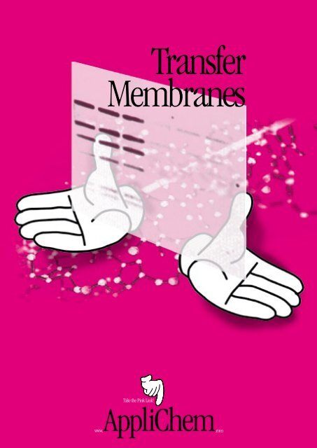
Transfer Membranes
Transfer Membranes
Transfer Membranes

Create successful ePaper yourself
Turn your PDF publications into a flip-book with our unique Google optimized e-Paper software.
<strong>Transfer</strong><br />
<strong>Membranes</strong><br />
Take the Pink Link!<br />
www.<br />
.com
a b o u t u s<br />
s o m e t h i n g<br />
Vision<br />
AppliChem was founded with the aim of supplying chemicals for chemical, biological,<br />
pharmaceutical and clinical research. It was also intended that AppliChem's products should<br />
be available worldwide.<br />
Experience<br />
Our chemists have had many years of in-depth experience and offer a sound partnership<br />
in helping to solve your problems in the lab. With you or for you – we want to develop new<br />
products. As well as flexibility, we assure you of strict confidentiality in all your projects.<br />
Assortment<br />
We prepare and provide you with chemicals and reagents including even those not listed<br />
in our current catalogs. When talking of “chemicals” in the widest sense of the word,<br />
we offer the service ‘all products – one supplier’.<br />
Quality<br />
Thanks to our quality management system, with AppliChem as your supplier you gain a<br />
decisive advantage over your competitors. Our products will fulfil your expectations and<br />
your individual, particular requirements are our business.<br />
AppliChem is continuously gaining new customers, due to the exact and constant quality,<br />
as well as to the advantageous prices, of our products and services. AppliChem is a reliable<br />
partner. Our quality control department provides detailed documentation on request.
c o n t e n t s<br />
Introduction 2<br />
Reprobe Nitrocellulose Supported <strong>Transfer</strong> Membrane 3<br />
Pure Nitrocellulose Unsupported <strong>Transfer</strong> Membrane 3<br />
PVDF-Star (Polyvinylidene Fluoride) <strong>Transfer</strong> <strong>Membranes</strong> 4<br />
Reprobe Nylon Positively Charged <strong>Transfer</strong> Membrane 5<br />
Pure Nylon Neutral <strong>Transfer</strong> Membrane 6<br />
<strong>Transfer</strong> <strong>Membranes</strong> Supplied by AppliChem 7<br />
AppliChem <strong>Transfer</strong> <strong>Membranes</strong> by Application 8<br />
You know your application, we know the membrane you require<br />
Selecting a <strong>Transfer</strong> Membrane 9<br />
Troubleshooting Guide 10<br />
Procedures and features of hybridization membranes plus troubleshooting guide<br />
Protocols and Technical Papers – Survey 13<br />
Protocols for DNA and RNA Applications 15<br />
1 Southern (DNA) and Northern (RNA) Hybridizations with Reprobe Nylon Positively Charged <strong>Membranes</strong> 15<br />
2 Southern (DNA) and Northern (RNA) Hybridizations with Pure Nylon Neutral <strong>Membranes</strong> 8<br />
3 Southern (DNA) and Northern (RNA) Hybridizations with Nitrocellulose <strong>Membranes</strong> 21<br />
4 Non-Radioactive Detection Systems with Pure Nylon Neutral 24<br />
5 Non-Radioactive Detection Systems with Nitrocellulose <strong>Membranes</strong> 26<br />
6 Alkaline Blotting with Reprobe Nylon Positively Charged <strong>Transfer</strong> Membrane 28<br />
7 Colony Hybridizations and Plaque Lifts with Nylon <strong>Membranes</strong> 29<br />
8 Colony Hybridizations and Plaque Lifts with Nitrocellulose <strong>Transfer</strong> <strong>Membranes</strong> 30<br />
9 Colony Hybridizations and Plaque Lifts with Pure Nylon Neutral <strong>Membranes</strong> 31<br />
10 RNA Probes to be Used in Northern and Southern Blotting 32<br />
11 Oligonucleotide Probes to be Used in Northern and Southern Blotting 33<br />
12 Nucleic Acid Dot/Slot Blotting with Nylon <strong>Membranes</strong> 32<br />
13 Nucleic Acid Dot/Slot Blotting with Nitrocellulose <strong>Membranes</strong> 35<br />
Protocols for Protein Applications 36<br />
14 Western (Protein) Blotting with Nitrocellulose <strong>Membranes</strong> 36<br />
15 Western Blotting from a SDS-PAGE System 37<br />
16 Protein Sequencing From A Western Blot 39<br />
17 Stripping PVDF-Star (Polyvinylidene Fluoride) <strong>Transfer</strong> Membrane 39<br />
18 <strong>Transfer</strong> of High Molecular Weight Proteins from a Non-Denaturing Gel System<br />
to the PVDF-Star (Polyvinylidene Fluoride) Membrane 40<br />
19 <strong>Transfer</strong> of Basic Proteins from an Acid/Urea Gel System at Acidic pH<br />
to the PVDF-Star (Polyvinylidene Fluoride) <strong>Transfer</strong> Membrane 40<br />
20 Protein Dot Slot Blotting with PVDF (Polyvinylidene Fluoride) <strong>Transfer</strong> <strong>Membranes</strong> 41<br />
21 Blocking with Blocking Reagent CA 42<br />
22 Blocking with Blocking Reagent GE 43<br />
Recipes for Buffers used in Nucleic Acid <strong>Transfer</strong>s 44<br />
Related Products: Chemicals Used For Blotting 45<br />
Appendix: Data from the General Catalog 46<br />
Blocking Reagents 46<br />
<strong>Transfer</strong> <strong>Membranes</strong> 50<br />
© 2008 AppliChem • <strong>Transfer</strong> <strong>Membranes</strong>
Introduction<br />
i n t r o d u c t i o n<br />
AppliChem supplies a range of transfer membranes designed and<br />
tested specifically for RNA, DNA and protein analysis. AppliChem also<br />
provides our customers with tried and proven protocols developed to<br />
obtain consistent reproducible and dependable results when used with<br />
AppliChem membranes. AppliChem highly recommends that you follow<br />
these protocols for optimal transfers.<br />
Products<br />
Reprobe Nitrocellulose Supported <strong>Transfer</strong> Membrane<br />
Outperforms unsupported nitrocellulose when extensively reprobing or performing colony and plaque lift.<br />
Some of the applications include DNA, RNA, and protein assays.<br />
Pure Nitrocellulose Unsupported <strong>Transfer</strong> Membrane<br />
Unsupported membrane of choice used for all protein or immunoblotting applications.<br />
PVDF-Star (Polyvinylidene Fluoride) <strong>Transfer</strong> Membrane<br />
Naturally hydrophobic, unsupported PVDF has the highest binding capacity for western transfers,<br />
amino acid analysis and protein sequencing.<br />
Reprobe Nylon Positively Charged <strong>Transfer</strong> Membrane<br />
Next generation and outperforms previous membranes with high signal strength and low background attributes.<br />
Inherently charged nylon membrane. Tested and performs well with radioactive or non-radioactive procedures.<br />
Pure Nylon Neutral <strong>Transfer</strong> Membrane<br />
Used where a neutral or single probe test is desired.<br />
<strong>Transfer</strong> <strong>Membranes</strong> • AppliChem © 2008
Reprobe Nitrocellulose Supported <strong>Transfer</strong> Membrane<br />
● Supported for procedures requiring rigorous handling<br />
● High sensitivities, low backgrounds<br />
● Strong – will not curl, bend or crack after baking<br />
Applications<br />
● Northerns, Southerns<br />
● Multiple re-hybridizations<br />
● Colony/plaque lifts<br />
Pure Nitrocellulose Unsupported <strong>Transfer</strong> Membrane<br />
● For procedures that require optimum resolution<br />
● Membrane of choice for all protein or immunoblotting<br />
applications<br />
● Cellulose acetate free –<br />
Applications<br />
● Westerns<br />
● Dot/slot blots<br />
● Radiolabeled detection systems<br />
● Chemiluminescent<br />
● Biotinylated detection systems<br />
Product Description<br />
Reprobe Nitrocellulose Supported is an internally supported nitrocellulose membrane that combines the binding characteristics<br />
of nitrocellulose with the strength of nylon. Reprobe Nitrocellulose Supported is ideal for applications where nitrocellulose<br />
has been used in the past such as DNA/RNA/Protein transfers. However, it outperforms unsupported nitrocellulose<br />
when extensive handling is required such as reprobing or colony and plaque lifts.<br />
The applied, unique process of impregnating pure nitrocellulose polymer onto an inert polyester web produces a very<br />
strong, dimensionally stable hybridization membrane. The inert web provides dimensional strength to prevent curling,<br />
cracking and tearing even after baking. This ensures the highest binding nitrocellulose membrane, approximately<br />
100 µg/cm 2 . There is nothing to lower the binding capacity such as cellulose acetate or other additives.<br />
This combination is especially important in colony and plaque lifts and multiple reprobings or any other procedure requiring<br />
rigorous handling. Membrane breakage is eliminated, reducing the costs due to wasted time, materials, and lost data. Many<br />
researchers have reprobed Reprobe Nitrocellulose Supported as many as five times.<br />
In all protocols using nitrocellulose, Reprobe Nitrocellulose Supported may be substituted without making any changes in<br />
the protocol while maintaining the same high signal and low backgrounds.<br />
All membranes are Triton free and do not contain any other cytotoxins.<br />
assuring high binding and sensitivity<br />
● Low background<br />
● Easily blocked<br />
● Nucleic acid binding is 100 µg/cm 2<br />
● Northerns<br />
● Protein & immunoblotting<br />
● Southerns<br />
● Dot/slot blots<br />
Product Description<br />
Pure Nitrocellulose is a pure unsupported nitrocellulose and is the membrane of choice for all protein or immunoblotting<br />
applications. Pure Nitrocellulose unsupported does not contain cellulose acetate, a low binding polymer. This assures the<br />
highest binding and sensitivity, 100 µg/cm 2 .<br />
Nitrocellulose exhibits the highest sensitivity with very low backgrounds in all transfers, especially in protein blotting. Unlike<br />
PVDF, nitrocellulose wets out naturally, does not require methanol, and will not turn hydrophobic during the transfer<br />
process. Nitrocellulose is very easily blocked and does not need the many blocking steps required with PVDF.<br />
Excellent results will be obtained with all detection systems: antibody/antigen, radiolabeled, biotinylated, and chemiluminescent,<br />
giving you a great amount of flexibility in designing your procedure.<br />
product information<br />
© 2008 AppliChem • <strong>Transfer</strong> <strong>Membranes</strong>
PVDF-Star (Polyvinylidene Fluoride) <strong>Transfer</strong> Membrane<br />
● Ideal for use in protein binding applications such as western blots, solid phase assays and immunoblotting procedures<br />
● Naturally hydrophobic unsupported membrane<br />
● Superior strength, can withstand aggressive handling or be used with automated equipment without breaking or tearing<br />
● High range of chemical compatibility, resistant to most commonly used chemicals and chemically aggressive solvents.<br />
Chemical compatibility allows the use of all commonly used stains<br />
● Low backgrounds, high sensitivity detects low-level components<br />
● Highest binding capacity eliminating sample “blow through”, binds a wide range of fragment sizes<br />
● BSA binding capacity is 125 µg/cm 2<br />
● Low extractables, ensures tests will be clean with consistent results<br />
● Hydrophobic, resists water<br />
● Lot-to-lot consistency, quality checks ensure consistent binding for dependable results every time<br />
Applications<br />
● Western transfers<br />
● Protein sequencing<br />
● Peptide mapping<br />
● Amino acid analysis<br />
● Dot/slot blots<br />
Product Description<br />
PVDF-Star (Polyvinylidene Fluoride) membrane is a naturally hydrophobic, unsupported transfer membrane. It has a high<br />
binding capacity and low backgrounds and is ideal for use in protein binding applications such as Western blots, solid phase<br />
assays, immuno blotting procedures, amino acid analysis and protein sequencing.<br />
Exceptional quality control procedures throughout the manufacturing process eliminates “lot-to-lot” variation and greatly<br />
minimizes “bald spot” problems. PVDF assures reproducible results with maximum sensitivity.<br />
Protein can be electroblotted from a variety of gel matrices onto PVDF-Star. Because of its high sensitivity and strong binding<br />
affinity, protein will not pass through the membrane. The broad chemical compatibility of PVDF-Star allows the use of all<br />
commonly used stains such as: Amido Black, Colloidal Gold, Coomassie Blue, India Ink and Ponceau-S. PVDF-Star will not<br />
degrade, distort or shrink when using high concentrations of methanol for destaining. The exceptional tensile strength allows<br />
for multiple reprobings and easy removal of target bands without concern for the membrane tearing, fracturing or curling.<br />
PVDF-Star exhibits efficient transfer of proteins in a wide range of molecular weights, from 5k Daltons to 700k Daltons. The<br />
binding capacity averages 125 µg/cm 2 for larger globular proteins such as immunoglobulins and higher binding capacities<br />
for small peptides.<br />
Specifications<br />
Thickness 40 – 250 µm<br />
BSA Protein Binding 125 µg/cm 2 for Immunoglobulins<br />
Pore Size Range 0.2 and 0.45 µm<br />
p r o d u c t<br />
<strong>Transfer</strong> <strong>Membranes</strong> • AppliChem © 2008
Reprobe Nylon Positively Charged <strong>Transfer</strong> Membrane<br />
● Ideal for use in multiple reprobings and nucleic acid transfers under alkaline conditions<br />
● Specifically designed for multiple reprobings: Inherently charged; enhances nucleic acid binding – even under alkaline<br />
conditions. Supported, has added strength and durability preventing distortion or contamination in multiple reprobings<br />
● Greater binding capacity with a binding capacity of 450 mg/cm 2 , Reprobe Nylon Positively Charged <strong>Transfer</strong> Membrane<br />
can bind a wide range of fragment sizes and can still be easily blocked for low background<br />
● Excellent signal better retains DNA resulting in a strong signal using smaller quantities of DNA<br />
● Hydrophilic, eliminates the need for wetting agents that can potentially interfere with biological processes<br />
● Lot-to-lot consistency, quality checks ensure lot-to-lot consistency, both down and across the polyester web,<br />
for dependable results every time<br />
Applications<br />
● Radiolabeled & non-radiolabeled detection systems<br />
● Southern transfers<br />
● Northern transfers<br />
● Micro arrays<br />
● Macro arrays<br />
● Chemiluminescent systems<br />
● Multiple Reprobings<br />
● Alkaline Blotting<br />
● UV Crosslinking<br />
Product Description<br />
Reprobe Nylon Positively Charged transfer membrane is an inherently charged, naturally hydrophilic nylon membrane, a<br />
pure polymer impregnated by an inert polyester web. It is specifically designed to allow for numerous reprobings and use<br />
in nucleic acid transfers under alkaline conditions. Reprobe Nylon Positively Charged transfer membrane has been tested to<br />
provide consistent results through 12 or more reprobings.<br />
The high binding capacity of 450 mg/cm 2 makes Reprobe Nylon Positively Charged transfer membrane ideal for all Southern<br />
and Northern applications, including alkaline blotting. Reprobe Nylon Positively Charged transfer membrane is ideally suited<br />
for all probes both radioactive and non-radioactive, including chemiluminescent and biotinylated detection systems.<br />
Reprobe Nylon Positively Charged offers significantly increased binding, maximum “lot-to-lot” consistency, and excellent<br />
signal retention. This higher sensitivity enables researchers to obtain reliable results using smaller quantities of DNA samples<br />
and allows them to conserve valuable or expensive samples and save them for future use.<br />
Given that smaller samples and higher density formats or micro arrays are becoming more common, the Reprobe Nylon<br />
Positively Charged <strong>Transfer</strong> Membrane is especially well suited for genomic research. Even with small DNA samples, Reprobe<br />
Nylon Positively Charged <strong>Transfer</strong> Membrane is able to retain a strong signal after multiple reprobings, providing easier<br />
automated reading of signals and more discrimination.<br />
The unique qualities of Reprobe Nylon Positively Charged <strong>Transfer</strong> Membrane provide a lower background and higher signal<br />
assuring reliable results the first time a transfer is executed.<br />
Product Characteristics<br />
Sterilization<br />
Gamma irradiation or Ethylene Oxide (EtO)<br />
USP Class VI Testing<br />
Passed<br />
Thickness 65 – 125 µm<br />
Extractables
Pure Nylon Neutral <strong>Transfer</strong> Membrane<br />
The purity and consistency of the Pure Nylon Neutral <strong>Transfer</strong> Membrane, coupled with its added durability<br />
and signal retention, make it an ideal membrane for use in medical research, scientific studies or<br />
test confirmations where precise biological pattern replications, such as DNA and RNA transfers, are<br />
integral to the success of the procedure.<br />
product information<br />
● Untreated nylon performs optimally in standard techniques<br />
● Single probes<br />
● Ideal for use in nucleic acids transfers and protein binding<br />
● Supported Nylon membrane, has added strength and durability preventing distortion or contamination in multiple<br />
reprobings<br />
● High binding capacity, with a nucleic acid binding capacity of approx. 350 µg/cm 2 , Pure Nylon Neutral <strong>Transfer</strong> Membrane<br />
can bind a wide range of fragment sizes, increasing the efficiency of transfers<br />
● Hydrophilic, which eliminates the need for wetting agents that can potentially interfere with biological processes<br />
● Lot-to-lot consistency, quality checks ensure lot-to-lot consistency, both down and across the polyester web, for<br />
dependable results every time<br />
Applications<br />
● Radiolabeled & non-radiolabeled detection systems ● Southern transfers ● Northern transfers<br />
● UV Crosslinking ● Colony lifts ● Plaque lifts ● Library Screenings ● Single Probe ● Protein binding<br />
● Micro arrays ● Macro arrays ● Dot/Slot blots<br />
Product Description<br />
Pure Nylon Neutral is a pure polymer impregnated by an inert polyester web. It is naturally hydrophilic and optimized<br />
for protein binding and for high, reproducible binding of nucleic acids. This supported nylon membrane, is optimal for<br />
all traditional DNA and RNA transfers. Pure Nylon Neutral has a higher binding capacity (approx. 350 µg/cm 2 ) than nitrocellulose<br />
membrane and binds a greater range of fragment sizes.<br />
Pure Nylon Neutral membrane is naturally hydrophilic, eliminating the need for wetting agents that can potentially interfere<br />
with biological processes. Pure Nylon Neutral membrane wets out quickly and evenly and will never turn hydrophobic<br />
during hybridization.<br />
UV crosslinking Pure Nylon Neutral is an excellent way to attach nucleic acids. By exposing a nylon membrane to a UV source,<br />
a covalent bond forms between the membrane and the DNA or RNA, which maximizes signal retention after reprobing.<br />
By the unique impregnation process, Pure Nylon Neutral has many advantages. Nylon is impregnated onto an integral<br />
polyester support web giving the nylon added dimensional strength, which prevents cracking, tearing, curling, and breaking.<br />
This is essential to protocols requiring aggressive handling, such as colony and plaque lifts and multiple reprobings. Using<br />
Pure Nylon Neutral by avoiding membrane breakage can save a significant amount of time and money.<br />
Product Characteristics<br />
Sterilization<br />
USP Class VI Testing<br />
Gamma irradiation or Ethylene Oxide (EtO)<br />
Passed<br />
Thickness 65 – 125 µm<br />
Extractables
<strong>Transfer</strong> <strong>Membranes</strong> Supplied by AppliChem<br />
Membrane Features Benefits Choose By Detection<br />
Systems<br />
Nylon<br />
Pure Nylon Neutral<br />
<br />
Reprobe Nylon Positively<br />
Charged<br />
<br />
Nitrocellulose<br />
Pure Nitrocellulose<br />
Unsupported<br />
<br />
<br />
Reprobe Nitrocellulose<br />
Supported <br />
<br />
<br />
<br />
<br />
<br />
Polyvinylidene Fluoride<br />
PVDF-Star<br />
<br />
<br />
Pure Nylon Membrane<br />
Positively Charged Nylon<br />
Membrane<br />
Pure Nitrocellulose<br />
Supported Pure<br />
Nitrocellulose<br />
Hydrophobic PVDF<br />
Membrane<br />
Untreated nylon performs<br />
optimally in standard<br />
techniques like Northerns<br />
and Southerns.<br />
Binding capacity<br />
350 µg/cm 2 .<br />
Inherently charged nylon<br />
membrane specifically<br />
designed for Multiple<br />
Reprobings.<br />
Binding capacity<br />
450 µg/cm 2 .<br />
Pure nitrocellulose is<br />
the membrane of choice<br />
for protein and immunoblotting<br />
techniques,<br />
as well as any other<br />
procedures that require<br />
optimum resolution.<br />
Binding capacity<br />
100 µg/cm 2 .<br />
Supported pure nitrocellulose<br />
is used in<br />
procedures requiring<br />
the highest sensitivities,<br />
low backgrounds and<br />
rigorous handling.<br />
The membrane can be<br />
reprobed many times.<br />
Binding capacity<br />
100 µg/cm 2 .<br />
Hydrophobic PVDF<br />
membrane is designed<br />
for protein sequencing,<br />
western transfers and<br />
amino acid analysis.<br />
Binding capacity<br />
125 µg/cm 2 .<br />
Radiolabeled & Non-<br />
Radiolabeled Detection<br />
Systems<br />
Radiolabeled Detection<br />
Systems<br />
Radiolabeled,<br />
Chromogenic and<br />
Chemiluminescent<br />
Detection Systems<br />
Radiolabeled Detection<br />
Systems<br />
Chemiluminescent and<br />
Biotinylated Detection<br />
Systems<br />
Chemical compatibility<br />
allows the use of all<br />
commonly used stains<br />
Choose By Procedures<br />
s u p p l y<br />
Northerns<br />
Southerns<br />
UV Crosslinking<br />
Colony/Plaque Lifts<br />
Library Screenings<br />
Northern<br />
Southerns<br />
Multiple Reprobings<br />
Alkaline Blotting<br />
Westerns<br />
Protein &<br />
Immunoblotting<br />
Northerns<br />
Southerns<br />
Northerns<br />
Southerns<br />
Multiple<br />
Rehybridizations<br />
Colony and Plaque Lifts<br />
Western <strong>Transfer</strong>s<br />
Protein Sequencing<br />
Amino Acid Analysis<br />
© 2008 AppliChem • <strong>Transfer</strong> <strong>Membranes</strong>
AppliChem <strong>Transfer</strong> <strong>Membranes</strong> by Application<br />
Application Membrane Catalog Number<br />
Alkaline Reprobe Nylon Positively Charged A5255<br />
Blotting<br />
Amino Acid Pure Nitrocellulose Unsupported A5250 or A5239<br />
Analysis PVDF-Star A5243<br />
Biotinylated Pure Nylon Neutral A4399 or A5248<br />
Probes Reprobe Nitrocellulose Supported A5237 or A5242<br />
Cell Blotting Reprobe Nylon Positively Charged A5255<br />
Pure Nylon Neutral<br />
A4399 or A5248<br />
Cell Labeling Pure Nylon Neutral A4399 or A5248<br />
Chemilumines- Pure Nylon Neutral<br />
A4399 or A5248<br />
cent Probes Reprobe Nitrocellulose Supported A5237 or A5242<br />
Chromogenic Pure Nitrocellulose Unsupported A5250 or A5239<br />
Reprobe Nitrocellulose Supported A5237 or A5242<br />
Colony Lift Pure Nylon Neutral A4399 or A5248<br />
Reprobe Nitrocellulose Supported gridded discs *<br />
DNA Reprobe Nylon Positively Charged A5255<br />
Hybridization Pure Nylon Neutral<br />
A4399 or A5248<br />
Reprobe Nitrocellulose Supported A5237 or A5242<br />
Dot/Slot Reprobe Nylon Positively Charged A5255<br />
Blotting Reprobe Nitrocellulose Supported A5237 or A5242<br />
Drug Binding Pure Nitrocellulose Unsupported A5250 or A5239<br />
Assay Reprobe Nitrocellulose Supported A5237 or A5242<br />
Hormone Pure Nitrocellulose Unsupported A5250 or A5239<br />
Binding Reprobe Nitrocellulose Supported A5237 or A5242<br />
Immuno- Pure Nitrocellulose Unsupported A5250 or A5239<br />
blotting PVDF-Star A5243<br />
Library Pure Nylon Neutral A4399 or A5248<br />
Screening Reprobe Nitrocellulose Supported A5237 or A5242<br />
Multiple Reprobe Nylon Positively Charged A5255<br />
Reprobings<br />
Application Membrane Catalog Number<br />
Northern Reprobe Nylon Positively Charged A5255<br />
<strong>Transfer</strong>s Pure Nylon Neutral A4399 or A5248<br />
Reprobe Nitrocellulose Supported A5237 or A5242<br />
Pure Nitrocellulose Unsupported A5250 or A5239<br />
Non-Radio- Pure Nylon Neutral A4399 or A5248<br />
active Probes Reprobe Nitrocellulose Supported A5237 or A5242<br />
Nucleic Acid Reprobe Nylon Positively Charged A5255<br />
<strong>Transfer</strong> Pure Nylon Neutral A4399 or A5248<br />
Reprobe Nitrocellulose Supported A5237 or A5242<br />
Pure Nitrocellulose Unsupported A5250 or A5239<br />
Plaque Lifts Reprobe Nitrocellulose Supported gridded discs *<br />
Protein Pure Nitrocellulose Unsupported A5250 or A5239<br />
Blotting PVDF-Star A5243<br />
Protein Pure Nitrocellulose Unsupported A5250 or A5239<br />
Precipitation<br />
Reprobe Nitrocellulose Supported A5237 or A5242<br />
Protein PVDF-Star 0.1 µm pore size *<br />
Sequencing<br />
Protein Pure Nitrocellulose Unsupported A5250 or A5239<br />
<strong>Transfer</strong> PVDF-Star A5243<br />
Radiolabeled Reprobe Nylon Positively Charged<br />
Probe<br />
A5255<br />
Reprobe Nitrocellulose Supported A5237 or A5242<br />
RNA Reprobe Nylon Positively Charged A5255<br />
Hybridizations Reprobe Nitrocellulose Supported A5237 or A5242<br />
Solid Phase PVDF-Star A5243<br />
Assay<br />
Southern Reprobe Nylon Positively Charged A5255<br />
<strong>Transfer</strong>s<br />
Reprobe Nitrocellulose Supported A5237 or A5242<br />
Pure Nylon Neutral<br />
A4399 or A5248<br />
UV Pure Nylon Neutral A4399 or A5248<br />
Crosslinking<br />
Vacuum Reprobe Nylon Positively Charged A5255<br />
Blotting Reprobe Nitrocellulose Supported A5237 or A5242<br />
Westerns Pure Nitrocellulose Unsupported A5250 or A5239<br />
PVDF-Star<br />
A5243<br />
* available on request<br />
applications<br />
<strong>Transfer</strong> <strong>Membranes</strong> • AppliChem © 2008
s e l e c t i o n<br />
Selecting a <strong>Transfer</strong> Membrane<br />
Selecting the appropriate membrane is critical to the success of a nucleic acid or protein transfer procedure. AppliChem<br />
supplies several types of membranes for hybridization technology, each exhibiting different performance characteristics<br />
which can directly affect the outcome of a specific technique. Below are some of the more frequently performed procedures<br />
and features of hybridization membranes.<br />
Rehybridizations AppliChem offers membranes recommended for rehybridization procedures: Pure Nylon Neutral,<br />
Reprobe Nylon Positively Charged and Reprobe Nitrocellulose Supported, a supported pure nitrocellulose.<br />
Reprobe Nylon Positively Charged membranes can be most frequently reprobed. On nylon membranes, the number of<br />
reprobing steps is a function of the amount of hydrolysis to which the membrane is exposed during the protocol, and the<br />
additive effects of hot water, sodium hydroxide and an acidic environment. Sodium hydroxide solutions deteriorate the nylon<br />
matrix and are not recommended in procedures where reprobing steps are required.<br />
The polyester support web used in manufacturing Reprobe Nitrocellulose Supported allows the membrane to be reprobed<br />
several times. Because the binding capacity of nitrocellulose is less than that of nylon (100 µg/cm 2 vs. 400 µg/cm 2 ),<br />
the potential number of rehybridizations is fewer. See below for more details (Southern (DNA) and Northern (RNA)<br />
Hybridizations)<br />
Non-Radioactive Probes Reprobe Nylon Positively Charged may be used particularily where multiple probing is involved.<br />
UV Crosslinking For covalent binding of nucleic acids to a transfer membrane, AppliChem membranes can be UV<br />
Crosslinked by following the manufacturer's instructions. It is particularly recommended when working with short<br />
fragments, small samples, or low numbers of base pairs, because of the improved resolution this technique offers.<br />
Protein Blotting Pure Nitrocellulose Unsupported and PVDF-Star membranes are recommended for use in protein blotting.<br />
Nitrocellulose membranes are able to be more thoroughly blocked, reducing the high background potential associated with<br />
protein blotting. PVDF membranes are more resistant to the harsh chemicals used in Edman degradation.<br />
Alkaline Blotting For more rapid transfers, an alkaline blotting procedure can be used with Reprobe Nylon membranes.<br />
Alkaline blotting is not recommended when reprobing is required. See Protocol 6 for more details on Alkaline Blotting with<br />
Reprobe Nylon Positively Charged <strong>Transfer</strong> Membrane.<br />
Staining Procedures Reprobe Nitrocellulose Supported, Pure Nitrocellulose Unsupported and PVDF-Star membranes<br />
are recommended for procedures that require a staining step with India Ink, Coomassie Blue, Colloidal Gold, or any other<br />
commonly used stain. Nylon membranes irreversibly bind many stains.<br />
Reducing Backgrounds There are many sources of background problems, or low signal-to-noise ratios. Some of the most<br />
common include: contaminated probes, contaminated hybridization solutions, and incorrectly chosen stringency levels.<br />
Nonfat milk should not be used as a blocking agent as it may increase nonspecific binding. Nylon membranes have a much<br />
higher binding capacity than nitrocellulose membranes, and a greater potential for backgrounds. AppliChem membranes are<br />
all manufactured by strict quality control procedures, ensuring a uniform membrane with consistently low backgrounds.<br />
Troubleshooting Common Blotting Problems Many blotting problems can be eliminated by observing the following<br />
recommendations.<br />
Blotchy or incomplete transfers are caused by poor contact between the gel and the membrane. Even after careful smoothing<br />
of the membrane to the gel, incomplete degassing of transfer solutions can cause air pockets to form. Evolving gas from<br />
Tris or, in the case of protein transfers, methanol, can disrupt the tight contact necessary between the membrane and the<br />
gel for successful transfers.<br />
Smeared or skewed bands are often caused by uneven contact between the gel and the membrane, or the membrane and<br />
the chromatography paper. To avoid this problem, roll a pipet down the membrane after it has been applied to the gel, and<br />
once again over the chromatography paper after it has been applied to the membrane. Do not move the membrane until the<br />
transfer is complete, as this will cause smearing.<br />
© 2008 AppliChem • <strong>Transfer</strong> <strong>Membranes</strong>
Troubleshooting Guide and Application Tips<br />
Problems and Solutions<br />
Unsuccesful Rehybridizations<br />
My membrane is<br />
deteriorating during<br />
the rehybridization<br />
procedure?<br />
My application demands<br />
an extensive number<br />
of reprobes and I’m<br />
losing signal?<br />
My probe is not stripping<br />
from the membrane,<br />
how should I change my<br />
procedure?<br />
If so, what type of membrane are you using? AppliChem offers three types of membranes recommended for<br />
rehybridization procedures: Pure Nylon Neutral, Reprobe Nylon Positively Charged, and Reprobe Nitrocellulose<br />
Supported, a supported pure nitrocellulose.<br />
Pure Nylon Neutral membranes have largely replaced nitrocellulose because they are more resilient during<br />
applications requiring multiple reprobes. Reprobe Nitrocellulose Supported (a supported nitrocellulose) was<br />
developed for the same reason. The polyester support web used in manufacturing Reprobe Nitrocellulose<br />
Supported allows the membrane to be reprobed several times. Reprobe Nylon Positively Charged is similar in<br />
resiliency to Pure Nylon Neutral.<br />
If so, what type of membrane are you using? Because the binding capacity of nitrocellulose is less than that of<br />
nylon (100 µg/cm 2 vs. 400 µg/cm 2 ), the potential number of rehybridizations is fewer as compared to nylon<br />
membranes. Likewise, the binding capacity of charged nylon (Reprobe Nylon Positively Charged) membranes<br />
is generally greater than standard nylon, and subsequently charged nylon may have advantages during multiple<br />
reprobes. The number of reprobing steps is a function of the amount of hydrolysis to which the membrane is<br />
exposed during the protocol, and the additive effects of hot water, sodium hydroxide and an acidic environment.<br />
Did you let the membrane dry after the initial probe was applied? Drying causes irreversible binding of DNA to<br />
microporous membranes. If this has occurred, look through the helpful tips listed below.<br />
My probe won’t strip<br />
from the membrane,<br />
how can I rescue<br />
this blot?<br />
The nucleic acid did<br />
not transfer completely<br />
to the membrane,<br />
what should I do?<br />
My Signal is low,<br />
what are the common<br />
reasons for this?<br />
Try preparing a new probe and using a different detection protocol. For example, if you prepared a biotinylated<br />
probe and detected with a streptavidin conjugate, omit the biotin-streptavidin step during rehybridization by<br />
using a directly conjugated probe, such as an alkaline phosphatase conjugated probe. If you used a radioactive<br />
probe, use a chemiluminescent system to detect after the next hybridization (or vice versa). If you have enough<br />
time and are using radioactive probes (e.g. Protocol 11), simply let your first probe decay before the second<br />
round of hybridization.<br />
Signal Problems<br />
Blotchy or incomplete transfers are caused by poor contact between the gel and the membrane. Even after careful<br />
smoothing of the membrane to the gel, incomplete degassing of transfer solutions can cause air pockets to form.<br />
Evolving gas from Tris or, in the case of protein transfers, methanol, can disrupt the tight contact necessary<br />
between the membrane and the gel for successful transfers.<br />
When you have low signal, it is best to check your reagents by performing extra controls. The most common<br />
reason for poor signal is a bad probe. Prepare a new probe and perform a dot blot comparing the old and new<br />
probes. Do you see a difference between the probes? Even nonradioactive probes can deteriorate during storage.<br />
Is the signal weak for the new probe as well? Then your detection enzymes may be bad or the reagents used to<br />
prepare the probe are bad. You might also blot a small amount of unlabeled complementary DNA and hybridize<br />
to the new probe. Are you seeing signal from the blotted probe but not the hybridized DNA? If so there could<br />
be a problem with your hybridization protocol, such as the wash temperature or your buffers. If you’re using<br />
nonradioactive detection methods, test your enzymes and substrates as well.<br />
t r o u b l e s h o<br />
10 <strong>Transfer</strong> <strong>Membranes</strong> • AppliChem © 2008
Background Problems<br />
Reprobe Nylon Positively Charged may be used particularily where multiple probing is involved. Additionally,<br />
AppliChem's positively charged Reprobe Nylon membrane shows superior results as compared to competitor<br />
membranes when non-radioactive detection is used.<br />
Did you make up a new probe? If so, was there adequate separation of the unincorporated label from the incorporated?<br />
Are you using old solutions? There may be contamination. Usually in these cases it is best to prepare new<br />
solutions, new probes and use new reagents. This is often the fastest way to get your system working again.<br />
Miscellaneous<br />
No. Slight color changes in AppliChem's new positively charged membranes are expected and have no effect<br />
on results. These color changes will vary according to the blotting procedure used and the pH of solutions.<br />
AppliChem uses this color change to ensure quality during the manufacturing procedure.<br />
High Backgrounds<br />
Poor agitation during prehybridization and hybridization steps can lead to insufficient blocking of the entire<br />
membrane. Due to the strength of the internal support web, Reprobe Nitrocellulose Supported, Pure Nylon<br />
Neutral, and Reprobe Nylon Positively Charged transfer membranes can withstand higher levels of agitation<br />
without tearing or ripping. Incorrect probe concentration can occur when using dextran sulfate in hybridization<br />
or prehybridization solutions. Dextran sulfate causes the effective concentration of the probe to increase because<br />
it excludes the probe from the volume of solution the dextran sulfate polymer occupies. When using dextran<br />
sulfate, lower the probe solutions to less than 10 ng/ml of the solution. When not using dextran sulfate, maintain<br />
the optimum probe concentration at 25 – 40 ng/ml of solution.<br />
Residual agarose on membranes can cause a fuzzy background to appear on blots. Be sure to wash nylon<br />
membranes with 5X SSPE at 60°C, after the immobilization step. Due to the strength of the membrane, supported<br />
membranes (Reprobe Nitrocellulose Supported, Pure Nylon Neutral and Reprobe Nylon Positively Charged<br />
<strong>Transfer</strong> <strong>Membranes</strong>) can be more easily washed without tearing or ripping.<br />
I switched to a<br />
non-radioactive detection<br />
system, and now<br />
I have high background,<br />
what should I do?<br />
Everything was working<br />
fine and now suddenly<br />
I have high backgrounds,<br />
Why?<br />
My membrane changed<br />
color during my blotting<br />
procedure, should I be<br />
concerned?<br />
Troubleshooting Gel Casting Procedures<br />
Troubleshooting blotting problems begins with the correct gel casting procedures. Skewed, streaked, incomplete,<br />
or non-uniform transfers can be the results of poorly cast gels. The following recommendations are made for<br />
setting up the gel. Gels greater than 4mm thick can interfere with the free transfer of nucleic acids.<br />
Be sure that the gel tray is level before casting the gel. If the surface is not level, non-uniform transfers may result.<br />
Maintain a gel casting temperature of 55°C – 70°C degrees, and be sure that the gel particles are completely<br />
dissolved. Undissolved agarose particles can result in streaked or skewed bands.<br />
Immediately after gel casting and solidification, submerge the gel slab in electrophoresis buffer. This will prevent<br />
the formation of an impermeable “skin” over the surface of the gel which can inhibit transfer of nucleic acids<br />
from the gel.<br />
After setting up the blotting assembly, be sure to:<br />
● Invert the gel so that the underside of the gel is the side in contact with the membrane.<br />
● Allow the transfer solutions enough time to “breathe”, so that they may degas completely. Incompletely<br />
degassed transfer solutions evolve gas after the blotting assembly is set up, and can cause air bubbles between<br />
the membrane and gel that can impede the transfer of nucleic acids.<br />
o t i n g & t i p s<br />
© 2008 AppliChem • <strong>Transfer</strong> <strong>Membranes</strong> 11
Background Problems<br />
While there are several ways to decontaminate probe solutions, the following methods are two of the most efficient.<br />
The second method can be rapidly performed with minimum effort.<br />
Method 1:<br />
Phenol/Chloroform extract the probe to remove unincorporated nucleotides, proteins, and other contaminants.<br />
Method 2:<br />
Clean the probe by adding a small volume of the hybridization buffer to the probe and filtering it through a Cameo 25AS low<br />
protein binding cellulose acetate syringe filter (GE Osmonics Catalog #DDA02025S0; available at AppliChem). Contaminants<br />
in the probe solution will be held back by the 0.2 µm filter with no probe loss caused by nonspecific binding to the filtration<br />
membrane.<br />
Probe length is also a factor contributing to background levels seen on transfer membranes. Between 250 – 800 base<br />
pairs is the recommended optimum length of a probe; probe lengths smaller or larger than this can lead to a low signalto-noise<br />
ratio. Probes smaller than 250 base pairs often bind poorly and may require less stringent hybridization and wash<br />
procedures. Probes larger than 800 base pairs may contain a wider variety of size classes, which can lead to extraneous<br />
binding to the transfer membrane.<br />
Hybridization Solution Related Background Problems<br />
Contaminated hybridization solutions are another common source of background problems. Hybridization solutions should<br />
be filtered with a pure cellulose acetate Cameo 25AS syringe filter ( GE Osmonics Catalog #DDA02025S0; available at<br />
AppliChem), to remove contaminants.<br />
Additionally, all solutions and buffers should be made fresh before each transfer with sterile, double-distilled, deionized<br />
water, and very high grade reagents. After fresh buffers are made, they should be filtered with a Cameo 25AS syringe filter<br />
(GE Osmonics Catalog #DDA02025S0; available at AppliChem) to ensure that no contaminants remain in the solution.<br />
Formamide-based hybridization solutions are a frequent source of background noise, and the formamide must be freshly<br />
made and deionized.<br />
DNA Dependent Background<br />
This type of background is caused by nonspecific annealing of the probe to the bound DNA or RNA, and can be eliminated<br />
by adjusting the stringency of both the hybridization and wash steps, and by the addition of heterologous DNA or RNA<br />
to the hybridization solutions. During the hybridization and prehybridization steps, a concentration of 0.1 – 1 % SDS is<br />
recommended for nylon membranes. Because the final wash step after the hybridization is the most important, AppliChem<br />
recommends:<br />
Northerns: 0.1X SSPE, 0.1 – 0.5 % SDS, for 30 minutes at 65°C.<br />
Southerns: 0.1X SSPE, 0.5 – 1 % SDS, for 30 minutes at 65°C.<br />
Optimized Blocking Solutions<br />
A concentration of 5 – 7X Denhardt’s solution is recommended for use with nylon membranes. Exceeding this level can<br />
lead to quenching of the signal.<br />
troubleshooting&tipsProbe Related<br />
Backgrounds Associated with Reprobing<br />
A follow-up autoradiograph after probe removal is strongly recommended to determine if the probe has been fully stripped.<br />
Otherwise, backgrounds can appear in blots that have not been fully erased.<br />
12 <strong>Transfer</strong> <strong>Membranes</strong> • AppliChem © 2008
Protocols and Technical Papers – Survey<br />
Selecting the appropriate membrane is critical to the success of protein transfer and nucleic acid procedures. Using the<br />
correct protocol with AppliChem membranes insures you that your procedure will perform with optimal results.<br />
AppliChem provides the following tried and proven protocols developed by MSI to obtain consistent reproducible and<br />
dependable results when used with AppliChem’ membranes.<br />
1 Southern (DNA) and Northern (RNA) Hybridizations <br />
with Reprobe Nylon Positively Charged <strong>Membranes</strong><br />
Southern (DNA) and northern (RNA) protocol for our charged nylon – Reprobe Nylon Positively Charged<br />
Membrane – a membrane manufactured for your multi-probe needs.<br />
2 Southern (DNA) and Northern (RNA) Hybridizations<br />
Run Southern DNA and Northern (RNA) Hybridizations with Pure Nylon Neutral membranes<br />
3 Southern (DNA) and Northern (RNA) Hybridizations<br />
Protocol for use with Reprobe Nitrocellulose Supported and Pure Nitrocellulose Unsupported membranes.<br />
4 Non-Radioactive Detection Systems with Pure Nylon Neutral<br />
Procedure for non-radioactive systems – Reprobe Nylon Positively Charged may be used particulary<br />
where multiple probing is involved.<br />
5 Non-Radioactive Detection Systems with Nitrocellulose <strong>Membranes</strong><br />
with Reprobe Nitrocellulose Supported <strong>Membranes</strong> and Pure Nitrocellulose Unsupported<br />
6 Alkaline Blotting with Reprobe Nylon Positively Charged <strong>Transfer</strong> Membrane<br />
<strong>Membranes</strong> with very high binding capacities are necessary for the success alkaline blotting<br />
7 Colony Hybridizations and Plaque Lifts with Nylon <strong>Membranes</strong><br />
Perform colony hybridizations and plaque lifts with Pure Nylon and Reprobe Nylon <strong>Membranes</strong><br />
8 Colony Hybridizations and Plaque Lifts with Nitrocellulose <strong>Transfer</strong> <strong>Membranes</strong><br />
Perform colony hybridizations and plaque lifts with Reprobe Nitrocellulose Supported and Pure Nitrocellulose<br />
Unsupported <strong>Transfer</strong> <strong>Membranes</strong>.<br />
9 Colony Hybridizations and Plaque Lifts with Pure Nylon Neutral <strong>Membranes</strong><br />
Perform colony hybridizations and plaque lifts with Pure Nylon Neutral <strong>Membranes</strong><br />
10 RNA Probes to be Used in Northern and Southern Blotting<br />
Use Pure Nylon Neutral in northern and southern blotting<br />
11 Oligonucleotide Probes to be Used in Northern and Southern Blotting<br />
Use Pure Nylon Neutral membranes in Northern and Southern Blotting<br />
12 Nucleic Acid Dot/Slot Blotting with Nylon <strong>Membranes</strong><br />
Purify DNA or RNA with Pure Nylon Neutral membranes<br />
13 Nucleic Acid Dot/Slot Blotting with Nitrocellulose <strong>Membranes</strong><br />
Purify DNA or RNA with Reprobe Nitrocellulose Supported and Pure Nitrocellulose Unsupported <strong>Transfer</strong><br />
<strong>Membranes</strong>.<br />
14 Western (Protein) Blotting with Nitrocellulose <strong>Membranes</strong><br />
For use with Reprobe Nitrocellulose Supported and Pure Nitrocellulose Unsupported <strong>Transfer</strong> <strong>Membranes</strong>.<br />
p r o t o c o l s<br />
© 2008 AppliChem • <strong>Transfer</strong> <strong>Membranes</strong> 13
p r o t o c o l s<br />
15 Western Blotting from a SDS-PAGE System<br />
Use PVDF-Star transfer membranes for Western Blotting from a SDS-PAGE System<br />
16 Protein Sequencing From A Western Blot<br />
Use PVDF-Star for protein sequencing from a Western Blot<br />
17 Stripping PVDF-Star (Polyvinylidene Fluoride) <strong>Transfer</strong> Membrane<br />
A stripping procedure that will not adversely alter membrane bound proteins<br />
18 <strong>Transfer</strong> of High Molecular Weight Proteins from a Non-Denaturing Gel System <br />
to the PVDF-Star (Polyvinylidene Fluoride) Membrane<br />
Use PVDF-Star transfer membranes in non-denaturing gel systems<br />
19 <strong>Transfer</strong> of Basic Proteins from an Acid/Urea Gel System at Acidic pH <br />
to the PVDF-Star (Polyvinylidene Fluoride) <strong>Transfer</strong> Membrane<br />
Use PVDF-Star transfer membranes as an alternative to nitrocellulose in Western Blotting applications<br />
20 Protein Dot Slot Blotting with PVDF (Polyvinylidene Fluoride) <strong>Transfer</strong> <strong>Membranes</strong><br />
Perform Dot Slot Blotting with PVDF-Star transfer membranes<br />
21 Blocking with Blocking Reagent CA<br />
22 Blocking with Blocking Reagent GE<br />
14 <strong>Transfer</strong> <strong>Membranes</strong> • AppliChem © 2008
Protocols for DNA and RNA Applications<br />
Protocol 1: Southern (DNA) and Northern (RNA) Hybridizations<br />
with Reprobe Nylon Positively Charged <strong>Membranes</strong><br />
Gel Preparation<br />
S o u t h e r n s<br />
Run DNA on an agarose gel with a running buffer of TAE or TBE. Rinse gel with dH 2O. Fragment DNA by immersing the gel<br />
in 0.2 N HCl for 10 minutes. Denature DNA by soaking gel in 1.0 M NaCl/0.5 M NaOH for 30 minutes. Neutralize the gel by<br />
soaking in 0.5 M Tris · HCl (pH 8.0), 0.5 M NaCl for 30 minutes.<br />
N o r t h e r n s<br />
Run RNA under denaturing conditions in a Glyoxal, Formaldehyde, or Methyl Mercuric Hydroxide gel. Gel should be<br />
0.8 – 1.5 % agarose, 2.5 – 5.0 mm thick. Stain with 33 µg/ml acridine orange in 10 mM NaPO 4 (pH 6.5), then destain<br />
3 x 15 minutes in buffer. Or, stain with ethidium bromide, 1 µg/ml in 50 mM NaOH for 25 minutes, then destain in 200 mM<br />
NaOAc (pH 4.0), 2 x 20 minutes.<br />
Membrane Preparation<br />
Float membrane on distilled water, then immerse until thoroughly wet. <strong>Transfer</strong> the membranes to 20X SSC prior to blotting.<br />
Capillary <strong>Transfer</strong><br />
When starting new experiments, it is highly recommended that controls be performed to ensure complete transfer. Stain<br />
the gel with ethidium bromide before and after transfer. Add 10 ml of a 10 mg/ml Ethidium Bromide stock to about 500<br />
ml of Running Buffer. Soak the gel with gentle agitation for 20 minutes. If low levels of DNA need be visualized, destain the<br />
gel for 20 to 40 minutes.<br />
Use a <strong>Transfer</strong> Buffer of 20X SSC. Cut three pieces of filter paper (chromatography grade) 7.6 cm (three inches) longer than<br />
the glass plate to be used for the capillary transfer. Place on top of a glass plate and then saturate it with <strong>Transfer</strong> Buffer.<br />
Place the gel on top of the filter paper. The bottom surface of the gel should face up. Remove all air bubbles between the gel<br />
and blotting surface with a gloved hand or by rolling a pipette across the surface. Place the membrane over the gel, again<br />
removing all air bubbles. Place 2 – 5 pieces of filter paper cut to the size of the gel over the assembly. Place the glass plate<br />
and the gel assembly on top of a glass baking tray filled with transfer buffer. Allow the bottom layer of filter paper to overhang<br />
into the transfer solution in the glass baking tray.<br />
Place a 5 cm (2 inch) stack of paper towels on top of the gel assembly and secure it with a light weight. Make sure that the<br />
filter paper under the gel is completely saturated. The wicking action of the solution through the gel and up the paper towels<br />
allows the solution to transfer the DNA or RNA molecules to the membrane. Throughout the transfer, do not allow the paper<br />
on top of the gel to contact the paper below the gel. If the paper towels become saturated with transfer buffer, replace them<br />
with dry ones. After transfer, stain the gel with 0.5 µg/ml ethidium bromide to check transfer efficiency.<br />
01<br />
protocol<br />
protocols<br />
Alternative <strong>Transfer</strong> Systems<br />
Vacuum blotting, semi-dry electroblotting, bi-directional transfers, and positive pressure blotting systems can all be used<br />
with AppliChem nylon membranes. Follow manufacturers instructions.<br />
Immobilization<br />
After blotting, wash the membranes in 5X SSPE at 60°C for 5 minutes. This is an optional step to remove residual agarose.<br />
The nucleic acid on the membrane is immobilized by drying thoroughly. This is accomplished by baking the membrane for<br />
15 minutes at 80°C. It is not necessary to use a vacuum oven.<br />
As an additional immobilization step, the dry membrane may be exposed to 5 mJ/cm 2 260 nm UV irradiation using a UV<br />
crosslinker. A slight improvement in signal may be observed after UV irradiation. If the immobilization procedure is skipped,<br />
the signal will be reduced. For applications involving large amounts of applied DNA (e.g., 10 ng of Hind III digested Lambda<br />
DNA), drying may not be required.<br />
Hybridization Procedure<br />
There are various formats used for hybridization: tubes in a hybridization oven, Tupperware in a shaking waterbath, or<br />
sealed bags in an oven. Heat-sealable bags conserve solution and generate less volume of radioactive waste (if radioactive<br />
detection procedures are employed.) Tubes within hybridization ovens also have this advantage. Tupperware use within a<br />
shaking waterbath uses more hybridization solution, but may help to achieve complete hybridization. The choice depends<br />
© 2008 AppliChem • <strong>Transfer</strong> <strong>Membranes</strong> 15
protocol<br />
01<br />
on available resources within the laboratory and on individual preference.<br />
All hybridization solutions should be filtered before use with a sterile Cameo 25AS, 0.22 µm supported cellulose acetate filter<br />
(GE Osmonics catalog #DDA02025S0; available at AppliChem). (NOTE: Use only cellulose acetate filters; other membrane<br />
types may not perform comparably). The low binding Cameo 25AS will filter prehybridization and hybridization solutions<br />
without nonspecifically binding essential components of these solutions.<br />
Before prehybridization wet the blot in dH 2O and then transfer the blot to whichever salt solution is used for hybridization.<br />
(For example, if 6X SSPE is used during hybridization, then wet the blot in dH 2O and transfer to 6X SSPE.<br />
Prehybridization<br />
AppliChem recommends that blots be placed in prehybridization solution for 30 minutes at 65°C. Place blots individually<br />
into prehybridization solution if many blots are being processed simultaneously. Use at least 0.25 ml/cm 2 of the following<br />
prehybridization buffers, complementary to the probe. Typically Salmon Sperm DNA is phenol/chloroform extracted,<br />
sheared, and heat denatured.<br />
S o u t h e r n P r e h y b r i d i z a t i o n S o l u t i o n<br />
Denhardt’s solution<br />
6X SSC<br />
N o r t h e r n P r e h y b r i d i z a t i o n S o l u t i o n<br />
5x Denhardt’s solution<br />
5x SSPE<br />
1.0 % SDS 0.1 – 0.5 % SDS<br />
100 µg/ml denatured DNA* 00 µg/ml denatured DNA*<br />
* Denatured DNA is typically Salmon Sperm DNA, phenol/chloroform extracted, sheared, and heat denatured.<br />
There are many variations on the hybridization procedure. AppliChem does not recommend inclusion of 10 % dextran<br />
sulfate or 10 % PEG unless fast hybridization times (e.g., 4 hours) are required. Also, dextran sulfate should not be used<br />
with oligonucleotide probes. Solutions containing 7 % SDS are not recommended. SSPE or SSC may be used, and the<br />
concentration may be as low as 2X or as high as 6X. The denatured DNA used depends on the application; it must not be<br />
complementary to the probe.<br />
Hybridization<br />
Remove prehybridization solution and replace with hybridization solution.<br />
Hybridization temperature should be determined by the presence of formamide in the hybridization solution.<br />
Hybridization Temperature Chart<br />
Te m p e r a t u r e % F o r m a m i d e H y b r i d i z a t i o n S o l u t i o n<br />
42°C 50 % Low Temperature Hybridization Solution<br />
65°C 0 % High Temperature Hybridization Solution<br />
Low Temperature Hybridization Solution<br />
S o u t h e r n s<br />
N o r t h e r n s<br />
50 % formamide 50 % formamide<br />
5X Denhardt’s solution<br />
5X Denhardt’s solution<br />
6X SSPE<br />
5X SSPE<br />
0.2 % SDS 0.2 % SDS<br />
100 µg/ml denatured DNA* 00 µg/ml denatured DNA*<br />
* Denatured DNA is typically Salmon Sperm DNA, phenol/chloroform extracted, sheared, and heat denatured.<br />
p r o<br />
16 <strong>Transfer</strong> <strong>Membranes</strong> • AppliChem © 2008
High Temperature Hybridization Solution<br />
S o u t h e r n s<br />
N o r t h e r n s<br />
5X Denhardt’s solution<br />
6X SSC<br />
5X Denhardt’s solution<br />
5X SSPE<br />
1 % SDS 0.5 % SDS<br />
100 µg/ml denatured DNA* 100 µg/ml denatured DNA*<br />
01<br />
protocol<br />
* Denatured DNA is typically Salmon Sperm DNA, phenol/chloroform extracted, sheared, and heat denatured.<br />
Alternative probes (oligonucleotide, RNA, or biotinylated probes) may also be used. See Protocol 11 for “Oligonucleotide<br />
Probes to be Used in Northern and Southern Blotting”. Clean probe solutions by adding a small amount of hybridization<br />
solution to the probe, and filter it through a Cameo 25AS supported cellulose acetate syringe filter (GE Osmonics catalog<br />
#DDA02025S0; available at AppliChem) to eliminate any contaminants before they come into contact with the transfer membrane.<br />
The probe is typically used in excess during hybridization procedures. Calculate the amount of bound target DNA and<br />
be sure the total amount of probe is at least 10 fold greater. Probe concentration should not exceed 20 ng/ml. Increase the<br />
volume of the hybridization reaction if necessary so probe is in excess. Single copy genes or low copy message may require<br />
1 – 5 x 10 6 cpm/ml. Radioactive probes should be labeled no more than 24 hours before hybridization. Nonradioactive<br />
probes may be stored at -20°C for up to 6 months. Denature the probe by placing it into 100 to 500 ml prehybridization<br />
solution (or TE buffer) and heating to 95°C for 5 minutes. Place on ice. Add the probe to the prehybridization solution to<br />
make hybridization solution. Hybridize at least 12 hours if PEG or dextran sulfate are not used. Usually this step is carried<br />
out overnight.<br />
Post Hybridization Washing Steps<br />
Briefly rinse the blots in 2X SSC. Then, wash twice 15 minutes with 2X SSC, 1 % SDS at 65°C and twice 15 minutes in<br />
0.1X SSC, 1 % SDS at 65°C. <strong>Transfer</strong> to 2X SSC after washing. Use at least 1 ml of wash solution per square centimeter for all<br />
membranes. There are many variations on washing procedures. More or less stringent conditions may be used depending<br />
on the application. The washing procedure works for most applications.<br />
After washing, remove excess moisture with paper towels. Washes for northern blots sometimes substitute SSPE for SSC.<br />
Autoradiography and Detection<br />
Important: If the membrane is to be rehybridized, do not allow it to dry past this point. This will cause irreversible binding<br />
of probe to the membrane. For radioactive probes, wrap the membrane in plastic wrap and expose film at -70°C in a<br />
cassette with an intensifying screen for 25 – 60 hours. For nonradioactive probes, follow the manufacturer’s recommended<br />
procedure. Alkaline phosphatase conjugated streptavidin enzymes with biotin-labeled probes work well with AppliChem's<br />
Reprobe Nylon membranes.<br />
Probe Removal (Stripping)<br />
Important: Do not allow the membrane to dry. Wash in 0.4 M NaOH for 30 min at 45°C with moderate agitation. Wash in<br />
Membrane Neutralization Solution (1.5 M NaCl; 1 M Tris · HCl, pH 7.4) for 15 minutes at room temperature.<br />
When using this procedure for the first time, confirm probe removal by re-exposing film. If non-radioactive methods are<br />
used to confirm probe removal, the membrane should be blocked with prehybridization solution for 30 minutes before<br />
detection, otherwise high background levels will be encountered.<br />
The success of probe removal depends on the amount of applied DNA. This procedure has been tested for 12 to 15 rounds<br />
of stripping and reprobing with up to 1 ng of applied DNA.<br />
t o c o l s<br />
© 2008 AppliChem • <strong>Transfer</strong> <strong>Membranes</strong> 17
protocol<br />
02<br />
Protocol 2: Southern (DNA) and Northern (RNA) Hybridizations<br />
with Pure Nylon Neutral <strong>Membranes</strong><br />
Gel Preparation<br />
S o u t h e r n s<br />
Run DNA on an agarose gel with a running buffer of TAE or TBE. Rinse gel with dH 2O. If necessary, fragment DNA by<br />
immersing the gel in 0.25 N HCl for 8 – 10 minutes. Denature DNA by soaking gel in 1.0 M NaCl/0.5 M NaOH two times for<br />
20 minutes each. Neutralize the gel by soaking in 0.5 M Tris (pH 7.5), 1.5 M NaCl two time for 20 minutes each.<br />
N o r t h e r n<br />
Run RNA under denaturing conditions in a Glyoxal, Formaldehyde, or Methyl Mercuric Hydroxide gel. Gel should be<br />
0.8 – 1.5 % agarose, 2.5 – 5.0 mm thick. Stain with 33 µg/ml acridine orange in 10 mM NaPO 4 (pH 6.5), then destain<br />
3 x 15 minutes in buffer. Or, stain with ethidium bromide, 1 µg/ml in 50 mM NaOH for 25 minutes, then destain in 200 mM<br />
NaOAc (pH 4.0), 2 x 20 minutes.<br />
protocols<br />
Membrane Preparation<br />
Float membrane on distilled water, then immerse until thoroughly wet. Soak the membrane in the transfer buffer until use.<br />
Capillary <strong>Transfer</strong><br />
Use a transfer buffer of 10X SSPE or SSC.<br />
Cut three pieces of filter paper (chromatography grade) 7.5 cm (3 inches) longer than the glass plate to be used for the<br />
capillary transfer. Saturate the filter paper with transfer buffer and place on top of a glass plate.<br />
Place the gel on top of the filter paper, and the membrane over the gel. Place 5 pieces of filter paper cut to the size of the<br />
gel over the assembly. Throughout the transfer, do not allow the paper on top of the gel to contact the paper below the gel.<br />
This is done by placing strips of Parafilm around the sides of the gel.<br />
Place the glass plate and the gel assembly on top of a glass baking tray filled with transfer buffer. Allow the bottom layer<br />
of filter paper to overhang into the transfer solution in the glass baking tray. Place a 5 cm (2 inch) stack of paper towels<br />
on top of the gel assembly and secure it with a light weight. Make sure that the filter paper under the gel is completely<br />
saturated. The wicking action of the solution through the gel and up the paper towels allows the solution to transfer the DNA<br />
or RNA molecules to the membrane.<br />
Secure plastic wrap over the entire assembly and place in a cold room for 3 hours to overnight. If the paper towels become<br />
saturated with transfer buffer, replace them with dry ones. After transfer, stain the gel with 0.5 µg/ml ethidium bromide to<br />
check transfer efficiency.<br />
Alternative <strong>Transfer</strong> Systems<br />
Vacuum blotting, semi-dry electroblotting, bi-directional transfers, and positive pressure blotting systems can all be used<br />
with AppliChem nylon membranes. Follow manufacturers instructions.<br />
Immobilization<br />
After blotting, wash the membranes in 5X SSPE at 60°C for 5 minutes. This is an optional step to remove residual agarose.<br />
The membrane can be immobilized by baking or UV crosslinking.<br />
To bake the membrane a vacuum oven or convection oven may be used, place the membrane in a 65° – 80°C oven for<br />
1 hour or until the membrane is completely dry.<br />
The membrane may be UV crosslinked by exposing the membrane to a controlled UV source. Follow the instructions of<br />
the manufacturer of the crosslinker or expose a damp membrane to a transilluminator. The total exposure should be<br />
120 mJ/cm. Increased exposure will cause a decrease in signal intensity after reprobing, proportional to the amount of<br />
overexposure.<br />
Hybridization Procedure<br />
Hybridization is most commonly done in heat-sealable bags in order to conserve solution and protect researchers from<br />
exposure to radioactivity. All hybridization solutions should be filtered before use with a Cameo 25AS, 0.22 µm supported<br />
cellulose acetate filter (GE Osmonics catalog #DDA02025S0; available at AppliChem). (NOTE: Use only cellulose acetate<br />
filters; other membrane types may not perform comparably). The low binding Cameo 25AS will filter prehybridization and<br />
hybridization solutions without nonspecifically binding essential components of these solutions.<br />
18 <strong>Transfer</strong> <strong>Membranes</strong> • AppliChem © 2008
Prehybridization<br />
This step should always be carried out at the temperature of the hybridization. Place the membrane in a heat-sealable bag<br />
without the probe in 0.25 ml/cm 2 of the following prehybridization buffer:<br />
02<br />
protocol<br />
S o u t h e r n P r e h y b r i d i z a t i o n S o l u t i o n N o r t h e r n P r e h y b r i d i z a t i o n S o l u t i o n<br />
6X SSPE<br />
5X Denhardt's solution<br />
5X SSPE<br />
50 % Formamide<br />
0.5 – 1.0 % SDS 0.1 – 0.5 % SDS<br />
50 µg/ml denatured DNA 00 µg/ml denatured DNA<br />
10 % Dextran Sulfate 5X Denhardt's solution<br />
Shake one to two hours, at 42°C.<br />
Hybridization<br />
Remove prehybridization solution completely from bag, and add the hybridization solution.<br />
Hybridization temperature should be determined by the presence of formamide in the hybridization solution.<br />
Hybridization Temperature Chart<br />
Te m p e r a t u r e % F o r m a m i d e H y b r i d i z a t i o n S o l u t i o n<br />
42°C 50 % Low Temperature Hybridization Solution<br />
65°C 0 % High Temperature Hybridization Solution<br />
protocols<br />
Low Temperature Hybridization Solution<br />
Southern<br />
Northern<br />
50 % formamide 50 % formamide<br />
5X Denhardt's solution<br />
5X Denhardt's solution<br />
6X SSPE<br />
5X SSPE<br />
0.2 % SDS 0.2 % SDS<br />
100 µg/ml denatured DNA 00 µg/ml denatured DNA<br />
10 % dextran sulfate 0 % dextran sulfate<br />
High Temperature Hybridization Solution<br />
Southern<br />
Northern<br />
5X Denhardt's solution<br />
5X Denhardt's solution<br />
6X SSPE<br />
5X SSPE<br />
0.5 % SDS 0.5 % SDS<br />
50 µg/ml denatured DNA 00 – 200 µg/ml denatured DNA<br />
10 % dextran sulfate 0 % dextran sulfate<br />
NOTE: When using nylon membranes, the potential for backgrounds is greater. Increased volumes of<br />
Denhardt's solution and SDS can help further block the membrane.<br />
Dextran sulfate is a rate enhancer for probes larger than 200 base pairs and should not be used with<br />
oligonucleotide probes.<br />
Alternative probes (oligonucleotide, RNA, or biotinylated probes) may also be used.<br />
Clean probe solutions by adding a small amount of hybridization solution to the probe, and filter it<br />
through a Cameo 25AS supported cellulose acetate syringe filter (GE Osmonics catalog #DDA02025S0;<br />
available at AppliChem), to eliminate any contaminants before they come into contact with the transfer<br />
membrane.<br />
Denature the probe by boiling in TE buffer for 5 minutes, or incubate with 0.1 volume of 1 N NaOH at<br />
37°C for 5 minutes. Place on ice.<br />
© 2008 AppliChem • <strong>Transfer</strong> <strong>Membranes</strong> 19
protocol<br />
02<br />
Add the decontaminated probe to the hybridization solution in the heat-sealable bag and reseal.<br />
Probe concentration should not exceed 20 ng/ml. Single copy genes or low copy message may require<br />
1 – 5 x 10 6 cpm/ml. Probes should be labeled no more than 24 hours before hybridization.<br />
Hybridize 12 hours to overnight. Important: If the membrane is to be rehybridized, do not allow it to<br />
dry past this point. This will cause irreversible binding to the membrane.<br />
Post Hybridization<br />
Wash temperature should be 25°C below the Tm (melting temperature of the hybrid). If the homology between the probe<br />
and membrane-bound DNA is inexact, the wash temperature should be lower.<br />
Stringency Washing Procedure<br />
Low Stringency (for inexact matching)<br />
2 x 15 minutes with 1X SSC, 0.1 % SDS at room temperature<br />
2 x 15 minutes with 1X SSC, 0.1 % SDS at 37°C<br />
Medium Stringency<br />
2 x 15 minutes with 5X SSC, 0.5 % SDS at room temperature<br />
2 x 15 minutes with 1X SSC, 0.5 – 1.0 % SDS at 37°C<br />
1 x 15 minutes with 0.1X SSC, 1.0 % SDS at 37°C<br />
High Stringency (for perfect hybrids)<br />
2 x 15 minutes with 5X SSC, 0.5 % SDS at room temperature<br />
2 x 15 minutes with 1X SSC, 0.5 – 1.0 % SDS at 37°C<br />
3 x 15 minutes with 0.1X SSC, 1.0 % SDS at 65°C<br />
NOTE: With nylon membranes, a medium or high stringency washing procedure is recommended to control potential<br />
background problems.<br />
Use 0.5 ml of wash solution per square centimeter for all membranes. After washing, remove excess moisture with paper<br />
towels. Washes for Northern blots should substitute SSPE for SSC in all wash steps.<br />
Autoradiography<br />
Wrap the membrane in plastic wrap and autoradiograph at -70°C in a cassette with an intensifying screen while slightly<br />
damp. Expose the membrane for 25 – 60 hours.<br />
Probe Removal<br />
Do not allow the membrane to dry if a rehybridization step is intended. Wash in 5 mM Tris · HCl (pH 8.0), 0.2 mM EDTA,<br />
0.05 % pyrophosphate, 0.1 Denhardt's for 1 – 2 hours at 65°C. Rinse in 1X SSPE. Or, wash in 50 % formamide, 6X SSPE at<br />
65°C for 30 minutes. Rinse in 2X SSPE.<br />
p r o t o c o l s<br />
20 <strong>Transfer</strong> <strong>Membranes</strong> • AppliChem © 2008
Protocol 3: Southern (DNA) and Northern (RNA) Hybridizations Nitrocellulose<br />
<strong>Membranes</strong> with Reprobe Nitrocellulose Supported, Pure Nitrocellulose Unsupported <strong>Transfer</strong> <strong>Membranes</strong><br />
Gel Preparation<br />
S o u t h e r n s<br />
Run DNA on an agarose gel with a running buffer of TAE or TBE. If necessary, fragment gel in 0.25 N HCl. Rinse gel with dH 2O<br />
and denature DNA by soaking gel in 1.0 M NaCl/0.5 M NaOH 2 times for 20 minutes each. Neutralize the gel by soaking in<br />
0.5 M Tris (pH 7.5), 1.5 M NaCl two times for 20 minutes each.<br />
N o r t h e r n s<br />
Run RNA under denaturing conditions in a Glyoxal, Formaldehyde, or Methyl Mercuric Hydroxide gel. Gel should be<br />
0.8 – 1.5 % agarose, 2.5 – 5.0 mm thick. Stain with 33 µg/ml acridine orange in 10 mM NaPO 4 (pH 6.5), then destain<br />
3 x 15 minutes in buffer, or stain with ethidium bromide, 1 µg/ml in 50 mM NaOH for 25 minutes, then destain in 200 mM<br />
NaOAc (pH 4.0), 2 x 20 minutes.<br />
Membrane Preparation<br />
Float membrane on distilled water, then immerse until wet thoroughly. If wetting is not immediate, heat water until just under<br />
boiling temperature. Soak in the transfer buffer until use.<br />
Gel Pretreatment<br />
S o u t h e r n s : Not necessary.<br />
N o r t h e r n s : Pretreatment, such as fragmentation of RNA, is unnecessary.<br />
Capillary <strong>Transfer</strong><br />
Use a transfer buffer of 20X SSPE or SSC.<br />
Cut 3 pieces of filter paper (chromatography grade) 7.5 cm (3 inches) longer than the glass plate to be used for the capillary<br />
transfer. Saturate the filter paper with transfer buffer and place on top of a glass plate.<br />
Place gel on top of the filter paper, and the membrane over the gel. Place 5 pieces of filter paper cut to the size of the gel<br />
over the assembly. Throughout the transfer, do not allow the paper on top of the gel to contact the paper below the gel. This<br />
is done by placing strips of Parafilm around the sides of the gel.<br />
Place the glass plate and the gel assembly on top of a glass baking tray filled with transfer buffer. Allow the bottom layer of<br />
filter paper to overhang into the transfer solution in the glass baking tray. Place a 5 cm (2 inch) stack of paper towels on<br />
top of the gel assembly and secure with a light weight. Make sure that the filter paper under the gel is completely saturated.<br />
The wicking action of the solution through the gel and up the paper towels allows the solution to transfer the DNA or RNA<br />
molecules to the membrane.<br />
Secure plastic wrap over the entire assembly and place in a cold room for 3 hours to overnight. If the paper towels become<br />
saturated with transfer buffer, replace them with dry ones. After transfer, stain the gel with 0.5 µg/ml ethidium bromide to<br />
check transfer efficiency.<br />
03<br />
protocol<br />
protocols<br />
Alternative <strong>Transfer</strong> Systems<br />
Vacuum blotting, semi-dry electroblotting, bidirectional transfers, and positive pressure blotting systems can all be used with<br />
AppliChem nitrocellulose membranes. Follow manufacturers instructions.<br />
Immobilization<br />
After blotting, wash the membranes in 5X SSPE at 60°C for 5 minutes. This is an optional step to remove residual agarose.<br />
The membrane can be immobilized by backing or UV crosslinking.<br />
To bake the membrane a vacuum oven or convection oven may be used, place the membrane in a 65° – 80°C oven for<br />
1 hour or until the membrane is completely dry.<br />
The membrane may be UV crosslinked by exposing the membrane to a controlled UV source. Follow the instructions of<br />
the manufacturer of the crosslinker or expose a damp membrane to a controlled UV source, total exposure should be<br />
120 mJ/cm. Increased exposure will cause a decrease in signal intensity after reprobing, proportional to the amount of<br />
overexposure.<br />
© 2008 AppliChem • <strong>Transfer</strong> <strong>Membranes</strong> 21
protocol<br />
03<br />
Hybridization Procedure<br />
Hybridization is most commonly done in heat-sealable bags in order to conserve solution and protect researchers from<br />
exposure to radioactivity. All hybridization solutions should be filtered before use with Cameo 25AS, 0.22 µm supported<br />
cellulose acetate filter (GE Osmonics catalog #DDA02025S0; available at AppliChem) (NOTE: Use only cellulose acetate<br />
filters; other membrane types may not perform comparably). The low binding Cameo 25AS will filter prehybridization and<br />
hybridizaton solutions without nonspecifically binding essential components of these solutions.<br />
protocols<br />
Prehybridization<br />
This step should always be carried out at the temperature of the hybridization. Place the membrane in a heat-sealable bag<br />
without the probe in 0.1 ml/cm 2 of the following prehybridization buffers.<br />
Southern Prehybridization Solution<br />
Northern Prehybridization Solution<br />
5X SSPE<br />
5X SSPE<br />
50 % Formamide 50 % Formamide<br />
5X Denhardt's solution<br />
5X Denhardt's solution<br />
0.5 % SDS 0.1 – 0.5 % /SDS<br />
100 µg/ml denatured DNA 00 µg/ml denatured DNA<br />
10 % dextran sulfate 0 % dextran sulfate<br />
Nitrocellulose membranes should be wet with 1X SSPE, 0.1 % SDS before prehybridization step.<br />
Shake 1 to 2 hours.<br />
Hybridization<br />
Remove prehybridization solution completely from bag, and add the hybridization solution.<br />
Hybridization temperature should be determined by the presence of formamide in the hybridization solution.<br />
Hybridization Temperature Chart<br />
Te m p e r a t u r e % F o r m a m i d e H y b r i d i z a t i o n S o l u t i o n<br />
42°C 50 % Low-Temperature Hybridization Solution<br />
65°C 0 % High-Temperature Hybridization Solution<br />
Low-Temperature Hybridization Solution<br />
Southerns<br />
Northerns<br />
50 % Formamide 50 % Formamide<br />
5X Denhardt's solution<br />
5X Denhardt's solution<br />
6X SSPE<br />
5X SSPE<br />
0.2 % SDS 0.2 % SDS<br />
100 – 200 µg/ml denatured DNA 00 – 200 µg/ml denatured DNA<br />
10 % dextran sulfate 0 % dextran sulfate<br />
High-Temperature Hybridization Solution<br />
Southerns<br />
Northerns<br />
5X Denhardt's solution<br />
5X Denhardt's solution<br />
6X SSPE<br />
5X SSPE<br />
0.5 % SDS 0.5 % SDS<br />
50 µg/ml denatured DNA 00 µg/ml denatured DNA<br />
10 % dextran sulfate 0 % dextran sulfate<br />
Dextran sulfate is a rate enhancer for probes larger than 200 base pairs and should not be used with oligonucleotide probes.<br />
Alternative probes (oligonucleotide, RNA, or biotinylated probes) may also be used.<br />
22 <strong>Transfer</strong> <strong>Membranes</strong> • AppliChem © 2008
Clean probe solutions by adding a small amount of hybridization solution to the probe, and filter it through a Cameo 25AS<br />
supported cellulose acetate syringe filter (GE Osmonics catalog #DDA02025S0; available at AppliChem), to eliminate any<br />
contaminants before they come into contact with the transfer membrane.<br />
Denature the probe by boiling in TE buffer for 5 minutes, or incubate with 0.1 volume of 1 N NaOH at 37°C for 5 minutes.<br />
Place on ice.<br />
Add the decontaminated probe to the hybridization solution in the heat-sealable bag and reseal. Probe concentration should<br />
not exceed 20 ng/ml. Single copy genes or low copy message may require 1 – 5 x 10 6 cpm/ml. Probes should be labeled<br />
no more than 24 hours before hybridization.<br />
Hybridize 12 hours to overnight. Important: If the membrane is to be rehybridized, do not allow it to dry past this point. This<br />
will cause irreversible binding to the membrane.<br />
Post Hybridization<br />
Wash temperature should be 25°C below the Tm (melting temperature of the hybrid). If the homology between the probe<br />
and membrane-bound DNA is inexact, the wash temperature should be lower.<br />
Stringency Washing Procedure<br />
Low Stringency (for inexact matching)<br />
2 x 15 minutes with 1X SSC, 0.1 % SDS at room temperature<br />
2 x 15 minutes with 1X SSC, 0.1 % SDS at 37°C<br />
Medium Stringency<br />
2 x 15 minutes with 5X SSC, 0.5 % SDS at room temperature<br />
2 x 15 minutes with 1X SSC, 0.5 – 1.0 % SDS at 37°C<br />
1 x 15 minutes with 0.1X SSC, 1.0 % SDS at 37°C<br />
High Stringency(for perfect hybrids)<br />
2 x 15 minutes with 5X SSC, 0.5 % SDS at room temperature<br />
2 x 15 minutes with 1X SSC, 0.5 – 1.0 % SDS at 37°C<br />
3 x 15 minutes with 0.1X SSC, 1.0 % SDS at 65°C<br />
NOTE: With nitrocellulose membranes, an initial low stringency washing procedure is recommended.<br />
Use 0.5 ml of wash solution per square centimeter for all membranes. After washing, remove excess moisture with paper<br />
towels.<br />
Autoradiography<br />
Wrap the membrane in plastic wrap and autoradiograph at -70°C in a cassette with an intensifying screen while slightly<br />
damp. Expose the membrane for 25 – 60 hours.<br />
Probe Removal<br />
Do not allow the membrane to dry if a rehybridization step is intended.<br />
For Pure Nitrocellulose Unsupported: Boil 0.1X SSPE or SCC, 0.1 % SDS and add blot after removing from heat. Check for<br />
residual probe activity.<br />
For Reprobe Nitrocellulose Supported: Boil in distilled water for 5 minutes. Check for residual probe activity. If necessary,<br />
boil for additional 5 minutes.<br />
03<br />
protocol<br />
p r o t o c o l s<br />
© 2008 AppliChem • <strong>Transfer</strong> <strong>Membranes</strong> 23
protocol<br />
04<br />
Protocol 4: Non-Radioactive Detection Systems with Pure Nylon Neutral<br />
There are many advantages when hybridizing with non-radioactive probes. The disposal problems associated with radioactive<br />
materials are eliminated with the use of nonisotopic probes. Biotinylated probes are more stable than isotopic probes, and<br />
have better signal-to-noise ratios. Increased amounts of probe can be used, cutting hybridization times by several hours.<br />
Pure Nylon Neutral is a chemically optimized hybridization membrane that demonstrates superior binding and reduced<br />
backgrounds when using biotinylated probe systems. Pure Nylon Neutral is the recommended membrane when working with<br />
chemiluminescent detection systems. Charge-modified nylon membranes are difficult to use with non-radioactive detection<br />
systems because background noise can obscure the bands. Reprobe Nylon Positively Charged may be used particularily<br />
where multiple probing is involved.<br />
Gel<br />
Run an agarose gel in TBE buffer, and stain with ethidium bromide (1.0 µg/ml) for 15 minutes.<br />
Membrane Preparation<br />
Submerge the membrane in deionized water to moisten, and soak in 10X SSC for 30 minutes prior to transfer.<br />
For electrophoretic transfers, soak in 1X TAE.<br />
Gel Preparation<br />
Depurinate the gel in 0.25 M HCl for 30 minutes while gently agitating, with one exchange at 15 minutes. For preparations<br />
involving whole plasmids, extend the depurination time to 40 minutes, with exchanges at 15 and 30 minutes. Denature the<br />
gel in 1.5 M NaCl, 0.5 M NaOH for 1 hour with one exchange at 30 minutes. Place the gel in 1.5 M NaCl, 1 M Tris · HCl<br />
(pH 8.0) for 1 hour with one exchange at 30 minutes in order to neutralize the gel.<br />
<strong>Transfer</strong><br />
<strong>Transfer</strong> the DNA to the membrane by Southern blotting (either vacuum or capillary) and restain the gel with ethidium<br />
bromide to determine transfer efficiency. See Protocol 1 for Southern (DNA) and Northern (RNA) Hybridizations with<br />
Reprobe Nylon Positively Charged <strong>Membranes</strong>.<br />
Immobilization<br />
After blotting, wash the membrane in 5X SSPE at 60°C for 5 minutes. This is an optional step to remove residual agarose.<br />
Bake membrane at 80°C until dry or UV crosslink per manufacturer's instructions. Do not exceed 120 mJ/cm 2 . Increased<br />
exposure will cause a decrease in signal intensity after reprobing, proportional to the amount of overexposure.<br />
Probe Preparation<br />
Ideally, probes should be between 400 – 600 base pairs. Probe sizing gels, using the appropriate size standards, should be<br />
run with the probes after labelling.<br />
Prehybridization<br />
Prewash the membrane in 2X SSC, 1 % SDS at 42°C for 1 hour.<br />
Block membranes in the prehybridization solution for 30 – 40 minutes at room temperature.<br />
Prehybridization Buffer<br />
Tris-buffered saline/pH 7.5 (0.1 M Tris · HCl, 0.5 M NaCl)<br />
1.0 % Casein<br />
3.0 % Liquid Hipure Gelatin<br />
0.05 % Tween-20<br />
p r o t o c o l s<br />
24 <strong>Transfer</strong> <strong>Membranes</strong> • AppliChem © 2008
Hybridization<br />
Convert the probe to its single stranded form by adding an equal volume of 0.2 N NaOH and incubate at 37°C 1 – 2 hours.<br />
Remove the prehybridization buffer, add the hybridization buffer with the probe, and incubate at 42°C 12 hours or overnight<br />
with gentle agitation.<br />
04<br />
protocol<br />
Hybridization Buffer<br />
45 % deionized formamide<br />
1X Denhardt’s solution<br />
5X SSC<br />
0.1 % SDS<br />
10 % Dextran Sulfate<br />
20 mM Phosphate buffer/pH 7.0<br />
p r o t o c o l s<br />
Washes<br />
When the hybridization is complete, wash the membrane in small trays in the following wash solutions:<br />
0.16X SSC, 0.1 % SDS, 2 times for 3 minutes each at room temperature<br />
0.16X SSC, 0.1 % SDS, 2 times for 3 minutes each at 64°C;<br />
Rinse in 2X SSC, 2 times for 3 minutes each at room temperature.<br />
Post Hybridization<br />
Block the filter by rinsing for one minute in Buffer 1, followed by an incubation step in 3 % Fraction V BSA in freshly made<br />
Buffer 1 for one hour at 65°C. Repeat incubation step with fresh buffer for an additional 30 minutes.<br />
Buffer 1<br />
0.1 M Tris · HCl (pH 7.5)<br />
0.1 M NaCl<br />
2 mM Magnesium Chloride<br />
0.05 % Triton-X 100<br />
Detection<br />
Remove the membrane from the hybridization solution, and wash in TBS buffer with 5 % Tween-20 for 5 minutes. Additional<br />
washes in TBS buffer may be required.<br />
Add the streptavidin-enzyme complex according to manufacturer’s instructions.<br />
Recommended Buffer Solutions<br />
Alkaline Phosphatase Buffer Solution<br />
Horseradish Peroxidase Substrate Buffer Solution<br />
100 mM Tris · HCl (pH 9.1) 2 mg/ml Tetramethyl Benzidene in<br />
100 mM NaCl 00 % Ethanol and H 2O 2 at 0.0014 %<br />
5 mM MgCl 2 0.01 M Sodium Citrate<br />
5 mM Nitro Blue Tetrazolium 0.01 M EDTA (pH 5.0)<br />
3 mM 5-bromo-4-chloro-3-indolyl-phosphate (BCIP)<br />
Post Detection<br />
After the membranes are adequately stained, rinse briefly in deionized water. Dry the membranes between filter paper, or<br />
bake in a vacuum oven.<br />
© 2008 AppliChem • <strong>Transfer</strong> <strong>Membranes</strong> 25
protocol<br />
05<br />
Protocol 5: Non-Radioactive Detection Systems with Nitrocellulose <strong>Membranes</strong><br />
with Reprobe Nitrocellulose Supported <strong>Membranes</strong> and Pure Nitrocellulose Unsupported<br />
There are many advantages when hybridizing with non-radioactive probes. The disposal problems associated with radioactive<br />
probes are eliminated with the use of non-isotopic probes. Biotinylated probes are more stable than isotopic probes,<br />
and have better signal-to-noise ratios. Increased amounts of probe can be used, cutting hybridization times by several hours.<br />
Reprobe Nitrocellulose Supported membranes are strong and easy to use with biotinylated detection systems. Traditional<br />
nitrocellulose membranes can sometimes be difficult to use because the fragility of the membrane can cause blue lines and<br />
spots when using biotinylated probes. However, they require no excessive blocking steps.<br />
Gel<br />
Run an agarose gel in TBE buffer, and stain with ethidium bromide (1.0 µg/ml) for 15 minutes.<br />
Membrane Preparation<br />
Submerge the membrane in deionized water to moisten, and then soak in 20X SSC, for 30 minutes before transfer.<br />
Gel Preparation<br />
Depurinate the gel in 0.25 M HCl for 30 minutes while gently agitating, with one exchange at 15 minutes. For preparations<br />
involving whole plasmids, extend the depurination time to 40 minutes, with exchanges at 15 and 30 minutes. Denature the<br />
gel in 1.5 M NaCl, 0.5 M NaOH for 1 hour with one exchange at 30 minutes. Place the gel in 1.5 M NaCl, 1 M Tris · HCl<br />
(pH 8.0) for 1 hour with one exchange at 30 minutes in order to neutralize the gel.<br />
<strong>Transfer</strong><br />
<strong>Transfer</strong> the DNA to the membrane by Southern blotting, (either vacuum or capillary) and restain the gel with ethidium<br />
bromide to determine transfer efficiency.<br />
Immobilization<br />
Bake the membrane in a vacuum oven at 80°C for two hours.<br />
Probe Preparation<br />
Ideally, probes should be between 400 – 600 base pairs. Probe sizing gels, using the appropriate size standards, should be<br />
run with the probes after labelling.<br />
Prehybridization<br />
Incubate the blot in a heat-sealable bag with 0.1 ml/cm 2 prehybridization buffer (without the probe) for four hours at 42°C.<br />
Prehybridization Solution<br />
50 % Formamide<br />
5X SSC<br />
5X Denhardt's solution<br />
25 mM sodium phosphate (pH 6.5)<br />
Denatured salmon sperm DNA (0.5 mg/ml)<br />
Hybridization<br />
Convert the probe to its single stranded form by adding an equal volume of 0.2 N NaOH and incubate at 37°C. Remove the<br />
prehybridization buffer, add the hybridization buffer with the probe, and incubate at 42°C overnight with gentle agitation.<br />
Hybridization Buffer<br />
45 % Formamide<br />
5X SSC<br />
5X Denhardt's solution<br />
20 mM sodium phosphate (pH 6.5)<br />
Denatured salmon sperm DNA (0.2 mg/ml)<br />
Biotinylated probe (heat denatured)<br />
p r o<br />
26 <strong>Transfer</strong> <strong>Membranes</strong> • AppliChem © 2008
Washes<br />
When the hybridization is complete, wash the membrane in small trays in the following wash solutions:<br />
2X SSC, 0.1 % SDS 6 minutes with one exchange at 3 minutes at room temperature<br />
0.2X SSC, 0.1 % SDS 6 minutes with one exchange at 3 minutes at room temperature<br />
0.16X SSC, 0.1 % SDS 30 minutes with one exchange at 15 minutes at room temperature<br />
05<br />
protocol<br />
Post Hybridization<br />
Block the filter by rinsing for one minute in Buffer 1, followed by an incubation step in 3 % Fraction V BSA in freshly made<br />
Buffer 1 for one hour at 65°C.<br />
Buffer 1 Buffer 2<br />
2 mM Magnesium Chloride 0.1 M Tris · HCl (pH 9.5)<br />
0.1 M Tris · HCl (pH 7.5) 0.1 M NaCl<br />
0.1 M NaCl 50 mM Magnesium Chloride<br />
0.05 % Triton-X 100<br />
Chromogenic Detection<br />
Expose the blot to 7.0 ml of streptavidin detection solution (prepare using 1.0 µl streptavidin-alkaline phosphatase stock<br />
solution per 1.0 ml of Buffer 1) per 100 cm 2 of filter area for 5 minutes/side with gentle mixing. Wash the membrane in<br />
Buffer 1 for 30 minutes (with one exchange at 15 minutes) and Buffer 2 for 10 minutes.<br />
Visualization<br />
For each 100 cm 2 of membrane, prepare a solution containing 33 µl of Nitro Blue Tetrazolium (75 mg/ml in 70 %<br />
dimethylformamide) and 25 µl of 5-bromo-4-chloro-3-indolyl-phosphate (BCIP; 50 mg/ml in dimethylformamide) in 7.5<br />
ml of the incubation mixture referenced above (Buffer 1 with 3 % Fraction V BSA). Place the membrane and solution in a<br />
heat-sealable bag in the dark. Color development should occur within 30 minutes.<br />
Post Detection<br />
Stop the reaction by allowing the membrane to soak for 1 – 2 minutes in a solution of 20 mM Tris (pH 7.5), 0.5 mM<br />
disodium EDTA, followed by baking for 1 – 2 minutes at 80°C.<br />
Alkaline Phosphatase Buffer Solution<br />
Horseradish Peroxidase Substrate Buffer Solution<br />
100 mM Tris · HCl (pH 9.1) 2 mg/ml Tetramethyl Benzidene in<br />
100 mM NaCl 00 % Ethanol and H 2O 2 at 0.0014 %<br />
5 mM MgCl 2 0.01 M Sodium Citrate<br />
5 mM Nitro Blue Tetrazolium 0.01 M EDTA (pH 5.0)<br />
3 mM 5-bromo-4-chloro-3-indolyl-phosphate (BCIP)<br />
After membranes are adequately stained, rinse briefly in deionized water. Dry the membranes between filter paper, or bake<br />
in a vacuum oven.<br />
t o c o l s<br />
© 2008 AppliChem • <strong>Transfer</strong> <strong>Membranes</strong> 27
protocol<br />
06<br />
Protocol 6: Alkaline Blotting with Reprobe Nylon Positively Charged <strong>Transfer</strong><br />
Membrane<br />
<strong>Membranes</strong> with very high binding capacities are necessary for the success of this technique. For this reason, Reprobe Nylon<br />
membranes are recommended for use in alkaline blotting.<br />
Membrane Preparation<br />
Wet Reprobe Nylon in dH 2O, then soak in freshly prepared transfer buffer (0.4 M NaOH/0.6 M NaCl).<br />
p r o t o c o l s<br />
Gel Preparation<br />
Soak the agarose gel in transfer buffer for 15 minutes with gentle agitation. Use a 4:1 ratio of buffer to gel. Fragment the DNA<br />
by incubation in 0.25 N HCl for 15 minutes (use a 2:1 acid-to-gel ratio) and then soak in transfer buffer for 30 minutes.<br />
<strong>Transfer</strong><br />
Capillary blot DNA to Reprobe Nylon membrane (see Protocol 1 for the Southern blotting procedure).<br />
Neutralization<br />
Neutralize membranes in 0.5 M Tris · HCl (pH 7.0), 1 M NaCl for 15 minutes at room temperature.<br />
Immobilization<br />
Bake membrane at 80°C until dry or UV crosslink per manufacturer’s instructions. Do not exceed 120 mJ/cm 2 . Increased<br />
exposure will cause a decrease in signal intensity after reprobing, proportional to the amount of overexposure.<br />
Hybridization<br />
Hybridize by standard Southern procedures for nylon membranes (see Protocol 1 for additional information).<br />
28 <strong>Transfer</strong> <strong>Membranes</strong> • AppliChem © 2008
Protocol 7: Colony Hybridizations and Plaque Lifts with Nylon <strong>Membranes</strong><br />
with Pure Nylon Neutral and Reprobe Nylon Positively Charged <strong>Transfer</strong> <strong>Membranes</strong>.<br />
Membrane Preparation<br />
If necessary, sterilize the membrane between two pieces of filter paper in the autoclave for 15 minutes. Allow the membrane<br />
to wet on the agar surface (or appropriate medium) on the petri dish. Mark the membrane so that future orientation is<br />
assured.<br />
Colony <strong>Transfer</strong><br />
Step 1:<br />
Incubate cell colonies and plaques at 37°C until they are 0.5 – 1.0 mm in diameter. Alternatively, the cell colonies may be<br />
grown after the membrane and appropriate cell media have been added to the plate.<br />
Step 2:<br />
Replication. With forceps, lift the membrane from the surface of the plate and place it colony/plaque side up on two sheets<br />
of dry filter paper. Carefully place a fresh membrane over the one with the colonies, and apply gentle pressure with a<br />
replication tool or glass plate. Avoid smearing the colonies when separating the membranes. To amplify plasmids, transfer<br />
the membrane to an agar plate with 200 – 250 µg/ml of chloramphenicol at 37°C for 10 hours.<br />
Plaque <strong>Transfer</strong><br />
Step 1:<br />
Cells should be plated with phage in soft agarose and incubated at 37°C until plaques are 0.2 mm. After incubation, chill<br />
plate at 4°C for 15 minutes to set the agarose.<br />
Step 2:<br />
Place membrane on the plate in complete contact with the agarose and assure orientation by marking. Allow phage to<br />
transfer for 2 minutes. Increase the transfer time if many transfers are to be performed.<br />
Isolation<br />
Lay membrane on filter paper prewet with 0.5 N NaOH (add 1.5 M NaCl for plaques) for 5 – 7 minutes to denature the<br />
nucleic acid.<br />
Immobilization Colonies<br />
Bake at 80°C until dry or UV crosslink per manufacturer's instructions. Neutralize the membrane on filter paper saturated<br />
with 1 M Tris · HCl (pH 8.0) for 5 minutes. Incubate on filter paper saturated with 1 M Tris · HCl (pH 8.0), 1.5 M NaCl for<br />
5 minutes. Wash in 2X SSC. Bake membranes for 15 – 30 minutes until dry at 80°C.<br />
Plaques<br />
Neutralize and incubate the membrane on filter paper saturated with 0.5 M Tris · HCl (pH 8.0), 1.5 M NaCl. Place the<br />
membrane on sheets wet with 2X SSC and blot dry. Bake membranes until dry for 15 – 30 minutes at 80°C.<br />
Hybridization<br />
Use 5 – 10 ml of prehybridization buffer solution per membrane, with gentle agitation. Prehybridize for 1 hour in a heatsealable<br />
bag.<br />
Prehybridization Buffer<br />
50 % Formamide<br />
1 – 3X Denhardt’s solution<br />
5X SSPE<br />
0.1 – 0.5 % SDS<br />
100 – 200 µg/ml denatured DNA*<br />
Hybridize in freshly made prehybridization buffer with 1 – 5 x 10 6 cpm/ml probe, or approximately 5 – 20 ng/ml. Incubate<br />
for 12 – 20 hours at 45°C.<br />
* Denatured DNA is typically Salmon Sperm DNA, phenol/chloroform extracted, sheared, and heat denatured.<br />
Detection<br />
Autoradiograph on X-ray film at -70°C for 48 hours or more.<br />
Probe Removal (Stripping)<br />
See probe removal details in the appropriate sections of this protocol please see Protocol 1.<br />
07<br />
protocols<br />
protocol<br />
© 2008 AppliChem • <strong>Transfer</strong> <strong>Membranes</strong> 29
protocol<br />
08<br />
Protocol 8: Colony Hybridizations and Plaque Lifts with Nitrocellulose <strong>Transfer</strong><br />
<strong>Membranes</strong> with Reprobe Nitrocellulose Supported, Pure Nitrocellulose Unsupported <strong>Membranes</strong><br />
Membrane Preparation<br />
If necessary, sterilize the membrane between 2 pieces of filter paper in the autoclave for 15 minutes. Allow the membrane<br />
to wet on the agar surface. Mark the membrane so that future orientation is assured.<br />
NOTE: Do not autoclave the membrane with the separator paper.<br />
Colony <strong>Transfer</strong><br />
Step 1:<br />
Incubate cell colonies and plaques at 37°C until they are 0.5 – 1.0 mm in diameter. Alternatively, the cell colonies may be<br />
grown after the membrane and appropriate cell media have been added to the plate.<br />
Step 2:<br />
Replication. With forceps, lift the membrane from the surface of the plate and place it colony/plaque side up on two sheets<br />
of dry filter paper. Carefully place a fresh membrane over the one with the colonies, and apply gentle pressure with a<br />
replication tool or glass plate. Avoid smearing the colonies when separating the membranes. To amplify plasmids, transfer<br />
the membrane to an agar plate with 200 – 250 µg/ml of chloramphenicol at 37°C for 10 hours.<br />
Plaque <strong>Transfer</strong><br />
Step 1:<br />
Cells should be plated with phage in soft agarose and incubated at 37°C until plaques are 0.2 mm. After incubation, chill<br />
plate at 4°C for 15 minutes to set the agarose.<br />
Step 2:<br />
Place membrane on the plate in complete contact with the agarose and assure orientation by marking. Allow phage to<br />
transfer for 5 minutes. Increase the transfer time if many transfers are to be performed.<br />
Isolation and Immobilization<br />
Lay membrane on filter paper prewet with 0.5 N NaOH (add 1.5 M NaCl for plaques) for 5 – 7 minutes to denature the<br />
nucleic acid.<br />
Colony Hybridization<br />
Neutralize the membrane on filter paper saturated with 1 M Tris · HCl (pH 8.0) for 5 minutes. Incubate on filter paper<br />
saturated with 1 M Tris · HCl (pH 8.0), 1.5 M NaCl for 5 minutes. Wash in 2X SSC.<br />
Plaques<br />
Neutralize and incubate the membrane on filter paper saturated with 0.5 M Tris · HCl (pH 8.0), 1.5 M NaCl. Place the<br />
membrane on sheets wet with 2X SSC and blot dry.<br />
Immobilization<br />
Bake membranes until dry for 15 – 30 minutes at 80°C. Nitrocellulose membranes must always be baked in a vacuum oven.<br />
Hybridization<br />
Use 5 – 10 ml of prehybridization buffer solution per membrane with gentle agitation. Prehybridize for 1 hour in a heatsealable<br />
bag.<br />
Prehybridization Buffer<br />
50 % Formamide<br />
5X SSPE<br />
1 – 3X Denhardt's solution<br />
0.1 – 0.5 % SDS<br />
100 – 200 µg/ml denatured DNA<br />
Hybridize in freshly made prehybridization buffer with 1 – 5 x 10 6 cpm/ml probe, or approximately 5 – 20 ng/ml. Incubate<br />
for 12 – 20 hours at 45°C.<br />
Detection<br />
Autoradiograph on X-ray film at -70°C for 48 hours or more.<br />
Probe Removal<br />
See Southern and Northern Hybridizations for reprobing procedures (see Protocol 3).<br />
30 <strong>Transfer</strong> <strong>Membranes</strong> • AppliChem © 2008<br />
p r o
Protocol 9: Colony Hybridization and Plaque Lifts with Pure Nylon Neutral<br />
<strong>Membranes</strong><br />
Membrane Preparation<br />
If necessary, sterilize the membrane between two pieces of filter paper in the autoclave for 15 minutes.<br />
09<br />
protocol<br />
Colony <strong>Transfer</strong><br />
Step 1:<br />
Incubate cell colonies and plaques at 37°C until they are 0.5 – 1.0 mm in diameter. Alternatively, the cell colonies may be<br />
grown after the membrane and appropriate cell media have been added to the plate. Place membranes on the plate with the<br />
center touching first so that the membranes wet from the center out to the edge. Assure orientation by marking.<br />
Step 2:<br />
Replication. With forceps, lift the membrane from the surface of the plate and place it colony/plaque side up on two sheets<br />
of dry filter paper. Carefully place a fresh membrane over the one with the colonies, and apply gentle pressure with a<br />
replication tool or glass plate. Avoid smearing the colonies when separating the membranes. To amplify plasmids, transfer<br />
the membrane to an agar plate with 200 – 250 µg/ml of chloramphenicol at 37°C for 10 hours.<br />
Plaque <strong>Transfer</strong><br />
Step 1:<br />
Cells should be plated with phage in soft agarose and incubated at 37°C until plaques are 0.2 mm. After incubation, chill<br />
plate at 4°C for 15 minutes to set the agarose.<br />
Step 2:<br />
Place membrane on the plate in complete contact with the agarose and assure orientation by marking. Allow phage to<br />
transfer for 5 minutes. Increase the transfer time if many transfers are to be performed.<br />
Isolation and Immobilization<br />
Lay membrane on plastic wrap with a pool of 0.5 N NaOH (add 1.0 M NaCl for plaques) for 10 minutes to denature the<br />
nucleic acid. Remove circles from plastic wrap and transfer them to filter paper to absorb excess moisture for 2 minutes. If<br />
filter paper absorbtion is incomplete, transfer the circles to fresh filter paper.<br />
Colonies<br />
Neutralize the membrane on plastic wrap with pools of 0.5 M Tris · HCl (pH 8.0), 0.5 M NaCl for 2 minutes and transfer<br />
them to the filter paper to absorb excess moisture. If filter paper absorbtion is incomplete, transfer the circles to fresh filter<br />
paper. Rinse in 2X SSC.<br />
Plaques<br />
Neutralize and incubate the membrane on filter paper saturated with 0.5 M Tris · HCl (pH 8.0), 1.5 M NaCl. Place the<br />
membrane on sheets wet with 2X SSC and blot dry. Bake membranes until dry for 15 – 30 minutes at 80°C.<br />
Hybridization<br />
See prehybridization, hybridization, and post-hybridization procedures within the Southern and Northern Hybridization<br />
section.<br />
Detection<br />
Autoradiograph on X-ray film at -70°C for 48 hours or more.<br />
Probe Removal<br />
See Southern and Northern Hybridizations for probe removal details.<br />
t o c o l s<br />
© 2008 AppliChem • <strong>Transfer</strong> <strong>Membranes</strong> 31
protocol<br />
10<br />
Protocol 10: RNA Probes to be Used in Northern and Southern Blotting<br />
RNA Probes with Pure Nylon Neutral <strong>Membranes</strong><br />
1. Pre-Hybridization Conditions:<br />
5X SSPE<br />
1.0 % SDS<br />
50 % Formamide<br />
10X Denhardt's Reagent<br />
Pre-Hybridization be performed for 1 – 2 hours between 50 – 60°C.<br />
NOTE: For probe denaturation, incubate the probe at 100°C for 5 minutes, then immediately chill on ice.<br />
2. Remove the Pre-Hybridization Buffer<br />
3. Hybridization Conditions:<br />
5x SSPE<br />
1.0 % SDS<br />
50 % Formamide<br />
20 ng/ml RNA Probe<br />
Hybridization should be performed in a shaker bath for 1 hour at TH.<br />
4. Washing Conditions: Wash the blot three times in 1X SSPE and 0.1% SDS for 20 minutes each at 65° C.<br />
5. Final Wash Conditions: Wash the blot in 0.1X SSPE and 0.1% SDS for 20 minutes at 65° C.<br />
RNA Probes with Pure Nitrocellulose Unsupported and Reprobe Nitrocellulose<br />
Supported <strong>Membranes</strong><br />
1. Pre-Hybridization Conditions:<br />
6X SSPE<br />
0.5 % SDS<br />
5X Denhardt's Reagent<br />
Pre-Hybridization should be performed at 60°C for 30 minutes to 1 hour.<br />
2. Remove the Hybridization Buffer<br />
3. Hybridization Conditions:<br />
6X SSPE<br />
0.5 % SDS<br />
5X Denhardt's Reagent<br />
100 µg/ml RNA<br />
Hybridization should be carried out with 10 6 – 10 7 cpm/ml end labeled probe at TH for up to 20 hours.<br />
4. Washing Conditions: Wash the blot three times in 1X SSPE and 0.1 % SDS for 20 minutes at 65°C.<br />
5. Final Wash Conditions: Wash the blot in 0.1X SSPE and 0.1 % SDS for 20 minutes at 65°C.<br />
NOTE: Background removal is accomplished by incubating the blot in 1.0 µg/ml RNase A in 2X SSC for 20 minutes at room<br />
temperature. Next rinse the blot in 1X SSPE and 0.1 % SDS.<br />
p r o t<br />
32 <strong>Transfer</strong> <strong>Membranes</strong> • AppliChem © 2008
Protocol 11: Oligonucleotide Probes to be Used in Northern and Southern Blotting<br />
For Pure Nylon Neutral <strong>Membranes</strong><br />
Non Isotopic Detection Systems<br />
1. Pre-Hybridization Conditions:<br />
5X SSPE<br />
1.0 % SDS<br />
50 % Formamide<br />
5X Denhardt's Reagent<br />
Pre-Hybridization be performed at TH (Hybridization Temperature), for 1 – 2 hours.<br />
NOTE: High concentrations of blocking reagent such as Denhardt's will quench signal. Concentrations of up to 10X<br />
can be used. Some common substitutions for Denhardt's Reagent are Heparin, Gelatin, and commercially produced<br />
blocking powders. Non-fat dry milk is not recommended for use due to it's potential for quenching signal.<br />
2. Remove the Pre-Hybridization Buffer<br />
3. Hybridization Conditions:<br />
5X SSPE<br />
1.0 % SDS<br />
50 % Formamide<br />
20 µg/ml oligonucleotide<br />
Hybridization should be performed in a shaker bath for 1 hour at TH.<br />
4. Washing Conditions: Wash the blot twice for 5 minutes each in 500 ml 2X SSPE and 0.1% SDS at room<br />
temperature.<br />
5. Final Wash Conditions: Wash the blot twice for five minutes each in 500 ml of 0.1X SSPE at TH.<br />
Isotopic Detection Systems<br />
1. Pre-Hybridization Conditions:<br />
6X SSPE<br />
0.5 % SDS<br />
5X Denhardt's Reagent<br />
Pre-Hybridization should be carried out at TH for 30 minutes to 1 hour<br />
2. Remove the Pre-hybridization Buffer<br />
3. Hybridization Conditions:<br />
6X SSPE<br />
0.5 % SDS<br />
5X Denhardt's Reagent<br />
100 – 200 µg/ml tRNA<br />
Hybridization should be carried out with 10 6 – 10 7 cpm/ml end labeled probe at TH.<br />
4. Washing Conditions: Wash the blot three times in 1X SSPE and 0.1 % SDS for 20 minutes at 65°C.<br />
5. Final Wash Conditions: Wash the blot in 0.1X SSPE and 0.1 % SDS for 20 minutes at TH.<br />
11<br />
protocol<br />
o c o l s<br />
© 2008 AppliChem • <strong>Transfer</strong> <strong>Membranes</strong> 33
protocol<br />
12<br />
Protocol 12: Nucleic Acid Dot/Slot Blotting with Nylon <strong>Membranes</strong><br />
with Pure Nylon Neutral <strong>Membranes</strong><br />
Sample Preparation<br />
Purify DNA or RNA by cell lysis or by standard methods, and suspend in 50 µl of TE buffer (pH 8.0). For RNA, adjust the<br />
pH to 7.5.<br />
DNA: Denature DNA with the addition of 0.1 volume of 3 M NaOH. Incubate for 1 hour at 60°C. Cool and add 1 volume<br />
2 M NH 4OAc (pH 7.0), or 6X SSC.<br />
RNA: Denature RNA by adding 35 µl of 20X SSC, and 20 µl of 37 % formaldehyde. Incubate for 15 minutes at 60°C.<br />
Membrane Preparation<br />
Wet membranes in dH 2O, then soak in the appropriate buffer.<br />
DNA - Soak in 1 M NH 4OAc (pH 7.0), or 6X SSC.<br />
RNA - Soak in 20X SSC.<br />
Sample Application<br />
If using a filtration manifold, cut 2 pieces of filter paper to the size of the manifold plate and soak them in:<br />
DNA - 6X SSC<br />
RNA - 20X SSC<br />
Place filter paper, then membrane onto the sample plate and secure the dot blotting or slot blotting unit. Apply low vacuum,<br />
and wash the wells with either 500 µl of 1 M NH 4OAc (pH 7.0), or 6X SSC for DNA, 500 µl of 20X SSC for RNA. Apply sample<br />
(not to exceed 10 µg DNA or RNA/well), under low vacuum. Remove membrane. If working with RNA, rinse each well again<br />
with 500 µl of 20X SSC.<br />
If dotting directly onto the membrane, apply 2 – 5 µl samples to membrane placed on 2 sheets of dry filter paper. Allow to<br />
dry.<br />
Immobilization<br />
After blotting wash the membrane in 5X SSPE at 60°C for 5 minutes. This is an optional step to remove residual agarose.<br />
The membrane can be immobilized by baking or UV crosslinking.<br />
To bake the membrane a vacuum or convection oven may be used, place the membrane in a 65°– 80°C oven for 1 hour or<br />
until membrane is completely dry.<br />
The membrane may be UV crosslinked by exposing the membrane to a controlled UV source such as a transilluminator.<br />
Follow the instructions of the manufacturer of the transilluminator or expose a damp membrane to 120 mJ/cm. Increased<br />
exposure will cause a decrease in signal intensity after reprobing, proportional to the amount of the overexposure.<br />
Hybridization<br />
Hybridize by standard methods. See 9.) Southern and Northern procedures for more information.<br />
Detection<br />
Detect with isotopic or colorimetric probes. Nylon membranes can be scanned by laser densitometry.<br />
p r o t o<br />
34 <strong>Transfer</strong> <strong>Membranes</strong> • AppliChem © 2008
Protocol 13: Nucleic Acid Dot/Slot Blotting with Nitrocellulose <strong>Membranes</strong><br />
with Reprobe Nitrocellulose Supported, Pure Nitrocellulose Unsupported <strong>Membranes</strong><br />
Sample Preparation<br />
Purify DNA or RNA by cell lysis or by standard methods, and suspend in 50 µl of TE buffer (pH 8.0). For RNA adjust the<br />
pH to 7.5.<br />
DNA: Denature DNA with the addition of 0.1 volume of 3 M NaOH. Incubate for 1 hour at 60°C. Cool and add 1 volume<br />
2 M NH 4OAc (pH 7.0), or 6X SSC.<br />
RNA: Denature RNA by adding 35 µl of 20X SSC, and 20 µl of 37 % formaldehyde. Incubate for 15 minutes at 60°C.<br />
Wet membrane in dH 2O, then soak in the appropriate buffer.<br />
DNA - Soak in 1 M NH 4OAc (pH 7.0), or 6X SSC.<br />
RNA - Soak in 20X SSC.<br />
Sample Application<br />
If using a filtration manifold, cut 2 pieces of filter paper to the size of the manifold plate and soak them in:<br />
DNA - 6X SSC<br />
RNA - 20X SSC<br />
Place filter paper, then membrane onto the sample plate and secure the unit. Apply low vacuum, and wash wells with 500<br />
µl of 1 M NH 4OAc (pH 7.0), or 6X SSC for DNA, 500 µl of 20X SSC for RNA. Apply sample (not to exceed 10 µg DNA or<br />
RNA/well), under low vacuum. Remove membrane. If working with RNA, rinse each well again with 500 µl of 20X SSC.<br />
If dotting directly onto the membrane, apply 2 – 5 µl samples to membrane placed on 2 sheets of dry filter paper. Allow to<br />
dry.<br />
Immobilization<br />
Immobilize DNA by baking in a vacuum oven at 80°C for 15 minutes – 1 hour. Remove when dry.<br />
Hybridization<br />
Hybridize by standard methods. See Protocol 3 Southern and Northern procedures for more information.<br />
Detection<br />
Detect with isotopic or colorimetric probes. Densitometric analysis can be carried out on nitrocellulose membranes after<br />
clearing with paraffin oil.<br />
13<br />
protocol<br />
c o l s<br />
© 2008 AppliChem • <strong>Transfer</strong> <strong>Membranes</strong> 35
protocol<br />
Protocols for Protein Applications<br />
14<br />
Protocol 14: Western (Protein) Blotting with Nitrocellulose <strong>Membranes</strong><br />
with Reprobe Nitrocellulose Supported, Pure Nitrocellulose Unsupported <strong>Membranes</strong><br />
Protocols are often provided for Western blotting onto nitrocellulose and nylon membranes, but AppliChem recommends<br />
that most Western blot procedures be performed on AppliChem Pure Nitrocellulose Unsupported or Reprobe Nitrocellulose<br />
Supported membranes to reduce the potential for high backgrounds.<br />
Gel Preparation<br />
Gels may be stained before transfer with Coomassie Blue, or after transfer with Fast Green, Amido Black, or any other<br />
appropriate stain. Soak the gel for 1 hour in a transfer buffer made of: 25 mM Tris · HCl (pH 8.0), 0.15 M glycine, 20 %<br />
methanol.<br />
protocols<br />
Membrane Preparation <strong>Transfer</strong><br />
Completely soak the membrane in deionized water, and then in transfer buffer.<br />
Electroblotting<br />
Assemble the membrane and gel in the electroblotting unit.<br />
Place the membrane on the anode (positive) side of the gel. <strong>Transfer</strong> according to manufacturer's instructions. Remove and<br />
wash thoroughly with transfer buffer.<br />
Capillary Blotting<br />
Prepare gel assembly by the method of Southern. <strong>Transfer</strong> for 2 hours to overnight. Use transfer buffer of 10 mM Tris · HCl<br />
(pH 7.5).<br />
After the transfer step, determine transfer efficiency by staining the blot or gel by standard methods.<br />
Blocking Procedures<br />
Step 1: First Wash<br />
Block the blot in PBS buffer (0.9 % NaCl, 10 mM sodium phosphate, pH 7.2) containing 5 % nonfat dry milk for 1 hour,<br />
with gentle agitation. Tween-20 may also be added to enhance blocking.<br />
Step 2: Primary Antibody Binding<br />
Remove the PBS buffer solution from blot completely. Dilute the first antibody in 50 ml of fresh PBS buffer solution. Incubate<br />
the blot in the PBS blocking buffer/antibody solution for 1 hour at 37°C with gentle agitation. Use a ratio of 5 – 10 ml of<br />
solution to 100 cm 2 of membrane.<br />
Step 3: Second Wash<br />
Wash the membrane in 100 ml of fresh PBS buffer solution (without antibody) with 0.1 – 0.3 % Tween-20. Agitate in a<br />
shaker for 5 minutes. Repeat the wash step 2 times. (Note: Increasing the number of short washes reduces the potential<br />
for high backgrounds).<br />
Detection<br />
Thoroughly remove the PBS buffer solution and overlay the blot with an antispecies (second) antibody, or with protein A<br />
(radiolabeled or enzyme linked) for 1 – 2 hours at room temperature with gentle agitation.<br />
The final concentration of radiolabeled second antibody solution should be 1 – 2 x 10 5 dpm/ml of PBS buffer solution.<br />
Enzyme-linked second antibody solutions should be made at a 1:1000 titer in PBS buffer solution.<br />
Repeat the wash step described in the procedure above.<br />
Signal Development<br />
The choice of signal development method is dependent on the type of probe used. Radiolabeled probes are developed and<br />
quantitated by autoradiography. Enzyme-conjugated labels (horseradish peroxidase or alkaline phosphatase) are developed<br />
and quantitated with the appropriate substrate solution.<br />
Probe Removal<br />
Do not allow the filter to become dry, or irreversible binding of the probe will result.<br />
Wash the membrane at 60°C for 30 minutes in 0.05 M sodium phosphate (pH 6.5), 10.0 M urea, 0.1 M 2-mercaptoethanol,<br />
or wash the membrane in 0.2 M glycine · HCl, 0.5 M NaCl for 5 minutes. Rinse in 0.1 M NaOH or 0.5 M Tris for 10 minutes.<br />
36 <strong>Transfer</strong> <strong>Membranes</strong> • AppliChem © 2008
Protocol 15: Western Blotting from a SDS-PAGE System<br />
Tank Electroblotting<br />
After electrophoresis, place the SDS-PAGel in a transfer buffer of 25 mM Tris/192 mM Glycine in 15 % methanol, pH 8.2,<br />
to equilibrate for 15 – 20 minutes before blotting. Size and cut the PVDF-Star membrane to fit the SDS-PAGel. Pre-wet the<br />
membrane in 5 – 10 ml of 100 % methanol for 5 seconds. Place the membrane in 500 ml of water for 5 minutes to remove<br />
the methanol. Equilibrate with transfer buffer (500 ml, as described above) for 10 – 15 minutes prior to use in blotting.<br />
The PVDF-Star membrane must be kept wet at all times. If the membrane dries out, re-wet in methanol and water as<br />
described above, and proceed with the transfer. Assemble the blotting cassette as follows:<br />
Insert one half of the plastic cassette in a large dish containing transfer buffer. Place a piece of moistened filter paper on the<br />
cassette. Carefully put the gel onto the wet filter paper, and place the wet PVDF-Star membrane onto the gel, removing any<br />
trapped air bubbles. Overlay a second sheet of moistened filter paper on top of the PVDF-Star membrane, again removing<br />
any trapped air bubbles. Place a porous foam sheet on top to complete the sandwich and ensure uniform membrane contact<br />
with the gel, and insert the assembled cassette into the blotting apparatus. Be sure that the membrane side of the assembly<br />
faces the anode electrode in order that transfer from the gel to the membrane will take place.<br />
Fill the electroblotting apparatus with 4 – 5 liters of transfer buffer and connect the cooling coil to a suitable circulation<br />
cooling water bath. <strong>Transfer</strong> proteins from the SDS-PAGel to the PVDF-Star membrane at 70 V for 1 – 2 hours. After transfer,<br />
stain the membrane with Amido Black or Coomassie Blue for 10 minutes followed by rapid destaining in 50 % methanol, 10<br />
% acetic acid for 10 minutes, followed by a wash in distilled water.<br />
Semi-Dry Electroblotting<br />
After electrophoresis, place the SDS-PAGel in transfer buffer for 15 – 20 minutes to equilibrate before blotting. See the<br />
semi-dry electroblotter manufacturer's instructions for details on assembly and recommended buffers.<br />
<strong>Transfer</strong> times are dependent on the size of the proteins and percentage of gel used. In most cases, complete transfer is<br />
achieved in 30 minutes. <strong>Transfer</strong> times should never exceed one hour.<br />
Protein MW Range (Daltons)<br />
Estimated <strong>Transfer</strong> Time<br />
50,000 5 minutes<br />
50,000 – 200,000 30 minutes<br />
Up to 250,000<br />
40 minutes<br />
Note: Be sure to remove all trapped air bubbles to avoid bald spots in the blotted membrane.<br />
Also, when transferring from thin polyacrylamide gels, place the wetted transfer membrane onto the gel<br />
while still on the electrophoresis plate. Invert the plate and carefully place the membrane/gel sandwich<br />
onto wetted filter paper, then remove the glass plate and continue.<br />
Storage<br />
Membrane blots can be dried and stored at 4°C for use at a later date. Rewet the membrane by prewetting it in a small<br />
volume of 100 % methanol for 2 – 4 seconds then placing it in a large volume of deionized water to remove the methanol<br />
or, if exposure to methanol is to be avoided, the membrane blot can be taken directly to protein background “blocking”<br />
and then air dried. This coats the membrane with hydrophilic protein and allows easy rewetting in antibody incubation<br />
solution.<br />
Using this method requires that the membrane strips be stored at 4°C. The blots are stable for up to 1 year when stored dry.<br />
15<br />
protocol<br />
protocols<br />
Immunostaining<br />
Begin by incubating blots in a protein blocking solution of 5 % BSA in TBS for 1 hour at 37°C or, by incubating blots in<br />
0.5 % non-fat milk in PBS at 37°C for 1 hour. After incubation with the protein blocking solution, the blots should be washed<br />
for 5 minutes in a wash solution of 0.1 % (w/v) BSA in TBS. Repeat the wash cycle 3 times. Incubate with the primary<br />
monoclonal antibody, which has been diluted 1:1000 with an antibody incubation solution of 1 % BSA, 0.05 % Tween-20<br />
in TBS, for 2 hours at room temperature with gentle agitation. Repeat the wash step and incubate with one of the following<br />
enzyme-conjugated second antibody working solutions for 2 hours at room temperature.<br />
Horseradish Peroxidase (HRP) conjugated rabbit anti-mouse IgG (1:500 dilution in 1 % BSA, 0.05 % Tween-20 in TBS).<br />
Alkaline Phosphatase conjugated goat anti-mouse IgG (1:500 dilution in 1 % BSA, 0.05 % Tween-20 in TBS).<br />
Colloidal Gold labeled goat anti-mouse IgG (1:100 dilution in 1 % BSA, 0.05 % Tween-20 in TBS overnight at room temperature).<br />
© 2008 AppliChem • <strong>Transfer</strong> <strong>Membranes</strong> 37
protocol<br />
15<br />
Repeat wash step and then incubate with the appropriate substrate system to develop the colored reaction product. The<br />
development of the color can take 10 – 20 minutes at room temperature. Horseradish Peroxidase Substrate development<br />
is stopped by rinsing in water followed by drying the membrane. The developed blots should be stored dry, and protected<br />
from light.<br />
Enzyme Conjugate Substrates<br />
Horseradish Peroxidase<br />
0.8 mM AEC (3-amino-9-ethylcarbazole)<br />
29 mM sodium acetate<br />
0.2 mM thimerosal<br />
0.02 % hydrogen peroxide<br />
4CN (4-chloro-1-napthol)<br />
DAB (3, 3'-diaminobenzidine)<br />
Alkaline Phosphatase<br />
0.56 mM 5-bromo-4-chloro-3-indolyl-phosphate (BCIP)<br />
0.45 mM NitroBlue tetrazolium (NBT) in 10 mM Tris · HCl, pH 9.5<br />
Colloidal (silver enhancement)<br />
77 mM hydroquinone<br />
5.5 mM silver lactate in 100 mM citrate buffer pH 3.85 (make reagent in a darkened container).<br />
After silver enhancement is complete, soak the blot in fixing solution, for 5 minutes followed by a rinse in water.<br />
Storage<br />
Store blots dry. Avoid fading by protecting the blot from light.<br />
Protein Staining<br />
Low Sensitivity Detection (1 – 5 ng/band)<br />
Ponceau-S Dye<br />
After blotting, rinse PVDF-Star with dH 2O. Stain with 0.2 % of Ponceau-S for 1 minute. Rinse briefly with dH 2O to remove<br />
excess stain.<br />
To remove stain from protein bands, rinse briefly (5 minutes) in 0.1 N NaOH. This may be less successful with basic proteins<br />
and could lead to protein loss from the membrane.<br />
Amido Black or Coomassie Blue<br />
Membrane strips should be stained in Amido Black or Coomassie Blue for 10 to 15 minutes, followed by destaining for 10<br />
to 15 minutes in 45 % methanol/7 % acetic acid for Amido Black and 50 % methanol/10 % acetic acid for Coomassie Blue.<br />
Place the blots in 90 % methanol for 1 to 2 minutes to remove any residual stain.<br />
High Sensitivity Detection<br />
0.005 – 0.1 µg/band<br />
India Ink<br />
Place the blots in 0.1 M sodium phosphate buffered saline containing 0.3 % Tween-20 at 37°C for 1 hour. Wash in this buffer<br />
for 1/2 hour at room temperature, repeating the process 3 times. Stain overnight in 0.1 % India ink in the buffer solution<br />
and air dry for storage.<br />
Colloidal Gold<br />
Follow the above procedure using Colloidal Gold undiluted according to the manufacturer's instructions.<br />
p r o<br />
38 <strong>Transfer</strong> <strong>Membranes</strong> • AppliChem © 2008
Protocol 16: Protein Sequencing From A Western Blot<br />
AppliChem PVDF-Star membrane is preferentially chosen for applications requiring direct protein sequencing from a<br />
Western Blot because of the membrane’s chemical compatibility with the acidic environment necessary for use with protein<br />
sequencing equipment.<br />
For optimal results, prepare the samples and run protein samples on a polyacrylamide gel as indicated in Western Blotting<br />
PVDF-Star <strong>Transfer</strong> Membrane protocol.<br />
After electrophoresis is complete, prepare the membrane according to protocol and electroblot the proteins to the PVDF-Star<br />
membrane. Remove the membrane from the gel, rinse with dH 2O and stain with Coomassie Blue, Amido Black, or Ponceau-S<br />
to locate the bands of interest.<br />
Destain the membrane and carefully excise the band from the PVDF-Star membrane. Load the band into the sequencing<br />
cup of the sequencer apparatus according to manufacturer’s instructions. The use of polybrene is not necessary. This<br />
eliminates precycling and shortens run times while reducing sequencer background. Operate the sequencer according to<br />
manufacturer’s instructions. Yields should be in excess of 75 % of estimated blotted protein.<br />
16<br />
protocol<br />
Stain Staining Solution Destain<br />
Coomassie Blue 0.1 % Coomassie Blue R-250 in 1 % acetic acid/40 % MeOH 50 % MeOH in dH 2O<br />
Amido Black 0.1 % Amido Black in 1 % acetic acid/40 % MeOH dH 2O<br />
Ponceau-S 0.2 % Ponceau-S in 1 % acetic acid dH 2O<br />
Protocol 17: Stripping PVDF-Star (Polyvinylidene Fluoride) <strong>Transfer</strong> Membrane<br />
This stripping procedure will not adversely alter the membrane bound protein and is especially recommended when studying<br />
reduced or denatured proteins.<br />
1. Prewet the membrane with 100 % methanol, then incubate in 0.2 M glycine · HCl, pH 2.5 with 0.05 % Tween 20.<br />
2. Place the membrane in a sealed plastic container, and place in an agitating water bath at 80°C for one hour.<br />
Decant the old solution and replenish the bag with the same volume of fresh solution. Repeat the agitation for one hour.<br />
3. Wash the membrane in BBS-Tween 20, then re-block in 5 % BSA with BBS.<br />
17<br />
protocol<br />
t o c o l s<br />
© 2008 AppliChem • <strong>Transfer</strong> <strong>Membranes</strong> 39
protocol<br />
18PVDF-Star transfer membrane is an intelligent alternative to nitrocellulose in Western blotting applications. It offers greater<br />
Protocol 18: <strong>Transfer</strong> of High Molecular Weight Proteins from a Non-Denaturing Gel<br />
System to the PVDF-Star (Polyvinylidene Fluoride) Membrane<br />
mechanical strength, high protein binding capacity, and compatibility with moist protein staining protocols. Its greater<br />
chemical compatibility allows for use of a wide range of solvents for rapid destaining. A non-denaturing gel system in<br />
conduction with PVDF-Star makes it possible to quantitatively transfer high molecular weight proteins in the size range of<br />
67,000 to 669,000 daltons.<br />
protocols<br />
Electrophoresis Under non-Denaturing Conditions<br />
Dissolve samples in the electrophoresis buffer consisting of 90 mM EDTA, 0.005 % sodium azide, 10 % glycerol, and<br />
0.01 % bromophenol blue as tracking dye. Electrophoresis is carried out using 3 – 27 % T linear gradient gels. These gels<br />
are initially run at a constant voltage of 150 V for 1 hour, then increased to 200 V until the dye front reaches the end of the<br />
gel slab.<br />
Electroblotting<br />
After PAGE, electrotransfer is carried out in a Tris/glycine buffer system in the presence of methanol. The PVDF-Star<br />
membrane must be wet in methanol prior to transfer for 1 to 2 seconds to allow the hydrophobic surfaces to become fully<br />
wet in aqueous solutions. This is followed by a rinse in water and finally in transfer buffer. The membrane must remain wet<br />
at all times. If the membrane dries out, re-wet in methanol and water as described above and proceed with the transfer. Next,<br />
assemble the blotting cassette following the manufacturer's instructions. <strong>Transfer</strong> proteins from the gel to the membrane at<br />
200 V for 18 hours at 18°C. After electrotransfer the blot is washed four times, for 30 minutes in phosphate buffered saline<br />
solution containing Twen-20. The first wash should be done at 37°C, while the remaining three are at room temperature.<br />
Stain the blots according to the manufacturer's instructions. For general guidelines on Western blotting with AppliChem<br />
PVDF-Star membrane see Protocol 15.<br />
Storage<br />
Membrane blots can be dried and stored at +4°C for use at a later date. Rewet the membrane by placing the membrane in<br />
a small volume of 100 % methanol for 1 – 2 seconds, then placing it in a large volume of deionized water to remove the<br />
methanol. If exposure to methanol is to be avoided, the blot can be taken directly to protein “blocking” and then air dried.<br />
This will coat the membrane with hydrophilic protein and allows for rewetting in antibody incubation solution. When stored<br />
dry, the blots will remain stable for up to one year.<br />
protocol<br />
19PVDF-Star transfer membrane is an intelligent alternative to nitrocellulose in Western blotting applications. It offers greater<br />
Protocol 19: <strong>Transfer</strong> of Basic Proteins from an Acid/Urea Gel System at Acidic pH to<br />
the PVDF-Star (Polyvinylidene Fluoride) <strong>Transfer</strong> Membrane<br />
mechanical strength, while combining high protein binding capacity with low backgrounds. Due to the chemical compatibility<br />
of the PVDF-Star membrane, a wide range of solvents can be used for destaining.<br />
Protocol<br />
Begin by dissolving the protein in a solution of 0.9 M acetic acid, and 6.25 M urea. Volumes will depend on the total volume<br />
of the samples being used. It may also be beneficial to add 0.1 % of tracking dye, for example Pyronin-Y. This will make it<br />
easier to trace the proteins as they run down the gel.<br />
Load the solution onto a PAGel, and run the electrophoresis in 0.9 M acetic acid at 35 mA for 4 hours or until the dye<br />
reaches the bottom of the gel. Prior to transfer it is imperative to wet the PVDF-Star membrane for 1 – 2 seconds in 100 %<br />
methanol, followed by a rinse in water, and finally with transfer buffer. The membrane must remain wet at all times. If it dries<br />
out, the transfer can be adversely affected. The wetting steps can be repeated as necessary to prevent this from happening.<br />
Electroblotting can then be carried out at 150 V for 2 hours. For best results follow the manufacturing instructions accompanying<br />
your equipment. Membrane blots should then be washed in phosphate buffered saline containing 0.3 % Tween-20<br />
at 37°C. Two additional washes should follow both at room temperature for 30 minutes. Staining with any of the commonly<br />
used dyes can then be accomplished easily with PVDF-Star.<br />
40 <strong>Transfer</strong> <strong>Membranes</strong> • AppliChem © 2008
Colloidal Silver Enhancement<br />
Use 77 mM hydroquinone, 5.5 mM silver lactate in 100 mM citrate buffer at pH 3.85. Make reagent in a darkened container<br />
as it is photosensitive. After the enhancement is complete, soak the blot in fixing solution for 5 minutes followed by a rinse<br />
in water.<br />
Coomassie Blue or Amido Black<br />
Soak the membrane in either dye for 10 to 15 minutes. Destaining should immediately follow with 45 % methanol and 7 %<br />
acetic acid for Amido Black, or 50 % methanol and 10 % acetic acid for Coomassie blue. Destaining should continue for<br />
10 to 15 minutes. Placing the blots into 90 % methanol for 1 to 2 minutes after destaining will remove any residual stain.<br />
Ponceau S<br />
After blotting, rinse the PVDF-Star with deionized water. Apply 0.2 % Ponceau-S for 1 minute then rinse with deionized water<br />
to remove excess stain.<br />
Storage<br />
Membrane blots can be dried and stored at 4°C for use at a later date. Rewet the membrane by placing the membrane in<br />
a small volume of 100 % methanol for 1 – 2 seconds, then placing it in a large volume of deionized water to remove the<br />
methanol. If exposure to methanol is to be avoided, the blot can be taken directly to protein “blocking” and then air-dried.<br />
This will coat the membrane with hydrophilic protein and allows for rewetting in antibody incubation solution. When stored<br />
dry, the blots will remain stable for up to one year<br />
19<br />
protocol<br />
Protocol 20: Protein Dot Slot Blotting with PVDF (Polyvinylidene Fluoride) <br />
<strong>Transfer</strong> <strong>Membranes</strong><br />
Sample Preparation<br />
Suspend purified samples in a buffer solution such as PBS or TBS. If a filtration manifold is being used, the samples should<br />
be diluted to between 300 – 500 µl for a concentration of between 1 – 10 µg/spot. Serial dilutions of the sample can be<br />
performed to determine the optimal concentrations for subsequent binding assays. If a filtration manifold is not being used,<br />
volumes should be between 2 – 5 µl. Increase the sample concentration and reapply until the appropriate amount of sample<br />
is immobilized.<br />
Membrane Preparation<br />
Soak the membrane in 100 % methanol until complete saturation is reached, then soak the membrane in PBS or TBS until<br />
it is used.<br />
Sample Application<br />
Place the membrane as flat as possible on dry sheets of absorbent blotting paper. Apply 2 – 5 ml of sample to the membrane.<br />
If larger volumes are used, the samples should be allowed to spot dry prior to subsequent sample application. If a filtration<br />
manifold is being used, place two sheets of absorbent blotting paper in the filter support plate. The membrane should be<br />
placed on top of the absorbent paper and clamp the sample well plate into place. Apply low vacuum and wash the individual<br />
wells with 500 µl of sample buffer. Add sample to the well and filter through. Follow sample filtration with a wash of 500<br />
µl of sample buffer.<br />
20<br />
protocol<br />
p r o t o c o l s<br />
© 2008 AppliChem • <strong>Transfer</strong> <strong>Membranes</strong> 41
protocol<br />
21<br />
Protocol 21: Blocking with Blocking Reagent CA (A3409,0010)<br />
Blocking Reagent for Hydridization Assays and Western Blots<br />
Storage<br />
Room Temperature<br />
Introduction<br />
The Blocking Reagent CA is used in hybridization and detection procedures using non-radioactive nucleic acid probes, and<br />
for Western blots.<br />
When immunoassays and hybridization assays, such as dot blots, Western blots, Southern blots, or Northern blots are<br />
performed, there is nonspecific binding resulting in high background. In order to reduce the nonspecific binding, Blocking<br />
Reagent CA is used to “block” unbound sites left after immobilization of the specific protein or after the hybridization with<br />
non-radioactive probe. The Blocking Reagent CA improves sensitivity and reduces background.<br />
Note: Nonfat dry milk inhibits the streptavidin-biotin interaction due to its content of biotin<br />
1. Procedure<br />
P r o t e i n s<br />
For blotting applications such as Western blots and dot blots, add 0.2 % (w/v) Blocking Reagent CA into TBST or PBST, heat<br />
to 75 – 80°C in a water bath or microwave oven, and stir well until dissolved. The Blocking Reagent CA dissolves to give a<br />
milky solution. Use for blocking and for dilutions of antibodies.<br />
Note: Do not use the Blocking Reagent CA in PBST for alkaline phosphatase conjugate dilutions<br />
N u c l e i c A c i d s<br />
For hybridization applications add 0.2 % (w/v) Blocking Reagent CA to Tris-Saline buffer (100 mM Tris-Cl pH 7.5, 600 mM<br />
NaCl), heat to 60 – 65°C in a water bath or microwave oven, and stir well until dissolved. The Blocking Reagent CA dissolves<br />
to give a clear solution. Use for blocking after the wash steps, and before incubation in any enzyme-conjugate solution<br />
(e.g. Streptavidin-HRP, Streptavidin-AP).<br />
2. Optimization of Time Required for Blocking with the Blocking Reagent CA<br />
2.1. Cut 7 small squares of nitrocellulose or other suitable membrane.<br />
2.2. Label each square with a ball point pen in 10 minute increments (60, 50, 40, 30, 20, 10), and one without blocking.<br />
2.3. Place the first square (60) in a few ml of Blocking Reagent CA solution, and add successive squares at 10-minute<br />
intervals.<br />
2.4. Wash all squares in TBST, PBST or Tris-Saline buffer (100 mM Tris-Cl pH 7.5, 600 mM NaCl).<br />
2.5. Dilute the secondary antibody or streptavidin (HRP-conjugated) in Blocking Reagent CA solution.<br />
2.6. Incubate on shaker for 1 hour.<br />
2.7. Rinse in TBST, PBST or Tris-Saline buffer three times, 10 minutes each time.<br />
2.8. Detect with the Chemiluminescent Detection Kit for horseradish peroxidase (Order-No. A3417,1200).<br />
2.9 Evaluate background intensity in each square. Select the incubation time that gives the lowest background.<br />
p r o<br />
42 <strong>Transfer</strong> <strong>Membranes</strong> • AppliChem © 2008
Protocol 22: Blocking with Blocking Reagent GE (A4879)<br />
Blocking Reagent for Western Blots, ELISAs, surface coating & Gene-chip Technology (replaces acetylated<br />
BSA)<br />
CAS-No.: 68410-45-7<br />
pH-Value<br />
Source<br />
5.0 – 7.0 (20 %, RT)<br />
bovine<br />
Description highly purified gelatin proteins in pharmaceutical quality in accordance with DAC 1986,<br />
Storage<br />
Stability<br />
st. supplement 1989<br />
room temperature, dry place<br />
minimum 2 years at room temperature<br />
Introduction<br />
The Blocking Reagent GE is a fat-free and carbohydrate-free gelatin derivative. It has a better blocking capacity than bovine<br />
serum albumin (BSA), there is virtually no background in western blotting, especially in combination with enhanced<br />
chemiluminescence detection, and it increases the signal-to-noise ratio in ELISAs. The Blocking Reagent GE has an average<br />
molecular weight of 3 kD. It his highly soluble in aqueous solutions (up to 50 %) and it doesn't precipitate at +4°C in<br />
aqueous solutions.<br />
Standard Western-Blot Protocol<br />
● Perform SDS-polyacrylamide gel elctrophoresis (SDS-PAGE) on your sample of choice and transfer the proteins to<br />
either nitrocellulose or PVDF membranes.<br />
● Block residual binding sites by incubating the membrane in blocking buffer with the appropriate detergent and<br />
1 – 3 % of Blocking Reagent GE in either PBS (phosphate buffered saline) or TBS (Tris buffered saline) for<br />
25 – 60 minutes at room temperature and constant agitation.<br />
● Incubate the membrane with the appropriate first antibody in freshly prepared blocking solution either at<br />
room temperature for several hours or over night at +4°C with constant agitation.<br />
● After first antibody incubation wash the membrane several times with blocking buffer.<br />
● Incubate the membrane with secondary antibody of choice in blocking buffer for 1 – 2 hours at room temperature<br />
and consistent agitation.<br />
● Wash membrane several times in blocking buffer.<br />
● Wash membrane twice in PBS or TBS.<br />
● Use detection method of choice.<br />
22<br />
protocol<br />
t o c o l s<br />
© 2008 AppliChem • <strong>Transfer</strong> <strong>Membranes</strong> 43
e i p e s<br />
Recipes for Buffers used in Nucleic Acid <strong>Transfer</strong>s *<br />
Electrophoresis Buffers<br />
1. TAE (1X) 40 mM Tris-acetate, 1 mM EDTA, pH 8.0 pH adjustments can be made with glacial acetic acid<br />
2. TBE (1X) 45 mM Tris-base, 45 mM boric acid, 2 mM EDTA, pH 8.0<br />
3. MOPS Buffer (10X) 50 mM sodium acetate, 10 mM EDTA, 0.2 M 3-morpholinopropanesulfonic acid, pH 7.0<br />
4. E Buffer 5 mM sodium borate, 10 mM sodium sulfate, 1 mM EDTA, 50 mM boric acid, pH 8.2<br />
<strong>Transfer</strong> and Washing Buffers<br />
1. SSPE (1x) 0.18 M NaCl, 10 mM sodium phosphate, pH 7.7, 1 mM EDTA. To bring pH to 7.7, Na 2HPO 4 is<br />
added to NaH 2PO 4<br />
2. SSC (1x) 0.15 M NaCl, 15 mM sodium citrate, pH 7.0<br />
3. TBS (1x) 50 mM Tris, 150 mM NaCl<br />
Blocking Agents<br />
1. Denhardt's Reagent 0.02 % Ficoll (mw = 400,000), 0.02 % polyvinylpyrolidone (mw = 40,000),<br />
0.02 % BSA(Bovine Serum Albumin)<br />
Sample Buffers<br />
1. TE Buffer (1x) 10 mM Tris · HCl, pH 8.0, 1 mM EDTA<br />
2. SET Buffer (1x) 0.1 M NaCl, 10 mM Tris · HCl, pH 8.0, 1 mM EDTA<br />
pH adjustment to EDTA are made with NaOH<br />
pH adjustments to Tris solution is made with HCl<br />
* formulations recommended by AppliChem<br />
Related Products: Chemicals Used For Blotting<br />
AppliChem's General Catalog lists more than 2,700 biochemicals. Ask your local distributor for a copy of our catalog or<br />
order it at www.applichem.de.<br />
Acids and bases<br />
Albumins<br />
Amino acids<br />
Antibiotics/Antimycotics<br />
Biological Buffers<br />
Cell culture powder media<br />
Detergents<br />
DNA and RNA isolation kits<br />
Electrophoresis Chemicals<br />
Enzyme inhibitors<br />
Enzyme substrates<br />
Enzymes<br />
Indicators<br />
Molecular biology grade reagents<br />
Natural substance standards<br />
PCR grade reagents<br />
ready-to-use solutions and reagents<br />
Salts<br />
Solvents<br />
Stains<br />
Standard volumetric solutions<br />
Vitamins<br />
and many more…<br />
44 <strong>Transfer</strong> <strong>Membranes</strong> • AppliChem © 2008
Prod. No. Description<br />
Comments<br />
A1398 Acridine orange (C.I. 46005)<br />
Acrylamide, Bisacrylamide Probably the widest range of acrylamide –<br />
bisacrylamide mixtures available in the market.<br />
Agarose<br />
AppliChem offers 11 different types of agaroses<br />
A1399 Amido black 10 B (C.I. 20470)<br />
A2936 Ammonium acetate Molecular biology grade<br />
A4716 Ammonium acetate – Solution (5 M) Molecular biology grade<br />
A1117 BCIP 5-Bromo-4-chloro-3-indolyl phosphate<br />
p-toluidine salt<br />
Blocking reagents<br />
see next pages<br />
A3786 4-Chloro-1-naphthol<br />
Coomassie‚ Brilliant blue G-250 or R-250<br />
A0596 3,3'-Diaminobenzidine tetrahydrochloride DAB<br />
A4892 EDTA – Solution pH 8.0 (0.5 M) Molecular biology grade<br />
A1151 Ethidium bromide<br />
A2273 Ethidium bromide – Solution 0.07 % ready-to-use solution in dropper bottles<br />
(concentration 0.7 mg/ml)<br />
A1152 Ethidium bromide – Solution 1 % ready-to-use solution (concentration 1 mg/ml)<br />
A1401 Fast Green FCF (C.I. 42053)<br />
A2156 Formamide Molecular biology grade, deionized<br />
A1067 Glycine Molecular biology grade<br />
A2169 Glyoxal – Solution 40 % Molecular biology grade ready-to-use solution<br />
A3728 Hybridization Solution for Southern and Northern blots<br />
Loading buffers<br />
0 different loading buffers<br />
A4998 Magnesium chloride solution (1 M) Molecular biology grade<br />
A1108 β-Mercaptoethanol Molecular biology grade<br />
A1243 NBT Nitro blue tetrazolium chloride<br />
PBS buffer (Dulbecco's) – Powder powder mixtures (10X and 1X)<br />
Phosphate buffer<br />
see the different Sodium phosphates in the catalog<br />
A3099 Polyethylene glycol 6000 Molecular biology grade other PEG's available: PEG 200 – PEG 8000<br />
A1405 Ponceau S (C.I. 27195)<br />
A1406 Pyronin Y (C.I. 45005)<br />
SDS – Solutions Molecular biology grade ready-to-use solutions (10 % or 20 %)<br />
A2942 Sodium chloride Molecular biology grade<br />
A4522 tri-Sodium citrate dihydrate Molecular biology grade<br />
A3910 Sodium hydroxide pellets BioChemica<br />
A2197 Sodium pyrophosphate decahydrate BioChemica<br />
A4506 SSC buffer (20X) – Powder Molecular biology grade<br />
A1396 SSC buffer (20X) Molecular biology grade ready-to-use solution<br />
A1397 SSPE buffer (20X) Molecular biology grade ready-to-use solution<br />
TAE buffer ready-to-use solutions (50X or 10X)<br />
TBE buffer<br />
ready-to-use solutions (10X or 5X) or as powder Coomassie ® Brilliant blue<br />
A3840 3,3',5,5'-Tetramethyl benzidine TMB<br />
is a registered trademark<br />
A1278 Thimerosal<br />
Tris buffer (1 M) ready-to-use solutions (pH 6.8 – pH 8.8)<br />
of Imperial Industries PLC<br />
A1287 Triton‚ X-100 – Solution 10 % peroxide-free<br />
Tween ® 20 is a registered<br />
A4975 Triton‚ X-100 Molecular biology grade<br />
trademark of ICI America Inc.<br />
A1284 Tween‚ 20 – Solution 10 % peroxide-free<br />
Triton<br />
A4974 Tween‚ 20 Molecular biology grade<br />
X-100 is a registered<br />
trademark of Union Carbide<br />
A1049 Urea Molecular biology grade<br />
Company<br />
blotting products<br />
© 2008 AppliChem • <strong>Transfer</strong> <strong>Membranes</strong> 45
Albumins<br />
Ovalbumin HS-No.: 35029070 Storage: +4˚C<br />
LGK: 10 - 13<br />
Poison class (CH): F<br />
Albumin acetylated<br />
from bovine serum CAS-No.: 9048-46-8<br />
EC-No.: 2923225<br />
Specification:<br />
DNases/RNases/Proteases not detectable<br />
Assay (Protein) . . . . . . . . . . min. 98 %<br />
Heavy metals . . . . . . . . . . . max. 0.005 %<br />
Loss on drying . . . . . . . . . . max. 5 %<br />
g<br />
pH (1 %; 0.15 M NaCl) . approx. 4.5 (20˚C) Sodium acetate . max. 1 %<br />
Sulfated ash . . . . . . . . . max. 1.5 %<br />
Immunoglobulins . . . . . not detectable<br />
Other enzymes . . . . . . . not detectable<br />
Order-No. Quantity<br />
A0845,0100 100 mg<br />
A0845,0250 250 mg<br />
A0845,0001 1 g<br />
Comment: Albumin acetylated is very well suited for blotting experiments in molecular biology. The manufacturing process to gain acetylatedalbumin uses acetic anhydride and chaotropic<br />
agents which distroy all kind of enzyme activity.<br />
Blocking Reagent CA<br />
Blocking Reagent for Hybridization assays<br />
and Western blots<br />
HS-No.: 38220000<br />
Storage: RT<br />
• based on Casein from bovine (Origin: New Zealand)<br />
• used for blocking buffers and dilution of antibodies<br />
• Biotin-free! suited for streptavidin-biotin systems<br />
g<br />
Order-No. Quantity<br />
A3409,0010 10 g<br />
Comment: The Blocking Reagent CA is used in hybridization and detection procedures using non-radioactive nucleic probes, and for Western blots. When immunoassays and hybridization<br />
assays, such as dot blots, Western blots, Southern blots or Northern blots are performed, there is nonspecific binding resulting in high background. In order to reduce the<br />
nonspecific binding, the Blocking Reagent CA is used to "block" unbound sites left after immobilization of the specific protein or after the hybridization with non- radioactive probe.<br />
It improves sensitivity and reduces background. Nonfat dry milk inhibits the streptavidin-biotin interaction due to its content of biotin. Since Blocking Reagent CA doesn't contain<br />
biotin, it may be used in systems making use of the streptavidin-biotin interaction.<br />
Application:<br />
Proteins: For blotting applications such as Western blots and dot blots, add 0.2 % (w/v) Blocking Reagent CA into TBST (0.1 % Tween 20 in TBS (see below)) or PBST (0.1 %<br />
Tween in PBS), heat to 75 - 80˚C in a water bath or microwave oven, and stir well until dissolved. Blocking Reagent CA dissolves to give a milky solution. This reagent is used for<br />
blocking and for dilutions of antibodies. Do not use in PBST for alkaline phosphatase conjugate dilutions!<br />
Nucleic acids: For hybridization applications add 0.2 % (w/v) Blocking Reagent CA to Tris buffered saline (50 mM Tris · HCl; pH 7.5; 150 mM NaCl), heat to 60 - 65˚C in a water bath<br />
or microwave oven, and stir well until dissolved. Blocking Reagent CA dissolves to give a clear solution. Use for blocking after the wash steps, and before incubation in any enzymeconjugate<br />
solution (e.g. Streptavidin-HRP or Streptavidin-AP).<br />
Origin: The product is based upon casein, which is bovine origin. The product is manufactured in New Zealand, and only from animals born and raised in New Zealand. Therefore<br />
there is no possibility of BSE contamination.<br />
Blocking Reagent GE<br />
Gelatin proteins from bovine CAS-No.: 68410-45-7<br />
HS-No.: 35030010<br />
EC-No.: 2325546<br />
Storage: RT<br />
LGK: 10 - 13<br />
Poison class (CH): F<br />
WGK: nwg<br />
• based on modified gelatin<br />
• used for Western blots and Dot blots<br />
• suited for surface coatings<br />
• suited for Geb-Chip-Technology (subsitute for acetylated BSA)<br />
Specification:<br />
Protein content (dry subst.) . approx. 97 %<br />
Aerobic bacterial counts . . . max. 500/g<br />
Appearance (powder) . . . . . light colored<br />
Ash (550˚C) . . . . . . . . . . . . . max. 0.9 %<br />
Average molecular weight . . 3000 D<br />
Dry substance (105˚C, 16h) . 91.5 - 96.0 %<br />
Heavy metals (as Pb) . . . . . max. 40 ppm<br />
Casein (vitamin-free)<br />
Solubility (Water) . . . . . . up to 50 %<br />
from milk manufactured in New Zealand CAS-No.: 9000-71-9<br />
HS-No.: 35011090<br />
EC-No.: 2325551<br />
Specification:<br />
Assay (Protein) . . . . . . . . . . min. 95 %<br />
Amino nitrogen . . . . . . . . . . approx. 10 %<br />
Loss on drying . . . . . . . . . . max. 10 %<br />
Residue on ignition . . . . . . . max. 2.2 %<br />
Sulfated ash . . . . . . . . . . . . max. 2.2 %<br />
Biotin . . . . . . . . . . . . . . 0.009 mg/g<br />
Fat . . . . . . . . . . . . . . . . max. 1.5 %<br />
Folic acid . . . . . . . . . . . 0.4 mg/g<br />
Niacin . . . . . . . . . . . . . . 0.3 mg/g<br />
Pantothenic acid . . . . . . 4 mg/g<br />
Storage: RT<br />
LGK: 10 - 13<br />
Disposal: 3<br />
Riboflavin . . . . . 0.5 mg/g<br />
Thiamine · HCl . 0.2 mg/g<br />
Vitamin B 12 . . . . 0.008 mg/g<br />
Order-No. Quantity<br />
A4879,0010 10 g<br />
A4879,0025 25 g<br />
A4879,0050 50 g<br />
A4879,0100 100 g<br />
A4879,0250 250 g<br />
A4879,0500 500 g<br />
Order-No. Quantity<br />
A3275,0100 100 g<br />
A3275,0500 500 g<br />
A3275,1000 1 kg<br />
46 Blocking Reagents • AppliChem © 2008
Denhardt's - Solution (50X)<br />
Composition:<br />
Albumin Fraction V . . . . . . . 0.1 g/10 ml<br />
Ficoll 400 . . . . . . . . . . . . . . 0.1 g/10 ml<br />
Polyvinylpyrrolidone K30 . . . 0.1 g/10 ml<br />
g<br />
HS-No.: 38220000<br />
Storage: -20˚C<br />
Shipment: wet ice<br />
LGK: 10 - 13<br />
Order-No. Quantity<br />
A2248,0005 5 ml<br />
A2248,0010 10 ml<br />
Literature: (1) Denhardt, D.T. (1966) Biochem. Biophys. Res. Com.23, 641-646 A membrane-filter technique for the detection of complementary DNA.<br />
(2) Sambrook, J., Fritsch, E.F. & Maniatis, T. (1989) Molecular Cloning: A Laboratory Manual, 2nd Edition; p. 9.47-9.55, 6.12. Cold Spring Harbor Laboratory Press, Cold Spring<br />
Harbor, New York.<br />
Comment: Denhardt's solution is a blocking reagent for preventing the unspecific binding of nucleic acids to nitrocellulose or nylon membranes in hybridisation experiments. Denatured DNA<br />
binds to nitrocellulose membranes, but not free RNA. Pretreatment of the membrane with Denhardt's solution prevents the binding of single stranded or (unspecific) denatured<br />
DNA. The specific 'annealing' of denatured DNA to its complementary DNA is not inhibited. The use of Denhardt's reagent is recommended for Northern hybridisation, hybridisation<br />
of RNA, 'single copy Southern hybridisation' or hybridisation of DNA, immobilized on nylon membranes (2).<br />
Denhardt's solution is set up as a 50X stock solution (50X reagent: 5 g Ficoll 400, 5 g polyvinylpyrrolidone, 5 g BSA fraction V in water ad 500 ml), filtered and stored at -20˚C. This<br />
stock solution is diluted with 10X prehybridisation buffer (usually 6X SSC or 6X SSPE with 0.5 % SDS and 100 mg/ml denatured, sonified salmon sperm DNA; ref. 2).<br />
Denhardt's powder mixture (for 50X stock solution)<br />
Composition:<br />
Albumin Fraction V . . . . . . . 0.5 g/50 ml<br />
Ficoll 400 . . . . . . . . . . . . . . 0.5 g/50 ml<br />
Polyvinylpyrrolidone K30 . . . 0.5 g/50 ml<br />
HS-No.: 38220000<br />
Storage: +4˚C<br />
LGK: 10 - 13<br />
Order-No. Quantity<br />
A3792,0010 10 ml<br />
A3792,0050 50 ml<br />
Deoxyribonucleic acid from salmon sperms see Salmon sperm DNA sodium salt Page 4<br />
Dextran sulfate sodium salt<br />
M = approx. 500000 g/mol<br />
CAS-No.: 9011-18-1<br />
HS-No.: 39139080<br />
Storage: +4˚C<br />
hygroscopic<br />
Dextran sulfate sodium salt Molecular biology grade<br />
Specification:<br />
DNases/RNases/Proteases not detectable<br />
Loss on drying . . . . . . . . . . max. 10 %<br />
pH (1 %, H 2O) . . . . . . . . . . . 5.0 - 7.5<br />
Solubility (10 %, H 2O) . . . . . clear, colorless<br />
Sulfated ash . . . . . . . . . . . . 40 - 50 %<br />
Chloride . . . . . . . . . . . . . . . max. 0.01 %<br />
Fe . . . . . . . . . . . . . . . . . max. 0.0002 %<br />
Order-No. Quantity<br />
A4970,0010 10 g<br />
A4970,0025 25 g<br />
A4970,0050 50 g<br />
A4970,0100 100 g<br />
A4970,0250 250 g<br />
Dextran sulfate sodium salt<br />
Specification:<br />
Loss on drying . . . . . . . . . . max. 10 %<br />
pH (1 %; H 2O) . . . . . . . . . . . 5.0 - 7.5<br />
Solubility (10 %; H 2O) . . . . . clear, colorless<br />
Sulfated ash . . . . . . . . . . . . 40 - 50 %<br />
Chloride . . . . . . . . . . . . . . . max. 0.01 %<br />
g<br />
Fe . . . . . . . . . . . . . . . . . max. 0.0002 %<br />
Order-No. Quantity<br />
A2250,0025 25 g<br />
A2250,0050 50 g<br />
A2250,0100 100 g<br />
A2250,0250 250 g<br />
Literature: (1) Wetmur, J.G. (1975) Biopolymers 14, 2517-2524 Acceleration of DNA renaturation rates.<br />
(2) Wahl, G.M. et al. (1979) Proc. Natl. Acad. Sci. USA 76, 3683-3687 Efficient transfer of large DNA fragments from agarose gels to diazobenzyloxymethylpaper<br />
and rapid hybridization by using dextran sulfate.<br />
(3) Meinkoth, J. & Wahl, G. (1984) Anal. Biochem. 138, 267-284 Review: Hybridisation of immobilized nucleic acids.<br />
(4) Sambrook, J., Fritsch, E.F. & Maniatis, T. (1989) Molecular Cloning: A Laboratory Manual. 2nd Edition page 9.50. Cold Spring Harbor Laboratory Press. Cold Spring Harbor, New<br />
York.<br />
Comment: Dextran sulfate is a heparin-like polysaccharide, which improves the hybridisation rate of nucleic acids, depending on the probe employed, by a factor of 3 - 100 (2-4). This results<br />
from a reduction of the volume of the DNA-containing sample by the polymere, leading to a DNA-concentration (1, 4). Dextran sulfate should only be applied, when the hybridisation<br />
rate or the DNA-quantity is a limiting factor for the identification of 'rare' sequences. The highest effect with Dextran sulfate is observed with polynucleotides of >250 nucleotides in<br />
length. There is no improvement with oligonucleotides less than 14 bases in length. In addition, there is no advantage for the hybridisation of bacterial colonies or plaques. It even<br />
may result in a high back ground. The handling of Dextran sulfate is made more difficult by its high viscosity (4).<br />
A 50% stock solution is slightly yellow (2), the working concentration is 10%. If aqueous solutions of Dextran sulfate have to be autoclaved, they have to be buffered with sodium<br />
bicarbonate to prevent a decomposition during autoclaving.<br />
blocking reagents<br />
© 2008 AppliChem • Blocking Reagents 47
Gelatin powdered, pure Ph. Eur., USP<br />
Specification:<br />
Appearance of solution . . . . complies<br />
Ash . . . . . . . . . . . . . . . . . . . max. 2 %<br />
Gelatinising capacity . . . . . . complies<br />
Heavy metals (as Pb) . . . . . max. 0.001 %<br />
Identity . . . . . . . . . . . . . . . . complies<br />
g<br />
CAS-No.: 9000-70-8<br />
HS-No.: 35030010<br />
EC-No.: 2325546<br />
Loss on drying . . . . . . . max. 15 %<br />
Microbiological test . . . . max. 1000 CFU/g<br />
Odor / H2O-insol. subst. complies<br />
pH (1 %; H 2O) . . . . . . . . 3.8 - 7.6 (20˚C)<br />
Peroxides . . . . . . . . . . . max. 0.01 %<br />
Storage: RT<br />
LGK: 10 - 13<br />
Phenolic preservatives<br />
not detectable<br />
Sulfur dioxide . . max. 0.005 %<br />
As . . . . . . . . . . . max. 0.00008 %<br />
Order-No. Quantity<br />
A1693,0500 500 g<br />
A1693,1000 1 kg<br />
A1693,5000 5 kg<br />
Literature: (1) Heussen, C. & Dowdle. E.B. (1980) Anal. Biochem. 102, 196-202 Electrophoretic analysis of the plasminogen activator in polyacrylamide gels.<br />
(2) Sambrook, J., Fritsch, E.F. & Maniatis, T. (1989) Molecular Cloning: A Laboratory Manual. 2nd Edition page 18.44. Cold Spring Harbor Laboratory Press. Cold Spring Harbor,<br />
N.Y.<br />
(3) Kleiner, D.E. & Stetler-Stevenson, W.G. (1994) Anal. Biochem. 218, 325-329 Quantitative zymography: detection of gelatinase in picogram quantities.<br />
(4) Hassel, M. et al. (1996) Anal. Biochem. 242, 274-275 Extraction of proteins from protease-rich tissue without proteolysis.<br />
(5) Jones, B.L. et al. (1998) Anal. Biochem. 263, 214-220 Improved endoproteinase assay with gelatin or azogelatin.<br />
(6) Stommel, J.R. et al. (1997) BioTechniques 22, 1064-1066 Effect of gelatin and BSA in the PCR to increase RAPD.<br />
Gelatin proteins from bovine see Blocking Reagent GE Page 1<br />
Heparin sodium<br />
from porcine mucosa<br />
M = 8000 - 25000 g/mol<br />
CAS-No.: 9041-08-1<br />
HS-No.: 39139080<br />
Storage: RT R: 36/37/38-42/43<br />
S: 22-24-26-36/37 / WGK: 1<br />
harmful<br />
Poison class (CH): 3<br />
e<br />
Specification:<br />
Specification:<br />
Assay . . . . . . . . . . . . . . . . . min. 150 I.U. / mg<br />
Heavy metals (as Pb) . . . . . max. 0.003 %<br />
Loss on drying . . . . . . . . . . max. 8 %<br />
pH (1 %; H 2O) . . . . . . . . . . . 5.5 - 8.0 (20˚C)<br />
Solubility (1 %; H2O) . . . clear, colorless<br />
Sulfated ash . . . . . . . . . 30.0 - 43.0 %<br />
Na . . . . . . . . . . . . . . . . . 9.5 - 12.5 %<br />
Order-No. Quantity<br />
A3004,0250 250 mg<br />
A3004,0500 500 mg<br />
A3004,0001 1 g<br />
A3004,0005 5 g<br />
Milk powder see Nonfat dried milk powder Page 3<br />
Nonfat dried milk powder<br />
Skim milk, Milk powder HS-No.: 04021011<br />
EC-No.: 2710453<br />
Specification:<br />
Ash . . . . . . . . . . . . . . . . . . . 8.5 % – 0.5 %<br />
pH (H 2O) . . . . . . . . . . . . . . . 6.5 – 0.2 (20˚C)<br />
Protein . . . . . . . . . . . . . . . . 35.0 % – 1.0 %<br />
Total no. of germs . . . . . . . . max. 30000/g<br />
g<br />
Fat . . . . . . . . . . . . . . . . max. 1.0 %<br />
Lactose . . . . . . . . . . . . . 52.0 % – 1.0 %<br />
Storage: RT<br />
Order-No. Quantity<br />
A0830,0500 500 g<br />
A0830,1000 1 kg<br />
A0830,5000 5 kg<br />
Literature: (1) Johnson, D.A. et al. (1987) Gene Anal. Techn. 1, 3-8 Improved technique utilizing nonfat dry milk for analysis of proteins and nucleic acids<br />
transferred to nitrocellulose.<br />
(2) Harlow, E., Lane, D. (eds.) (1988) Antibodies A Laboratory Manual; Cold Spring Harbor Laboratory Press, Cold Spring Harbor, New York.<br />
(3) Sambrook, J., Fritsch, E.F. & Maniatis, T. (1989) Molecular Cloning: A Laboratory Manual, 2nd Edition. Cold Spring Harbor Laboratory Press, Cold Spring Harbor, New York.<br />
(4) Ausubel, F.A., Brent, R., Kingston, R.E., Moore, D.D., Seidman, J.G., Smith, J.A. & Struhl, K. (eds.) (1995) Current Protocols in Molecular Biology. Page 2.10.16 (Suppl. 42)<br />
Greene Publishing & Wiley-Interscience, New York.<br />
Ovalbumin see Albumins Page 1<br />
Plastone see Polyvinylpyrrolidone Page 3<br />
Polyvinylpyrrolidone<br />
PVP, Povidone, Plastone CAS-No.: 9003-39-8<br />
HS-No.: 39059990<br />
EC-No.: 2018004<br />
Storage: RT<br />
LGK: 10 - 13<br />
Disposal: 3<br />
Poison class (CH): F<br />
WGK: 1<br />
Polyvinylpyrrolidone (K90) Molecular biology grade<br />
M = approx. 360000 (g/mol)<br />
Specification:<br />
DNases/RNases/Proteases not detectable<br />
Assay (from N) . . . . . . . . . . min. 11.5 - 12.8 %<br />
Heavy metals (as Pb) . . . . . 0.001 %<br />
Loss on drying . . . . . . . . . . max 5 %<br />
g<br />
pH (10 %; H 2O) . . . . . . . 5.0 - 9.0<br />
Order-No. Quantity<br />
A2260,0250 250 g<br />
A2260,0500 500 g<br />
A2260,1000 1 kg<br />
Literature: (1) Denhardt, D.T. (1966) Biochem. Biophys. Res. Com. 23, 641-646 A Membrane-filter technique for the detection of complementary DNA.<br />
(2) Sambrook, J., Fritsch, E.F. & Maniatis, T. (1989) Molecular Cloning: A Laboratory Manual, 2nd Edition pages 9.47-9.55. Cold Spring Harbor Laboratory Press, Cold Spring<br />
Harbor, New York.<br />
Povidone see Polyvinylpyrrolidone Page 3<br />
PVP see Polyvinylpyrrolidone Page 3<br />
48 Blocking Reagents • AppliChem © 2008
Salmon sperm DNA sodium salt<br />
Deoxyribonucleic acid from salmon<br />
sperms<br />
lyophilized<br />
CAS-No.: 9007-49-2<br />
HS-No.: 29349089<br />
EC-No.: 3095666<br />
Storage: +4˚C<br />
Salmon sperm DNA sodium salt (sonified)<br />
Specification:<br />
DNases/RNases/Proteases not detectable<br />
Fragment size . . . . . . . . . . . 500 - 1000 bp<br />
Order-No. Quantity<br />
A2159,0001 1 g<br />
A2159,0005 5 g<br />
A2159,0010 10 g<br />
Salmon sperm DNA sodium salt<br />
Specification:<br />
DNases/RNases/Proteases not detectable<br />
g<br />
Literature:<br />
Order-No. Quantity<br />
A2160,0001 1 g<br />
A2160,0005 5 g<br />
A2160,0010 10 g<br />
(1) Sambrook, J., Fritsch, E.F. & Maniatis, T. (1989) Molecular Cloning: A Laboratory Manual, 2nd Edition; page 9.48. Cold Spring Harbor Laboratory Press, Cold Spring Harbor, New<br />
York.<br />
Comment: To prevent unspecific binding of the probe to the membrane in Southern- and Northern-Hybridisations salmon sperm DNA is added to the pre-hybridisation solution (Blotto,<br />
Denhardt´s etc.). Salmon sperm DNA is sonified (fragments of 500 - 1000 base pairs) and extracted with phenol : chloroform. The lyophilised DNA ist dissolved in sterile bidistilled<br />
water (stock solution e. g. 10 mg/ml) and stored at -20˚C (1). This DNA is used with a final concentration of 100 mg/ml.<br />
Skim milk see Nonfat dried milk powder Page 3<br />
blocking reagents<br />
© 2008 AppliChem • Blocking Reagents 49
Reprobe Nitrocellulose supported <strong>Transfer</strong> Membrane<br />
HS-No.: 84219900<br />
u Application<br />
• Southern and Northern blots; Dot/Slot blots<br />
• Multiple Reprobing<br />
• Colony lifts, Plaque lifts<br />
Storage: RT<br />
u Features and Benefits<br />
• Supported: Has added strength and durability allowing multiple reprobing<br />
• Greatest binding capacity of nitrocellulose with 100 mg/cm 2<br />
Reprobe Nitrocellulose supported 0.22 mm <strong>Transfer</strong> Membrane<br />
Specification:<br />
Nucleic Acid Binding . . . . . . 100 mg/cm 2<br />
Pore size . . . . . . . . . . . . . . . 0.22 mm<br />
supplied as sheets (20 cm x 20 cm), as discs (82 mm diameter) or as a roll (30 cm x 3 m)<br />
g<br />
Order-No. Quantity<br />
A5237,2020S5 5 Sheet<br />
A5237,8250D 50 Disc<br />
A5237,3030R 1 Roll<br />
Comment: Reprobe Nitrocellulose Supported <strong>Transfer</strong> Membrane - is an internally supported nitrocellulose membrane that combines the binding characteristics of nitrocellulose with the<br />
strength of nylon. Reprobe Nitrocellulose - supported is ideal for applications where nitrocellulose has been used in the past such as DNA / RNA / Protein transfers. However, it<br />
outperforms unsupported nitrocellulose when extensive handling is required such as reprobing or colony and plaque lifts. Due to a unique process of impregnating pure<br />
nitrocellulose polymer onto an inert polyester web produces a very strong, dimensionally stable hybridization membrane. The inert web provides dimensional strength to prevent<br />
curling, cracking and tearing even after baking. This ensures the highest binding nitrocellulose membrane, approximately 100 mg/cm 2 . There is nothing to lower the binding capacity<br />
such as cellulose acetate or other additives. This combination is especially important in colony and plaque lifts and multiple reprobings or any other procedure requiring rigorous<br />
handling. Membrane breakage is eliminated, reducing the cost due to wasted time, materials, and lost data. Many researchers have reprobed Reprobe Nitrocellulose Supported<br />
<strong>Transfer</strong> Membrane as many as five times. In all protocols using nitrocellulose, Reprobe Nitrocellulose - supported may be substituted without making any changes in the protocol<br />
while maintaining the same high signal and low backgrounds. All membranes are Triton free and do not contain any other cytotoxins.<br />
Pure Nitrocellulose unsupported <strong>Transfer</strong> Membrane<br />
HS-No.: 84219900<br />
u Application<br />
• Southern and Northern blots; Dot/Slot blots<br />
• Western blots<br />
• Immunoblotting<br />
Storage: RT<br />
u Features and Benefits<br />
• Low background, Easily blocked<br />
• Cellulose acetate free - assuring high binding and sensitivity<br />
Pure Nitrocellulose unsupported 0.22 mm <strong>Transfer</strong> Membrane<br />
Specification:<br />
Nucleic Acid Binding . . . . . . 100 mg/cm 2<br />
Pore size . . . . . . . . . . . . . . . 0.22 mm<br />
supplied as sheets (20 cm x 20 cm) or as a roll (30 cm x 3 m)<br />
g<br />
Order-No. Quantity<br />
A5250,2020S5 5 Sheet<br />
A5250,3030R 1 Roll<br />
Comment: Pure Nitrocellulose Unsupported <strong>Transfer</strong> membrane - is a pure unsupported nitrocellulose and is the membrane of choice for all protein or immunoblotting applications. Pure<br />
Nitrocellulose Unsupported <strong>Transfer</strong> membrane does not contain cellulose acetate, a low binding polymer. This assures the highest binding and sensitivity (100 mg/cm 2 ).<br />
Nitrocellulose exhibits the highest sensitivity with very low backgrounds in all transfers, especially in protein blotting. Unlike PVDF, nitrocellulose wets out naturally, does not require<br />
methanol, and will not turn hydrophobic during the transfer process. Nitrocellulose is very easily blocked and does not need the many blocking steps required with PVDF. Excellent<br />
results will be obtained with all detection systems: antibody/antigen, radiolabelled, biotinylated, and chemiluminescent, giving you a great amount of flexibility in designing your<br />
procedure.<br />
Pure Nitrocellulose unsupported 0.45 mm <strong>Transfer</strong> Membrane<br />
Specification:<br />
Nucleic Acid Binding . . . . . . 100 mg/cm 2<br />
Pore size . . . . . . . . . . . . . . . 0.45 mm<br />
supplied as sheets (20 cm x 20 cm or 30 cm x 50 cm), as discs (82 mm diameter) or as rolls (15 cm or 20 cm or 30 cm x 3 m)<br />
g<br />
Order-No. Quantity<br />
A5239,2020S5 5 Sheet<br />
A5239,8250D 50 Disc<br />
A5239,3015R 1 Roll<br />
A5239,3020R 1 Roll<br />
A5239,3050S5 5 Sheet<br />
A5239,3030R 1 Roll<br />
Comment: For further informations please see comments for A5250.<br />
t r a n s f e r<br />
50 <strong>Transfer</strong> <strong>Membranes</strong> • AppliChem © 2008
Pure Nylon Neutral <strong>Transfer</strong> Membrane<br />
HS-No.: 84219900<br />
Storage: RT<br />
u Application<br />
• Southern-, Northern-, alkaline blots; Micro and Macro arrays; Dot/Slot blots<br />
• Radiolabeled detection and chemiluminescent systems<br />
• Colony lifts, Plaque lifts, Library screening<br />
u Features and Benefits<br />
• Supported: Has added strength and durability preventing distortion or contamination in multiple reprobing<br />
• Greater binding capacity: With a binding capacity of 350 mg/cm 2 , this membrane can bind a wide range of fragment sizes, increasing the efficiency of transfers<br />
• Hydrophilic: Eliminates the need for wetting agents that can potentially interfere with biological processes<br />
Pure Nylon Neutral <strong>Transfer</strong> Membrane 0,22 mm (30 cm x 3 m)<br />
Specification:<br />
Max. operating temperature 180˚C<br />
Nucleic Acid Binding . . . . . . 350 mg/cm 2<br />
Pore size . . . . . . . . . . . . . . . 0.22 mm<br />
g<br />
Thickness . . . . . . . . . . . 65 - 125 mm<br />
Order-No. Quantity<br />
A4399,3030R 1 Roll<br />
Comment: This membrane is due to its tight pore size suited for the binding of lower base pairs, while the same type of Nylon membrane with a loose pore size of 0.45 mm is well suited for<br />
standard transfers.<br />
Pure Nylon Neutral <strong>Transfer</strong> Membrane 0.45 mm<br />
Specification:<br />
Max. operating temp. . . . . . 180˚C<br />
Nucleic Acid Binding . . . . . . 350 mg/cm 2<br />
Pore size . . . . . . . . . . . . . . . 0.45 mm<br />
Thickness . . . . . . . . . . . . . . 65 - 125 mm<br />
supplied as discs (82 mm or 137 mm diameter) or as a roll (30 cm x 3 m)<br />
g<br />
Order-No. Quantity<br />
A5248,8250D 50 Disc<br />
A5248,13750D 50 Disc<br />
A5248,3030R 1 Roll<br />
Comment: The Pure Nylon Neutral <strong>Transfer</strong> Membrane, a supported nylon membrane, is optimal for all traditional DNA and RNA transfers. Pure Nylon has a higher binding capacity (approx.<br />
350 mg/cm 2 ) than nitrocellulose membrane and binds a greater range of fragment sizes. Pure Nylon membrane is naturally hydrophilic, eliminating the need for wetting agents that<br />
can potentially interfere with biological processes. Pure Nylon membrane wets out quickly and evenly and will never turn hydrophobic during hybridization. UV crosslinking Pure<br />
Nylon is an excellent way to attach nucleic acids. By exposing a nylon membrane to a UV source, a covalent bond forms between the membrane and the DNA or RNA, which<br />
maximizes signal retention after reprobing. Due to a unique impregnation process, Pure Nylon has many advantages. Nylon is impregnated onto an integral polyester support web<br />
giving the nylon added dimensional strength, which prevents cracking, tearing, curling, and breaking. This is essential to protocols requiring aggressive handling, such as colony<br />
and plaque lifts and multiple reprobings. Using Pure Nylon by avoiding membrane breakage can save a significant amount of time and money.<br />
Reprobe Nylon Positively Charged <strong>Transfer</strong> Membrane 0.45 mm (30 cm x 3 m)<br />
Size: Roll 30 cm wide, 3 m long HS-No.: 84219900 Storage: RT<br />
u Application<br />
• Southern-, Northern-, alkaline blots; Micro and Macro arrays<br />
• Radiolabeled detection and chemiluminescent systems<br />
• multiple repobings<br />
u Features and Benefits<br />
• Inherently charged: Enhances nucleic acid binding - even under alkaline conditions<br />
• Supported: Has added strength and durability preventing distortion or contamination in multiple reprobing<br />
• Greater binding capacity: With a binding capacity of 450 mg/cm 2 , this membrane can bind a wide range of fragment sizes and can still be easily blocked for low<br />
background<br />
• Hydrophilic: Eliminates the need for wetting agents that can potentially interfere with biological processes<br />
• Excellent signal: Better retains DNA resulting in a strong signal using smaller quantities of DNA<br />
Specification:<br />
Nucleic Acid Binding . . . . . . 450 mg/cm 2 (BSA)<br />
Thickness . . . . . . . . . . . . . . 65 - 125 mm<br />
Pore size: optimized for DNA Binding<br />
g<br />
Order-No.<br />
A5255,3030R<br />
Quantity<br />
1 Roll<br />
Comment: The Reprobe Nylon transfer membrane is an inherently charged nylon membrane, specifically designed to allow for numerous reprobings. Reprobe Nylon has been tested to<br />
provide consistent results through 12 or more reprobings. The high binding capacity of 450 mg/cm 2 makes Reprobe Nylon ideal for all Southern and Northern applications, including<br />
alkaline blotting. Reprobe Nylon is ideally suited for all probes both radioactive and non-radioactive, including chemiluminescent and biotinylated detection systems. Reprobe<br />
Nylon offers significantly increased binding, maximum "lot-to-lot" consistency, and excellent signal retention. The inherent charge on this nylon membrane along with its hydrophilic<br />
nature make consistent repeatable results a reality for researchers. After 12 rounds of reprobing, Reprobe Nylon has a lower background and higher signal.<br />
Polyvinylidene fluoride transfermembran see PVDF-Star <strong>Transfer</strong> Membrane 0.45 mm Page 3<br />
m e m b r a n e s<br />
© 2008 AppliChem • <strong>Transfer</strong> <strong>Membranes</strong> 51
PVDF-Star <strong>Transfer</strong> Membrane 0.45 mm<br />
Polyvinylidene fluoride transfermembran HS-No.: 84219900 Storage: RT<br />
u Application<br />
• Western blots<br />
• Immunoblotting<br />
• Amino acid and protein analysis<br />
u Features and Benefits<br />
• Superior strength: Can withstand aggressive handling or be used with automated equipment without breaking or tearing.<br />
• Exceptional sensitivity: Detects low-level components<br />
• Hydrophobic: Resists water<br />
• High binding capacity: Binds a wide range of fragment sizes<br />
• High range of chemical compatibility: Resistant to most commonly used chemicals and chemically aggressive solvents<br />
Specification:<br />
Pore size . . . . . . . . . . . . . . . 0.45 mm<br />
Protein binding . . . . . . . . . . 125 mg/cm 2<br />
Thickness . . . . . . . . . . . . . . 140 - 250 mm<br />
supplied as sheets (20 cm x 20 cm) or as roll (30 cm x 3 m)<br />
g<br />
Order-No.<br />
A5243,2020S<br />
A5243,3030R<br />
Quantity<br />
5 Sheet<br />
1 Roll<br />
Comment: PVDF-Star is a naturally hydrophobic, unsupported polyvinylidene fluoride transfer membrane, designed to deliver the highest binding capacity combined with low backgrounds in<br />
western transfer, amino acid analysis and protein sequencing applications. Exceptional quality control procedures throughout the manufacturing process eliminates "lot-to-lot"<br />
variation and greatly minimizes "bald spot" problems. PVDF assures reproducible results with maximum sensitivity.<br />
Protein can be electroblotted from a variety of gel matrices onto PVDF-Star. Because of its high sensitivity and strong binding affinity, protein will not pass through the membrane.<br />
The broad chemical compatibility of PVDF-Star allows the use of all commonly used stains such as: Amido Black, Colloidal Gold, Coomassie Blue, India Ink and Ponceau-S. PVDF-<br />
Star will not degrade, distort or shrink when using high concentrations of methanol for destaining. The exceptional tensile strength allows for multiple reprobings and easy removal<br />
of target bands without concern for the membrane tearing, fracturing or curling.<br />
To assure you of the highest binding and to eliminate sample "blow through", regardless of how small your peptides may be, AppliChem will offer other pore size ratings on request.<br />
As standard product, AppliChem offers the 0.45 micron pore size.<br />
PVDF-Star exhibits efficient transfer of proteins in a wide range of molecular weights, from 5 kDaltons to 700 kDaltons. The binding capacity averages 125 mg/cm 2 for larger globular<br />
proteins such as immunoglobulins and higher binding capacities for small peptides.<br />
t r a n s f e r<br />
m e m b r a n e s<br />
52 <strong>Transfer</strong> <strong>Membranes</strong> • AppliChem © 2008
A25<br />
4t Matthes + Traut · Darmstadt<br />
AppliChem Scandinavia<br />
Lyngby Storcenter 66, lejl. 1203<br />
DK-2800 Kongens Lyngby, Denmark<br />
Phone 0045 39 64 46 66<br />
Fax 0045 39 64 34 81<br />
eMail service@applichem.dk<br />
Internet www.applichem.com<br />
AppliChem Asia Pte Ltd<br />
TradeHub 21 8 Boon Lay Way #03-10<br />
Singapore 609964 Singapore<br />
Phone 0065 65154616<br />
Fax 0065 65150220<br />
eMail sales-asia@applichem.com<br />
Internet www.applichem.com<br />
AppliChem, LLC<br />
3998 FAU Blvd., Suite 210<br />
Boca Raton, FL 33431 USA<br />
Phone 001 561 750 6120<br />
Fax 001 561 750 6140<br />
eMail sales@applichem.com<br />
Internet www.applichem.us<br />
Phone 1 800-620-7783 (T-Free North America)<br />
Fax 1 800-823-9903 (T-Free North America)<br />
There is another top address in Darmstadt:<br />
AppliChem GmbH Ottoweg 4 D - 64291 Darmstadt Phone +49 (0)6151 / 9357-0 Fax +49 (0)6151 / 9357-11<br />
eMail service@applichem.com internet www.applichem.com