Peri-implantitis - Institute for Dental Implants
Peri-implantitis - Institute for Dental Implants
Peri-implantitis - Institute for Dental Implants

You also want an ePaper? Increase the reach of your titles
YUMPU automatically turns print PDFs into web optimized ePapers that Google loves.
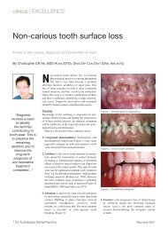
clinical | EXCELLENCE<br />
Failing implants,<br />
maintenance, recall<br />
By Christopher CK Ho, BDS (Hons), Grad.Dip.Clin.Dent (Oral <strong>Implants</strong>), M.Clin.Dent (Pros)<br />
Teck Tang, BDSc (Hons), D.Clin.Dent (<strong>Peri</strong>o), FRACDS (<strong>Peri</strong>o)<br />
The introduction of dental implants has<br />
expanded the armamentarium of dental practitioners<br />
in replacing missing teeth, however<br />
implant rehabilitation is no longer restricted to<br />
restoring function. <strong>Dental</strong> implants have become a<br />
multi-million dollar industry driven by bone augmentation,<br />
soft tissue management and aesthetic<br />
restorations. As dental practitioners, we constantly<br />
get bombarded with updates about the latest dental<br />
implants, new techniques and explanded indications<br />
from advertisements and other media. Very<br />
rarely however, do we hear of the complications<br />
that may arise with implant rehabilitation.<br />
In this article, we will deal with the issues of<br />
implant failures and complications and emphasise<br />
the importance of ongoing maintenance care.<br />
Implant failures<br />
Implant failures may be described as early or late.<br />
An early failure follows shortly after placement and<br />
osseointegration is never achieved. A late failure<br />
occurs in a successfully integrated implant some<br />
time after restoration.<br />
Early failures<br />
An early failure of an implant results from an inability<br />
to establish “an intimate bone-to-implant contact” or<br />
osseointegration (Quirynen et al. 2002). This means<br />
that problems have occurred with the bone healing<br />
process after implant placement. Commonly, it is<br />
related to traumatic surgery and occurrence of<br />
micromotion at the interface during the critical<br />
post-implantation phase (Esposito et al. 2000).<br />
Late failures<br />
Late implant failures have been associated with<br />
both peri-<strong>implantitis</strong> and/or occlusal overloading<br />
(van Steenberghe et al. 1990, Quirynen et al. 2002).<br />
Since the occlusal overload leads to failure shortly<br />
after implant fixture restoration, this could be considered<br />
to be an “intermediate failure” and should<br />
be easily averted by careful and judicious treatment<br />
planning. Thus true failures are most likely attributed<br />
to peri-implant infections.<br />
Implant survival and implant success<br />
There has always been some confusion in the literature<br />
in terms of defining implant survival and<br />
implant success. Implant survival means the presence<br />
of implants irrespective of the conditions of<br />
the implants. Implant success means the presence<br />
of implants with no interventions required. As yet,<br />
consensus agreement on criteria of success has not<br />
been achieved (van Steenberghe et al, 1999).<br />
138 Australasian <strong>Dental</strong> Practice January/February 2011
clinical | EXCELLENCE<br />
Figure 1. <strong>Peri</strong>-implant mucositis - bleeding on probing however no loss of bony attachment.<br />
Figure 2. <strong>Peri</strong>-<strong>implantitis</strong> - Inflammatory process characterised by early loss of peri-implant bone.<br />
Biological complications<br />
Biological complications may include all the soft tissue<br />
complications that may arise from the implant reconstructions<br />
such as hyperplasia, excessive tissue swelling, peri-implant<br />
mucositis and peri-<strong>implantitis</strong>. It has been reported that 28-56%<br />
of subjects with dental implants suffer peri-<strong>implantitis</strong> while 10-<br />
15% of subjects exhibit severe peri-<strong>implantitis</strong> (Zitzmann &<br />
Berglundh 2008). These figures are interestingly very similar to<br />
the ones <strong>for</strong> periodontitis.<br />
<strong>Peri</strong>-implant mucositis is defined as a reversible inflammatory<br />
process in the soft tissues surrounding a functioning implant, with<br />
no loss of bone (Figure 1). The inflammatory infiltrate adjacent<br />
to teeth and implants were found to be similar and suggests a<br />
similar host response in gingiva and peri-implant mucosa<br />
(Berglundh et al. 1992).<br />
<strong>Peri</strong>-<strong>implantitis</strong> is an inflammatory process characterised by<br />
additional loss of peri-implant bone (Figures 2-5). It is important<br />
to recognise that peri-<strong>implantitis</strong> is not a synonym <strong>for</strong> “failing<br />
implant” or “ailing implant”.<br />
Figure 3. Progression of peri-<strong>implantitis</strong> lesion with crater like<br />
loss of bone.<br />
Susceptibility <strong>for</strong> periodontitis and peri-<strong>implantitis</strong><br />
<strong>Peri</strong>odontitis is a multifactorial disease involving complex interactions<br />
between host and plaque, further modified by genetic and<br />
environmental factors. The most recognised factors related to susceptibility<br />
<strong>for</strong> periodontitis include plaque composition, smoking,<br />
genetics and various systemic conditions.<br />
January/February 2011 Australasian <strong>Dental</strong> Practice 139
clinical | EXCELLENCE<br />
Figure 4. Biologic and Technical Complications. <strong>Implants</strong> placed over 10 years ago, with technical complication in 16 implant with<br />
fractured abutment screw and biologic complications with peri-<strong>implantitis</strong> and subsequent loss of bone.<br />
Risk factors <strong>for</strong> peri-<strong>implantitis</strong><br />
• History of periodontitis<br />
• Smoking<br />
• Poor oral hygiene<br />
• Exposed threads<br />
• Exposed surface coatings (roughened surfaces)<br />
• Deep pockets (placed too deep, placed into deficiencies)<br />
• No plaque removal access (ridge lap crown,<br />
connected prostheses)<br />
Figure 5. Clinical picture of patient from Figure 4.<br />
Figure 6. Risk factors <strong>for</strong> peri-<strong>implantitis</strong>.<br />
Since the bacteria cannot differentiate between implants and<br />
teeth, it would be reasonable to assume that all other factors <strong>for</strong><br />
development of peri-<strong>implantitis</strong> remain the same <strong>for</strong> periodontitis.<br />
There<strong>for</strong>e, the susceptibility <strong>for</strong> peri-<strong>implantitis</strong> should be<br />
dependent on the susceptibility <strong>for</strong> periodontitis.<br />
Risk factors <strong>for</strong> peri-<strong>implantitis</strong><br />
Apart from a history of periodontitis, the other established risk factors<br />
<strong>for</strong> peri-<strong>implantitis</strong> include smoking and poor oral hygiene<br />
(Figure 6). It is imperative <strong>for</strong> clinicians to not only identify these<br />
risk factors but also to manage them to the best of their ability. This<br />
could include stabilising periodontal infections be<strong>for</strong>e implant<br />
therapy, providing ongoing maintenance care, delivering smoking<br />
cessation advice and also designing prosthesis that would allows<br />
easy oral hygiene practices (Figures 7 and 8).<br />
Does peri-<strong>implantitis</strong><br />
progress faster than periodontitis?<br />
<strong>Peri</strong>-<strong>implantitis</strong> is similar to periodontitis. They both involve<br />
alveolar bone loss. However, there are some differences. There<br />
is a zone of connective tissues being attached to the root<br />
surface in periodontitis. But in peri-<strong>implantitis</strong>, the connective<br />
tissue does not attach directly onto implants and there is<br />
no periodontal ligament, so the inflammatory lesion in<br />
peri-<strong>implantitis</strong> always extends closer to the bone surface<br />
(Figure 9) (Gualini & Berglundh 2003). There<strong>for</strong>e, it<br />
progresses faster and it is potentially a more aggressive disease<br />
and it is very hard to treat. Nevertheless, tissue degradation may<br />
be a slow process, as in chronic periodontitis, a function time<br />
exceeding 5 years <strong>for</strong> implants may be required to detect biological<br />
peri-implant complications.<br />
140 Australasian <strong>Dental</strong> Practice January/February 2011
clinical | EXCELLENCE<br />
Figure 7. Poor design of fixed prosthesis with buccal flange. This does not allow cleaning of the tissue fitting surface of the bridge<br />
with subsequent soft tissue inflammation evident from food trapping.<br />
Figure 8. All-ceramic fixed implant supported bridge with convex tissue fitting surface allowing cleansability by the patient with supefloss,<br />
waterpick or interdental aids.<br />
<strong>Implants</strong> in patients<br />
with periodontitis<br />
A number of studies have confirmed that<br />
patients with a history of periodontitis<br />
may yield lower success rates than<br />
patients without a history of periodontitis<br />
(Karoussis et al. 2004, Roos-Jansaker et<br />
al. 2006).<br />
Aggressive periodontitis<br />
So far, there are a few case reports <strong>for</strong><br />
implant therapy in aggressive periodontitis<br />
patients. The evidence is not<br />
conclusive and some reported failure and<br />
some reported success (Fardal et al. 1999,<br />
Malmstrom et al. 1990, Yalcin et al. 2001,<br />
Wu & Chee 2007).<br />
A study comparing implant success<br />
rates in aggressive periodontitis and<br />
chronic periodontitis patients showed that<br />
3-year implant success rate is slightly<br />
lower in aggressive periodontitis patients,<br />
but still well above 95% with strict periodontal<br />
maintenance regime (Mengel &<br />
Flores-de-Jacoby 2005).<br />
Refractory periodontitis<br />
For refractory periodontitis, if the periodontal<br />
infections are not under control,<br />
implant therapy should be delayed.<br />
Figure 9. <strong>Peri</strong>odontitis (left) vs peri-<strong>implantitis</strong> (right). Note there is no periodontal ligament<br />
attachment with implants (Image courtesy of Astratech).<br />
Maintenance and recall<br />
After successful periodontal and implant<br />
therapy, the patient should be offered<br />
an individually tailored maintenance care<br />
program. It is important to assess mobility,<br />
probing depth, bleeding on probing<br />
and suppuration during a recall visit.<br />
Radiographic and microbiological parameters<br />
are to be added, depending on the<br />
primary clinical findings. In addition, the<br />
occlusion of the suprastructures should<br />
not be overlooked.<br />
In the treatment of peri-<strong>implantitis</strong>,<br />
only limited scientific evidence is available<br />
to recommend any specific treatment<br />
modality. Most studies lack controls<br />
and randomization and are often<br />
handicapped by a small sample size.<br />
There are also limited reports on microbiological<br />
changes and histological changes<br />
following treatment.<br />
Nevertheless, a systematic approach <strong>for</strong><br />
monitoring tissues around implants in the<br />
prevention and treatment of peri-implant<br />
142 Australasian <strong>Dental</strong> Practice January/February 2011
clinical | EXCELLENCE<br />
PD ≤ 3mm<br />
PD 4 to 5mm<br />
Absence of plaque<br />
BOP negative<br />
Presence of plaque<br />
BOP+<br />
No treatment<br />
Mechanical<br />
debridement +<br />
polishing<br />
+<br />
Antiseptic<br />
cleansing<br />
A<br />
+<br />
B<br />
BOP+<br />
No bone loss<br />
+<br />
+<br />
Radiograph!<br />
PD ≥ 5mm<br />
BOP+<br />
Bone loss ≤ 2mm<br />
BOP+<br />
Bone loss > 2mm<br />
Systemic or local<br />
antibiotic therapy<br />
+<br />
Resective or<br />
regenerative<br />
surgery<br />
C<br />
+<br />
D<br />
Figure 10. Cumulative Interceptive Supportive Therapy (CIST) (Lang et al, 2004).<br />
Table 1. Cumulative Interceptive Supportive Therapy (CIST) modalities (Lang et al, 2004).<br />
A. Mechanical cleansing using rubber cups and polishing paster, acrylic scalers <strong>for</strong> chipping off calculus. Instruction <strong>for</strong> more<br />
effective oral hygiene practices.<br />
B. Antiseptic therapy. Rinses with 0.1% to 0.2% chlorhexidine digluconate <strong>for</strong> 30 seconds using approximately 10ml, <strong>for</strong><br />
3 to 4 weeks, supplemented by irrigating locally with chlorhexidine (preferably 0.2% to 0.5%) using a Luer syringe or local<br />
chlorhexidine gel application.<br />
C. Antibiotic therapy:<br />
1. Systemic ornidazole (2 x 500 mg/day) or metronidazole (3 x 250 mg/day) <strong>for</strong> 10 days or<br />
combination of metronidazole (500 mg/day) plus amoxicillin (375 mg/day) <strong>for</strong> 10 days.<br />
2. Local: application of antibiotics using controlled release devices <strong>for</strong> 10 days (25% Tetracycline fibres).<br />
D. Surgical approach:<br />
1. Regenerative surgery using abundant saline rinses at the defect, barrier membranes, close flap adaptation and careful<br />
post-surgical monitoring <strong>for</strong> several months. Plaque control is to be assured by applying chlorhexidine gels.<br />
2. Resective surgery. Apical repositioning of the flap following osteoplasty around the defect.<br />
disease has been recommended by Lang and coworkers in Berne,<br />
Switzerland. This systematic protocol, referred to as Cumulative<br />
Interceptive Supportive Therapy (CIST), contains four cumulative<br />
treatment modalities (A-D) (Figure 10, Table 1). Each step of<br />
the procedures is used in a sequential manner with increasing<br />
antibacterial intervention, combined with surgical<br />
resective/regenerative treatment (A+B+C+D). The CIST protocol<br />
has been shown to be effective in improvement of clinical and<br />
microbiological parameters in clinical studies (Mombelli et al.<br />
2001, Persson et al. 2006).<br />
144 Australasian <strong>Dental</strong> Practice January/February 2011
clinical | EXCELLENCE<br />
Conclusions<br />
<strong>Dental</strong> implants are an excellent option <strong>for</strong> replacing missing<br />
teeth, but problems may arise with such treatment. Early implant<br />
failures occur at a global rate of about 2.5%. Late implant failures<br />
are mainly related to peri-<strong>implantitis</strong>. In addition, technical and<br />
biological complications are common in implant therapy.<br />
Susceptibility <strong>for</strong> peri-<strong>implantitis</strong> is associated with susceptibility<br />
<strong>for</strong> periodontitis. There<strong>for</strong>e, every partially edentulous<br />
patient should receive appropriate periodontal screening and<br />
treatment prior to implant therapy. It is reasonable to place<br />
implants in periodontitis patients but they are at much greater<br />
risks of developing problems.<br />
Comprehensive treatment planning is paramount with regular<br />
recall and maintenance necessary to detect and intercept problems<br />
early. Instruction in oral hygiene and smoking cessation<br />
advice should be given. For periodontitis patients, regular supportive<br />
periodontal therapy and smooth and well contoured<br />
transmucosal abutments are required <strong>for</strong> the long term success of<br />
implant therapy.<br />
The cumulative interceptive supportive therapy protocol can be<br />
adopted in the prevention and treatment of peri-<strong>implantitis</strong>. However,<br />
there is limited scientific evidence to recommend any<br />
specific treatment modalities <strong>for</strong> peri-<strong>implantitis</strong>, with more clinical<br />
controlled trials required <strong>for</strong> the management of this problem.<br />
References<br />
1. Berglundh T, Persson L, Klinge B. (2002) A systematic review of the incidence of<br />
biological and technical complications in implant dentistry reported in prospective<br />
longitudinal studies of at least 5 years. Clinical Oral <strong>Implants</strong> Research 29 (Suppl.<br />
3): 197–212.<br />
2. Brägger U, Karoussis I, Persson R, Pjetursson B, Salvi G, Lang N. P. (2005) Technical<br />
and biological complications/failures with single crowns and fixed partial dentures<br />
on implants: a 10-year prospective cohort study. Clin. Oral Impl. Res. 326-334.<br />
3. Esposito M, Thomsen P, Sennerby L, Lekholm U. (2000) Histopathologic observations<br />
on late oral implant failures. Clin Implant Dent Relat Res. 2, 18-32.<br />
4. Fardal O, Johannessen AC, Olsen I. (1999) Severe, rapidly progressing peri<strong>implantitis</strong>.<br />
Journal of Clinical <strong>Peri</strong>odontology 26, 313–317.<br />
5. Gualini F, Berglundh T. (2003) Immunohistochemical characteristics of inflammatory<br />
lesions at implants. J Clin <strong>Peri</strong>o 30, 14-18.<br />
6. Karoussis K, Muller S, Salvi GE, Heitz-Mayfield L.J, Bragger U, Lang N.P.<br />
(2004) Association between periodontal and peri-implant conditions: a 10-year<br />
prospective study. Clinical Oral <strong>Implants</strong> Research 15, 1–7.<br />
7. Lang NP, Berglundh T, Heitz-Mayfield LJ, Pjetursson BE, Salvi GE, Sanz M.<br />
Consensus statements and recommended clinical procedures regarding implant survival<br />
and complications. Int J Oral Maxillofac <strong>Implants</strong> 2004;19 Suppl:150–154.<br />
8. Lang NP, Wilson TG, Corbet EF. (2000) Biological complications with dental<br />
implants: their prevention, diagnosis and treatment. Clinical Oral <strong>Implants</strong> Research<br />
11 (Suppl. 1): 146–155.<br />
9. Mengel R, Flores-de-Jacoby L. (2005) <strong>Implants</strong> in patients treated <strong>for</strong> generalized<br />
aggressive and chronic periodontitis: a 3-year prospective longitudinal study. J <strong>Peri</strong>odontol.<br />
76, 534-43.<br />
10. Mengel R, Schröder T, Flores-de-Jacoby L. (2001) Osseointegrated implants in<br />
patients treated <strong>for</strong> generalized chronic periodontitis and generalized aggressive<br />
periodontitis: 3- and 5-year results of a prospective long-term study. J <strong>Peri</strong>odontol.<br />
72, 977-89.<br />
11. Mombelli A, Feloutzis A, BraggerU, Lang, N.P. (2001) Treatment of peri<strong>implantitis</strong><br />
by local delivery of tetracycline. Clinical, microbiological and<br />
radiological results. Clinical Oral <strong>Implants</strong> Research 14, 404–411.<br />
12. Persson GR, Salvi GE, Heitz-Mayfield LJ, Lang NP. (2006) Antimicrobial<br />
therapy using a local drug delivery system (Arestin) in the treatment of peri-<strong>implantitis</strong>.<br />
I: Microbiological outcomes. Clin Oral <strong>Implants</strong> Res. 17, 386-93.<br />
13. Quirynen M, De Soete M, van Steenberghe D. (2002) Infectious risks <strong>for</strong> oral<br />
implants: a review of the literature. Clinical Oral <strong>Implants</strong> Research 13, 1–19.<br />
14. Roos-Jansaker AM, Renvert H, Lindahl C, Renvert S. (2006) Nine- to fourteenyear<br />
follow up of implant treatment. Part iii: factors associated with peri-implant<br />
lesions. Journal of Clinical <strong>Peri</strong>odontology 33. 296–301.<br />
15. van Steenberghe, D., Quirynen, M. & Naert, I. (1999) Survival and success rates<br />
with oral endosseous implants. In: Lang, N. P., Attstrom, R. & Lindhe, J. (eds). Proceedings<br />
of the 3de European Workshop on <strong>Peri</strong>odontology. 242–254. Berlin:<br />
Quintessence.<br />
16. Wu AY, Chee W. (2007) Implant-supported reconstruction in a patient with generalized<br />
aggressive periodontitis. J <strong>Peri</strong>odontol. 78, 777-82.<br />
17. Yalcin S, Yalçin F, Günay Y, Bellaz B, Onal S, Firatli E. (2001) Treatment of<br />
aggressive periodontitis by osseointegrated dental implants. A case report. J <strong>Peri</strong>odontol.<br />
72, 411-6.<br />
18. Zitzmann NU, Berglundh T. Definition and prevalence of peri-implant diseases.<br />
(2008) J Clin <strong>Peri</strong>odontol. 35(8 Suppl), 286-91.<br />
About the authors<br />
Dr Christopher Ho received his Bachelor of <strong>Dental</strong> Surgery with<br />
First Class Honours at the University of Sydney. He has completed<br />
postgraduate studies in the Graduate Diploma in Clinical<br />
Dentistry in Oral <strong>Implants</strong> at the University of Sydney and Masters<br />
of Clinical Dentistry in Prosthodontics with Distinction from<br />
Kings College, London. Dr Ho is a lecturer on aesthetic and<br />
implant dentistry locally and internationally and is involved with<br />
the evaluation and development of new dental products and materials.<br />
He is a faculty member with the UCLA/Global <strong>Institute</strong> <strong>for</strong><br />
<strong>Dental</strong> Education teaching in the one year Master programs in<br />
Esthetic Dentistry and Implant Dentistry. Dr Ho’s research interests<br />
are in immediate placement and loading of dental implants.<br />
He has a referral-based private practice in prosthodontic and<br />
implant dentistry in Sydney, Australia.<br />
Dr Teck Tang graduated with his Bachelor of <strong>Dental</strong> Science with<br />
Honours at the University of Western Australia. He then completed<br />
his specialist training in <strong>Peri</strong>odontics at the University of<br />
Adelaide. He was a recipient of the APA scholarship and received<br />
a prize <strong>for</strong> his research publication. He is a member of various<br />
organisations including the Australasian Osseointegration<br />
Society (ASO), Australian Society of <strong>Peri</strong>odontology (ASP) and<br />
ITI (International Team <strong>for</strong> Implantology). Currently, Dr Tang<br />
works in restricted practices, specialising in <strong>Peri</strong>odontics and<br />
Implant Dentistry. His interests range from management of periodontal<br />
diseases including periodontal regeneration to placing<br />
implants in patients with periodontitis.<br />
146 Australasian <strong>Dental</strong> Practice January/February 2011