Screening of Soil and Sheep Faecal Samples for Predacious Fungi ...
Screening of Soil and Sheep Faecal Samples for Predacious Fungi ...
Screening of Soil and Sheep Faecal Samples for Predacious Fungi ...

You also want an ePaper? Increase the reach of your titles
YUMPU automatically turns print PDFs into web optimized ePapers that Google loves.
Shams Ghahfarokhi et al.<br />
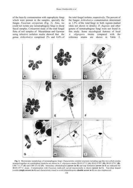
<strong>of</strong> the heavily contamination with saprophytic fungi<br />
which were present in the samples, specially the<br />
fungus Fusarium oxysporum (Fig. 2). Also, we<br />
could not isolate any nematophagous fungi in sheep<br />
faecal samples. Concurrent study <strong>of</strong> the total fungal<br />
flora <strong>of</strong> soil samples <strong>of</strong> Maz<strong>and</strong>aran <strong>and</strong> Garmsar<br />
using selective isolation media showed that the<br />
genus Arthrobotrys comprised 2% <strong>and</strong> 0.4% <strong>of</strong><br />
the total fungal isolates, respectively. The percent <strong>of</strong><br />
the fungus Arthrobotrys contamination determined<br />
as 1.5% <strong>of</strong> the total fungi in both regions studied<br />
(data not shown in details). D. flagrans <strong>and</strong> other<br />
genera <strong>of</strong> nematophagous fungi were not found in<br />
this study. Some mycological features <strong>of</strong> local<br />
A. oligospora strains compared with the<br />
reference strains are shown in Table 2.<br />
Fig. 2. Microscopic morphology <strong>of</strong> nematophagous fungi. Characteristic conidial structures including egg-like two celled conidia<br />
arranged together on conidiophore denticles are shown in A. oligospora strains IRAN 877 C (A), IRAN 878 C (D), IRAN 879 C (B),<br />
CBS 111.37 (E) <strong>and</strong> CBS 251.82 (F). Intercalary chlamidospore (arrow in H) <strong>and</strong> trap <strong>for</strong>mation (arrow in I) are indicated <strong>for</strong><br />
IRAN 877 C strain. Contamination with Fusarium multi-cell curved macroconidia is shown (arrow in C). Tear-drop shaped<br />
conidia (single arrows in G) <strong>and</strong> characteristic round to ovoid chlamidospores (double arrow in G) are also emphasized.<br />
138