Arnold Chiari Malformation with Holocord Syringomyelia ... - IJPMR
Arnold Chiari Malformation with Holocord Syringomyelia ... - IJPMR
Arnold Chiari Malformation with Holocord Syringomyelia ... - IJPMR

Create successful ePaper yourself
Turn your PDF publications into a flip-book with our unique Google optimized e-Paper software.
123<br />
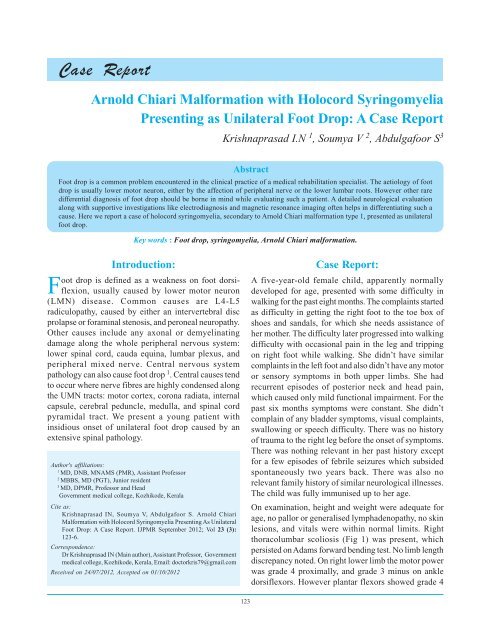
Case Report<br />
<strong>Arnold</strong> <strong>Chiari</strong> <strong>Malformation</strong> <strong>with</strong> <strong>Holocord</strong> <strong>Syringomyelia</strong><br />
Presenting as Unilateral Foot Drop: A Case Report<br />
Krishnaprasad I.N 1 , Soumya V 2 , Abdulgafoor S 3<br />
Abstract<br />
Foot drop is a common problem encountered in the clinical practice of a medical rehabilitation specialist. The aetiology of foot<br />
drop is usually lower motor neuron, either by the affection of peripheral nerve or the lower lumbar roots. However other rare<br />
differential diagnosis of foot drop should be borne in mind while evaluating such a patient. A detailed neurological evaluation<br />
along <strong>with</strong> supportive investigations like electrodiagnosis and magnetic resonance imaging often helps in differentiating such a<br />
cause. Here we report a case of holocord syringomyelia, secondary to <strong>Arnold</strong> <strong>Chiari</strong> malformation type 1, presented as unilateral<br />
foot drop.<br />
Key words : Foot drop, syringomyelia, <strong>Arnold</strong> <strong>Chiari</strong> malformation.<br />
Introduction:<br />
Foot drop is defined as a weakness on foot dorsiflexion,<br />
usually caused by lower motor neuron<br />
(LMN) disease. Common causes are L4-L5<br />
radiculopathy, caused by either an intervertebral disc<br />
prolapse or foraminal stenosis, and peroneal neuropathy.<br />
Other causes include any axonal or demyelinating<br />
damage along the whole peripheral nervous system:<br />
lower spinal cord, cauda equina, lumbar plexus, and<br />
peripheral mixed nerve. Central nervous system<br />
pathology can also cause foot drop 1 . Central causes tend<br />
to occur where nerve fibres are highly condensed along<br />
the UMN tracts: motor cortex, corona radiata, internal<br />
capsule, cerebral peduncle, medulla, and spinal cord<br />
pyramidal tract. We present a young patient <strong>with</strong><br />
insidious onset of unilateral foot drop caused by an<br />
extensive spinal pathology.<br />
Author's affiliations:<br />
1<br />
MD, DNB, MNAMS (PMR), Assistant Professor<br />
2<br />
MBBS, MD (PGT), Junior resident<br />
3<br />
MD, DPMR, Professor and Head<br />
Government medical college, Kozhikode, Kerala<br />
Cite as:<br />
Krishnaprasad IN, Soumya V, Abdulgafoor S. <strong>Arnold</strong> <strong>Chiari</strong><br />
<strong>Malformation</strong> <strong>with</strong> <strong>Holocord</strong> <strong>Syringomyelia</strong> Presenting As Unilateral<br />
Foot Drop: A Case Report. <strong>IJPMR</strong> September 2012; Vol 23 (3):<br />
123-6.<br />
Correspondence:<br />
Dr Krishnaprasad IN (Main author), Assistant Professor, Government<br />
medical college, Kozhikode, Kerala, Email: doctorkris79@gmail.com<br />
Received on 24/07/2012, Accepted on 01/10/2012<br />
Case Report:<br />
A five-year-old female child, apparently normally<br />
developed for age, presented <strong>with</strong> some difficulty in<br />
walking for the past eight months. The complaints started<br />
as difficulty in getting the right foot to the toe box of<br />
shoes and sandals, for which she needs assistance of<br />
her mother. The difficulty later progressed into walking<br />
difficulty <strong>with</strong> occasional pain in the leg and tripping<br />
on right foot while walking. She didn’t have similar<br />
complaints in the left foot and also didn’t have any motor<br />
or sensory symptoms in both upper limbs. She had<br />
recurrent episodes of posterior neck and head pain,<br />
which caused only mild functional impairment. For the<br />
past six months symptoms were constant. She didn’t<br />
complain of any bladder symptoms, visual complaints,<br />
swallowing or speech difficulty. There was no history<br />
of trauma to the right leg before the onset of symptoms.<br />
There was nothing relevant in her past history except<br />
for a few episodes of febrile seizures which subsided<br />
spontaneously two years back. There was also no<br />
relevant family history of similar neurological illnesses.<br />
The child was fully immunised up to her age.<br />
On examination, height and weight were adequate for<br />
age, no pallor or generalised lymphadenopathy, no skin<br />
lesions, and vitals were <strong>with</strong>in normal limits. Right<br />
thoracolumbar scoliosis (Fig 1) was present, which<br />
persisted on Adams forward bending test. No limb length<br />
discrepancy noted. On right lower limb the motor power<br />
was grade 4 proximally, and grade 3 minus on ankle<br />
dorsiflexors. However plantar flexors showed grade 4<br />
September Issue-2012 (3rd Proof)<br />
123
124<br />
power. Left lower limb and both upper limbs showed<br />
grade 5 power. Deep tendon reflexes were normally<br />
elicitable from all four limbs, except right ankle jerk<br />
which was sluggish. Plantar reflex was not elicitable on<br />
right side. Mild wasting of hypothenar muscles and mild<br />
clubbing were noticed on both hands (Fig 2). Grip was<br />
moderate and appropriate for her age. There were no<br />
sensory findings in all four limbs or trunk. Cranial nerves<br />
and cerebellar function tests were <strong>with</strong>in normal limits.<br />
<strong>IJPMR</strong> 2012 Sep; 23(3) : 123-6<br />
Nerve conduction study was performed in all four limbs.<br />
CMAP and SNAP were recorded from median, ulnar,<br />
radial, peroneal, tibial and sural nerves. Bilateral median<br />
CMAP and SNAP were found to be <strong>with</strong>in normal limits.<br />
Low amplitude CMAPs were recorded from both ulnar<br />
nerves suggestive of an axonal loss pattern, whereas<br />
corresponding SNAPs were <strong>with</strong>in normal limits. Both<br />
tibial CMAPs were <strong>with</strong>in normal limits. Low amplitude<br />
axonal loss pattern was recorded from both peroneal<br />
nerve CMAPs, <strong>with</strong> normal SNAPs. Sural SNAP also<br />
recorded normally. F waves were either absent or latency<br />
prolonged from all motor nerves suggested. The whole<br />
NCS picture pointed towards an anterior horn cell<br />
involvement, and further work up <strong>with</strong> MRI was planned<br />
X-ray of spine (Fig 3) showed thoracolumbar scoliosis<br />
towards right <strong>with</strong> apex at T12 and absent rotation.<br />
Cobb’s angle measured was 30 degrees. No other<br />
abnormalities were detected.<br />
MRI of the spine (Fig 4) <strong>with</strong> post contrast enhancement<br />
showed extensive syringohydromyelia involving the<br />
Fig 1- Thoracolumbar Scoliosis<br />
Fig 2- Wasting of Hypothenar Muscle<br />
Fig 3- Straight X-ray Showing Scoliosis<br />
September Issue-2012 (3rd Proof)
<strong>Arnold</strong> <strong>Chiari</strong> <strong>Malformation</strong> <strong>with</strong> holocord <strong>Syringomyelia</strong> – Krishnaprasad IN et al<br />
125<br />
Fig 4- MRI Showing Involvement of Spinal Cord<br />
entire cord from C2 to T10 <strong>with</strong> no abnormal contrast<br />
enhancement. Herniation of cerebellar tonsils noted 4<br />
mm below foramen magnum. The findings were<br />
consistant <strong>with</strong> <strong>Arnold</strong> <strong>Chiari</strong> malformation type 1.<br />
Discussion:<br />
Syrinxes associated <strong>with</strong> congenital foramen magnum<br />
encroachment (<strong>Chiari</strong> malformations) may result from<br />
either central canal dilation (i.e,communicating<br />
syringomyelia or hydromyelia) or from an eccentric<br />
syrinx cavity in the gray matter of the cord (noncommunicating<br />
syringomyelia). Because the two types<br />
of syrinxes are often indistinguishable by imaging<br />
studies, particularly later in the disease, they may be<br />
referred to as syringohydromyelia or<br />
hydrosyringomyelia.<br />
<strong>Chiari</strong> I malformation is a congenital downward<br />
displacement of the cerebellar tonsils through the<br />
foramen magnum, into the cervical subarachnoid space.<br />
<strong>Chiari</strong> II malformation is downward displacement of the<br />
cerebellar vermis, pons, and medulla into the foramen<br />
magnum and elongation of the fourth ventricle. <strong>Chiari</strong> I<br />
malformation typically presents clinically in young<br />
adults, whereas <strong>Chiari</strong> II malformation presents in<br />
infants and is usually associated <strong>with</strong><br />
myelomeningocoele and hydrocephalus. <strong>Chiari</strong> I<br />
malformation is the most common cause of<br />
syringomyelia. Although it usually presents clinically<br />
in young adult years, it may manifest first in an infant<br />
or older adult. The duration from onset of symptoms to<br />
diagnosis is typically 3 to 7 years.<br />
The earliest symptom is often headache or neck or arm<br />
pain aggravated by straining or coughs; neck pain may<br />
be accompanied by torticollis. Neurologic findings<br />
depend on the structures involved. The syrinx is often<br />
noted at the C4–6 bony levels but may extend rostrally<br />
or caudally the full length of the cord (holocord<br />
syringomyelia). Dissociated sensory loss in a cape like<br />
distribution over the neck and arms is characteristic,<br />
because crossing spinothalamic tract fibres carrying pain<br />
and temperature sensation are most affected, whereas<br />
posterior column sensory fibres are spared. Hand and<br />
arm weakness develop as lower motor neurons of the<br />
cervical cord are affected. Long-tract myelopathic<br />
symptoms develop in the lower limbs <strong>with</strong> further<br />
expansion of the cervical syrinx.<br />
Worsening scoliosis is common, particularly in<br />
childhood-onset syringomyelia of <strong>Chiari</strong> I malformation.<br />
Other associated craniocervical abnormalities like<br />
Klippel–Feil anomaly (congenital fusion of cervical<br />
vertebrae) and atlanto-occipital assimilation may<br />
coexist. Syrinx extension into the brainstem can lead to<br />
lower cranial nerve, cerebellar and respiratory problems.<br />
Rostral or caudal extension of the syrinx may result from<br />
rapid changes in intraspinal pressure, such as those<br />
caused by coughing, straining, or sneezing.<br />
<strong>Chiari</strong> II malformation often presents in infancy as<br />
stridor, weak cry, nystagmus, and apnoea or in childhood<br />
as gait abnormality, spasms, worsening incoordination,<br />
and nystagmus. Later, it may lead to loss of head control,<br />
arm weakness, spasms, and tetraparesis.<br />
Differential diagnosis for syringomyelia of <strong>Chiari</strong> I<br />
malformation includes multiple sclerosis; spinal<br />
muscular atrophy, amyotrophic lateral sclerosis;<br />
spinocerebellar ataxias, cervical disc and post-traumatic<br />
syringomyelia from trauma, arteriovenous malformation,<br />
arachnoiditis or meningitis, neurofibromatosis, and from<br />
spinal cord or brainstem tumours.<br />
MRI confirms the diagnosis, but asymptomatic tonsillar<br />
herniation is common. Because cerebellar tonsils retract<br />
upward <strong>with</strong> age, MRI interpretation depends on ageappropriate<br />
controls. Tonsillar herniations greater than<br />
September Issue-2012 (3rd Proof)
126<br />
6 mm are significant up to age 10 (In our patient it was<br />
4 mm), greater than 5 mm for ages 10 to 30, and greater<br />
than 4 mm for ages over 30. However, 30 per cent of<br />
persons <strong>with</strong> cerebellar herniations of 5 to 10 mm are<br />
asymptomatic.<br />
Treatment of <strong>Chiari</strong> I malformation involves posterior<br />
fossa decompression <strong>with</strong> a suboccipital craniectomy,<br />
<strong>with</strong> or <strong>with</strong>out dural patch grafting, and cervical<br />
laminectomy <strong>with</strong> fenestration or shunting of the syrinx<br />
cavity. In patients <strong>with</strong> mild symptoms, 70 to 80 per<br />
cent report improvement. Some authors 2 suggest that<br />
direct surgery of the syrinx should be undertaken only<br />
after craniocervical decompression has failed.<br />
A “top-down” approach has been suggested for surgical<br />
treatment of <strong>Chiari</strong> II malformation: shunt for<br />
hydrocephalus, then posterior fossa decompression for<br />
<strong>Chiari</strong> malformation, then syringopleural shunt for<br />
syringomyelia if needed.<br />
After posterior decompression, <strong>with</strong> or <strong>with</strong>out syrinx<br />
shunting, 50 per cent or more patients improve; in those<br />
<strong>with</strong> syrinxes, about one-third improves, one-third<br />
stabilises, and one-third deteriorates further after surgery.<br />
Postoperative neurologic decline can result from<br />
recurrent syringomyelia, occipital C1–C2 instability,<br />
pseudomeningocele, tethering of the spinal cord,<br />
meningitis, and extradural abscess.<br />
Conclusion:<br />
Bilateral progressive foot drop as a manifestation of<br />
holocord syringomyelia has been reported in the<br />
literature 3-5 . In all those reports, however there were<br />
other manifestations of typical syringomyelia which<br />
helped in differentiating the condition. In our patient,<br />
despite extensive spinal involvement the manifestations<br />
are limited to scattered motor findings and scoliosis.<br />
Other prominent feature of syringomyelia, the<br />
dissociated sensory loss, was conspicuously absent and<br />
inexplicable. Since nerve conduction studies showed<br />
evidence of moderate axonal loss in the CMAPs from<br />
peroneal and ulnar nerves bilaterally <strong>with</strong> abnormality<br />
in F waves, a pathology involving the anterior horn cells<br />
was suspected. Those findings along <strong>with</strong> the presence<br />
of scoliosis, prompted us to do a spinal work up <strong>with</strong><br />
MRI which helped us to establish the diagnosis. This<br />
case stresses the importance of conducting a<br />
comprehensive clinical examination, ably supported by<br />
appropriate investigations, in any patient presenting <strong>with</strong><br />
a seemingly peripheral lesion.<br />
We took a neurosurgery opinion for the child for any<br />
surgical intervention. But since the clinical features were<br />
static at present and child was not significantly disabled,<br />
it was decided to manage conservatively at present, and<br />
consider decompression only if clinical worsening<br />
observed on follow-up. The child was given an ankle<br />
foot orthosis to aid ambulation and a molded spinal<br />
jacket to prevent spinal curve progression, and is<br />
currently under active follow-up.<br />
References:<br />
<strong>IJPMR</strong> 2012 Sep; 23(3) : 123-6<br />
1. Westhout FD, Pare´ LS, Linskey ME. Central causes of foot<br />
drop: rare and underappreciated differential diagnoses. J Spinal<br />
Cord Med 2007; 30: 62-6.<br />
2. Little JW. <strong>Syringomyelia</strong>. Lin VW, Cardenas DD In: editor<br />
Spinal Cord Medicine: Principles and Practice. New York;<br />
2003: 504-5.<br />
3. Muhn N, Baker SK, Hollenberg RD, Meaney BF, Tarnopolsky<br />
MA. <strong>Syringomyelia</strong> presenting as rapidly progressive foot drop.<br />
J Clin Neuromuscular Disord 2002; 3: 133-4.<br />
4. J Kumar, A Kumar, S Gupta. <strong>Chiari</strong> I malformation <strong>with</strong><br />
holocord syrinx. J Neurol Neurosurg Psychiatry 2007; 78: 146.<br />
5. Saifudheen K, Jose J, Gafoor VA. <strong>Holocord</strong> syringomyelia<br />
presenting as rapidly progressive foot drop. J Neurosci Rural<br />
Pract 2011; 2: 195-6.<br />
September Issue-2012 (3rd Proof)