

Witzelsucht after Right Putaminal Hemorrhage: A Case Report
Witzelsucht after Right Putaminal Hemorrhage: A Case Report
Witzelsucht after Right Putaminal Hemorrhage: A Case Report

You also want an ePaper? Increase the reach of your titles
YUMPU automatically turns print PDFs into web optimized ePapers that Google loves.
<strong>Witzelsucht</strong> <strong>after</strong> <strong>Right</strong> <strong>Putaminal</strong> <strong>Hemorrhage</strong>: A <strong>Case</strong> <strong>Report</strong><br />
INTRODUCTION<br />
The German “witzelsucht” means a tendency to tell<br />
inappropriate and poor jokes. Patients with witzelsucht<br />
have a habitual routine of telling jokes and pointless stories<br />
filled with jocularity in socially inappropriate circumstances,<br />
and applying many witticisms and quips to<br />
conversations, while being oneself inordinately entertained<br />
thereby. But, surprisingly, they are paradoxically<br />
insensitive to humor. More than 100 years ago, witzelsucht<br />
was described following right frontal tumors (1) .<br />
Here we present a case with witzelsucht and hypersexuality<br />
<strong>after</strong> a hemorrhagic stroke.<br />
From the Department of Neurology, National Cheng Kung<br />
University Medical Center, Tainan, Taiwan.<br />
Received April 25, 2005. Revised June 3, 2005.<br />
Accepted August 5, 2005.<br />
Ying-Chu Chen, Chi-Yu Tseng, and Ming-Chyi Pai<br />
Abstract- <strong>Witzelsucht</strong> is a tendency to tell inappropriate and poor jokes. It usually occurs <strong>after</strong> a focal<br />
lesion involving orbitofrontal cortical or paramedian thalamic regions, especially on the right side. Here we<br />
report a 56-year-old man developing witzelsucht and hypersexuality <strong>after</strong> a right putaminal hemorrhage.<br />
The hematoma extended to the sublenticular part of posterior internal capsule and mesencephalon. The<br />
hemorrhage might have disconnected the fibers in the ascending reticular systems, and the fibers between<br />
paramedian thalamus and orbitofrontal cortex, and thus could be responsible for the patient’s rare clinical<br />
manifestations.<br />
Key Words: Humor, Hypersexuality, Orbitofrontal lobe, Paramedian thalamus, <strong>Putaminal</strong> hemorrhage,<br />
<strong>Witzelsucht</strong><br />
Acta Neurologica Taiwanica Vol 14 No 4 December 2005<br />
Acta Neurol Taiwan 2005;14:195-200<br />
CASE REPORT<br />
195<br />
A right-handed 56-year-old man, KS, with 8 years<br />
of formal education, was a factory owner. He had a history<br />
of hypertension without medication for many years,<br />
but no history of stroke or psychiatric illness. KS suffered<br />
from an acute onset of left hemiparesis, sensory<br />
loss on the left side of his body, and dysarthria in a<br />
morning <strong>after</strong> playing mahjong overnight. The paresis<br />
of extremities progressed rapidly to dense paralysis<br />
within several minutes, and he became dull. On arrival<br />
at the emergency room, KS presented with, in addition<br />
to the aforementioned symptoms, evident left central<br />
Reprint requests and correspondence to: Ming-Chyi Pai, MD,<br />
PhD. Division of Behavioral Neurology, Department of<br />
Neurology, National Cheng Kung University Hospital, No.<br />
138, Sheng Li Road, Tainan, Taiwan.<br />
E-mail: pair@mail.ncku.edu.tw
196<br />
type facial palsy, dysphagia, and a dense visual field<br />
defect on his left side. KS was oriented and responded<br />
properly, although slowly. He did not undergo surgical<br />
intervention because the hematoma was small.<br />
In the following days, KS became alert gradually. He<br />
would complain of nasal obstruction, headache, and<br />
request oral intake to replace tube feeding which made<br />
him feel uncomfortable. On the 5th day, KS could speak<br />
for a longer period of time, yet developed inappropriate<br />
jokes and exaggerated similes. At that time, KS was alert<br />
and cooperative, and had no disorientation, delusions,<br />
hallucinations, or emotional liability. He had mild<br />
dysarthria and dysphagia, but no abnormal crying or<br />
laughters. He was euphoric, outspoken, prankish, and<br />
was so talkative that an interruption was usually needed<br />
to pull the conversation back to the topic or to complete<br />
a test. KS frequently spoke to attract others’ attention<br />
with a tendency of exaggeration. He also talked a lot of<br />
seeming witticisms and quips. On some occasions, he<br />
showed no smiles or laughter to the jokes he talked<br />
which made everyone laugh loudly, while on other occasions,<br />
he was not able to appreciate jokes from the others.<br />
However, the content of his chats was not bizarre.<br />
For instance, KS called his attending physician “Kong-<br />
Ming”, who was a famous prime minister in ancient<br />
China, and the residents and clerks “Kong-Ming’s generals<br />
and soldiers”. He also interpreted the medical team as<br />
a troop to fight his disease. KS had no anosognosia or<br />
asomatagnosia. Sometimes, he used right hand to raise<br />
the weak left arm repeatedly for rehabilitation, and asked<br />
doctors to do their best to take care of his weak limbs.<br />
He was concerned about the functional deficits, but<br />
talked of them humorously. Freehand copying of representational<br />
drawings, such as a clock-face or a flowerhead,<br />
was fair and showed no hemispatial neglect.<br />
Hypersexuality developed at the same time. KS<br />
would use erotic words when there were women nearby.<br />
He harassed young nurses and a middle aged caregiver,<br />
with amorous language, and would even touch them<br />
from time to time. KS called the caregiver “Honey”, and<br />
asked her to be his “ladylove”. He also wanted her to<br />
obey his orders, including being touched by him. He<br />
would become childish, or even angry, if she refused his<br />
Acta Neurologica Taiwanica Vol 14 No 4 December 2005<br />
requests. On one occasion, KS called a young female<br />
clerk “a little hen” in his vernacular language. He did not<br />
care about the complaints from others, and was unable to<br />
correct his inappropriate behaviors. KS’s family was surprised<br />
at his inappropriate jokes and the hypersexual<br />
behaviors, which were quite different from that he<br />
behaved before the stroke. Before the stroke, he did talk<br />
humorously sometimes. But he had never told jokes<br />
incongruous with the context, or behaved impolitely or<br />
brusquely to females.<br />
He was transferred to the rehabilitation ward on the<br />
11th day of hospitalization. Sertraline, a selective serotonin<br />
reuptake inhibitor, was given the next day at daily<br />
dose of 50 mg. Moderate reduction of the aberrant<br />
behaviors was noted. The effect, however, lasted for no<br />
more than three weeks. Two months later, sertraline was<br />
discontinued. The dense left hemiplegia improved gradually<br />
in the following two months when he could walk<br />
with a cane. The sensory loss on the left side of body<br />
also was much improved. However, KS’s wife still<br />
reported that the endless jokes of KS were not only inappropriate<br />
in terms of context, but also erotic or even<br />
obscene. Medications with venlafaxine, a serotonin and<br />
norepinephrine reuptake inhibitor, were instituted roughly<br />
four months <strong>after</strong> the stroke. Interestingly, the behavioral<br />
changes were noticeably reduced <strong>after</strong> the medication<br />
with venlafaxine at a daily dose of 37.5 mg for two<br />
weeks. In the following two months, inappropriate jokes<br />
and hypersexual behaviors were rarely noticed. No<br />
relapse of aberrant behaviors was noted before this<br />
report was written.<br />
INVESTIGATION<br />
Two hours <strong>after</strong> the stroke, a cerebral CT scan was<br />
done and showed a hematoma at the right putamen (Fig.<br />
A). The hematoma was roughly 8-10 ml in volume and<br />
extended into the posterior and lateral portion of thalamus<br />
as well as the rostral midbrain (Fig. B). A brain<br />
MRI was done on the 11th day of hospitalizatoion,<br />
showing extension of the hematoma to the posterior and<br />
lateral, but not paramedian, portion of the right thalamus<br />
(Fig. C). HMPAO-SPECT was performed also on the
A B<br />
C<br />
D E F<br />
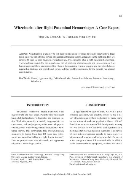
Figure. (A) The Brain CT image shows right putaminal hemorrhage with extension to the posterolateral portion of thalamus and<br />
internal capsule, as well as the midbrain (B), two hours affer the stroke; (C) The coronal section of T1-weighted brain MRI<br />
image shows a hematoma occupying the right subcortical region. Note that the paramedian thalamus is not involved; (D)<br />
A sketch depicts the ansa lenticularis and inferior thalamic peduncle on the coronal view of cerebrum; (E) The sagittal<br />
section of T1-weighted brain MRI image shows that the rostral midbrain is involved by the subcortical hematoma; (F) A<br />
sketch depicts the ascending reticular system and medial forebrain bundle on the sagittal view of diencephalons and midbrain.<br />
Abbreviations for (E) and (F): P: putamen, T: thalamus, AP: ansa peduncularis, ITP: inferior thalamic peduncle, C:<br />
corpus callosum, D: diencephalon, M: midbrain, DTN: dorsal tegmental nucleus, VTN: ventral tegmental nucleus, RN: red<br />
nucleus, LC: locus ceruleus, MFB: medial forebrain bundle.<br />
11th day, and showed perfusion defect in the right basal<br />
ganglion and thalamus, as well as hypoperfusion of right<br />
temporal and posterior parietal lobes. There was no<br />
reduction of perfusion in bilateral frontal lobes.<br />
Cognitive Ability Screening Instrument (CASI) (2)<br />
was performed on the 13th day, showing deficits in the<br />
domains of recent memory, orientation, abstract thinking,<br />
drawing, and verbal fluency. The score of Mini-<br />
Mental State Examination (MMSE) (3) was 23/30.<br />
DISCUSSION<br />
<strong>Witzelsucht</strong>: a disorder of humor<br />
Humor is a highly evolved cognitive ability, and,<br />
Acta Neurologica Taiwanica Vol 14 No 4 December 2005<br />
197<br />
like the other cognitive functions, it may be impaired by<br />
cerebral damages. The disorders of humor include<br />
inability to produce or appreciate jokes, and addiction of<br />
telling inappropriate jokes (4) . A striking example of the<br />
disorders of humor is witzelsucht, which comprises two<br />
seemingly paradoxical manifestations: excessive and<br />
inappropriate production of jokes, and impaired appreciation<br />
of humor. The most frequently mentioned anatomical<br />
location of the lesion responsible for witzelsucht is in<br />
the orbitofrontal region, especially on the right side (5-7) .<br />
Historically, the frontal lobe dysfunction has been related<br />
to personality changes, including pronounced alteration<br />
on the production of and response to humor (8,9) .<br />
Previous investigations on the effects of cerebral lesion
198<br />
on humor have suggested the important role of the right<br />
hemisphere in the appreciation of humor (10-13) . Shammi<br />
and Stuss also argued the unique role of the right frontal<br />
lobe in the integration of different cognitive and affective<br />
information, including complex human abilities<br />
such as episodic memory, self-awareness, and humor (4) .<br />
Several cases of witzelsucht with unilateral or bilateral<br />
paramedian thalamic lesions (14-18) have also been reported.<br />
The disorders of humor are distinct from disorders of<br />
laugh. Laugh is normally an expression of merriment<br />
with typical facial movements and clonic contractions of<br />
the expiratory muscles (19) . Disorders of laugh can occur<br />
when this expression is inappropriate or out of control .<br />
The most common disorders of laugh are associated with<br />
pseudobulbar palsy (20) . It can be triggered by trivial stimuli,<br />
and may be incongruent with the underlying mood,<br />
or even intermixed with crying (19) .<br />
Anatomical correlation between witzelsucht and<br />
subcortical lesions<br />
Our case, KS, had no previous history of psychiatric<br />
or personality disorder, and no emotional incontinence<br />
either. He was alert and cooperative <strong>after</strong> the stroke.<br />
Thus, his witzelsucht and hypersexuality are most likely<br />
consequences of the stroke. However, the hematoma did<br />
not involve the frontal lobe on either side (the most frequently<br />
reported anatomical locations responsible for<br />
such symptoms). Moreover, the paramedian thalamic<br />
structures, another previously reported location (14-18) , were<br />
also not damaged according to the MRI findings. The<br />
SPECT study did show a perfusion defect in the right<br />
thalamus, a finding perhaps ascribable to the mass effect<br />
of the hematoma. However, this defect is unlikely the<br />
major etiological factor for KS’s witzelsucht because of<br />
the persistence of the symptoms for 4 months.<br />
It has been known that emotion and its expression<br />
are highly related to the basal forebrain, orbitofrontal<br />
region, piriform cortex, and amygdala, all of which are<br />
connected with part of the diencephalon such as paramedian<br />
thalamus and hypothalamus (21-24) . The paramedian<br />
thalamic complex comprises mediodorsal, medioventral,<br />
and mass intermedia nuclei (25) . The mediodorsal nuclei<br />
consists of three components, namely, magnocellular,<br />
Acta Neurologica Taiwanica Vol 14 No 4 December 2005<br />
parvicellar, and paralaminar (26) , and may play a major<br />
role in emotional control. The magnocellular component<br />
has reciprocal connections with orbitofrontal and piriform<br />
cortex (27) . The connections comprise part of the<br />
ansa peduncularis, including the inferior thalamic peduncle<br />
and fibers interconnecting amygdala and hypothalamus<br />
(28,29) . In the cat, piriform cortex lesions may cause<br />
hypersexuality (30) . We therefore propose that disconnection<br />
between the paramedical thalamic complex and the<br />
frontal-limbic system might play a major role in the genesis<br />
of witzelsucht in KS, as the MRI study clearly<br />
showed involvement of the ansa peduncularis and the<br />
inferior thalamic peduncle (31) (Fig. C). The inferior thalamic<br />
peduncle goes through the sublenticular part of the<br />
posterior internal capsule which is ventral to putamen<br />
(29,31-33) (Fig. D). Consistently, cases with similar<br />
symptoms to those of KS have been reported <strong>after</strong> small<br />
lesions in the ventrostriatum and substantia<br />
innominata (31) .<br />
On the other hand, ascending reticular systems in the<br />
brainstem may also affect emotional expression (34) . For<br />
example, raphe nucleus, locus ceruleus, and some disseminated<br />
neurons in the mesencephalic tegmentum<br />
send serotonergic, adrenergic, and dopaminergic efferents<br />
through the medial forebrain bundle to the<br />
orbitofrontal cortex and diencephalon (35,36) (Fig. F).<br />
Specific mesencephalic reticular nuclei such as the ventral<br />
and dorsal tegmental nuclei send fibers to join the<br />
ascending systems in the median forebrain bundle for<br />
emotional modulation (34,37) . Because the hematoma of KS<br />
extended down to the mesencephalic tegmentum (Fig.<br />
E), it might have interrupted the ascending reticular systems.<br />
The temporary responsiveness to sertraline and a<br />
sustained relief of symptoms by venlafaxine could support<br />
the important role of the ascending serotonin system<br />
in the genesis of the behavioral symptoms in KS.<br />
CONCLUSION<br />
This is a case with not only unusual manifestations<br />
with a relatively common stroke, but also a unique localization<br />
for a special behavioral disorder. We further suggest<br />
that the frontal lobe syndrome-like symtoms in this
patient may result from disconnection of the fronto-limbic<br />
cortex from its related subcortical structures (including<br />
paramedian diencephalon and upper brainstem).<br />
These findings also support the essential role of right<br />
hemisphere in the production and appreciation of humor,<br />
as well as in emotional control.<br />
REFERENCES<br />
1. Oppenheim H. Zur Pathologie der Gehirngeschwulste.<br />
Archiv fur Psychiatrie und Nervenkrankheiten 1890;21:<br />
560-87,705-45.<br />
2. Teng EL, Hasegawa K, Homma A, et al. The Cognitive<br />
Ability Screening Instrument (CASI): a practical test for<br />
cross-cultural epidemiological studies of dementia. Int<br />
Psychogeriatr 1994;6:45-58.<br />
3. Folstein M, Folstein S, McHugh P. “Mini-Mental State”: a<br />
practical method for grading the cognitive impairment of<br />
patients for the clinician. J Psychiatry Res 1975;12:189-98.<br />
4. Shammi P, Stuss DT. Humour appreciation: a role of the<br />
right frontal lobe. Brain 1999;122:657-66.<br />
5. Vardi J, Finkelstein Y, Zlotogorski Z, et al. L’homme qui<br />
rit: inappropriate laughter and release phenomena of the<br />
frontal subdominant lobe. Behav Med 1994;20:44-6.<br />
6. Grafman J, Vance SC, Weingartner H, et al. The effects of<br />
lateralized frontal lesions on mood regulation. Brain 1986;<br />
109:1127-48.<br />
7. Coulson S, Lovett C. Handedness, hemispheric asymmetries,<br />
and joke comprehension. Cogn Brain Res 2004;19:<br />
275-88.<br />
8. Stuss DT, Benson DF. The Frontal Lobes. New York:<br />
Raven Press, 1986.<br />
9. Stuss DT, Gow CA, Hetherington CR. ‘No longer Gage’:<br />
frontal lobe dysfunction and emotional changes. J Consult<br />
Clin Psychol 1992;60:349-59.<br />
10. Wapner W, Hamby S, Gardner H. The role of the right<br />
hemisphere in the apprehension of complex linguistic materials.<br />
Brain Lang 1981;14:15-33.<br />
11. Brownell HH, Michel D, Powelson J, et al. Surprise but not<br />
coherence: sensitivity to verbal humor in right-hemisphere<br />
patients. Brain Lang 1983;18:20-7.<br />
12. Dagge M, Hartje W. Influence of contextual complexity on<br />
the processing of cartoons by patients with unilateral<br />
lesions. Cortex 1985;21:607-16.<br />
Acta Neurologica Taiwanica Vol 14 No 4 December 2005<br />
199<br />
13. Bihrle AM, Brownell HH, Powelson JA, et al.<br />
Comprehension of humorous and nonhumorous materials<br />
by left and right brain-damaged patients. Brain Cogn 1986;<br />
5:399-411.<br />
14. Spinella M. Hypersexuality and dysexecutive syndrome<br />
<strong>after</strong> a thalamic infarct. Int J Neurosci 2004;114:1581-90.<br />
15. Fukutake T, Akada K, Ito S, et al. Severe personality<br />
changes <strong>after</strong> unilateral left paramedian thalamic infarct.<br />
Eur Neurol 2002;47:156-60.<br />
16. Fukatsu R, Fujii T, Yamadori A, et al. Persisting childish<br />
behavior <strong>after</strong> bilateral thalamic infarcts. Eur Neurol 1997;<br />
37:230-5.<br />
17. Bogousslavsky J, Ferrazzini M, Regli F, et al. Manic delirium<br />
and frontal-like syndrome with paramedian infarction<br />
of the right thalamus. J Neurol Neurosurg Psychiatry 1988;<br />
51:116-9.<br />
18. Stuss DT, Guberman A, Nelson R, et al. The neuropsychology<br />
of paramedian thalamic infarction. Brain Cogn 1988;8:<br />
348-78.<br />
19. Mendez MF, Nakawatase TV, Brown CV. Involuntary<br />
laughter and inappropriate hilarity. J Neuropsychiatry Clin<br />
Neurosci 1999;11:253-8.<br />
20. Dark FL, McGrath JJ, Ron MA. Pathological laughing and<br />
crying. Aust N Z J Psychiatry 1996;30:472-9.<br />
21. Burruss JW, Hurley RA, Taber KH, et al. Functional neuroanatomy<br />
of the frontal lobe circuits. Radiology 2000;<br />
214:227-30.<br />
22. Lane RD, Reiman EN, Ahern GL, et al. Neuroanatomical<br />
correlates of happiness, sadness, and disgust. Am J<br />
Psychiatry 1997;154:926-33.<br />
23. Scott SK, Holmes A, Friston KJ, et al. A thalamo-prefrontal<br />
system for representation in executive response<br />
choice. Neuroreport 2000;11:1523-7.<br />
24. Aggleton JP, Mishkin M. Projections of the amygdala to the<br />
thalamus in the cynomolgus monkey. J Comp Neurol 1984;<br />
222:56-68.<br />
25. Dekaban A. Human thalamus. An anatomical developmental<br />
and pathological study-division of the human adult thalamus<br />
into nuclei by use the cyto-myelo-architectonic<br />
method. J Comp Neurol 1953;99:639-83.<br />
26. Giguere M, Goldman-Rakic PS. Mediodorsal nucleus:<br />
areal, laminar, and tangential distribution of afferents and<br />
efferents in the frontal lobe of rhesus monkeys. J Comp<br />
Neurol 1988;277:195-213.
200<br />
27. Goldman-Rakic PS, Porrino LJ. The primate mediodorsal<br />
(MD) nucleus and its projection to the frontal lobe. J Comp<br />
Neurol 1985;242:535-60.<br />
28. Girgis M. The role of the thalamus in the regulation of<br />
aggressive behavior. Int J Neurol 1971;8:327-51.<br />
29. Krettek JE, Price JL. A direct input from the amygdala to<br />
the thalamus and the cerebral cortex. Brain Res 1974;67:<br />
169-74.<br />
30. Green JD, Clemente CD, DeGroot J. Rhinencephalic<br />
lesions and behavior in cats. J Comp Neurol 1957;108:505-<br />
36.<br />
31. Sheps JG. The nuclear configuration and cortical connections<br />
of the human thalamus. J Comp Neurol 1945;83:1-56.<br />
32. Leonard CM. The connections of the dorsomedial nuclei.<br />
Brain Behav Evol 1972;6:524-41.<br />
33. Ray JP, Price JL. The organization of the thalamocortical<br />
Acta Neurologica Taiwanica Vol 14 No 4 December 2005<br />
connections of the mediodorsal thalamic nucleus in the rat,<br />
related to the ventral forebrain-prefrontal cortex topography.<br />
J Comp Neurol 1992;323:167-97.<br />
34. Nauta WJ. Hippocampal projections and related neural<br />
pathways to the midbrain in the cat. Brain 1958;81:319-40.<br />
35. Parent A, Descarries L, Beaudet A. Organization of ascending<br />
serotonin systems in the adult rat brain. A radioautographic<br />
study <strong>after</strong> intraventricular administration of [3H]<br />
5-hydroxytryptamine. Neuroscience 1981;6:115-38.<br />
36. Raisman G. The connexions of the septum. Brain 1966;89:<br />
317-48.<br />
37. Holstege G. Some anatomical observations on the projections<br />
from hypothalamus to brainstem and spinal cord: an<br />
HRP and autoradiographic tracing study in the cat. J Comp<br />
Neurol 1987;260:98-126.