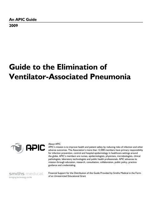
Guide to the Elimination of Ventilator-Associated Pneumonia - APIC
Guide to the Elimination of Ventilator-Associated Pneumonia - APIC
Guide to the Elimination of Ventilator-Associated Pneumonia - APIC

Create successful ePaper yourself
Turn your PDF publications into a flip-book with our unique Google optimized e-Paper software.
An <strong>APIC</strong> <strong>Guide</strong><br />
2009<br />
<strong>Guide</strong> <strong>to</strong> <strong>the</strong> <strong>Elimination</strong> <strong>of</strong><br />
Ventila<strong>to</strong>r-<strong>Associated</strong> <strong>Pneumonia</strong><br />
About <strong>APIC</strong><br />
<strong>APIC</strong>’s mission is <strong>to</strong> improve health and patient safety by reducing risks <strong>of</strong> infection and o<strong>the</strong>r<br />
adverse outcomes. The Association’s more than 12,000 members have primary responsibility<br />
for infection prevention, control and hospital epidemiology in healthcare settings around<br />
<strong>the</strong> globe. <strong>APIC</strong>’s members are nurses, epidemiologists, physicians, microbiologists, clinical<br />
pathologists, labora<strong>to</strong>ry technologists and public health pr<strong>of</strong>essionals. <strong>APIC</strong> advances its<br />
mission through education, research, consultation, collaboration, public policy, practice<br />
guidance and credentialing.<br />
Financial Support for <strong>the</strong> Distribution <strong>of</strong> this <strong>Guide</strong> Provided by Smiths Medical in <strong>the</strong> Form<br />
<strong>of</strong> an Unrestricted Educational Grant
For additional resources, please visit http://www.apic.org/<strong>Elimination</strong><strong>Guide</strong>s.<br />
Look for o<strong>the</strong>r <strong>to</strong>pics in <strong>APIC</strong>’s <strong>Elimination</strong> <strong>Guide</strong> Series, including:<br />
• Ca<strong>the</strong>ter-Related Bloodstream Infections<br />
• Ca<strong>the</strong>ter-Related Urinary Tract Infections<br />
• Clostridium difficile<br />
• Mediastinitis<br />
• MRSA in Long-Term Care<br />
Copyright © 2009 by <strong>APIC</strong><br />
All rights reserved. No part <strong>of</strong> this publication may be reproduced, s<strong>to</strong>red in a retrieval system, or transmitted, in any form or by<br />
any means, electronic, mechanical, pho<strong>to</strong>copying, recording, or o<strong>the</strong>rwise, without prior written permission <strong>of</strong> <strong>the</strong> publisher.<br />
All inquires about this document or o<strong>the</strong>r <strong>APIC</strong> products and services may be addressed <strong>to</strong>:<br />
<strong>APIC</strong> Headquarters<br />
1275 K Street, NW<br />
Suite 1000<br />
Washing<strong>to</strong>n, DC 20005<br />
Phone: 202.789.1890<br />
Email: <strong>APIC</strong>info@apic.org<br />
Web: www.apic.org<br />
ISBN: 1-933013-43-5
<strong>Guide</strong> <strong>to</strong> <strong>the</strong> <strong>Elimination</strong> <strong>of</strong> Ventila<strong>to</strong>r-<strong>Associated</strong> <strong>Pneumonia</strong><br />
Table <strong>of</strong> Contents<br />
1. Acknowledgments .....................................................................4<br />
2. Conflict <strong>of</strong> Interest . . . . . . . . . . . . . . . . . . . . . . . . . . . . . . . . . . . . . . . . . . . . . . . . . . . . . . . . . . . . . . . . . . . . 5<br />
3. <strong>Guide</strong> Overview .......................................................................6<br />
4. Problem Identification . . . . . . . . . . . . . . . . . . . . . . . . . . . . . . . . . . . . . . . . . . . . . . . . . . . . . . . . . . . . . . . . . 9<br />
5. Surveillance Definitions . . . . . . . . . . . . . . . . . . . . . . . . . . . . . . . . . . . . . . . . . . . . . . . . . . . . . . . . . . . . . . . 16<br />
6. Risk Assessment .....................................................................21<br />
7. Surveillance Plan .....................................................................28<br />
8. Prevention Strategies . . . . . . . . . . . . . . . . . . . . . . . . . . . . . . . . . . . . . . . . . . . . . . . . . . . . . . . . . . . . . . . . . 33<br />
9. Putting It All Toge<strong>the</strong>r ................................................................42<br />
ASSOCIATION FOR PROFESSIONALS IN INFECTION CONTROL AND EPIDEMIOLOGY 3
<strong>Guide</strong> <strong>to</strong> <strong>the</strong> <strong>Elimination</strong> <strong>of</strong> Ventila<strong>to</strong>r-<strong>Associated</strong> <strong>Pneumonia</strong><br />
Acknowledgments<br />
<strong>APIC</strong> acknowledges <strong>the</strong> valuable contributions <strong>of</strong> <strong>the</strong> following individuals:<br />
Authors<br />
Linda R. Greene, RN, MPS, CIC<br />
Direc<strong>to</strong>r <strong>of</strong> Infection Prevention<br />
Rochester General Health System<br />
Rochester, NY<br />
Kathleen Sposa<strong>to</strong>, RN, BSN, CIC<br />
Direc<strong>to</strong>r, Infection Prevention and Control<br />
Glens Falls Hospital<br />
Glens Falls, NY<br />
Reviewers<br />
Michelle R. Farber, RN, CIC<br />
Infection Control Specialist<br />
Mercy Community Hospital<br />
Coon Rapids, MN<br />
Teresa M. Ful<strong>to</strong>n, RN, MSN, CIC, CNLCP<br />
Direc<strong>to</strong>r, Quality and Patient Safety<br />
Whidbey General Hospital<br />
Coupeville, WA<br />
Robert A. Garcia, BS, MT(ASCP), CIC<br />
President and CEO<br />
Enhanced Epidemiology<br />
Valley Stream, NY<br />
4 ASSOCIATION FOR PROFESSIONALS IN INFECTION CONTROL AND EPIDEMIOLOGY
<strong>Guide</strong> <strong>to</strong> <strong>the</strong> <strong>Elimination</strong> <strong>of</strong> Ventila<strong>to</strong>r-<strong>Associated</strong> <strong>Pneumonia</strong><br />
Conflict <strong>of</strong> Interest<br />
Authors and reviewers <strong>of</strong> this <strong>Guide</strong> were asked <strong>to</strong> complete an <strong>APIC</strong> Conflict-<strong>of</strong>-Interest Disclosure Statement.<br />
Linda R. Greene, RN, MPS, CIC has nothing <strong>to</strong> disclose.<br />
Kathleen Sposa<strong>to</strong>, RN, BSN, CIC has nothing <strong>to</strong> disclose.<br />
Michelle R. Farber, RN, CIC has nothing <strong>to</strong> disclose.<br />
Robert A. Garcia, BS, MT(ASCP), CIC has nothing <strong>to</strong> disclose.<br />
Teresa M. Ful<strong>to</strong>n, RN, MSN, CIC, CNLCP serves on I-Flow corporate speakers bureau.<br />
ASSOCIATION FOR PROFESSIONALS IN INFECTION CONTROL AND EPIDEMIOLOGY 5
<strong>Guide</strong> <strong>to</strong> <strong>the</strong> <strong>Elimination</strong> <strong>of</strong> Ventila<strong>to</strong>r-<strong>Associated</strong> <strong>Pneumonia</strong><br />
<strong>Guide</strong> Overview<br />
The purpose <strong>of</strong> this guide is <strong>to</strong> provide evidence-based practice guidelines for <strong>the</strong> elimination <strong>of</strong> ventila<strong>to</strong>rassociated<br />
pneumonia (VAP).<br />
<strong>Pneumonia</strong> accounts for approximately 11% <strong>to</strong> 15% <strong>of</strong> all hospital-associated infections (HAIs) and 27% and 24%<br />
<strong>of</strong> all infections acquired in <strong>the</strong> medical intensive care unit (MICU) and coronary care unit (CCU), respectively. 1<br />
The primary risk fac<strong>to</strong>r for <strong>the</strong> development <strong>of</strong> hospital-associated bacterial pneumonia is mechanical ventilation<br />
(with its requisite endotracheal intubation). Rates <strong>of</strong> VAP vary depending on <strong>the</strong> type <strong>of</strong> ICU, and may range from<br />
zero <strong>to</strong> 16 per 1000 ventila<strong>to</strong>r days. Highest rates were identified in trauma ICUs, as reported in <strong>the</strong> 2008 National<br />
Healthcare Safety Network (NHSN) report. 2<br />
The VAP Infection Prevention and Control Program<br />
An effective facility-wide infection prevention and control program is comprised <strong>of</strong> many components and<br />
interventions that can reduce <strong>the</strong> risk <strong>of</strong> VAP in acutely ill patients. This guide will provide strategies and <strong>to</strong>ols that<br />
can be used for VAP prevention. Recent quality improvement initiatives suggest that many cases <strong>of</strong> VAP might<br />
be prevented by careful attention <strong>to</strong> <strong>the</strong> process <strong>of</strong> care. The successful management <strong>of</strong> patients on ventila<strong>to</strong>rs is<br />
necessary <strong>to</strong> ensure <strong>the</strong> best possible outcomes for individual patients while reducing <strong>the</strong> morbidity and mortality<br />
associated with <strong>the</strong>se infections.<br />
Components <strong>of</strong> a Successful Program<br />
Accountability for VAP prevention activities is outlined in <strong>the</strong> 2008 Strategies <strong>to</strong> Prevent Ventila<strong>to</strong>r-<strong>Associated</strong><br />
<strong>Pneumonia</strong> in Acute Care Hospitals and is outlined here. 3<br />
1. The hospital’s chief executive <strong>of</strong>ficer and senior management are responsible for ensuring that <strong>the</strong> healthcare<br />
system supports an infection prevention and control program <strong>to</strong> effectively prevent VAP.<br />
2. Senior management is accountable for ensuring that an adequate number <strong>of</strong> trained personnel are assigned<br />
<strong>to</strong> <strong>the</strong> infection prevention and control program.<br />
3. Senior management is accountable for ensuring that healthcare personnel, including licensed and<br />
nonlicensed personnel, are competent <strong>to</strong> perform <strong>the</strong>ir job responsibilities.<br />
4. Direct healthcare providers (e.g., physicians, nurses, aides, and <strong>the</strong>rapists) and ancillary personnel (e.g.,<br />
environmental services and equipment processing personnel) are responsible for ensuring that appropriate<br />
infection prevention and control practices are used at all times (including hand hygiene, standard and<br />
transmission-based or expanded precautions, cleaning and disinfection <strong>of</strong> equipment and <strong>the</strong> environment,<br />
aseptic technique when suctioning secretions and handling respira<strong>to</strong>ry <strong>the</strong>rapy equipment, patient<br />
positioning, sedation and weaning pro<strong>to</strong>cols, and oral care).<br />
5. Hospital and unit leaders are responsible for ensuring that personnel are accountable for <strong>the</strong>ir actions.<br />
6. The person who manages <strong>the</strong> infection prevention and control program is responsible for ensuring that an<br />
active program <strong>to</strong> identify VAP is implemented, that data on VAP are analyzed and regularly provided <strong>to</strong><br />
those who can use <strong>the</strong> information <strong>to</strong> improve <strong>the</strong> quality <strong>of</strong> care (e.g., unit staff, clinicians, and hospital<br />
administra<strong>to</strong>rs), and that evidence-based practices are incorporated in<strong>to</strong> <strong>the</strong> program.<br />
7. Healthcare personnel are accountable for ensuring that appropriate training and educational programs <strong>to</strong><br />
prevent VAP are developed and provided <strong>to</strong> medical staff, patients, and families.<br />
6 ASSOCIATION FOR PROFESSIONALS IN INFECTION CONTROL AND EPIDEMIOLOGY
<strong>Guide</strong> <strong>to</strong> <strong>the</strong> <strong>Elimination</strong> <strong>of</strong> Ventila<strong>to</strong>r-<strong>Associated</strong> <strong>Pneumonia</strong><br />
The role <strong>of</strong> <strong>the</strong> infection preventionist in <strong>the</strong> effort <strong>to</strong> reduce <strong>the</strong> incidence <strong>of</strong> VAP includes policy and best practice<br />
subject matter expertise, provision <strong>of</strong> surveillance data and risk assessment, consultation on infection prevention<br />
interventions, and facilitation <strong>of</strong> VAP-related improvement projects. It is important that <strong>the</strong> infection preventionist<br />
communicates and networks with all members <strong>of</strong> <strong>the</strong> patient care team regarding VAP-related infection prevention.<br />
Providing subject matter expertise <strong>to</strong> those involved with clinical management <strong>of</strong> <strong>the</strong> patients, including physicians,<br />
physician assistants, and nurse practitioners, is essential. An understanding <strong>of</strong> <strong>the</strong> elements <strong>of</strong> surveillance definitions<br />
compared with clinical definitions is important. Anes<strong>the</strong>siologists, respira<strong>to</strong>ry care, hospitalists, emergency department<br />
physicians, and medical residents are examples <strong>of</strong> individuals involved in intubation. Nursing staff and o<strong>the</strong>r members<br />
<strong>of</strong> <strong>the</strong> healthcare team are responsible for care <strong>of</strong> <strong>the</strong> patient on a ventila<strong>to</strong>r. Therefore, success <strong>of</strong> a prevention project<br />
requires that <strong>the</strong>se personnel are fully engaged and committed <strong>to</strong> this important patient safety initiative. Obtaining<br />
<strong>the</strong> resources that will engage direct care providers in <strong>the</strong> VAP quality/performance improvement activities is a critical<br />
component <strong>of</strong> intervention development. Key players must be held accountable for compliance with <strong>the</strong> prevention<br />
strategies and interventions. This can be facilitated through moni<strong>to</strong>ring and reporting <strong>of</strong> <strong>the</strong> results <strong>of</strong> <strong>the</strong> intervention<br />
on a consistent basis, and instituting additional improvements when appropriate.<br />
Basic Infection Prevention and Antimicrobial Stewardship<br />
Although this guide focuses on infection prevention related <strong>to</strong> VAP use, it is necessary <strong>to</strong> look at more global<br />
interventions that have an impact on HAIs such as VAP. The basics <strong>of</strong> infection prevention and control are<br />
necessary underpinnings <strong>of</strong> programs, policies, and pro<strong>to</strong>cols that impact HAIs (appropriate hand hygiene,<br />
environmental and equipment considerations, compliance with standard and transmission-based precautions, etc.).<br />
One component <strong>of</strong> HAI prevention deserves added attention in this guide. As highlighted in <strong>the</strong> Centers for<br />
Disease Control and Prevention’s (CDC) Campaign <strong>to</strong> Prevent Antimicrobial Resistance in Healthcare Settings, a<br />
program for antimicrobial stewardship in any healthcare setting (acute or long-term care) has potential for positive<br />
impact on all HAIs. The combination <strong>of</strong> effective antimicrobial stewardship with a comprehensive infection<br />
control program has been shown <strong>to</strong> limit <strong>the</strong> emergence and transmission <strong>of</strong> antimicrobial-resistant bacteria. A<br />
secondary goal <strong>of</strong> antimicrobial stewardship is <strong>to</strong> reduce healthcare costs without adversely impacting quality <strong>of</strong><br />
care.<br />
Antimicrobial stewardship can play a role in minimizing <strong>the</strong> potential adverse outcomes <strong>of</strong> <strong>the</strong>se occurrences.<br />
Inappropriate choice and utilization <strong>of</strong> antimicrobials has well documented adverse effects on patients and can lead<br />
<strong>to</strong> development <strong>of</strong> multidrug resistance in <strong>the</strong> healthcare setting. Preparing a facility- or unit-based antibiogram<br />
can demonstrate <strong>the</strong> changes in antimicrobial resistance and susceptibility patterns that develop over time, and<br />
can be used <strong>to</strong> track and moni<strong>to</strong>r changes. It should be noted that rates <strong>of</strong> hospital-acquired pneumonia (HAP)<br />
due <strong>to</strong> multidrug-resistant (MDR) pathogens have increased dramatically in hospitalized patients, especially in<br />
ICU and transplant patients. Multidisciplinary development <strong>of</strong> evidence-based practice guidelines incorporating<br />
local microbiology and resistance patterns can improve antimicrobial utilization. <strong>Guide</strong>line implementation can be<br />
facilitated through provider education and feedback on antimicrobial use and patient outcomes. 4<br />
The CDC/Healthcare Infection Control Practices Advisory Committee (HICPAC) Management <strong>of</strong> Multidrug-<br />
Resistant Organisms in Healthcare Settings, 2006 (MDRO <strong>Guide</strong>) recommends that “systems are in place <strong>to</strong><br />
promote optimal treatment <strong>of</strong> infections and appropriate antimicrobial use.” Pro<strong>to</strong>cols for initial empiric <strong>the</strong>rapy<br />
have emerged as a potentially effective means <strong>of</strong> avoiding unnecessary antibiotic administration while increasing<br />
<strong>the</strong> likelihood <strong>of</strong> initially appropriate <strong>the</strong>rapy. It is beyond <strong>the</strong> purview <strong>of</strong> this <strong>Guide</strong> <strong>to</strong> explore appropriate empiric<br />
and <strong>the</strong>rapeutic antibiotic selections for VAP. However, guidelines from <strong>the</strong> American Thoracic Society can be<br />
accessed online at http://ajrccm.atsjournals.org/cgi/content/full/171/4/388. 5<br />
ASSOCIATION FOR PROFESSIONALS IN INFECTION CONTROL AND EPIDEMIOLOGY 7
<strong>Guide</strong> <strong>to</strong> <strong>the</strong> <strong>Elimination</strong> <strong>of</strong> Ventila<strong>to</strong>r-<strong>Associated</strong> <strong>Pneumonia</strong><br />
References<br />
1<br />
Kollef M. Prevention <strong>of</strong> hospital-associated pneumonia and ventila<strong>to</strong>r-associated pneumonia. Crit Care Med<br />
2004;32(6):1396–1405.<br />
2<br />
Edwards JR, Peterson KD, Andrus ML, et al. National Healthcare Safety Network (NHSN) Report, data summary for<br />
2006 through 2007. Am J Infect Control 2008;36(9):609–626.<br />
3<br />
C<strong>of</strong>fin S, Klompas M, Classen D, et al. Strategies <strong>to</strong> prevent ventila<strong>to</strong>r-associated pneumonia in acute care hospitals. Infect<br />
Control and Hosp Epidemiol 2008;29:S31–S40.<br />
4<br />
Dillet TH, Owens RC, McGowan JE Jr, et al. Infectious Diseases Society <strong>of</strong> America and <strong>the</strong> Society for Healthcare<br />
Epidemiology <strong>of</strong> America guidelines for developing an institutional program <strong>to</strong> enhance antimicrobial stewardship. Clin<br />
Infect Dis 2007;44:159–177.<br />
5<br />
American Thoracic Society; Infectious Diseases Society <strong>of</strong> America. <strong>Guide</strong>lines for <strong>the</strong> management <strong>of</strong> adults with hospitalacquired,<br />
ventila<strong>to</strong>r-associated, and healthcare-associated pneumonia. Am J Resp Crit Care Med 2005;171:388–416.<br />
8 ASSOCIATION FOR PROFESSIONALS IN INFECTION CONTROL AND EPIDEMIOLOGY
<strong>Guide</strong> <strong>to</strong> <strong>the</strong> <strong>Elimination</strong> <strong>of</strong> Ventila<strong>to</strong>r-<strong>Associated</strong> <strong>Pneumonia</strong><br />
Problem Identification<br />
VAP is associated with increased lengths <strong>of</strong> ICU and hospital stay, increased mortality rates, and increased costs.<br />
In a metaanalysis <strong>of</strong> research articles published between 1990 and March 1, 2004, morbidity (as evidenced by<br />
increased length-<strong>of</strong>-stay [LOS] in <strong>the</strong> ICU), mortality, and costs associated with VAP were evaluated. Analysis <strong>of</strong><br />
LOS reports demonstrated that VAP was associated with a mean increase in ICU length-<strong>of</strong>-stay <strong>of</strong> 6.1 days.<br />
The authors reviewed nine reports related <strong>to</strong> mortality, four <strong>of</strong> which did not attribute significant mortality <strong>to</strong> <strong>the</strong><br />
diagnosis. The remaining five studies reported an excess mortality that varied from 15% <strong>to</strong> 50% when compared<br />
with control cases. 1 Luna et al. reported that mortality rates vary with patient population and infecting organism;<br />
mortality increases when <strong>the</strong> infecting organism is MDR. 2 O<strong>the</strong>r sources report increased mortality associated with<br />
untreated or inadequately treated infection. Increased costs associated with VAP were dependent on length-<strong>of</strong>stay,<br />
with a range <strong>of</strong> approximately $10,000 <strong>to</strong> $40,000. Table 3-1 summarizes <strong>the</strong> findings <strong>of</strong> several studies that<br />
investigated <strong>the</strong> effects <strong>of</strong> VAP on morbidity, mortality, and/or cost.<br />
Findings from o<strong>the</strong>r studies include variability <strong>of</strong> mortality rates associated with HAP, due <strong>to</strong> underlying disease<br />
and etiology. Mortality attributed <strong>to</strong> HAP is highly dependent on <strong>the</strong> institution <strong>of</strong> appropriate antibiotic <strong>the</strong>rapy,<br />
virulence <strong>of</strong> pathogen, and host defenses, with case fatality rates being highest in mechanically ventilated patients<br />
with high severity <strong>of</strong> illness and infection caused by nonfermentative Gram-negative bacilli. 3,4 Additionally, HAP<br />
attributable <strong>to</strong> MDR microorganisms was significantly associated with mortality. 5<br />
Table 3-1. Studies Investigating <strong>the</strong> Effect <strong>of</strong> VAP on Morbidity, Mortality, and Cost<br />
Publication<br />
Date<br />
Investiga<strong>to</strong>r Study Number <strong>of</strong><br />
Patients with<br />
VAP<br />
2008 Brilli et al. 6 The business<br />
case for<br />
preventing VAP<br />
in pediatric<br />
intensive care<br />
unit patients<br />
2005 Kollef et al. 7 Epidemiology<br />
and outcomes<br />
<strong>of</strong> HAP: results<br />
from a large<br />
U.S. database <strong>of</strong><br />
culture-positive<br />
pneumonia<br />
2005 Cocanour<br />
et al. 8<br />
Cost <strong>of</strong> a VAP<br />
in a shock<br />
trauma ICU<br />
2005 Safdar et al. 1 Clinical and<br />
economic<br />
consequence <strong>of</strong><br />
VAP<br />
13<br />
(retrospective<br />
matched<br />
case-control<br />
study)<br />
499<br />
(retrospective<br />
matched<br />
cohort study)<br />
70<br />
(case control<br />
study)<br />
Effect <strong>of</strong> VAP<br />
on Morbidity<br />
# mean hospital<br />
LOS; mean<br />
attributable # in<br />
LOS due <strong>to</strong> VAP<br />
8.7 days<br />
Effect <strong>of</strong><br />
VAP<br />
on Mortality<br />
Cost<br />
<strong>Associated</strong><br />
with VAP<br />
Not studied $51,157<br />
(attributable<br />
VAP costs)<br />
# LOS 29.3% $150,841<br />
(mean hospital<br />
charge)<br />
# ventila<strong>to</strong>r days<br />
and ICU LOS<br />
Not applicable Significantly<br />
longer ICU LOS<br />
Mortality rate<br />
doubled in<br />
patients with<br />
VAP<br />
$57,000<br />
(excess<br />
hospital cost)<br />
$13,647<br />
(estimated<br />
attributable<br />
VAP cost,<br />
using upper<br />
limit <strong>of</strong> LOS)<br />
Comment<br />
Study compared<br />
communityacquired<br />
pneumonia<br />
(CAP), healthcareassociated<br />
pneumonia (HCAP),<br />
HAP, and VAP<br />
ASSOCIATION FOR PROFESSIONALS IN INFECTION CONTROL AND EPIDEMIOLOGY 9
<strong>Guide</strong> <strong>to</strong> <strong>the</strong> <strong>Elimination</strong> <strong>of</strong> Ventila<strong>to</strong>r-<strong>Associated</strong> <strong>Pneumonia</strong><br />
Publication<br />
Date<br />
2003 Warren<br />
et al. 3<br />
Investiga<strong>to</strong>r Study Number <strong>of</strong><br />
Patients with<br />
VAP<br />
Outcome and<br />
attributable<br />
cost <strong>of</strong> VAP<br />
among ICU<br />
patients in<br />
a suburban<br />
medical center<br />
2002 Rello et al. 4 Epidemiology<br />
and outcomes<br />
<strong>of</strong> VAP in a<br />
large U.S.<br />
database<br />
2001 Bregeon<br />
et al. 5<br />
2001 Bercault<br />
et al. 9<br />
1999 Heyland<br />
et al. 10<br />
Is VAP an<br />
independent<br />
risk fac<strong>to</strong>r for<br />
death?<br />
Mortality rate<br />
attributable<br />
<strong>to</strong> ventila<strong>to</strong>rassociated<br />
nosocomial<br />
pneumonia in<br />
an adult ICU<br />
The attributable<br />
morbidity and<br />
mortality <strong>of</strong><br />
VAP in <strong>the</strong><br />
critically ill<br />
patient<br />
819<br />
(prospective<br />
cost analysis)<br />
842<br />
(retrospective<br />
matched<br />
cohort study)<br />
39<br />
(matched-pair<br />
case-control<br />
study)<br />
135<br />
(prospective<br />
matched,<br />
risk-adjusted<br />
cohort study)<br />
175<br />
(prospective<br />
matched<br />
cohort study)<br />
Effect <strong>of</strong> VAP<br />
on Morbidity<br />
# incidence <strong>of</strong><br />
sepsis, ICU LOS,<br />
hospital LOS<br />
# duration <strong>of</strong><br />
mechanical<br />
ventilation,<br />
ICU LOS, <strong>to</strong>tal<br />
hospital LOS<br />
Not studied<br />
Effect <strong>of</strong><br />
VAP<br />
on Mortality<br />
Cost<br />
<strong>Associated</strong><br />
with VAP<br />
# mortality $11,897<br />
(attributable<br />
cost <strong>of</strong> VAP)<br />
No significant<br />
difference<br />
VAP is not an<br />
independent<br />
risk for #<br />
mortality<br />
# ICU LOS # mortality<br />
risk (absolute<br />
risk # 5.8%;<br />
relative risk #<br />
32.3%)<br />
# ICU LOS<br />
(4.3 days)<br />
# mortality<br />
risk (absolute<br />
risk #5.8%;<br />
relative risk<br />
#32.3%)<br />
$40,000<br />
(increase<br />
in inpatient<br />
billed charges)<br />
Not studied<br />
Not studied<br />
Comment<br />
Renal failure, bone<br />
marrow failure,<br />
and treatment with<br />
corticosteroids<br />
were independent<br />
risk fac<strong>to</strong>rs for<br />
mortality<br />
Included patients<br />
ventilated for ≥48<br />
hours; attributable<br />
risk <strong>of</strong> VAP appears<br />
<strong>to</strong> vary with patient<br />
population and<br />
infecting organism<br />
Pathogenesis and Epidemiology <strong>of</strong> VAP<br />
<strong>Pneumonia</strong> Definitions<br />
<strong>Pneumonia</strong> is classified as community-acquired (CAP), healthcare-associated (HCAP), HAP, or VAP. VAP is a<br />
sub-classification <strong>of</strong> HAP, if <strong>the</strong> patient is hospitalized during <strong>the</strong> period <strong>of</strong> mechanical ventilation. CAP is defined<br />
as pneumonia for which <strong>the</strong> first positive bacterial culture is obtained within 48 hours <strong>of</strong> admission <strong>to</strong> <strong>the</strong> hospital<br />
and <strong>the</strong> patient does not have risk fac<strong>to</strong>rs for HAP. HCAP occurs when <strong>the</strong> patient’s first positive bacterial culture is<br />
obtained within 48 hours <strong>of</strong> admission and <strong>the</strong> patient has any <strong>of</strong> <strong>the</strong> following risk fac<strong>to</strong>rs: admission source indicates<br />
a transfer from ano<strong>the</strong>r healthcare facility; patient has received hemodialysis, wound, or infusion <strong>the</strong>rapy as an<br />
outpatient; patient was previously hospitalized for at least 3 days within <strong>the</strong> past 90 days prior <strong>to</strong> current admission; or<br />
<strong>the</strong> patient is immunocompromised due <strong>to</strong> underlying disease or <strong>the</strong>rapy (HIV, chemo<strong>the</strong>rapy). HAP is pneumonia<br />
in which <strong>the</strong> patient’s first positive bacterial culture is obtained more than 48 hours after admission <strong>to</strong> <strong>the</strong> hospital.<br />
According <strong>to</strong> this source, VAP is pneumonia that develops in a mechanically ventilated patient with a first positive<br />
bacterial culture beyond 48 hours after hospital admission or tracheal intubation, whichever occurred first. 11 It is noted<br />
10 ASSOCIATION FOR PROFESSIONALS IN INFECTION CONTROL AND EPIDEMIOLOGY
<strong>Guide</strong> <strong>to</strong> <strong>the</strong> <strong>Elimination</strong> <strong>of</strong> Ventila<strong>to</strong>r-<strong>Associated</strong> <strong>Pneumonia</strong><br />
that this definition <strong>of</strong> VAP differs from <strong>the</strong> NHSN surveillance definition <strong>of</strong> VAP, as <strong>the</strong> NHSN definition does not<br />
require a 48-hour period <strong>of</strong> intubation and ventilation before pneumonia can be considered ventila<strong>to</strong>r-associated.<br />
HAP, whe<strong>the</strong>r or not associated with mechanical ventilation, is generally a secondary endogenous infection.<br />
Although exogenous sources <strong>of</strong> infectious microorganisms exist, it is typically <strong>the</strong> patient’s own colonizing flora<br />
that is implicated in infection.<br />
In <strong>the</strong> healthy individual, <strong>the</strong> lower respira<strong>to</strong>ry tract is a sterile site and <strong>the</strong> body possesses many defense<br />
mechanisms <strong>to</strong> maintain that state. Mechanical barriers, humoral and cell-mediated immunity, and phagocyte<br />
activity act <strong>to</strong> defend against bacterial invasion <strong>of</strong> lung tissue. Human saliva contains components that demonstrate<br />
antimicrobial properties and helps <strong>to</strong> regulate <strong>the</strong> composition <strong>of</strong> oral flora.<br />
Fac<strong>to</strong>rs that may interfere with <strong>the</strong> host’s defenses and predispose <strong>to</strong> respira<strong>to</strong>ry infection include alterations in<br />
level <strong>of</strong> consciousness, cigarette smoke, alcohol intake, viral infections, sepsis, endotracheal tubes, nasogastric<br />
tubes, respira<strong>to</strong>ry <strong>the</strong>rapy devices, hypoxemia, acidosis, <strong>to</strong>xic inhalations, pulmonary edema, uremia, malnutrition,<br />
immunosuppressive agents, and mechanical obstruction. 12 Inadequate salivary flow in intubated patients causes<br />
xeros<strong>to</strong>mia, which may contribute <strong>to</strong> mucositis and colonization <strong>of</strong> <strong>the</strong> oropharynx with Gram-negative bacteria. 13<br />
Advanced age predisposes <strong>the</strong> individual <strong>to</strong> development <strong>of</strong> pneumonia due <strong>to</strong> a less efficient cough reflex and<br />
changes in humoral immunity and cell-mediated immune function. The patient who is immunosuppressed due <strong>to</strong><br />
disease state or treatment modality is also at increased risk for development <strong>of</strong> infection.<br />
The intubated patient is <strong>of</strong>ten a critically ill individual with many risk fac<strong>to</strong>rs that contribute <strong>to</strong> <strong>the</strong> development <strong>of</strong><br />
pneumonia. Risk fac<strong>to</strong>rs for VAP can be classified as modifiable or nonmodifiable, as well as patient-related and<br />
treatment-related.<br />
Nonmodifiable risk fac<strong>to</strong>rs for VAP include male gender, preexisting pulmonary disease, coma, AIDS, head trauma,<br />
and multi-organ system failure. Nonmodifiable treatment-related risk fac<strong>to</strong>rs include neurosurgical procedures,<br />
intracranial pressure moni<strong>to</strong>ring, re-intubation, and transportation out <strong>of</strong> an ICU. Intubation and mechanical<br />
ventilation are prerequisites for <strong>the</strong> diagnosis <strong>of</strong> VAP. Modifiable risk fac<strong>to</strong>rs include duration <strong>of</strong> ventilation;<br />
risk varies over time, being greatest early in <strong>the</strong> ventila<strong>to</strong>r period and decreasing as ventila<strong>to</strong>r LOS progresses.<br />
Nasotracheal intubation is associated with <strong>the</strong> development <strong>of</strong> sinusitis and should be avoided. Supine position is also<br />
associated with an increased risk <strong>of</strong> VAP, especially in <strong>the</strong> presence <strong>of</strong> simultaneous enteral feeding. Enteral feeding<br />
itself is a risk fac<strong>to</strong>r for VAP, mainly due <strong>to</strong> an increased risk <strong>of</strong> aspiration. But, because <strong>the</strong> alternative, parenteral<br />
nutrition, is associated with even greater risk (<strong>of</strong> bloodstream infection), it is advised <strong>to</strong> feed critically ill patients<br />
enterally as early as possible. 14–16 Oropharyngeal colonization has been identified as a risk fac<strong>to</strong>r for <strong>the</strong> development<br />
<strong>of</strong> VAP. Evidence indicates that <strong>the</strong> oropharynx acts as a reservoir for bacteria that are subsequently aspirated in<strong>to</strong> <strong>the</strong><br />
lower respira<strong>to</strong>ry tract. Colonization <strong>of</strong> <strong>the</strong> oropharynx progresses rapidly in ICU patients and occurs more frequently<br />
in patients who go on <strong>to</strong> develop VAP. Additionally, bacteria in dental plaque act as a major contribu<strong>to</strong>r <strong>to</strong> infection<br />
<strong>of</strong> <strong>the</strong> respira<strong>to</strong>ry tract. Stress ulcer prophylaxis, in <strong>the</strong> form <strong>of</strong> H2-antagonists and antacids, has been recognized<br />
as a risk fac<strong>to</strong>r for VAP. Some studies indicate that prior antibiotic <strong>the</strong>rapy is a risk fac<strong>to</strong>r for VAP, and antibiotics<br />
predispose patients <strong>to</strong> colonization and subsequent infection with antibiotic-resistant organisms. 17–20<br />
VAP is divided in<strong>to</strong> early- and late-onset disease. Early-onset VAP occurs during <strong>the</strong> first 4 days <strong>of</strong> <strong>the</strong> patient’s<br />
admission and is <strong>of</strong>ten caused by Strep<strong>to</strong>coccus pneumonia, Haemophilus influenza, or Moraxella catarrhalis. By comparison,<br />
late-onset VAP occurs beyond 4 days after admission and is more commonly caused by Pseudomonas aeruginosa,<br />
Acine<strong>to</strong>bacter or Enterobacter spp., or methicillin-resistant Staphylococcus aureus (MRSA). Many <strong>of</strong> <strong>the</strong> organisms<br />
associated with late-onset VAP are resistant <strong>to</strong> multiple antibiotics or have MDR strains. Staphylococcus aureus is isolated<br />
in 20% <strong>to</strong> 40% <strong>of</strong> cases and is especially common in persons taking drugs by injection; in patients with neurological<br />
ASSOCIATION FOR PROFESSIONALS IN INFECTION CONTROL AND EPIDEMIOLOGY 11
<strong>Guide</strong> <strong>to</strong> <strong>the</strong> <strong>Elimination</strong> <strong>of</strong> Ventila<strong>to</strong>r-<strong>Associated</strong> <strong>Pneumonia</strong><br />
Table 3-2. Host Defenses (Pulmonary)<br />
Location<br />
Upper Airways<br />
Nasopharynx<br />
Oropharynx<br />
Conducting Airways<br />
Trachea, bronchii<br />
Lower Airways<br />
Terminal airways, alveoli<br />
Defense Mechanism<br />
Nasal hair<br />
Turbinates<br />
Upper airway ana<strong>to</strong>my<br />
Mucociliary apparatus<br />
IgA secretion<br />
Saliva<br />
Sloughing <strong>of</strong> epi<strong>the</strong>lial cells<br />
Bacterial interference<br />
Complement production<br />
Coughing, epiglottic reflexes<br />
Airway branching<br />
Mucociliary apparatus<br />
Immunoglobulin production<br />
Airway surface liquid<br />
Alveolar lining fluid<br />
Cy<strong>to</strong>kines<br />
Alveolar macrophages<br />
Polymorphonuclear leukocytes<br />
Cell-mediated immunity<br />
(From Breese Hall C, Mcbride J. Bronchiolitis. In Mandell G, Bennett J, Dolin R, eds. Principles<br />
and Practice <strong>of</strong> Infectious Diseases. Philadelphia: Churchill Livings<strong>to</strong>ne, 2005:820.)<br />
disease, <strong>the</strong>rmal injury, or wound infection; and in patients who have received prior antibiotic <strong>the</strong>rapy or have had a<br />
prolonged stay in <strong>the</strong> ICU. Compared with patients with VAP caused by methicillin-susceptible Staphylococcus aureus<br />
(MSSA), those in whom <strong>the</strong> causative organism is MRSA are <strong>of</strong>ten older and are significantly more likely <strong>to</strong> have had<br />
previous chronic lung disease, antibiotic <strong>the</strong>rapy, steroid <strong>the</strong>rapy, and greater than 6 days <strong>of</strong> mechanical ventilation.<br />
Bacteremia, shock, and mortality are usually higher in <strong>the</strong> MRSA group. In many patients, VAP is caused by<br />
multiple organisms (polymicrobial). Aerobic Gram-negative bacilli, including Escherichia coli, Klebsiella pneumonia,<br />
Enterobacter spp., Serratia spp., Pseudomonas aeruginosa, and Acine<strong>to</strong>bacter spp., are most frequently isolated,<br />
particularly in patients with late-onset disease or those with serious underlying disease. 21,22 According <strong>to</strong> <strong>the</strong><br />
NHSN’s annual summary 23 <strong>of</strong> resistant pathogens associated with HAIs, <strong>the</strong> following organisms were identified<br />
as causing VAP (in order <strong>of</strong> most <strong>to</strong> least frequent with percentage <strong>of</strong> isolates in paren<strong>the</strong>ses):<br />
Staphylococcus aureus (24.4%)<br />
Pseudomonas aeruginosa (16.3%)<br />
Enterobacter spp. (8.4%)<br />
Acine<strong>to</strong>bacter baumannii (8.4%)<br />
Klebsiella pneumonia (7.5%)<br />
Escherichia coli (4.6%)<br />
Candida spp. (2.7%)<br />
Klebsiella oxy<strong>to</strong>ca (2.2%)<br />
Coagulase-negative Staphylococcus (1.3%)<br />
12 ASSOCIATION FOR PROFESSIONALS IN INFECTION CONTROL AND EPIDEMIOLOGY
<strong>Guide</strong> <strong>to</strong> <strong>the</strong> <strong>Elimination</strong> <strong>of</strong> Ventila<strong>to</strong>r-<strong>Associated</strong> <strong>Pneumonia</strong><br />
O<strong>the</strong>r unspecified organisms accounted for 23.1% <strong>of</strong> causative organisms (see Table 3-3). The epidemiology and<br />
pathogenesis <strong>of</strong> VAP is changing as hospitalized patients are now older and have more comorbidities, immune<br />
system dysfunction, invasive procedures, and exposure <strong>to</strong> antibiotics. Patients are more mobile and more likely<br />
<strong>to</strong> reside in short- and long-term care facilities, increasing <strong>the</strong>ir potential for colonization, person-<strong>to</strong>-person<br />
transmission, and infection with MDR pathogens.<br />
Summary <strong>of</strong> epidemiologic and pathogenic points. 15,24<br />
• The incidence <strong>of</strong> VAP is 3- <strong>to</strong> 10-fold greater than pneumonia in nonventilated patients.<br />
• VAP occurs in 8% <strong>to</strong> 28% <strong>of</strong> patients undergoing mechanical ventilation.<br />
• In <strong>the</strong> healthy individual, <strong>the</strong> lower respira<strong>to</strong>ry tract is a sterile body site.<br />
• The body possesses several defense mechanisms <strong>to</strong> prevent contamination <strong>of</strong> <strong>the</strong> lungs.<br />
• Disease processes, treatment modalities and personal habits or practices (i.e., cigarette smoke, alcohol<br />
intake) can impair <strong>the</strong> body’s natural defense mechanisms, predisposing <strong>the</strong> individual <strong>to</strong> lower respira<strong>to</strong>ry<br />
infection.<br />
• Mechanical ventilation is <strong>the</strong> primary risk fac<strong>to</strong>r for development <strong>of</strong> VAP for several reasons:<br />
o<br />
The endotracheal tube itself acts as a conduit from <strong>the</strong> upper respira<strong>to</strong>ry tract <strong>to</strong> <strong>the</strong> lower respira<strong>to</strong>ry<br />
tract.<br />
o<br />
Secretions collect on and around <strong>the</strong> endotracheal cuff; leakage <strong>of</strong> this fluid is <strong>the</strong> primary mechanism<br />
<strong>of</strong> infection <strong>of</strong> <strong>the</strong> lower respira<strong>to</strong>ry tract.<br />
o<br />
Sedation <strong>of</strong> patients who are mechanically ventilated inhibits <strong>the</strong> natural ability <strong>to</strong> clear secretions.<br />
o<br />
Patients undergoing mechanical ventilation are frequently fed via <strong>the</strong> nasogastric route, providing a<br />
source <strong>of</strong> fluid for aspiration and micro-aspiration.<br />
o<br />
Critically ill patients, especially those who are unstable with regard <strong>to</strong> neurologic or cardiac status, are<br />
<strong>of</strong>ten maintained in a supine position.<br />
o<br />
Activity is frequently limited during <strong>the</strong> period <strong>of</strong> mechanical ventilation.<br />
• VAP risk is greatest early on in ventilation, and diminishes over time.<br />
• VAP is frequently bacteriological in origin, especially in <strong>the</strong> immunocompromised patient.<br />
• Colonization <strong>of</strong> <strong>the</strong> oropharynx and dental surfaces act as a reservoir <strong>of</strong> bacteria that ultimately gain access<br />
<strong>to</strong> <strong>the</strong> lower respira<strong>to</strong>ry tract in patients undergoing mechanical ventilation.<br />
Table 3-3. Organisms <strong>Associated</strong> with VAP<br />
Early-Onset VAP<br />
Late-Onset VAP<br />
CDC NHSN 2006–2007 Summary Data<br />
(within first 4 days <strong>of</strong> admission) (after day 4)<br />
Strep<strong>to</strong>coccus pneumonia Pseudomonas aeruginosa Staphylococcus aureus (24.4%)<br />
Haemophilus influenza Acine<strong>to</strong>bacter spp. Pseudomonas aeruginosa (16.3%)<br />
Moraxella catarrhalis Enterobacter spp. Enterobacter spp. (8.4%)<br />
Methicillin-resistant<br />
Staphylococcus aureus<br />
Acine<strong>to</strong>bacter baumannii (8.4%)<br />
Klebsiella pneumonia (7.5%)<br />
Escherichia coli (4.6%)<br />
Candida spp. (2.7%)<br />
Klebsiella oxy<strong>to</strong>ca (2.2%)<br />
Coagulase-negative Staphylococcus (1.3%)<br />
O<strong>the</strong>r (23.1%)<br />
ASSOCIATION FOR PROFESSIONALS IN INFECTION CONTROL AND EPIDEMIOLOGY 13
<strong>Guide</strong> <strong>to</strong> <strong>the</strong> <strong>Elimination</strong> <strong>of</strong> Ventila<strong>to</strong>r-<strong>Associated</strong> <strong>Pneumonia</strong><br />
• Sources <strong>of</strong> pathogens for HAP include healthcare devices, <strong>the</strong> environment (air, water, equipment, and<br />
fomites), and commonly <strong>the</strong> transfer <strong>of</strong> microorganisms between <strong>the</strong> patient and staff or o<strong>the</strong>r patients,<br />
most frequently via <strong>the</strong> hands <strong>of</strong> healthcare workers. Never<strong>the</strong>less, most HAP is considered <strong>to</strong> be a<br />
secondary endogenous infection (resulting from <strong>the</strong> patient’s own colonizing flora).<br />
• Rates <strong>of</strong> Legionella pneumophila pneumonia vary considerably among hospitals, and disease occurs more<br />
commonly with serogroup 1 when <strong>the</strong> water supply is colonized or <strong>the</strong>re is ongoing construction.<br />
• Inhalation or direct inoculation <strong>of</strong> pathogens in<strong>to</strong> <strong>the</strong> lower airway, hema<strong>to</strong>genous spread from infected<br />
intravenous ca<strong>the</strong>ters or o<strong>the</strong>r infectious sites, and bacterial translocation from <strong>the</strong> gastrointestinal tract<br />
lumen are uncommon pathogenic mechanisms.<br />
• Infected bi<strong>of</strong>ilm in <strong>the</strong> endotracheal tube with subsequent embolization <strong>to</strong> distal airways may be important<br />
in <strong>the</strong> pathogenesis <strong>of</strong> VAP.<br />
• The s<strong>to</strong>mach and sinuses may be potential reservoirs <strong>of</strong> hospital-acquired pathogens that contribute <strong>to</strong><br />
bacterial colonization <strong>of</strong> <strong>the</strong> oropharynx, but <strong>the</strong>ir contribution is controversial, may vary by <strong>the</strong> population<br />
at risk, and may be decreasing with <strong>the</strong> changing pathogenesis <strong>of</strong> HAP.<br />
• VAP is divided in<strong>to</strong> early- and late-onset illness:<br />
o<br />
Early-onset VAP is typically caused by antibiotic-susceptible bacteria, including Strep<strong>to</strong>coccus<br />
pneumonia, Haemophilus influenza, and Moraxella catarrhalis.<br />
o<br />
Late-onset VAP is more likely <strong>to</strong> be caused by antibiotic-resistant bacteria, including Pseudomonas<br />
aeruginosa, Acine<strong>to</strong>bacter spp., Enterobacter spp., and MRSA.<br />
o<br />
Early-onset VAP that occurs in patients who have had healthcare exposure (treatment in a dialysis or<br />
wound care center, hospital admission <strong>of</strong> more than 3 days in <strong>the</strong> past 90 days, or residence in a longterm<br />
care facility) is more likely <strong>to</strong> follow <strong>the</strong> microbiological pattern <strong>of</strong> late-onset VAP.<br />
• The prevalence <strong>of</strong> MDR pathogens varies by patient population, hospital, and type <strong>of</strong> ICU, which<br />
emphasizes <strong>the</strong> need for local surveillance data.<br />
• Rates <strong>of</strong> polymicrobial VAP are especially high in patients with acute respira<strong>to</strong>ry distress syndrome (ARDS).<br />
• In <strong>the</strong> immunocompromised patient, infection with viral or fungal agents is more common than in <strong>the</strong><br />
patient whose immune status is competent.<br />
References<br />
1<br />
Safdar N, Dezfulian C, Collard H, et al. Clinical and economic consequences <strong>of</strong> ventila<strong>to</strong>r-associated pneumonia: A<br />
systematic review. Crit Care Med 2005;33:2184–2193.<br />
2<br />
Luna C, Blanzaco D, Niederman M, et al. Resolution <strong>of</strong> ventila<strong>to</strong>r-associated pneumonia: Prospective evaluation <strong>of</strong> <strong>the</strong><br />
clinical pulmonary infection score as an early clinical predic<strong>to</strong>r <strong>of</strong> outcome. Crit Care Med 2003;31:676–682.<br />
3<br />
Warren D, Shukla S, Olsen M, et al. Outcome and attributable cost <strong>of</strong> ventila<strong>to</strong>r-associated pneumonia among intensive<br />
care unit patients in a suburban medical center. Crit Care Med 2003;31:1312–1317.<br />
4<br />
Rello J, Ollendor D, Oster G, et al. Epidemiology and outcomes <strong>of</strong> ventila<strong>to</strong>r-associated pneumonia in a large U.S.<br />
database. Chest 2002;122:2115–2121.<br />
5<br />
Bregeon F, Ciais V, Carret V, et al. Is ventila<strong>to</strong>r-associated pneumonia an independent risk fac<strong>to</strong>r for death? Anes<strong>the</strong>siology<br />
2001;94:554–560.<br />
6<br />
Brilli R, Sparling K, Lake M, et al. The business case for preventing ventila<strong>to</strong>r-associated pneumonia in pediatric intensive<br />
care unit patients. Jt Comm J Qual Patient Saf 2008;34:629–638.<br />
14 ASSOCIATION FOR PROFESSIONALS IN INFECTION CONTROL AND EPIDEMIOLOGY
<strong>Guide</strong> <strong>to</strong> <strong>the</strong> <strong>Elimination</strong> <strong>of</strong> Ventila<strong>to</strong>r-<strong>Associated</strong> <strong>Pneumonia</strong><br />
7<br />
Kollef M, Shorr A, Tabak Y, et al. Epidemiology and outcomes <strong>of</strong> health care-associated pneumonia: Results from a large<br />
U.S. database <strong>of</strong> culture-positive pneumonia. Chest 2005;128:3854–3862.<br />
8<br />
Cocanour C, Ostrosky-Zeichner L, Peninger M, et al. Cost <strong>of</strong> a ventila<strong>to</strong>r-associated pneumonia in a shock trauma<br />
intensive care unit. Surgical Infections 2005;6:65–72.<br />
9<br />
Bercault N, Boulain T. Mortality rate attributable <strong>to</strong> ventila<strong>to</strong>r-associated nosocomial pneumonia in an adult intensive care<br />
unit: A prospective case-control study. Crit Care Med 2001;29:2303–2309.<br />
10<br />
Heyland D, Cook D, Griffith L, et al. The attributable morbidity and mortality <strong>of</strong> ventila<strong>to</strong>r-associated pneumonia in <strong>the</strong><br />
critically ill patient. Am J Respir Crit Care Med 1999;159:1249–1256.<br />
11<br />
Kollef M. Nosocomial pneumonia. In Kollef M, Bedient T, Isakow W, Witt C, eds. The Washing<strong>to</strong>n Manual <strong>of</strong> Critical<br />
Care. Philadelphia: Lippincott, Williams and Wilkins, 2007:261.<br />
12<br />
Breese Hall C, Mcbride J. Bronchiolitis. In Mandell G, Bennett J, Dolin R, eds. Principles and Practice <strong>of</strong> Infectious Diseases.<br />
Philadelphia: Churchill Livings<strong>to</strong>ne, 2005:820.<br />
13<br />
Dennesen P, van der Ven A, Vlasveld M, et al. Inadequate salivary flow and poor oral mucosa status in intubated intensive<br />
care unit patients. Crit Care Med 2003;31:781–786.<br />
14<br />
Chastre J, Fagon J. State <strong>of</strong> <strong>the</strong> art: Ventila<strong>to</strong>r-associated pneumonia. Am J Respir Crit Care Med 2002;165:867–903.<br />
15<br />
American Thoracic Society; Infectious Diseases Society <strong>of</strong> America. <strong>Guide</strong>lines for <strong>the</strong> management <strong>of</strong> adults with hospitalacquired,<br />
ventila<strong>to</strong>r-associated, and healthcare-associated pneumonia. Am J Resp Crit Care Med 2005;171:388–416.<br />
16<br />
Wunderink R, Mayhall CG, Gilbert C. Methodology for clinical investigation <strong>of</strong> ventila<strong>to</strong>r-associated pneumonia:<br />
Epidemiology and <strong>the</strong>rapeutic intervention. Chest 1992;102:580–588.<br />
17<br />
Garcia R. A review <strong>of</strong> <strong>the</strong> possible role <strong>of</strong> oral and dental colonization on <strong>the</strong> occurrence <strong>of</strong> health care-associated<br />
pneumonia: Underappreciated risk and a call for interventions. Am J Infect Control 2005;33:527–541.<br />
18<br />
Garrouste-Oregeas M, Chevret S, Arlet G, et al. Oropharyngeal or gastric colonization and nosocomial pneumonia in adult<br />
intensive care unit patients: A prospective study based on genomic DNA analysis. Am J Resp Crit Care Med 1997;156:1647–1655.<br />
19<br />
Bonten M, Bergmans D, Ambergen A, et al. Risk fac<strong>to</strong>rs for pneumonia, and colonization <strong>of</strong> respira<strong>to</strong>ry tract and s<strong>to</strong>mach<br />
in mechanically ventilated ICU patients. Am J Resp Crit Care Med 1996;154:1339–1346.<br />
20<br />
Scannapieco F. Role <strong>of</strong> oral bacteria in respira<strong>to</strong>ry infection. J Periodon<strong>to</strong>l 1999;70:793–802.<br />
21<br />
Craven D. Epidemiology <strong>of</strong> ventila<strong>to</strong>r-associated pneumonia. Chest 2000;117:186–187.<br />
22<br />
Weber D, Rutala W, Sickbert-Bennett E, et al. Microbiology <strong>of</strong> ventila<strong>to</strong>r-associated pneumonia compared with that <strong>of</strong><br />
hospital-acquired pneumonia. Infect Control and Hosp Epidemiol 2007;28:825–831.<br />
23<br />
Hidron A, Edward J, Patel J, et al. Antimicrobial-resistant pathogens associated with health care-associated infections:<br />
Annual summary <strong>of</strong> data reported <strong>to</strong> <strong>the</strong> national healthcare safety network at <strong>the</strong> Centers for Disease Control and<br />
Prevention, 2006-2007. Infect Control and Hosp Epidem 2008;29:996–1011.<br />
24<br />
Trouillet J, Chastre J, Vuagnat A, et al. Ventila<strong>to</strong>r-associated pneumonia caused by potentially drug-resistant bacteria. Am J<br />
Respir Crit Care Med 1998;157:531–539.<br />
ASSOCIATION FOR PROFESSIONALS IN INFECTION CONTROL AND EPIDEMIOLOGY 15
<strong>Guide</strong> <strong>to</strong> <strong>the</strong> <strong>Elimination</strong> <strong>of</strong> Ventila<strong>to</strong>r-<strong>Associated</strong> <strong>Pneumonia</strong><br />
Surveillance Definitions<br />
The definition <strong>of</strong> VAP is <strong>of</strong>ten a subject <strong>of</strong> controversy and may be <strong>the</strong> most subjective <strong>of</strong> all <strong>the</strong> device-associated<br />
infection definitions. It is important for <strong>the</strong> infection preventionist <strong>to</strong> note that <strong>the</strong>re is a distinction between<br />
clinical and surveillance definitions. The clinical diagnosis <strong>of</strong> VAP is <strong>of</strong>ten made when <strong>the</strong> patient has a new or<br />
progressive lung infiltrate plus at least two <strong>of</strong> <strong>the</strong> following three criteria: fever, purulent sputum, or leukocy<strong>to</strong>sis.<br />
For surveillance purposes, most hospital epidemiologists and infection preventionists use <strong>the</strong> VAP definition<br />
published by <strong>the</strong> NHSN. The NHSN surveillance definition utilizes three categories <strong>of</strong> criteria, including clinical,<br />
radiological, and microbiological data when indicated. The use <strong>of</strong> this standardized surveillance definition enables<br />
<strong>the</strong> organization <strong>to</strong> utilize data for comparative purposes. However, despite <strong>the</strong> use <strong>of</strong> a common definition,<br />
significant inter-observer variability has been noted. Helping clinicians understand that differences exist between<br />
clinical and surveillance definitions is an important step in engaging members <strong>of</strong> <strong>the</strong> healthcare team in VAP<br />
prevention improvement plans. 1–3<br />
NHSN Definitions <strong>of</strong> VAP<br />
NHSN definitions utilize three specific types <strong>of</strong> pneumonia: clinically defined pneumonia (PNU1), pneumonia<br />
with specific labora<strong>to</strong>ry findings (PNU2), and pneumonia in immunocompromised patients (PNU3). Listed in <strong>the</strong><br />
following text are general comments applicable <strong>to</strong> all specific types <strong>of</strong> pneumonia, along with abbreviations used in<br />
<strong>the</strong> algorithms.<br />
The NHSN definitions can be found online at: http://www.cdc.gov/nhsn/PDFs/pscManual/6pscVAPcurrent.pdf.<br />
Figure 4-1 summarizes <strong>the</strong> definitions.<br />
The NHSN reviews comments on VAP and has followed comments on <strong>the</strong> <strong>APIC</strong> Listserv. Clarification <strong>of</strong> <strong>the</strong><br />
definition was provided in <strong>the</strong> May 2007 NHSN newsletter, found online at http://www.cdc.gov/ncidod/dhqp/<br />
nhsn_newsletters.html.<br />
A pneumonia should be considered an HA-VAP if it is <strong>the</strong> result <strong>of</strong> “aspiration during or near <strong>the</strong> time<br />
<strong>of</strong> intubation.” The authors noted that <strong>the</strong> CDC has been clear on this subject since National Nosocomial<br />
Infections Surveillance System (NNIS)/NHSN began collecting data using <strong>the</strong> revised pneumonia criteria in<br />
January 2002. <strong>Pneumonia</strong> due <strong>to</strong> intubation should be evaluated in order <strong>to</strong> develop preventative strategies and<br />
is considered an HAI.<br />
The following questions and answers are posted in response <strong>to</strong> participants in a quality improvement project in<br />
New York State; available online at: http://jeny.ipro.org/showthread.php?t=2025.<br />
VAP Prevention (VAPP) Project FAQs<br />
Question I: If <strong>the</strong> patient is intubated pre-admission, how should we determine <strong>the</strong> VAP?<br />
If <strong>the</strong> patient was symp<strong>to</strong>m-free at <strong>the</strong> time <strong>of</strong> <strong>the</strong> intubation by <strong>the</strong> paramedic or emergency department<br />
and meets <strong>the</strong> NHSN criteria/algorithm for VAP, it is a positive device-associated pneumonia. However, if<br />
<strong>the</strong> patient was intubated and received care at ano<strong>the</strong>r hospital and subsequently transferred <strong>to</strong> your facility,<br />
<strong>the</strong>n you need <strong>to</strong> apply <strong>the</strong> 48-hour rule. Only pneumonias appearing 48 hours post-admission would be<br />
considered a VAP.<br />
16 ASSOCIATION FOR PROFESSIONALS IN INFECTION CONTROL AND EPIDEMIOLOGY
<strong>Guide</strong> <strong>to</strong> <strong>the</strong> <strong>Elimination</strong> <strong>of</strong> Ventila<strong>to</strong>r-<strong>Associated</strong> <strong>Pneumonia</strong><br />
Figure 4-1. <strong>Pneumonia</strong> flow diagram. (From CDC/NHSN Manual, March 2009.)<br />
ASSOCIATION FOR PROFESSIONALS IN INFECTION CONTROL AND EPIDEMIOLOGY 17
<strong>Guide</strong> <strong>to</strong> <strong>the</strong> <strong>Elimination</strong> <strong>of</strong> Ventila<strong>to</strong>r-<strong>Associated</strong> <strong>Pneumonia</strong><br />
Question II: If a VAP occurs within 48 hours <strong>of</strong> intubation, is it considered hospital-acquired?<br />
Yes, <strong>the</strong> development <strong>of</strong> a VAP can occur within 48 hours <strong>of</strong> intubation.<br />
Question III: What is <strong>the</strong> minimum time frame?<br />
There is no minimum period <strong>of</strong> time that <strong>the</strong> ventila<strong>to</strong>r must be in place in order for <strong>the</strong> pneumonia <strong>to</strong> be<br />
ventila<strong>to</strong>r-associated except for <strong>the</strong> transferred patient in example in Question I.<br />
Question IV: Do we call it a VAP if <strong>the</strong> patient aspirated on intubation?<br />
If <strong>the</strong> patient was symp<strong>to</strong>m-free and had obvious aspiration at <strong>the</strong> time <strong>of</strong> <strong>the</strong> intubation, it is a hospital-associated<br />
event. If <strong>the</strong> patient met VAP criteria, <strong>the</strong> answer is yes.<br />
Question V: What is <strong>the</strong> definition <strong>of</strong> a VAP?<br />
It is a pneumonia that occurs in a patient who was intubated and ventilated at <strong>the</strong> time <strong>of</strong>, or within 48 hours<br />
before, <strong>the</strong> onset <strong>of</strong> pneumonia.<br />
Question VI: I rarely have a VAP defined as a PNU2 or PNU3. What am I doing wrong?<br />
You are not doing anything wrong. In general, <strong>the</strong> majority <strong>of</strong> VAPs identified through surveillance fall in<strong>to</strong><br />
PNU1. This is because most VAPs are clinically diagnosed without specific lab findings <strong>to</strong> confirm <strong>the</strong> exact<br />
etiology that would place <strong>the</strong>m in<strong>to</strong> <strong>the</strong> PNU2 category.<br />
Question VII: Why do we use PNU1, PNU2, and PNU3?<br />
PNU1 is <strong>the</strong> domain where all “clinically” defined pneumonias are tracked; clinically defined meaning <strong>the</strong> use<br />
<strong>of</strong> chest x-rays along with <strong>the</strong> patient’s signs and symp<strong>to</strong>ms. PNU2 tracks <strong>the</strong> pneumonias with specific lab<br />
confirmation (positive blood or pleural cultures, quantitative cultures, polymerase chain reaction, antibodies, etc.)<br />
and PNU3 tracks <strong>the</strong> pneumonias in immunocompromised patients.<br />
Question VIII: Is it correct that <strong>the</strong> first step is a chest x-ray finding?<br />
Correct. You are looking for a new or progressive and persistent infiltrate, consolidation, cavitation, or<br />
pneuma<strong>to</strong>celes. The o<strong>the</strong>r clarification comes with determining if <strong>the</strong> patient is with or without underlying disease.<br />
If <strong>the</strong> patient does not have underlying disease, one or more serial x-rays with one <strong>of</strong> <strong>the</strong> findings is enough. If <strong>the</strong><br />
patient does have underlying disease, two or more serial x-rays with findings are necessary.<br />
In patients with pulmonary or cardiac disease, <strong>the</strong> diagnosis <strong>of</strong> pneumonia may be difficult. Again, in <strong>the</strong>se difficult<br />
cases with underlying disease, serial chest x-rays must be examined <strong>to</strong> help separate infectious from noninfectious<br />
causes (e.g., pulmonary edema).<br />
O<strong>the</strong>r helpful tips:<br />
• <strong>Pneumonia</strong> has a rapid onset and progression but it does not resolve quickly.<br />
• X-ray changes related <strong>to</strong> pneumonia can persist for several weeks.<br />
• If <strong>the</strong> x-ray changes resolve quickly, it suggests that <strong>the</strong> patient does not have pneumonia, but ra<strong>the</strong>r a<br />
noninfectious process.<br />
18 ASSOCIATION FOR PROFESSIONALS IN INFECTION CONTROL AND EPIDEMIOLOGY
<strong>Guide</strong> <strong>to</strong> <strong>the</strong> <strong>Elimination</strong> <strong>of</strong> Ventila<strong>to</strong>r-<strong>Associated</strong> <strong>Pneumonia</strong><br />
Question IX: Since radiologists frequently will not put a diagnosis on x-rays, should we provide education?<br />
It is always important <strong>to</strong> provide all members <strong>of</strong> <strong>the</strong> clinical team with information and education on clinical care<br />
requirements, practices, and information. However, radiologists do not diagnose. They usually do not know <strong>the</strong><br />
patient and may have limited his<strong>to</strong>ry <strong>of</strong> that patient. Their focus is on thoroughly analyzing and describing <strong>the</strong><br />
x-ray findings. It is <strong>the</strong> attending physician’s responsibility, frequently in conjunction with o<strong>the</strong>r providers, <strong>to</strong><br />
make <strong>the</strong> determination based on <strong>the</strong> x-ray report in conjunction with <strong>the</strong> his<strong>to</strong>ry, physical assessment, and o<strong>the</strong>r<br />
findings.<br />
O<strong>the</strong>r helpful tips:<br />
In addition <strong>to</strong> infiltration, consolidation, cavitation, and pneuma<strong>to</strong>cele ≤1 year, <strong>the</strong> following o<strong>the</strong>r x-rays<br />
descriptions can also be indicative <strong>of</strong> pneumonia:<br />
• Focal opacification<br />
• Patchy density<br />
• Air space disease<br />
There is no minimum period <strong>of</strong> time that <strong>the</strong> ventila<strong>to</strong>r must be in place in order for <strong>the</strong> pneumonia <strong>to</strong> be<br />
considered ventila<strong>to</strong>r-associated. The definition includes patients who are intubated and ventilated at <strong>the</strong> time <strong>of</strong>,<br />
or within 48 hours before, <strong>the</strong> onset <strong>of</strong> pneumonia.<br />
1. Physician diagnosis <strong>of</strong> pneumonia alone is not an acceptable criterion for HCAP.<br />
2. Although specific criteria are included for infants and children, pediatric patients may meet any <strong>of</strong> <strong>the</strong> o<strong>the</strong>r<br />
pneumonia specific site criteria.<br />
3. VAP (i.e., pneumonia in persons who had a device <strong>to</strong> assist or control respiration continuously through<br />
a tracheos<strong>to</strong>my or by endotracheal intubation within <strong>the</strong> 48-hour period before <strong>the</strong> onset <strong>of</strong> infection,<br />
inclusive <strong>of</strong> <strong>the</strong> weaning period) should be so designated when reporting data.<br />
4. When assessing a patient for presence <strong>of</strong> pneumonia, it is important <strong>to</strong> distinguish among changes in<br />
clinical status due <strong>to</strong> o<strong>the</strong>r conditions such as myocardial infarction, pulmonary embolism, respira<strong>to</strong>ry<br />
distress syndrome, atelectasis, malignancy, chronic obstructive pulmonary disease, hyaline membrane<br />
disease, bronchopulmonary dysplasia, etc. Also, care must be taken when assessing intubated patients <strong>to</strong><br />
distinguish among tracheal colonization, upper respira<strong>to</strong>ry tract infections (e.g., tracheobronchitis), and<br />
early-onset pneumonia.<br />
Finally, it should be recognized that it may be difficult <strong>to</strong> determine HCAP in <strong>the</strong> elderly, infants, and<br />
immunocompromised patients because such conditions may mask typical signs or symp<strong>to</strong>ms associated<br />
with pneumonia. Alternate specific criteria for <strong>the</strong> elderly, infants, and immunocompromised patients have<br />
been included in this definition <strong>of</strong> HCAP. (See Fig. 4-1 and http://www.cdc.gov/ncidod/dhqp/nhsn_<br />
newsletters.html.)<br />
5. HCAP can be characterized by its onset: early or late. Early-onset pneumonia occurs during <strong>the</strong> first 4 days<br />
<strong>of</strong> hospitalization and is <strong>of</strong>ten caused by Moraxella catarrhalis, H. influenzae, and S pneumoniae. Causative<br />
agents <strong>of</strong> late-onset pneumonia are frequently Gram-negative bacilli or S. aureus, including MRSA. Viruses<br />
(e.g., influenza A and B or respira<strong>to</strong>ry syncytial virus) can cause early- and late-onset nosocomial pneumonia,<br />
whereas yeasts, fungi, legionellae, and Pneumocystis carinii are usually pathogens <strong>of</strong> late-onset pneumonia.<br />
6. <strong>Pneumonia</strong> due <strong>to</strong> gross aspiration (e.g., in <strong>the</strong> setting <strong>of</strong> intubation in <strong>the</strong> emergency room or operating<br />
room) is considered healthcare-associated if it meets any specific criteria and was not clearly present or<br />
incubating at <strong>the</strong> time <strong>of</strong> admission <strong>to</strong> <strong>the</strong> hospital.<br />
ASSOCIATION FOR PROFESSIONALS IN INFECTION CONTROL AND EPIDEMIOLOGY 19
<strong>Guide</strong> <strong>to</strong> <strong>the</strong> <strong>Elimination</strong> <strong>of</strong> Ventila<strong>to</strong>r-<strong>Associated</strong> <strong>Pneumonia</strong><br />
7. Multiple episodes <strong>of</strong> HCAP may occur in critically ill patients with lengthy hospital stays. When<br />
determining whe<strong>the</strong>r <strong>to</strong> report multiple episodes <strong>of</strong> HCAP in a single patient, look for evidence <strong>of</strong><br />
resolution <strong>of</strong> <strong>the</strong> initial infection. The addition <strong>of</strong> or change in pathogen alone is not indicative <strong>of</strong> a new<br />
episode <strong>of</strong> pneumonia. The combination <strong>of</strong> new signs and symp<strong>to</strong>ms and radiographic evidence or o<strong>the</strong>r<br />
diagnostic testing is required.<br />
8. Positive Gram stain for bacteria and positive KOH (potassium hydroxide) mount for elastin fibers and/or<br />
fungal hyphae from appropriately collected sputum specimens are important clues that point <strong>to</strong>ward <strong>the</strong><br />
etiology <strong>of</strong> <strong>the</strong> infection. However, sputum samples are frequently contaminated with airway colonizers and<br />
<strong>the</strong>refore must be interpreted cautiously. In particular, Candida is commonly seen on stain, but infrequently<br />
causes HCAP.<br />
References<br />
1<br />
Horan TC, Andrus M, Duceck MA. CDC/NHSN surveillance definition <strong>of</strong> health care-associated infection and criteria for<br />
specific types <strong>of</strong> infections in <strong>the</strong> acute care setting. Am J Infect Control 2008;35:309–332.<br />
2<br />
Centers for Disease Control and Prevention. Outline for healthcare-associated infection surveillance. Available<br />
online at http://www.cdc.gov/ncidod/dhqp/nhsn_documents.html and http://www.cdc.gov/ncidod/dhqp/pdf/nhsn/<br />
OutlineForHAISurveillance.pdf. Accessed April 13, 2009.<br />
3<br />
Centers for Disease Control and Prevention. NHSN manual: Patient safety component pro<strong>to</strong>cols. Available online at<br />
http://www.cdc.gov/ncidod/dhqp/nhsn_documents.html and http://www.cdc.gov/ncidod/dhqp/pdf/nhsn/NHSN_Manual_<br />
PatientSafetyPro<strong>to</strong>col_CURRENT.pdf. Accessed April 13, 2009.<br />
20 ASSOCIATION FOR PROFESSIONALS IN INFECTION CONTROL AND EPIDEMIOLOGY
<strong>Guide</strong> <strong>to</strong> <strong>the</strong> <strong>Elimination</strong> <strong>of</strong> Ventila<strong>to</strong>r-<strong>Associated</strong> <strong>Pneumonia</strong><br />
Risk Assessment<br />
In order <strong>to</strong> focus surveillance efforts, it is important <strong>to</strong> conduct a risk assessment for VAP. The purpose <strong>of</strong><br />
performing an infection control risk assessment (ICRA) is <strong>to</strong> guide <strong>the</strong> development <strong>of</strong> a surveillance, prevention,<br />
and control program plan that is based on ICU-specific or specialty unit-specific data. To develop a VAP risk<br />
assessment, <strong>the</strong> following elements must be available:<br />
• His<strong>to</strong>rical data from <strong>the</strong> ICU or specialty areas<br />
• Demographics <strong>of</strong> <strong>the</strong> patient population<br />
• Results <strong>of</strong> moni<strong>to</strong>ring or o<strong>the</strong>r quality improvement activities<br />
Baseline VAP Risk Assessment<br />
Surveillance performed for <strong>the</strong> VAP risk assessment provides <strong>the</strong> information needed <strong>to</strong> identify whe<strong>the</strong>r VAP<br />
is increasing, decreasing, or remaining <strong>the</strong> same in an ICU, on a designated specialty unit, in a clinical service, or<br />
in an o<strong>the</strong>rwise defined population. Processes used <strong>to</strong> capture <strong>the</strong> data must be standardized so that statistical<br />
evaluation is relevant and comparative over time. When facilities utilize <strong>the</strong> NHSN definition, it is important that<br />
<strong>the</strong> definition be applied consistently over time. Facilities that utilize <strong>the</strong> NHSN definition may evaluate <strong>the</strong>ir<br />
performance based on comparative data that are available online at http://www.cdc.gov/ncidod/dhqp/pdf/nhsn/<br />
2008NHSNReport.pdf.<br />
Also note that NHSN-defined VAP is not comparable <strong>to</strong> data mined from administrative data.<br />
Conducting <strong>the</strong> VAP Risk Assessment<br />
The following steps outline tips for conducting a VAP risk assessment and may be helpful for organizations.<br />
I. Assess compliance with patient care practices<br />
1. Does <strong>the</strong> organization routinely collect data on<br />
process measures related <strong>to</strong> VAP?<br />
Process measures may include:<br />
• Hand hygiene compliance<br />
• Sedation interruption<br />
• Assessment <strong>of</strong> readiness <strong>to</strong> wean<br />
• Maintenance <strong>of</strong> semirecumbent positioning<br />
• Oral care<br />
2. If so, do <strong>the</strong> results <strong>of</strong> <strong>the</strong>se data demonstrate<br />
compliance <strong>to</strong> recommended practices?<br />
3. Are results <strong>of</strong> <strong>the</strong> measures reported <strong>to</strong><br />
senior leadership, nursing leadership, and care<br />
providers?<br />
4. Are <strong>the</strong>re written policies, pro<strong>to</strong>cols, or pathways<br />
that describe <strong>the</strong> recommended practices for<br />
prevention <strong>of</strong> VAP?<br />
Yes<br />
Yes<br />
Yes<br />
Yes<br />
No<br />
No<br />
No<br />
No<br />
ASSOCIATION FOR PROFESSIONALS IN INFECTION CONTROL AND EPIDEMIOLOGY 21
<strong>Guide</strong> <strong>to</strong> <strong>the</strong> <strong>Elimination</strong> <strong>of</strong> Ventila<strong>to</strong>r-<strong>Associated</strong> <strong>Pneumonia</strong><br />
If <strong>the</strong>re are no data <strong>to</strong> demonstrate adherence <strong>to</strong> patient care practices, <strong>the</strong> following process measures may be<br />
helpful. These process measures have been recommended in C<strong>of</strong>fin S et al. 1<br />
It is important <strong>to</strong> emphasize that <strong>the</strong> responsibility for process moni<strong>to</strong>ring should be part <strong>of</strong> <strong>the</strong> clinical leader’s<br />
responsibilities and should not be <strong>the</strong> sole responsibility <strong>of</strong> <strong>the</strong> infection preventionist.<br />
1) Compliance with hand hygiene guidelines for all clinicians who deliver care <strong>to</strong> patients undergoing<br />
ventilation:<br />
a. Collect hand hygiene data on a sample <strong>of</strong> healthcare personnel from all disciplines providing hands-on<br />
care <strong>to</strong> patients undergoing ventilation, including physicians, nurses, respira<strong>to</strong>ry <strong>the</strong>rapists, and o<strong>the</strong>rs<br />
who may provide direct care.<br />
b. Identify time frame in which <strong>to</strong> collect this sample (e.g., weekly, daily for specified period <strong>of</strong> time).<br />
Preferred measure for hand hygiene compliance<br />
I. Numera<strong>to</strong>r: number <strong>of</strong> observed appropriate hand hygiene episodes performed by healthcare personnel<br />
II. Denomina<strong>to</strong>r: number <strong>of</strong> observed opportunities for hand hygiene<br />
III. Multiply by 100 so that <strong>the</strong> measure is expressed as a percentage<br />
Example:<br />
Month<br />
2008<br />
Unit<br />
Number <strong>of</strong> appropriate hand<br />
hygiene episodes observed<br />
Number <strong>of</strong> observed opportunities<br />
January MICU 67 100 67%<br />
February MICU 68 100 68%<br />
March MICU 87 100 87%<br />
Percentage compliance<br />
2) Compliance with daily sedation interruption and assessment <strong>of</strong> readiness <strong>to</strong> wean:<br />
Assessment should be performed by chart review <strong>of</strong> a sample <strong>of</strong> all patients currently undergoing ventilation.<br />
Evidence <strong>of</strong> daily documentation on <strong>the</strong> patient’s chart, bedside paperwork, or electronic medical record <strong>of</strong> a<br />
sedation interruption and assessment <strong>of</strong> readiness <strong>to</strong> wean should be present unless clinically contraindicated.<br />
Perform assessments at regular intervals.<br />
Preferred measure <strong>of</strong> compliance with sedation interruption and assessment <strong>of</strong> readiness <strong>to</strong> wean<br />
I. Numera<strong>to</strong>r: number <strong>of</strong> patients undergoing ventilation with daily documentation <strong>of</strong> consideration <strong>of</strong><br />
sedation interruption and assessment <strong>of</strong> readiness <strong>to</strong> wean or contraindication<br />
II. Denomina<strong>to</strong>r: number <strong>of</strong> patients undergoing ventilation<br />
III. Multiply by 100 so that <strong>the</strong> measure is expressed as a percentage<br />
3) Compliance with regular antiseptic oral care (e.g., every 2 <strong>to</strong> 4 hours, <strong>to</strong>oth brushing every 6 hours):<br />
Assessment should be performed by chart review <strong>of</strong> a sample <strong>of</strong> all patients currently undergoing ventilation.<br />
Perform assessments at regular intervals.<br />
22 ASSOCIATION FOR PROFESSIONALS IN INFECTION CONTROL AND EPIDEMIOLOGY
<strong>Guide</strong> <strong>to</strong> <strong>the</strong> <strong>Elimination</strong> <strong>of</strong> Ventila<strong>to</strong>r-<strong>Associated</strong> <strong>Pneumonia</strong><br />
Preferred measure <strong>of</strong> assessment <strong>of</strong> compliance with antiseptic oral care<br />
I. Numera<strong>to</strong>r: number <strong>of</strong> patients undergoing ventilation with daily documentation <strong>of</strong> regular oral care<br />
according <strong>to</strong> product instructions<br />
II. Denomina<strong>to</strong>r: number <strong>of</strong> patients undergoing ventilation<br />
III. Multiply by 100 so that <strong>the</strong> measure is expressed as a percentage<br />
4) Compliance with semirecumbent positioning for all eligible patients:<br />
Assessment should be performed for all patients currently undergoing ventilation by direct observation <strong>of</strong> <strong>the</strong><br />
position <strong>of</strong> <strong>the</strong> head <strong>of</strong> bed. Perform assessments at regular intervals. Exclude patients who are not eligible for<br />
semirecumbent positioning (e.g., select neurosurgery patients, increased intracranial pressure, severe hypotension,<br />
patients who require Trendelenburg position).<br />
Preferred measure <strong>of</strong> assessment <strong>of</strong> semirecumbent positioning compliance<br />
I. Numera<strong>to</strong>r: number <strong>of</strong> patients undergoing ventilation who are in a semirecumbent position (30- <strong>to</strong><br />
45-degree elevation <strong>of</strong> <strong>the</strong> head <strong>of</strong> <strong>the</strong> bed) at <strong>the</strong> time <strong>of</strong> observation<br />
II. Denomina<strong>to</strong>r: number <strong>of</strong> patients undergoing ventilation who are eligible <strong>to</strong> be in a semirecumbent<br />
position<br />
III. Multiply by 100 so that <strong>the</strong> measure is expressed as a percentage<br />
Overall assessment <strong>of</strong> patient care processes: Is <strong>the</strong>re an effective organizational program that reflects compliance<br />
<strong>to</strong> recommended practices?<br />
II. Outcome Assessment<br />
Assess baseline outcome data (see section on definitions in previous section).<br />
Step 1. Decide on <strong>the</strong> time period for your analysis. It may be a month, a quarter, 6 months, a year, or some o<strong>the</strong>r<br />
period.<br />
Step 2. Select <strong>the</strong> patient population for analysis; for example, <strong>the</strong> type <strong>of</strong> location (MICU, surgical ICU [SICU]).<br />
Step 3. Select <strong>the</strong> infections <strong>to</strong> be used in <strong>the</strong> numera<strong>to</strong>r. They must be site-specific and must have occurred in <strong>the</strong><br />
selected patient population. Their date <strong>of</strong> onset must be during <strong>the</strong> selected time period.<br />
Step 4. Determine <strong>the</strong> number <strong>of</strong> device days <strong>to</strong> be used as <strong>the</strong> denomina<strong>to</strong>r <strong>of</strong> <strong>the</strong> rate. Device days are <strong>the</strong> <strong>to</strong>tal<br />
number <strong>of</strong> days <strong>of</strong> exposure <strong>to</strong> <strong>the</strong> ventila<strong>to</strong>r in <strong>the</strong> selected population during <strong>the</strong> selected time period.<br />
If information is not available electronically, <strong>the</strong> collection <strong>of</strong> denomina<strong>to</strong>r data may be facilitated by <strong>the</strong> respira<strong>to</strong>ry<br />
<strong>the</strong>rapy department or clinical staff. The number <strong>of</strong> patients who are on a ventila<strong>to</strong>r should be collected at <strong>the</strong> same<br />
time each day.<br />
The outcome measure should be stratified by type <strong>of</strong> ICU. Determine how <strong>the</strong> rate for that particular ICU or stepdown<br />
unit relates <strong>to</strong> comparative data from <strong>the</strong> NHSN. What percentile is <strong>the</strong> specific ICU in comparison with<br />
NHSN data? Percentiles from organizations that are NHSN members will be au<strong>to</strong>matically reported when VAP<br />
rates are generated through <strong>the</strong> output options menu. O<strong>the</strong>r organizations can compare rates with NHSN data<br />
by utilizing <strong>the</strong> most recent NHSN published data. If NHSN criteria are not used, compare rates over time. Rates<br />
cannot be used for comparison purposes and can only be compared <strong>to</strong> one’s own progress over time.<br />
ASSOCIATION FOR PROFESSIONALS IN INFECTION CONTROL AND EPIDEMIOLOGY 23
<strong>Guide</strong> <strong>to</strong> <strong>the</strong> <strong>Elimination</strong> <strong>of</strong> Ventila<strong>to</strong>r-<strong>Associated</strong> <strong>Pneumonia</strong><br />
Table 5-1. VAP Moni<strong>to</strong>ring Form<br />
VAP MONITORING FORM<br />
HEALTHCARE EPIDEMIOLOGY<br />
Date Admit: Transfer From: Place Intubation: Service:<br />
Patient demographics<br />
Date Intubated: Date Extubated:<br />
Date Trached: Date mechanical vent. discontinued:<br />
DATE 1 2 3 4 5 6 7 8 9 10 11 12 13 14 15 16<br />
Ventila<strong>to</strong>r Days<br />
Purulent Sputum<br />
Crackles/ Wheezes<br />
Temperature 38.3°C = 101°F<br />
ET Present E = ET<br />
T = Trach<br />
Presence <strong>of</strong> Inline Suction Cath.<br />
Use <strong>of</strong> External Suction Cath.<br />
Frequency <strong>of</strong> Suctioning<br />
External Feeding (specify site)<br />
Legend: √ = Applicable / Present PRISM<br />
0 = Not Applicable / Absent<br />
(From Francina Singh, RN, BScN, MPH, CIC, S<strong>to</strong>ny Brook University Medical Center, S<strong>to</strong>ny Brook, NY.)<br />
24 ASSOCIATION FOR PROFESSIONALS IN INFECTION CONTROL AND EPIDEMIOLOGY
<strong>Guide</strong> <strong>to</strong> <strong>the</strong> <strong>Elimination</strong> <strong>of</strong> Ventila<strong>to</strong>r-<strong>Associated</strong> <strong>Pneumonia</strong><br />
Table 5-1. VAP Moni<strong>to</strong>ring Form (continued)<br />
VAP MONITORING FORM<br />
HEALTHCARE EPIDEMIOLOGY<br />
Date Admit: Transfer From: Place Intubation: Service:<br />
Patient demographics<br />
Date Intubated: Date Extubated:<br />
Date Trached: Date mechanical vent. discontinued:<br />
DATE 17 18 19 20 21 22 23 24 25 26 27 28 29 30<br />
Ventila<strong>to</strong>r Days<br />
Purulent Sputum<br />
Crackles/ Wheezes<br />
Temperature 38.3°C = 101°F<br />
ET Present E = ET<br />
T = Trach<br />
Presence <strong>of</strong> Inline Suction Cath.<br />
Use <strong>of</strong> External Suction Cath.<br />
Frequency <strong>of</strong> Suctioning<br />
External Feeding (specify site)<br />
Legend: √ = Applicable / Present PRISM<br />
0 = Not Applicable / Absent<br />
ASSOCIATION FOR PROFESSIONALS IN INFECTION CONTROL AND EPIDEMIOLOGY 25
<strong>Guide</strong> <strong>to</strong> <strong>the</strong> <strong>Elimination</strong> <strong>of</strong> Ventila<strong>to</strong>r-<strong>Associated</strong> <strong>Pneumonia</strong><br />
Table 5-2. Example <strong>of</strong> Utilizing Published Data <strong>to</strong> Calculate <strong>the</strong> Cost <strong>of</strong> an HAI<br />
Year Unit Total number<br />
<strong>of</strong> VAPs<br />
Total excess<br />
cost per case<br />
Total excess costs<br />
attributable <strong>to</strong> VAP<br />
2008 SICU 18 $9,969 2 18 cases X $9,969 = $ 179,442<br />
Year Unit Total number<br />
<strong>of</strong> VAPS<br />
Total excess<br />
length-<strong>of</strong>-stay<br />
per case<br />
Total excess length-<strong>of</strong>-stay<br />
attributable <strong>to</strong> VAP<br />
2008 SICU 18 6 days 3 108 days excess LOS<br />
III. Financial Impact<br />
Determine <strong>the</strong> financial impact <strong>of</strong> a VAP.<br />
Organizations may elect <strong>to</strong> use published data or calculate <strong>the</strong> financial impact based on actual excess costs. It is<br />
helpful <strong>to</strong> partner with <strong>the</strong> finance department <strong>to</strong> determine <strong>the</strong> actual incremental cost <strong>of</strong> a VAP. The incremental<br />
cost is <strong>the</strong> difference between <strong>the</strong> average cost for similar admissions without an infection and <strong>the</strong> average cost for<br />
admissions with an infection. For example, if <strong>the</strong> average cost for a patient with a VAP is $52,000 and <strong>the</strong> average<br />
cost for a similar admission with no infection is $32,000, <strong>the</strong>n <strong>the</strong> incremental cost for <strong>the</strong> VAP is $20,000. Tools<br />
such as <strong>APIC</strong>’S HAI cost calcula<strong>to</strong>r <strong>to</strong>ol provide tables and graphs which may assist <strong>the</strong> infection preventionist in<br />
calculating <strong>the</strong> cost <strong>of</strong> specific HAIs such as VAP.<br />
IV. Information from Quality and Risk Assessment Activities<br />
Data from mortality reviews, sentinel events (unexpected death due <strong>to</strong> VAP), and o<strong>the</strong>r information from quality<br />
reviews should be included in <strong>the</strong> risk assessment.<br />
V. Evaluating <strong>the</strong> Risk Assessment and Developing a Surveillance Plan<br />
Once <strong>the</strong> VAP risk assessment is completed, it is used as part <strong>of</strong> <strong>the</strong> overall organizational risk assessment. Note<br />
that <strong>the</strong> risk assessment should be conducted by a multidisciplinary team.<br />
Table 5-3. Example <strong>of</strong> Infection Control Risk Assessment: Blank<br />
Device-<strong>Associated</strong><br />
Infections<br />
Urinary tract<br />
infection (UTI)<br />
VAP<br />
Surgical site<br />
infection (SSI)<br />
Benchmark<br />
High<br />
Risk<br />
High<br />
Volume<br />
Potential Negative<br />
Outcome<br />
National<br />
Initiative<br />
Relative risk 0–3: 3 = high risk, 0 = no risk .<br />
(From Shannon Oriola, RN, COHN, CIC, Sharp Metropolitan Medical Center, San Diego, CA.)<br />
26 ASSOCIATION FOR PROFESSIONALS IN INFECTION CONTROL AND EPIDEMIOLOGY<br />
Financial<br />
Incentive<br />
Risk<br />
Rating
<strong>Guide</strong> <strong>to</strong> <strong>the</strong> <strong>Elimination</strong> <strong>of</strong> Ventila<strong>to</strong>r-<strong>Associated</strong> <strong>Pneumonia</strong><br />
Using <strong>the</strong> Tool<br />
The following is a hypo<strong>the</strong>tical example <strong>of</strong> how <strong>the</strong> <strong>to</strong>ol may be used based on <strong>the</strong> information obtained in <strong>the</strong> risk<br />
assessment steps:<br />
Benchmark: VAP rate is in 90th percentile compared with NHSN data.<br />
Assessment: Risk score is 3. VAP is a high outlier compared with NHSN data.<br />
High risk: VAP is associated with significant morbidity and mortality. Internal process measures show poor<br />
compliance <strong>to</strong> hand hygiene and o<strong>the</strong>r process measures.<br />
Assessment: Risk score is 3.<br />
High Volume: The number <strong>of</strong> cases has risen since last year and ventila<strong>to</strong>r utilization ratio is well above NHSN data.<br />
Assessment: Risk score 3.<br />
Potential negative outcome: Morbidity and mortality reviews demonstrate attributable mortality.<br />
Assessment: Risk score 3.<br />
National Initiative: This is not part <strong>of</strong> publicly reported data. Currently not associated with Centers for Medicare<br />
and Medicaid Services (CMS) measures.<br />
Assessment: Risk score 0.<br />
Financial incentives: Excess cost $179,000. Excess LOS 108 days.<br />
Assessment: Risk score 2.<br />
Infection Control Risk Assessment<br />
Table 5-4. Example <strong>of</strong> Infection Control Risk Assessment<br />
Device-<strong>Associated</strong><br />
Infections<br />
Benchmark<br />
High<br />
Risk<br />
High<br />
Volume<br />
Potential Negative<br />
Outcome<br />
National<br />
Initiative<br />
Financial<br />
Incentive<br />
UTI (ICU) 0 1 3 1 3 2 10<br />
VAP 3 3 3 3 0 2 14<br />
CLAB (ICU) 0 3 3 3 3 3 15<br />
Relative risk 0–3.<br />
Risk<br />
Rating<br />
Organizations may elect <strong>to</strong> predetermine a score that would indicate a high priority based on <strong>the</strong> risk assessment<br />
(e.g., risk rating above 10) or <strong>the</strong>y may choose <strong>to</strong> identify those measures with <strong>the</strong> highest scores.<br />
References<br />
1<br />
C<strong>of</strong>fin S, Klompas M, Classen D, et al. Strategies <strong>to</strong> prevent ventila<strong>to</strong>r-associated pneumonia in acute care hospitals. Infec<br />
Control and Hosp Epidemiol 2008;29:S31–S40.<br />
2<br />
S<strong>to</strong>ne PW, Braccia D, Larson E. Systematic review <strong>of</strong> economic analyses <strong>of</strong> health care-associated infections. Am J Infect<br />
Control 2005;33(9):501–509.<br />
3<br />
Rello J, Ollendor D, Oster G, et al. Epidemiology and outcomes <strong>of</strong> ventila<strong>to</strong>r-associated pneumonia in a large U.S.<br />
database. Chest 2002;122:2115–2121.<br />
ASSOCIATION FOR PROFESSIONALS IN INFECTION CONTROL AND EPIDEMIOLOGY 27
<strong>Guide</strong> <strong>to</strong> <strong>the</strong> <strong>Elimination</strong> <strong>of</strong> Ventila<strong>to</strong>r-<strong>Associated</strong> <strong>Pneumonia</strong><br />
Surveillance Plan<br />
“Surveillance is a systematic method <strong>of</strong> collecting, consolidating, and analyzing data concerning <strong>the</strong> distribution<br />
and determinates <strong>of</strong> a given disease or event, followed by <strong>the</strong> dissemination <strong>of</strong> that information <strong>to</strong> those who can<br />
improve <strong>the</strong> outcomes.” 1 It is a dynamic and essential element <strong>of</strong> an effective infection prevention and control<br />
program.<br />
The surveillance plan is, in part, determined by <strong>the</strong> ICRA, completed at least annually, providing direction for <strong>the</strong><br />
infection prevention and control program for <strong>the</strong> facility. The risk assessment is a global assessment <strong>of</strong> infectionrelated<br />
vulnerability that is specific <strong>to</strong> a particular institution and is based on geographic location, services provided,<br />
populations served, and environmental issues.<br />
In addition <strong>to</strong> <strong>the</strong> identification <strong>of</strong> potential infection-related hazards, <strong>the</strong> ICRA outlines interventions and<br />
strategies for abatement <strong>of</strong> risk, process measures <strong>to</strong> assess compliance with those interventions and strategies,<br />
outcome measures for <strong>the</strong> determination <strong>of</strong> effectiveness <strong>of</strong> <strong>the</strong> interventions and strategies, and an indication <strong>of</strong><br />
prioritization <strong>of</strong> risk and abatement. Just as <strong>the</strong> infection prevention and control program surveillance plan is a<br />
dynamic entity, so, <strong>to</strong>o, is <strong>the</strong> risk assessment.<br />
When developing a surveillance program, <strong>the</strong> following steps are essential:<br />
1. Selection <strong>of</strong> surveillance methodology; for example, <strong>to</strong>tal house, targeted, or a combination<br />
methodology. Targeted surveillance represents <strong>the</strong> method that maximizes infection prevention<br />
and control resources by focusing on particular care units, invasive procedures, and organisms <strong>of</strong><br />
epidemiological significance. Targeted surveillance programs and plans typically focus on highvolume,<br />
high-risk procedures and on those HAIs and adverse outcomes that are potentially preventable<br />
(e.g., VAP).<br />
2. Definition <strong>of</strong> <strong>the</strong> population(s) at risk for a particular infection or adverse outcome based on <strong>the</strong> facility<br />
ICRA. Criteria used <strong>to</strong> define <strong>the</strong> outcome should reflect generally accepted definitions <strong>of</strong> <strong>the</strong> disease or<br />
event moni<strong>to</strong>red. Published criteria for identification <strong>of</strong> HAIs are available. In <strong>the</strong> case <strong>of</strong> VAP, <strong>the</strong> NHSN<br />
definition is utilized <strong>to</strong> define <strong>the</strong> outcome. Criteria utilized <strong>to</strong> define a surveillance case are not necessarily<br />
diagnostic criteria. The infection preventionist must determine prioritization <strong>of</strong> events or indica<strong>to</strong>rs <strong>to</strong><br />
moni<strong>to</strong>r based on <strong>the</strong> ICRA. In some instances, <strong>the</strong> choice is determined by state-mandated, infection<br />
reporting requirements or by regula<strong>to</strong>ry bodies. High-volume, high-risk procedures or processes merit<br />
attention in <strong>the</strong> surveillance plan.<br />
3. Criteria used <strong>to</strong> conduct surveillance must remain consistent in order <strong>to</strong> collect meaningful data. Unless<br />
criteria and measurement methodologies remain consistent over time, data collected will be <strong>of</strong> little value<br />
in assessing <strong>the</strong> need for and <strong>the</strong> impact <strong>of</strong> interventions and strategies designed <strong>to</strong> improve outcomes.<br />
If benchmark data are <strong>to</strong> be used for comparison, it is important <strong>to</strong> maintain intra-agency consistency. If<br />
NHSN benchmark data will be used for comparison with <strong>the</strong> facility’s outcome data, NHSN surveillance<br />
criteria must be utilized.<br />
4. Once <strong>the</strong> at-risk population and surveillance criteria have been determined, data elements that will be<br />
collected must be identified. Data elements will depend on <strong>the</strong> event being moni<strong>to</strong>red and should include<br />
case identifiers and those elements that will allow <strong>the</strong> infection preventionist <strong>to</strong> determine if a case meets<br />
<strong>the</strong> established criteria. Data collection may be ei<strong>the</strong>r concurrent or retrospective. Each method has<br />
advantages as well as disadvantages. Concurrent data collection has <strong>the</strong> potential <strong>to</strong> initiate interventions<br />
while <strong>the</strong>re is an opportunity <strong>to</strong> affect <strong>the</strong> patient’s outcome; retrospective surveillance may provide more<br />
28 ASSOCIATION FOR PROFESSIONALS IN INFECTION CONTROL AND EPIDEMIOLOGY
<strong>Guide</strong> <strong>to</strong> <strong>the</strong> <strong>Elimination</strong> <strong>of</strong> Ventila<strong>to</strong>r-<strong>Associated</strong> <strong>Pneumonia</strong><br />
comprehensive data, as <strong>the</strong> medical record will be more complete. Sources <strong>of</strong> surveillance data may include<br />
<strong>the</strong> medical record, admission records, microbiologic and radiologic reports, records <strong>of</strong> device days, and<br />
pharmacy reports, among o<strong>the</strong>rs.<br />
5. Determination <strong>of</strong> data analysis methods or <strong>the</strong> statistical measures that will be used <strong>to</strong> analyze <strong>the</strong> data<br />
collected. If rates and ratios are utilized, numera<strong>to</strong>r and denomina<strong>to</strong>r data must be defined. Whenever<br />
possible, <strong>the</strong> same methodology as a nationally validated surveillance system should be used as a<br />
comparison. This comparison presupposes alignment <strong>of</strong> surveillance criteria, as well as <strong>the</strong> data collection<br />
methodology. Numera<strong>to</strong>r data will be cases <strong>of</strong> adverse outcome; denomina<strong>to</strong>r data will represent a<br />
measurement <strong>of</strong> risk for that outcome. In <strong>the</strong> case <strong>of</strong> VAP, <strong>the</strong> numera<strong>to</strong>r will be cases <strong>of</strong> VAP and <strong>the</strong><br />
denomina<strong>to</strong>r will be ventila<strong>to</strong>r days (representing <strong>the</strong> period <strong>of</strong> risk). Calculation <strong>of</strong> a rate also includes<br />
multiplication by a fac<strong>to</strong>r <strong>of</strong> 1000 in <strong>the</strong> case <strong>of</strong> device-associated infection.<br />
6. Data for <strong>the</strong> indica<strong>to</strong>r(s) chosen will be collected consistently in an appropriate time frame; for example,<br />
a month, a quarter, a year. For events that occur with some frequency, monthly reporting may be<br />
appropriate; for those outcomes that occur rarely, a longer observation period will provide data that are<br />
more meaningful.<br />
7. The final, and perhaps most valuable, step in <strong>the</strong> surveillance plan is <strong>the</strong> reporting back <strong>of</strong> data collected<br />
<strong>to</strong> those who can have an impact on <strong>the</strong> outcome. The following example describes denomina<strong>to</strong>r and<br />
numera<strong>to</strong>r collection methodologies, as well as an example <strong>of</strong> a simple report. 1,2<br />
Determining Denomina<strong>to</strong>r Days for VAP<br />
Denomina<strong>to</strong>r data for VAP will be ventila<strong>to</strong>r days. Typically, <strong>the</strong> number <strong>of</strong> patients with invasive devices<br />
(ventila<strong>to</strong>rs, central venous access devices, and indwelling urinary ca<strong>the</strong>ters) is counted at <strong>the</strong> same time each day.<br />
A ventila<strong>to</strong>r is defined as a device <strong>to</strong> assist or control respiration continuously, inclusive <strong>of</strong> <strong>the</strong> weaning period,<br />
through a tracheos<strong>to</strong>my or by endotracheal intubation. Lung expansion devices such as intermittent positivepressure<br />
breathing (IPPB), nasal positive end-expira<strong>to</strong>ry pressure (PEEP), and continuous nasal positive airway<br />
pressure (CPAP, hypoCPAP) are not considered ventila<strong>to</strong>rs unless delivered via tracheos<strong>to</strong>my or endotracheal<br />
intubation.<br />
Example<br />
In <strong>the</strong> Rosewood General Hospital MICU, device days are counted at 2400 hours daily. During <strong>the</strong> month <strong>of</strong> July,<br />
<strong>the</strong>re were 5 patients who were intubated and mechanically ventilated:<br />
Patient A: intubated at 2200 hours on July 6, remained intubated and mechanically ventilated until 0730<br />
hours on July 15 = 9 ventila<strong>to</strong>r days<br />
Patient B: intubated at 0800 hours on July 9, remained intubated and mechanically ventilated until 2330 hours<br />
on July 9 = 0 ventila<strong>to</strong>r days<br />
Patient C: intubated at 0900 hours on July 9, remained intubated and mechanically ventilated until 1000<br />
hours on July 31 = 22 ventila<strong>to</strong>r days<br />
Patient D: intubated at 0100 hours on July 12, remained intubated and mechanically ventilated until 2330<br />
hours on July 25 = 13 ventila<strong>to</strong>r days<br />
Patient E: intubated at 1330 hours on July 12, remained intubated and mechanically ventilated until 0600<br />
hours on August 6 = 20 ventila<strong>to</strong>r days (in July)<br />
ASSOCIATION FOR PROFESSIONALS IN INFECTION CONTROL AND EPIDEMIOLOGY 29
<strong>Guide</strong> <strong>to</strong> <strong>the</strong> <strong>Elimination</strong> <strong>of</strong> Ventila<strong>to</strong>r-<strong>Associated</strong> <strong>Pneumonia</strong><br />
Table 6-1. Example Device Days Log: Rosewood General Hospital, July<br />
Date Foley Ca<strong>the</strong>ter Days Central Line Days Ventila<strong>to</strong>r Days<br />
1 5 2 0<br />
2 6 2 0<br />
3 4 2 0<br />
4 3 3 0<br />
5 8 3 0<br />
6 5 3 1<br />
7 5 3 1<br />
8 5 3 1<br />
9 5 1 2<br />
10 5 5 2<br />
11 8 5 2<br />
12 8 5 4<br />
13 8 5 4<br />
14 8 5 4<br />
15 2 5 3<br />
16 0 5 3<br />
17 6 7 3<br />
18 7 5 3<br />
19 4 2 3<br />
20 9 7 3<br />
21 4 1 3<br />
22 2 1 3<br />
23 1 0 3<br />
24 5 4 3<br />
25 8 7 2<br />
26 6 5 2<br />
27 9 8 2<br />
28 10 9 2<br />
29 4 2 2<br />
30 5 4 2<br />
31 7 6 1<br />
Total 172 125 64<br />
The number <strong>of</strong> ventila<strong>to</strong>r days for <strong>the</strong> unit for <strong>the</strong> month <strong>of</strong> July is 64.<br />
In this example from Rosewood General Hospital, all invasive devices are included on <strong>the</strong> data collection<br />
form. This compilation <strong>of</strong> information may make data collection and documentation simpler, depending on <strong>the</strong><br />
data collection methodology <strong>of</strong> a particular unit. In o<strong>the</strong>r instances, device days may be collected separately<br />
30 ASSOCIATION FOR PROFESSIONALS IN INFECTION CONTROL AND EPIDEMIOLOGY
<strong>Guide</strong> <strong>to</strong> <strong>the</strong> <strong>Elimination</strong> <strong>of</strong> Ventila<strong>to</strong>r-<strong>Associated</strong> <strong>Pneumonia</strong><br />
Table 6-2. Example VAP Data and Rates: Rosewood General Hospital<br />
Month Jan Feb Mar Apr May Jun Jul Aug Sep Oct Nov Dec<br />
VAP cases 2 0 1 1 2 1 1 0 1 2 0 1<br />
Vent days 105 78 92 73 300 139 64 85 90 123 167 201<br />
VAP rate 19.0 0 10.8 13.6 6.6 7.2 15.6 0 11.1 16.2 0 4.9<br />
Quarterly VAP rate 10.9 7.8 8.4 6.1<br />
(ventila<strong>to</strong>r devices, central ca<strong>the</strong>ters, and indwelling urinary ca<strong>the</strong>ters) and those numbers may be tracked on<br />
separate forms. Often, <strong>the</strong> respira<strong>to</strong>ry <strong>the</strong>rapy department will moni<strong>to</strong>r ventila<strong>to</strong>r days, but those numbers<br />
may not be collected in <strong>the</strong> same manner as described by CDC (i.e., counting numbers <strong>of</strong> patients undergoing<br />
mechanical ventilation at <strong>the</strong> same time each day).<br />
In <strong>the</strong> MICU, during <strong>the</strong> month <strong>of</strong> July, Patient A met criteria for pneumonia on July 12. Because Patient A was<br />
intubated and mechanically ventilated on July 12, <strong>the</strong> pneumonia is described as ventila<strong>to</strong>r-associated. No o<strong>the</strong>r<br />
mechanically ventilated patients met <strong>the</strong> pneumonia criteria in July.<br />
Rate calculation: 1 VAP/64 ventila<strong>to</strong>r days × 1000 (fac<strong>to</strong>r) = .0156 × 1000 = 15.6. Thus, <strong>the</strong> VAP rate<br />
for Rosewood General Hospital’s MICU for <strong>the</strong> month <strong>of</strong> July is 15.6, or 15.6 VAP cases for every 1000<br />
ventila<strong>to</strong>r days. Because <strong>the</strong> number <strong>of</strong> ventila<strong>to</strong>r days for this MICU is relatively small, it may be more<br />
meaningful <strong>to</strong> expand <strong>the</strong> time period for reporting purposes. Quarterly data reporting would probably<br />
provide a more accurate VAP surveillance representation. Table 6-2 and Figures 6-1 and 6-2 summarize 12<br />
months <strong>of</strong> VAP rates.<br />
Figure 6-1. Data represented in graph form, by month, with trend line.<br />
ASSOCIATION FOR PROFESSIONALS IN INFECTION CONTROL AND EPIDEMIOLOGY 31
<strong>Guide</strong> <strong>to</strong> <strong>the</strong> <strong>Elimination</strong> <strong>of</strong> Ventila<strong>to</strong>r-<strong>Associated</strong> <strong>Pneumonia</strong><br />
Figure 6-2. Data represented in graph form, by quarter with trend line.<br />
References<br />
1<br />
Meehan Arias K. Surveillance. <strong>APIC</strong> Text <strong>of</strong> Infection Control and Epidemiology. Washing<strong>to</strong>n, DC: Association for<br />
Pr<strong>of</strong>essionals in Infection Control and Epidemiology, 2005:3-1–3-18.<br />
2<br />
Scheckler W, Brimhall D, Buck A, et al. Requirements for infrastructure and essential activities <strong>of</strong> infection control and<br />
epidemiology in hospitals: A consensus report. Infect Cont and Hosp Epidem 1998;19:114–124.<br />
32 ASSOCIATION FOR PROFESSIONALS IN INFECTION CONTROL AND EPIDEMIOLOGY
<strong>Guide</strong> <strong>to</strong> <strong>the</strong> <strong>Elimination</strong> <strong>of</strong> Ventila<strong>to</strong>r-<strong>Associated</strong> <strong>Pneumonia</strong><br />
Prevention Strategies<br />
Most prevention strategies focus on three main issues: aspiration, colonization <strong>of</strong> <strong>the</strong> aerodigestive tract, and<br />
contaminated equipment. Because few studies have evaluated <strong>the</strong> prevention <strong>of</strong> VAP in children, <strong>the</strong> majority <strong>of</strong><br />
<strong>the</strong>se recommendations stem from studies that were performed in adults. 1<br />
Reduction <strong>of</strong> Bacterial Colonization<br />
Perhaps <strong>the</strong> most effective means <strong>of</strong> preventing VAP caused by exogenous microorganisms is consistent and<br />
thorough hand hygiene. Hand hygiene forms <strong>the</strong> underpinnings <strong>of</strong> an effective infection prevention and control<br />
program. All healthcare personnel should perform hand antisepsis before and after contact with patients. Hand<br />
antisepsis should also be performed before and after contact with <strong>the</strong> patient’s respira<strong>to</strong>ry equipment and items in<br />
<strong>the</strong> patient’s room, and after contact with respira<strong>to</strong>ry secretions. Gloves should be worn if contact with respira<strong>to</strong>ry<br />
secretions or contaminated objects is anticipated, and appropriate hand antisepsis should be performed before and<br />
after glove use.<br />
The Endotracheal Tube<br />
Intubation and mechanical ventilation increase <strong>the</strong> risk <strong>of</strong> HCAP 6- <strong>to</strong> 21-fold, and should be avoided whenever<br />
possible. 2 Noninvasive positive pressure ventilation, using ei<strong>the</strong>r a full face mask or a nasal mask, can decrease <strong>the</strong><br />
risk <strong>of</strong> aspiration around an artificial airway but is only useful for short-term ventilation.<br />
Orotracheal tubes are preferred over nasotracheal intubation <strong>to</strong> prevent sinusitis and reduce <strong>the</strong> risk <strong>of</strong> VAP. Nasal<br />
obstruction with an endotracheal tube may prevent <strong>the</strong> clearance <strong>of</strong> secretions from <strong>the</strong> sinuses, resulting in <strong>the</strong><br />
development <strong>of</strong> sinusitis. However, causality between sinusitis and VAP has not been firmly established. 3 A cuffed<br />
endotracheal tube with at least 20 cm <strong>of</strong> H 2<br />
O should be maintained <strong>to</strong> reduce <strong>the</strong> chance that <strong>the</strong> patient will<br />
aspirate secretions that accumulate above <strong>the</strong> cuff. 4<br />
Secretions are common in <strong>the</strong> upper airways <strong>of</strong> intubated patients and pool above <strong>the</strong> endotracheal tube cuff,<br />
allowing for leakage <strong>of</strong> contaminated secretions in<strong>to</strong> <strong>the</strong> lower airway. The effect <strong>of</strong> using an endotracheal tube<br />
that has a separate dorsal lumen, which allows continuous aspiration <strong>of</strong> <strong>the</strong> subglottic secretions, has been studied.<br />
In a metaanalysis, continuous subglottic secretion drainage was effective in preventing early-onset VAP (VAP<br />
developing within 4 days), although none <strong>of</strong> <strong>the</strong> studies showed a corresponding effect on mortality rate, LOS in<br />
<strong>the</strong> ICU, or duration <strong>of</strong> mechanical ventilation. 5<br />
A more recent article by Bouza et al. involved a randomized control study over a two-year period in major heart<br />
surgery patients. The study found that continuous aspiration <strong>of</strong> subglottic secretions in those patients receiving<br />
mechanical ventilation for more than 48 hours reduced <strong>the</strong> incidence <strong>of</strong> ventila<strong>to</strong>r-associated pneumonia as well<br />
as ICU stay, duration <strong>of</strong> mechanical ventilation and antibiotic consumption. The study concludes that continuous<br />
aspiration <strong>of</strong> subglottic secretions should be encouraged at least in patients undergoing major heart surgery. 6<br />
Role <strong>of</strong> Contamination<br />
Contaminated equipment and environmental contamination are risk fac<strong>to</strong>rs for VAP. A large number <strong>of</strong><br />
prospective, randomized trials have shown that <strong>the</strong> frequency <strong>of</strong> ventila<strong>to</strong>r circuit change does not affect <strong>the</strong><br />
incidence <strong>of</strong> VAP. Condensate collecting in <strong>the</strong> ventila<strong>to</strong>r circuit becomes contaminated from patient secretions<br />
ASSOCIATION FOR PROFESSIONALS IN INFECTION CONTROL AND EPIDEMIOLOGY 33
<strong>Guide</strong> <strong>to</strong> <strong>the</strong> <strong>Elimination</strong> <strong>of</strong> Ventila<strong>to</strong>r-<strong>Associated</strong> <strong>Pneumonia</strong><br />
and can inadvertently be flushed in<strong>to</strong> <strong>the</strong> lower airway or <strong>to</strong> in-line medication nebulizers when <strong>the</strong> patient<br />
turns or changes position. 2 Care must be taken <strong>to</strong> remove condensate from ventila<strong>to</strong>r tubings. Staff should<br />
collaborate with <strong>the</strong> respira<strong>to</strong>ry <strong>the</strong>rapy department <strong>to</strong> ensure such findings are not <strong>the</strong> result <strong>of</strong> a technical issue<br />
(e.g., vent setting, filter condition). Passive humidifiers or heat-moisture exchanges have been shown <strong>to</strong> decrease<br />
colonization. However, recommendations for use remain an unresolved issue since <strong>the</strong>re is no evidence proving<br />
<strong>the</strong>ir effect on VAP. 3<br />
Recommended strategies <strong>to</strong> minimize contamination <strong>of</strong> mechanical ventila<strong>to</strong>r equipment include 1 :<br />
• Use sterile water <strong>to</strong> rinse reusable respira<strong>to</strong>ry equipment.<br />
• Remove condensate from ventila<strong>to</strong>ry circuits before repositioning <strong>the</strong> patient. Keep <strong>the</strong> ventila<strong>to</strong>ry circuit<br />
closed during condensate removal.<br />
• Change <strong>the</strong> ventila<strong>to</strong>ry circuit only when visibly soiled or malfunctioning.<br />
• S<strong>to</strong>re and disinfect respira<strong>to</strong>ry <strong>the</strong>rapy equipment properly. Whenever possible, use steam sterilization (by<br />
au<strong>to</strong>claving) or high-level disinfection by wet heat pasteurization at >158°F (>70°C) for 30 minutes for<br />
reprocessing semicritical equipment or devices (e.g., items that come in<strong>to</strong> direct or indirect contact with<br />
mucous membranes <strong>of</strong> <strong>the</strong> lower respira<strong>to</strong>ry tract) that are not sensitive <strong>to</strong> heat and moisture. Use lowtemperature<br />
sterilization methods (as approved by <strong>the</strong> Office <strong>of</strong> Device Evaluation, Center for Devices and<br />
Radiologic Health, Food and Drug Administration) for equipment or devices that are heat- or moisturesensitive.<br />
After disinfection, proceed with appropriate rinsing, drying, and packaging, taking care not <strong>to</strong><br />
contaminate <strong>the</strong> disinfected items in <strong>the</strong> process. S<strong>to</strong>re items in a clean area away from exposure <strong>to</strong> dust,<br />
excess heat, or moisture. 7<br />
HICPAC’s complete recommendations for sterilization, disinfection, and maintenance <strong>of</strong> respira<strong>to</strong>ry equipment<br />
and categorization according <strong>to</strong> strength <strong>of</strong> evidence can be accessed online at http://www.cdc.gov/mmwr/preview/<br />
mmwrhtml/rr5303a1.htm.<br />
Decreasing <strong>the</strong> Duration <strong>of</strong> Intubation<br />
One <strong>of</strong> <strong>the</strong> simplest methods <strong>to</strong> reduce <strong>the</strong> risk <strong>of</strong> VAP is <strong>to</strong> extubate patients as soon as possible. Numerous<br />
randomized and observational studies have shown that <strong>the</strong> longer an endotracheal tube remains in place, <strong>the</strong> greater <strong>the</strong><br />
risk <strong>of</strong> developing VAP. 4 There is also an incremental cost associated with mechanical ventilation. Dasta et al. calculated<br />
a mean incremental cost <strong>of</strong> mechanical ventilation at $1,522 per day. Interventions that focus on reducing <strong>the</strong> duration<br />
<strong>of</strong> mechanical ventilation could lead <strong>to</strong> reductions not only in <strong>the</strong> incidence <strong>of</strong> VAP, but in <strong>to</strong>tal hospital cost. 8<br />
The patient’s readiness for weaning and <strong>the</strong> appropriateness <strong>of</strong> spontaneous breathing trials should be assessed on<br />
a daily basis. Strategies such as improved methods <strong>of</strong> sedation and <strong>the</strong> use <strong>of</strong> pro<strong>to</strong>cols <strong>to</strong> facilitate and accelerate<br />
weaning have been recommended <strong>to</strong> reduce <strong>the</strong> incidence <strong>of</strong> VAP. Kress et al. conducted a randomized, controlled<br />
trial in 128 adult mechanically ventilated patients receiving continuous infusion <strong>of</strong> sedative agents in a MICU.<br />
Daily interruption resulted in a significant reduction in time spent on mechanical ventilation. The duration <strong>of</strong><br />
mechanical ventilation decreased from 7.3 days <strong>to</strong> 4.9 days (p = .004). 9 More recent studies have demonstrated that<br />
pro<strong>to</strong>col-driven daily spontaneous breathing trials have resulted in improved extubation rates without an increased<br />
incidence <strong>of</strong> re-intubation. 10,11<br />
Positioning<br />
Supine patient positioning may facilitate aspiration. The risk <strong>of</strong> aspiration may be reduced by elevating <strong>the</strong> head<br />
<strong>of</strong> <strong>the</strong> bed 30 <strong>to</strong> 45 degrees (semirecumbent positioning). Multivariable analysis <strong>of</strong> risk fac<strong>to</strong>rs associated with<br />
VAP found up <strong>to</strong> a 67% reduction in VAP among patients maintained in <strong>the</strong> semirecumbent position compared<br />
34 ASSOCIATION FOR PROFESSIONALS IN INFECTION CONTROL AND EPIDEMIOLOGY
<strong>Guide</strong> <strong>to</strong> <strong>the</strong> <strong>Elimination</strong> <strong>of</strong> Ventila<strong>to</strong>r-<strong>Associated</strong> <strong>Pneumonia</strong><br />
Figure 7-1. Sample ventila<strong>to</strong>r bundle collection sheet. (From Institute for Healthcare Improvement.)<br />
Date<br />
Bed/<br />
Patient<br />
Initials<br />
Head<strong>of</strong>-<strong>the</strong>bed<br />
Daily<br />
Sedation<br />
Vacation<br />
Daily<br />
Assessment <strong>of</strong><br />
Readiness <strong>to</strong><br />
Wean<br />
Daily<br />
Spontaneous<br />
Breathing Trial<br />
PUD<br />
Prophylaxis<br />
DVT<br />
Prophylaxis<br />
£ £ £ £ £ £<br />
£ £ £ £ £ £<br />
£ £ £ £ £ £<br />
£ £ £ £ £ £<br />
£ £ £ £ £ £<br />
£ £ £ £ £ £<br />
£ £ £ £ £ £<br />
£ £ £ £ £ £<br />
£ £ £ £ £ £<br />
£ £ £ £ £ £<br />
£ £ £ £ £ £<br />
£ £ £ £ £ £<br />
£ £ £ £ £ £<br />
£ £ £ £ £ £<br />
£ £ £ £ £ £<br />
£ £ £ £ £ £<br />
£ £ £ £ £ £<br />
£ £ £ £ £ £<br />
£ £ £ £ £ £<br />
with a control group who were maintained in a supine position. 12 However, consistency in positioning may play a<br />
role in <strong>the</strong> outcome. A more recent randomized control study assessed <strong>the</strong> feasibility <strong>of</strong> maintaining <strong>the</strong> head <strong>of</strong><br />
bed elevation on a consistent basis. In this study, backrest elevation was measured continuously with a moni<strong>to</strong>rlinked<br />
device during <strong>the</strong> first week <strong>of</strong> <strong>the</strong> study <strong>to</strong> assess average elevation. Patients who were assigned <strong>to</strong> <strong>the</strong><br />
semirecumbent group were not continuously maintained at 45 degrees and rates <strong>of</strong> ventila<strong>to</strong>r pneumonia were<br />
equal in both <strong>the</strong> semirecumbent and <strong>the</strong> control groups. 13<br />
Suggestions for maintaining a semirecumbent position for ventilated patients are posted on <strong>the</strong> Institute for<br />
Healthcare Improvement (IHI) Web site (http://www.ihi.org/IHI/Topics/CriticalCare/IntensiveCare/Changes/<br />
Implement<strong>the</strong>Ventila<strong>to</strong>rBundle.htm) as well as http://www.apic.org/eliminationguides.<br />
• Place a reminder poster on <strong>the</strong> wall at <strong>the</strong> head <strong>of</strong> <strong>the</strong> patient's bed.<br />
• Provide feedback on compliance.<br />
ASSOCIATION FOR PROFESSIONALS IN INFECTION CONTROL AND EPIDEMIOLOGY 35
<strong>Guide</strong> <strong>to</strong> <strong>the</strong> <strong>Elimination</strong> <strong>of</strong> Ventila<strong>to</strong>r-<strong>Associated</strong> <strong>Pneumonia</strong><br />
• Add head-<strong>of</strong>-<strong>the</strong>-bed elevation <strong>to</strong> patient’s daily goals sheet.<br />
• Mark bed with tape <strong>to</strong> 45 degrees.<br />
• Mark head-<strong>of</strong>-<strong>the</strong>-bed elevation on <strong>the</strong> ICU record.<br />
Mouth Care<br />
HICPAC recommends <strong>the</strong> development and implementation <strong>of</strong> a comprehensive oral hygiene program, potentially<br />
with <strong>the</strong> inclusion <strong>of</strong> an antiseptic agent, for settings where patients are at risk for HAP. Pathogens colonize <strong>the</strong><br />
teeth and oral mucosa. Oral suctioning prevents oral secretions from pooling, and <strong>to</strong>oth brushing removes <strong>the</strong> plaque<br />
that promotes bacterial growth. In a metaanalysis, <strong>the</strong> incidence <strong>of</strong> VAP was significantly reduced by oral antiseptics<br />
such as chlorhexidine (relative risk [RR] 0.56, 95% confidence interval [CI] 0.39–0.81), but not oral applications <strong>of</strong><br />
antibiotics. 14 Strong studies have demonstrated VAP reduction when institutions have added a comprehensive oral–<br />
dental care program as components <strong>to</strong> <strong>the</strong>ir bundle. 15–17 These pro<strong>to</strong>cols did not contain use <strong>of</strong> an antibacterial such as<br />
chlorhexidine. Garcia published a comprehensive review on <strong>the</strong> role <strong>of</strong> oral and dental colonization in which he notes<br />
that <strong>the</strong> cumulative evidence strongly supports new interventions as part <strong>of</strong> a comprehensive oral and dental care plan,<br />
and that oral colonization may be an under-appreciated risk in <strong>the</strong> development <strong>of</strong> VAP. 18<br />
Figure 7-3 can help you assess and document an oral hygiene program.<br />
Nutrition<br />
Enteral feeding tubes can increase <strong>the</strong> risk <strong>of</strong> aspiration. 1 Care providers should moni<strong>to</strong>r <strong>the</strong> patient’s <strong>to</strong>lerance<br />
<strong>of</strong> gastric feedings, auscultate for bowel sounds, and measure abdominal girth frequently. Residual gastric volume<br />
should be measured at least every 4 hours during continuous feedings and before each intermittent feeding <strong>to</strong><br />
decrease <strong>the</strong> likelihood <strong>of</strong> gastric distension and aspiration. Less than 200 mL is generally considered an acceptable<br />
amount <strong>of</strong> gastric residual volume. Several studies have found an association between <strong>the</strong> aspiration <strong>of</strong> gastric<br />
contents and VAP, suggesting that <strong>the</strong> avoidance <strong>of</strong> gastric over-distention may reduce this occurrence. 19<br />
Measures aimed at avoiding gastric over-distention include reducing <strong>the</strong> use <strong>of</strong> narcotics and anticholinergic<br />
agents, using gastrointestinal motility agents, supplying enteral nutrition with smaller bore feeding tubes, and<br />
administering feeding solutions directly in<strong>to</strong> <strong>the</strong> small bowel instead <strong>of</strong> <strong>the</strong> s<strong>to</strong>mach. A metaanalysis found that<br />
small bowel feeding was associated with a reduction in gastroesophageal regurgitation, an increase in calories<br />
absorbed, and a shorter time <strong>to</strong> achieving <strong>the</strong> target dose <strong>of</strong> nutrition. 20 Small bowel feeding was associated with an<br />
overall reduction in pneumonia. There are currently no formal recommendations on <strong>the</strong> optimal type <strong>of</strong> feeding. 1<br />
Mobility<br />
Although <strong>the</strong> complications <strong>of</strong> immobility are well described in <strong>the</strong> literature, critically ill patients are <strong>of</strong>ten subjected<br />
<strong>to</strong> prolonged periods <strong>of</strong> bed rest. Mechanical ventilation is not a contraindication <strong>to</strong> getting patients out <strong>of</strong> bed. Some<br />
reviewers have concluded that a mobility pro<strong>to</strong>col for critically ill patients may provide <strong>the</strong> structure and impetus<br />
<strong>to</strong> progress patient activities in a systematic manner that prevents bed rest–related complications. Future research is<br />
needed <strong>to</strong> illuminate <strong>the</strong> best methods and timing <strong>to</strong> optimize <strong>the</strong> functional abilities <strong>of</strong> critically ill patients. 21<br />
A mobility pro<strong>to</strong>col is provided in Figure 7-4 as an example for systematically advancing patient activity.<br />
Technology<br />
Bi<strong>of</strong>ilms form on <strong>the</strong> endotracheal tube causing bacterial contamination. Certain bacteria, such as Pseudomonas<br />
species, appear <strong>to</strong> be more capable <strong>of</strong> forming bi<strong>of</strong>ilms in <strong>the</strong> presence <strong>of</strong> abnormal airway mucosa. Novel technologies<br />
36 ASSOCIATION FOR PROFESSIONALS IN INFECTION CONTROL AND EPIDEMIOLOGY
<strong>Guide</strong> <strong>to</strong> <strong>the</strong> <strong>Elimination</strong> <strong>of</strong> Ventila<strong>to</strong>r-<strong>Associated</strong> <strong>Pneumonia</strong><br />
Head <strong>of</strong> bed<br />
elevated at<br />
least 30<br />
degrees at all<br />
times<br />
If unable <strong>to</strong><br />
bend at <strong>the</strong><br />
hip, use<br />
reverse<br />
Trendelenburg<br />
Figure 7-2. Sample head-<strong>of</strong>-<strong>the</strong>-bed elevation poster. (From Michelle Farber, RN, CIC, Mercy Community Hospital, Coon Rapids, MN.)<br />
ASSOCIATION FOR PROFESSIONALS IN INFECTION CONTROL AND EPIDEMIOLOGY 37
<strong>Guide</strong> <strong>to</strong> <strong>the</strong> <strong>Elimination</strong> <strong>of</strong> Ventila<strong>to</strong>r-<strong>Associated</strong> <strong>Pneumonia</strong><br />
Patient Name:<br />
Medical Record Number:<br />
Date:<br />
Mouth Care Assessment and Documentation<br />
Assessment Scale 1–4 Comments<br />
Teeth Clean 1<br />
Plaque/debris in localized area 2<br />
Plaque/debris along gum line 3<br />
Ill-fitting dentures/caries 4<br />
Tongue Pink and moist 1<br />
Coated 2<br />
Shiny/red 3<br />
Blistered/cracked 4<br />
Lips Smooth/moist 1<br />
Dry/cracked 2<br />
Bleeding 3<br />
Ulcerated 4<br />
Mucous membranes Pink and moist 1<br />
Reddened/coated 2<br />
White areas 3<br />
Ulcerated/bleeding 4<br />
Total score<br />
8 or below: Mouth care every 4 hours<br />
9 and above: Mouth care every 2 hours<br />
Activity<br />
Monday<br />
Date ____<br />
Tuesday<br />
Date ____<br />
Wednesday<br />
Date ____<br />
Thursday<br />
Date ____<br />
Friday<br />
Date ____<br />
Saturday<br />
Date ____<br />
Sunday<br />
Date ____<br />
Brush teeth<br />
Q 12<br />
Initials<br />
0800_____<br />
Initials<br />
0800_____<br />
Initials<br />
0800_____<br />
Initials<br />
0800_____<br />
Initials<br />
0800_____<br />
Initials<br />
0800_____<br />
Initials<br />
0800_____<br />
2000_____<br />
2000_____<br />
2000_____<br />
2000_____<br />
2000_____<br />
2000_____<br />
2000_____<br />
Provide oral care every 2 <strong>to</strong><br />
4 hours with antiseptic<br />
Time and<br />
initials<br />
Time and<br />
initials<br />
Time and<br />
initials<br />
Time and<br />
initials<br />
Time and<br />
initials<br />
Time and<br />
initials<br />
Time and<br />
initials<br />
Apply mouth moisturizer <strong>to</strong><br />
oral mucosa<br />
and lips<br />
_________<br />
_________<br />
_________<br />
_________<br />
_________<br />
_________<br />
_________<br />
_________<br />
_________<br />
_________<br />
_________<br />
_________<br />
_________<br />
_________<br />
_________<br />
_________<br />
_________<br />
_________<br />
_________<br />
_________<br />
_________<br />
_________<br />
_________<br />
_________<br />
_________<br />
_________<br />
_________<br />
_________<br />
_________<br />
_________<br />
_________<br />
_________<br />
_________<br />
_________<br />
_________<br />
_________<br />
_________<br />
_________<br />
_________<br />
_________<br />
_________<br />
_________<br />
_________<br />
_________<br />
_________<br />
_________<br />
_________<br />
_________<br />
_________<br />
_________<br />
_________<br />
_________<br />
_________<br />
_________<br />
_________<br />
_________<br />
_________<br />
_________<br />
_________<br />
_________<br />
_________<br />
_________<br />
_________<br />
_________<br />
_________<br />
_________<br />
_________<br />
_________<br />
_________<br />
_________<br />
Suction orally as necessary<br />
Comments and daily<br />
assessment score<br />
Figure 7-3. Mouth care assessment and documentation form. (From Linda R. Greene, RN, MPS, CIC, Rochester General Health<br />
System, Rochester, NY.)<br />
38 ASSOCIATION FOR PROFESSIONALS IN INFECTION CONTROL AND EPIDEMIOLOGY
<strong>Guide</strong> <strong>to</strong> <strong>the</strong> <strong>Elimination</strong> <strong>of</strong> Ventila<strong>to</strong>r-<strong>Associated</strong> <strong>Pneumonia</strong><br />
Intensive Care Progressive Mobility <strong>Guide</strong>lines<br />
Goal <strong>of</strong> Early Mobilization:<br />
Promote mechanical ventila<strong>to</strong>r weaning process<br />
Reduce ICU and Hospital LOS<br />
Prevent physical deconditioning<br />
Prevent Ventila<strong>to</strong>r-<strong>Associated</strong> <strong>Pneumonia</strong> (VAP)<br />
Prevent Pressure Ulcers<br />
Maintain/achieve preadmission activity level<br />
Enhance Patient physical and psychological well<br />
being<br />
Moni<strong>to</strong>r for Physical Therapy /<br />
Occupational Therapy Consult:<br />
OT consult on admission, <strong>the</strong>n weekly follow-up<br />
evaluation<br />
PT consult when patient is able <strong>to</strong> cooperate with<br />
activity <strong>of</strong> begins SBT (Spontaneous Breathing Trials)<br />
Document all Mobility on Flow Sheet<br />
Level I Modified Mobility Process<br />
Criteria: Admission <strong>to</strong> Intensive Care Unit or Progressive Care Unit<br />
•Reposition and Turn Q 2 Hrs<br />
•AROM/PROM<br />
•Splints and/or boots (alternate) for contracture prevention<br />
•HOB @ 30 degrees<br />
Advance mobility using progressive Algorithm Level as Pt.<br />
<strong>to</strong>lerates. Reassess q 12 hours<br />
Exclusion criteria for advancing mobility level:<br />
•Lobar collapse or atelectasis, excessive secretions and/or:<br />
•Fi0 2<br />
>50% with Peep > 10<br />
•SaO 2<br />
<strong>Guide</strong> <strong>to</strong> <strong>the</strong> <strong>Elimination</strong> <strong>of</strong> Ventila<strong>to</strong>r-<strong>Associated</strong> <strong>Pneumonia</strong><br />
aimed at limiting <strong>the</strong> formation <strong>of</strong> bi<strong>of</strong>ilm include a silver-coated endotracheal tube, which was associated with<br />
delayed occurrence <strong>of</strong> VAP in a large randomized trial. 22 No significant differences were observed between <strong>the</strong> two<br />
groups in durations <strong>of</strong> intubation, ICU stay, hospital stay, mortality, and frequency and severity <strong>of</strong> adverse events. At<br />
<strong>the</strong> present time, <strong>the</strong> use silver-coated endotracheal tubes lacks enough evidence <strong>to</strong> demonstrate cause and effect. 1<br />
Role <strong>of</strong> Gastric Acidity<br />
The risk <strong>of</strong> VAP is increased by decreasing gastric acidity, which can result in greater gastric colonization with<br />
pathogenic bacteria. Stress ulcer prophylaxis decreases gastric acidity. Because ventilated patients are at high<br />
risk for stress ulcers, prevention <strong>of</strong> peptic ulcers is a common strategy. A randomized, controlled trial compared<br />
three strategies <strong>of</strong> stress ulcer prophylaxis (ranitidine, aluminum hydroxide/magnesium hydroxide antacid, and<br />
sucralfate). 23 The incidence <strong>of</strong> late-onset (more than 4 days after intubation) pneumonia was significantly lower<br />
with sucralfate compared with pH-altering drugs (5% vs. 16% with antacids and 21% with ranitidine). Patients<br />
who received sucralfate had a lower median gastric pH and less frequent gastric colonization compared with <strong>the</strong><br />
o<strong>the</strong>r groups. Never<strong>the</strong>less, when patients with pH >4 were evaluated separately, <strong>the</strong> patients receiving sucralfate<br />
still exhibited lower rates <strong>of</strong> gastric colonization, suggesting that sucralfate may possess intrinsic antibacterial<br />
activity. There was a trend <strong>to</strong>ward an increased incidence <strong>of</strong> gastric bleeding in patients taking sucralfate compared<br />
with antacids and ranitidine, but this difference was not statistically significant.<br />
Key Prevention Strategies<br />
• Pay strict attention <strong>to</strong> hand hygiene and basic infection prevention strategies.<br />
• Avoid unnecessary antibiotics.<br />
• Perform routine antiseptic mouth care.<br />
• Prevent aspiration <strong>of</strong> contaminated secretions: maintain semirecumbent positioning.<br />
• Shorten duration <strong>of</strong> mechanical ventilation: apply weaning pro<strong>to</strong>cols and optimal use <strong>of</strong> sedation.<br />
• Avoid routine ventila<strong>to</strong>r changes.<br />
• Remove condensate from ventila<strong>to</strong>ry circuits. Keep <strong>the</strong> ventila<strong>to</strong>ry circuit closed during condensate removal.<br />
• Disinfect and s<strong>to</strong>re respira<strong>to</strong>ry <strong>the</strong>rapy equipment properly.<br />
• Minimize gastric distension.<br />
• Educate healthcare personnel who care for patients undergoing ventilation about VAP.<br />
• Perform direct observation <strong>of</strong> compliance with VAP-specific process measures.<br />
• Conduct regular surveillance for outcomes measures.<br />
References<br />
1<br />
C<strong>of</strong>fin S, Klompas M, Classen D, et al. Strategies <strong>to</strong> prevent ventila<strong>to</strong>r-associated pneumonia in acute care hospitals. Infec<br />
Control and Hosp Epidemiol 2008;29:S31–S40.<br />
2<br />
Kollef M. Prevention <strong>of</strong> hospital-associated pneumonia and ventila<strong>to</strong>r-associated pneumonia. Crit Care Med<br />
2004;32(6):1396–1405.<br />
3<br />
American Thoracic Society; Infectious Disease Society <strong>of</strong> America. <strong>Guide</strong>lines for <strong>the</strong> management <strong>of</strong> adults with hospitalacquired,<br />
ventila<strong>to</strong>r-associated, and healthcare-associated pneumonia. Am J Resp Crit Care Med 2005;171;388–416.<br />
4<br />
Cook D, De Jonghe B, Brochard L, et al. Influence <strong>of</strong> airway management on ventila<strong>to</strong>r-associated pneumonia: evidence<br />
from randomized trials. JAMA 1998;279:781–787.<br />
40 ASSOCIATION FOR PROFESSIONALS IN INFECTION CONTROL AND EPIDEMIOLOGY
<strong>Guide</strong> <strong>to</strong> <strong>the</strong> <strong>Elimination</strong> <strong>of</strong> Ventila<strong>to</strong>r-<strong>Associated</strong> <strong>Pneumonia</strong><br />
5<br />
Mahul P, Auboyer C, Jospe R, et al. Prevention <strong>of</strong> nosocomial pneumonia in intubated patients: respective role <strong>of</strong><br />
mechanical subglottic secretions drainage and stress ulcer prophylaxis. Intensive Care Med 1992;18:20–25.<br />
6<br />
Bouza E, Perez M, Munoz P, et al. Continuous aspiration <strong>of</strong> subglottic secretions in <strong>the</strong> prevention <strong>of</strong> ventila<strong>to</strong>r-associated<br />
pneumonia in <strong>the</strong> pos<strong>to</strong>perative period <strong>of</strong> major heart surgery. Chest 2008;134:938–946.<br />
7<br />
Tablan O. <strong>Guide</strong>lines for preventing health care-associated pneumonia, 2003: recommendations <strong>of</strong> CDC and <strong>the</strong><br />
Healthcare Infection Control Practices Advisory Committee. MMWR Recomm Rep 2004;53(RR03):1–36.<br />
8<br />
Dasta JF, McLaughlin TP, Mody SH, et al. Daily cost <strong>of</strong> an intensive care unit day: <strong>the</strong> contribution <strong>of</strong> mechanical<br />
ventilation. Crit Care Med 2005;33(6):1266–1271.<br />
9<br />
Robertson TE, Sona C, Schallom L, et al. Improved extubation rates and earlier liberation from mechanical ventilation with<br />
implementation <strong>of</strong> a daily spontaneous-breathing trial pro<strong>to</strong>col. Am Coll Surg 2008;206(3):489–495.<br />
10<br />
Nisim AA, Margulies DR, Wilson MT, et al. A 2-minute pre-extubation pro<strong>to</strong>col for ventilated intensive care unit<br />
patients. Am J Surg 2008;196(6):890–895.<br />
11<br />
Robertson TE, Mann HJ, Hyzy R, et al. Multicenter implementation <strong>of</strong> a consensus-developed, evidence-based,<br />
spontaneous breathing trial pro<strong>to</strong>col. Partnership for Excellence in Critical Care. Crit Care Med 2008;36(10):2753–2762.<br />
12<br />
Drakulovic MB, Torres A, Bauer TT, et al. Supine body position as a risk fac<strong>to</strong>r for nosocomial pneumonia in mechanically<br />
ventilated patients: a randomized trial. Lancet 1999;354:1851–1858.<br />
13<br />
van Nieuwenhoven CA, Vanderbroucke-Grauls C, van Tiel FH, et al. Feasibility and effects <strong>of</strong> <strong>the</strong> semirecumbent position<br />
<strong>to</strong> prevent ventila<strong>to</strong>r-associated pneumonia: a randomized study. Crit Care Med 2006;34:396–402.<br />
14<br />
Barry A, Davidson P, Masters J, et al. Systematic literature review <strong>of</strong> oral hygiene practices for intensive care patients<br />
receiving mechanical ventilation. Am J Crit Care 2007;16(6):352–361.<br />
15<br />
Schlader B, S<strong>to</strong>tt K, Lloyd RC. The effect <strong>of</strong> a comprehensive oral care pro<strong>to</strong>col on patients at risk for ventila<strong>to</strong>r-associated<br />
pneumonia. J Adv Health Care 2002;4:27–30.<br />
16<br />
Fields LB. Oral care intervention <strong>to</strong> reduce incidence <strong>of</strong> ventila<strong>to</strong>r associated pneumonia in <strong>the</strong> neurologic intensive care<br />
unit. J Neurosci Nurs 2008;40:291–298.<br />
17<br />
Sona CS, Zack JE, Schallom ME, et al. The impact <strong>of</strong> a simple, low-cost oral care pro<strong>to</strong>col on ventila<strong>to</strong>r-associated<br />
pneumonia rates in a surgical intensive care unit. J Intensive Care Med 2009;24(1):54–62.<br />
18<br />
Garcia R. A review <strong>of</strong> <strong>the</strong> possible role <strong>of</strong> oral and dental colonization on <strong>the</strong> occurrence <strong>of</strong> healthcare-associated<br />
pneumonia: underappreciated risk and a call for interventions. Am J Infect Cont 2005;33(9):527–541.<br />
19<br />
Pruitt, W, Jacobs N. Can you prevent ventila<strong>to</strong>r associated-pneumonia? Nurs Manage 2005;(Suppl):4–7.<br />
20<br />
Heyland DK, Drover JW, Dhaliwal R, et al. Optimizing <strong>the</strong> benefits and minimizing <strong>the</strong> risks <strong>of</strong> enteral nutrition in <strong>the</strong><br />
critically ill: Role <strong>of</strong> small bowel feeding. JPEN J Parenter Enteral Nutr 2002;26(6 Suppl):S51–S55.<br />
21<br />
Timmerman R. Mobility pro<strong>to</strong>col for critically ill adults. Dimens Crit Care Nurs 2007;26(5):175–179.<br />
22<br />
Koelff, M, Afesa B. Silver-coated endotracheal tubes and incidence <strong>of</strong> ventila<strong>to</strong>r-associated pneumonia: <strong>the</strong> NASCENT<br />
randomized trial. JAMA 2008;300(7):805–813.<br />
23<br />
Prod’hom G, Leuenberger P, Koerfer J, et al. Nosocomial pneumonia in mechanically ventilated patients receiving antacid,<br />
ranitidine, or sucralfate. Ann Intern Med 1994;120:653.<br />
ASSOCIATION FOR PROFESSIONALS IN INFECTION CONTROL AND EPIDEMIOLOGY 41
<strong>Guide</strong> <strong>to</strong> <strong>the</strong> <strong>Elimination</strong> <strong>of</strong> Ventila<strong>to</strong>r-<strong>Associated</strong> <strong>Pneumonia</strong><br />
Putting It All Toge<strong>the</strong>r<br />
In early 2001, IHI collaborated with VHA (formerly known as Volunteer Hospitals <strong>of</strong> America) <strong>to</strong> design a<br />
care model in ICUs. A group <strong>of</strong> faculty, including intensivists and improvement leaders, convened <strong>to</strong> determine<br />
improvement priorities for large-scale ICU redesign and worked with 13 participating ICUs. Care <strong>of</strong> ventilated<br />
patients was identified as a <strong>to</strong>p priority. They identified that ventila<strong>to</strong>r patients are at high risk for several<br />
serious complications: VAP, venous thromboembolism (VTE), and stress-induced gastrointestinal bleeding.<br />
Four elements <strong>of</strong> care for prevention <strong>of</strong> <strong>the</strong>se adverse events in vented patients were supported by solid levelone<br />
trials 1 :<br />
1. Elevation <strong>of</strong> <strong>the</strong> head-<strong>of</strong>-<strong>the</strong>-bed <strong>to</strong> between 30 and 45 degrees.<br />
2. Daily “sedative interruption” and daily assessment <strong>of</strong> readiness <strong>to</strong> extubate.<br />
3. Peptic ulcer disease (PUD) prophylaxis.<br />
4. Deep venous thrombosis (DVT) prophylaxis (unless contraindicated).<br />
Although several hospitals have reported reduced VAP rates through <strong>the</strong> implementation <strong>of</strong> a consistent approach<br />
<strong>to</strong> all four elements <strong>of</strong> <strong>the</strong> “bundle,” IHI has clarified that this is a ventila<strong>to</strong>r bundle and not a VAP bundle. For<br />
example, PUD prophylaxis was included because <strong>of</strong> stress associated with a ventila<strong>to</strong>r and not for VAP prevention.<br />
Since <strong>the</strong>se patients are <strong>of</strong>ten NPO—can have nothing by mouth—and are in a stressful environment, prevention<br />
<strong>of</strong> peptic ulcers is one complication on which <strong>the</strong> group chose <strong>to</strong> work.<br />
Likewise, DVT prophylaxis is an important complication <strong>to</strong> consider for patients who are mechanically<br />
ventilated in <strong>the</strong> ICU, but does not have a strong link <strong>to</strong> VAP prevention on its own. The goal was <strong>to</strong> get<br />
<strong>the</strong> team on <strong>the</strong> same page by reviewing and implementing <strong>the</strong> four bundle items for all patients unless<br />
contraindicated and, in those cases, ensuring team discussion and documentation. 1 Recently, <strong>the</strong> Agency for<br />
Healthcare Research and Quality (AHRQ) noted that although several improvement leaders assert that setting<br />
<strong>the</strong> target <strong>of</strong> complete adherence <strong>to</strong> a bundle <strong>of</strong> processes “sets <strong>the</strong> bar high” and motivates overall system<br />
redesign ra<strong>the</strong>r than targeted single-process interventions, <strong>the</strong>re are currently no data <strong>to</strong> support this claim. IHI<br />
also recommends specific strategies for implementing <strong>the</strong> “bundles,” such as audit and feedback <strong>of</strong> infection<br />
rates and all-or-none measurements, and use <strong>of</strong> multidisciplinary rounds and setting daily patient goals for ICU<br />
patients. The AHRQ document concluded that <strong>the</strong> very limited published data do not allow evaluation <strong>of</strong> <strong>the</strong><br />
effectiveness <strong>of</strong> <strong>the</strong>se strategies. However, it noted that <strong>the</strong> recommendations <strong>of</strong> <strong>the</strong> “100,000 Lives” campaign<br />
are being widely implemented in U.S. hospitals, providing an excellent opportunity for conducting higher quality<br />
studies <strong>to</strong> determine effective implementation strategies. 2<br />
Educational Programs<br />
Education <strong>of</strong> healthcare personnel is widely viewed as a fundamental measure in reducing VAP. 3,4 A recent<br />
systematic review <strong>of</strong> educational interventions for HAIs included six studies which described <strong>the</strong> effects <strong>of</strong> an<br />
educational intervention on VAP rates. All six were pre- and postintervention studies. 5 The review concluded<br />
that <strong>the</strong> implementation <strong>of</strong> educational interventions may reduce HAIs considerably; however, randomized trials<br />
using validated educational interventions are recommended <strong>to</strong> determine <strong>the</strong> independent effect <strong>of</strong> education on<br />
reducing <strong>the</strong>se infections. Studies are summarized in Table 8-1.<br />
42 ASSOCIATION FOR PROFESSIONALS IN INFECTION CONTROL AND EPIDEMIOLOGY
<strong>Guide</strong> <strong>to</strong> <strong>the</strong> <strong>Elimination</strong> <strong>of</strong> Ventila<strong>to</strong>r-<strong>Associated</strong> <strong>Pneumonia</strong><br />
Table 8-1. Summary <strong>of</strong> Studies Analyzing <strong>the</strong> Impact <strong>of</strong> Educational Interventions on HAI Rates<br />
Location/<br />
Year<br />
1993<br />
U.S.<br />
2002<br />
U.S.<br />
2003<br />
U.S.<br />
2004<br />
Pakistan<br />
2005<br />
Thailand<br />
2006<br />
Argentina<br />
First<br />
Author<br />
Kellighan 6<br />
Zack 7<br />
Babcock 8<br />
Salahuddin 9<br />
Danchaivijitir 10<br />
Rosenthal 11<br />
Study<br />
Design<br />
Pre-post<br />
intervention<br />
Pre-post<br />
intervention<br />
Pre-post<br />
intervention<br />
Pre-post<br />
intervention<br />
Pre-post<br />
intervention<br />
Pre-post<br />
intervention<br />
Target<br />
Population<br />
Nursing,<br />
respira<strong>to</strong>ry<br />
<strong>the</strong>rapy<br />
Nursing,<br />
respira<strong>to</strong>ry<br />
<strong>the</strong>rapy<br />
Nursing,<br />
respira<strong>to</strong>ry<br />
<strong>the</strong>rapy, MD<br />
Setting Target<br />
Infection<br />
Infection<br />
Rate<br />
Before<br />
MSICU VAP 7 cases/100<br />
patients<br />
ICU VAP 12.6/1000<br />
vent. days<br />
ICU VAP 8.75/1000<br />
vent. days<br />
After<br />
3 cases/100<br />
patients<br />
5.7/1000<br />
vent. days<br />
4.74/1000<br />
vent. days<br />
RR<br />
P<br />
Value<br />
0.36 (0.11–0.65)<br />
p =.02<br />
0.4 (NR)<br />
p =
<strong>Guide</strong> <strong>to</strong> <strong>the</strong> <strong>Elimination</strong> <strong>of</strong> Ventila<strong>to</strong>r-<strong>Associated</strong> <strong>Pneumonia</strong><br />
outcomes data for more than 15,000 patients in 51 U.S. hospital ICUs showed that those with high nurse staffing<br />
levels (<strong>the</strong> average was 17 registered nurse hours per patient day) had a lower incidence <strong>of</strong> infections. 15 Reports in<br />
<strong>the</strong> literature also emphasize that engaging committed leaders, including physician and nursing champions, and<br />
leveraging executive support are essential <strong>to</strong> successful programs.<br />
Targeting Zero – Success S<strong>to</strong>ries and Case Studies<br />
As healthcare has attempted <strong>to</strong> move from silos <strong>of</strong> care driven by specialized groups <strong>to</strong> collaborative groups<br />
and integrated systems, it is imperative that both processes and products are designed and implemented in <strong>the</strong><br />
most effective and efficient manner <strong>to</strong> achieve <strong>the</strong> desired outcomes. Central <strong>to</strong> this <strong>the</strong>me is <strong>the</strong> philosophy<br />
<strong>of</strong> Targeting Zero whereby every healthcare institution should work <strong>to</strong>ward a goal <strong>of</strong> zero healthcare-associated<br />
infections. While not all HAIs are preventable, <strong>APIC</strong> believes that all organizations should set <strong>the</strong> aspirational<br />
goal <strong>of</strong> elimination and strive for zero infections. Every HAI impacts <strong>the</strong> life <strong>of</strong> a patient and a family, and even<br />
one should be considered <strong>to</strong>o many. The following examples demonstrate how <strong>the</strong> target zero philosophy has been<br />
implemented in specific organizations:<br />
Critical Event Analysis<br />
Michelle Farber, RN, CIC, Infection Control Specialist, operationalized <strong>the</strong> targeting zero philosophy at Mercy<br />
Community Hospital in Coon Rapids, MN by implementing <strong>the</strong> IHI bundles, using an infection prevention<br />
and control goal sheet, and fostering interdepartmental relationships. Farber’s team standardized <strong>the</strong> use <strong>of</strong> an<br />
endotracheal tube with separate dorsal lumen for continuous aspiration <strong>of</strong> subglottic secretions, hardwired practices<br />
for moni<strong>to</strong>ring head-<strong>of</strong>-bed elevation and mouth care, and involved all team members in <strong>the</strong> process <strong>to</strong> identify<br />
and mitigate risk. When a single VAP occurs, <strong>the</strong> team uses a Critical Event Analysis <strong>to</strong>ol <strong>to</strong> identify risk fac<strong>to</strong>rs<br />
and ways in which compliance may have broken down due <strong>to</strong> processes that could not support prevention. The<br />
<strong>to</strong>ol helps focus <strong>the</strong> group’s efforts on <strong>the</strong> case by looking at <strong>the</strong> patient’s hospital course and reviewing processes<br />
<strong>of</strong> care and compliance <strong>to</strong> standardized pro<strong>to</strong>cols. Each analysis is posted for all <strong>to</strong> see. This approach has resulted<br />
in sustaining reduced incidences <strong>of</strong> VAP from nine months <strong>to</strong> more than one year with no VAP. In <strong>the</strong> event <strong>of</strong><br />
VAP, <strong>the</strong> team would take it personally. Farber notes that it is important <strong>to</strong> not only identify opportunities for<br />
improvement but also <strong>to</strong> celebrate when positive findings occur.<br />
Focus on Teamwork<br />
Dr. Marilyn Kole and her interdisciplinary ICU team at Cape Coral Hospital, a part <strong>of</strong> <strong>the</strong> Lee Memorial<br />
Health System in Ft. Meyers, FL, knew that targeting zero was possible when <strong>the</strong>y noted <strong>the</strong> success <strong>of</strong> ano<strong>the</strong>r<br />
small hospital in reaching and maintaining a zero rate <strong>of</strong> VAP. She credits her hospital’s reduction in VAP <strong>to</strong><br />
<strong>the</strong> involvement <strong>of</strong> everyone on <strong>the</strong> team in <strong>the</strong> implementation <strong>of</strong> evidence-based practices. In addition <strong>to</strong> staff<br />
involvement, Stephen Streed, MS, CIC, System Direc<strong>to</strong>r <strong>of</strong> Epidemiology, noted that <strong>the</strong> team includes patients<br />
and families. Patient education, including hand hygiene, is promoted on family rounding. The overall improvement<br />
process has increased awareness among staff and built a sense <strong>of</strong> pride, particularly following <strong>the</strong> organization’s<br />
extended success in combating VAP. Ownership has taken root in everyone, not just <strong>the</strong> clinical staff. If a VAP case<br />
were <strong>to</strong> occur, everyone would be distressed, and staff would conduct an almost root-cause-analysis-level exploration<br />
<strong>of</strong> <strong>the</strong> individual and <strong>the</strong> case. Dr. Kole and Streed both recommend that hospitals prioritize and start small.<br />
System Approach <strong>to</strong> Reducing VAP<br />
When Lisa Tikusis, RN, BSN, CIC, and her team at St. James Hospital in Olympia Fields, IL began <strong>the</strong>ir<br />
project in 2005; VAP was common in <strong>the</strong> ICUs. Tikusis and her team <strong>to</strong>ok a systems approach <strong>to</strong> VAP reduction<br />
which meant not only implementing best practices, but introducing products that helped support <strong>the</strong>se practices.<br />
They started with educational videos for <strong>the</strong> staff and introduced <strong>the</strong> VAP bundle. As aspiration is an important<br />
44 ASSOCIATION FOR PROFESSIONALS IN INFECTION CONTROL AND EPIDEMIOLOGY
<strong>Guide</strong> <strong>to</strong> <strong>the</strong> <strong>Elimination</strong> <strong>of</strong> Ventila<strong>to</strong>r-<strong>Associated</strong> <strong>Pneumonia</strong><br />
risk fac<strong>to</strong>r in <strong>the</strong> development <strong>of</strong> VAP, <strong>the</strong> team realized that prepackaged oral care products would promote<br />
standardized mouth care practices and assure consistency. Additionally, because <strong>the</strong> accumulation <strong>of</strong> respira<strong>to</strong>ry<br />
secretions in <strong>the</strong> subglottic space is a well-proven cause <strong>of</strong> VAP, <strong>the</strong> team decided that <strong>the</strong>ir prevention strategy<br />
should also include aspiration <strong>of</strong> secretions from <strong>the</strong> subglottic space and techniques <strong>to</strong> avoid leakage between <strong>the</strong><br />
endotracheal tube and <strong>the</strong> tracheal wall. They implemented an endotracheal tube with a separate dorsal lumen for<br />
continuous aspiration <strong>of</strong> subglottic secretions in all trauma patients. Tikusis notes that products have <strong>to</strong> be used in<br />
<strong>the</strong> right setting and that <strong>the</strong>re are learning curves associated with implementation. They made “walking rounds”<br />
<strong>to</strong> assure appropriate implementation <strong>of</strong> products and practices. Tikusis has realized a nine-fold reduction in VAP<br />
rates as a result <strong>of</strong> this system approach <strong>to</strong> VAP reduction.<br />
The Power <strong>of</strong> Persistence<br />
Coretha Weaver, BSN, CIC and <strong>the</strong> Infection Prevention Team at Erlanger Health System in Chattanooga,<br />
Tennessee <strong>to</strong>ok on a huge challenge <strong>of</strong> changing longstanding practices in <strong>the</strong> care <strong>of</strong> ventila<strong>to</strong>r patients.<br />
Erlanger had not been able <strong>to</strong> conduct VAP surveillance since 1999 and realized that despite limited resources,<br />
prevention <strong>of</strong> VAP was an important patient safety goal. She and <strong>the</strong> team persisted at driving a VAP reduction<br />
program utilizing existing resources and developing a cross-disciplinary, cross-department team <strong>to</strong> lead this<br />
initiative. They created a VAP reduction training video and required every staff member <strong>to</strong> view <strong>the</strong> video or<br />
an on line training program. The Infection Prevention staff worked with frontline care providers <strong>to</strong> incorporate<br />
daily practices in<strong>to</strong> <strong>the</strong> routine standard <strong>of</strong> care. In addition <strong>to</strong> education, daily sedation vacation, daily<br />
assessment for weaning, standardized mouth care, head <strong>of</strong> bed pro<strong>to</strong>cols and an endotracheal tube with suction<br />
lumen and evacuation port were implemented. More than two years after <strong>the</strong>y initiated this program, leadership<br />
began <strong>to</strong> realize and embrace <strong>the</strong>se efforts; allocating additional dollars <strong>to</strong> <strong>the</strong>se prevention efforts. The VAP<br />
rate plummeted; from a baseline <strong>of</strong> 13 cases per 1,000 vent days <strong>to</strong> less than 1 case per 1,000 vent days. In July<br />
and August <strong>of</strong> 2008 – <strong>the</strong>re were no VAP cases reported. While <strong>the</strong> effectiveness can be evaluated by improved<br />
patient outcomes; it also affected <strong>the</strong> bot<strong>to</strong>m line; generating a savings <strong>of</strong> $528,000 in a single year, based on <strong>the</strong><br />
estimated cost <strong>of</strong> VAP.<br />
Key Prevention Strategies<br />
• Pinpoint opportunities: identify early innova<strong>to</strong>rs, key units, and target metrics.<br />
• Engage key people: leverage executive support and identify nursing and physician champions.<br />
• Incorporate changes in<strong>to</strong> <strong>the</strong> routine standard <strong>of</strong> care: hardwire practices.<br />
• Communicate consistently: disseminate results <strong>of</strong> process and outcome measures.<br />
• Connect <strong>to</strong> purpose: help staff understand how simple actions connect <strong>to</strong> outcomes.<br />
• Review deviations: review all cases <strong>to</strong> identify opportunities for improvement and system issues.<br />
References<br />
1<br />
Institute for Healthcare Improvement. Implement <strong>the</strong> ventila<strong>to</strong>r bundle. Available online at http://www.ihi.org/IHI/<br />
Topics/CriticalCare/IntensiveCare/Changes/Implement<strong>the</strong>Ventila<strong>to</strong>rBundle.htm. Accessed April 13, 2009.<br />
2<br />
Ranji SR, Shetty K, Posley KA, et al. Prevention <strong>of</strong> Healthcare-<strong>Associated</strong> Infections. Vol. 6: Closing <strong>the</strong> quality gap:<br />
a critical analysis <strong>of</strong> quality improvement strategies. AHRQ Publication No. 04(07)-0051-6. Rockville, MD: Agency for<br />
Healthcare Research and Quality. Available online at http://www.ahrq.gov/downloads/pub/evidence/pdf/qualgap6/hainfgap<br />
.pdf. Accessed April 13, 2009.<br />
3<br />
C<strong>of</strong>fin S, Klompas M, Classen D, et al. Strategies <strong>to</strong> prevent ventila<strong>to</strong>r-associated pneumonia in acute care hospitals. Infec<br />
Control and Hosp Epidemiol 2008;29:S31–S40.<br />
ASSOCIATION FOR PROFESSIONALS IN INFECTION CONTROL AND EPIDEMIOLOGY 45
<strong>Guide</strong> <strong>to</strong> <strong>the</strong> <strong>Elimination</strong> <strong>of</strong> Ventila<strong>to</strong>r-<strong>Associated</strong> <strong>Pneumonia</strong><br />
4<br />
American Thoracic Society; Infectious Diseases Society <strong>of</strong> America. <strong>Guide</strong>lines for <strong>the</strong> management <strong>of</strong> adults with<br />
hospital-acquired, ventila<strong>to</strong>r-associated, and healthcare-associated pneumonia. Am J Resp Crit Care Med 2005;171;<br />
388–416.<br />
5<br />
Safdar N, Abad C. Educational interventions for prevention <strong>of</strong> healthcare-associated infection: a systematic review. Crit<br />
Care Med 2008;36(3):933–940.<br />
6<br />
Kelleghan SI, Salemi C, Padilla S, et al. An effective continuous quality improvement approach <strong>to</strong> <strong>the</strong> prevention <strong>of</strong><br />
ventila<strong>to</strong>r associated pneumonia. Am J Infect Control 1993;21:322–330.<br />
7<br />
Zack JE, Garrison T, Trovillion E, et al. Effect <strong>of</strong> an education program aimed at reducing <strong>the</strong> occurrence <strong>of</strong> ventila<strong>to</strong>rassociated<br />
pneumonia. Crit Care Med 2002;30:2407–2412.<br />
8<br />
Babcock HM, Zack JE, Garrison T, et al. An educational intervention <strong>to</strong> reduce ventila<strong>to</strong>r-associated pneumonia in an<br />
integrated health system: A comparison <strong>of</strong> effects. Chest 2004;125:2224–2231.<br />
9<br />
Salahuddin N, Zafar A, Sukhyani L, et al. Reducing ventila<strong>to</strong>r-associated pneumonia rates through a staff education<br />
programme. J Hosp Infect 2004;57:223–227.<br />
10<br />
Danchaivijitr S, Assanasen S, Apisarnthanarak A, et al. Effect <strong>of</strong> an education program on <strong>the</strong> prevention <strong>of</strong> ventila<strong>to</strong>rassociated<br />
pneumonia: A multicenter study. J Med Assoc Thai 2005;88(Suppl 10):S36–S41.<br />
11<br />
Rosenthal VD, Guzman S, Crnich C. Impact <strong>of</strong> an infection control program on rates <strong>of</strong> ventila<strong>to</strong>r-associated pneumonia<br />
in intensive care units in two Argentinean hospitals. Am J Infect Control 2006;34:58–63.<br />
12<br />
Bonello RS, Fletcher CE, Becker WK, et al. An intensive care unit quality improvement collaborative in nine Department<br />
<strong>of</strong> Veterans Affairs hospitals: Reducing ventila<strong>to</strong>r-associated pneumonia and ca<strong>the</strong>ter-related bloodstream infection rates. Jt<br />
Comm J Qual Patient Saf 2008;34:639–645.<br />
13<br />
O’Keefe-McCarthy S, Santiago C, Lau G. Ventila<strong>to</strong>r-associated pneumonia bundled strategies: an evidence based practice.<br />
Worldviews Evid Based Nurs 2008;5:193–204.<br />
14<br />
Youngest P, Carroll M, Farber M, et al. Implementing a ventila<strong>to</strong>r bundle in a community hospital. Jt Comm J Qual<br />
Patient Saf 2007;33:219–225.<br />
15<br />
S<strong>to</strong>ne P, Mooney-Kane C, Larson EL, et al. Nurse working conditions and patient safety outcomes. Med Care 2007;45:<br />
571-578.<br />
46 ASSOCIATION FOR PROFESSIONALS IN INFECTION CONTROL AND EPIDEMIOLOGY
<strong>Guide</strong> <strong>to</strong> <strong>the</strong> <strong>Elimination</strong> <strong>of</strong> Ventila<strong>to</strong>r-<strong>Associated</strong> <strong>Pneumonia</strong><br />
CRITICAL EVENT ANALYSIS<br />
Describe patient his<strong>to</strong>ry.<br />
WHAT CAN WE LEARN FROM THIS?<br />
The Patient<br />
The Course<br />
Describe clinical course <strong>of</strong> patient and <strong>the</strong> hospital-acquired infection detail.<br />
Positive Findings - Celebrate!<br />
Summarize documentation or observed compliance with infection prevention measures.<br />
Opportunities for Improvement<br />
Summarize infection prevention measures that could have prevented infection.<br />
Lessons Learned<br />
Share lessons learned from this patient and how compliance or procedure changes may prevent infection in<br />
o<strong>the</strong>r patients.<br />
(From Michelle R. Farber, RN, CIC, Mercy Community Hospital, Coon Rapids, MN)<br />
ASSOCIATION FOR PROFESSIONALS IN INFECTION CONTROL AND EPIDEMIOLOGY 47