Kingdom Eubacteria, Archaebacteria with Cyanobacteria
Kingdom Eubacteria, Archaebacteria with Cyanobacteria
Kingdom Eubacteria, Archaebacteria with Cyanobacteria

Create successful ePaper yourself
Turn your PDF publications into a flip-book with our unique Google optimized e-Paper software.
<strong>Kingdom</strong> <strong>Eubacteria</strong>, <strong>Archaebacteria</strong><br />
<strong>with</strong> <strong>Cyanobacteria</strong><br />
Once, scientists placed all life forms into two kingdoms: Plantae and Animalia. Included in the<br />
<strong>Kingdom</strong> Plantae were bacteria, blue-green algae, fungi, and protistans. Today, the consensus is life<br />
can be divided into six kingdoms: Euacteria, <strong>Archaebacteria</strong>, Protista, Fungi, Plantae and Animalia.<br />
Note that even this breakdown of life forms does not include viruses, prions and viroids. We’ll stick<br />
<strong>with</strong> the six kingdom approach for this exercise.<br />
<strong>Eubacteria</strong><br />
This is a large grouping of<br />
organisms, far more extensive<br />
than <strong>Archaebacteria</strong>. Campbell<br />
(1993) provides the following<br />
breakdown.<br />
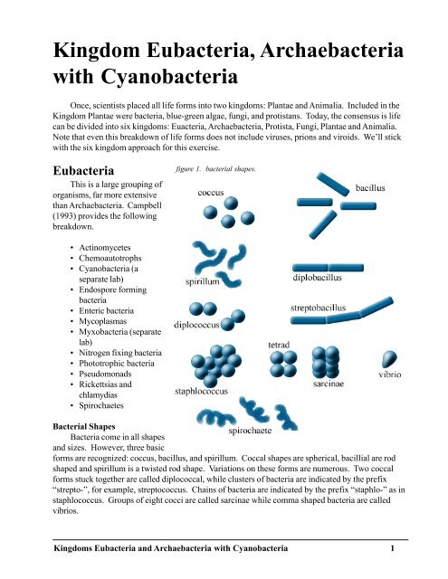
figure 1. bacterial shapes.<br />
• Actinomycetes<br />
• Chemoautotrophs<br />
• <strong>Cyanobacteria</strong> (a<br />
separate lab)<br />
• Endospore forming<br />
bacteria<br />
• Enteric bacteria<br />
• Mycoplasmas<br />
• Myxobacteria (separate<br />
lab)<br />
• Nitrogen fixing bacteria<br />
• Phototrophic bacteria<br />
• Pseudomonads<br />
• Rickettsias and<br />
chlamydias<br />
• Spirochaetes<br />
Bacterial Shapes<br />
Bacteria come in all shapes<br />
and sizes. However, three basic<br />
forms are recognized: coccus, bacillus, and spirillum. Coccal shapes are spherical, bacillial are rod<br />
shaped and spirillum is a twisted rod shape. Variations on these forms are numerous. Two coccal<br />
forms stuck together are called diplococcal, while clusters of bacteria are indicated by the prefix<br />
“strepto-”, for example, streptococcus. Chains of bacteria are indicated by the prefix “staphlo-” as in<br />
staphlococcus. Groups of eight cocci are called sarcinae while comma shaped bacteria are called<br />
vibrios.<br />
<strong>Kingdom</strong>s <strong>Eubacteria</strong> and <strong>Archaebacteria</strong> <strong>with</strong> <strong>Cyanobacteria</strong> 1
Actinomycetes<br />
Although the suffix “mycetes” refers to fungi, these have been removed from the fungal classification<br />
and placed in the <strong>Eubacteria</strong>. These are most often found in soils, as Streptomyces and Mycobacterium.<br />
Observe the prepared slide of Streptomyces, a Gram+, nonmotile organism <strong>with</strong> coenocytic<br />
hyphae. From this genus, we produce the antibiotic streptomycin.<br />
Chemoautotrophic Bacteria<br />
Many of you have sensed at least one species of these if you have been in the Keys and smelled<br />
the rather disagreeable odor of rotten eggs as you drive Highway 1. The smell is hydrogen sulfide<br />
H 2<br />
S, a compound used by some forms to produce energy. It is noticeably worse when someone<br />
disturbs mangroves. Example genera include Nitrobactera and Nitrosomonas. These bacteria oxidize<br />
inorganic substances to produce energy for food production. Some of the inorganic materials used<br />
are NH 3<br />
, NO 3-<br />
, H 2<br />
S, S, and Fe +3 . They utilize these inorganic materials, along <strong>with</strong> carbon in carbon<br />
dioxide to manufacture carbohydrates and other compounds. If prepared slides are available,<br />
observe the characteristics of this group.<br />
Endospore Forming Bacteria<br />
These bacteria form internalized spores which may “hatch” when environmental conditions<br />
permit. They may be either aerobic or anaerobic and typically are Gram positive (more on this later),<br />
flagellated rods. Genera include Bacillus and Clostridium. Clostridium is the bacterium that causes<br />
gangrene. Observe a prepared slide of Clostridium botulinum, rod-shaped and which produces<br />
powerful exotoxins. This bacterium causes botulism or food poisoning.<br />
Enteric Bacteria<br />
For a while, little was known about these bacteria since they are facultatively anaerobic and are<br />
difficult to culture (at least some forms). The most well known is Escherichia coli which lives in our<br />
small intestine in a symbiotic relationship producing vitamin K for us. They are Gram negative.<br />
Some are quite harmless, and in the case of the E. coli that inhabits our small intestine, quite beneficial.<br />
Other strains of E. coli and other genera are quite pathogenic. Other genera include Salmonella<br />
(responsible for Salmonella poisoning) and Vibrio cholerae, responsible for the potentially deadly<br />
disease cholera. Observe the prepared slide of Vibrio, a Gram- rod.<br />
Mycoplasmas<br />
First, unlike all the rest of eubacteria, these don’t have any cell walls. They are essentially a<br />
mass of protoplasm, albeit a very small mass of protoplasm (0.10-0.25 µm), some of the smallest of<br />
all cells. These are saprobic and animal pathogens for the most part, however, they have been implicated<br />
in lethal yellowing of palms here in south Florida.<br />
Myxobacteria<br />
Strange is the best description of these. They are groups of individual cells that feed over a<br />
substrate and move by gliding. Once environmental conditions are harsh, the individual cells congregate<br />
to form “fruiting” structures which eventually release spores. More will be discussed about this<br />
group later in another lab. A typical genus is Myxococcus.<br />
<strong>Kingdom</strong>s <strong>Eubacteria</strong> and <strong>Archaebacteria</strong> <strong>with</strong> <strong>Cyanobacteria</strong> 2
Nitrogen Fixing Bacteria<br />
The most studied of these include mutualistic<br />
species that live in nodules on the roots of<br />
leguminous plants (beans). They are capable<br />
of removing atmospheric nitrogen (N 2<br />
) and<br />
converting it to a form more usable as nitrites<br />
and nitrates. In addition to mutualistic species,<br />
there are free-living species as well. Typical<br />
genera are Azotobacter and Rhizobium.<br />
Observe the prepared slide of Rhizobium<br />
leguminosarum, a Gram–, motile rod<br />
found in the nodules of roots of legumes. This<br />
bacterium fixes atmospheric nitrogen.<br />
Figure 2: The Nitrogen Cycle.<br />
Phototrophic Anaerobic Bacteria<br />
In lecture, you will hear about the differences<br />
between cyanobacteria and eubacteria.<br />
Much of the comparison involves the photosynthetic<br />
process of this group <strong>with</strong> cyanobacteria.<br />
There are numerous groups <strong>with</strong>in this,<br />
including purple sulfur bacteria, green sulfur<br />
bacteria and the genera Rhodospirillum.<br />
Observe the prepared slide of Rhodospirillum rubrum, a Gram–, motile, photosynthetic spiral.<br />
Upon exposure to light, this organism turns pink.<br />
Pseudomonads<br />
Probably, an entire course could be figure 3. Bacterial flagellum.<br />
offered in this diverse group, particularly the<br />
genus Pseudomonas. This genus is found in<br />
virtually all aquatic and soil habitats. The<br />
genus is characterized by rod-shaped, Gram<br />
negative cells that have flagella “tufted” at<br />
one end (lophotrichous). They are<br />
chemoheterotrophs.<br />
Many bacteria (not cyanobacteria)<br />
produce flagella. Neither bacteria nor<br />
cyanobacteria produce cilia. Both may<br />
produce a slime or mucous coat around<br />
themselves for protection. The flagellum is<br />
composed of a protein called flagellin and<br />
episolon-N-methyl lysine (Wistreich and<br />
Lechtman 1980). The flagellum is constructed of several rings, a hook-like structure and a long<br />
filament. It is attached to the inner and outer membranes (the outer only if present). Observe the<br />
prepared slide of the flagellar stained bacterium.<br />
<strong>Kingdom</strong>s <strong>Eubacteria</strong> and <strong>Archaebacteria</strong> <strong>with</strong> <strong>Cyanobacteria</strong> 3
Rickettsias and Chlamydias<br />
All of these are obligate parasites <strong>with</strong> Gram negative walls. Usually there are at least two hosts<br />
(arthropod and mammal). Rickettsia rickettsia is the causative agent of Rocky Mountain Spotted<br />
Fever (the vector is the deer tick) and Chlamydia is a pathogenic bacterium passed between birds and<br />
humans. If a specimen is available, observe Rickettsia and Chlamydia.<br />
Spirochaetes<br />
These are helical cells that have cork-screw like movements due to their internal flagellar filaments.<br />
They may be free-living or parasites as Treponema pallidum, the causative agent of syphilis.<br />
The original test for syphilis was to look for the bacterium after treatment <strong>with</strong> a dye which fluoresced.<br />
More sophisticated tests are now available. Observe the prepared slide of Treponema<br />
pallidum.<br />
<strong>Archaebacteria</strong><br />
Although the term achaio comes from the Greek for ancient, <strong>Archaebacteria</strong> probably evolved,<br />
like Bacteria and eukaryotes from a common ancestor. However, <strong>Archaebacteria</strong> are more related to<br />
the eukaryotes than to the Bacteria.<br />
Several features separate the archaebacteria from eubacteria.<br />
• no peptidoglycan in cell walls<br />
• unique lipid bilayer of cell membranes<br />
• RNA polymerase more similar to eukaryotes<br />
• ribosomal protein more similar to eukaryotes (Campbell 1993).<br />
<strong>Archaebacteria</strong> also grow in extreme habitats as salt flats, hot, acidic aquatic environments and<br />
anaerobic environments. From this, Campbell (1993) breaks the <strong>Archaebacteria</strong> down into 3 subgroups:<br />
methanogens, extreme halophiles, and thermoacidophiles.<br />
Methanogens<br />
Methanogens are noted for their use of H 2<br />
to reduce CO 2<br />
into methane gas (CH 4<br />
). Methanogens<br />
may be obligately anaerobic and may actually be destroyed by exposure to oxygen, a principle many<br />
water treatment facilities put into play when they aerate sewerage.<br />
Methanogens produce marsh gas, which may be seen bubbling up from stagnant bodies of water<br />
(either that or it’s oxygen from photosynthesis). You can actually collect the gas and burn it off and<br />
many a person thinks they have seen UFO’s in the Everglades when in reality it’s burning marsh gas.<br />
There is also plenty of methane produced by cattle and termites which is pumped into the atmosphere.<br />
Some suggest the huge cattle population may actually be cause for global warming by placing<br />
more methane into the atmosphere.<br />
Halophiles<br />
Look for these in salt water environments where the salt concentration is far above the ocean, as<br />
the Great Salt Lake in Utah and the Dead Sea in the Middle East. Sea water may have a salt concentration<br />
of around 3.3% and halophilic <strong>Archaebacteria</strong> may survive 10 times the salt concentration of<br />
the sea (Campbell 1993).<br />
Thermoacidophiles<br />
As the name implies, these bacteria may <strong>with</strong>stand quite hot temperatures and low pH. It is not<br />
unusual for these forms to survive temperatures of around 80° Celsius and a pH of 2 (Campbell<br />
<strong>Kingdom</strong>s <strong>Eubacteria</strong> and <strong>Archaebacteria</strong> <strong>with</strong> <strong>Cyanobacteria</strong> 4
1993).<br />
Observe any genera of <strong>Archaebacteria</strong> available. Compare them to the <strong>Eubacteria</strong> just<br />
observed.<br />
<strong>Cyanobacteria</strong><br />
In the past, <strong>Cyanobacteria</strong> were grouped <strong>with</strong> plants and called blue-green algae. The word<br />
cyan comes from the Gr kyanos which means blue. Evidence today from electron microscopy and<br />
biochemistry suggest they are more bacteria-like (Bold 1973). They are ubiquitous; occurring in<br />
aerial, terrestrial, and aquatic habitats. There are approximately 150 genera <strong>with</strong> 1,500 species (Bold<br />
1973).<br />
Many botanists argue the blue-greens are indeed plants and not bacteria. Their beliefs are based<br />
on three characteristics of blue-greens: (1) bacteria do not contain chlorophyll a while the blue-greens<br />
do, (2) the blue-greens are more differentiated than bacteria and (3) bacteria do not produce oxygen<br />
during photosynthesis while plants do.<br />
Reasons for including the <strong>Cyanobacteria</strong> in the bacteria group include (1) they have a cell wall<br />
composed of murein, similar in nature to the cell walls of bacteria (2) they have a single strand of<br />
DNA for a genome and (3) it is a prokaryotic cell (Bold 1973). See table 1 on page 8 for a comparison.<br />
What do you think? Is it a bacterium, archaebacterium, or blue-green alga?<br />
Pigments<br />
The presence or absence of pigments in species is a critical factor in identification. Pigments<br />
associated <strong>with</strong> the cyanobacteria include the water insoluble pigments chlorophyll a and the carotenoids.<br />
Water soluble pigments include phycocyanin, phycoerythrin, and allophycocyanin.<br />
Gelatinous Sheath<br />
One characteristic shared by all<br />
members of the <strong>Cyanobacteria</strong> is the<br />
presence of a layer of slimy material<br />
of varying thickness and consistency,<br />
the gelatinous sheath. Electron<br />
microscopy has determined it is<br />
composed of “fibrillar material<br />
embedded <strong>with</strong>in an amorphous<br />
matrix” (Bold 1973). It can be<br />
shown by staining the alga in diluted<br />
India ink or methylene blue (Bold<br />
1973). Obtain a living culture of<br />
Gloeocapsa and make a wet mount.<br />
Observe first <strong>with</strong>out stain and then<br />
stain <strong>with</strong> methylene blue to see the<br />
gelatinous sheath. Save the slide for<br />
comparison <strong>with</strong> Chroococcus.<br />
Gloeocapsa (Gr. gloia, glue + L. capsa, a box or case) is best found on moist rocks in shady<br />
areas, walls, and on the outside of flower pots. It gives the appearance of a light green paint on the<br />
surface of the object. You generally do not find Gloeocapsa growing as a single cell due to the<br />
<strong>Kingdom</strong>s <strong>Eubacteria</strong> and <strong>Archaebacteria</strong> <strong>with</strong> <strong>Cyanobacteria</strong> 5
abundant cell division and the gelatinous sheath.<br />
Food Storage<br />
The photosynthetic storage product of plants is starch. However, there are many varieties of<br />
starch. <strong>Cyanobacteria</strong> form cyanophycean starch in structures called alpha granules (Bold 1973).<br />
This starch is actually glycogen (Bold 1973) more appropriately associated <strong>with</strong> animal starch (and in<br />
some bacteria).<br />
Growth Patterns<br />
The patterns of growth exhibited<br />
by the blue-greens take several<br />
forms. Chroococcus is unicellular.<br />
Gloeocapsa is unicellular also but<br />
thick gelatinous sheathes prevent the<br />
cells from separating and thus<br />
Gloeocapsa appears colonial.<br />
Make a wet mount of<br />
Chroococcus, stain it <strong>with</strong> methylene<br />
blue and compare it to Gloeocapsa.<br />
Merismopedia forms flat plates of<br />
cells and Lyngbya and Gleotrichia form<br />
filamentous structures.<br />
Nostoc (name used by Paracelsus)<br />
is also commonly known as star jelly or<br />
witches’ butter (Bold 1973). It is often<br />
found growing as a jelly-like mass on the<br />
ground. To the layperson, Nostoc is<br />
virtually identical to another filamentous<br />
genus, Anabaena (Gr. anabainein, to<br />
arise). To the scientist, the way to tell<br />
the difference is the more abundant<br />
gelatinous sheath in Nostoc and the size<br />
of hormogonia. A hormogonium consists<br />
of a series of cells divided from<br />
another series of cells by dead cells.<br />
Both Nostoc and Anabaena produce<br />
akinetes and heterocysts. Both are capable<br />
of nitrogen fixation like some of the eubacteria.<br />
Akinetes are spores and are highly<br />
resistant to environmental stresses and<br />
some have germinated after many years in<br />
storage (Bold 1973). The heterocysts also<br />
serve as the boundaries for hormogonia and<br />
are capable of fixing nitrogen.<br />
Make a wet mount of either Nostoc<br />
or Anabaena, whichever is provided. Observe and compare to a prepared slide of the same. Try to<br />
identify the following: cell wall, akinete, heterocyst, sheath, hormogonium.<br />
<strong>Kingdom</strong>s <strong>Eubacteria</strong> and <strong>Archaebacteria</strong> <strong>with</strong> <strong>Cyanobacteria</strong> 6
Anabaena and another blue-green,<br />
Microcystis, are the main species found in<br />
water blooms in the Everglades and Lake<br />
Okeechobee. Water blooms are rapid<br />
algae growth that often results in oxygen<br />
depletion and death of aquatic life, especially<br />
fish.<br />
How is it a plant which evolves<br />
oxygen during photosynthesis can deplete<br />
oxygen in a lake and<br />
cause fish kills?<br />
______________________________________________________________<br />
_<br />
Anabaena may also be found as an<br />
endophyte (living inside another plant). The<br />
common water fern Azolla is found in many<br />
of South Florida’s canals growing as a green<br />
“carpet” on the surface. Inside the fronds of<br />
the fern, you may find Anabaena. It seems to<br />
be a mutualistic relationship in that Anabaena<br />
is capable of nitrogen fixation. Apparently,<br />
Anabaena provides nitrates and nitrites for<br />
the fern and the fern provides a place for<br />
Anabaena to live.<br />
Oscillatoria (L. oscillare, to swing) is<br />
aptly named. It performs an oscillating<br />
motion in aquatic suspension. Make a wet<br />
mount and observe the gentle back and<br />
forth swaying of the filaments. Oscillatoria<br />
is most often found in floating mats or<br />
strings in aquatic habitats or on damp soil<br />
(Bold 1973). It has a very thin sheath and<br />
cells are divided by hormogonia. Note the<br />
dead or biconcave cells.<br />
Lyngbya (named in honor of the Danish<br />
phycologist Lyngbye) is difficult to distinguish<br />
from Oscillatoria except for one main feature.<br />
The gelatinous sheath in Lyngbya is extensive<br />
whereas it is quite thin in Oscillatoria.<br />
Lyngbya does not produce the oscillating motion seen in Oscillatoria but it does produce hormogonia<br />
separated by dead cells. Observe a prepared slide of both Oscillatoria and Lyngbya and compare the<br />
two.<br />
<strong>Kingdom</strong>s <strong>Eubacteria</strong> and <strong>Archaebacteria</strong> <strong>with</strong> <strong>Cyanobacteria</strong> 7
Table 1. Comparison of Bacteria and <strong>Cyanobacteria</strong> from Pritchard and Bradt (1984).<br />
STRUCTURE/FUNCTION<br />
BACTERIA<br />
CYANOBACTERI A<br />
Membrane<br />
Bound Organelles<br />
none<br />
none<br />
DNA<br />
no<br />
histone s<br />
no histone s<br />
Ribosomes<br />
Cell Wall<br />
sedimentation coefficient<br />
70s<br />
muramic acid,<br />
diaminopimelic acid and<br />
other organic acids<br />
sedimentation coefficient<br />
70s<br />
muramic acid,<br />
diaminopimelic acid,<br />
glucoasamines, alanine,<br />
glutamic acid<br />
Lysozyme<br />
Reaction<br />
dissolves<br />
cell wall<br />
dissolves cell wall<br />
Sexual Activity<br />
Nitrogen<br />
Light<br />
Fixation<br />
Receptive Molecules<br />
bacterial genetic<br />
recombination<br />
(transformation,<br />
transduction, or<br />
conjugation0<br />
some bacterial species<br />
bacteriochlorophyll s<br />
recombination in a<br />
mutant species<br />
few<br />
some species, especially<br />
those <strong>with</strong> heterocysts<br />
chlorophyll<br />
a , carotenoids,<br />
phycobilins<br />
Photosynthetic<br />
Environment<br />
anaerobic<br />
and aerobic<br />
anaerobic and aerobic<br />
Photosynthetic By-Products<br />
Materials Required for<br />
Photosynthesis<br />
Vegetative Reproduction<br />
hydrogen, sulfur, organic<br />
compounds<br />
H 2<br />
, H S,<br />
CO<br />
, organic<br />
2 2<br />
compounds, light<br />
spores, fission,<br />
fragmentation<br />
oxygen<br />
H 2<br />
O,<br />
CO<br />
, minerals, light<br />
2<br />
spores, fission,<br />
fragmentation of<br />
hormogones, akinetes<br />
<strong>Kingdom</strong>s <strong>Eubacteria</strong> and <strong>Archaebacteria</strong> <strong>with</strong> <strong>Cyanobacteria</strong> 8
References Cited<br />
Bold HC. 1973. Morphology of plants, 3rd ed. New York: Harper & Row. p 134-145.<br />
Campbell NA. 1993. Biology, 3rd ed. Benjamin/Cummings. Redwood City, CA. 1190 pp.<br />
Ppritchard HN, Bradt PT. 1984. Biology of nonvascular plants. St. Louis: Times Mirror/Mosby.<br />
Wistreich GA, Lechtman MD. 1980. Microbiology, 3rd ed. New York: Macmillan, CA., p 122.<br />
<strong>Kingdom</strong>s <strong>Eubacteria</strong> and <strong>Archaebacteria</strong> <strong>with</strong> <strong>Cyanobacteria</strong> 9