Sample Exam 2 - Biology Courses Server
Sample Exam 2 - Biology Courses Server
Sample Exam 2 - Biology Courses Server

You also want an ePaper? Increase the reach of your titles
YUMPU automatically turns print PDFs into web optimized ePapers that Google loves.
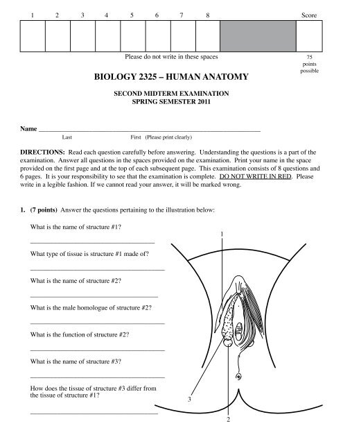
1 2 3 4 5 6 7 8 Score<br />
Please do not write in these spaces<br />
BIOLOGY 2325 – HUMAN ANATOMY<br />
75<br />
points<br />
possible<br />
SECOND MIDTERM EXAMINATION<br />
SPRING SEMESTER 2011<br />
Name __________________________________________________________________<br />
Last<br />
First (Please print clearly)<br />
DIRECTIONS: Read each question carefully before answering. Understanding the questions is a part of the<br />
examination. Answer all questions in the spaces provided on the examination. Print your name in the space<br />
provided on the first page and at the top of each subsequent page. This examination consists of 8 questions and<br />
6 pages. It is your responsibility to see that the examination is complete. DO NOT WRITE IN RED. Please<br />
write in a legible fashion. If we cannot read your answer, it will be marked wrong.<br />
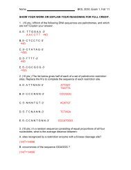
1. (7 points) Answer the questions pertaining to the illustration below:<br />
What is the name of structure #1<br />
_____________________________________<br />
1<br />
What type of tissue is structure #1 made of<br />
________________________________________<br />
What is the name of structure #2<br />
______________________________________<br />
What is the male homologue of structure #2<br />
________________________________________<br />
What is the function of structure #2<br />
________________________________________<br />
What is the name of structure #3<br />
________________________________________<br />
How does the tissue of structure #3 differ from<br />
the tissue of structure #1<br />
______________________________________<br />
3<br />
2
Name _____________________________________<br />
2. (15 points) Draw the following nervous traces on the<br />
illustration below. When drawing the neurons, you<br />
must clearly draw the location of their cell body and<br />
synaptic junctions. The synaptic junctions must be<br />
represented like the pictures that you studied in the<br />
lecture manual. You must also label the neurons.<br />
At Level 1, draw and label a reflex pathway to a<br />
skeletal muscle served by the ventral ramus. Color<br />
each neuron a different color and clearly show the cell<br />
bodies and synapse locations.<br />
Level 1<br />
At Level 2, draw and label a sympathetic neuron<br />
reflex from and to a blood vessel in the epaxial body<br />
wall. Color each neuron a different color and clearly<br />
show the cell bodies and synapse locations.<br />
Level 2<br />
At Level 3, draw and label a sympathetic neuron<br />
pathway from and to a blood vessel in the descending<br />
colon. Color each neuron a different color and clearly<br />
show the cell bodies and synapse locations.<br />
Level 3<br />
At Level 4, draw and label a parasympathetic neuron<br />
pathway from and to the taenia coli muscle of the<br />
descending colon. Color each neuron a different color<br />
and clearly show the cell bodies and synapse locations.<br />
Level 4<br />
In this illustration what spinal levels are represented<br />
by:<br />
Level 1 _____________________________<br />
Level 2 _____________________________<br />
Level 3 _____________________________<br />
Level 4 _____________________________<br />
2
Name _____________________________________<br />
3. (9 points) One of the major themes of this portion of the course was the pattern of the body wall muscles<br />
of the trunk. The table below depicts the pattern of design of the body wall and has an empty box for each<br />
muscle in the body wall. Fill in each of the boxes with the correct muscle to complete the table.<br />
Pattern Ventral Lateral<br />
Outermost<br />
Lateral<br />
External<br />
Lateral<br />
Middle<br />
Lateral<br />
Internal<br />
Subvertebral<br />
Thorax Abdomen<br />
3
Name _____________________________________<br />
4. (14 points) Trace a red blood cell on a continuous route from the heart to the right transversus thoracis muscle then back to<br />
the heart then to the greater curvature of the stomach at its pyloric end then back to the heart and then to the right testis then back<br />
again to the heart and then to the right subcostal muscle in the uppermost intercostal space and then once more back to the heart.<br />
There are no blockages so your trace must traverse normal pathways of flow. Your trace must consist of a columnar list of<br />
the vessels through which the red blood cell passes as it moves to and from each of these structures. Use the space in the columns<br />
below. You do not need to list the structures of the heart in the trace, nor do you have to write arterioles, capillaries, venules.<br />
Heart<br />
Right testis<br />
Right transversus thoracis muscle<br />
Heart<br />
Heart<br />
Right subcostal muscle in<br />
upper intercostal space<br />
Greater curvature of pyloric end of stomach<br />
Heart<br />
Heart<br />
4
Name _____________________________________<br />
5. (8 points) In the spaces below, label the numbered structures in the illustration and answer the question<br />
below the numbered lines.<br />
1. ________________________________<br />
2. ________________________________<br />
3. ________________________________<br />
4. ________________________________<br />
5. ________________________________<br />
What named tubes run through structure #1<br />
__________________________________<br />
__________________________________<br />
1<br />
5<br />
What named tube runs through structure #3<br />
____________________________________<br />
2<br />
3<br />
4<br />
6. (6 points) Answer the questions about the illustration below.<br />
2<br />
A<br />
B<br />
What type of neuron is depicted above _____________________________________________________<br />
What is the name of the processes indicated by #1 ____________________________________________<br />
Where in the peripheral nervous system would you find structure #2 ______________________________<br />
If letter A represents the peripheral end of this cell and letter B is the end in the spinal cord, what would the<br />
end indicated by the A be associated with ___________________________________________________<br />
When process B projects into the spinal cord, name two types of neurons it can synapse with.<br />
1<br />
_________________________________________ and _________________________________________<br />
5
Name _____________________________________<br />
7. (11 points) Fill in the blanks in the sentences below.<br />
The ___________________________ nerve distributes parasympathetic efferent neurons to the thoracic<br />
viscera and arises from the _____________________________.<br />
Name one primary retroperitoneal organ. __________________________________________________<br />
Name four secondary retroperitoneal organs:<br />
_____________________________________________________<br />
_____________________________________________________<br />
_____________________________________________________<br />
_____________________________________________________<br />
The _______________________ is the peritoneal fold that runs between the stomach and the liver.<br />
At ovulation the egg breaks the surface of the ovary and enters the _______________________________.<br />
What named cells form the myelin sheaths of the peripheral nervous system ______________________<br />
The adoral branch of the splenic artery is the _____________________________________<br />
8. (5 points) In the boxes below pick a color and fill the box and then clearly illustrate each structure on the<br />
picture with that color. You must clearly color the entire extent of each of the named structures. Also, with<br />
a labeled arrow clearly indicate the location of the pleural cavities and pericardial cavity. In the spaces<br />
below list three mediastinal structures that are visible in this illustration other than the heart and its anatomy.<br />
visceral pleura<br />
parietal pleura<br />
visceral pericardium<br />
parietal pericardium<br />
fibrous pericardium<br />
Mediastinal structures:<br />
__________________<br />
__________________<br />
__________________<br />
6