Rhizoctonia solani.pdf - RAC-1 - Mardi
Rhizoctonia solani.pdf - RAC-1 - Mardi
Rhizoctonia solani.pdf - RAC-1 - Mardi

You also want an ePaper? Increase the reach of your titles
YUMPU automatically turns print PDFs into web optimized ePapers that Google loves.
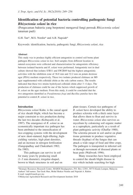
J. Trop. Agric. and Fd. Sc. 38(2)(2010): 249– 256<br />
G.H. Tan, M.S. Nordin and A.B. Napsiah<br />
Identification of potential bacteria controlling pathogenic fungi<br />
<strong>Rhizoctonia</strong> <strong>solani</strong> in rice<br />
(Pengecaman bakteria yang berpotensi mengawal fungi perosak <strong>Rhizoctonia</strong> <strong>solani</strong><br />
tanaman padi)<br />
G.H. Tan*, M.S. Nordin* and A.B. Napsiah*<br />
Keywords: identification, bacteria, pathogenic fungi, <strong>Rhizoctonia</strong> <strong>solani</strong>, rice<br />
Abstract<br />
This study was to produce highly efficient antagonists to control soil borne plant<br />
pathogen <strong>Rhizoctonia</strong> <strong>solani</strong> in rice. Soil samples from different location in<br />
natural ecosystem were collected and characterization for antagonistic efficiency<br />
between isolated bacteria and R. <strong>solani</strong> were performed. Antagonistic test in dual<br />
culture showed that isolates S3B11 and SW2B49 had the highest antagonistic<br />
activities with the inhibition zone of 36.0 mm and 32.2 mm on potato dextrose<br />
agar (PDA) medium respectively. These two isolates produced chitinase on M9<br />
agar supplemented with colloidal chitin as the sole carbon source. The results<br />
indicated that these two strains hydrolyzed colloidal chitin after 3–4 days. The<br />
production of chitinase could be one of the factors which suppressed growth of<br />
R. <strong>solani</strong> on the agar medium. From this study, it could be concluded that the<br />
two antagonists identified as Pseudomonas fragi and Bacillus pumilus have the<br />
potential to control R. <strong>solani</strong> in rice.<br />
Introduction<br />
<strong>Rhizoctonia</strong> <strong>solani</strong> Kuhn. is the causal agent<br />
of rice sheath blight, which has become a<br />
major constraint to rice production during<br />
the last two decades (Kobayashi et al.<br />
1997). The emergence of R. <strong>solani</strong> as an<br />
economically important rice pathogen has<br />
been attributed to the intensification of<br />
rice-cropping systems with the development<br />
of new short-statured, high-tillering, highyielding<br />
varieties, high plant densities,<br />
and an increase in nitrogen fertilization<br />
(Gangopadhay and Chakrabarti 1982;<br />
Ou 1985).<br />
This pathogen can survive in soil<br />
for many years by producing small<br />
(1–3 mm diameter), irregular-shaped,<br />
brown to black structures in soil and on<br />
plant tissues. Certain rice pathogens of<br />
R. <strong>solani</strong> have developed the ability to<br />
produce sclerotia with a thick outer layer<br />
that allows them to float and survive in<br />
water. <strong>Rhizoctonia</strong> <strong>solani</strong> also survives as<br />
mycelium by colonizing soil organic matter<br />
as a saprophyte, particularly as a result of<br />
plant pathogenic activity (Ghaffar 1988).<br />
The sclerotia present in soil and/or on plant<br />
tissue germinate to produce vegetative<br />
threads (hyphae) of the fungus that can<br />
attack a wide range of food and fibre crops.<br />
The pathogen is transported in infected soil<br />
or through movement of diseased plants or<br />
bean pods (Wallwork 1996).<br />
Many researches have been employed<br />
to control the sheath blight disease in<br />
rice which include searching for high<br />
*Strategic Resources Research Centre, MARDI Headquarters, P.O. Box 12301, 50774 Kuala Lumpur, Malaysia<br />
Authors’ full names: Tan Geok Hun, Mohd Shukor Nordin and Napsiah A. Rahim<br />
E-mail: tangh@mardi.gov.my<br />
©Malaysian Agricultural Research and Development Institute 2010<br />
249
Potential bacteria for controlling <strong>Rhizoctonia</strong> <strong>solani</strong><br />
quality seeds and avoid seeds that may be<br />
contaminated with the pathogen (Farr et al.<br />
1989), preparing for planting areas to<br />
increase drainage and prevent water<br />
accumulation in the field and addition of<br />
compost and organic fertilizers to decrease<br />
disease levels (Hawksworth et al. 1995).<br />
Fungicides such as methyl thiophanate,<br />
PCNB (pentachloronitrobenzene) and<br />
chlorothalonil have also been applied to<br />
reduce the disease infection. However, so far<br />
none of them is found to be effective against<br />
this disease in the long term.<br />
Biological control has been developed<br />
as an alternative to reduce usage of chemical<br />
pesticides to control various plant diseases.<br />
By using this strategy, microorganisms<br />
themselves or the antibiotics and degrading<br />
enzymes that they produce can be used<br />
directly against various plant pathogens.<br />
Many strains of B. subtilis have shown to<br />
be potential biocontrol agent against fungal<br />
pathogens. It was reported that the principle<br />
mechanism of this antifungal involves the<br />
production of antibiotics (Fravel 1988). In<br />
addition, B. subtilis strains produce volatiles<br />
that antagonise a range of soil-borne plant<br />
pathogen including R. <strong>solani</strong> and Pythium<br />
ultimum. Thus, the aim of this study was<br />
to observe the effectiveness of selected<br />
microbes to control R. <strong>solani</strong> in rice.<br />
Materials and methods<br />
Isolation and preparation of bacterial<br />
culture<br />
Bacterial culture was isolated from<br />
various sources such as sewages, soils<br />
and water samples from Pontian, Tok<br />
Bok waterfall, Kelantan and Cameron<br />
Highlands. The isolation and screening of<br />
pure bacterial culture followed the common<br />
Microbiological Protocol as described by<br />
Sambrook et al. (1989) and cultured on<br />
NBTA medium (nutrient agar supplemented<br />
with 0.025 g bromothymol blue and 0.04 g<br />
2,3,5-triphenyltetrazolium chloride per<br />
litre, AmBresco, USA). A loopful of 24 h<br />
bacterial culture was added to a 250 ml<br />
conical flask containing 100 ml of tryptic<br />
soy broth and shaken for 24 h at ambient<br />
temperature (~28 °C). Subsequently, the<br />
bacterial suspension was spun at 4,000 x g<br />
for 5 min and the supernatant was<br />
discarded. Sterile phosphate buffer (50 mM,<br />
pH 7.2) was added to the pellets and<br />
mixed thoroughly to obtain a concentrated<br />
suspension of the bacterial symbiont.<br />
Different cell concentrations were then<br />
prepared by dilution of sterile phosphate<br />
buffer (50 mM), and the cell number in each<br />
suspension was established by counting the<br />
colony forming unit (cfu/ml).<br />
Fungi preparation<br />
The isolate of R. <strong>solani</strong> was obtained<br />
from sheath blight disease in rice from<br />
Biotechnology Research Centre, MARDI.<br />
The fungi was cultured on PDA (Potato<br />
Dextrose Agar, AmBresco, USA) and<br />
incubated in the dark at ambient temperature<br />
(~28 °C) for 5 days for complete<br />
sporulation, and later stored at 4 °C.<br />
Inhibition of fungal growth by bacteria<br />
One-week-old R. <strong>solani</strong> culture was cut<br />
into 5 mm diameter discs and transferred<br />
to the PDA petri dishes. In each dish, the<br />
same agar medium was inoculated with a<br />
5 mm diameter disc of antagonist positioned<br />
diametrically opposite to the 5 mm diameter<br />
disc of the R. <strong>solani</strong>. The distance between<br />
discs was approximately 60 mm. The<br />
cultures were incubated at 28 ± 3 °C and<br />
measurement was recorded after 4 days of<br />
incubation. The radial growth was measured<br />
after 1 week of the observation. The<br />
efficiency of antagonist in suppressing radial<br />
growth was calculated as follows: [C-T]/C x<br />
100 where C is radial growth measurement<br />
of the pathogen in the negative control and<br />
T is radial growth of the pathogen in the<br />
presence of antagonist. The disc soaked in<br />
distilled water was set as negative control.<br />
Each test has three replications.<br />
Detection of chitinolytic activity<br />
In order to test the chitinolytic activity<br />
on plates, cells of bacteria strains were<br />
250
G.H. Tan, M.S. Nordin and A.B. Napsiah<br />
inoculated on a chitinase detection agar<br />
plate which was prepared by mixing 10 g<br />
of colloidal chitin and 20 g of agar in M9<br />
medium (25.6 g Na 2<br />
HPO 4<br />
7H 2<br />
O, 6.0 g<br />
KH 2<br />
PO 4<br />
, 1.0 g NaCl, 2.0 g NH 4<br />
Cl, 1 litre).<br />
The plates were incubated at 30 °C for<br />
7 days until clearing zones could be detected<br />
around the colonies. Each test has three<br />
replications.<br />
Identification of potential bacteria using<br />
16S rDNA analysis<br />
A loopful of bacterial cell was picked up<br />
and suspended in TE buffer [100 mM Tris<br />
(pH 8.0), 30 mM EDTA (pH 8.0), 360 µl].<br />
Lsozyme (50 mg/ml, 20 µl) was added into<br />
the tube and incubated at 37 °C for 30 min.<br />
SDS [10% (w/v), 40 µl] was added, and<br />
the tube was inverted for 5–6 times, then<br />
incubated at 55 °C for 10 min. Phenol:<br />
chloroform: isoamyalcohol [25:24:1, 400 µl]<br />
was added and mixed well. The mixture was<br />
spun at 13,000 rpm for 15 min. The upper<br />
solution was transferred to a new microtube.<br />
The same procedure was repeated if the<br />
solution was not clear. Sodium acetate<br />
(3 M, 1/10 volume) and 2 volume of cold<br />
ethanol was then added. DNA was then<br />
pooled by glass rod and dried for 5–10 min<br />
until DNA became clear. Finally, the DNA<br />
was dissolved in 100–200 µl of sterilized<br />
distilled water and stored at –20 °C.<br />
Polymerase Chain Reaction (PCR)<br />
of the 16S rDNA for the bacterial isolates<br />
was conducted and the PCR reaction was<br />
carried out as followed: sterile distilled<br />
water (59.5 ml); 10X PCR buffer (Promega,<br />
USA, 10.0 ml); 25 mM MgCl 2<br />
(Promega,<br />
USA, 8.0 ml); 2 mM dNTP mix (Promega,<br />
USA, 10.0 ml); universal primers (Forward:<br />
5’ GAG TTT GAT CCT GC TCA G 3’;<br />
Reverse: 5’ GTT ACC TTG TTA CGA CTT<br />
3’] Invitrogen, USA, 10 pmol/µl, 4.0 ml);<br />
Taq Polymerase (5U/ml; 0.5 ml) and genomic<br />
DNA as template (4 ml). The PCR tubes was<br />
then put into therma cycler and preheat at<br />
94 °C for 3 min, followed by 25 cycles of<br />
denaturing at 94 °C for 1 min, annealing<br />
at 50 °C for 1 min, extension at 72 °C for<br />
2 min and final elongation at 72 °C for<br />
3 min. The reactions were kept at 4 ºC until<br />
loaded onto the gel. The PCR product was<br />
purified, sequenced and compared with the<br />
16S rDNA sequence of bacteria from the<br />
NCBI Gene Bank nucleotide sequence. The<br />
sequence and identity of each isolate was<br />
then double confirmed by constructing the<br />
Phylogenetic Tree using BioEdit (CA) and<br />
MEGA 4.0.2 software (UK).<br />
Results and discussion<br />
The interaction between target fungi<br />
(R. <strong>solani</strong>) and bacteria culture was recorded<br />
after 4 days of incubation once the two<br />
cultures were placed on the same petri<br />
dish. A total of 25 bacteria isolates (out of<br />
43 isolated from different sources) were<br />
examined as potential inhibitor against<br />
R. <strong>solani</strong> with different antagonistic<br />
efficiency (Figure 1). Based on their<br />
performance, isolate S3B11 and SW2B49<br />
showed the highest inhibition performance<br />
against R. <strong>solani</strong> as shown in Plate 1.<br />
These two isolates were further tested<br />
for chitinolytic production on M9 media<br />
supplemented with 0.1% colloidal chitin.<br />
The result showed the appearance of clear<br />
zone around the colony as shown in Plate 2<br />
indicating the chitinase activity of the<br />
culture.<br />
Several bacteria were reported to<br />
have the potential to produce thermo stable<br />
chitinase (Sakai et al. 1998). In this study,<br />
Pseudomonas fragi and Bacillus pumilus<br />
required 3 to 4 days to produce thermo<br />
stable chitinase when incubated at 60 °C.<br />
A similar finding was reported by Khiyami<br />
and Masmali (2008) where production<br />
of chitinase by P. aeruginosa K-187 and<br />
Bacillus strain MH-1 took about 4 days<br />
at 58 °C. Chitinase is able to degrade<br />
chitin in the fungi cell wall and inhibited<br />
growth of the fungi. Chitinase producing<br />
microorganisms have been reported as<br />
biocontrol agent for different kinds of<br />
plant fungal diseases (Chernin et al. 1995,<br />
Freeman et al. 2004; Liu et al. 2008).<br />
251
Potential bacteria for controlling <strong>Rhizoctonia</strong> <strong>solani</strong><br />
40<br />
35<br />
30<br />
Inhibition zone (mm)<br />
25<br />
20<br />
15<br />
10<br />
5<br />
0<br />
J4B33<br />
J4E2<br />
J2B31<br />
M2B31<br />
J6B41<br />
M2B27<br />
S1B6<br />
S2B3<br />
S3B11<br />
S3B12<br />
S4B17<br />
S2B2<br />
Bacteria isolates<br />
Figure 1. Inhibition zone (mm) formed by bacteria isolates against <strong>Rhizoctonia</strong> <strong>solani</strong> on plate after<br />
1 week of incubation. Assays were performed in triplicates and the error bars represent the standard<br />
error from the arithmetic mean<br />
S1B23<br />
S4B17<br />
S4B21<br />
TB1<br />
TB7<br />
TB8<br />
TB9<br />
J5B34<br />
FP1B52<br />
SW2B49<br />
FP3B57<br />
FP1B50<br />
FP1B51<br />
Control<br />
Disc without bacteria, soaked<br />
in distilled water<br />
(a)<br />
(1)<br />
(b)<br />
(2)<br />
Plate 1. Some examples of the antagonistic effect of bacteria in dual culture. (a) The control disc<br />
without bacteria. (b) Disc soaked in (1) S3B11 and (2) SW2B49, a zone of inhibition in a lawn of<br />
<strong>Rhizoctonia</strong> <strong>solani</strong> surrounds a spot-inoculum of antagonists (indicated by arrow)<br />
252
G.H. Tan, M.S. Nordin and A.B. Napsiah<br />
(a)<br />
Plate 2. (a). Detection of chitinase production of S3B11 on M9 medium containing 0.1% colloidal chitin,<br />
without congo red, (b). The clearing zones of colloidal chitin formed by chitinase, stained with 0.1%<br />
Congo red solution (indicated by black arrow)<br />
(b)<br />
Based on previous studies, there<br />
are more than 167 biological compounds<br />
produced by Bacillus species that are active<br />
against bacteria, fungi, protozoa and viruses<br />
(Berdy 1974; Katz and Demain 1977;<br />
Karuse et al. 1990; Cordovilla et al. 1993).<br />
Most of the antibacterias are peptides and<br />
are active against Gram-positive species<br />
while compounds such as polymyxin and<br />
colistin are active against Gram-negative<br />
species. Bacillus subtilis BNI strain is<br />
effective against Macrophomina phaseolina<br />
(Smith et al. 1986), R. <strong>solani</strong> and P. ultimum<br />
(Wright and Thompson 1985; Fiddaman and<br />
Rossal 1993). Bacillus cereus 28-9 excreted<br />
two chitinases which had inhibitory activity<br />
against Botritis elipitica (Huang et al. 2005).<br />
Based on 16S rDNA sequence analysis,<br />
the isolate S3B11 was identified as Bacillus<br />
pumilus and SW2B49 as Pseudomonas<br />
fragi. These sequences were further<br />
confirmed by constructing the phylogenetic<br />
tree to correlate with the family tree of<br />
both species. The known genus sequence of<br />
Bacillus and Pseudomonas were downloaded<br />
from the Gene Bank and computed using<br />
BioEdit (CA) and MEGA 4.0.2 software<br />
(UK). The final phylogenetic trees<br />
(Figure 2) showed that the new sequence of<br />
S3B11 and SW2B49 are closely related to<br />
Bacillus pumillus with 99% and Pseudomoas<br />
fragi with 100% of similarity respectively.<br />
Table 1. The identification of positive antagonists<br />
against <strong>Rhizoctonia</strong> <strong>solani</strong> by 16S rDNA analysis<br />
Bacteria<br />
isolates<br />
ID (% similarity with NCBI<br />
Gene Bank)<br />
J4B33 Bacillus thuringiensis (98%)<br />
J4E2 Bacillus sp. (80%)<br />
J2B31 Bacillus sp. (80%)<br />
M2B31 Bacillus sp. (80%)<br />
J6B41 Bacillus fusiformis (95%)<br />
M2B27 Kluyvera Georgiana (90%)<br />
S1B6 Baccilus cereus (95%)<br />
S2B3 Lysinibacillus sphaericus (90%)<br />
S3B12 Bacillus thuringiensis (90%)<br />
S4B17 Bacillus sp. (80%)<br />
S2B2 Lysinibacillus sphaericus (90%)<br />
S1B23 Bacillus macroides (98%)<br />
S4B21 Bacillus sp. (80%)<br />
TB1 Bacillus sp. (80%)<br />
TB7 Bacillus cereus (95%)<br />
TB8 Bacillus sp. (85%)<br />
TB9 Bacillus sp. (85%)<br />
J5B34 Bacillus cereus (95%)<br />
FP1B52 Corynebacterium urealyticum (85%)<br />
FP3B57 Aeromonas allosaccharophila (80%)<br />
FP1B50 Aeromonas sp. (80%)<br />
FP1B51 Bacillus cereus (90%)<br />
253
Potential bacteria for controlling <strong>Rhizoctonia</strong> <strong>solani</strong><br />
78<br />
95<br />
73<br />
45<br />
21<br />
67<br />
85 54<br />
99<br />
gi|7209528|dbj|AB0211811| Bacillus atrop<br />
gi|7209545|dbj|AB0211981| Bacillus valli<br />
gi|37722425|gb|AY1494731| Bacillus polyf<br />
82 gi|50812173:980911361 Bacillus subtilis<br />
gi|7209538|dbj|AB0211911| Bacillus mojav<br />
99<br />
88<br />
gi|53774186|gb|AY6036572| Bacillus axarq<br />
81 gi|53774185|gb|AY6036562| Bacillus malac<br />
gi|56182545:990811452 Bacillus lichenifo<br />
S3B11<br />
99 gi|27497686|gb|AY1678791| Bacillus pumilus<br />
gi|21912394|emb|AJ4196291|BLU419629 Baci<br />
99 gi|7209539|dbj|AB0211921| Bacillus mycoi<br />
gi|7209546|dbj|AB0211991| Bacillus weihe<br />
gi|50196905933510841 Bacillus anthracis<br />
gi|42779081:933510842 Bacillus cereus AT<br />
86 gi|2331222|gb|AF0131211| Bacillus pseudo<br />
52 gi|73254772|gb|DQ1489521| Bacillus alkal<br />
gi|10636212|emb|AJ2500561|BFU250056 Baci<br />
gi|289294|gb|L140131|BACRRNAGD Bacillus<br />
gi|1240022|emb|X807251| Ecoli ATCC 11775<br />
0.02<br />
(a) Bacillus genus<br />
78<br />
gi|12406960|emb|AJ2493821| Pseudomonas f<br />
79 gi|21726935|emb|AJ4928271| Pseudomonas c<br />
38 gi|4164003|gb|AF1003211|AF100321 Pseudom<br />
gi|7384769|dbj|AB0305831| Pseudomonas al<br />
gi|4138390|emb|AJ0115041| Pseudomonas ab<br />
95<br />
50<br />
77 gi|1907092|emb|Z766521| Pagarici 16S rRNA<br />
100 SW2B49<br />
gi|10567504|gb|AF094733AF094733 Pseudomonas fragi<br />
29<br />
53 gi|27901525|emb|AJ5376011| Pseudomonas a<br />
56 gi|4433332|dbj|D840091|PSEIAM08 Pseudomo<br />
48<br />
99 gi|8778106|gb|AF2689681| Pseudomonas bre<br />
gi|2909329|emb|X995401| Pseudomonas angu<br />
99<br />
gi|78038894|emb|AM1145271| Pseudomonas b<br />
gi|60359782|dbj|AB1894521| Pseudomonas a<br />
0.02<br />
29<br />
41<br />
(b) Pseudomonas genus<br />
gi|45418|emb|X066841| Pseudomonas aerugi<br />
gi|4433330|dbj|D840061|PSEIAM05 Pseudomo<br />
gi|4165397|dbj|AB0213911| Pseudomonas bo<br />
gi|1240022|emb|X807251| Ecoli ATCC 11775<br />
Figure 2. Phylogenetic tree of (a) Bacillus genus and (b) Pseudomonas genus. S3B11 and SW2B49<br />
are closely related to Bacillus pumillus with 99% and Pseudomoas fragi with 100% of similarity,<br />
respectively<br />
254
G.H. Tan, M.S. Nordin and A.B. Napsiah<br />
The other isolates were also identified using<br />
the same method and listed in Table 1.<br />
However, the phylogenetic data of isolate<br />
was not shown.<br />
Conclusion<br />
This study concluded that out of 25<br />
bacteria isolates from various sources, two<br />
antagonists namely S3B11 and SW2B49<br />
inhibit the growth of R. <strong>solani</strong> due to the<br />
production of chitinase enzyme in the<br />
medium. They were identified as Bacillus<br />
pumilus and Pseudomonas fragi. Their<br />
potential in suppressing fungal growth<br />
should be exploited as a complement or an<br />
alternative to chemical control for sheath<br />
blight disease in rice.<br />
Acknowledgement<br />
The authors would like to thank MARDI<br />
and MOSTI for funding the study under<br />
the Bilateral Project between MARDI and<br />
Malaysia-Agriculture Genetic Institute<br />
(AGI). Many thanks to Dr Marzuki Hashim<br />
and Ms Noriha Mat Amin, Biotechnology<br />
Research Centre, MARDI for providing the<br />
R. <strong>solani</strong> culture.<br />
References<br />
Berdy, J. (1974). Recent developments of antibiotic<br />
research and classification of antibiotics<br />
according to chemical structure. Adv Appl<br />
Microbiol 18: 309–406<br />
Chernin, L., Ismailov, Z., Haran, S. and<br />
Chet, I. (1995). Chitinolytic Enterobacter<br />
agglomerans antagonistic to fungal plant<br />
pathogens. Appl. Environ. Microbiol. 61:<br />
1720–1726<br />
Cordovilla, P., Valdivia, E., Gonzalez-Segura, A.,<br />
Galvez, A., Martinez-Bueno, M. and<br />
Maqueda, M. (1993). Antagonistic action<br />
of the bacterium Bacillus licheniformis M-4<br />
toward the amoeba Naegleria fowleri. J.<br />
Eukaryot Microbiol 40: 323–328<br />
Farr, D.F., Bills, G.F., Chamuris, G.P. and Rossman,<br />
A.Y. (1989). Fungi on plants and plant<br />
products in the United States. St. Paul,<br />
Minnesota: APS Press<br />
Fravel, D.R. (1988). Role of antibiosis in the<br />
biocontrol of diseases. Ann. Rev. Phytopathol.<br />
26: 75–91<br />
Fiddaman, P.J. and Rossal, S. (1993). The<br />
production of antifungal volatiles by Bacillus<br />
subtilis. J. Bacteriol. 74: 119–126<br />
Freeman, S., Minzm, O., Kolesnik, I., Barbul, O.,<br />
Zveibil, A., Maymon, M., Nitzani, Y.,<br />
Kirshner, B., Rav-David, D., Bilu, A.,<br />
Dag, A., Shafir, S. and Elad, Y. (2004).<br />
Trichoderma biocontrol of Colletotrichum<br />
acutatum and Botrytis cinerea and survival<br />
in strawberry. Eur. J. Plant Pathol. 110:<br />
361–370<br />
Gangopadhay, S. and Chakrabarti, N.K. (1982).<br />
Sheath blight on rice. Rev. Plant Pathol. 61:<br />
451–460<br />
Ghaffar, A. (1988). Soil borne disease research<br />
centre, Final Research report. Department of<br />
Botany University of Karachi 75270 Pakistan.<br />
111 p.<br />
Hawksworth, D.L., Kirk, P.M., Sutton, B.C. and<br />
Pegler, D.N. (1995). Ainsworth and Bisby’s<br />
Dictionary of the Fungi. CAB International.<br />
Cambridge, Oxon, UK: University Press<br />
Huang, C.J., Wang, T.K., Chung, S.C. and<br />
Chen, C.Y. (2005). Identification of an<br />
antifungal chitinase from a potential<br />
biocontrol agent, Bacillus cereus 28–9.<br />
J. Biochem. Molecular. Biol. 38(10): 82–88<br />
Karuse, N., Tenyo, O., Kobaru, S., Kamei, H.,<br />
Miyaki, T., Konishi, M. and Oki, T. (1990).<br />
Pumilucidin, a complex of new antiviral<br />
antibiotics. Production, isolation, chemical<br />
properties, structure and biological activity.<br />
J. Antibiot 43: 267–280<br />
Katz, E. and Demain, A.L. (1977). The peptide<br />
antibiotics of Bacillus: chemistry, biogenesis<br />
and possible functions. Bacteriol Rev. 40:<br />
449–474<br />
Khiyami, M. and Masmali, I. (2008). Characteristics<br />
of thermostable chitinase enzymes of Bacillus<br />
licheniformis isolated from red palm weavil<br />
gut. Australian Journal of Basic and Applied<br />
Sciences 2: 943–948<br />
Kobayashi, T., Mew, T.W. and Hashiba, T. (1997).<br />
Relationship between incidence of rice<br />
sheath blight and primary inoculum in the<br />
Philippines: Mycelia in plant debris and<br />
sclerotia. Ann. Phytopathol. Soc. Jpn. 63:<br />
324 –327<br />
Liu, Z.H., Yang, Q., Hu, S., Zhang, J.D. and Ma, J.<br />
(2008). Cloning and characterization of a<br />
novel chitinase gene (chi6) from Chaetomium<br />
globosum and identification of its biological<br />
activity. Appl. Microbial Biotechnol. 80:<br />
241–252<br />
Ou, S.H. (1985). Rice diseases. Richmond, Surrey,<br />
Great Britain: Commonwealth Mycological<br />
Institute<br />
255
Potential bacteria for controlling <strong>Rhizoctonia</strong> <strong>solani</strong><br />
Sambrook, J., Fritsch, E.F. and Maniatis, T. (1989).<br />
Molecular cloning: a laboratory manual.<br />
Cold Spring Harbour, New York: Cold Spring<br />
Harbor Laboratory Press.<br />
Sakai, K., Yakota, A., Kurokawa, H., Wakayama, M.<br />
and Moriguchi, M. (1998). Purification<br />
and characterization of three thermostable<br />
endochitinases of Bacillus noble strain MH-1<br />
isolated from chitin containing compost. Appl.<br />
Environ. Microbiol. 64(9): 3340–3397<br />
Smith, M.D., Flicknger, J.L., Lineberger, D.W.<br />
and Schmidth, D.B. (1986). Protoplast<br />
transformation in coryneform Bacteria<br />
and introduction of an oLAmylase Gene<br />
from Bacillus amyloliquefaciens into<br />
Brevibacterium lactofermentum. Appl.<br />
Environ. Microbiol. 51(3): 3634–3639<br />
Wallwork, H. (1996). Cereal root and crown<br />
diseases, p. 14 –16. Perth, Australia:<br />
Kondinin Group<br />
Wright, S.J.L. and Thompson, R.J. (1985). Bacillus<br />
volatiles antagonize cyanobacteria. FEMS<br />
Microbiol. Lett. 30: 263–267<br />
Abstrak<br />
Kajian ini bertujuan untuk menghasilkan antagonis yang cekap mengawal fungi<br />
perosak dalam tanaman padi, iaitu <strong>Rhizoctonia</strong> <strong>solani</strong>. Pensampelan tanah dan<br />
pengasingan bakteria dari beberapa lokasi dan tindak balas antagonis antara<br />
pencilan bakteria dengan R. <strong>solani</strong> telah dijalankan. Ujian antagonis menunjukkan<br />
bahawa pencilan S3B11 dan SW2B49 mempamerkan aktiviti antagonis yang<br />
paling tinggi dengan menghasilkan zon jernih masing-masing pada 36.0 mm<br />
dan 32.2 mm dalam medium agar dektrosa kentang (PDA). Kedua-dua pencilan<br />
mampu menghasilkan kitinase dalam agar M9 yang ditambahkan dengan kitin<br />
berkoloid sebagai sumber karbon. Keputusan menunjukkan kedua-dua pencilan<br />
tersebut mampu menghidrolisis kitin berkoloid selepas 3–4 hari. Penghasilan<br />
kitinas merupakan salah satu faktor yang merencatkan pertumbuhan R. <strong>solani</strong><br />
pada medium agar. Daripada kajian ini, kedua-dua antagonis yang dikenal pasti<br />
sebagai Pseudomonas fragi dan Bacillus pumilus berpotensi mengawal R. <strong>solani</strong><br />
dalam tanaman padi.<br />
Accepted for publication on 27 October 2010<br />
256