Optimisation in CT - CT users group
Optimisation in CT - CT users group
Optimisation in CT - CT users group
Create successful ePaper yourself
Turn your PDF publications into a flip-book with our unique Google optimized e-Paper software.
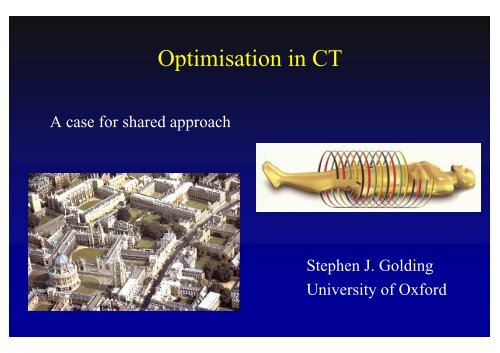
A case for shared approach<br />
<strong>Optimisation</strong> <strong>in</strong> <strong>CT</strong><br />
Stephen J. Gold<strong>in</strong>g<br />
University of Oxford
10 mm sections<br />
20 second exposure<br />
60 second reconstruction<br />
Body <strong>CT</strong> 1979:
Body <strong>CT</strong> 2007<br />
Submillimetre sections Subsecond exposure<br />
“<strong>in</strong>stant” reconstruction<br />
64 slice, 128, 256…….<br />
Data volume a problem
<strong>CT</strong> is now our major radiation<br />
protection challenge
Impact of new technique<br />
Tasks performed better<br />
Tasks performed more<br />
easily<br />
New applications
Cl<strong>in</strong>ical Benefits of MS<strong>CT</strong>:<br />
Speed : - image quality<br />
traditional applications<br />
Cover : - Reduced anatomical misregistration
Cl<strong>in</strong>ical Benefits of MS<strong>CT</strong>:<br />
new applications<br />
Multiphase enhancement<br />
<strong>CT</strong> Angiography<br />
<strong>CT</strong> Urography<br />
3D/virtual reality
Hepatic enhancement; carc<strong>in</strong>oma of pancreas
<strong>CT</strong> angiography
The Era of 3D/Virtual Reality/4D
Cl<strong>in</strong>ical benefits of MD<strong>CT</strong><br />
Multiphase enhancement<br />
<strong>CT</strong> Angiography<br />
<strong>CT</strong> Urography<br />
3D/Virtual reality<br />
Screen<strong>in</strong>g
Small pulmonary nodules:<br />
detection at chest <strong>CT</strong> and outcome<br />
• Chest <strong>CT</strong> 3445<br />
• Inclusion criteria 344<br />
• Characterisation 87<br />
• Benign 77<br />
• Malignant 10 (0.29%)<br />
• Primary neoplasm 9 (0.03%)<br />
Benjam<strong>in</strong> et al, 2003
Acute Appendicitis: effect of <strong>in</strong>creased<br />
use of <strong>CT</strong> on select<strong>in</strong>g patients earlier<br />
“With <strong>in</strong>creased <strong>CT</strong> use there were less severe<br />
imag<strong>in</strong>g f<strong>in</strong>d<strong>in</strong>gs, <strong>in</strong>clud<strong>in</strong>g absence of<br />
periappendiceal strand<strong>in</strong>g, and a significant<br />
decrease <strong>in</strong> surgical-pathologic severity of<br />
appendiceal disease and hospital stay.”<br />
Raptopoulos et al, 2003
Applications have risen dramatically<br />
s<strong>in</strong>ce 2000.<br />
Many young patients, benign disease<br />
But that this not the only problem
Skull<br />
Limbs<br />
Chest<br />
Abdomen<br />
IVU<br />
Lumbar sp<strong>in</strong>e<br />
<strong>CT</strong><br />
0 5 10 15 20 25 30<br />
% contribution<br />
collective dose<br />
frequency
1995 30%<br />
1998 40%<br />
<strong>CT</strong>: contributions to dose<br />
Shrimpton & Wall<br />
Shrimpton & Edyvean
Mettler at al, 2001<br />
11% of exam<strong>in</strong>ations<br />
67% of dose<br />
11% <strong>in</strong> children<br />
Hart and Wall, 2004<br />
47% of dose<br />
7% of exam<strong>in</strong>ations
Factors of 10-40<br />
Factors of 8-20<br />
Dose variance<br />
(Shrimpton et al 1991)<br />
(Olerud 1997)<br />
Exam<strong>in</strong>ation technique: variations<br />
<strong>Optimisation</strong> of technique is now our major challenge
N o o f p a ts<br />
140<br />
120<br />
100<br />
80<br />
60<br />
40<br />
20<br />
0<br />
100<br />
300<br />
JRH <strong>CT</strong> - DLP Dec 00 and Jan 01<br />
mGy-cm<br />
500<br />
700<br />
900<br />
1100<br />
1300<br />
1500<br />
1700<br />
1900<br />
2100
<strong>CT</strong>PA<br />
<strong>CT</strong>A<br />
Abdo/pel<br />
Chest<br />
Ch/Abdo<br />
Bra<strong>in</strong><br />
0 500 1000 1500 2000
JRH cases over 1000 mGy-cm<br />
Bra<strong>in</strong> 17<br />
Bra<strong>in</strong> + 4 parts 1<br />
Bra<strong>in</strong> + 3 parts 1<br />
Bra<strong>in</strong> + 2 parts 6<br />
Bra<strong>in</strong> +1 part 5<br />
3 trunk parts 5<br />
2 trunk parts 13<br />
1 trunk part 9<br />
<strong>CT</strong>A 4
Reasons for variable practice<br />
Cl<strong>in</strong>ical <strong>in</strong>dications<br />
Grow<strong>in</strong>g applications<br />
Poor knowledge/practice<br />
Workload pressure<br />
Inexperience
Is this the age of imag<strong>in</strong>g over-kill?<br />
Cl<strong>in</strong>ical/workload pressures motivate<br />
aga<strong>in</strong>st quality/protection<br />
<strong>Optimisation</strong> of practice now represents<br />
the major challenge <strong>in</strong> dose reduction.<br />
The evidence base for practice change is<br />
weak
To what extent should technology<br />
alter technique?<br />
Has disease changed?<br />
Have diagnostic criteria changed?<br />
Will cl<strong>in</strong>ical management change?<br />
Are there risk limitation implications?
EC Directive 97/43<br />
Justification<br />
<strong>Optimisation</strong><br />
Audit<br />
National law 2000<br />
Is it right to regard these<br />
as separate processes?
<strong>Optimisation</strong><br />
• Equipment – quality control<br />
• Equipment – advances<br />
• Exam<strong>in</strong>ations – threshold exposure (DRLs)<br />
• Practice – optimisation <strong>in</strong>cludes justification
Automatic Exposure Control (AEC)<br />
• All modern manufacturers<br />
• Dose depends on tube rotation<br />
time and tube current (to a<br />
lesser extent kV)<br />
• Most common to vary mAs<br />
• preset algorithm
Oxford Experience: 1, 4, 8, 16 slice<br />
DLP<br />
mGycm<br />
1200<br />
1000<br />
800<br />
600<br />
400<br />
200<br />
0<br />
BRAIN CAP CHEST <strong>CT</strong> 3DU <strong>CT</strong> KUB <strong>CT</strong>PA Facial<br />
Exam type<br />
S<strong>in</strong>gle Slice<br />
4 Slice<br />
8 Slice<br />
16 slice
Modify<strong>in</strong>g the exam<strong>in</strong>ation<br />
• Extent – should be practised <strong>in</strong> all cases<br />
• BUT: modern practice/workload motivates aga<strong>in</strong>st<br />
• Exposure<br />
• The aim is to complete the exam<strong>in</strong>ation with the<br />
m<strong>in</strong>imum threshold exposure<br />
• BUT – the evidence base for m<strong>in</strong>imum exposure is<br />
weak.
Modify<strong>in</strong>g extent – what can be done?<br />
• Frequent audit aga<strong>in</strong>st DRL<br />
• Cont<strong>in</strong>ual audit/challenge<br />
• Important role for Physicist - proactive
Modify<strong>in</strong>g exposure<br />
<strong>CT</strong> of the chest: m<strong>in</strong>imal tube current (50%)<br />
Mayo et al, 1995<br />
Low dose <strong>CT</strong> <strong>in</strong> orbital trauma (90%)<br />
Radiation dose reduction <strong>in</strong> <strong>CT</strong> (96%)<br />
Jackson & Whitehouse, 1993<br />
Starck et al, 1998
A Scientific Basis for Dose Reduction <strong>in</strong><br />
Multislice <strong>CT</strong> of the Face: H.Nwume, 2002<br />
• Phantom<br />
• 8 steps, 10-80 mA<br />
• 4 slice: pitch 3, 6<br />
• 8 slice: pitch 0.67, 1.67<br />
• Scor<strong>in</strong>g
Image quality<br />
Im age quality<br />
4.5<br />
4.5<br />
3.5<br />
2.5<br />
1.5<br />
0.5<br />
4<br />
3.5<br />
3<br />
2.5<br />
2<br />
1.5<br />
1<br />
0.5<br />
0<br />
4<br />
3<br />
2<br />
1<br />
0<br />
Comparison of subjective image quality<br />
relative to scann<strong>in</strong>g dose. Axial Images<br />
Results – 8-slice<br />
Pitch 0.625<br />
Pitch 1.675<br />
0 10 20 30<br />
<strong>CT</strong>DIw (mGy)<br />
Comparison of subjective image quality<br />
relative to scann<strong>in</strong>g dose. 3D Images<br />
Pitch 0.625<br />
pitch 1.625<br />
0 5 10 15<br />
<strong>CT</strong>DIw (mGy)<br />
Image quality<br />
Image quality<br />
12<br />
10<br />
8<br />
6<br />
4<br />
2<br />
0<br />
12<br />
10<br />
8<br />
6<br />
4<br />
2<br />
0<br />
Comparison of objective image quality<br />
relative to scann<strong>in</strong>g dose. Axial Images<br />
0 10 20 30<br />
<strong>CT</strong>DIw (mGy)<br />
Pitch 0.625<br />
Pitch 1.675<br />
Comparison of objective image quality<br />
relative to scann<strong>in</strong>g dose. 3D Images<br />
Pitch 0.625<br />
Pitch 1.675<br />
0 10 20 30<br />
<strong>CT</strong>DIw (mGy)
HR<strong>CT</strong> of the face: conclusions:<br />
Acceptable axial images at 40mA<br />
Acceptable 3D images at 10mA<br />
(Manufacturer’s recommendation: 140mA)
New work: Cervical and Lumbar Sp<strong>in</strong>e<br />
Trauma<br />
• Reduce dose without degrad<strong>in</strong>g fracture detectability<br />
• Phantoms built with artificial fractures<br />
• Order of vertebrae randomised each time<br />
• Scans with tube current 120 -10 mA
Sp<strong>in</strong>e Phantoms 2
Fracture Image
Exposure <strong>in</strong>fluences<br />
noise and therefore<br />
contrast resolution<br />
Soft tissue lesion<br />
detection is the real<br />
anxiety
Experiment 2: dose reduction <strong>in</strong> the bra<strong>in</strong><br />
Creation of “lesions”<br />
Variable attenuation<br />
Variable size<br />
Variable position
Bra<strong>in</strong> “lesions”: the answer<br />
Commercial jelly<br />
(orange)<br />
Bubblewrap
7<br />
6<br />
5<br />
4<br />
3<br />
2<br />
1<br />
0<br />
10<br />
20<br />
30<br />
40<br />
Scores, large lesions<br />
50<br />
60<br />
70<br />
80<br />
90<br />
Tube current<br />
100<br />
110<br />
120<br />
130<br />
140
8<br />
7<br />
6<br />
5<br />
4<br />
3<br />
2<br />
1<br />
0<br />
10<br />
20<br />
30<br />
40<br />
Scores, small lesions<br />
50<br />
60<br />
70<br />
80<br />
90<br />
Tube current<br />
100<br />
110<br />
120<br />
130<br />
140
Experiment 2: prelim<strong>in</strong>ary conclusions<br />
Large lesions: dose may be reduced 50%<br />
Small lesions: further study needed
European Commission Study Group<br />
1994 to 2007<br />
7 countries<br />
Nationally paired Physicist/Radiologist
<strong>CT</strong> work<strong>in</strong>g <strong>group</strong> – 4 th Framework<br />
• European guidel<strong>in</strong>es<br />
• Quality criteria for<br />
computed tomography<br />
1999
5 th Framework Programme Concerted<br />
Guidel<strong>in</strong>es for MS<strong>CT</strong><br />
European Field Survey<br />
MS<strong>CT</strong> dosimetry<br />
Action<br />
Assessment of patient dose <strong>in</strong> <strong>CT</strong>
14 applications<br />
53 <strong>in</strong>stitutions<br />
8 countries<br />
European Field Survey
Abdomen, abscess
Effective dose, mSv<br />
30<br />
25<br />
20<br />
15<br />
10<br />
5<br />
0<br />
Liver metastasis,<br />
colorectal carc<strong>in</strong>oma<br />
Sequence 4<br />
Sequence 3<br />
Sequence 2<br />
Sequence 1<br />
1 3 5 7 9 11 13 15 17 19 21<br />
Hospital #
European Field Survey<br />
Large difference <strong>in</strong> protocols<br />
Large difference <strong>in</strong> parameters<br />
Scanned range<br />
No. of series/repeats<br />
Tube current<br />
Section thickness<br />
Great dose reduction potential exists
6 th Framework Programme<br />
Prospective studies; applications MS<strong>CT</strong>: justification<br />
Automatic exposure controls: optimisation<br />
Paediatric MS<strong>CT</strong>: justification/optimisation<br />
New approaches to MS<strong>CT</strong> dosimetry: audit<br />
Website: www.msct.eu
<strong>CT</strong> Quality Criteria Document - MS<strong>CT</strong><br />
Technical pr<strong>in</strong>ciples<br />
Cl<strong>in</strong>ical pr<strong>in</strong>ciples<br />
Good technique<br />
Guidel<strong>in</strong>es on dose<br />
paediatrics<br />
26 applications
Justification <strong>in</strong> <strong>Optimisation</strong><br />
• Exam<strong>in</strong>ation necessary?<br />
• Exam<strong>in</strong>ation the right one?<br />
• Exam<strong>in</strong>ation the right extent/quality?<br />
• i.e. more than just: ? <strong>CT</strong> <strong>in</strong>dicated?
What is the cl<strong>in</strong>ical question?<br />
Surgeon needs to know:<br />
? depressed orbital floor<br />
? extent
Delayed diagnosis: ultra-low dose<br />
10-20mA DLP 40mGy-cm
Increas<strong>in</strong>g the availability of both<br />
ultrasound and MRI will reduce reliance<br />
upon techniques <strong>in</strong>volv<strong>in</strong>g x-rays,<br />
particularly for young patients at higher<br />
risk.<br />
How far can the UK follow this<br />
advice?<br />
NRPB (1990)
Effective justification<br />
Is there a case for us<strong>in</strong>g non-radiation tests <strong>in</strong> the<br />
first <strong>in</strong>stance?<br />
Irrespective of sensitivity?<br />
Pre-radiation screen<strong>in</strong>g?<br />
Is this compatible with British health care?
“Black Bone” MRI<br />
Can MRI replace <strong>CT</strong>?<br />
In what circumstances is the greater resolution<br />
of <strong>CT</strong> for cortical bone essential to<br />
management?
Volume acquisition<br />
Acquisition time 3 m<strong>in</strong>s<br />
Gradient echo<br />
5° flip angle<br />
PD weight<strong>in</strong>g<br />
In-phase TE<br />
256 2 512 2
MM<br />
160<br />
140<br />
120<br />
100<br />
80<br />
60<br />
40<br />
20<br />
0<br />
Comparison Direct, <strong>CT</strong> and MRI<br />
Landmark
The Future<br />
More evidence is needed<br />
Dose audit is mandatory<br />
Further surveys/studies<br />
Cont<strong>in</strong>ued/updated advice<br />
Vigilance!
If you seek to regulate the people by law they will<br />
learn how to stay out of gaol but feel no shame.<br />
If lead by virtue and propriety they will feel<br />
shame and become good.<br />
Kongzi (Confucius), 551-459 BC


