Temporal difference models describe higher-order learning in humans
Temporal difference models describe higher-order learning in humans
Temporal difference models describe higher-order learning in humans

Create successful ePaper yourself
Turn your PDF publications into a flip-book with our unique Google optimized e-Paper software.
letters to nature<br />
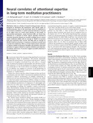
Figure 1 Experimental design and temporal <strong>difference</strong> model. a, The experimental design<br />
expressed as a Markov cha<strong>in</strong>, giv<strong>in</strong>g four separate trial types. b, <strong>Temporal</strong> <strong>difference</strong><br />
value. As <strong>learn<strong>in</strong>g</strong> proceeds, earlier cues learn to make accurate value predictions (that is,<br />
weighted averages of the f<strong>in</strong>al expected pa<strong>in</strong>). c, <strong>Temporal</strong> <strong>difference</strong> prediction error;<br />
dur<strong>in</strong>g <strong>learn<strong>in</strong>g</strong> the prediction error is transferred to earlier cues as they acquire the<br />
ability to make predictions. In trial types 3 and 4, the substantial change <strong>in</strong> prediction<br />
elicits a large positive or negative prediction error. (For clarity, before and mid-<strong>learn<strong>in</strong>g</strong> are<br />
shown only for trial type 1.)<br />
trials, the expectation evoked by the first cue would be reversed by<br />
the second. This allowed us to study the neural implementation of<br />
both the expectations themselves, and their reversals.<br />
Two important aspects of most accounts of prediction <strong>learn<strong>in</strong>g</strong><br />
are the predictions themselves (termed values) and the errors <strong>in</strong><br />
those predictions 9 . Figure 1b shows the predictions associated with<br />
each of the trial types 1–4. These predictions are calculated and<br />
revised as new stimuli are presented. Figure 1c shows the associated<br />
prediction error. The nature of this signal, which treats ongo<strong>in</strong>g<br />
changes <strong>in</strong> predicted values on an exact par with actual affective<br />
outcomes, has helped to expla<strong>in</strong> data on dopam<strong>in</strong>e cell activity. This<br />
prediction error signal drives <strong>learn<strong>in</strong>g</strong> by specify<strong>in</strong>g how the<br />
predictions should change. In appetitive condition<strong>in</strong>g the dopam<strong>in</strong>e<br />
projection to the ventral striatum is believed to be a critical<br />
substrate for this signal, although apart from theoretical speculations<br />
about opponent process<strong>in</strong>g 16 , the equivalent for aversive<br />
condition<strong>in</strong>g is less clear. As <strong>in</strong> earlier work on appetitive condition<strong>in</strong>g,<br />
we used the temporal <strong>difference</strong> model to generate<br />
regressors based on the values and prediction errors appropriate<br />
to each <strong>in</strong>dividual subject 13 . Statistical parametric mapp<strong>in</strong>g of the<br />
regression coefficients permits identification of regions associated<br />
with, and <strong>in</strong> receipt of, <strong>in</strong>formation about predictions. Indeed, the<br />
temporal <strong>difference</strong> value was (negatively) correlated with the<br />
reaction times across subjects for the high-valued cues<br />
(P , 0.001), even when consider<strong>in</strong>g the second-<strong>order</strong> cue alone<br />
(P , 0.01). This result provides strong evidence that behavioural<br />
re<strong>in</strong>forcement occurs <strong>in</strong> a manner consistent with the temporal<br />
<strong>difference</strong> model.<br />
The prediction error was highly correlated with activity <strong>in</strong> both<br />
the right and the left ventral putamen (Fig. 2). Correlations were<br />
also noted <strong>in</strong> the right head of the caudate, the left substantia nigra,<br />
the cerebellum (bilaterally) and the right anterior <strong>in</strong>sula cortex<br />
(Fig. 2). Figure 3 shows the estimated responses <strong>in</strong> the right ventral<br />
putamen. As the most straightforward model coupl<strong>in</strong>g prediction<br />
error to BOLD signal would predict, positive (Fig. 3a) and negative<br />
(Fig. 3b) prediction errors at various times <strong>in</strong> the trial are clearly<br />
represented, as is the biphasic form of the prediction error (Fig. 3c).<br />
We also <strong>in</strong>vestigated the representation of the temporal <strong>difference</strong><br />
value (comb<strong>in</strong><strong>in</strong>g the predicted and the actual pa<strong>in</strong> value, for<br />
reasons of analysis) by <strong>in</strong>clud<strong>in</strong>g the temporal <strong>difference</strong> value<br />
term <strong>in</strong> our regression model. This revealed correlated activity <strong>in</strong><br />
the right anterior <strong>in</strong>sula cortex (Fig. 4a). The estimated response is<br />
illustrated <strong>in</strong> Fig. 4c. The importance of this structure <strong>in</strong> pa<strong>in</strong><br />
<strong>learn<strong>in</strong>g</strong> has previously been identified 5 . In addition, we found<br />
temporal <strong>difference</strong> value-related responses <strong>in</strong> the bra<strong>in</strong>stem<br />
(Fig. 4b). Precise anatomical localization of bra<strong>in</strong>stem activation<br />
is difficult with standard neuroimag<strong>in</strong>g, although we note a consistency<br />
with the probable location of the dorsal raphe nucleus. We<br />
also observed temporal <strong>difference</strong> value-related responses <strong>in</strong> the<br />
anterior c<strong>in</strong>gulate cortex and the right amygdala, which did not<br />
survive statistical correction for multiple comparisons.<br />
The strik<strong>in</strong>g resemblance between the BOLD signal <strong>in</strong> the ventral<br />
putamen and the temporal <strong>difference</strong> prediction error (Fig. 3) offers<br />
powerful support for the temporal <strong>difference</strong> model, <strong>in</strong> particular<br />
because this is a second-<strong>order</strong> paradigm. Other dynamic <strong>models</strong> of<br />
Figure 2 <strong>Temporal</strong> <strong>difference</strong> prediction error (statistical parametric maps). Areas<br />
coloured yellow/orange show significant correlation with the temporal <strong>difference</strong><br />
prediction error. Yellow represents the greatest correlation. Peak activations (MNI<br />
coord<strong>in</strong>ates and statistical z scores) are: right ventral putamen (put; (32, 0, 28),<br />
z ¼ 5.38); left ventral putamen (put; (230, 22, 24), z ¼ 3.93); right head of caudate<br />
(caud; (18, 20, 6), z ¼ 3.75); left substantia nigra (sn; (210, 210, 28), z ¼ 3.52); right<br />
anterior <strong>in</strong>sula (<strong>in</strong>s; (46, 22, 24), z ¼ 3.71); right cerebellum ((28, 246, 230),<br />
z ¼ 4.91); and left cerebellum ((234, 252, 228), z ¼ 4.42). R <strong>in</strong>dicates the right side.<br />
NATURE | VOL 429 | 10 JUNE 2004 | www.nature.com/nature 665<br />
© 2004 Nature Publish<strong>in</strong>g Group


![[F-18]-L-DOPA PET scan shows loss of dopaminergic neurons](https://img.yumpu.com/41721684/1/190x146/f-18-l-dopa-pet-scan-shows-loss-of-dopaminergic-neurons.jpg?quality=85)