A case of repeated gum boils with frequently yellowish foul ... - KIMS
A case of repeated gum boils with frequently yellowish foul ... - KIMS
A case of repeated gum boils with frequently yellowish foul ... - KIMS

Create successful ePaper yourself
Turn your PDF publications into a flip-book with our unique Google optimized e-Paper software.
BULLETIN OF THE KUWAIT INSTITUTE FOR MEDICAL SPECIALIZATION 2002;1:81-83<br />
A <strong>case</strong> <strong>of</strong> <strong>repeated</strong> <strong>gum</strong> <strong>boils</strong> <strong>with</strong> <strong>frequently</strong> <strong>yellowish</strong> <strong>foul</strong> discharge<br />
Essam I. Zaatar<br />
SELF-ASSESSMENT QUIZ<br />
A thirty-five year old female has presented in<br />
your clinic <strong>with</strong> a chief complaint <strong>of</strong> “a <strong>repeated</strong><br />
<strong>gum</strong> boil which <strong>frequently</strong> opens up<br />
<strong>with</strong> <strong>yellowish</strong> <strong>foul</strong> discharge related to the<br />
upper left front teeth towards the lip side.”<br />
Her medical history was found non contributory.<br />
The patient could not recall a history <strong>of</strong><br />
any trauma to her upper front teeth. The<br />
clinical examination <strong>of</strong> her involved teeth revealed<br />
intact clinical crowns, slightly darkened<br />
lateral incisor, and periodontal probing<br />
was found <strong>with</strong>in normal limits. The labial<br />
mucosa in her upper left anterior teeth region<br />
appeared inflamed <strong>with</strong> no observable swelling;<br />
an evidence <strong>of</strong> a healed sinus tract was<br />
noted on the attached gingival in the canine<br />
area. The palatal mucosa appeared normal.<br />
Questions<br />
1. Which tooth (teeth) is (are) involved in this<br />
problem<br />
2. What diagnostic tests would you perform<br />
3. What is your diagnosis for this patient<br />
4. How should this <strong>case</strong> be treated<br />
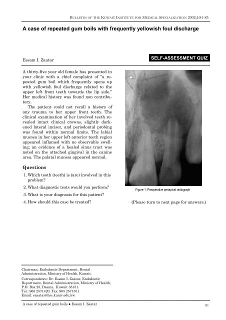
Figure 1. Preoperative periapical radiograph<br />
(Please turn to next page for answers.)<br />
Chairman, Endodontic Department, Dental<br />
Administration, Ministry <strong>of</strong> Health, Kuwait.<br />
Correspondence: Dr. Essam I. Zaatar, Endodontic<br />
Department, Dental Administration, Ministry <strong>of</strong> Health,<br />
P.O. Box 28, Dasma, Kuwait 35151.<br />
Tel.: 965 2571420; Fax: 965 2571531<br />
Email: ezaatar@hsc.kuniv.edu.kw<br />
A <strong>case</strong> <strong>of</strong> <strong>repeated</strong> <strong>gum</strong> <strong>boils</strong> ● Essam I. Zaatar<br />
81
BULLETIN OF THE KUWAIT INSTITUTE FOR MEDICAL SPECIALIZATION 2002;1:81-83<br />
Answers<br />
1. Which tooth (teeth) is (are) involved in this<br />
problem<br />
There is No answer for such a question. Unfortunately<br />
one might be easily deceived by<br />
the information presented on radiographs. It<br />
is imperative to appreciate the inherent limitation<br />
<strong>of</strong> the use <strong>of</strong> radiographs as they provide<br />
a two dimensional image <strong>of</strong> a three dimensional<br />
object. The size <strong>of</strong> the lesion and<br />
the number <strong>of</strong> teeth apparently involved on<br />
the radiographs should not be looked at as<br />
sole determining factors in treatment planning.<br />
Another limitation <strong>of</strong> radiographs is the<br />
personal variances in their interpretation. In<br />
a related study Gelfand and co-workers 1 have<br />
shown that only a little over 50% <strong>of</strong> the participating<br />
dentists have agreed in reading the<br />
same radiographs in less that 50% <strong>of</strong> the<br />
time! In the same study 22% <strong>of</strong> the dentists<br />
replied differently on the same radiograph<br />
when viewed twice.<br />
2. What diagnostic tests would you perform<br />
Diagnosis and treatment <strong>of</strong> <strong>case</strong>s <strong>of</strong> teeth<br />
<strong>with</strong> open apices and large periapical lesions<br />
should be performed following a systematic<br />
approach. Careful studying <strong>of</strong> the chief complaint<br />
and the history <strong>of</strong> the chief complaint<br />
usually provide dependable leads to follow.<br />
Failure to identify the involved tooth or teeth<br />
will be negatively reflected on the treatment<br />
outcome. The available literature indicates<br />
that no single diagnostic test can be totally<br />
reliable. All relevant tests should be performed<br />
before a clear treatment plan can be<br />
designed and executed. Shortcuts cannot be<br />
accepted in handling such <strong>case</strong>s.<br />
Before running the diagnostic tests, the<br />
operator should establish a communication<br />
base <strong>with</strong> the patient including what to expect<br />
and how to respond to the tests performed.<br />
The applied diagnostic tests in this <strong>case</strong><br />
include:<br />
A. THERMAL TESTS<br />
These tests include both cold and heat tests.<br />
They are indicative <strong>of</strong> pulpal status in terms<br />
<strong>of</strong> whether a pulp is healthy or not, or<br />
stressed as was called by Abou-Rass. 2 One<br />
82<br />
should also remember here that it has long<br />
been shown by Seltzer et al. that no relation<br />
could be linked between the clinical findings<br />
<strong>of</strong> these tests and the histological picture <strong>of</strong><br />
the pulp. 3<br />
Since the chief complaint <strong>of</strong> this <strong>case</strong> did<br />
not reflect any sensitivity to thermal<br />
changes, the objective <strong>of</strong> applying thermal<br />
tests here would be for the purpose <strong>of</strong> ruling<br />
out teeth <strong>with</strong> vital pulps and confirming the<br />
suspected lateral incisor. It is recommended<br />
to perform the tests first on a healthy tooth<br />
as a control, in order to make the patient familiar<br />
<strong>with</strong> the stimulus and to alert the operator<br />
about the normal pain threshold <strong>of</strong> the<br />
patient. The major point here is to avoid repeating<br />
the tests <strong>with</strong>in a short period <strong>of</strong><br />
time like seconds, in order to avoid yielding<br />
conflicting results.<br />
In this <strong>case</strong> all teeth in the area responded<br />
<strong>with</strong>in normal limits to both cold and heat<br />
tests, and failed to elicit any response when<br />
applied to the lateral incisor.<br />
B. ELECTRIC PULP TESTS<br />
The mode <strong>of</strong> action <strong>of</strong> the so called EPT<br />
(Electric Pulp Tester) is to induce pulpal response<br />
by electric excitation <strong>of</strong> nerve endings<br />
particularly the A-delta nerve fibers. The only<br />
information obtained from using these devices<br />
is whether the pulp is responsive or not, <strong>with</strong><br />
no attempt to differentiate degrees <strong>of</strong> pulpal<br />
pathosis. 4 It is advised to repeat the test several<br />
times before drawing a conclusion <strong>with</strong><br />
no fear <strong>of</strong> developing adaptation or habituation<br />
by the patient. 5 The old misconception <strong>of</strong><br />
the contraindication <strong>of</strong> the use <strong>of</strong> pulp testers<br />
on patients <strong>with</strong> pacemakers has recently<br />
been debated by Miller et al, who reported<br />
their safe use on these patients especially<br />
<strong>with</strong> the available new generations <strong>of</strong> EPT. 6<br />
In this patient, the EPT is used to assist in<br />
recognizing the non vital teeth in the area<br />
<strong>with</strong> clear understanding <strong>of</strong> the unreliable<br />
results <strong>of</strong> using it on teeth <strong>with</strong> open apices.<br />
C. PALPATION AND PERCUSSION<br />
These are not pulp vitality tests! Positive responses<br />
<strong>of</strong> these tests <strong>of</strong>ten indicate extension<br />
<strong>of</strong> pulpal inflammation to the periradicular<br />
tissue. It is important to note that <strong>case</strong>s <strong>with</strong><br />
chronic periapical inflammation <strong>of</strong>ten yield<br />
negative results <strong>with</strong> these tests.<br />
In this patient the result <strong>of</strong> the percussion<br />
and palpation tests revealed slight tenderness<br />
A <strong>case</strong> <strong>of</strong> <strong>repeated</strong> <strong>gum</strong> <strong>boils</strong> ● Essam I. Zaatar
BULLETIN OF THE KUWAIT INSTITUTE FOR MEDICAL SPECIALIZATION 2002;1:81-83<br />
especially in relation to the root apex <strong>of</strong> the<br />
lateral incisor.<br />
D. SINUS TRACING<br />
A size 35 gutta-percha cone is usually recommended<br />
to be used as a tracer to pin point the<br />
<strong>of</strong>fending tooth whenever a sinus tract is detected.<br />
One must consider that sinus tracts do<br />
not <strong>of</strong>ten lay directly beneath their openings<br />
on the surface.<br />
E. MOBILITY<br />
The test is aimed to explore the integrity <strong>of</strong><br />
the periodontal attachment.<br />
In this patient the evident amount <strong>of</strong> bone<br />
loss in the radiograph especially in the lateral<br />
incisor area reflected the need to explore the<br />
chances <strong>of</strong> tooth mobility.<br />
In this <strong>case</strong> the results were all <strong>with</strong>in normal<br />
limits.<br />
3. What is your diagnosis for this patient<br />
No conclusion should be drawn from the radiographs<br />
<strong>with</strong> regard to the nature <strong>of</strong> the<br />
pathosis; the commonly used terms ‘a cyst’ or<br />
‘a granuloma’ should only be concluded from<br />
the result <strong>of</strong> the histopathology <strong>of</strong> the biopsy<br />
taken whenever indicated.<br />
The history given by the patient and the<br />
results <strong>of</strong> the above tests indicated that the<br />
lateral incisor had lost its vitality some time<br />
before root formation was completed. The irritants<br />
from the necrotic pulp have triggered<br />
the periapical pathosis. This situation had led<br />
to the development <strong>of</strong> the so-called “chronic<br />
apical periodontitis”. With the history <strong>of</strong> intermittent<br />
discharge <strong>of</strong> pus through the sinus<br />
tract, the condition may also be called<br />
“chronic suppurative apical periodontitis”.<br />
4. How should this <strong>case</strong> be treated<br />
Endodontic therapy <strong>of</strong> the lateral incisor is<br />
the treatment <strong>of</strong> choice in this <strong>case</strong>. The procedures<br />
will be discussed in detail in the next<br />
issue <strong>of</strong> the Journal.<br />
References<br />
1. Gelfand M, Sunderman EJ, Goldman M. Reliability<br />
<strong>of</strong> radiographical interpretations. J Endodon<br />
1983;9:71-5.<br />
2. Abou-Rass M. The stressed pulp condition: an<br />
endodontic restorative diagnostic concept. J<br />
Prosthet Dent 1982;48:264-7.<br />
3. Seltzer S, Bender IB, Ziontz M. The dynamics<br />
<strong>of</strong> pulp inflammation: correlations between diagnostic<br />
data and actual histological findings<br />
in the pulp. Oral Surg Oral Med Oral Pathol<br />
1963;16:846.<br />
4. Lado EA, Richmond AF, Marks RG. Reliability<br />
and validity <strong>of</strong> a digital pulp tester as a standard<br />
for measuring sensory perception. J Endodon<br />
1988;14:352-6.<br />
5. Dal Santl FB, Throckmorton GS, Ellis III. Reproducibility<br />
<strong>of</strong> data from a hand-held digital<br />
pulp tester used on teeth and oral s<strong>of</strong>t tissue.<br />
Oral Surg Oral Med Oral Pathol 1992;72:103-8.<br />
6. Miller CA, Leonelli FM, Latham E. Selective<br />
interference <strong>with</strong> pacemaker activity by electrical<br />
dental devices. Oral Surg Oral Med Oral<br />
Pathol Oral Radiol Endod 1998;85:33-6.<br />
A <strong>case</strong> <strong>of</strong> <strong>repeated</strong> <strong>gum</strong> <strong>boils</strong> ● Essam I. Zaatar<br />
83