The Mtwo NiTi rotary system for root canal ... - Vdw-dental.com
The Mtwo NiTi rotary system for root canal ... - Vdw-dental.com
The Mtwo NiTi rotary system for root canal ... - Vdw-dental.com

Create successful ePaper yourself
Turn your PDF publications into a flip-book with our unique Google optimized e-Paper software.
industry _ grande I<br />
<strong>The</strong> <strong>Mtwo</strong> <strong>NiTi</strong> <strong>rotary</strong> <strong>system</strong><br />
<strong>for</strong> <strong>root</strong> <strong>canal</strong> preparation<br />
Author_ V.A. Malagino,N. M. Grande, G. Plotino & F. Somma, Italy<br />
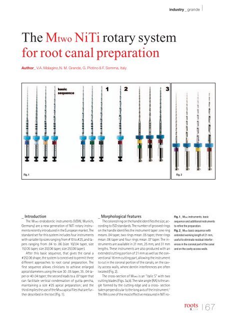
Fig. 1 Fig. 2<br />
_ Introduction<br />
<strong>The</strong> <strong>Mtwo</strong> endodontic instruments (VDW, Munich,<br />
Germany) are a new generation of <strong>NiTi</strong> <strong>rotary</strong> instruments<br />
recently introduced in the European market. <strong>The</strong><br />
standard set <strong>for</strong> this <strong>system</strong> includes four instruments<br />
with variable tip sizes ranging from #10 to #25, and tapers<br />
ranging from .04 to .06 (size 10/.04 taper, size<br />
15/.05 taper, size 20/.06 taper, size 25/.06 taper).<br />
After this basic sequence, that gives the <strong>canal</strong> a<br />
#25/.06 shape, the <strong>system</strong> is conceived to permit three<br />
different approaches to <strong>root</strong> <strong>canal</strong> preparation. <strong>The</strong><br />
first sequence allows clinicians to achieve enlarged<br />
apical diameters using the size 30 . 05 taper, 35 . 04 taper<br />
or 40 .04 taper; the second leads to a .07 taper that<br />
can facilitate vertical condensation of gutta-percha,<br />
maintaining a size #25 apical preparation; and the<br />
third implies the use of the <strong>Mtwo</strong>apical files that are further<br />
described in the text (Fig. 1).<br />
_ Morphological features<br />
<strong>The</strong> colored ring on the handle identifies the size, according<br />
to ISO standards. <strong>The</strong> number of grooved rings<br />
on the handle identifies the instrument taper: one ring<br />
means .04 taper, two rings mean .05 taper, three rings<br />
mean .06 taper and four rings mean .07 taper. <strong>The</strong> instruments<br />
are available in 21 mm, 25 mm, and 31 mm<br />
lengths. <strong>The</strong>se instruments are also produced with an<br />
extended cutting portion of 21 mm as well as the conventional<br />
16 mm cutting part, allowing the instrument<br />
to cut in the coronal portion of the <strong>canal</strong>s, on the cavity<br />
access walls, where dentin interferences are often<br />
located (Fig. 2).<br />
<strong>The</strong> cross-section of <strong>Mtwo</strong> is an “italic S” with two<br />
cutting blades (Figs. 3a,b). <strong>The</strong> rake angle (RA) is the angle<br />
<strong>for</strong>med by the cutting edge and a cross- section<br />
taken perpendicular to the long axis of the instrument. 1<br />
<strong>The</strong> RA is one of the most effective measured in <strong>NiTi</strong> ro-<br />
Fig. 1_ <strong>Mtwo</strong> instruments, basic<br />
sequence and additional instruments<br />
to refine the preparation.<br />
Fig. 2_ <strong>Mtwo</strong> basic sequence with<br />
extended working length of 21 mm,<br />
useful to eliminate residual interferences<br />
in the coronal part of the <strong>canal</strong><br />
and on the cavity access walls.<br />
<strong>root</strong>s<br />
3_2006<br />
I67
I industry _ grande<br />
Fig. 3a_ SEM image of <strong>Mtwo</strong> instrument<br />
cross-section, showing the two<br />
blade cutting surfaces resulting in an<br />
“Italic S” design.<br />
Fig. 3b_ Cross-section obtained by<br />
means of µCT scanning and reconstruction<br />
of an <strong>Mtwo</strong> size 25, .06 taper.<br />
Fig. 3c_ SEM image of an <strong>Mtwo</strong> 25 .06,<br />
the axial view shows the two cutting<br />
blade surfaces with efficient RA (200x).<br />
Fig. 3d_ SEM image of the non-cutting<br />
tip of an <strong>Mtwo</strong> instrument (200x).<br />
Fig. 3e_ SEM image of an <strong>Mtwo</strong> size<br />
#25 taper .06 in lateral view: the HA<br />
increases from apex to crown (50x).<br />
Fig. 3f_ <strong>Mtwo</strong> A1 lateral view: showing<br />
the cutting blade surface and the<br />
unique tip design with an exaggerated<br />
taper in the last millimeter (50 x).<br />
Fig. 3g_ <strong>Mtwo</strong> A1 tip: showing the innovative<br />
tip of these instruments with<br />
two straight blades not spiraled in the<br />
last apical millimeter (50 x).<br />
Fig. 4_ <strong>Mtwo</strong> R tip: showing the cutting<br />
surfaces of the tip (200 x).<br />
Fig. 5_ Pre-instrumentation (left) and<br />
post-instrumentation (right)<br />
<strong>root</strong> <strong>canal</strong> cross-section slides of a<br />
seoond upper premolar with oval<br />
anatomy obtained by means of µCT<br />
scanning and reconstruction (in collaboration<br />
with R. Bedini and R. Pecci<br />
– Italian National Institute of Health,<br />
Technology and Health Department,<br />
Rome, Italy)<br />
Fig. 6_ Superimposition of pre- (yellow)<br />
and post- (red) instrumentation<br />
µCT three dimensional reconstruction<br />
of a second lower premolar with<br />
oval and curved anatomy prepared<br />
with <strong>Mtwo</strong> <strong>system</strong>, it is possible to<br />
note in both mesio-distal and<br />
bucco-lingual views that a great part<br />
of the anatomy has been addressed<br />
by the mechanical action of the instruments<br />
(in collaboration with R.<br />
Bedini and R. Pecci – Italian National<br />
Institute of Health, Technology and<br />
Health Department, Rome, Italy).<br />
Fig. 7a_ Mesio distal view of pre-instrumentation<br />
and post-instrumentation<br />
µCT three dimensional reconstruction<br />
of a first lower molar prepared<br />
with <strong>Mtwo</strong> <strong>system</strong> (in collaboration<br />
with R. Bedini and R. Pecci -<br />
Italian National Institute of Health,<br />
Technology and Health Department,<br />
Rome, Italy).<br />
Fig. 3a Fig. 3b Fig. 3c Fig. 3d<br />
Fig. 3e<br />
Fig. 3f<br />
tary instruments, enhancing the cutting efficiency of<br />
this instrument (Fig. 3c). <strong>The</strong> tip is non-cutting (Fig. 3d).<br />
<strong>The</strong> helical angle (HA) or flute angle is defined as the<br />
angle <strong>for</strong>med by the instrument’s cutting surface and<br />
the dentin wall observed in longitudinal section. 2,3 <strong>The</strong><br />
HA is determined by the blade pitch of the instrument:<br />
the bigger it is, the more open the HA will be. A shorter<br />
blade pitch will determine a closer HA; a longer one will<br />
result in a more open HA. <strong>The</strong> HA of an instrument is an<br />
important parameter to determine not only the instrument’s<br />
cutting efficiency, but also its mechanical resistance<br />
and its dynamic features.<br />
<strong>The</strong> HA of <strong>Mtwo</strong> instruments is variable and specific<br />
<strong>for</strong> the different files (Fig. 3e).<br />
<strong>The</strong> HA is more open (greater) <strong>for</strong> the bigger sizes<br />
(less flutes <strong>for</strong> instrument length), and it decreases <strong>for</strong><br />
the smaller sizes (more flutes). This determines a<br />
greater cutting efficiency <strong>for</strong> the bigger sizes and a<br />
greater mechanical resistance together with a tendency<br />
to advance in the <strong>canal</strong> <strong>for</strong> the smaller ones. <strong>The</strong><br />
Fig. 4<br />
Fig. 3g<br />
flutes are deeper moving from the tip to the handle; increasing<br />
the capacity to remove debris coronally. Moreover,<br />
<strong>for</strong> the bigger file sizes (#20. 06, #25. 06) the HA is<br />
variable in the same instruments, it increases from the<br />
tip to the handle as does the spiral pitch, while it is constant<br />
<strong>for</strong> the smaller files, especially <strong>for</strong> the #10. 04, the<br />
first <strong>rotary</strong> instrument that is introduced in the <strong>root</strong><br />
<strong>canal</strong>. <strong>The</strong> variable HA reduces the tendency of the instrument<br />
to be sucked down into the <strong>canal</strong>.<br />
<strong>The</strong> tendency to advance spontaneously in the <strong>root</strong><br />
<strong>canal</strong> <strong>for</strong> the smaller instrument is necessary to<br />
progress in the <strong>canal</strong> during the first phase of the treatment.<br />
<strong>The</strong> operator should tend towards a pulling-out<br />
movement, holding back the instrument in rotation,<br />
enhancing the characteristic of removing debris and<br />
the cutting efficiency.<br />
_ <strong>Mtwo</strong> A and <strong>Mtwo</strong> R<br />
<strong>The</strong> <strong>Mtwo</strong><strong>system</strong> has been <strong>com</strong>pleted with three <strong>rotary</strong><br />
files specifically designed <strong>for</strong> apical preparation,<br />
68 I<br />
<strong>root</strong>s<br />
3_2006
industry _ grande I<br />
Fig. 5<br />
Fig. 7a<br />
Fig. 8a<br />
Fig. 6<br />
Fig. 7b<br />
the <strong>Mtwo</strong> A, and two files specifically designed <strong>for</strong> retreatment,<br />
the <strong>Mtwo</strong> R. <strong>The</strong> three apical files <strong>Mtwo</strong> A1,<br />
A2 and A3 vary in tip size and taper. <strong>The</strong> innovative feature<br />
of these instruments is the high taper of the last<br />
apical millimetre while the rest of the coronal portion<br />
is a 2% ISO taper (Fig. 3f). <strong>The</strong> A1 instrument has a tip<br />
size (D0) of 0.20 mm and 15% taper in the first millimeter,<br />
thus measuring 0.35 mm in D1. A2 instruments<br />
have a tip size of 0.25 mm and 15% taper in the first<br />
millimeter, thus measuring 0.40 mm in D1. A3 instrument<br />
presents a tip size of 0.25 mm and 20% taper in<br />
the first millimeter, thus measuring 0.45 mm in D1. <strong>The</strong><br />
remaining portion of these instruments, from D1 to<br />
D16, present a 2% taper. To obtain this design, the apical<br />
millimeter of the instrument is not produced in a<br />
spiral but has two straight blades (Fig. 3g). This design<br />
has been developed to obtain bigger preparation diameters<br />
in the apical portion of the <strong>root</strong> <strong>canal</strong>s, maintaining<br />
the anatomy of the apical <strong>for</strong>amen according to scientific<br />
evidence that the <strong>root</strong> <strong>canal</strong> diameters in the<br />
apical portion are bigger than the average <strong>root</strong> <strong>canal</strong><br />
preparations normally used. 4 -6<br />
<strong>The</strong> enhanced taper in<br />
the apical zone also provides a resistance <strong>for</strong>m against<br />
the condensation pressures of obturation and prevents<br />
the extrusion of filling material. 7<br />
<strong>The</strong> <strong>Mtwo</strong>R instruments are specifically designed <strong>for</strong><br />
the retreatment of obturation materials. <strong>The</strong> retreatment<br />
files are <strong>Mtwo</strong> R 15/.05 and <strong>Mtwo</strong> R 25/.05, presenting<br />
an active tip that allows clinicians to easily penetrate<br />
obturation material (Fig. 4).<br />
_ Operative sequence<br />
<strong>The</strong> <strong>Mtwo</strong> <strong>NiTi</strong> <strong>rotary</strong> instruments are used at<br />
300 rpm. <strong>Mtwo</strong> instruments are used in a simultaneous<br />
technique without any early coronal enlargement. 8 After<br />
a glide path has been established with a #10 stainless<br />
steel K-type file, instruments are each taken to the<br />
working length (WL) with light apical pressure. As soon<br />
as the clinician feels a binding sensation, he or she pulls<br />
the instrument away <strong>for</strong> 1 mm to 2 mm so that it can<br />
work passively in a brushing action to selectively remove<br />
the interferences and to advance towards the<br />
apex. <strong>The</strong> instruments are used with a lateral pressing<br />
movement in order to obtain a circumferential cut, and<br />
only allowed to rotate at length <strong>for</strong> few seconds<br />
(Figs. 5,6).<br />
<strong>The</strong> operative sequence suggested <strong>for</strong> these instruments<br />
is a crown-down technique, whereby the apex is<br />
reached by every <strong>NiTi</strong> instrument at each step. This<br />
means that this is a technique from the crown to the<br />
apex, but it first uses smaller instruments be<strong>for</strong>e using<br />
bigger ones, as is done in the step-back technique. <strong>The</strong><br />
inventor defines this as a “simultaneous technique,” as<br />
the entire length of the <strong>canal</strong> is approached at the same<br />
time. <strong>The</strong> instrument does not have to be <strong>for</strong>ced in; as<br />
soon as the clinician feels a binding sensation, he or she<br />
has to back the instrument away <strong>for</strong> 1 mm to 2 mm so<br />
that it can work passively to create the space necessary<br />
to go to the apex (Figs. 7a,b). Using the instruments with<br />
a lateral pressing movement (brushing, milling) the<br />
tendency to progress automatically in the <strong>canal</strong> (a sensation<br />
of being “sucked down”) increases its efficiency.<br />
<strong>The</strong> high flexibility and fatigue resistance9 of the <strong>Mtwo</strong><br />
instruments permits the use of this approach in severly<br />
curved <strong>root</strong> <strong>canal</strong>s with an efficient and safely action<br />
(Figs. 8,9).<br />
_ Conclusion<br />
An important consideration regarding the proposal<br />
<strong>for</strong> a simultaneous approach using <strong>NiTi</strong> <strong>rotary</strong> instru-<br />
Fig. 8b<br />
Fig. 8c<br />
Fig. 8d<br />
<strong>root</strong>s<br />
3_2006<br />
I69
I industry _ grande<br />
Fig. 7b_ Bucco lingual view of<br />
pre-instrumentation and<br />
post-instrumentation µCT three<br />
dimensional reconstruction of a first<br />
lower molar prepared with <strong>Mtwo</strong> <strong>system</strong><br />
(in collaboration with R. Bedini<br />
and R. Pecci – Italian National<br />
Institute of Health, Technology and<br />
Health Department, Rome, Italy).<br />
Fig. 8_ a) Right second upper premolar<br />
with acute pulp inflammation<br />
b) <strong>Mtwo</strong> #20 taper.06 to the working<br />
length, c) Obturation of the <strong>root</strong> <strong>canal</strong><br />
<strong>system</strong> in two projections<br />
Fig. 9_ a) Right first lower molar<br />
with chronic apical periodontitis<br />
b) Working length film to confirm the<br />
”electronic length”<br />
c,d) Obturation of the <strong>root</strong> <strong>canal</strong><br />
<strong>system</strong> in two projections, after the<br />
cleaning and shaping phases<br />
per<strong>for</strong>med with the <strong>Mtwo</strong> <strong>system</strong><br />
e) 1 year control<br />
Fig. 9b<br />
Fig. 9d<br />
Fig. 9a<br />
Fig. 9c<br />
Fig. 9e<br />
ments concerns differing points of view about the<br />
crown-down concept. In the simultaneous technique,<br />
the coronal portion is prepared be<strong>for</strong>e the apical one,<br />
using smaller instruments first. In the <strong>NiTi</strong> era, the<br />
crown-down concept is instead associated with the<br />
use of bigger instruments (eg, tip diameter, taper) <strong>for</strong><br />
the shaping of the coronal portion, followed by smaller<br />
instruments to advance toward the apex15. <strong>The</strong> use of<br />
the smaller instrument first is not in contrast to the<br />
crown-down approach because it is also a crowndown<br />
technique in which the <strong>canal</strong> is prepared starting<br />
from the coronal towards the apical portion, even if all<br />
instruments reach the apex. This new concept facilitates<br />
<strong>root</strong> <strong>canal</strong> shaping particularly in the most difficult<br />
cases, reducing the incidence of procedural errors<br />
that could occur in the first phase of the treatment in<br />
which the <strong>canal</strong> has to be negotiated with rigid stainless<br />
steel files to at least 0.20 mm. _<br />
_ Literature<br />
1.Arens D.<strong>The</strong> crown-down technique: a paradigm shift.<br />
Dent Today 1996;15:38.<br />
2. Buchanan LS.<strong>The</strong> art of endodontics: files of greater taper.<br />
Dent Today 1996;15:42.<br />
3. Buchanan LS. <strong>The</strong> standardized–taper <strong>root</strong> <strong>canal</strong> preparation,<br />
part 1: concept <strong>for</strong> variably tapered shaping instruments.<br />
Dent Today 1998;5:54.<br />
4.Wu MK,R'oris A,Barkis D,Wesselink PR.Prevalence and extent<br />
of long oval <strong>canal</strong>s in the apical third. Oral Surg Oral Med Oral<br />
Pathol Oral Radiol Endod 2000;89:739–743.<br />
5. Orstavik D, Kerekes K, Molven O. Effects of extensive apical<br />
reaming and calcium hydroxide dressing on bacterial infection<br />
during treatment of apical periodontitis:A pilot study.<br />
Int Endod J 1991;24:1–7.<br />
6. Card SJ, Sigurdsson A, Orstavik D, Trope M. <strong>The</strong> effectiveness<br />
of increased apical enlargement in reducing intra<strong>canal</strong> bacteria.<br />
J Endod 2002;28:779–783.<br />
7. Serota KS, Nahmias Y, Barnett F. Predictable endodontic success:<strong>The</strong><br />
apical control zone. Dent Today 2003;22:90–97.<br />
8. Foschi F, Nucci C, Montebugnoli L, Marchionni S, Breschi L,<br />
Malagnino VA,Prati C.SEM evaluation of <strong>canal</strong> wall dentine following<br />
use of <strong>Mtwo</strong> and ProTaper <strong>NiTi</strong> <strong>rotary</strong> instruments.<br />
Int Endod J 2004;37:832–839.<br />
9. Grande NM,Plotino G,Pecci R,Bedini R,Malagnino VA,Somma<br />
F. Cyclic fatigue resistance and three-dimensional analysis of<br />
instruments from two <strong>NiTi</strong> <strong>rotary</strong> <strong>system</strong>s.Int Endod J,in press.<br />
10. Grande NM, Plotino G, Butti A, Messina F, Pameijer CH, Somma<br />
F. Cross-sectional analysis of <strong>root</strong> <strong>canal</strong>s prepared with <strong>NiTi</strong> <strong>rotary</strong><br />
instruments and stainless steel reciprocating files.<br />
Oral Surg, Oral Med, Oral Pathol, Oral Radiol, Endodontol, in<br />
press.<br />
11. Plotino G,Grande NM,Sorci E,Malagnino VA,Somma F.A <strong>com</strong>parison<br />
of cyclic fatigue between used and new <strong>Mtwo</strong> <strong>NiTi</strong> <strong>rotary</strong><br />
instruments. Int Endod J, in press.<br />
12.Veltri M,Mollo A,Mantovani L,Pini P,Balleri P,Grandini S.A <strong>com</strong>parative<br />
study of Endoflare-Hero Shaper and <strong>Mtwo</strong> <strong>NiTi</strong> instruments<br />
in the preparation of curved <strong>root</strong> <strong>canal</strong>s.<br />
Int Endod J 2005;38:610–6.<br />
13. Schäfer E, Erler M, Dammaschke T. Comparative study on the<br />
shaping ability and cleaning efficiency of <strong>rotary</strong> <strong>Mtwo</strong> instruments.<br />
Part 1. Shaping ability in simulated curved <strong>canal</strong>s.<br />
Int Endod J 2006; 39:196–202.<br />
14. Schäfer E, Erler M, Dammaschke T. Comparative study on the<br />
shaping ability and cleaning efficiency of <strong>rotary</strong> <strong>Mtwo</strong> instruments.Part<br />
2.Cleaning effectiveness and shaping ability in severely<br />
curved <strong>root</strong> <strong>canal</strong>s of extracted teeth.<br />
Int Endod J 2006; 39:203–12.<br />
15. Ruddle CJ. Cleaning and shaping the <strong>root</strong> <strong>canal</strong> <strong>system</strong>. In: Cohen<br />
S, Burns RC. Pathways of the pulp. 8th ed. St. Louis:<br />
Mosby; 2002, 231–292.<br />
_author info<br />
Dr Malagnino is at the Department of Endodontics,<br />
University “G. D’Annunzio”, Chieti, Italy<br />
Dr Grande, Dr Plotino and Dr Somma are at the Department<br />
of Endodontics, Catholic University of Sacred<br />
Heart, Rome, Italy<br />
You may contact Dr Grande via e-mail at:<br />
nmg@fastwebnet.it<br />
<strong>root</strong>s<br />
70 I<br />
<strong>root</strong>s<br />
3_2006