2009 - Lehrstuhl für Medizintechnik - RWTH Aachen University
2009 - Lehrstuhl für Medizintechnik - RWTH Aachen University
2009 - Lehrstuhl für Medizintechnik - RWTH Aachen University

You also want an ePaper? Increase the reach of your titles
YUMPU automatically turns print PDFs into web optimized ePapers that Google loves.
Helmholtz-Institute for Biomedical Engineering<br />
<strong>RWTH</strong> <strong>Aachen</strong> <strong>University</strong><br />
<strong>2009</strong><br />
Medical Engineering<br />
Introduction<br />
The Chair of Medical Engineering (mediTEC) of the Faculty<br />
of Mechanical Engineering of the <strong>RWTH</strong> <strong>Aachen</strong> <strong>University</strong><br />
is especially engaged in basic research issues as well as application<br />
oriented aspects of computer assisted diagnosis<br />
and model guided therapy systems engineering. In this context<br />
the activities are concentrated on the following areas:<br />
image, signal and information processing as an essential basis<br />
for computer assisted model-based therapy planning,<br />
biomechanical modeling and simulation, surgical navigation<br />
and robotics, smart mechatronic instruments and devices,<br />
ultrasound technology and medical shock waves as well as<br />
ergonomics and safety in medicine.<br />
Actual projects in the domain of Orthopedic and Trauma<br />
Surgery, Neurosurgery, General Endoscopic Surgery,<br />
Cardiology, Interventional Radiology, Maxillofacial Surgery,<br />
Dental Therapy and Rehabilitation are ranging from feasibility<br />
studies (proof of concept) and system development to<br />
usability analyses and clinical field tests. The OrthoMIT project<br />
(minimal invasive orthopedic therapy; 7/2005-6/2010;<br />
24 partners; 14.5 M€ overall funding by the German<br />
Federal Ministry of Education and Research – BMBF) continues<br />
to be one major framework of our research activities.<br />
Additionally, various new research grants and industrial<br />
cooperation related to basic research issues as well as innovative<br />
application oriented concepts and patent applications<br />
have been established. As in 2007 and 2008, in <strong>2009</strong><br />
our team again received the Medical Technology Innovation<br />
Award of the German Federal Ministry of Education and<br />
Research (BMBF) for a novel approach in computer assisted<br />
surgical instrument control.<br />
Based on our long-standing research activities on ergonomics<br />
in medicine, we established a portfolio of tools and methods<br />
for usability engineering of medical products. Especially the<br />
application of our new method and software tool mAIXuse<br />
for human error risk analysis received a very positive response<br />
in cooperation with various industrial partners.<br />
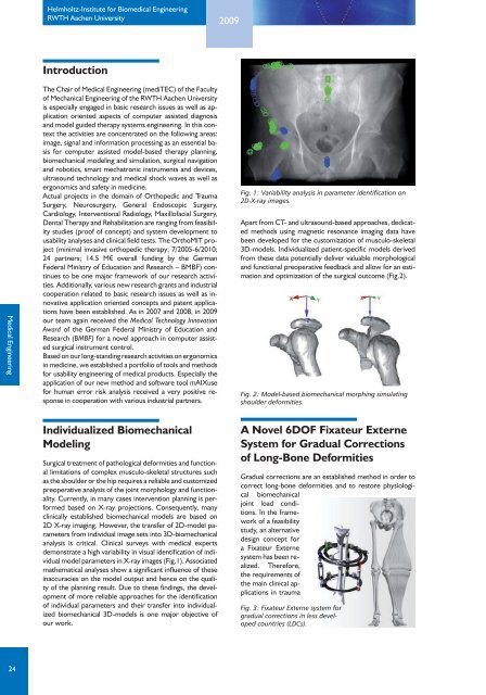
Fig. 1: Variability analysis in parameter identification on<br />
2D-X-ray images.<br />
Apart from CT- and ultrasound-based approaches, dedicated<br />
methods using magnetic resonance imaging data have<br />
been developed for the customization of musculo-skeletal<br />
3D-models. Individualized patient-specific models derived<br />
from these data potentially deliver valuable morphological<br />
and functional preoperative feedback and allow for an estimation<br />
and optimization of the surgical outcome (Fig.2).<br />
Fig. 2: Model-based biomechanical morphing simulating<br />
shoulder deformities.<br />
Individualized Biomechanical<br />
Modeling<br />
Surgical treatment of pathological deformities and functional<br />
limitations of complex musculo-skeletal structures such<br />
as the shoulder or the hip requires a reliable and customized<br />
preoperative analysis of the joint morphology and functionality.<br />
Currently, in many cases intervention planning is performed<br />
based on X-ray projections. Consequently, many<br />
clinically established biomechanical models are based on<br />
2D X-ray imaging. However, the transfer of 2D-model parameters<br />
from individual image sets into 3D-biomechanical<br />
analysis is critical. Clinical surveys with medical experts<br />
demonstrate a high variability in visual identification of individual<br />
model parameters in X-ray images (Fig.1). Associated<br />
mathematical analyses show a significant influence of these<br />
inaccuracies on the model output and hence on the quality<br />
of the planning result. Due to these findings, the development<br />
of more reliable approaches for the identification<br />
of individual parameters and their transfer into individualized<br />
biomechanical 3D-models is one major objective of<br />
our work.<br />
A Novel 6DOF Fixateur Externe<br />
System for Gradual Corrections<br />
of Long-Bone Deformities<br />
Gradual corrections are an established method in order to<br />
correct long-bone deformities and to restore physiological<br />
biomechanical<br />
joint load conditions.<br />
In the framework<br />
of a feasibility<br />
study, an alternative<br />
design concept for<br />
a Fixateur Externe<br />
system has been realized.<br />
Therefore,<br />
the requirements of<br />
the main clinical applications<br />
in trauma<br />
Fig. 3: Fixateur Externe system for<br />
gradual corrections in less developed<br />
countries (LDCs).<br />
24