2009 - Lehrstuhl für Medizintechnik - RWTH Aachen University
2009 - Lehrstuhl für Medizintechnik - RWTH Aachen University
2009 - Lehrstuhl für Medizintechnik - RWTH Aachen University

Create successful ePaper yourself
Turn your PDF publications into a flip-book with our unique Google optimized e-Paper software.
<strong>2009</strong><br />
Helmholtz-Institute for Biomedical Engineering<br />
<strong>RWTH</strong> <strong>Aachen</strong> <strong>University</strong><br />
Chair of Medical Engineering<br />
Faculty of Mechanical Engineering<br />
Innovative<br />
Technology<br />
for Smart<br />
Therapy<br />
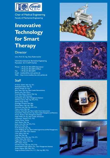
Director<br />
Univ.-Prof. Dr.-Ing. Klaus Radermacher<br />
Helmholtz-Institute for Biomedical Engineering<br />
Pauwelsstr. 20, D-52074 <strong>Aachen</strong><br />
Phone: +49 (0) 241 80-23870 (Secretary)<br />
+49 (0) 241 80-23873 (Office)<br />
Fax: +49 (0) 241 80-22870<br />
Email: meditec@hia.rwth-aachen.de<br />
Web : http://www.meditec.hia.rwth-aachen.de<br />
Staff<br />
Al Hares, Ghaith, Dipl.-Ing. (SY)<br />
Bisplinghoff, Stefan, Dipl.-Ing.<br />
Becker, Michael, Dr. med.<br />
Belei, Peter, Dipl.-Ing. (Team Leader Biomechanics)<br />
Benzko, Julia, Dipl.-Inform.<br />
Chuembou Pekam, Fabrice, Dipl.-Ing.<br />
Crets, Hans, CAD Technician<br />
Dannenberg, Kirsa, Team Management Assistant<br />
Dell’Anna, Jasmin, Dipl.-Ing. (FH), M.Sc.<br />
Dienst, Svenja, Trainee<br />
Drexhage, Christine, Dipl.-Biol.<br />
Elfring, Robert, Dipl.-Ing., MBA<br />
Eschweiler, Jörg, Dipl.-Ing. (FH), M.Sc.<br />
Fieten, Lorenz, Dipl.-Ing.<br />
Follmann, Axel, Dipl.-Ing. (Team Leader Smart Instruments)<br />
Fuente Klein, Matias de la, Dr.-Ing. (Team Leader Navigation and Robotics)<br />
Ginova, Teodora, Team Management Assistant<br />
Heger, Stefan, Dr.-Ing. (Team Leader Ultrasound)<br />
Hananouchi, Takehito, M.D., PhD<br />
Hübner, Moritz, Trainee<br />
Ibach, Bastian, Dipl.-Ing.<br />
Jansen, Arne, Dipl.-Ing. (FH), M.Sc.<br />
Janß, Armin, Dipl.-Ing.<br />
Korff, Alexander, Dipl.-Ing.<br />
Lauer, Wolfgang, Dr.-Ing. (Team Leader Ergonomics and Risk Management)<br />
Leucht, Gero, Math.-Tech. Assistant<br />
Linke, Sabine, Dipl.-Ing.<br />
Long, Yifei, Dipl.-Ing.<br />
Maciejasz, Pawel, Dipl.-Ing., M.Sc. (PL)<br />
Müller, Meiko, Dipl.-Inform.<br />
Niggemeyer, Martin, Dipl.-Ing.<br />
Palm, Fabian, Trainee<br />
Schmidt, Frauke, Dipl.-Ing. (FH), M.Sc.<br />
Stockschläder-Krüger, Sabine, M.A., Team Management Assistant<br />
Strake, Melanie, Dipl.-Math. (FH)<br />
Vargas da Silveira Cunha-Cruz, V. C., Dipl.-Ing. (BR), M.Sc.<br />
23
Helmholtz-Institute for Biomedical Engineering<br />
<strong>RWTH</strong> <strong>Aachen</strong> <strong>University</strong><br />
<strong>2009</strong><br />
Medical Engineering<br />
Introduction<br />
The Chair of Medical Engineering (mediTEC) of the Faculty<br />
of Mechanical Engineering of the <strong>RWTH</strong> <strong>Aachen</strong> <strong>University</strong><br />
is especially engaged in basic research issues as well as application<br />
oriented aspects of computer assisted diagnosis<br />
and model guided therapy systems engineering. In this context<br />
the activities are concentrated on the following areas:<br />
image, signal and information processing as an essential basis<br />
for computer assisted model-based therapy planning,<br />
biomechanical modeling and simulation, surgical navigation<br />
and robotics, smart mechatronic instruments and devices,<br />
ultrasound technology and medical shock waves as well as<br />
ergonomics and safety in medicine.<br />
Actual projects in the domain of Orthopedic and Trauma<br />
Surgery, Neurosurgery, General Endoscopic Surgery,<br />
Cardiology, Interventional Radiology, Maxillofacial Surgery,<br />
Dental Therapy and Rehabilitation are ranging from feasibility<br />
studies (proof of concept) and system development to<br />
usability analyses and clinical field tests. The OrthoMIT project<br />
(minimal invasive orthopedic therapy; 7/2005-6/2010;<br />
24 partners; 14.5 M€ overall funding by the German<br />
Federal Ministry of Education and Research – BMBF) continues<br />
to be one major framework of our research activities.<br />
Additionally, various new research grants and industrial<br />
cooperation related to basic research issues as well as innovative<br />
application oriented concepts and patent applications<br />
have been established. As in 2007 and 2008, in <strong>2009</strong><br />
our team again received the Medical Technology Innovation<br />
Award of the German Federal Ministry of Education and<br />
Research (BMBF) for a novel approach in computer assisted<br />
surgical instrument control.<br />
Based on our long-standing research activities on ergonomics<br />
in medicine, we established a portfolio of tools and methods<br />
for usability engineering of medical products. Especially the<br />
application of our new method and software tool mAIXuse<br />
for human error risk analysis received a very positive response<br />
in cooperation with various industrial partners.<br />
Fig. 1: Variability analysis in parameter identification on<br />
2D-X-ray images.<br />
Apart from CT- and ultrasound-based approaches, dedicated<br />
methods using magnetic resonance imaging data have<br />
been developed for the customization of musculo-skeletal<br />
3D-models. Individualized patient-specific models derived<br />
from these data potentially deliver valuable morphological<br />
and functional preoperative feedback and allow for an estimation<br />
and optimization of the surgical outcome (Fig.2).<br />
Fig. 2: Model-based biomechanical morphing simulating<br />
shoulder deformities.<br />
Individualized Biomechanical<br />
Modeling<br />
Surgical treatment of pathological deformities and functional<br />
limitations of complex musculo-skeletal structures such<br />
as the shoulder or the hip requires a reliable and customized<br />
preoperative analysis of the joint morphology and functionality.<br />
Currently, in many cases intervention planning is performed<br />
based on X-ray projections. Consequently, many<br />
clinically established biomechanical models are based on<br />
2D X-ray imaging. However, the transfer of 2D-model parameters<br />
from individual image sets into 3D-biomechanical<br />
analysis is critical. Clinical surveys with medical experts<br />
demonstrate a high variability in visual identification of individual<br />
model parameters in X-ray images (Fig.1). Associated<br />
mathematical analyses show a significant influence of these<br />
inaccuracies on the model output and hence on the quality<br />
of the planning result. Due to these findings, the development<br />
of more reliable approaches for the identification<br />
of individual parameters and their transfer into individualized<br />
biomechanical 3D-models is one major objective of<br />
our work.<br />
A Novel 6DOF Fixateur Externe<br />
System for Gradual Corrections<br />
of Long-Bone Deformities<br />
Gradual corrections are an established method in order to<br />
correct long-bone deformities and to restore physiological<br />
biomechanical<br />
joint load conditions.<br />
In the framework<br />
of a feasibility<br />
study, an alternative<br />
design concept for<br />
a Fixateur Externe<br />
system has been realized.<br />
Therefore,<br />
the requirements of<br />
the main clinical applications<br />
in trauma<br />
Fig. 3: Fixateur Externe system for<br />
gradual corrections in less developed<br />
countries (LDCs).<br />
24
<strong>2009</strong><br />
Helmholtz-Institute for Biomedical Engineering<br />
<strong>RWTH</strong> <strong>Aachen</strong> <strong>University</strong><br />
and orthopedic surgery, the biomechanical constraints of<br />
long bone gradual corrections as well as the specific boundary<br />
conditions regarding manufacturing in less developed<br />
countries (LDCs) have been taken into account. In these<br />
countries, common systems often are not available due<br />
to their high costs. As a consequence the main objective<br />
of this project was the development of a novel versatile<br />
system that can be manufactured under simple, low-cost<br />
conditions.<br />
Smart Spine Navigation<br />
Based on our Zero-Dose X-ray navigation approach an<br />
efficient dose reduction can be achieved in computer assisted<br />
navigation systems by using patient individualized<br />
deformable models. After a successful evaluation of the<br />
system on lower extremities, our efforts now have been<br />
extended towards spine applications, with an even higher<br />
potential for efficient dose reduction and increased<br />
safety.<br />
Due to higher anatomical complexity and the presence of<br />
sensible structures nearby, new registration methods and<br />
dedicated user guidance are necessary. The new Zero-<br />
Dose spine module will be an essential basis for the upcoming<br />
SpinePilot project starting in 2010.<br />
been carried out. Additionally, user-centered usability tests<br />
have been performed.<br />
Furthermore, an extensive parameter study of the sawing<br />
process was carried out. In laboratory trials the impact of<br />
these parameters on the soft tissue preserving capabilities<br />
of the saw, the cutting efficiency and the manual control of<br />
the instrument were systematically investigated.<br />
Based on the results of these studies the STS-system<br />
was built up consisting of the planning system, the<br />
real-time control unit and the semiautomatic instrument<br />
itself. Ongoing research is focused on preclinical<br />
and clinical studies demonstrating the system’s overall<br />
performance.<br />
Modular Minirobot<br />
Fig. 5: Semiautomatic<br />
trepanation<br />
instrument (top)<br />
and close-up of<br />
cutting process<br />
(bottom).<br />
Surgical instruments have to meet strict hygienic demands.<br />
Hence, specific sterility requirements as well as potential<br />
risks related to hygienic reprocessing have to be considered<br />
regarding the instrumental design.<br />
Medical Engineering<br />
Fig. 4: Concept study of a mechatronic system for spine<br />
surgery using the Zero-Dose X-ray navigation approach.<br />
Protection of Dura Mater Using a<br />
Smart Trepan<br />
A semiautomatic trepanation system (Smart Trepan) based<br />
on a new soft tissue preserving saw and an autonomous<br />
control of the cutting depth has been developed for minimal<br />
invasive skull surgery.<br />
The protection of the dura mater and a minimized cutting<br />
gap are the major objectives of this project. The synergistic<br />
control concept of the Smart Trepan System (STS)<br />
combines the accuracy and time efficiency of a computer<br />
controlled device with the surgeon’s experience and cognitive<br />
capabilities.<br />
Usability and safety of the semiautomatic handheld device<br />
are of utmost importance. Therefore, prospective risk and<br />
usability analysis based on our mAIXuse technique have<br />
Fig. 6: Modular structure of the MINARO-System with<br />
basic modules, application-specific kinematic modules and<br />
two instrument modules (right) and modules “contaminated”<br />
with a fluorescent fluid during cleanability study (left).<br />
At the Chair of Medical Engineering a fully sterilizable<br />
modular minirobot (MINARO) is developed. The modular<br />
structure allows for easy adaptation to different orthopedic<br />
as well as non-orthopedic applications. During<br />
the design of the system the influence of reprocessing on<br />
25
Helmholtz-Institute for Biomedical Engineering<br />
<strong>RWTH</strong> <strong>Aachen</strong> <strong>University</strong><br />
<strong>2009</strong><br />
Medical<br />
Engineering<br />
the robotic functions and the reliable cleanability were<br />
reviewed.<br />
To analyze its cleanability the robot was “contaminated” by<br />
a fluorescent fluid and cleaned in a standardized automatic<br />
decontamination process. As a result of the evaluation potential<br />
hygienic risks related to the robot design could be<br />
identified.<br />
iShunt – an Intelligent<br />
Mechatronic Implant for the<br />
Therapy of Hydrocephalus<br />
The human brain is surrounded by an aqueous fluid. In case<br />
of hydrocephalus, the inner fluid chambers are enlarged and<br />
the intracranial pressure is severely raised, leading to brain<br />
damage if untreated. The common therapy is the implantation<br />
of so called shunt systems, which regulate the intracranial<br />
pressure by controlled drainage of liquor. Until now<br />
there are no mechatronic shunt systems available that allow<br />
for automatic adjustment of drainage to physiological conditions.<br />
Thus, over- and under-drainage often occur due to<br />
false opening pressures leading to brain damage and pain for<br />
the patient. The major objectives of the iShunt project, initiated<br />
by the Chair of Medical Information Technology of our<br />
institute, are the development and evaluation of new concepts<br />
for mechatronic shunt systems. In the context of this<br />
project, our team is especially responsible for the development<br />
of the mechatronic valve systems and components.<br />
Fig. 7: Principle of an implanted shunt valve together with<br />
conceptual studies on potential design solutions.<br />
Non-Invasive Diagnosis of<br />
an Imminent Compartment<br />
Syndrome<br />
The compartment syndrome, an enormous pressure increase<br />
in a muscle compartment, is one of the most common<br />
traumatological complications in case of fractured<br />
extremities, leading to a severe blockage of microcirculation.<br />
Common therapy consists of a fasciotomy, where the<br />
affected member is surgically opened on its whole length and<br />
kept open until the pressure has decreased (Fig. 8). In order<br />
to avoid complications and to<br />
ensure an appropriate treatment,<br />
an early and reliable diagnosis<br />
is essential. Until now,<br />
there are no objective and reliable<br />
non-invasive diagnostic<br />
methods available. In the context<br />
of this research project,<br />
new non-invasive methods<br />
for the diagnosis of an imminent<br />
compartment syndrome<br />
are developed and evaluated.<br />
IDA – Intraoral Data Acquisition<br />
Using Ultrasound Micro-Scanning<br />
The conventional manufacturing process of dental prostheses<br />
based on casted gypsum plasters is error-prone,<br />
time consuming and expensive. CAD/CAM based optical<br />
scanners have been launched for extra-oral or intraoral<br />
digitization of prepared teeth promising high accuracy<br />
and more comfort for the patients. Nevertheless, sub-gingival<br />
preparations cannot be scanned by optical systems<br />
without the exposure of the preparation boundaries,<br />
and blood and saliva influence the accuracy of the digitization.<br />
Moreover, unintentional<br />
reflections, e.g.<br />
from the dentin layer, have<br />
to be compensated by applying<br />
powder on the teeth<br />
which yields additional error<br />
sources and expenses.<br />
The objective of the interdisciplinary<br />
IDA-project is<br />
the development of an ultrasound-based<br />
intraoral micro-scanner,<br />
replacing the<br />
conventional casting process<br />
without the drawbacks<br />
of current optical scanners.<br />
Fig. 8: Fasciotomy in case of<br />
a compartment syndrome.<br />
Fig. 9: First laboratory<br />
study of the IDA concept.<br />
Ultrasound is able to pass soft tissue, blood as well as saliva<br />
and does not use ionizing radiation. The patented concept<br />
of the micro-scanner consists of a cost-efficient high<br />
frequency transducer and a mechanically mounted five degrees<br />
of freedom (DOF) micro-reflector. The kinematic<br />
design, the ultrasound hardware as well as the signal and<br />
image processing are major foci of the project. The IDAproject<br />
is the winner of the “Medical Technology Innovation<br />
Award” (2008) funded by the German Federal Ministry of<br />
Education and Research (BMBF).<br />
Haptics in Computer Assisted<br />
Surgery<br />
CT-based pin-point procedures in the spine area require<br />
high accuracy and human expertise. The success of these<br />
procedures mainly depends on visual and haptic cues and<br />
is strongly related to the individual experience and skills of<br />
the physician. To reduce radiation exposure of the patients<br />
26
<strong>2009</strong><br />
Helmholtz-Institute for Biomedical Engineering<br />
<strong>RWTH</strong> <strong>Aachen</strong> <strong>University</strong><br />
Fig. 10: CT-based pin-point telehaptic<br />
manipulator.<br />
and physicians as well<br />
as to optimize the<br />
workflow, a miniaturized<br />
master-slave<br />
tele-manipulator for<br />
interventional radiology<br />
is currently being<br />
developed. The<br />
slave-manipulator is<br />
controlled by a master<br />
device with force<br />
feedback (Phantom<br />
1.5 6-DOF High-<br />
Force) enabling a hybrid image- and force-based navigation<br />
and motion control of the slave manipulator during needle<br />
placement. Hence, repeated CT-scans potentially can<br />
be avoided.<br />
Fig. 12: VDE application<br />
recommendation:<br />
risk management<br />
for IT networks<br />
incorporating medical<br />
devices in operating<br />
theatres.<br />
Myocardial Vitality Assessment<br />
For patients with low left ventricular ejection fraction,<br />
a precise differentiation of vital and avital myocardial tissue<br />
is therapeutically and prognostically very important. At<br />
present, cardiac MRT is the gold standard of this process.<br />
However, its clinical application is limited due to time loss,<br />
high costs and limited availability. In a pilot study, the feasibility<br />
of using cardiac shock waves to activate hibernating<br />
myocardial tissue and the use of 2D-Strain Ultrasound<br />
analysis to detect the change of cardiac contraction is analyzed<br />
in in-vitro experiments in cooperation with the<br />
Clinic for Anesthesiology, Clinic for Internal Medicine I<br />
(both <strong>University</strong> Hospital <strong>Aachen</strong>) and the Shock Wave<br />
Laboratory <strong>Aachen</strong>.<br />
Model-Based Usability- and Risk<br />
Analysis<br />
Various studies concerning critical events in the medical context<br />
have proven that in most of the cases use deficiencies are<br />
the cause for human failure, especially when new and complex<br />
technical equipment is involved. International standards<br />
such as IEC 60601-1-6 and IEC 62366 have defined a comprehensive<br />
usability engineering (UE) process for market approval<br />
of medical devices including usability specification and<br />
mandatory usability validation with intended user groups.<br />
Medical Engineering<br />
Fig. 11: Fluorescence microscopy following shock wave<br />
application.<br />
Risk Management of Integrated<br />
Surgical Work Systems<br />
In the OrthoMIT project, a concept of a modular and<br />
flexible integrated surgical workstation based on the<br />
service oriented architecture (SOA) paradigm has been<br />
developed. In the context of modular system architectures,<br />
risk management is the most important aspect.<br />
The exemplary application of our concept according<br />
to IEC 80001 has been published as a VDE application<br />
recommendation for risk management in medical ITnetworks.<br />
Fig. 13: Model-based usability evaluation with the mAIXuse<br />
tool for a commercial planning and navigation system.<br />
In the framework of the BMWi-funded AiF / FQS INNORISK<br />
project a novel method for the design and risk assessment<br />
of Human-Machine-Interfaces (HMI) was developed and<br />
evaluated together with several industrial partners. Based<br />
on these results, the related software tool was further developed<br />
and evaluated in the framework of the OrthoMIT<br />
project. The new software tool (mAIXuse) enables a formal-analytical<br />
usability evaluation and HMI-related risk analysis<br />
already at an early stage in the developmental process.<br />
mAIXuse can be used prospectively in the context of the design<br />
process as well as for the analysis, redesign or validation<br />
of existing HMIs.<br />
Based on a formal task modeling approach with an integrated<br />
error analysis (based on taxonomies of human failure) interactive<br />
use process sequences are characterized and their potential<br />
impact on the overall HMI process can be investigated.<br />
27
Helmholtz-Institute for Biomedical Engineering<br />
<strong>RWTH</strong> <strong>Aachen</strong> <strong>University</strong><br />
<strong>2009</strong><br />
Medical Engineering<br />
The application of dedicated assessment algorithms enables<br />
an efficient and reliable analysis and provides an automated<br />
documentation of the evaluation results according to normative<br />
and regulatory standards.<br />
On the basis of these techniques and tools, training courses<br />
as well as the conduction of usability and risk assessment<br />
studies (e.g. with the mAIXuse method) are offered<br />
in cooperation with the<br />
CeMPEG e.V. as a service<br />
for medical device manufacturers<br />
and hospitals.<br />
Fig. 14: Ergonomic assessment<br />
of planning<br />
systems for<br />
orthopedic<br />
interventions:<br />
gaze path<br />
based on<br />
eyetracking<br />
data (l.) and<br />
evaluation in<br />
the usability<br />
lab (r.).<br />
Within our recently expanded usability laboratory comprehensive<br />
user-oriented interaction evaluation and workflow<br />
assessment regarding medical devices can be performed.<br />
Synchronized, video based task and working posture analysis<br />
as well as logging of relevant physiological data (e.g.<br />
EMG, ECG, EDA, breathing frequency…) can be provided<br />
in lab settings as well as directly in the OR environment.<br />
Remote and mobile head-mounted eye-tracking devices<br />
are used to document and analyze gaze data during human<br />
interaction with specific user interfaces or complete interventional<br />
processes.<br />
Acknowledgements<br />
Apart from basic funds and industrial cooperation, in <strong>2009</strong><br />
research at mediTEC has been substantially funded by:<br />
• the German Research Foundation (DFG) within the<br />
Priority Program SPP1124 “Medizinische Navigation<br />
und Robotik”<br />
• the German Federal Ministry of Education and<br />
Research (BMBF) within the research program<br />
SOMIT “Schonendes Operieren mit innovativer<br />
Technik” as well as within the research program<br />
“Innovationswettbewerb <strong>Medizintechnik</strong>”<br />
Awards<br />
• Fieten, L., Pikkemaat, R., Leonhardt, S. Radermacher,<br />
K.: Medical Innovation Award <strong>2009</strong> of the German<br />
Federal Ministry of Education and Research (BMBF)<br />
• Heger, S., Fieten, L., Mumme, T., Wirtz, D.-C.,<br />
Radermacher, K.: Best Technical Poster Award <strong>2009</strong><br />
of the International Society of Computer Assisted<br />
Orthopedic Surgery<br />
Selected Publications<br />
[1] Bruners, P., Penzkofer, T., Nagel, M., Elfring, R., Gronloh, N., Schmitz-<br />
Rode, T., Günther, RW, Mahnken, AH.: Electromagnetic Tracking for<br />
CT-guided Spine Interventions: Phantom, Ex-vivo and In-vivo Results.<br />
In: European Radiology, Vol. 19 (4), pp. 990-994, <strong>2009</strong>.<br />
[2] Fieten, L., Eschweiler, J., Heger, S., Kabir, K.,Gravius, S., de la<br />
Fuente, M., Radermacher, K.: Surface-based determination of the<br />
pelvic coordinate system. In: SPIE Conference Series, Vol. 7261,<br />
pp. 726138-01-726138-10, <strong>2009</strong>.<br />
[3] Fieten, L., Schmieder, K., Engelhardt, M., Pasalic, L., Radermacher,<br />
K., Heger, St.: Fast and accurate registration of cranial CT images<br />
with A-mode-ultrasound. In: Int J CARS, Vol. 4, pp. 225-237,<br />
<strong>2009</strong>.<br />
[4] Hananouchi, T., Belei, P., Strake, M., Jansen, A., Radermacher, K.:<br />
Effect of Femoroplasty, cement augmentation into the proximal<br />
femur, as a prophylactic procedure for preventing hip fracture. In:<br />
J. of Japan Society of Computer Aided Surgery, Vol. 11, pp. 182-<br />
183, <strong>2009</strong>.<br />
[5] Janß, A. Lauer, W., Radermacher, K.: Bewertung sicherheitskritischer<br />
Systeme im OP. In: Zeitschrift für interaktive und kooperative<br />
Medien (i-com), Vol. 8, pp. 32-37, <strong>2009</strong>.<br />
[6] Schmitt, R., Rauchenberger, J., Radermacher, K., Lauer, W., Janß,<br />
A.: Neue Wege für das Risikomanagement b. d. Entwicklung risikosensitiver<br />
Produkte. FQS/DGQ, Vol. 88-04, <strong>2009</strong>.<br />
[7] Heger S.: Intraoperative Struktur- und Geometrieerfassung mittels<br />
A-Mode Ultraschall in der computerunterstützten Chirurgie. In:<br />
<strong>Aachen</strong>er Beiträge zur <strong>Medizintechnik</strong>, Shaker-Verlag <strong>Aachen</strong>, <strong>2009</strong><br />
(Fakultät für Maschinenwesen der <strong>RWTH</strong> <strong>Aachen</strong>, Berichter: Univ.-<br />
Prof. Dr.-Ing. K. Radermacher, Univ.-Prof. Dr. rer. nat. G. Rau)<br />
Team<br />
28