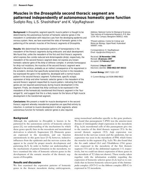
Muscles in the Drosophila second thoracic segment are patterned ...
Muscles in the Drosophila second thoracic segment are patterned ...
Muscles in the Drosophila second thoracic segment are patterned ...

Create successful ePaper yourself
Turn your PDF publications into a flip-book with our unique Google optimized e-Paper software.
222 Research Paper<br />
<strong>Muscles</strong> <strong>in</strong> <strong>the</strong> <strong>Drosophila</strong> <strong>second</strong> <strong>thoracic</strong> <strong>segment</strong> <strong>are</strong><br />
<strong>patterned</strong> <strong>in</strong>dependently of autonomous homeotic gene function<br />
Sudipto Roy, L.S. Shashidhara* and K. VijayRaghavan<br />
Background: In <strong>Drosophila</strong>, <strong>segment</strong>-specific muscle pattern is thought to be<br />
determ<strong>in</strong>ed by <strong>the</strong> autonomous function of homeotic selector genes <strong>in</strong> <strong>the</strong><br />
mesoderm <strong>in</strong> comb<strong>in</strong>ation with <strong>in</strong>ductive cues from <strong>the</strong> develop<strong>in</strong>g epidermis and<br />
nervous system. Here, we have exam<strong>in</strong>ed <strong>the</strong> roles of homeotic genes <strong>in</strong> <strong>the</strong><br />
pattern<strong>in</strong>g of <strong>the</strong> somatic muscles of <strong>the</strong> <strong>thoracic</strong> <strong>segment</strong>s of <strong>Drosophila</strong>.<br />
Results: We determ<strong>in</strong>ed <strong>the</strong> expression patterns of homeoprote<strong>in</strong>s <strong>in</strong> <strong>the</strong><br />
mesoderm of <strong>the</strong> <strong>thoracic</strong> <strong>segment</strong>s dur<strong>in</strong>g embryonic and adult development.<br />
We found that, unlike <strong>the</strong> mesoderm of <strong>the</strong> first and third <strong>thoracic</strong> <strong>segment</strong>s<br />
which express Sex combs reduced and Antennapedia (Antp), respectively, <strong>the</strong><br />
mesoderm of <strong>the</strong> <strong>second</strong> <strong>thoracic</strong> <strong>segment</strong> does not express any known<br />
homeotic selector gene of <strong>the</strong> Antp or bithorax complex. In animals homozygous<br />
for Antp null mutations, <strong>the</strong> muscles of <strong>the</strong> <strong>second</strong> <strong>thoracic</strong> <strong>segment</strong> were<br />
affected <strong>in</strong> <strong>the</strong> embryo, probably as an <strong>in</strong>direct consequence of its requirement <strong>in</strong><br />
<strong>the</strong> ectoderm. Animals that specifically lacked Antp function <strong>in</strong> <strong>the</strong> mesoderm,<br />
but expressed <strong>the</strong> gene <strong>in</strong> <strong>the</strong> epidermis, developed with a normal muscle<br />
pattern <strong>in</strong> <strong>the</strong> <strong>second</strong> <strong>thoracic</strong> <strong>segment</strong>. Fur<strong>the</strong>rmore, specific ectopic<br />
expression of Antp and o<strong>the</strong>r homeotic selector genes <strong>in</strong> <strong>the</strong> mesoderm of <strong>the</strong><br />
<strong>second</strong> <strong>thoracic</strong> <strong>segment</strong> respecified its muscle pattern, <strong>in</strong>dicat<strong>in</strong>g that <strong>the</strong>se<br />
genes <strong>are</strong> not required autonomously dur<strong>in</strong>g muscle development <strong>in</strong> this<br />
<strong>segment</strong>. F<strong>in</strong>ally, we showed that Antp cont<strong>in</strong>ues to be expressed <strong>in</strong> <strong>the</strong><br />
mesoderm of <strong>the</strong> homeotically transformed third <strong>thoracic</strong> <strong>segment</strong> <strong>in</strong> <strong>the</strong> ‘fourw<strong>in</strong>ged<br />
fly’, and suggest that this is a likely reason for <strong>the</strong> failure of flight muscle<br />
development <strong>in</strong> <strong>the</strong> transformed <strong>segment</strong>.<br />
Address: National Centre for Biological Sciences,<br />
Tata Institute of Fundamental Research, P.O. Box<br />
1234, IISc Campus, Bangalore 560012, India.<br />
*Present address: Centre for Cellular and<br />
Molecular Biology, Uppal Road, Hyderabad<br />
500007, India.<br />
Correspondence: K. VijayRaghavan<br />
Email: vijay@ncbs.tifrbng.res.<strong>in</strong><br />
Received: 28 November 1996<br />
Revised: 28 January 1997<br />
Accepted: 11 February 1997<br />
Published: 5 March 1997<br />
Electronic identifier: 0960-9822-007-00222<br />
Current Biology 1997,7:222–227<br />
© Current Biology Ltd ISSN 0960-9822<br />
Conclusions: We present a model for muscle development <strong>in</strong> <strong>the</strong> <strong>second</strong><br />
<strong>thoracic</strong> <strong>segment</strong> whereby mesodermal properties <strong>are</strong> specified entirely by<br />
<strong>in</strong>duction, <strong>in</strong> contrast to muscle development <strong>in</strong> o<strong>the</strong>r <strong>segment</strong>s, where<br />
autonomous function for homeotic selector genes is also required.<br />
Background<br />
Although <strong>the</strong> epidermis <strong>in</strong> <strong>Drosophila</strong> is known to be<br />
<strong>patterned</strong> by <strong>the</strong> autonomous activity of homeotic selector<br />
genes [1], our understand<strong>in</strong>g of <strong>the</strong> mechanisms by which<br />
<strong>the</strong>se genes specify fates <strong>in</strong> <strong>the</strong> mesoderm and mesodermal<br />
derivatives is relatively fragmentary [2]. Homeotic genes<br />
<strong>are</strong> expressed <strong>in</strong> <strong>the</strong> mesoderm, and can function<br />
autonomously <strong>in</strong> this germ layer [3–5]; however, <strong>in</strong>ductive<br />
<strong>in</strong>fluences from <strong>the</strong> epidermis and <strong>the</strong> nervous system also<br />
appear to be crucial for proper muscle development and<br />
pattern<strong>in</strong>g [6,7]. In order to fur<strong>the</strong>r our understand<strong>in</strong>g of<br />
<strong>the</strong> mechanisms of pattern formation <strong>in</strong> <strong>the</strong> mesoderm, we<br />
have exam<strong>in</strong>ed <strong>the</strong> roles of homeotic selector genes <strong>in</strong> <strong>the</strong><br />
development of <strong>the</strong> somatic muscles of <strong>the</strong> <strong>thoracic</strong> <strong>segment</strong>s<br />
of <strong>Drosophila</strong>.<br />
Results and discussion<br />
We first exam<strong>in</strong>ed <strong>the</strong> expression patterns of homeotic<br />
genes <strong>in</strong> <strong>the</strong> <strong>thoracic</strong> mesoderm dur<strong>in</strong>g embryogenesis,<br />
us<strong>in</strong>g monoclonal antibodies specific to <strong>the</strong> gene products.<br />
We found that para<strong>segment</strong> 5 (PS5) was <strong>the</strong> anterior-most<br />
doma<strong>in</strong> of Antennapedia (Antp) expression <strong>in</strong> <strong>the</strong> mesoderm<br />
(Fig. 1a,b); mesodermal cells of this para<strong>segment</strong> give rise<br />
to <strong>the</strong> muscles of <strong>the</strong> third <strong>thoracic</strong> <strong>segment</strong> (T3). In <strong>the</strong><br />
<strong>second</strong> <strong>thoracic</strong> <strong>segment</strong> (T2), Antp expression was<br />
detected <strong>in</strong> <strong>the</strong> nervous system and <strong>the</strong> epidermis, but no<br />
expression was seen <strong>in</strong> <strong>the</strong> mesoderm of PS4, which forms<br />
<strong>the</strong> muscles <strong>in</strong> this <strong>segment</strong> (Fig. 1a,b). We also found that<br />
<strong>the</strong> Sex combs reduced (Scr) and Ultrabithorax (Ubx) genes<br />
were expressed <strong>in</strong> <strong>the</strong> mesoderm of <strong>the</strong> first <strong>thoracic</strong><br />
<strong>segment</strong> (T1) and <strong>in</strong> <strong>the</strong> abdom<strong>in</strong>al <strong>segment</strong>s 1–5, respectively,<br />
but we could not detect expression of <strong>the</strong>se genes <strong>in</strong><br />
<strong>the</strong> mesoderm of T2 (data not shown). These results, taken<br />
toge<strong>the</strong>r with <strong>the</strong> <strong>in</strong>formation available regard<strong>in</strong>g <strong>the</strong><br />
expression of homeotic selector genes [1,2], lead us to conclude<br />
that no known homeotic selector gene of <strong>the</strong> Antennapedia<br />
complex (ANT-C) or <strong>the</strong> bithorax complex (BX-C) is<br />
expressed <strong>in</strong> T2 mesoderm.
Research Paper Homeotic gene function <strong>in</strong> <strong>Drosophila</strong> <strong>thoracic</strong> muscle development Roy et al. 223<br />
Figure 1<br />
Figure 2<br />
(a)<br />
T2<br />
T3<br />
Lateral<br />
muscles<br />
Ventral<br />
muscles<br />
(b) (c) (d)<br />
(e)<br />
(f)<br />
Antp is expressed <strong>in</strong> <strong>the</strong> T3 mesoderm, but not <strong>in</strong> T2 mesoderm dur<strong>in</strong>g<br />
embryogenesis. (a) Stage 12 embryo show<strong>in</strong>g expression of Antp <strong>in</strong><br />
<strong>the</strong> mesodermal cells of T3 (posterior-most arrow). Expression is<br />
absent from T1 and T2 mesoderm (anterior-most and middle arrow,<br />
respectively). (b) Stage 13 embryo show<strong>in</strong>g Antp expression <strong>in</strong> <strong>the</strong><br />
develop<strong>in</strong>g somatic muscles <strong>in</strong> T3 (vertical arrow). Expression <strong>in</strong> <strong>the</strong><br />
central nervous system of T1 and T2 <strong>segment</strong>s is <strong>in</strong>dicated by<br />
horizontal arrows. In (a), anterior is to <strong>the</strong> left and dorsal is top; <strong>in</strong> (b),<br />
anterior is top.<br />
Embryos that <strong>are</strong> homozygous mutant for a null allele of<br />
Antp, Antp w10 , exhibited severe disorganization of <strong>the</strong><br />
muscle pattern <strong>in</strong> T2 and T3 (Fig. 2b,c). Because Antp is<br />
not expressed <strong>in</strong> T2 mesoderm, we reasoned that <strong>the</strong><br />
muscle defects seen <strong>in</strong> T2 were due to <strong>in</strong>direct effects on<br />
<strong>the</strong> mesoderm of <strong>the</strong> absence of Antp function <strong>in</strong> <strong>the</strong> overly<strong>in</strong>g<br />
ectoderm. Fur<strong>the</strong>rmore, because Antp is expressed<br />
<strong>in</strong> T3 mesoderm and ectoderm [8], and has been shown to<br />
be required <strong>in</strong> T3 ectoderm [9,10], <strong>the</strong> effects on T3<br />
muscle pattern <strong>in</strong> Antp w10 embryos must have been caused<br />
by a comb<strong>in</strong>ation of <strong>the</strong> autonomous requirement of Antp<br />
<strong>in</strong> <strong>the</strong> mesoderm and <strong>the</strong> <strong>in</strong>ductive <strong>in</strong>fluences of Antp<br />
from <strong>the</strong> ectoderm. In order to test this reason<strong>in</strong>g, we<br />
selectively removed Antp function from <strong>the</strong> mesoderm,<br />
us<strong>in</strong>g mosaic embryos which expressed wild-type Antp <strong>in</strong><br />
<strong>the</strong> ectoderm only; for <strong>the</strong>se experiments, we used an<br />
ectoderm-specific GAL4 l<strong>in</strong>e, e22c [11], and <strong>the</strong> UAS-Antp<br />
transgene [12], <strong>in</strong> an o<strong>the</strong>rwise Antp null genetic background<br />
(Fig. 2d). We were able to dist<strong>in</strong>guish Antp mutant<br />
embryos by <strong>the</strong> absence of <strong>the</strong> first midgut constriction<br />
(Fig. 2e,f). In <strong>the</strong> presence of ectodermal Antp expression<br />
<strong>in</strong> such embryos, we observed that <strong>the</strong> muscle pattern <strong>in</strong><br />
T2 was completely rescued (Fig. 2d), <strong>in</strong>dicat<strong>in</strong>g that<br />
muscle pattern<strong>in</strong>g <strong>in</strong> this <strong>segment</strong> has a non-autonomous<br />
requirement for Antp.<br />
Analysis of T2- and T3-specific muscle patterns <strong>in</strong> loss-of-function Antp<br />
mutants and <strong>in</strong> mosaic embryos that have Antp function selectively<br />
removed from <strong>the</strong> mesoderm. (a) Schematic representation of wild-type<br />
muscle patterns <strong>in</strong> T2 and T3 <strong>segment</strong>s. The four lateral muscles<br />
(lateral transverse 1–4) <strong>are</strong> represented <strong>in</strong> red rectangles and <strong>the</strong>ir<br />
patterns <strong>are</strong> similar <strong>in</strong> both <strong>segment</strong>s; <strong>the</strong> ventral muscle patterns <strong>are</strong><br />
illustrated below <strong>the</strong> lateral muscles. We analyzed <strong>the</strong> phenotypes of<br />
<strong>the</strong> lateral muscles <strong>in</strong> Antp mutants and <strong>in</strong> mosaic embryos with Antp<br />
function selectively removed from <strong>the</strong> mesoderm, because <strong>the</strong><br />
phenotypic changes <strong>in</strong> <strong>the</strong>se <strong>are</strong> strik<strong>in</strong>g and least susceptible to mis<strong>in</strong>terpretation.<br />
The schematic of muscle patterns has been adapted<br />
from Bate [2]. (b) Wild-type pattern of lateral muscles <strong>in</strong> T2 (left arrow)<br />
and T3 (right arrow) <strong>in</strong> a stage 16 embryo. (c) A representative example<br />
of disrupted muscle pattern <strong>in</strong> T2 and T3 (asterisks) <strong>in</strong> embryos<br />
homozygous for a null allele of Antp (Antp w10 ). Although we have<br />
highlighted only <strong>the</strong> lateral muscles, <strong>the</strong>se embryos exhibit drastic<br />
disorganization of almost all muscle fibres <strong>in</strong> T2 and T3. (d) Rescued<br />
lateral muscles <strong>in</strong> T2 of an Antp w10 embryo (left arrow) <strong>in</strong> which Antp<br />
function was provided only <strong>in</strong> <strong>the</strong> ectoderm us<strong>in</strong>g <strong>the</strong> e22c-GAL4 driver<br />
and <strong>the</strong> UAS-Antp transgene <strong>in</strong> an o<strong>the</strong>rwise Antp null background<br />
(n = 25 mutant embryos were identified, half of which were expected to<br />
carry both <strong>the</strong> e22c-GAL4 transgene and <strong>the</strong> UAS-Antp transgene;<br />
transformation of mutant phenotypes to wild-type was observed <strong>in</strong><br />
n = 11 animals). Comp<strong>are</strong> <strong>the</strong> rescued lateral muscles <strong>in</strong> this figure with<br />
<strong>the</strong> wild-type pattern (left arrow, Fig. 2b) and mutant pattern <strong>in</strong> Antp null<br />
embryos (left asterisk, Fig. 2c). Note also <strong>the</strong> rescue of <strong>the</strong> pattern of T3<br />
lateral muscles <strong>in</strong> <strong>the</strong>se embryos (right arrow). Comp<strong>are</strong> this rescued<br />
pattern with <strong>the</strong> wild-type T3 pattern (right arrow, Fig. 2b) and mutant<br />
pattern (right asterisk, Fig. 2c). In <strong>the</strong>se embryos, we also observed<br />
rescue of o<strong>the</strong>r muscle fibres <strong>in</strong> both T2 and T3. Antp w10 homozygous<br />
mutant embryos can be easily identified on <strong>the</strong> basis of midgut<br />
constriction patterns [23,24]. See text and <strong>the</strong> Materials and methods<br />
section for details. (e) Pattern of midgut constrictions <strong>in</strong> stage 16 wildtype<br />
embryo (arrowheads). (f) Pattern of midgut constrictions <strong>in</strong> a stage<br />
16 Antp w10 embryo (arrowheads). Note <strong>the</strong> absence of <strong>the</strong> first midgut<br />
constriction. In (a–f), anterior is left.
224 Current Biology, Vol 7 No 4<br />
Figure 3<br />
Ga<strong>in</strong>-of-function phenotype for Antp <strong>in</strong> T2<br />
somatic muscles <strong>in</strong> <strong>the</strong> embryo. (a) Schematic<br />
representation of <strong>the</strong> wild-type pattern of<br />
ventral muscles <strong>in</strong> T2 and T3. Note <strong>the</strong><br />
characteristic presence of three extra muscle<br />
fibres <strong>in</strong> T3 (p<strong>in</strong>k, ventral longitud<strong>in</strong>al 4; blue,<br />
ventral oblique 3; and green, ventral acute 1)<br />
and <strong>the</strong>ir absence from T2. These three extra<br />
fibres <strong>are</strong> <strong>the</strong> only differences <strong>in</strong> muscle<br />
patterns between T2 and T3. As an <strong>in</strong>dication<br />
of <strong>the</strong> transformation of T2 muscle pattern <strong>in</strong>to<br />
that of T3 follow<strong>in</strong>g misexpression of Antp <strong>in</strong><br />
T2 mesoderm, we chose to score for <strong>the</strong><br />
appearance of <strong>the</strong>se T3-specific ventral fibres<br />
<strong>in</strong> T2. The schematic of muscle patterns has<br />
been adapted from Bate [2]. (b) Wild-type<br />
pattern of ventral muscles <strong>in</strong> T2 and T3. Note<br />
<strong>the</strong> presence of <strong>the</strong> ventral acute fibre <strong>in</strong> T3 as<br />
shown by <strong>the</strong> arrow, represented <strong>in</strong> green <strong>in</strong><br />
<strong>the</strong> schematic <strong>in</strong> (a), and its absence (asterisk)<br />
from T2. (c) Schematic representation of <strong>the</strong><br />
transformation of T2 muscle pattern to that of<br />
T3 after Antp misexpression <strong>in</strong> T2 mesoderm.<br />
Note <strong>the</strong> T3-specific ventral muscles <strong>in</strong> T2. (d)<br />
UAS-Antp expression driven by <strong>the</strong> 24B<br />
(a) T2 T3 (c) T2 T3<br />
(b)<br />
Wild type<br />
mesodermal GAL4 driver [13] results <strong>in</strong> <strong>the</strong><br />
transformation of T2 muscle pattern to a T3-<br />
like pattern. Note <strong>the</strong> presence of a ventral<br />
(d)<br />
Antp misexpression<br />
acute muscle <strong>in</strong> T2 (asterisk) and its normal<br />
counterpart <strong>in</strong> T3 (arrow). In (a–d), anterior is<br />
left.<br />
We also exam<strong>in</strong>ed muscle patterns <strong>in</strong> embryos where Antp<br />
was expressed throughout <strong>the</strong> mesoderm us<strong>in</strong>g a mesoderm-specific<br />
GAL4 stra<strong>in</strong>, 24B [13], and <strong>the</strong> UAS-Antp<br />
transgene <strong>in</strong> a wild-type background. As Antp is normally<br />
expressed <strong>in</strong> T3 mesoderm, we predicted that misexpression<br />
of Antp <strong>in</strong> T3 mesoderm should have no effect on <strong>the</strong><br />
muscles of this <strong>segment</strong>, and <strong>in</strong>deed we did not see any<br />
effect on T3 muscle development (Fig. 3d, comp<strong>are</strong> with<br />
Figure 4<br />
Antp is not expressed <strong>in</strong> <strong>the</strong> adult muscle<br />
precursors associated with <strong>the</strong> T2 w<strong>in</strong>g<br />
imag<strong>in</strong>al disc, or <strong>in</strong> <strong>the</strong> develop<strong>in</strong>g T2 pupal<br />
musculature, but is expressed <strong>in</strong> <strong>the</strong> haltere<br />
imag<strong>in</strong>al disc myoblasts <strong>in</strong> T3 and <strong>in</strong> <strong>the</strong><br />
develop<strong>in</strong>g T3 pupal muscles. (a) Notal region<br />
of <strong>the</strong> T2 w<strong>in</strong>g imag<strong>in</strong>al disc show<strong>in</strong>g absence<br />
of Antp expression <strong>in</strong> adult myoblasts (<strong>the</strong><br />
position of <strong>the</strong>se myoblasts is shown by <strong>the</strong><br />
star). The nuclear sta<strong>in</strong><strong>in</strong>g that is visible along<br />
<strong>the</strong> anterior marg<strong>in</strong> represents Antp expression<br />
<strong>in</strong> <strong>the</strong> epidermal cells of <strong>the</strong> disc (for fur<strong>the</strong>r<br />
details, see [12]). (b) Antp expression <strong>in</strong> T3<br />
myoblasts associated with <strong>the</strong> haltere disc<br />
(arrowhead). (c) Antp expression <strong>in</strong> myoblasts<br />
associated with <strong>the</strong> homeotically transformed<br />
haltere disc of <strong>the</strong> triple mutant (arrow). As <strong>in</strong><br />
wild-type w<strong>in</strong>g discs, expression was also seen<br />
<strong>in</strong> <strong>the</strong> anterior epidermal cells (data not shown).<br />
(d) A 24 h APF (after puparium formation)<br />
pupal preparation show<strong>in</strong>g Antp expression <strong>in</strong><br />
<strong>the</strong> myoblasts that <strong>are</strong> assembl<strong>in</strong>g to form <strong>the</strong><br />
T3-specific adult musculature (th<strong>in</strong> arrow). Note<br />
<strong>the</strong> absence of Antp expression <strong>in</strong> <strong>the</strong><br />
myoblasts and develop<strong>in</strong>g myofibres of <strong>the</strong><br />
IFMs of T2 (thick arrow). (e) Antp expression <strong>in</strong><br />
<strong>the</strong> migrat<strong>in</strong>g myoblasts <strong>in</strong> <strong>the</strong> homeotically<br />
transformed T3 <strong>segment</strong> <strong>in</strong> a 24 h APF pupa of<br />
a triple mutant (arrowheads). Only <strong>the</strong><br />
expanded HT3 <strong>segment</strong> is shown <strong>in</strong> this figure.<br />
The <strong>in</strong>ter-<strong>segment</strong>al nerve associated with<br />
myoblasts, which is now transformed towards a<br />
T2 phenotype, is shown by an asterisk. The<br />
brown sta<strong>in</strong><strong>in</strong>g <strong>in</strong> (d,e) represents Antp<br />
expression <strong>in</strong> <strong>the</strong> myoblasts of T3 and HT3,<br />
respectively. The w<strong>in</strong>g disc <strong>in</strong> (a) is shown at<br />
higher magnification than <strong>the</strong> haltere and<br />
transformed haltere discs <strong>in</strong> (b,c). In (a–c),<br />
anterior is left; <strong>in</strong> (d,e) anterior is top.
Research Paper Homeotic gene function <strong>in</strong> <strong>Drosophila</strong> <strong>thoracic</strong> muscle development Roy et al. 225<br />
Figure 5<br />
Ectopic expression of Antp <strong>in</strong> T2 mesoderm dur<strong>in</strong>g pupal development<br />
results <strong>in</strong> disruption of flight muscle development. (a) SG29.1-GAL4<br />
driven UAS-lacZ expression <strong>in</strong> <strong>the</strong> pupal myoblasts of <strong>the</strong> IFMs at 12 h<br />
APF. Asterisks <strong>in</strong>dicate <strong>the</strong> positions of <strong>the</strong> three larval muscles that<br />
serve as templates for <strong>the</strong> development of one group of IFMs, <strong>the</strong><br />
dorsal-longitud<strong>in</strong>al muscles (DLMs). (b) SG29.1-GAL4 driven UAS-<br />
Antp expression <strong>in</strong> <strong>the</strong> pupal myoblasts of <strong>the</strong> IFMs at 12 h APF<br />
(arrowheads). (c) Wild-type hemithorax show<strong>in</strong>g <strong>the</strong> morphology of <strong>the</strong><br />
IFMs. The star <strong>in</strong>dicates one of <strong>the</strong> six DLM fibres. (d) Hemithorax of a<br />
SG29.1; UAS-Antp fly show<strong>in</strong>g disruption of <strong>the</strong> IFMs. Note complete<br />
absence of <strong>the</strong> DLMs. Some of <strong>the</strong> rema<strong>in</strong><strong>in</strong>g dorso-ventral muscles<br />
<strong>are</strong> visible. In (a,b), anterior is top; <strong>in</strong> (c,d), anterior is top right corner.<br />
3b). However, as Antp is nei<strong>the</strong>r expressed nor functional <strong>in</strong><br />
T2 mesoderm, misexpression of Antp <strong>in</strong> this mesoderm<br />
could affect muscle development, transform<strong>in</strong>g T2 muscle<br />
pattern towards a T3 pattern; this was found to be <strong>the</strong> case<br />
(Fig. 3d, comp<strong>are</strong> with 3b). Taken toge<strong>the</strong>r, <strong>the</strong>se results<br />
suggest that Antp has no autonomous mesodermal function<br />
<strong>in</strong> <strong>the</strong> development and pattern<strong>in</strong>g of T2-specific musculature<br />
<strong>in</strong> <strong>the</strong> embryo. They also show that Antp is <strong>the</strong><br />
homeotic selector gene required for autonomous specification<br />
of <strong>segment</strong>al identity <strong>in</strong> T3 mesoderm and, when misexpressed<br />
<strong>in</strong> T2 mesoderm, can transform <strong>the</strong> pattern of T2<br />
ventral muscles <strong>in</strong>to that of a T3-like identity.<br />
Analysis of mosaic embryos, <strong>in</strong> which Antp function was<br />
provided only <strong>in</strong> <strong>the</strong> ectoderm but was specifically absent<br />
from <strong>the</strong> mesodermal cells, also showed a significant app<strong>are</strong>nt<br />
restoration of T3 muscle pattern (Fig. 2d), even though<br />
Antp is normally expressed <strong>in</strong> <strong>the</strong> mesodermal cells and<br />
muscles of this <strong>segment</strong>. This is not surpris<strong>in</strong>g because T2<br />
and T3 muscle patterns <strong>are</strong> very similar, except for three<br />
ventral muscle fibres that develop <strong>in</strong> T3 and <strong>are</strong> absent<br />
from T2 (Fig. 3a). Thus, when Antp was expressed <strong>in</strong> <strong>the</strong><br />
ectoderm of Antp – animals, T3 resembled T2, <strong>in</strong> that Antp<br />
was expressed <strong>in</strong> <strong>the</strong> ectoderm and no homeotic selector<br />
gene was expressed <strong>in</strong> <strong>the</strong> mesoderm. In addition, misexpression<br />
of Antp <strong>in</strong> T2 mesodermal cells resulted <strong>in</strong> <strong>the</strong><br />
ectopic development of T3-like ventral fibres <strong>in</strong> T2, suggest<strong>in</strong>g<br />
that Antp normally modifies <strong>the</strong> ‘ground plan’ of T2<br />
muscles <strong>in</strong>to a T3-specific pattern (Fig. 3c,d).<br />
We next determ<strong>in</strong>ed whe<strong>the</strong>r <strong>the</strong> mesodermal profile of<br />
Antp expression dur<strong>in</strong>g pupal development had <strong>the</strong> same<br />
<strong>segment</strong>al restrictions as that seen <strong>in</strong> <strong>the</strong> embryo. The <strong>in</strong>direct<br />
flight muscles (IFMs) develop <strong>in</strong> <strong>the</strong> T2 <strong>segment</strong> and<br />
<strong>the</strong>ir progenitors <strong>are</strong> myoblasts located on <strong>the</strong> T2 w<strong>in</strong>g<br />
imag<strong>in</strong>al discs [14]. These adult muscles offer a good model<br />
system for study<strong>in</strong>g <strong>the</strong> genetics of <strong>segment</strong>-specific<br />
muscle pattern<strong>in</strong>g, and <strong>the</strong>ir normal development has been<br />
well documented [15,16]. We found that <strong>the</strong> T2 w<strong>in</strong>g disc<br />
myoblasts did not express Antp (Fig. 4a), whereas <strong>the</strong><br />
myoblasts on <strong>the</strong> T3 haltere disc did express Antp (Fig. 4b).<br />
Animals homozygous for three Ubx mutations — anterobithorax,<br />
abx; bithorax 3 , bx 3 ; postbithorax, pbx — show a transformation<br />
of T3 towards T2: such ‘triple-mutant’ adults have<br />
two pairs of w<strong>in</strong>gs (<strong>the</strong> ‘four-w<strong>in</strong>ged fly’). However, <strong>the</strong><br />
homeotically transformed T3 (HT3) has only rudimentary<br />
IFMs [17,18]. We had shown earlier that <strong>the</strong> transformation<br />
of <strong>the</strong> ectoderm from T3 to T2 <strong>in</strong> <strong>the</strong>se triple-mutant<br />
animals causes <strong>the</strong> transformation of key aspects of muscle<br />
development: <strong>the</strong> number of myoblasts on <strong>the</strong> HT3 w<strong>in</strong>g<br />
imag<strong>in</strong>al disc and <strong>the</strong> pattern of migration of <strong>the</strong>se<br />
myoblasts dur<strong>in</strong>g pupal development <strong>are</strong> transformed<br />
towards a T2 identity [7]. These results demonstrated that<br />
<strong>in</strong>ductive <strong>in</strong>fluences <strong>are</strong> necessary but not sufficient for<br />
muscle transformation. As <strong>the</strong> four-w<strong>in</strong>ged fly has rudimentary/no<br />
flight muscles <strong>in</strong> HT3, we had argued that a critical<br />
mesodermal transformation had not taken place, because<br />
T3 mesoderm-specific selector gene expression and function<br />
is unaltered <strong>in</strong> <strong>the</strong> triple mutant [7]. Our current observations<br />
show that Antp is <strong>the</strong> selector gene <strong>in</strong> T3 mesoderm,<br />
but we <strong>are</strong> unable to identify <strong>the</strong> expression or function of<br />
any known selector gene <strong>in</strong> T2 mesoderm. In <strong>the</strong> Ubx<br />
triple-mutant larvae, we found that myoblasts on HT3 w<strong>in</strong>g<br />
imag<strong>in</strong>al discs cont<strong>in</strong>ued to express Antp (Fig. 4c), whereas<br />
myoblasts on T2 w<strong>in</strong>g discs did not (Fig. 4a).<br />
Dur<strong>in</strong>g <strong>the</strong> pupal development of wild-type animals, Antp is<br />
expressed <strong>in</strong> T3 mesoderm but is absent <strong>in</strong> <strong>the</strong> T2 mesoderm<br />
(Fig. 4d). In Ubx triple-mutant pupae, Antp expression<br />
cont<strong>in</strong>ues to be seen <strong>in</strong> HT3 myoblasts (Fig. 4e). If this<br />
cont<strong>in</strong>ued expression is a factor <strong>in</strong> <strong>the</strong> failure of IFM development<br />
to take place <strong>in</strong> HT3, <strong>the</strong>n we predicted that misexpression<br />
of Antp <strong>in</strong> T2 mesoderm dur<strong>in</strong>g pupal<br />
development could disrupt IFM development. The misexpression<br />
of Antp <strong>in</strong> <strong>the</strong> IFM progenitor myoblasts (Fig. 5a)
226 Current Biology, Vol 7 No 4<br />
Figure 6<br />
(a) Wild type (b) Antp – (c) Antp – ; GAL4/UAS-Antp <br />
expression <strong>in</strong> ectoderm<br />
Epidermis<br />
(d) Wild type; GAL4/UAS-Antp<br />
misexpression <strong>in</strong> mesoderm<br />
Develop<strong>in</strong>g<br />
muscle<br />
Myoblasts<br />
Schematic illustration of <strong>the</strong> importance of <strong>the</strong> identities of <strong>the</strong><br />
ectoderm and <strong>the</strong> mesoderm <strong>in</strong> <strong>the</strong> specification of muscle pattern <strong>in</strong><br />
<strong>the</strong> T2 of <strong>Drosophila</strong>. (a) In wild-type animals, Antp is expressed <strong>in</strong> <strong>the</strong><br />
T2 ectoderm (as shown <strong>in</strong> green) and is required to specify T2 pattern,<br />
whereas no homeotic gene is expressed <strong>in</strong> <strong>the</strong> mesoderm. T2<br />
mesoderm does not require any autonomous function of homeotic<br />
selector genes for <strong>the</strong> specification of its <strong>segment</strong>al identity. (b) In<br />
Antp null mutants, Antp expression is effectively removed only from <strong>the</strong><br />
ectoderm (represented by grey-coloured epidermis), because it is not<br />
normally expressed <strong>in</strong> <strong>the</strong> mesoderm. Absence of Antp <strong>in</strong> <strong>the</strong> ectoderm<br />
results <strong>in</strong> <strong>the</strong> disruption of muscle development (represented by <strong>the</strong><br />
distorted develop<strong>in</strong>g muscle fibre) as <strong>segment</strong>al properties of<br />
ectoderm and mesoderm do not match. (c) In Antp null mutant animals<br />
<strong>in</strong> which <strong>the</strong> GAL4/UAS system is used to replace Antp expression <strong>in</strong><br />
<strong>the</strong> epidermis (green) but not <strong>in</strong> <strong>the</strong> mesoderm (grey), muscle<br />
development is unaffected, <strong>in</strong>dicat<strong>in</strong>g <strong>the</strong> lack of requirement of Antp<br />
<strong>in</strong> <strong>the</strong> mesoderm of T2. (d) Misexpression of Antp <strong>in</strong> wild-type<br />
mesoderm, us<strong>in</strong>g <strong>the</strong> GAL4/UAS system (represented by greencoloured<br />
myoblasts and develop<strong>in</strong>g muscles), alters mesodermal<br />
identity and affects normal muscle development (represented by <strong>the</strong><br />
distorted develop<strong>in</strong>g muscle fibre), aga<strong>in</strong> because <strong>segment</strong>al identities<br />
of mesoderm (now T3-like) and ectoderm (T2) <strong>are</strong> not matched.<br />
was achieved us<strong>in</strong>g a GAL4 driver called SG29.1 (generated<br />
by an <strong>in</strong>sertion <strong>in</strong> <strong>the</strong> scalloped locus; B.V. Shyamala<br />
and K.V.R., unpublished observations). When Antp was<br />
expressed under <strong>the</strong> control of <strong>the</strong> SG29.1 GAL4 driver<br />
from <strong>the</strong> UAS-Antp transgene, Antp prote<strong>in</strong> could be<br />
detected <strong>in</strong> <strong>the</strong> T2 myoblasts as <strong>the</strong>y migrated over <strong>the</strong><br />
develop<strong>in</strong>g pupal epidermis (Fig. 5b). This Antp misexpression<br />
<strong>in</strong> T2 mesoderm disrupted IFM development<br />
(Fig. 5c,d). We have observed that Ubx, when misexpressed<br />
<strong>in</strong> T2 mesoderm dur<strong>in</strong>g pupation, also disrupts<br />
IFM development (data not shown). The failure of IFM<br />
development to proceed when Antp is expressed <strong>in</strong> T2<br />
mesoderm cannot be due to an <strong>in</strong>ductive effect from <strong>the</strong><br />
ectoderm, because <strong>the</strong> GAL4 driver is not expressed <strong>in</strong><br />
<strong>the</strong> presumptive notum <strong>in</strong> <strong>the</strong> w<strong>in</strong>g disc or <strong>in</strong> <strong>the</strong> differentiat<strong>in</strong>g<br />
notum overly<strong>in</strong>g <strong>the</strong> IFMs. Whereas <strong>the</strong> effect on<br />
IFM development seen <strong>in</strong> SG29.1; UAS-Antp animals<br />
could, <strong>in</strong> <strong>the</strong>ory, be <strong>the</strong> result of <strong>the</strong> expression of a posteriorly<br />
act<strong>in</strong>g homeotic gene <strong>in</strong> an anterior <strong>segment</strong>, this is<br />
unlikely because we observe a similar failure of IFM<br />
development when <strong>the</strong> anteriorly act<strong>in</strong>g gene Scr is<br />
expressed <strong>in</strong> T2 mesoderm (data not shown). Thus, for<br />
T2-specific IFM development to proceed normally, it is<br />
necessary both that no homeotic gene of <strong>the</strong> ANT-C or<br />
BX-C is expressed <strong>in</strong> <strong>the</strong> mesoderm of this <strong>segment</strong>, and<br />
that <strong>the</strong> ectoderm is of a T2 identity.<br />
Conclusions<br />
Our data show that no homeotic selector gene of <strong>the</strong> ANT-<br />
C or BX-C is expressed or functional <strong>in</strong> T2 mesoderm.<br />
Indeed, misexpression of Antp, <strong>the</strong> T3 mesoderm selector<br />
gene, <strong>in</strong> T2 mesoderm disrupts muscle development <strong>in</strong><br />
both <strong>the</strong> wild-type embryo and adult. Thus, <strong>in</strong> <strong>the</strong> <strong>second</strong><br />
<strong>thoracic</strong> <strong>segment</strong> of <strong>Drosophila</strong>, homeotic selector genes do<br />
not autonomously specify aspects of muscle pattern, but<br />
exert <strong>the</strong>ir <strong>in</strong>fluence through <strong>in</strong>ductive <strong>in</strong>teractions from<br />
<strong>the</strong> ectoderm.<br />
It has been shown previously that, when <strong>the</strong> functions of<br />
almost all <strong>the</strong> homeotic genes of <strong>the</strong> ANT-C and BX-C <strong>in</strong><br />
<strong>the</strong> <strong>Drosophila</strong> embryo <strong>are</strong> removed, <strong>the</strong> epidermis of all<br />
<strong>segment</strong>s is transformed to an epigenetic ‘ground state’,<br />
which resembles T2 epidermis [9,12,19,20]. We suggest<br />
that <strong>the</strong> T2 muscle pattern represents a ground state for <strong>the</strong><br />
somatic mesoderm, where no homeotic selector gene of <strong>the</strong><br />
ANT-C or <strong>the</strong> BX-C is active to specify muscle pattern<br />
autonomously (Fig. 6). (It is possible that one or several of<br />
<strong>the</strong> non-homeotic selector genes, for example teashirt, could<br />
be <strong>in</strong>volved <strong>in</strong> <strong>the</strong> specification of muscle pattern <strong>in</strong> this<br />
<strong>segment</strong> [21].) Our data suggest that, <strong>in</strong> <strong>the</strong> homeotically<br />
transformed HT3 <strong>segment</strong> of <strong>the</strong> ‘four-w<strong>in</strong>ged’ fly, <strong>the</strong><br />
IFMs would develop with a more T2-like phenotype if<br />
Antp expression were ablated <strong>in</strong> <strong>the</strong> mesoderm of this
Research Paper Homeotic gene function <strong>in</strong> <strong>Drosophila</strong> <strong>thoracic</strong> muscle development Roy et al. 227<br />
<strong>segment</strong>, or if <strong>the</strong> effectiveness with which <strong>the</strong> HT3 ectoderm<br />
is transformed towards <strong>the</strong> T2 phenotype were<br />
<strong>in</strong>creased, <strong>the</strong>reby ‘overpower<strong>in</strong>g’ Antp function <strong>in</strong> <strong>the</strong><br />
HT3 mesoderm. The <strong>second</strong> possibility may expla<strong>in</strong> <strong>the</strong><br />
presence of IFM-like muscles, albeit at a very low frequency,<br />
<strong>in</strong> <strong>the</strong> HT3 of animals that <strong>are</strong> abx, bx 3 , pbx <strong>in</strong> trans<br />
with a deficiency for <strong>the</strong> entire BX-C [22].<br />
Materials and methods<br />
Fly stocks<br />
Canton-S stra<strong>in</strong> was used as <strong>the</strong> wild-type stock <strong>in</strong> all control experiments<br />
unless o<strong>the</strong>rwise mentioned. The UAS-Antp transgenic stra<strong>in</strong> was generously<br />
provided by Sean Carroll (University of Wiscons<strong>in</strong>, Madison). The<br />
SG29.1 GAL4 l<strong>in</strong>e was generated <strong>in</strong> our laboratory by mobilization of a<br />
pGAL4 element onto <strong>the</strong> scalloped locus. The UAS-lacZ and 24B-GAL4<br />
l<strong>in</strong>es were obta<strong>in</strong>ed from Andrea Brand and Norbert Perrimon (Harvard<br />
Medical School, Boston, USA). The Antp w10 mutant stock was obta<strong>in</strong>ed<br />
from <strong>the</strong> <strong>Drosophila</strong> Stock Centre, Bloom<strong>in</strong>gton, USA. Four-w<strong>in</strong>ged flies<br />
that <strong>are</strong> homozygous for abx, bx 3 , pbx mutations <strong>in</strong> <strong>the</strong> Ubx gene were<br />
generated as described previously [7]. The e22c-GAL4 stra<strong>in</strong> was<br />
obta<strong>in</strong>ed from <strong>the</strong> <strong>Drosophila</strong> Stock Centre, Bloom<strong>in</strong>gton, USA, and it<br />
expresses GAL4 specifically <strong>in</strong> <strong>the</strong> ectoderm dur<strong>in</strong>g embryogenesis [11].<br />
We exam<strong>in</strong>ed its ability to drive Antp expression us<strong>in</strong>g <strong>the</strong> UAS-Antp<br />
transgene and Antp-specific antibodies and found that it could promote<br />
uniform levels of Antp expression all over <strong>the</strong> embryonic ectoderm.<br />
Mesodermal mosaic for Antp<br />
In order to remove Antp specifically from <strong>the</strong> mesoderm and analyze its<br />
effect on T2 muscle development, we collected embryos from a cross<br />
of male flies of <strong>the</strong> genotype e22c-GAL4/ + ; Antp w10/+ with females of<br />
<strong>the</strong> genotype UAS-Antp; Antp w10/+ . We were able to identify unambiguously<br />
embryos that were homozygous for <strong>the</strong> Antp w10 mutation on<br />
<strong>the</strong> basis of midgut morphology (Fig. 2e,f) and <strong>the</strong>se embryos were<br />
analyzed for rescue of T2 muscle pattern by sta<strong>in</strong><strong>in</strong>g with a rabbit antifly<br />
muscle myos<strong>in</strong> antibody (n = 25). Approximately half of <strong>the</strong>se<br />
embryos (n = 11/25) showed rescue of muscle pattern <strong>in</strong> both<br />
hemi<strong>segment</strong>s of T2 because <strong>the</strong>y had one copy of <strong>the</strong> e22c-GAL4<br />
and one copy of <strong>the</strong> UAS-Antp transgenes.<br />
Histochemical sta<strong>in</strong><strong>in</strong>g for -galactosidase activity<br />
-galactosidase activity from <strong>the</strong> UAS-lacZ transgene was assayed by<br />
standard procedures [15].<br />
Immunohistochemistry and microscopy<br />
Embryos, larval imag<strong>in</strong>al discs and pupal tissue were prep<strong>are</strong>d for<br />
immunohistochemistry as described previously [5,7]. The anti-Antp<br />
monoclonal antibody was a gift from Danny Brower (University of<br />
Arizona, Tucson) and was used at a dilution of 1:50. The rabbit anti-fly<br />
muscle myos<strong>in</strong> antibody, for <strong>the</strong> detection of embryonic muscle patterns,<br />
was used at a dilution of 1:500 and was obta<strong>in</strong>ed from Dan<br />
Kiehart (Duke University, Durham). Fluorophore-conjugated appropriate<br />
<strong>second</strong>ary antibodies were purchased from Jackson Immuno Research<br />
Laboratory Inc. The Vectasta<strong>in</strong> Elite Kit (Vector Laboratories) was used<br />
for <strong>in</strong>direct immunoperoxidase assays. After immunolabell<strong>in</strong>g, <strong>the</strong><br />
preparations were analyzed by laser scann<strong>in</strong>g confocal or differential<br />
<strong>in</strong>terference contrast microscopy.<br />
Analysis of IFM morphology<br />
Adult hemithoraces were dehydrated through alcohol grades, cle<strong>are</strong>d <strong>in</strong><br />
methyl salicylate, mounted <strong>in</strong> Canada Balsam and <strong>the</strong> morphology of <strong>the</strong><br />
IFMs was visualized us<strong>in</strong>g polarized light optics.<br />
Acknowledgements<br />
We thank A.H. Brand, N. Perrimon, S.B. Carroll and T. Kaufman for fly stocks;<br />
D. Brower, D. Kiehart and R. White for antibodies; K. White for use of confocal<br />
microscope and V. Rodrigues and anonymous reviewers for comments on<br />
<strong>the</strong> manuscript. This work was supported by grants from <strong>the</strong> National Centre<br />
for Biological Sciences and from <strong>the</strong> Department of Science and Technology,<br />
Govt. of India.<br />
References<br />
1. Mart<strong>in</strong>ez Arias A: Development and pattern<strong>in</strong>g of <strong>the</strong> larval<br />
epidermis of <strong>Drosophila</strong>. In The Development of <strong>Drosophila</strong><br />
melanogaster Vol. 1. Edited by Bate M, Mart<strong>in</strong>ez Arias A. New York:<br />
Cold Spr<strong>in</strong>g Harbor Laboratory Press; 1993:517–608.<br />
2. Bate M: The mesoderm and its derivatives. In The Development of<br />
<strong>Drosophila</strong> melanogaster Vol. 2. Edited by Bate M, Mart<strong>in</strong>ez Arias A.<br />
New York: Cold Spr<strong>in</strong>g Harbor Laboratory Press; 1993:1013–1090.<br />
3. Hooper J: Homeotic gene expression <strong>in</strong> muscles of <strong>Drosophila</strong><br />
larvae. EMBO J 1986, 5:2321–2329.<br />
4. Greig S, Akam M: Homeotic genes autonomously specify one<br />
aspect of pattern <strong>in</strong> <strong>the</strong> <strong>Drosophila</strong> mesoderm. Nature 1993,<br />
362:630–632.<br />
5. Michelson AM: Muscle pattern diversification <strong>in</strong> <strong>Drosophila</strong> is<br />
determ<strong>in</strong>ed by <strong>the</strong> autonomous function of homeotic genes <strong>in</strong> <strong>the</strong><br />
embryonic mesoderm. Development 1994, 120:755-768.<br />
6. Lawrence PA, Johnston P: The muscle pattern of a <strong>segment</strong> of<br />
<strong>Drosophila</strong> may be determ<strong>in</strong>ed by neurons and not by contribut<strong>in</strong>g<br />
myoblasts. Cell 1986, 45:505–513.<br />
7. Fernandes J, Celniker SE, Lewis EB, VijayRaghavan K: Muscle<br />
development <strong>in</strong> <strong>the</strong> four-w<strong>in</strong>ged <strong>Drosophila</strong> and <strong>the</strong> role of <strong>the</strong><br />
Ultrabithorax gene. Curr Biol 1994, 4:957–964.<br />
8. Carroll SB, Laymon RA, McCutcheon MA, Riley P, Scott MP: The<br />
localization and regulation of Antennapedia prote<strong>in</strong> expression <strong>in</strong><br />
<strong>Drosophila</strong> embryos. Cell 1986, 47:113–122.<br />
9. Struhl G: Genes controll<strong>in</strong>g <strong>segment</strong>al specification <strong>in</strong> <strong>the</strong><br />
<strong>Drosophila</strong> thorax. Proc Natl Acad Sci USA 1982, 79:7380–7384.<br />
10. Wakimoto BT, Kaufman TC: Analysis of larval <strong>segment</strong>ation <strong>in</strong> lethal<br />
genotypes associated with Antennapedia gene complex <strong>in</strong><br />
<strong>Drosophila</strong> melanogaster. Dev Biol 1981, 81:51–64.<br />
11. Lawrence PA, Bodmer R, V<strong>in</strong>cent J-P: Segmental pattern<strong>in</strong>g of heart<br />
precursors <strong>in</strong> <strong>Drosophila</strong>. Development 1995, 121:4303–4308.<br />
12. Carroll SB, Wea<strong>the</strong>rbee SD, Langeland JA: Homeotic genes and <strong>the</strong><br />
regulation and evolution of <strong>in</strong>sect w<strong>in</strong>g number. Nature 1995,<br />
375:58–61.<br />
13. Brand AH, Perrimon N: Targeted gene expression as a means of<br />
alter<strong>in</strong>g cell fates and generat<strong>in</strong>g dom<strong>in</strong>ant phenotypes.<br />
Development 1993, 118:401–415.<br />
14. Bate M, Rushton E, Currie DA: Cells with persistent twist expression<br />
<strong>are</strong> <strong>the</strong> embryonic precursors of adult muscles <strong>in</strong> <strong>Drosophila</strong>.<br />
Development 1991, 113:79–89.<br />
15. Fernandes J, Bate M, VijayRaghavan K: The development of <strong>the</strong><br />
<strong>in</strong>direct flight muscles of <strong>Drosophila</strong>. Development 1991,<br />
113:67–77.<br />
16. Fernandes J, VijayRaghavan K: The development of <strong>the</strong> <strong>in</strong>direct flight<br />
muscle <strong>in</strong>nervation <strong>in</strong> <strong>Drosophila</strong> melanogaster. Development 1993,<br />
118:215–227.<br />
17. Ferrus A, Kankel DR: Cell l<strong>in</strong>eage relationships <strong>in</strong> <strong>Drosophila</strong><br />
melanogaster: <strong>the</strong> relationships of cuticular to <strong>in</strong>ternal tissues. Dev<br />
Biol 1981, 85:485–504.<br />
18. Egger MD, Harris S, Peng B, Schneiderman A, Wyman RJ:<br />
Morphometric analysis of <strong>thoracic</strong> muscles <strong>in</strong> wild type and<br />
bithorax <strong>Drosophila</strong>. Anat Rec 1990, 226:373–382.<br />
19. Lewis EB: A gene complex controll<strong>in</strong>g <strong>segment</strong>ation <strong>in</strong> <strong>Drosophila</strong>.<br />
Nature 1978, 276:565–570.<br />
20. Struhl G: Role of <strong>the</strong> esc + gene product <strong>in</strong> ensur<strong>in</strong>g <strong>the</strong> selective<br />
expression of <strong>segment</strong>-specific homeotic genes <strong>in</strong> <strong>Drosophila</strong>. J<br />
Embryol Exp Morphol 1983, 76:297–331.<br />
21. Fasano L, Roder L, Core N, Alexandre E, Vola C, Jacq B, Kerridge, S:<br />
The gene teashirt is required <strong>in</strong> <strong>the</strong> development of <strong>Drosophila</strong><br />
embryonic trunk <strong>segment</strong>s and encodes a prote<strong>in</strong> with widely<br />
spaced z<strong>in</strong>c f<strong>in</strong>ger motifs. Cell 1991, 64:63–79.<br />
22. Schneiderman AM, Tao ML, Wyman RJ: Duplication of <strong>the</strong> escaperesponse<br />
neural pathway by mutation of <strong>the</strong> bithorax complex.Dev<br />
Biol 1993, 157:455–473.<br />
23. Tremml G, Bienz M: Homeotic gene expression <strong>in</strong> <strong>the</strong> visceral<br />
mesoderm of <strong>Drosophila</strong> embryos. EMBO J 1989, 8:2677–2685.<br />
24. Reuter R, Scott, MP: Expression and function of <strong>the</strong> homeotic genes<br />
Antennapedia and Sex combs reduced <strong>in</strong> <strong>the</strong> embryonic midgut of<br />
<strong>Drosophila</strong>. Development 1990, 109:289–303.<br />
Because Current Biology operates a ‘Cont<strong>in</strong>uous Publication<br />
System’ for Research Papers, this paper has been published<br />
via <strong>the</strong> <strong>in</strong>ternet before be<strong>in</strong>g pr<strong>in</strong>ted. The paper can be<br />
accessed from http://biomednet.com/cbiology/cub — for<br />
fur<strong>the</strong>r <strong>in</strong>formation, see <strong>the</strong> explanation on <strong>the</strong> contents page.