The Biomechanics of Control in Upper-Extremity Prostheses
The Biomechanics of Control in Upper-Extremity Prostheses
The Biomechanics of Control in Upper-Extremity Prostheses

You also want an ePaper? Increase the reach of your titles
YUMPU automatically turns print PDFs into web optimized ePapers that Google loves.
BIOMECHANICS OF CONTROL 11<br />
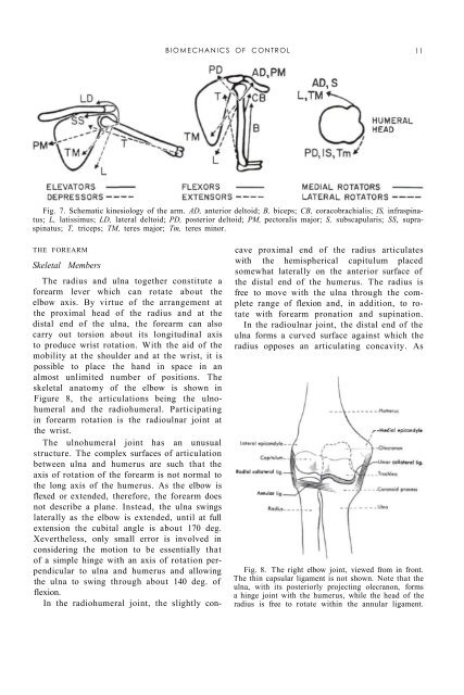
Fig. 7. Schematic k<strong>in</strong>esiology <strong>of</strong> the arm. AD, anterior deltoid; B, biceps; CB, coracobrachialis; IS, <strong>in</strong>frasp<strong>in</strong>atus;<br />
L, latissimus; LD, lateral deltoid; PD, posterior deltoid; PM, pectoralis major; S, subscapularis; SS, suprasp<strong>in</strong>atus;<br />
T, triceps; TM, teres major; Tm, teres m<strong>in</strong>or.<br />
THE FOREARM<br />
Skeletal Members<br />
<strong>The</strong> radius and ulna together constitute a<br />
forearm lever which can rotate about the<br />
elbow axis. By virtue <strong>of</strong> the arrangement at<br />
the proximal head <strong>of</strong> the radius and at the<br />
distal end <strong>of</strong> the ulna, the forearm can also<br />
carry out torsion about its longitud<strong>in</strong>al axis<br />
to produce wrist rotation. With the aid <strong>of</strong> the<br />
mobility at the shoulder and at the wrist, it is<br />
possible to place the hand <strong>in</strong> space <strong>in</strong> an<br />
almost unlimited number <strong>of</strong> positions. <strong>The</strong><br />
skeletal anatomy <strong>of</strong> the elbow is shown <strong>in</strong><br />
Figure 8, the articulations be<strong>in</strong>g the ulnohumeral<br />
and the radiohumeral. Participat<strong>in</strong>g<br />
<strong>in</strong> forearm rotation is the radioulnar jo<strong>in</strong>t at<br />
the wrist.<br />
<strong>The</strong> ulnohumeral jo<strong>in</strong>t has an unusual<br />
structure. <strong>The</strong> complex surfaces <strong>of</strong> articulation<br />
between ulna and humerus are such that the<br />
axis <strong>of</strong> rotation <strong>of</strong> the forearm is not normal to<br />
the long axis <strong>of</strong> the humerus. As the elbow is<br />
flexed or extended, therefore, the forearm does<br />
not describe a plane. Instead, the ulna sw<strong>in</strong>gs<br />
laterally as the elbow is extended, until at full<br />
extension the cubital angle is about 170 deg.<br />
Xevertheless, only small error is <strong>in</strong>volved <strong>in</strong><br />
consider<strong>in</strong>g the motion to be essentially that<br />
<strong>of</strong> a simple h<strong>in</strong>ge with an axis <strong>of</strong> rotation perpendicular<br />
to ulna and humerus and allow<strong>in</strong>g<br />
the ulna to sw<strong>in</strong>g through about 140 deg. <strong>of</strong><br />
flexion.<br />
In the radiohumeral jo<strong>in</strong>t, the slightly concave<br />
proximal end <strong>of</strong> the radius articulates<br />
with the hemispherical capitulum placed<br />
somewhat laterally on the anterior surface <strong>of</strong><br />
the distal end <strong>of</strong> the humerus. <strong>The</strong> radius is<br />
free to move with the ulna through the complete<br />
range <strong>of</strong> flexion and, <strong>in</strong> addition, to rotate<br />
with forearm pronation and sup<strong>in</strong>ation.<br />
In the radioulnar jo<strong>in</strong>t, the distal end <strong>of</strong> the<br />
ulna forms a curved surface aga<strong>in</strong>st which the<br />
radius opposes an articulat<strong>in</strong>g concavity. As<br />
Fig. 8. <strong>The</strong> right elbow jo<strong>in</strong>t, viewed from <strong>in</strong> front.<br />
<strong>The</strong> th<strong>in</strong> capsular ligament is not shown. Note that the<br />
ulna, with its posteriorly project<strong>in</strong>g olecranon, forms<br />
a h<strong>in</strong>ge jo<strong>in</strong>t with the humerus, while the head <strong>of</strong> the<br />
radius is free to rotate with<strong>in</strong> the annular ligament.