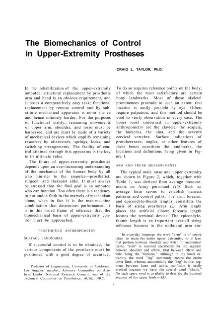
The Biomechanics of Control in Upper-Extremity Prostheses
The Biomechanics of Control in Upper-Extremity Prostheses
The Biomechanics of Control in Upper-Extremity Prostheses

You also want an ePaper? Increase the reach of your titles
YUMPU automatically turns print PDFs into web optimized ePapers that Google loves.
<strong>The</strong> <strong>Biomechanics</strong> <strong>of</strong> <strong>Control</strong><br />
<strong>in</strong> <strong>Upper</strong>-<strong>Extremity</strong> <strong>Prostheses</strong><br />
CRAIG L. TAYLOR, Ph.D. 1<br />
In the rehabilitation <strong>of</strong> the upper-extremity<br />
amputee, structural replacement by prosthetic<br />
arm and hand is an obvious requirement, and<br />
it poses a comparatively easy task; functional<br />
replacement by remote control and by substitute<br />
mechanical apparatus is more elusive<br />
and hence <strong>in</strong>f<strong>in</strong>itely harder. For the purposes<br />
<strong>of</strong> functional utility, rema<strong>in</strong><strong>in</strong>g movements<br />
<strong>of</strong> upper arm, shoulder, and torso must be<br />
harnessed, and use must be made <strong>of</strong> a variety<br />
<strong>of</strong> mechanical devices which amplify rema<strong>in</strong><strong>in</strong>g<br />
resources by alternators, spr<strong>in</strong>gs, locks, and<br />
switch<strong>in</strong>g arrangements. <strong>The</strong> facility <strong>of</strong> control<br />
atta<strong>in</strong>ed through this apparatus is the key<br />
to its ultimate value.<br />
<strong>The</strong> future <strong>of</strong> upper-extremity prosthetics<br />
depends upon an ever-<strong>in</strong>creas<strong>in</strong>g understand<strong>in</strong>g<br />
<strong>of</strong> the mechanics <strong>of</strong> the human body by all<br />
who m<strong>in</strong>ister to the amputee—prosthetist,<br />
surgeon, and therapist alike. It must always<br />
be stressed that the f<strong>in</strong>al goal is an amputee<br />
who can function. Too <strong>of</strong>ten there is a tendency<br />
to put undue faith <strong>in</strong> the marvels <strong>of</strong> mechanism<br />
alone, when <strong>in</strong> fact it is the man-mach<strong>in</strong>e<br />
comb<strong>in</strong>ation that determ<strong>in</strong>es performance. It<br />
is <strong>in</strong> this broad frame <strong>of</strong> reference that the<br />
biomechanical basis <strong>of</strong> upper-extremity control<br />
must be approached.<br />
PROSTHETICS ANTHROPOMETRY<br />
SURFACE LANDMARKS<br />
If successful control is to be obta<strong>in</strong>ed, the<br />
various components <strong>of</strong> the prosthesis must be<br />
positioned with a good degree <strong>of</strong> accuracy.<br />
1<br />
Pr<strong>of</strong>essor <strong>of</strong> Eng<strong>in</strong>eer<strong>in</strong>g, University <strong>of</strong> California,<br />
Los Angeles; member, Advisory Committee on Artificial<br />
Limbs, National Research Council, and <strong>of</strong> the<br />
Technical Committee on Prosthetics, ACAL, NRC.<br />
4<br />
To do so requires reference po<strong>in</strong>ts on the body,<br />
<strong>of</strong> which the most satisfactory are certa<strong>in</strong><br />
bony landmarks. Most <strong>of</strong> these skeletal<br />
prom<strong>in</strong>ences protrude to such an extent that<br />
location is easily possible by eye. Others<br />
require palpation, and this method should be<br />
used to verify observation <strong>in</strong> every case. <strong>The</strong><br />
bones most concerned <strong>in</strong> upper-extremity<br />
anthropometry are the clavicle, the scapula,<br />
the humerus, the ulna, and the seventh<br />
cervical vertebra. Surface <strong>in</strong>dications <strong>of</strong><br />
protuberances, angles, or other features <strong>of</strong><br />
these bones constitute the landmarks, the<br />
locations and def<strong>in</strong>itions be<strong>in</strong>g given <strong>in</strong> Figure<br />
1.<br />
ARM AND TRUNK MEASUREMENTS<br />
<strong>The</strong> typical male torso and upper extremity<br />
are shown <strong>in</strong> Figure 2, which, together with<br />
Table 1, was derived from average measurements<br />
on Army personnel (16). Such an<br />
average form serves to establish harness<br />
patterns and control paths. <strong>The</strong> arm, forearm,<br />
and epicondyle-thumb lengths 2<br />
constitute the<br />
basis <strong>of</strong> siz<strong>in</strong>g prostheses (2). Arm length<br />
places the artificial elbow; forearm length<br />
locates the term<strong>in</strong>al device. <strong>The</strong> epicondylethumb<br />
length is an important over-all siz<strong>in</strong>g<br />
reference because <strong>in</strong> the unilateral arm am-<br />
2<br />
In everyday language the word "arm" is <strong>of</strong> course<br />
taken to mean the entire upper extremity, or at least<br />
that portion between shoulder and wrist. In anatomical<br />
terms, "arm" is reserved specifically for the segment<br />
between shoulder and elbow, that between elbow and<br />
wrist be<strong>in</strong>g the "forearm." Although <strong>in</strong> the lower extremity<br />
the word "leg" commonly means the entire<br />
lower limb, whereas anatomically the "leg" is that segment<br />
between knee and ankle, confusion is easily<br />
avoided because we have the special word "shank."<br />
No such spare word is available to describe the humeral<br />
segment <strong>of</strong> the upper limb.—ED.
BIOMECHANICS OF CONTROL 5<br />
putee it is customary<br />
to match hook length<br />
(and, <strong>in</strong> the case <strong>of</strong> the<br />
artificial hand, thumb<br />
length) to the length <strong>of</strong><br />
the natural thumb (Fig.<br />
3). <strong>The</strong> bilateral arm<br />
amputee can be sized<br />
from body height by<br />
means <strong>of</strong> the Carlyle<br />
formulas (3), which employ<br />
factors derived<br />
from average body proportions.<br />
FUNCTIONAL<br />
ANATOMY<br />
<strong>The</strong> human torso,<br />
shoulder, and upper extremity<br />
are exceed<strong>in</strong>gly<br />
complex structures. In<br />
any deal<strong>in</strong>g with these<br />
elements <strong>of</strong> anatomy,<br />
therefore, it is desirable<br />
to sort out from the<br />
mass <strong>of</strong> detail those<br />
features important to<br />
the particular area <strong>of</strong><br />
study and application.<br />
Where prosthetic controls<br />
are concerned, the<br />
mechanism <strong>of</strong> movement is the central subject<br />
<strong>of</strong> consideration. This functional anatomy<br />
treats <strong>of</strong> the aspects <strong>of</strong> bone, jo<strong>in</strong>t, and muscle<br />
structure that together determ<strong>in</strong>e the modes<br />
and ranges <strong>of</strong> motion <strong>of</strong> the parts. It is a<br />
descriptive science, and while to escape dependence<br />
upon nomenclature is therefore<br />
impossible, the purpose here is to convey a<br />
basic understand<strong>in</strong>g <strong>of</strong> the operation <strong>of</strong> the<br />
upper-extremity mechanisms without undue<br />
use <strong>of</strong> specialized term<strong>in</strong>ology. In any case,<br />
the reader should have available basic anatomical<br />
references such as Gray's Anatomy (13)<br />
or k<strong>in</strong>esiology texts such as those <strong>of</strong> Ste<strong>in</strong>dler<br />
(17) and <strong>of</strong> Holl<strong>in</strong>shead (9).<br />
ELEMENTARY MOTIONS OF THE UPPER EX<br />
TREMITY<br />
<strong>The</strong> geometry <strong>of</strong> each jo<strong>in</strong>t is complex, and<br />
most movements <strong>in</strong>volve an <strong>in</strong>teraction <strong>of</strong> two<br />
Fig. 1. Bones and external landmarks <strong>in</strong> the upper extremity. Def<strong>in</strong>itions: seventh<br />
cervical vertebra, most prom<strong>in</strong>ent vertebra <strong>in</strong> the neck region; acromion, extreme<br />
lateral edge <strong>of</strong> the bony shelf <strong>of</strong> the shoulder; <strong>in</strong>ferior angle <strong>of</strong> scapula, lowest po<strong>in</strong>t<br />
on shoulder blade; epicondyles, lateral and medial bony po<strong>in</strong>ts at the pivot <strong>of</strong> the<br />
elbow; ulnar styloid, project<strong>in</strong>g po<strong>in</strong>t on little-f<strong>in</strong>ger side <strong>of</strong> the wrist.<br />
or more jo<strong>in</strong>ts. Consequently, a motion<br />
nomenclature based on jo<strong>in</strong>t movements would<br />
be unnecessarily complicated. More simply, the<br />
motion <strong>of</strong> each part upon its proximal jo<strong>in</strong>t<br />
may be described with respect to the pr<strong>in</strong>cipal<br />
planes which <strong>in</strong>tersect at that jo<strong>in</strong>t. In this<br />
system, moreover, one may def<strong>in</strong>e a standard<br />
position <strong>in</strong> which the trunk is erect, the arms<br />
hang with their axes vertical, the elbows are<br />
flexed to 90 deg., and the wrist planes are<br />
vertical to assume the "shake-hands" position.<br />
Figure 4 presents the angular movements<br />
possible <strong>in</strong> the three planes <strong>of</strong> space. <strong>The</strong><br />
shoulder-on-chest, arm-on-shoulder, and handon-wrist<br />
actions take place through two angles,<br />
as if mov<strong>in</strong>g about a universal jo<strong>in</strong>t. Geometrically,<br />
the arm motions are more precisely<br />
def<strong>in</strong>ed by a spherical coord<strong>in</strong>ate system where<br />
the segment position is given by longitude and
6 TAYLOR<br />
Fig. 2. Basic anthropometry <strong>of</strong> the male torso and upper extremity. See Table 1.<br />
colatitude angles. For descriptive purposes,<br />
however, the anatomical nomenclature is commonly<br />
used. It should be recognized that, for<br />
multiaxial jo<strong>in</strong>ts, flexion-extension and elevation-depression<br />
angles describe motions <strong>in</strong><br />
the major orthogonal planes only, and <strong>in</strong>termediate<br />
angular excursions must be thought <strong>of</strong><br />
as comb<strong>in</strong>ations <strong>of</strong> these motions.<br />
<strong>The</strong> simplified movement system depicted <strong>in</strong><br />
Figure 4 is <strong>in</strong>complete <strong>in</strong> many ways. Not <strong>in</strong>cluded<br />
are such movements as twist<strong>in</strong>g <strong>of</strong> the<br />
shoulder due to various scapular movements,<br />
anterior-posterior sw<strong>in</strong>gs <strong>of</strong> the arm <strong>in</strong> positions<br />
<strong>of</strong> partial elevation, and the slightly<br />
conical surface <strong>of</strong> revolution <strong>of</strong> forearm<br />
flexion. 3<br />
<strong>The</strong>se details may, however, be<br />
3<br />
It deserves to be noted here that, taken literally,<br />
expressions such as "forearm flexion-extension," "arm<br />
flexion-extension," and "humeral flexion-extension"<br />
represent questionable nomenclature. To "flex" means<br />
to "bend." Limb segments do not bend very readily
BIOMECHANICS OF CONTROL 7<br />
Fig. 3. Correct lengths for upper-extremity prostheses.<br />
In the unilateral case, hook length is made to co<strong>in</strong>cide<br />
with normal thumb length, as is also the thumb<br />
length <strong>of</strong> the artificial hand. For bilateral arm amputees,<br />
A = 0.19 X (body height); B + C = 0.21 X<br />
(body height). After Carlyle (J).<br />
ignored <strong>in</strong> the <strong>in</strong>terest <strong>of</strong> the simplicity <strong>of</strong><br />
description that is adequate for the purposes<br />
<strong>of</strong> upper-extremity prosthetics.<br />
THE SHOULDER GIRDLE<br />
Skeletal Members and Jo<strong>in</strong>ts<br />
<strong>The</strong> scapula and clavicle are the chief bones<br />
mak<strong>in</strong>g up the shoulder girdle. Secondarily, the<br />
proximal portion <strong>of</strong> the humerus may be <strong>in</strong>cluded,<br />
s<strong>in</strong>ce the close <strong>in</strong>terarticulation <strong>of</strong> all<br />
three bones at the shoulder jo<strong>in</strong>t gives a considerable<br />
degree <strong>of</strong> coord<strong>in</strong>ated activity among<br />
them and also extends to the complex as a<br />
whole the actions <strong>of</strong> many <strong>of</strong> the muscles<br />
<strong>in</strong>sert<strong>in</strong>g on the <strong>in</strong>dividual members.<br />
without break<strong>in</strong>g. Jo<strong>in</strong>ts are designed for flexion. In<br />
the lower extremity, for example, one speaks not <strong>of</strong><br />
"shank flexion" but <strong>of</strong> "knee flexion," not <strong>of</strong> "thigh<br />
flexion" but <strong>of</strong> "hip flexion." That is, one uses "flexion"<br />
or "extension" not with reference to motion <strong>of</strong> the<br />
distal segment but with reference to the more proximal<br />
jo<strong>in</strong>t. Although Webster accepts the expression "to<br />
flex the arm," he obviously uses the word "arm" <strong>in</strong><br />
the everyday sense <strong>of</strong> mean<strong>in</strong>g the entire upper extremity,<br />
or at least that portion between shoulder and<br />
wrist. Because this loose term<strong>in</strong>ology <strong>in</strong> the upper extremity<br />
is so widely established, not only among workers<br />
<strong>in</strong> prosthetics, it is used throughout this issue <strong>of</strong> ARTI<br />
FICIAL LIMBS, with the understand<strong>in</strong>g that "forearm<br />
flexion" means "elbow flexion," "arm flexion" and<br />
"humeral flexion" mean "flexion <strong>of</strong> the glenohumeral<br />
jo<strong>in</strong>t (and associated structures) " See page 9 et<br />
seq.—ED.<br />
Details <strong>of</strong> the skeletal anatomy <strong>in</strong>volved are<br />
shown <strong>in</strong> Figure 5. <strong>The</strong>re are <strong>in</strong> the system two<br />
jo<strong>in</strong>ts and one pseudo jo<strong>in</strong>t. In the sternoclavicular<br />
jo<strong>in</strong>t, the clavicle articulates with<br />
the sternum <strong>in</strong> a somewhat saddle-shaped<br />
juncture recessed <strong>in</strong> a concavity with<strong>in</strong> the<br />
sternum. <strong>The</strong> biaxial surfaces permit movements<br />
<strong>in</strong> two planes. Ligaments cross<strong>in</strong>g the<br />
jo<strong>in</strong>t prevent displacement <strong>of</strong> the clavicle<br />
anteriorly and laterally. <strong>The</strong> elevation-depression<br />
range is 50 to 60 deg., the flexion-extension<br />
range from 25 to 35 deg.<br />
In the acromioclavicular jo<strong>in</strong>t, the distal<br />
end <strong>of</strong> the clavicle articulates with the scapula<br />
<strong>in</strong> an elliptical juncture which permits a balland-socket<br />
type <strong>of</strong> action. <strong>The</strong> acromioclavicular<br />
ligaments b<strong>in</strong>d the jo<strong>in</strong>t directly.<br />
Strong ligaments from the clavicle to the<br />
coracoid process give important additional<br />
stabilization. <strong>The</strong> range <strong>of</strong> movement is small,<br />
be<strong>in</strong>g only about 10 deg. <strong>in</strong> the frontal and<br />
sagittal planes.<br />
<strong>The</strong> pseudo jo<strong>in</strong>t, the scapulothoracic, is a<br />
muscular suspension which holds the scapula<br />
aga<strong>in</strong>st the thoracic wall but which at the same<br />
time permits translatory and rotatory movements.<br />
A large factor <strong>in</strong> ma<strong>in</strong>ta<strong>in</strong><strong>in</strong>g this jo<strong>in</strong>t<br />
<strong>in</strong> position is barometric pressure, which is estimated<br />
to act upon it with a force <strong>of</strong> 170 lb.<br />
Muscles and Movements<br />
<strong>The</strong> complex arrangement <strong>of</strong> bony elements<br />
is rivaled by the <strong>in</strong>volved nature <strong>of</strong> the muscles<br />
<strong>of</strong> the shoulder girdle and by the <strong>in</strong>tricate<br />
ways <strong>in</strong> which they act upon it. <strong>The</strong> schematic<br />
view <strong>of</strong> Figure 6 presents the fundamentals.<br />
Elevation <strong>of</strong> the shoulder is seen to be brought<br />
about pr<strong>in</strong>cipally by elevators and downward<br />
rotators <strong>of</strong> the scapula, such as the upper<br />
trapezius, the levator scapulae, and the rhomboids.<br />
Although the rhomboids assist <strong>in</strong><br />
elevation, they do not contribute to upward<br />
rotation. Depression <strong>of</strong> the shoulder is mediated<br />
by muscles <strong>in</strong>serted on the scapula, the
Tig. 4. Simplified movement system <strong>in</strong> the upper extremity. Wrist flexion is omitted s<strong>in</strong>ce ord<strong>in</strong>arily it is not<br />
<strong>in</strong>volved <strong>in</strong> upper-extremity controls.
clavicle, and the proximal end <strong>of</strong><br />
the humerus. Anteriorly the lower<br />
fibers <strong>of</strong> the pectoralis major, the<br />
pectoralis m<strong>in</strong>or, and the subclavius,<br />
and posteriorly the lower<br />
trapezius and latissimus, act as<br />
depressors.<br />
Rotation <strong>of</strong> the scapula upward<br />
(i.e., right scapula, viewed from<br />
the rear, rotates counterclockwise)<br />
or downward (i.e., right scapula,<br />
viewed from the rear, rotates<br />
clockwise) is brought about by a<br />
special comb<strong>in</strong>ation <strong>of</strong> the elevators<br />
and depressors. As shown <strong>in</strong><br />
Figure 6, two portions <strong>of</strong> the trapezius,<br />
together with the serratus,<br />
cause upward rotation. Conversely,<br />
the pectorals, the latissimus, and<br />
the rhomboids cooperate to cause<br />
downward rotation. As will be seen<br />
later (page 13), the mechanical<br />
pr<strong>in</strong>ciple <strong>of</strong> the couple applies <strong>in</strong><br />
these rotatory actions upon the<br />
scapula.<br />
Flexion and extension <strong>of</strong> the<br />
shoulder <strong>in</strong>volve as pr<strong>in</strong>cipal elements<br />
the abduction and adduction, respectively,<br />
<strong>of</strong> the scapula. <strong>The</strong> flexor muscles act<strong>in</strong>g<br />
on the shoulder complex are the pectoralis<br />
major and m<strong>in</strong>or, which sw<strong>in</strong>g the clavicle and<br />
acromion forward. <strong>The</strong> serratus anterior aids<br />
strongly by abduct<strong>in</strong>g the scapula. <strong>The</strong> extensors,<br />
placed posteriorly, <strong>in</strong>clude the latissimus,<br />
which pulls posteriorly and medially on the<br />
humerus, and the trapezius and rhomboids,<br />
which pull medially on the scapula.<br />
<strong>The</strong> forward and backward shrugg<strong>in</strong>g <strong>of</strong> the<br />
shoulders with abduction and adduction, together<br />
with some upward and downward<br />
rotation <strong>of</strong> the scapulae, constitutes a major<br />
control source. Even <strong>in</strong> above-elbow amputees<br />
who use humeral flexion for forearm lift and<br />
for term<strong>in</strong>al-device operation at low elbow<br />
angles (page 22), scapular abduction is<br />
utilized for term<strong>in</strong>al-device operation at<br />
large angles <strong>of</strong> elbow flexion (e.g., when the<br />
term<strong>in</strong>al device is near the mouth). In shoulder<br />
amputees, both these operations depend<br />
wholly upon scapular abduction augmented by<br />
upward rotation.<br />
BIOMECHANICS OF CONTROL 9<br />
Fig. 5. Skeletal anatomy <strong>of</strong> the shoulder region, a, Anterior view.<br />
b, Posterior view.<br />
THE ARM<br />
<strong>The</strong> Humerus and the Glenohumeral Jo<strong>in</strong>t<br />
<strong>The</strong> humerus, together with its jo<strong>in</strong>t at the<br />
shoulder, comprises the skeletal mach<strong>in</strong>ery <strong>of</strong><br />
the arm. As noted <strong>in</strong> Figure 4, it is capable <strong>of</strong><br />
flexion-extension, elevation-depression, and<br />
rotation upon its proximal jo<strong>in</strong>t. <strong>The</strong> glenoid<br />
cavity, a lateral process on the scapula, receives<br />
the spherical surface <strong>of</strong> the humeral<br />
head. <strong>The</strong> glenohumeral articulation is therefore<br />
<strong>of</strong> true ball-and-socket character. <strong>The</strong><br />
fibrous jo<strong>in</strong>t capsule is remarkable <strong>in</strong> that it<br />
envelops the humeral head and the glenoid<br />
marg<strong>in</strong>s <strong>in</strong> complete but rather loose fashion,<br />
so that a wide range <strong>of</strong> movement is possible.<br />
To some extent barometric pressure, but to<br />
larger extent the musculature spann<strong>in</strong>g the<br />
jo<strong>in</strong>t, is responsible for keep<strong>in</strong>g the articular<br />
surfaces together <strong>in</strong> all angular positions. A<br />
group <strong>of</strong> muscles <strong>in</strong>clud<strong>in</strong>g the subscapularis,<br />
the suprasp<strong>in</strong>atus, and the <strong>in</strong>frasp<strong>in</strong>atus function<br />
pr<strong>in</strong>cipally <strong>in</strong> this hold<strong>in</strong>g action.
10 TAYLOR<br />
Fig. 6. Schematic k<strong>in</strong>esiology <strong>of</strong> the shoulder girdle. L, latissimus;<br />
LS, levator scapulae; LT, lower trapezius; MT, medial trapezius;<br />
PM, pectoralis major; Pm, pectoralis m<strong>in</strong>or; RM, rhomboid major;<br />
Rm, rhomboid m<strong>in</strong>or; SA, serratus anterior; SC, subclavius; UT,<br />
upper trapezius.<br />
Muscles and Movements<br />
<strong>The</strong> k<strong>in</strong>esiology <strong>of</strong> the arm is closely associated<br />
with that <strong>of</strong> the shoulder girdle, nearly all<br />
natural movements <strong>in</strong>volv<strong>in</strong>g a coord<strong>in</strong>ated<br />
movement between arm and shoulder. It is<br />
helpful, however, first to describe the pure<br />
movements <strong>of</strong> the arm. Schematics <strong>of</strong> the<br />
muscles act<strong>in</strong>g upon the arm are presented <strong>in</strong><br />
Figure 7. Elevation is effected by the lateral<br />
deltoid and the suprasp<strong>in</strong>atus, depression by<br />
the latissimus, the pectoralis major, the long<br />
head <strong>of</strong> the triceps, and the teres major. In<br />
both actions, the contributions <strong>of</strong> <strong>in</strong>dividual<br />
muscles differ accord<strong>in</strong>g to the<br />
angle <strong>of</strong> the arm. And it should be<br />
noted that, with <strong>in</strong>sertions near the<br />
pivot po<strong>in</strong>t <strong>of</strong> the humeral head,<br />
the rotatory moments are proportionately<br />
small, thus account<strong>in</strong>g for<br />
the large number <strong>of</strong> muscles necessary<br />
to give adequate jo<strong>in</strong>t torques.<br />
Arm flexion and extension are<br />
brought about by two groups <strong>of</strong><br />
muscles. <strong>The</strong> biceps, the coracobrachialis,<br />
the anterior deltoid,<br />
and the clavicular fibers <strong>of</strong> the pectoralis<br />
major mediate flexion, while<br />
the posterior deltoid, the long head<br />
<strong>of</strong> the triceps, the latissimus, and<br />
the teres major effect extension.<br />
Rotation <strong>of</strong> the arm depends upon<br />
muscles that <strong>in</strong>sert on the surface<br />
<strong>of</strong> the humerus and then pass anteriorly<br />
or posteriorly around it to<br />
impart medial or lateral torsion.<br />
As would be expected, rotational<br />
forces are greatest when the arm<br />
hangs at the side; torque is reduced<br />
drastically when the arm is elevated<br />
over the head and the twist<strong>in</strong>g<br />
angles <strong>of</strong> the muscles tend to disappear.<br />
Comb<strong>in</strong>ed Arm and Shoulder<br />
Movements<br />
In most natural arm movements,<br />
such as arm elevation, arm flexion,<br />
forward reach<strong>in</strong>g, and to-and-fro<br />
sw<strong>in</strong>gs <strong>of</strong> the partially elevated<br />
arm, both arm and shoulder girdle<br />
participate. In full arm elevation <strong>of</strong> 180 deg.,<br />
for example, 120 deg. are contributed by rotation<br />
<strong>of</strong> the arm on the glenohumeral jo<strong>in</strong>t,<br />
60 deg. are contributed by upward rotation <strong>of</strong><br />
the scapula (17). In forward reach<strong>in</strong>g, <strong>in</strong>volv<strong>in</strong>g<br />
partial arm flexion, the shoulder<br />
flexes and the scapula abducts and rotates<br />
slightly. Properly managed, this motion, the<br />
common flexion control motion <strong>of</strong> both the<br />
above- and the below-elbow amputee (pages<br />
19-22) can give marked gracefulness to<br />
prosthetic operation.
BIOMECHANICS OF CONTROL 11<br />
Fig. 7. Schematic k<strong>in</strong>esiology <strong>of</strong> the arm. AD, anterior deltoid; B, biceps; CB, coracobrachialis; IS, <strong>in</strong>frasp<strong>in</strong>atus;<br />
L, latissimus; LD, lateral deltoid; PD, posterior deltoid; PM, pectoralis major; S, subscapularis; SS, suprasp<strong>in</strong>atus;<br />
T, triceps; TM, teres major; Tm, teres m<strong>in</strong>or.<br />
THE FOREARM<br />
Skeletal Members<br />
<strong>The</strong> radius and ulna together constitute a<br />
forearm lever which can rotate about the<br />
elbow axis. By virtue <strong>of</strong> the arrangement at<br />
the proximal head <strong>of</strong> the radius and at the<br />
distal end <strong>of</strong> the ulna, the forearm can also<br />
carry out torsion about its longitud<strong>in</strong>al axis<br />
to produce wrist rotation. With the aid <strong>of</strong> the<br />
mobility at the shoulder and at the wrist, it is<br />
possible to place the hand <strong>in</strong> space <strong>in</strong> an<br />
almost unlimited number <strong>of</strong> positions. <strong>The</strong><br />
skeletal anatomy <strong>of</strong> the elbow is shown <strong>in</strong><br />
Figure 8, the articulations be<strong>in</strong>g the ulnohumeral<br />
and the radiohumeral. Participat<strong>in</strong>g<br />
<strong>in</strong> forearm rotation is the radioulnar jo<strong>in</strong>t at<br />
the wrist.<br />
<strong>The</strong> ulnohumeral jo<strong>in</strong>t has an unusual<br />
structure. <strong>The</strong> complex surfaces <strong>of</strong> articulation<br />
between ulna and humerus are such that the<br />
axis <strong>of</strong> rotation <strong>of</strong> the forearm is not normal to<br />
the long axis <strong>of</strong> the humerus. As the elbow is<br />
flexed or extended, therefore, the forearm does<br />
not describe a plane. Instead, the ulna sw<strong>in</strong>gs<br />
laterally as the elbow is extended, until at full<br />
extension the cubital angle is about 170 deg.<br />
Xevertheless, only small error is <strong>in</strong>volved <strong>in</strong><br />
consider<strong>in</strong>g the motion to be essentially that<br />
<strong>of</strong> a simple h<strong>in</strong>ge with an axis <strong>of</strong> rotation perpendicular<br />
to ulna and humerus and allow<strong>in</strong>g<br />
the ulna to sw<strong>in</strong>g through about 140 deg. <strong>of</strong><br />
flexion.<br />
In the radiohumeral jo<strong>in</strong>t, the slightly concave<br />
proximal end <strong>of</strong> the radius articulates<br />
with the hemispherical capitulum placed<br />
somewhat laterally on the anterior surface <strong>of</strong><br />
the distal end <strong>of</strong> the humerus. <strong>The</strong> radius is<br />
free to move with the ulna through the complete<br />
range <strong>of</strong> flexion and, <strong>in</strong> addition, to rotate<br />
with forearm pronation and sup<strong>in</strong>ation.<br />
In the radioulnar jo<strong>in</strong>t, the distal end <strong>of</strong> the<br />
ulna forms a curved surface aga<strong>in</strong>st which the<br />
radius opposes an articulat<strong>in</strong>g concavity. As<br />
Fig. 8. <strong>The</strong> right elbow jo<strong>in</strong>t, viewed from <strong>in</strong> front.<br />
<strong>The</strong> th<strong>in</strong> capsular ligament is not shown. Note that the<br />
ulna, with its posteriorly project<strong>in</strong>g olecranon, forms<br />
a h<strong>in</strong>ge jo<strong>in</strong>t with the humerus, while the head <strong>of</strong> the<br />
radius is free to rotate with<strong>in</strong> the annular ligament.
12 TAYLOR<br />
the forearm goes through a pronation-sup<strong>in</strong>ation<br />
range <strong>of</strong> about 170 deg., the radius<br />
"sw<strong>in</strong>gs like a gate" about the distal end <strong>of</strong> the<br />
ulna.<br />
Muscles and Movements<br />
As shown <strong>in</strong> Figure 9, the musculature for<br />
provid<strong>in</strong>g forearm flexion and extension is<br />
comparatively simple, while that for pronation-sup<strong>in</strong>ation<br />
is somewhat more <strong>in</strong>volved.<br />
Flexion <strong>of</strong> the forearm is effected pr<strong>in</strong>cipally<br />
by the biceps, orig<strong>in</strong>at<strong>in</strong>g on the scapula<br />
and <strong>in</strong>sert<strong>in</strong>g on the radius, and by the<br />
brachialis, spann<strong>in</strong>g the elbow from humerus<br />
to ulna. Secondarily, the brachioradialis and<br />
other muscles, orig<strong>in</strong>at<strong>in</strong>g distally on the<br />
humerus and cours<strong>in</strong>g down the forearm, contribute<br />
to flexion. Extension is largely the<br />
function <strong>of</strong> the triceps, orig<strong>in</strong>at<strong>in</strong>g on both the<br />
scapula and humerus and <strong>in</strong>sert<strong>in</strong>g on the<br />
leverlike olecranon process <strong>of</strong> the ulna. A small<br />
extensor action is added by the anconeus.<br />
Rotation <strong>of</strong> the forearm is a function <strong>of</strong><br />
many muscles. Some, such as the sup<strong>in</strong>ator,<br />
evidently are designed for the purpose, while<br />
others, as for example the f<strong>in</strong>ger flexors, have<br />
different pr<strong>in</strong>cipal functions, the contribution<br />
to forearm rotation be<strong>in</strong>g only <strong>in</strong>cidental.<br />
Figure 9 presents the major rotatory muscles<br />
only. Sup<strong>in</strong>ation is mediated by the brachioradialis,<br />
the sup<strong>in</strong>ator brevis, and the<br />
biceps, pronation by the pronators quadratus<br />
Fig. 9. Schematic k<strong>in</strong>esiology <strong>of</strong> the forearm. A,<br />
anconeus; B, biceps; BR, brachialis; BrR, brachioradialis;<br />
PT, pronator teres; PQ, pronator quadratus;<br />
Su, sup<strong>in</strong>ator; T, triceps.<br />
and teres. Of great importance to upper-extremity<br />
prosthetics is the fact that rotation <strong>of</strong><br />
the forearm is a function <strong>of</strong> total forearm<br />
length. With successively shorter stumps, not<br />
only are the rotation limits <strong>of</strong> the radius and<br />
ulna reduced, but also the contributions <strong>of</strong><br />
muscles are elim<strong>in</strong>ated as their <strong>in</strong>sertions are<br />
sectioned.<br />
MUSCULOSKELETAL<br />
MECHANISMS<br />
<strong>The</strong> upper extremity hav<strong>in</strong>g been considered<br />
from the standpo<strong>in</strong>t <strong>of</strong> functional and descriptive<br />
anatomy, attention may now be<br />
turned to a more mechanical view <strong>of</strong> its operations.<br />
Typical elements <strong>of</strong> mechanism <strong>in</strong> the<br />
upper extremity <strong>in</strong>clude jo<strong>in</strong>ts (bear<strong>in</strong>g surfaces),<br />
jo<strong>in</strong>t-l<strong>in</strong><strong>in</strong>g secretions (lubricants),<br />
bones (levers and couple members), tendons<br />
(transmission cables), and muscles (motors).<br />
<strong>The</strong> arrangement <strong>of</strong> these elements makes up a<br />
complex mach<strong>in</strong>ery capable <strong>of</strong> such diverse<br />
activities as precise orientation <strong>in</strong> space, performance<br />
<strong>of</strong> external work, f<strong>in</strong>e digital manipulations,<br />
and so on.<br />
TYPICAL JOINT MECHANICS<br />
<strong>The</strong> elbow jo<strong>in</strong>t embodies the essential<br />
structures <strong>of</strong> diarthrodial jo<strong>in</strong>ts. <strong>The</strong> bear<strong>in</strong>g<br />
surfaces are covered with a th<strong>in</strong> layer <strong>of</strong><br />
articular cartilage that is cont<strong>in</strong>uous with the<br />
synovial membrane l<strong>in</strong><strong>in</strong>g the whole jo<strong>in</strong>t<br />
capsule. Subsynovial pads <strong>of</strong> fat serve to fill<br />
up the chang<strong>in</strong>g spaces that occur dur<strong>in</strong>g<br />
movement <strong>of</strong> the jo<strong>in</strong>t (Fig. 10). It is believed<br />
that these fatty deposits serve as "pad oilers"<br />
to ma<strong>in</strong>ta<strong>in</strong> the cont<strong>in</strong>uous film <strong>of</strong> synovial<br />
fluid over the articular surfaces (4). This<br />
fluid conta<strong>in</strong>s muc<strong>in</strong> (a glycoprote<strong>in</strong> which<br />
serves as a lubricant for the jo<strong>in</strong>t) and other<br />
material constitut<strong>in</strong>g a nutritional medium<br />
for the articular cartilage. Considerable uncerta<strong>in</strong>ty<br />
exists concern<strong>in</strong>g the method <strong>of</strong><br />
formation and distribution <strong>of</strong> the fluid to the<br />
jo<strong>in</strong>t, but its mechanical function is clear and<br />
the normal jo<strong>in</strong>t performs as a well-oiled<br />
bear<strong>in</strong>g.<br />
BONES AND THEIR MECHANICAL FUNCTION<br />
<strong>The</strong> bones <strong>of</strong> the upper extremity, besides<br />
form<strong>in</strong>g a support for s<strong>of</strong>t tissue, provide a<br />
system <strong>of</strong> levers which makes the arm an im-
BIOMECHANICS OF CONTROL 13<br />
theory have dealt largely with the frequent<br />
and <strong>in</strong>cautious extension <strong>of</strong> the concept. It is<br />
now believed that genetic and growth factors<br />
determ<strong>in</strong>e the essential form and dimensions <strong>of</strong><br />
bone. Mechanical stresses serve secondarily<br />
to mold and modify it to give added strength<br />
where stresses are greatest. One must grant<br />
from even a superficial exam<strong>in</strong>ation <strong>of</strong> the<br />
<strong>in</strong>ternal structure <strong>of</strong> bone that Nature has<br />
done an admirable job <strong>of</strong> design<strong>in</strong>g for maximum<br />
strength with m<strong>in</strong>imum weight.<br />
Fig. 10. Typical change <strong>in</strong> jo<strong>in</strong>t spaces with flexionextension,<br />
as revealed by the elbow. Redrawn from<br />
Ste<strong>in</strong>dler (17), after Fick. A, Gap <strong>of</strong> the medial border<br />
<strong>of</strong> the olecranon surface with elbow <strong>in</strong> extreme extension.<br />
B, Gap <strong>of</strong> the lateral border <strong>of</strong> the olecranon <strong>in</strong><br />
extreme flexion.<br />
portant mechanism for the performance <strong>of</strong><br />
gross work, such as lift<strong>in</strong>g, sl<strong>in</strong>g<strong>in</strong>g, and<br />
thrust<strong>in</strong>g. <strong>The</strong> arm bones serve further<br />
as positioners <strong>of</strong> the hand, <strong>in</strong> which other,<br />
f<strong>in</strong>er bones constitute the <strong>in</strong>tricate articulated<br />
framework <strong>of</strong> the manipulative mechanism.<br />
Two ma<strong>in</strong> features <strong>of</strong> bones merit discussion<br />
here—their <strong>in</strong>ternal composition and construction<br />
and their external shape and adaptations<br />
that permit them to serve as members<br />
<strong>of</strong> mechanical systems.<br />
Internal<br />
Structure<br />
<strong>The</strong>re is much evidence that the gross <strong>in</strong>ternal<br />
structure <strong>of</strong> bone is em<strong>in</strong>ently suited to<br />
withstand the mechanical stresses placed upon<br />
it by the compressive loads <strong>of</strong> weight-bear<strong>in</strong>g,<br />
by the tensions <strong>of</strong> tendons and ligaments, and<br />
by the lateral pressures <strong>of</strong> adjacent tissues (4).<br />
<strong>The</strong> nature and orientation <strong>of</strong> the trabeculae<br />
<strong>in</strong> cancellous bone have, for example, long been<br />
held, <strong>in</strong> theory, to provide the maximum<br />
strength along the l<strong>in</strong>es <strong>of</strong> major stresses. This<br />
idea, orig<strong>in</strong>ally suggested by von Meyer,<br />
has been championed by many, <strong>in</strong>clud<strong>in</strong>g<br />
Koch, who carried out a stress analysis on the<br />
femur (12). Objections to the von Meyer<br />
Members <strong>of</strong> Mechanical Systems<br />
<strong>The</strong> second pr<strong>in</strong>cipal feature <strong>of</strong> bones, that<br />
<strong>of</strong> serv<strong>in</strong>g as rigid members <strong>in</strong> a complex <strong>of</strong><br />
mechanical systems, is the one that has engaged<br />
the most attention. It is surpris<strong>in</strong>g that<br />
the simple lever concepts <strong>of</strong> Archimedes have<br />
persisted <strong>in</strong> anatomy and k<strong>in</strong>esiology texts to<br />
the present day. Thus, the forearm-flexor<br />
system is said to act as a third-class lever, the<br />
extensor system as a first-class lever. Although<br />
these assertions are <strong>of</strong> course true, both <strong>of</strong><br />
these systems are, <strong>in</strong> the more complete<br />
language <strong>of</strong> Newtonian mechanics, parts <strong>of</strong><br />
force-couple systems <strong>in</strong> which equal and<br />
opposite components <strong>of</strong> force are transmitted<br />
through the bones and jo<strong>in</strong>ts (Fig. 11). Elftman<br />
(7) has emphasized this view. <strong>The</strong><br />
magnitude <strong>of</strong> the couple is given by the product<br />
<strong>of</strong> the force (either <strong>of</strong> the equal but opposite<br />
forces) and the distance between them, which<br />
also is numerically equal to the torque <strong>of</strong> the<br />
muscle force. <strong>The</strong> concept <strong>of</strong> the couple calls<br />
attention to the existence <strong>of</strong> the equal and<br />
opposite forces <strong>in</strong> jo<strong>in</strong>ts and emphasizes the<br />
loads placed upon them by muscular work.<br />
Another and more complicated application<br />
<strong>of</strong> the couple is seen <strong>in</strong> scapular rotation.<br />
Here, as described by Inman el al. (11) and as<br />
shown <strong>in</strong> Figure 12,<br />
the pull <strong>of</strong> the lower<br />
fibers <strong>of</strong> the serratus anterior<br />
upon the scapula<br />
is such as to give it<br />
Fig. 11. Force couples at<br />
the elbow. Tensile forces<br />
<strong>in</strong> biceps and brahialis are<br />
associated with equal, opposite,<br />
and parallel forces<br />
through the jo<strong>in</strong>t.
14 TAYLOR<br />
Fig. 12. Muscle forces<br />
act<strong>in</strong>g on the shoulder, anterior<br />
view. <strong>The</strong> trapezius,<br />
act<strong>in</strong>g diagonally, gives a<br />
supportive component. Fy,,<br />
and a horizontal component,<br />
Fx, which together<br />
with the opposite force from<br />
the serratus, 5, comprise an<br />
upward rotatory force<br />
couple on the scapula.<br />
upward rotation,<br />
while the thrust <strong>of</strong><br />
the clavicle, act<strong>in</strong>g<br />
through the acromioclavicular<br />
jo<strong>in</strong>t,<br />
holds a pivot for the<br />
rotation. Simultaneously,<br />
the pull <strong>of</strong> the<br />
upper trapezius fibers<br />
causes the clavicle<br />
to undergo angular<br />
rotation about<br />
the sternoclavicular<br />
jo<strong>in</strong>t. <strong>The</strong> result is<br />
that, at least through<br />
the first 90 deg. <strong>of</strong><br />
arm elevation, the<br />
motion is shared by<br />
coord<strong>in</strong>ated angular<br />
rotations <strong>of</strong> scapula,<br />
clavicle, and humerus.<br />
As a basic<br />
part <strong>of</strong> this rotatory<br />
action, the scapula<br />
acts as the moment<br />
arm <strong>of</strong> a force couple, the trapezius and serratus<br />
provid<strong>in</strong>g components <strong>of</strong> force which are equal<br />
and opposite.<br />
TENDONS AND MUSCLES<br />
<strong>The</strong> specific functions <strong>of</strong> tendons are to<br />
concentrate the pull <strong>of</strong> a muscle with<strong>in</strong> a small<br />
transverse area, to allow muscles to act from a<br />
distance, and <strong>in</strong> some <strong>in</strong>stances to transmit<br />
the pull <strong>of</strong> a muscle through a changed pathway.<br />
<strong>The</strong> mechanical importance <strong>of</strong> this tissue<br />
is nowhere more evident than <strong>in</strong> the arm, where<br />
a large degree <strong>of</strong> versatility <strong>of</strong> motion <strong>in</strong> the<br />
segment distal to each jo<strong>in</strong>t is preserved by<br />
"remot<strong>in</strong>g" the action <strong>of</strong> muscles through<br />
slender, cablelike tendons over jo<strong>in</strong>ts. By this<br />
means l<strong>in</strong>es <strong>of</strong> pull are brought near the jo<strong>in</strong>t<br />
axes, thus provid<strong>in</strong>g a lever arm consistent<br />
with the tensile force <strong>of</strong> the muscle at all jo<strong>in</strong>t<br />
angles and also giv<strong>in</strong>g at low jo<strong>in</strong>t angles an<br />
<strong>in</strong>creased angular motion for a given l<strong>in</strong>ear<br />
contraction. Other advantages <strong>of</strong> remot<strong>in</strong>g the<br />
muscles are seen <strong>in</strong> the forearm and hand. In<br />
order to afford the variety and complexity <strong>of</strong><br />
<strong>in</strong>terdigital movements, many <strong>in</strong>dependent<br />
muscle units are necessary, and critical space<br />
problems are avoided because muscles such as<br />
the common flexors and extensors <strong>of</strong> the f<strong>in</strong>gers<br />
are placed at some distance up the forearm.<br />
<strong>The</strong> predom<strong>in</strong>ant function <strong>of</strong> tendon as a<br />
tension member <strong>in</strong> series with muscle, which is<br />
a tension motor, is seen <strong>in</strong> early growth stages.<br />
An undifferentiated cellular reticulum <strong>of</strong><br />
connective tissue is everywhere found <strong>in</strong><br />
embryonic tissue. <strong>The</strong> parent cells are fibroblasts;<br />
they elaborate and extrude the collagenous<br />
material <strong>of</strong> which white fibers are<br />
made (4). At this po<strong>in</strong>t the presence <strong>of</strong> mechanical<br />
tensions <strong>in</strong> the tissue <strong>in</strong>fluences the rate,<br />
amount, and direction <strong>of</strong> the resultant fiber<br />
formation. At maturity the tendon is composed<br />
almost entirely <strong>of</strong> white collagen fibers,<br />
closely packed <strong>in</strong> parallel bundles, to form a<br />
cablelike strand. It is conta<strong>in</strong>ed with<strong>in</strong> a<br />
sheath which forms a loose cover<strong>in</strong>g lubricated<br />
cont<strong>in</strong>uously by a muc<strong>in</strong>ous fluid to<br />
reduce friction with surround<strong>in</strong>g tissues.<br />
Mutual adjustment <strong>of</strong> the characteristics <strong>of</strong><br />
muscle and tendon is shown <strong>in</strong> many respects.<br />
<strong>The</strong> musculotend<strong>in</strong>ous juncture varies with the<br />
arrangement <strong>of</strong> the muscle fiber. It shows a<br />
simple series arrangement for fusiform muscles<br />
like the biceps, or it comprises a distributed<br />
attachment zone by cont<strong>in</strong>uation <strong>of</strong> the<br />
tendon <strong>in</strong>to <strong>in</strong>tramuscular septa where p<strong>in</strong>niform<br />
fibers may <strong>in</strong>sert (Fig. 13). In some unexpla<strong>in</strong>ed<br />
way the relative lengths <strong>of</strong> muscle<br />
and associated tendon are so composed that<br />
the shorten<strong>in</strong>g range <strong>of</strong> the muscle is that<br />
necessary to move the segment distal to the<br />
jo<strong>in</strong>t through its maximum range (8). <strong>The</strong><br />
capacity to adapt the ratio <strong>of</strong> muscle length to<br />
tendon length has been demonstrated <strong>in</strong> an<br />
experiment <strong>in</strong> which the pathway <strong>of</strong> the<br />
tibialis anterior tendon <strong>in</strong> the rabbit was<br />
shortened. <strong>The</strong> result was that the tendon<br />
shortened while the muscle lengthened to rega<strong>in</strong><br />
the normal jo<strong>in</strong>t range (4).<br />
<strong>The</strong> relative strengths <strong>of</strong> muscle and <strong>of</strong><br />
tendon also show an approximate compatibility,<br />
the tensile strength <strong>of</strong> tendon, measured<br />
at from 8700 to 18,000 lb. per sq. <strong>in</strong>. (6), be<strong>in</strong>g<br />
greater than that for muscle. Strength tests <strong>of</strong><br />
excised muscle-tendon systems show that<br />
failure commonly occurs <strong>in</strong> the belly <strong>of</strong> the<br />
muscle, or at the musculotend<strong>in</strong>ous juncture,
BIOMECHANICS OF CONTROL 15<br />
Fig. 13. Muscle fiber patterns. A, Fusiform. B,<br />
Bip<strong>in</strong>niform.<br />
or at the bone-tendon juncture, but never<br />
exclusively <strong>in</strong> the tendon itself. Analysis <strong>of</strong><br />
cl<strong>in</strong>ical cases <strong>in</strong>dicates that muscle is still the<br />
site <strong>of</strong> failure even when it is maximally<br />
tensed (14). It is clear, then, that <strong>of</strong> the muscletendon<br />
comb<strong>in</strong>ation the tendon is normally<br />
always the stronger.<br />
FOREARM-FLEXOR MECHANICS<br />
<strong>The</strong> forearm-flexor system<br />
is well suited to serve as an<br />
example <strong>of</strong> biomechanics because<br />
the bone-jo<strong>in</strong>t system<br />
comprises a simple uniaxial<br />
h<strong>in</strong>ge while the flexor muscles,<br />
though five <strong>in</strong> number,<br />
can be reduced to a s<strong>in</strong>gle<br />
equivalent muscle whose<br />
geometry and dynamics can<br />
be specified from measurement<br />
data. Figure 14 illustrates<br />
the lever system on<br />
which the equivalent muscle acts. <strong>The</strong> angle<br />
between the axis <strong>of</strong> the muscle and that <strong>of</strong> the<br />
forearm bones, i.e., the "angle <strong>of</strong> pull,"<br />
theoretically ranges from 0 deg. at full extension<br />
to 90 deg. at 100 deg. <strong>of</strong> elbow angle,<br />
and s<strong>in</strong>ce the moment arm is cont<strong>in</strong>uously<br />
proportional to the s<strong>in</strong>e <strong>of</strong> the angle <strong>of</strong> pull the<br />
mechanical advantage <strong>of</strong> the lever also is<br />
proportional to it.<br />
<strong>The</strong>re are <strong>of</strong> course departures from this<br />
idealized geometry. For one th<strong>in</strong>g, the angle <strong>of</strong><br />
pull and the elbow angle are not exactly<br />
equal. Moreover, at small elbow angles the<br />
torque component does not actually drop to<br />
zero because the muscles must always pass over<br />
the elbow jo<strong>in</strong>t at some f<strong>in</strong>ite distance from its<br />
center. F<strong>in</strong>ally, the force-length curve (10)<br />
<strong>of</strong> the equivalent muscle must also be taken<br />
<strong>in</strong>to account <strong>in</strong> express<strong>in</strong>g the effective torque.<br />
For these and other reasons, actual torque<br />
measurements take precedence over theoretical<br />
calculations, and the composite curve <strong>of</strong><br />
Figure 14 has been plotted from the results <strong>of</strong><br />
a number <strong>of</strong> <strong>in</strong>vestigators. Whereas the<br />
moment arm peaks at an elbow angle <strong>of</strong> 100<br />
deg., the muscle force is decl<strong>in</strong><strong>in</strong>g throughout<br />
the elbow-flexion range, and the net effect,<br />
as reported by Miller (15), is a maximum<br />
torque <strong>of</strong> about 625 lb.-<strong>in</strong>. at from 80 to 90<br />
deg. Clarke and Bailey (5) found a peak <strong>of</strong><br />
about 400 lb.-<strong>in</strong>. at between 70 and 80 deg.,<br />
and the author has obta<strong>in</strong>ed 550 lb.-<strong>in</strong>. just<br />
under 90 deg. <strong>in</strong> a group <strong>of</strong> subjects. Wilkie's<br />
data give a value <strong>of</strong> about 525 lb.-<strong>in</strong>. at 80<br />
Fig. 14. Forearm-flexor mechanics.<br />
Insert gives the geometry<br />
<strong>of</strong> the idealized flexor system.
16 TAYLOR<br />
deg., measured on himself (22). <strong>The</strong>se variations<br />
can be expla<strong>in</strong>ed as result<strong>in</strong>g from the<br />
effect <strong>of</strong> a limited sampl<strong>in</strong>g <strong>of</strong> an <strong>in</strong>herently<br />
variable characteristic. Greater consistency<br />
probably could be obta<strong>in</strong>ed <strong>in</strong> a larger series<br />
<strong>of</strong> measurements.<br />
MAXIMUM TORQUES IN MAJOR ACTIONS<br />
Because they express the fundamental output<br />
characteristics, and because they are most<br />
easily measured, the muscle torques about the<br />
major jo<strong>in</strong>ts represent the most significant<br />
and practical aspects <strong>of</strong> the statics and dynamics<br />
<strong>of</strong> the musculoskeletal system. Not<br />
only is muscular power a concept <strong>of</strong> uncerta<strong>in</strong><br />
validity but also it is very difficult to measure.<br />
<strong>The</strong> comb<strong>in</strong>ed effect <strong>of</strong> muscle and lever, however,<br />
can easily be measured <strong>in</strong> many subjects,<br />
so that statistical stability can be achieved <strong>in</strong><br />
the results. Because muscle agonists change<br />
length with jo<strong>in</strong>t angle, and because they are<br />
thus caused to work on different parts <strong>of</strong> their<br />
length-tension diagrams, jo<strong>in</strong>t torques vary as<br />
a function <strong>of</strong> jo<strong>in</strong>t angle. As demonstrated by<br />
Clarke (5), this phenomenon, shown <strong>in</strong> Figure<br />
14 for the forearm-flexor system, holds more<br />
or less for all major actions about the jo<strong>in</strong>ts.<br />
But these details may be neglected <strong>in</strong> summariz<strong>in</strong>g<br />
the maximum torques throughout the<br />
upper-extremity system (Table 2).<br />
THE FUNCTIONAL ROLE OF SOCKETS<br />
<strong>The</strong> socket is the foundation <strong>of</strong> the upperextremity<br />
prosthesis. It obta<strong>in</strong>s purchase upon<br />
the most distal segment <strong>of</strong> the rema<strong>in</strong><strong>in</strong>g<br />
member and should be stable, though comfortable,<br />
<strong>in</strong> its fit with this member. <strong>The</strong><br />
socket must bear weight both axially and <strong>in</strong> all<br />
lateral directions. It is the attachment member<br />
for mechanical components and for control<br />
guides and reta<strong>in</strong>er po<strong>in</strong>ts. Hence the socket<br />
must be a sound structural member as well as<br />
a custom-fit, body-mat<strong>in</strong>g part. F<strong>in</strong>ally, the<br />
socket extends the control function <strong>of</strong> the<br />
member to which it is fitted, giv<strong>in</strong>g movement<br />
and direction to the prosthesis. In any discussion<br />
<strong>of</strong> prosthetic controls, therefore, the<br />
start<strong>in</strong>g po<strong>in</strong>t is the socket.<br />
<strong>The</strong> requirement <strong>of</strong> formability and strength<br />
<strong>in</strong> sockets has been met satisfactorily by the<br />
<strong>in</strong>troduction <strong>of</strong> polyester lam<strong>in</strong>ates (3,20).<br />
<strong>The</strong>se materials permit close match<strong>in</strong>g <strong>of</strong> the<br />
stump impression, and variations <strong>in</strong> strength<br />
can be <strong>in</strong>troduced by <strong>in</strong>creas<strong>in</strong>g the number <strong>of</strong><br />
lam<strong>in</strong>ate layers. <strong>The</strong> double-wall construction<br />
(3) provides a stump-fitted <strong>in</strong>ner wall, with an<br />
outer wall that can be designed to structural<br />
uniformity and cosmetic requirement. Siz<strong>in</strong>g<br />
to achieve this aim has now been reduced to<br />
standard practice (20). F<strong>in</strong>ally, the texture<br />
and color<strong>in</strong>g <strong>of</strong> the plastic lam<strong>in</strong>ate can be<br />
controlled to achieve satisfactory cosmetic<br />
results.<br />
Table 2
THE BELOW-ELBOW SOCKET<br />
<strong>The</strong> peculiar feature <strong>of</strong> the forearm, that<br />
pronation-sup<strong>in</strong>ation is a function <strong>of</strong> the whole<br />
forearm length, places a special limitation on<br />
the below-elbow socket. Although for stability<br />
<strong>in</strong> flexion the whole rema<strong>in</strong><strong>in</strong>g forearm stump<br />
is best sheathed <strong>in</strong> the socket, to do so prohibits<br />
forearm rotation. In the case <strong>of</strong> the<br />
longer below-elbow stumps, therefore, some<br />
sacrifice <strong>in</strong> stability can be afforded <strong>in</strong> the<br />
<strong>in</strong>terest <strong>of</strong> reta<strong>in</strong><strong>in</strong>g forearm rotation. <strong>The</strong><br />
proximal portion <strong>of</strong> the socket is fitted loosely<br />
to give freedom for forearm rotation while the<br />
distal portion is fitted snugly to provide a stable<br />
grip. Figure 15 shows the amount <strong>of</strong> forearm<br />
rotation available at various levels <strong>of</strong> the<br />
natural forearm and that rema<strong>in</strong><strong>in</strong>g <strong>in</strong> belowelbow<br />
amputees <strong>of</strong> various types. Because <strong>of</strong><br />
torsion <strong>of</strong> the flesh, however, and because <strong>of</strong><br />
slippage between the sk<strong>in</strong> and the socket,<br />
effective socket rotation is lost <strong>in</strong> stumps which<br />
are only 50 percent <strong>of</strong> forearm length. <strong>The</strong><br />
effective socket rotation rema<strong>in</strong><strong>in</strong>g <strong>in</strong> the<br />
wrist-disarticulation case is only about 90 deg.<br />
Further adaptations <strong>of</strong> below-elbow sockets<br />
to suit the functional requirements at the<br />
various levels are shown <strong>in</strong> Figure 16. In the<br />
long below-elbow stump, the elliptical crosssection<br />
<strong>of</strong> the forearm near the wrist permits a<br />
"screw-driver" fit <strong>of</strong> the socket to yield the<br />
Fig. 15. Below-elbow amputee types, based on average forearm<br />
length, epicondyle to styloid. After Taylor (18).<br />
BIOMECHANICS OF CONTROL 17<br />
maximum <strong>in</strong> rotational stability. With the<br />
shorter stumps, the possibility <strong>of</strong> effective<br />
rotation is reduced and is lost completely at<br />
about 50 percent <strong>of</strong> forearm length. At this<br />
level, the problem <strong>of</strong> forearm rotation is outweighed<br />
by that <strong>of</strong> provid<strong>in</strong>g flexion stability.<br />
Dependence upon a rigid or semirigid h<strong>in</strong>ge<br />
system is necessary <strong>in</strong> the short below-elbow<br />
stump, and f<strong>in</strong>ally, <strong>in</strong> the very short stump,<br />
effective forearm flexion is so reduced that a<br />
split socket with step-up h<strong>in</strong>ge becomes a<br />
necessity.<br />
<strong>The</strong> goal <strong>of</strong> below-elbow socket design is to<br />
rega<strong>in</strong> as completely as possible the control<br />
function <strong>of</strong> the forearm, which <strong>in</strong>cludes (a)<br />
position<strong>in</strong>g <strong>of</strong> the hand by forearm flexion and<br />
(b) hand rotation by means <strong>of</strong> pronationsup<strong>in</strong>ation.<br />
In the below-elbow prosthesis,<br />
adequate forearm flexion is obta<strong>in</strong>ed rather<br />
easily; rotation is limited to the potential<br />
available <strong>in</strong> the longer stumps. Manual wrist<br />
rotation, <strong>of</strong> course, supplements the rema<strong>in</strong><strong>in</strong>g<br />
natural rotation. In the below-elbow prosthesis,<br />
then, control <strong>of</strong> the term<strong>in</strong>al device <strong>in</strong><br />
space depends <strong>in</strong> fair measure upon the role <strong>of</strong><br />
the socket <strong>in</strong> preserv<strong>in</strong>g the residual flexion<br />
and rotation <strong>of</strong> the below-elbow stump.<br />
THE ABOVE-ELBOW SOCKET<br />
Unlike the below-elbow case, the aboveelbow<br />
stump presents no problem <strong>of</strong> dim<strong>in</strong>ish<strong>in</strong>g<br />
rotation with dim<strong>in</strong>ish<strong>in</strong>g<br />
stump length because arm rotation<br />
is conf<strong>in</strong>ed wholly to the glenohumeral<br />
jo<strong>in</strong>t. Socket design for<br />
the above-elbow case is therefore<br />
related pr<strong>in</strong>cipally to the requirement<br />
<strong>of</strong> fitt<strong>in</strong>g the stump closely<br />
so that the humeral lever can be<br />
fully effective <strong>in</strong> controll<strong>in</strong>g the<br />
prosthesis. Figure 17 shows the<br />
m<strong>in</strong>or variations correspond<strong>in</strong>g to<br />
above-elbow type, <strong>in</strong>clud<strong>in</strong>g the elbow<br />
disarticulation. Sockets for the<br />
latter must take account <strong>of</strong> the<br />
bulbous end <strong>of</strong> the stump. <strong>The</strong>y<br />
must provide snug fit around the<br />
epicondyle projections but ma<strong>in</strong>ta<strong>in</strong><br />
sufficient room <strong>in</strong> the region<br />
just above, where the stump<br />
cross-section is reduced, to permit
18 TAYLOR<br />
Fig. 16. Schematics <strong>of</strong> below-elbow prostheses. For each type, an <strong>in</strong>sert<br />
gives the cross-sectional anatomy 1 <strong>in</strong>. from the end <strong>of</strong> the stump. Sections<br />
are taken from the normal anatomy <strong>of</strong> the forearm. Sockets, h<strong>in</strong>ges, cuffs,<br />
and suspensions are for a, s<strong>in</strong>gle socket; b, rotation type; c, double-wall<br />
socket; and d, split socket. After Taylor (18).<br />
<strong>in</strong>sertion <strong>of</strong> the stump <strong>in</strong> the socket. In both<br />
the elbow-disarticulation and the standard<br />
above-elbow cases, the upper marg<strong>in</strong> <strong>of</strong> the<br />
socket is term<strong>in</strong>ated below the acromion<br />
for freedom <strong>of</strong> movement at the shoulder.<br />
In the short above-elbow case, the socket<br />
is carried up over the acromion to obta<strong>in</strong><br />
additional stabilization and suspension from<br />
the shoulder, as required by<br />
the very limited stump area.<br />
<strong>The</strong> control function <strong>of</strong> the<br />
above-elbow socket is tw<strong>of</strong>old.<br />
As <strong>in</strong> the below-elbow<br />
case, the socket extends the<br />
slump to the next more distal<br />
jo<strong>in</strong>t and thus gives range and<br />
direction to this component<br />
upon which the position<strong>in</strong>g <strong>of</strong><br />
the still more distal segments<br />
depends. But <strong>in</strong> addition to<br />
this feature, the above-elbow<br />
socket also has a power function.<br />
Through its attachments<br />
to shoulders and torso,<br />
it provides the forces and displacements<br />
needed to produce<br />
forearm flexion, term<strong>in</strong>aldevice<br />
operation, and elbow<br />
lock. To fulfill these functions,<br />
the socket must have<br />
stable purchase on the stump<br />
<strong>in</strong> both flexion and extension.<br />
Hence, for elbow-disarticulation<br />
and above-elbow types, the socket<br />
should cont<strong>in</strong>ue to the axillary level; for shortabove-elbow<br />
amputees, it should come up<br />
over the acromion (Fig. 17). F<strong>in</strong>ally, medial<br />
and lateral rotation <strong>of</strong> the socket are necessary<br />
for further functional position<strong>in</strong>g. Close<br />
fit and good suspension are required to give<br />
stability <strong>in</strong> these actions.<br />
Fig. 17. Schematics <strong>of</strong> above-elbow sockets, <strong>in</strong>clud<strong>in</strong>g elbow disarticulation. For each type, an <strong>in</strong>sert gives the<br />
cross-sectional anatomy at the <strong>in</strong>dicated level. Dashed l<strong>in</strong>es show stump contour and <strong>in</strong>ner wall <strong>of</strong> the socket.<br />
Standard and short above-elbow cases have a double-wall socket.
BIOMECHANICS OF CONTROL 19<br />
THE SHOULDER SOCKET<br />
In the range <strong>of</strong> amputation sites from<br />
transection <strong>of</strong> the humeral neck to complete<br />
removal <strong>of</strong> the shoulder girdle, the socket form<br />
changes from shoulder cap to thoracic saddle.<br />
As displayed <strong>in</strong> Figure 18, the bear<strong>in</strong>g area <strong>in</strong>creases<br />
as the rema<strong>in</strong><strong>in</strong>g shoulder elements are<br />
reduced; similarly, the amount <strong>of</strong> "buildout"<br />
needed to preserve shoulder outl<strong>in</strong>e <strong>in</strong>creases<br />
with <strong>in</strong>creas<strong>in</strong>g amputation loss. With<br />
disarticulations and all more extreme losses,<br />
sectional plates may be <strong>in</strong>troduced at the<br />
axillary parasagittal plane. This arrangement<br />
makes it possible to fabricate the prosthesis<br />
<strong>in</strong> two sections, a matter <strong>of</strong> considerable<br />
advantage to the limbmaker, and it also<br />
affords the functional advantage <strong>of</strong> a preposition<br />
swivel <strong>of</strong> the humeral section upon the<br />
saddle section to simulate flexion-extension<br />
<strong>of</strong> the arm.<br />
<strong>The</strong> functional aspects <strong>of</strong> the shoulder socket<br />
are to some extent secondary to the structural;<br />
yet there are certa<strong>in</strong> def<strong>in</strong>ite functional ends<br />
to be served. Shoulder and scapular mobility<br />
<strong>in</strong> elevation, flexion, and extension should<br />
be preserved to the highest possible degree.<br />
In humeral-neck and shoulder-disarticulation<br />
cases, aid can be given to the shrug control<br />
(biscapular abduction), and at least a small<br />
range <strong>of</strong> motion can be given to the elbow, but<br />
<strong>of</strong> course no such function can be expected <strong>in</strong><br />
forequarter or partial-forequarter amputees.<br />
stabilized on shoulder or torso, pass over a<br />
voluntarily movable shoulder or arm part, and<br />
thus provide relative motions with respect to<br />
the orig<strong>in</strong>. At the movable po<strong>in</strong>t, the control<br />
cable enters the Bowden-type hous<strong>in</strong>g, which<br />
transmits the relative motion <strong>in</strong>dependent <strong>of</strong><br />
movements <strong>of</strong> the distal segments. <strong>Control</strong>s<br />
may be used s<strong>in</strong>gly or <strong>in</strong> comb<strong>in</strong>ation, depend<strong>in</strong>g<br />
upon the level <strong>of</strong> amputation, amputee<br />
preference, and other practical considerations.<br />
Besides the relative motions between various<br />
segments <strong>of</strong> the human body, still another<br />
source <strong>of</strong> energy for operation <strong>of</strong> upper-extremity<br />
prostheses can be made available by<br />
the surgical procedure known as c<strong>in</strong>eplasty<br />
(1,19), <strong>in</strong> which a sk<strong>in</strong>-l<strong>in</strong>ed tunnel is fashioned<br />
<strong>in</strong> the belly <strong>of</strong> a muscle group. In various experimental<br />
programs conducted both here and<br />
abroad, muscle tunnels have been made <strong>in</strong> the<br />
forearm flexors, the forearm extensors, the<br />
biceps, the triceps, and the pectoralis major.<br />
Of all the various comb<strong>in</strong>ations tried, the<br />
biceps tunnel <strong>in</strong> below-elbow amputees has<br />
proved to be the most successful. Failure <strong>of</strong><br />
other c<strong>in</strong>eplasty systems has been due <strong>in</strong> some<br />
cases to <strong>in</strong>ability <strong>of</strong> designers to overcome the<br />
mechanical problems <strong>in</strong>volved <strong>in</strong> harness<strong>in</strong>g<br />
the energy thus provided and <strong>in</strong> other cases to<br />
the <strong>in</strong>herent properties <strong>of</strong> the particular muscle<br />
group concerned. In the below-elbow case, use<br />
<strong>of</strong> the biceps tunnel elim<strong>in</strong>ates the need for<br />
shoulder harness and permits operation <strong>of</strong> the<br />
MAJOR ARM AND SHOULDER<br />
CONTROLS<br />
<strong>The</strong> common method <strong>of</strong> operation<br />
<strong>of</strong> upper-extremity<br />
prostheses is by means <strong>of</strong><br />
shoulder harness which provides<br />
suspension and which<br />
also transmits force and excursion<br />
for control motions.<br />
In this manner such operations<br />
as forearm flexion-extension,<br />
term<strong>in</strong>al-device operation,<br />
and elbow lock are<br />
managed. Figure 19 presents<br />
the essential features <strong>of</strong> the<br />
major harness controls. In<br />
pr<strong>in</strong>ciple, each effective control<br />
must beg<strong>in</strong> with a po<strong>in</strong>t<br />
Fig. 18. Schematics <strong>of</strong> shoulder sockets. Solid l<strong>in</strong>es show residual bony<br />
structure, dashed l<strong>in</strong>es the body contour and <strong>in</strong>ner wall <strong>of</strong> the socket. Disarticulation<br />
and forequarter sockets may be two-piece with sectional plates<br />
at a.
20 TAYLOR<br />
Fig. 19. Major harness controls. <strong>The</strong> po<strong>in</strong>ts stabilized by harness (x) are beg<strong>in</strong>n<strong>in</strong>g po<strong>in</strong>ts for the control<br />
cable, which passes <strong>in</strong>to a Bowden-type hous<strong>in</strong>g at movable po<strong>in</strong>ts (•). <strong>The</strong> relative motion is transmitted via the<br />
Bowden cable to distal po<strong>in</strong>ts on the prosthesis.<br />
prosthesis with the stump <strong>in</strong> any position. It<br />
has given excellent results <strong>in</strong> many <strong>in</strong>stances<br />
and has been made available to those beneficiaries<br />
<strong>of</strong> the Veterans Adm<strong>in</strong>istration who<br />
can make effective use <strong>of</strong> the procedure.<br />
<strong>The</strong> c<strong>in</strong>eplasty tunnel <strong>in</strong> the biceps <strong>of</strong> the<br />
average male will provide sufficient force and<br />
excursion to operate modern term<strong>in</strong>al devices—an<br />
average maximum force <strong>of</strong> 50 lb.<br />
and 1 1/2 <strong>in</strong>. <strong>of</strong> useful excursion. It is not un-
BIOMECHANICS OF CONTROL 21<br />
usual for some <strong>in</strong>dividuals to be able to build<br />
up the force available to a value <strong>in</strong> excess <strong>of</strong><br />
100 lb., but such a high force normally is not<br />
required.<br />
THE NATURE AND OPERATION OF CONTROL<br />
SYSTEMS<br />
<strong>The</strong> Below-Elbow S<strong>in</strong>gle-<strong>Control</strong> System<br />
<strong>The</strong> s<strong>in</strong>gle control for the below-elbow amputee<br />
is powered by arm flexion to provide<br />
term<strong>in</strong>al-device operation. This control motion,<br />
used by the above-elbow amputee also,<br />
depends upon a coord<strong>in</strong>ated flexion <strong>of</strong> the<br />
humerus and abduction <strong>of</strong> the scapula on the<br />
amputated side; little shoulder activity is required<br />
on the sound side. It is substantially the<br />
same motion as that used <strong>in</strong> normal unilateral<br />
reach<strong>in</strong>g. <strong>The</strong> displacements <strong>of</strong> humerus and<br />
scapula are additive, so that the result<strong>in</strong>g<br />
motion is quite natural. With full Bowdencable<br />
transmissions <strong>of</strong> power from arm cuff to<br />
forearm socket, there is no <strong>in</strong>fluence <strong>of</strong> elbow<br />
angle, and the operation is mastered easily by<br />
all amputees with stumps <strong>of</strong> 35 percent or<br />
more <strong>of</strong> normal forearm length.<br />
<strong>The</strong> Below-Elbow Dual-<strong>Control</strong> System 4<br />
In harness<strong>in</strong>g below-elbow stumps shorter<br />
than 35 percent <strong>of</strong> normal forearm length, it<br />
generally is necessary to use an auxiliary type<br />
<strong>of</strong> lift to help the amputee flex the forearm.<br />
This procedure is applicable to a split-socket<br />
type <strong>of</strong> prosthesis. It merely is an adaptation<br />
<strong>of</strong> the above-elbow dual-control system (page<br />
4<br />
Although the term<strong>in</strong>ology commonly used to describe<br />
the several control systems could well afford to<br />
be better systematized, it is adopted here because it<br />
is now so well established throughout the field <strong>of</strong> prosthetics.<br />
One may th<strong>in</strong>k <strong>of</strong> "dual control" as mean<strong>in</strong>g<br />
that two control sources are <strong>in</strong>volved <strong>in</strong> the provision<br />
<strong>of</strong> all necessary functions, but accord<strong>in</strong>g to convention<br />
it means that two functions, specifically elbow flexion<br />
and term<strong>in</strong>al-device operation, are provided by a s<strong>in</strong>gle<br />
control source, the third function, elbow lock, if needed,<br />
be<strong>in</strong>g managed by an additional control source. Yet<br />
"triple control" (page 22) <strong>in</strong> the accepted sense means<br />
not that three functions are furnished by a s<strong>in</strong>gle control<br />
source but that three control sources are used to<br />
provide three functions, one for each.—ED.<br />
22) us<strong>in</strong>g a lever loop positioned on the<br />
forearm section so that arm flexion may be<br />
utilized to assist <strong>in</strong> forearm lift. <strong>The</strong> cable<br />
hous<strong>in</strong>g is split and assembled so that when the<br />
arm is flexed the elbow will flex. <strong>The</strong> elbow<br />
h<strong>in</strong>ge has no lock<strong>in</strong>g mechanism, the short<br />
below-elbow stump be<strong>in</strong>g used to stabilize the<br />
forearm. Normally, sufficient torque is available<br />
about the elbow axis to give adequate<br />
stability <strong>in</strong> all usable ranges.<br />
In prescrib<strong>in</strong>g for a new amputee with this<br />
level <strong>of</strong> amputation, it might be advisable<br />
first to have the amputee try a split-type<br />
prosthesis without the below-elbow dualcontrol<br />
system. If, at time <strong>of</strong> <strong>in</strong>itial checkout,<br />
the amputee cannot lift his forearm, or if he<br />
compla<strong>in</strong>s <strong>of</strong> pa<strong>in</strong>ful contact with his stump,<br />
then <strong>of</strong> course the dual system is <strong>in</strong>dicated.<br />
After the assist lift has been worn for some<br />
time, the rema<strong>in</strong><strong>in</strong>g muscles <strong>of</strong> the stump may<br />
have hypertrophied, <strong>in</strong> which case the amputee<br />
might be able to discard the dual system<br />
and convert to the below-elbow s<strong>in</strong>gle control.<br />
<strong>The</strong> Below-Elbow Biceps-C<strong>in</strong>eplasty System<br />
Force and excursion provided by the biceps<br />
muscle tunnel are harnessed by <strong>in</strong>sert<strong>in</strong>g <strong>in</strong>to<br />
the tunnel a cyl<strong>in</strong>drical p<strong>in</strong> <strong>of</strong> a nontoxic material<br />
and attach<strong>in</strong>g a cable to each end <strong>of</strong> the<br />
p<strong>in</strong>. As <strong>in</strong> the other types <strong>of</strong> control systems,<br />
the Bowden-cable pr<strong>in</strong>ciple is employed to<br />
ma<strong>in</strong>ta<strong>in</strong> a constant effective distance between<br />
the source <strong>of</strong> energy and the mechanism to be<br />
operated, regardless <strong>of</strong> relative motions occurr<strong>in</strong>g<br />
between body segments. In order that<br />
conventional term<strong>in</strong>al devices may be employed,<br />
it is necessary to jo<strong>in</strong> the two cables<br />
before attachment to the mechanism. Several<br />
devices for mak<strong>in</strong>g this coupl<strong>in</strong>g are available<br />
commercially.<br />
Suspension <strong>of</strong> the socket is provided by an<br />
arm cuff, which is attached to the socket by any<br />
<strong>of</strong> the various h<strong>in</strong>ges normally used <strong>in</strong> fabrication<br />
<strong>of</strong> below-elbow prostheses. <strong>The</strong> arm<br />
cuff is fashioned <strong>in</strong> such a manner that forces<br />
tend<strong>in</strong>g to pull the prosthesis from the stump<br />
are absorbed by the condyles <strong>of</strong> the elbow<br />
rather than by the muscle tunnel.
22 TAYLOR<br />
<strong>The</strong> Above-Elbow Dual-<strong>Control</strong> System<br />
In above-elbow amputees, the humeral<br />
stump furnishes the motive power for the three<br />
operations <strong>of</strong> the prosthesis—flexion <strong>of</strong> the<br />
forearm, operation <strong>of</strong> the term<strong>in</strong>al device, and<br />
management <strong>of</strong> the elbow lock. <strong>The</strong> first two<br />
operations are so l<strong>in</strong>ked mechanically that a<br />
s<strong>in</strong>gle control motion, arm flexion, produces<br />
either term<strong>in</strong>al-device operation or forearm<br />
flexion, depend<strong>in</strong>g on whether the elbow is<br />
locked or unlocked (Fig. 20). Although the<br />
control motion by arm flexion <strong>in</strong> the aboveelbow<br />
case is similar to that described for the<br />
below-elbow amputee, there are several differences.<br />
Because the cable passes through a<br />
lever loop on the forearm to give torque about<br />
the elbow, it is affected by elbow position. As<br />
the forearm is flexed, arm-flexion excursion is<br />
used up, and the excursion needed to operate<br />
the term<strong>in</strong>al device must come from scapular<br />
abduction (shrug), as <strong>in</strong> shoulder cases.<br />
Typically, the above-elbow amputee manages<br />
a full range <strong>of</strong> free forearm flexion by a normal<br />
arm-flexion movement. But <strong>in</strong> the elbowangle<br />
range <strong>of</strong> from 90 to 135 deg., with elbow<br />
locked for term<strong>in</strong>al-device operation, he must<br />
call upon supplementary excursions from<br />
biscapular abduction. With the term<strong>in</strong>al device<br />
at the mouth, practically all operation<br />
depends upon shoulder shrug.<br />
In the above-elbow dual-control system,<br />
operation <strong>of</strong> the elbow lock depends upon<br />
humeral extension and associated coord<strong>in</strong>ations.<br />
When the forearm has been flexed to the<br />
position desired, the elbow lock is engaged by<br />
the arm-extension movement. Skill is needed<br />
Fig. 20. Operation <strong>of</strong> above-elbow and shoulder dual controls.<br />
to ma<strong>in</strong>ta<strong>in</strong> tension on the arm-flexion cable so<br />
that the arm does not drop dur<strong>in</strong>g the lock<strong>in</strong>g<br />
control motion. Well-tra<strong>in</strong>ed amputees elevate<br />
the arm moderately to compensate for<br />
the humeral extension and thus ma<strong>in</strong>ta<strong>in</strong> the<br />
elbow angle. <strong>The</strong> extension control motion is<br />
complex. <strong>The</strong> humerus is simultaneously extended<br />
and elevated so that it moves obliquely<br />
to the side. Dur<strong>in</strong>g this phase, the po<strong>in</strong>t <strong>of</strong> the<br />
shoulder must be stabilized, or even moved<br />
forward, and the trapezius is bulged by downward<br />
rotation <strong>of</strong> the scapula (Fig. 21).<br />
<strong>The</strong> Above-Elbow Triple-<strong>Control</strong> System<br />
<strong>The</strong> triple-control system has been devised<br />
to separate term<strong>in</strong>al-device operation from<br />
forearm lift. When the dual-control system is<br />
used, the amputee must select, by the use <strong>of</strong><br />
the elbow lock, either term<strong>in</strong>al-device operation<br />
or forearm lift<strong>in</strong>g. By separat<strong>in</strong>g forearm<br />
flexion and term<strong>in</strong>al-device operation, the<br />
triple control makes it possible for the term<strong>in</strong>al<br />
device to be controlled by an <strong>in</strong>dependent body<br />
motion. Although <strong>in</strong> general an above-elbow<br />
amputee fitted with triple control has an elbow<br />
lock, a few such cases are able to separate prehension<br />
from forearm flexion without use <strong>of</strong> the<br />
lock<br />
A control cable from the term<strong>in</strong>al device is<br />
so attached and positioned that biscapular<br />
abduction or merely shoulder shrug will<br />
operate the term<strong>in</strong>al device through its full<br />
range <strong>of</strong> prehension. To lift the forearm the<br />
amputee uses arm flexion. Elbow-lock operation<br />
is accomplished <strong>in</strong> the same manner<br />
as <strong>in</strong> the dual-control system,<br />
that is, by arm extension.<br />
It is apparent that this arrangement<br />
will work best with<br />
a comparatively stable socket<br />
and a relatively long aboveelbow<br />
stump. <strong>The</strong> chief advantage<br />
<strong>of</strong> the triple-control<br />
system is that at full forearm<br />
flexion the term<strong>in</strong>al device<br />
may still be operated through<br />
its complete range.
BIOMECHANICS OF CONTROL 23<br />
on relative motion between shoulders and<br />
pelvis; the nudge control, requir<strong>in</strong>g either<br />
manual or ch<strong>in</strong> operation; extreme shoulder<br />
flexion on the sound side; and extension <strong>of</strong> the<br />
shoulder on the amputated side complete the<br />
array <strong>of</strong> known feasible possibilities. It is<br />
evident that with this class <strong>of</strong> amputees control<br />
motions will be slower and deliberately<br />
sequential. <strong>The</strong>y are therefore necessarily<br />
more noticeable and awkward.<br />
Fig. 21. Coord<strong>in</strong>ated control motions for elbow<br />
lock. Simultaneously the humerus is both extended<br />
(a) and abducted (b) while the shoulder is depressed<br />
(c) and the trapezius is bulged (d) by downward rotation<br />
<strong>of</strong> the scapula.<br />
<strong>The</strong> Shoulder Dual-<strong>Control</strong> System<br />
In the absence <strong>of</strong> the humeral lever, the<br />
shoulder becomes the major power source,<br />
biscapular abduction controll<strong>in</strong>g both forearm<br />
and term<strong>in</strong>al device <strong>in</strong> the dual-control<br />
system. <strong>The</strong> control path courses horizontally<br />
across the scapulae, and either opposite-axilla<br />
loop or basic chest-strap harness<br />
(page 46) captures the action satisfactorily.<br />
<strong>The</strong> comb<strong>in</strong>ation afforded by the dual pr<strong>in</strong>ciple<br />
also is illustrated <strong>in</strong> Figure 20.<br />
<strong>The</strong> shoulder amputee has a special difficulty<br />
<strong>in</strong> obta<strong>in</strong><strong>in</strong>g the comb<strong>in</strong>ation <strong>of</strong> full<br />
forearm flexion and term<strong>in</strong>al-device operation<br />
because, unlike the above-elbow amputee, who<br />
can add the excursions <strong>of</strong> humeral flexion and<br />
scapular abduction, he must obta<strong>in</strong> all movement<br />
from biscapular abduction. Shoulder<br />
amputees with broad shoulders and wide<br />
chests usually achieve this action satisfactorily;<br />
others must accept the limitation <strong>of</strong><br />
partial term<strong>in</strong>al-device operation at full<br />
forearm flexion. Partial-shoulder and forequarter<br />
amputees must depend upon the<br />
sound shoulder entirely, and <strong>in</strong> this case<br />
the action range <strong>of</strong> the term<strong>in</strong>al device<br />
typically is limited to not more than 90 deg.<br />
<strong>of</strong> forearm flexion.<br />
In shoulder amputees, operation <strong>of</strong> the<br />
elbow lock must be managed by various special<br />
arrangements. <strong>The</strong> waist control, utiliz<strong>in</strong>g<br />
shoulder elevation; the per<strong>in</strong>eal strap, based<br />
<strong>The</strong> Shoulder Triple-<strong>Control</strong> System<br />
<strong>The</strong> harness required for the triple-control<br />
shoulder-disarticulation system consists <strong>of</strong> a<br />
chest strap for forearm flexion, a waist strap to<br />
operate the elbow lock, and an oppositeshoulder<br />
loop for prehension. <strong>The</strong> amputee<br />
must have excellent scapular abduction and<br />
must be able to separate it from extreme<br />
opposite-shoulder shrug, and he must have<br />
available good shoulder elevation on the<br />
amputated side. <strong>The</strong> chief advantage <strong>of</strong> the<br />
triple control <strong>in</strong> the shoulder-disarticulation<br />
case is identical to that <strong>of</strong> the triple control <strong>in</strong><br />
the above-elbow case, namely, that the term<strong>in</strong>al<br />
device may be operated fully <strong>in</strong> the<br />
vic<strong>in</strong>ity <strong>of</strong> the mouth. To operate the prosthesis<br />
from an extended position, the amputee<br />
first produces biscapular abduction, thus<br />
rais<strong>in</strong>g the forearm. <strong>The</strong>n, with the forearm<br />
held <strong>in</strong> place, he elevates the shoulder on the<br />
amputated side to lock the elbow. To operate<br />
the term<strong>in</strong>al device, he then flexes the sound<br />
shoulder. Excursion for term<strong>in</strong>al-device operation<br />
is thus unaffected by forearm flexion.<br />
Unfortunately this system must be restricted<br />
to humeral-neck and shoulder-disarticulation<br />
cases. For lack <strong>of</strong> sufficient excursion<br />
on the amputated side, it is unlikely that a<br />
forequarter amputee would be able to use<br />
triple control.<br />
MECHANICAL APPLICATION OF THE MAJOR<br />
CONTROLS<br />
To elucidate practical amputee biomechanics,<br />
it is necessary to refer to several<br />
aspects <strong>of</strong> the connect<strong>in</strong>g mechanism between<br />
amputee and prosthesis <strong>in</strong> the power-transmission<br />
system. Of first importance are the<br />
proximal reta<strong>in</strong>ers, which are located at the
24 TAYLOR<br />
po<strong>in</strong>t where the cable from the shoulder<br />
harness enters the cable hous<strong>in</strong>g. <strong>The</strong>se reta<strong>in</strong>ers<br />
are the beg<strong>in</strong>n<strong>in</strong>g po<strong>in</strong>ts <strong>of</strong> the transmission<br />
systems <strong>in</strong>dicated <strong>in</strong> Figure 19. In<br />
both below- and above-elbow cases, the<br />
proximal reta<strong>in</strong>er is positioned <strong>in</strong> accordance<br />
with the ratios shown <strong>in</strong> Figure 22. For all<br />
above-elbow stumps <strong>of</strong> greater than 50 percent<br />
<strong>of</strong> acromion-to-epicondyle length, the proximal<br />
reta<strong>in</strong>er po<strong>in</strong>t is placed slightly lower than<br />
half way down the arm, the reason be<strong>in</strong>g that<br />
the control passes naturally through this po<strong>in</strong>t<br />
<strong>in</strong> its course from opposite shoulder, across<br />
the scapula, and thence to the lever loop on the<br />
forearm shell. <strong>The</strong> humeral lever power is<br />
quite adequate at this po<strong>in</strong>t (Table 3), and no<br />
practical advantage is ga<strong>in</strong>ed by a lower<br />
placement. With above-elbow stumps less<br />
than 50 percent as long as the normal arm<br />
length, acromion to epicondyle, the proximal<br />
reta<strong>in</strong>ers must be placed at the level <strong>of</strong> the<br />
stump end <strong>in</strong> order to prevent undue tipp<strong>in</strong>g<br />
<strong>of</strong> the socket, as would occur if forces developed<br />
beyond the end <strong>of</strong> the stump.<br />
In shoulder cases, the control path is directed<br />
horizontally at approximately the midscapular<br />
level and brought to the arm section at the<br />
axilla. <strong>The</strong> control motion is purely biscapular<br />
abduction, and consequently the proximal<br />
reta<strong>in</strong>er is placed on the prosthesis at the<br />
midscapular level. <strong>The</strong> result<strong>in</strong>g<br />
force and excursion<br />
are given <strong>in</strong> Table 3.<br />
Arm-extension forces are<br />
potentially quite high, as<br />
also shown <strong>in</strong> Table 3. Because<br />
only 2 to 6 lb. <strong>of</strong> force<br />
and 1/2 <strong>in</strong>. <strong>of</strong> excursion are<br />
required to operate an elbow<br />
lock, normally there is a generous<br />
power excess. <strong>The</strong> pr<strong>in</strong>cipal<br />
concern <strong>in</strong> harness<strong>in</strong>g<br />
arm-extension control is to<br />
obta<strong>in</strong> operation with m<strong>in</strong>imal<br />
movement and thus to<br />
avoid awkwardness.<br />
CONCLUSION<br />
<strong>The</strong> central purpose <strong>of</strong> this article has been<br />
to outl<strong>in</strong>e the biomechanical basis <strong>of</strong> control<br />
<strong>in</strong> upper-extremity prostheses. Consequently,<br />
emphasis has been placed upon the normal and<br />
residual functional anatomy and k<strong>in</strong>esiology<br />
underly<strong>in</strong>g this service. <strong>The</strong> particularized<br />
biomechanics <strong>of</strong> prosthesis control has been<br />
def<strong>in</strong>ed, and the limitations <strong>in</strong>curred <strong>in</strong><br />
amputations at high levels have been stressed.<br />
<strong>The</strong> major message is that a thorough understand<strong>in</strong>g<br />
<strong>of</strong> the motions <strong>of</strong> control available<br />
to each type <strong>of</strong> patient is necessary to the<br />
proper prescription, fitt<strong>in</strong>g, and tra<strong>in</strong><strong>in</strong>g <strong>of</strong> the<br />
upper-extremity amputee. Thus only can full<br />
advantage be taken <strong>of</strong> the improved functional<br />
features to be found <strong>in</strong> modern arm components.<br />
Fig. 22. Location <strong>of</strong> the proximal<br />
reta<strong>in</strong>er for both above- and<br />
below-elbow cases.
BIOMECHANICS OF CONTROL 25<br />
LITERATURE CITED<br />
1. Alldredge, Rufus H., Verne T. Inman, Hyman<br />
Jampol, Eugene F. Murphy, and August W.<br />
Spittler, <strong>The</strong> techniques <strong>of</strong> c<strong>in</strong>eplasly, Chapter 3<br />
<strong>in</strong> Klopsteg and Wilson's Human limbs and their<br />
substitutes, McGraw-Hill, New York, 1954.<br />
2. Carlyle, L. C, Us<strong>in</strong>g body measurements to determ<strong>in</strong>e<br />
proper lengths <strong>of</strong> artificial arms, Memorandum<br />
Report No. 15, Department <strong>of</strong> Eng<strong>in</strong>eer<strong>in</strong>g,<br />
University <strong>of</strong> California (Los Angeles), 1951.<br />
3. Carlyle, Lester, Fitt<strong>in</strong>g the artificial arm, Chapter<br />
19 <strong>in</strong> Klopsteg and Wilson's Human limbs and<br />
their substitutes, McGraw-Hill, New York, 1954.<br />
4. Clark, W. E. Le Gros, <strong>The</strong> tissues <strong>of</strong> the body; an<br />
<strong>in</strong>troduction to the study <strong>of</strong> anatomy, 3rd ed.,<br />
Clarendon Press, Oxford, 1952.<br />
5. Clarke, H. Harrison, and <strong>The</strong>odore L. Bailey,<br />
Strength curves for fourteen jo<strong>in</strong>t movements, J.<br />
Assoc. Phys. & Ment. Rehab., 4(2):12 (1950).<br />
6. Cronkite, Alfred Eugene, <strong>The</strong> tensile strength <strong>of</strong><br />
human tendons, Anat. Rec, 64:173 (1936).<br />
7. Elftman, H , Skeletal and muscular systems: structure<br />
and function, <strong>in</strong> Medical Physics, O. Glasser<br />
el al., eds., Vol. I, p. 1420, Year Book Publishers,<br />
Inc., Chicago, 1944.<br />
8. Ha<strong>in</strong>es, R. W., On muscles <strong>of</strong> full and <strong>of</strong> short action,<br />
J. Anat., 69:20 (1934).<br />
9. Holl<strong>in</strong>shead, W. H., Functional anatomy <strong>of</strong> the<br />
limbs and back; a text for students <strong>of</strong> physical<br />
therapy and others <strong>in</strong>terested <strong>in</strong> the locomotor apparatus,<br />
Saunders, Philadelphia, 1951.<br />
10. Inman, Verne T., and H. J. Ralston, <strong>The</strong> mechanics<br />
<strong>of</strong> voluntary muscle, Chapter 11 <strong>in</strong> Klopsteg<br />
and Wilson's Human limbs and their substitutes,<br />
McGraw-Hill, New York, 1954<br />
11. Inman, V. T , J. B. deC M. Saunders, and L. C.<br />
Abbott, Observations on the function <strong>of</strong> the<br />
shoulder jo<strong>in</strong>t, J. Bone & Jo<strong>in</strong>t Surg., 26:1 (1944).<br />
12. Koch, John C, <strong>The</strong> laws <strong>of</strong> bone architecture, Am.<br />
J. Anat., 21:177 (1917).<br />
13. Lewis, Warren H., ed., Gray's anatomy <strong>of</strong> the human<br />
body, 24th ed. revised, Lea and Febiger, Philadelphia,<br />
1942.<br />
14. McMaster, Paul E., Tendon and muscle ruptures;<br />
cl<strong>in</strong>ical and experimental studies on the causes and<br />
location <strong>of</strong> subcutaneous ruptures, J. Bone &<br />
Jo<strong>in</strong>t Surg., 15:705 (1933).<br />
15. Miller, D. P., A mechanical analysis <strong>of</strong> certa<strong>in</strong> lever<br />
muscles <strong>in</strong> man, Ph.D. dissertation, Graduate<br />
School, Yale University, New Haven, Conn.,<br />
1942.<br />
16. Newman, R. W., and R. M White, Reference<br />
anthropometry <strong>of</strong> Army men, Report No. 180,<br />
Quartermaster Climatic Research Laboratory,<br />
Lawrence, Mass., 1951.<br />
17. Ste<strong>in</strong>dler, Arthur, K<strong>in</strong>esiology <strong>of</strong> the human body<br />
t<strong>in</strong>der normal and pathological conditions, Charles<br />
C Thomas, Spr<strong>in</strong>gfield, Ill., 1955.<br />
18. Taylor, Craig L., <strong>The</strong> biomechanics <strong>of</strong> the normal<br />
and <strong>of</strong> the amputated upper extremity, Chapter 7<br />
<strong>in</strong> Klopsteg and Wilson's Human limbs and their<br />
substitutes, McGraw-Hill, New York, 1954.<br />
19. Taylor, Craig L., <strong>Control</strong> design and prosthetic<br />
adaptations to biceps and pectoral c<strong>in</strong>eplasly,<br />
Chapter 12 <strong>in</strong> Klopsteg and Wilson's Human<br />
limbs and their substitutes, McGraw-Hill, New<br />
York, 1954.<br />
20. University <strong>of</strong> California (Los Angeles), Department<br />
<strong>of</strong> Eng<strong>in</strong>eer<strong>in</strong>g, Manual <strong>of</strong> upper extremity<br />
prosthetics, R. Deane Aylesworth, ed., 1952.<br />
21. Unpublished data, UCLA.<br />
22. Wilkie, D. R., <strong>The</strong> relation between force and velocity<br />
<strong>in</strong> human muscle, J. Physiol., 110:249 (1949)